neuropsychology - lumen.luc.edu · 1 neuropsychology neuroscience 2009 9/28/09 margaret primeau,...
TRANSCRIPT
1
NEUROPSYCHOLOGYNeuroscience 2009
9/28/09
Margaret Primeau, PhD, ABPP/CNProfessor, Psychiatry & Behavioral Neurosciences
Overview
• What is neuropsychology?
– Sample tests
– What is “normal”?
• Example: evaluation of dementia
– Not always Alzheimer’s disease: Case E
– Goal is to help with correct diagnosis and assist with treatment
Definition
Neuropsychological testing uses behavioral measures to assess skills and abilities that relate to brain functioning. Most neuropsychological tests have been developed to measure “higher cerebral functioning,” so they usually focus on cognitive skills and abilities.
These tests have been developed to help diagnose brain damage or brain dysfunction in some patients and to help ascertain the behavioral effects of brain damage in others. Such evaluation can provide information about cognitive strengths and weaknesses within an individual and the areas in which an individual’s functioning may differ from that of the normal population. This type of evaluation is most commonly conducted on patients with neurologic disorders.
--from “Neuropsychological Testing” by Laetitia L. Thompson, PhD
(chapter 6 in Psychiatric Secrets)

2
Definition
• So, Neuropsychology assesses brain-behavior relations by means of (cognitive) tests for patients with neurological problems.
• Or, as one youngster understood it…
Nerdopsychology!
• Too nerdy to be a cool therapist
• But into data!
– Quantitative orientation to behavioral neuroscience
Fluency
• You have one minute to list all the different things you can find at the supermarket.
• GO!

3
Attention
Tap your pencil as quickly as you can every time you see a
letter except for “X”
How do we know what is normal?
Just like getting a lab test…
Ex: prolactin level
9 ng/mL Low = 3 High = 29
Result Normal Range

4
Just like getting a lab test…
Ex: prolactin level, 39 yo female
Reference range
– Male: 2.1 - 17.7
– Female non-pregnant: 2.8 - 29.2
– Pregnant: 9.7 - 209.0
– Postmenopausal: 1.8 - 20.3
9 ng/mL (± .32) Low = 3 High = 29
How do we know what is normal?
• Compare patient to him/herself
– Premorbid functioning
• Education, Occupation
• Daily living: e.g., driving
• Lifespan databases for NP tests
– Corrections for age, education, race/ethnic
– Norms: age intervals ≈ 5 years
Normal Curve for IQ scores
Wechsler Memory Scale—Third Ed. © 1997 by Harcourt Assessment. Reproduced with permission.
IQ = 115 (84th percentile)
Medical students!
5
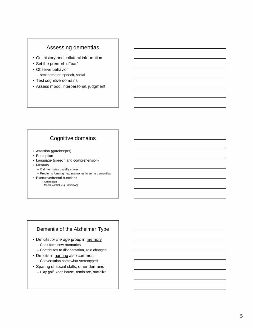
Assessing dementias
• Get history and collateral information
• Set the premorbid “bar”
• Observe behavior
– sensorimotor, speech, social
• Test cognitive domains
• Assess mood, interpersonal, judgment
Cognitive domains
• Attention (gatekeeper)
• Perception
• Language (speech and comprehension)
• Memory– Old memories usually spared
– Problems forming new memories in some dementias
• Executive/frontal functions• Abstraction
• Mental control (e.g., inhibition)
Dementia of the Alzheimer Type
• Deficits for the age group in memory
– Can’t form new memories
– Contributes to disorientation, role changes
• Deficits in naming also common
– Conversation somewhat stereotyped
• Sparing of social skills, other domains
– Play golf, keep house, reminisce, socialize
6
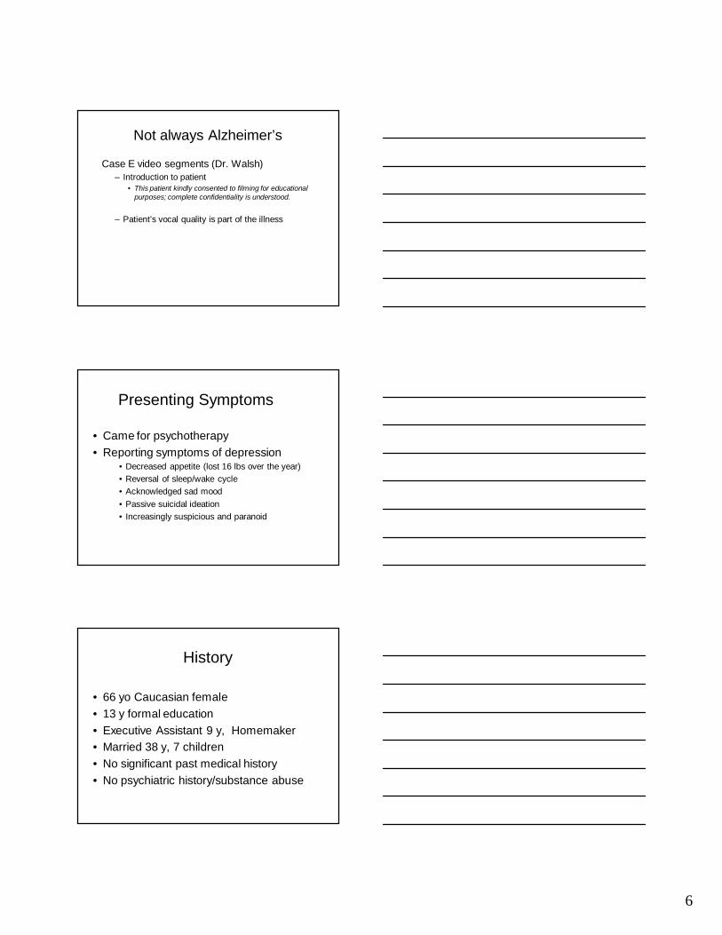
Not always Alzheimer’s
Case E video segments (Dr. Walsh)
– Introduction to patient• This patient kindly consented to filming for educational
purposes; complete confidentiality is understood.
– Patient’s vocal quality is part of the illness
Presenting Symptoms
• Came for psychotherapy
• Reporting symptoms of depression• Decreased appetite (lost 16 lbs over the year)
• Reversal of sleep/wake cycle
• Acknowledged sad mood
• Passive suicidal ideation
• Increasingly suspicious and paranoid
History
• 66 yo Caucasian female
• 13 y formal education
• Executive Assistant 9 y, Homemaker
• Married 38 y, 7 children
• No significant past medical history
• No psychiatric history/substance abuse
7
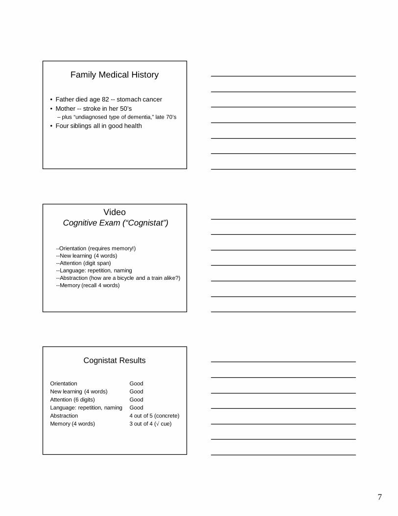
Family Medical History
• Father died age 82 -- stomach cancer
• Mother -- stroke in her 50’s
– plus “undiagnosed type of dementia,” late 70’s
• Four siblings all in good health
VideoCognitive Exam (“Cognistat”)
--Orientation (requires memory!)
--New learning (4 words)
--Attention (digit span)
--Language: repetition, naming
--Abstraction (how are a bicycle and a train alike?)
--Memory (recall 4 words)
Cognistat Results
Orientation
New learning (4 words)
Attention (6 digits)
Language: repetition, naming
Abstraction
Memory (4 words)
Good
Good
Good
Good
4 out of 5 (concrete)
3 out of 4 (√ cue)
8
Other Test Results
Impairments
• Low Reasoning/Abstraction
• Mental control impaired – Slow and perseverative on “Supermarket”
– Problems with planning, inhibition
– Could not do “Tap except for X”
– Unable to “shift” on Trail Making B
Presenting Neurologic Symptoms (2003)
• Progression of gait difficulties/falls
• Severe dysarthria
• Dystonic posturing of hands and feet
• Ataxic saccadic eye movements
• Poor voluntary upward + downward gaze
• Swallowing difficulties (liquids)
Neuroimaging Data
8/24/00 MRI
Essentially normal brain MRI
6/7/04 MRI
Mild diffuse cortical atrophy with no lobar predominance.

9
Epidemiology and courseSteele-Richardson-Olszewski syndrome
• Prevalence (M ≥ F)
– 1.4 / 100,000 over age 60 (v. PD: 5/100,000)
• Etiology
– Idiopathic: no familial, geographic, environmental, or ethnic patterns seen
– Tauopathic: chromosome 17
• Average survival: 9.7 y
• Death secondary to falls or aspiration
PSP
• Characteristic features (+ in case E)
– Broad based gait
– Axial rigidity +
– Toppling (backward) falls +
– Nuchal dystonia
– “Facial spasticity” (masked, apraxic) +
– Dysarthria (apraxia of speech) +
– ? Frontal release signs
– ? Utilization behavior, pallilalia
Criteria• NINDS-SPSP “Probable PSP”
Vertical supranuclear gaze palsy
+
Prominent postural instability with falls
within first year of onset
=
100% positive predictive value
10
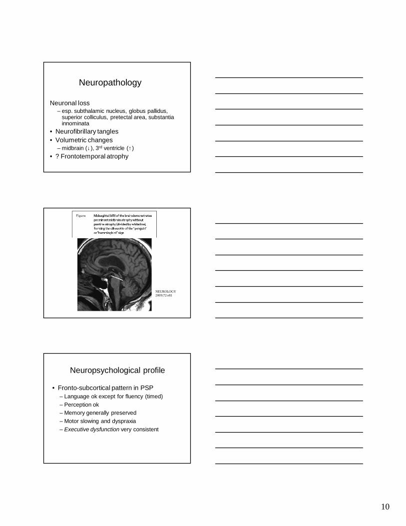
Neuropathology
Neuronal loss– esp. subthalamic nucleus, globus pallidus,
superior colliculus, pretectal area, substantia innominata
• Neurofibrillary tangles
• Volumetric changes– midbrain (↓), 3rd ventricle (↑)
• ? Frontotemporal atrophy
NEUROLOGY 2009;72:e81
Neuropsychological profile
• Fronto-subcortical pattern in PSP
– Language ok except for fluency (timed)
– Perception ok
– Memory generally preserved
– Motor slowing and dyspraxia
– Executive dysfunction very consistent
11
PSP versus Alzheimer’s
Dementia profile (matched on severity)
– Better memory in PSP than AD
• Mesial temporal structures OK
• Also somewhat better memory than Parkinsons and Lewy Body dementias
– Worse initiation than AD• Basal ganglia and midbrain networks…
PSP versus Alzheimer’s
• Affective symptoms
– Apathy more frequent in PSP
– Mild depression and anxiety (as in PD)
• Sleep disturbance worse than in AD
– Insomnia and frequent awakening
– Related to dysphagia, mobility, depression, nocturia
Lauterbach, 2004
Treatment
• Movement disorder (rigidity, bradykinesia)
– Some patients respond temporarily to levodopa
– Donepezil (AChE) worsened self-care skills
– Botulinum toxin for blepharospasm
• Cognition
– No benefit from AChE inhibitors that help AD
• Mood
– Anticholinergics, SSRIs or tricyclics may help
• Apathy
– Psychostimulant? in theory
12
Summary of Case E
• Affective disorder as presenting problem
• Dementia was mild
– Executive dysfunction
– Suspicion and reduced insight
• Dx Progressive Supranuclear Palsy
– History, course, MRI, NPT typical for PSP
• Good family support and adaptation
QUIZ
How is “normal” determined?
A. If I can do it, it’s normal.
B. Patient is normal if no abnormal signs are present.
C.Test results are compared to age group norms.
D. Normal means no change from past level of skills.
QUIZ
What unique information does the neuropsychological exam provide?
A. Test results quantify specific cognitive skills.
B. The tests measure mental status.
C. Test results can localize brain abnormality.
D. The exam determines the etiology of the brain disease.
13
QUIZ
How can the exam help determine whether an elderly patient is impaired or is just old?
A. Test norms are age-specific.
B. Medical and sensorimotor problems are taken into account.
C. The premorbid “bar” is set individually.
D. All of the above.
QUIZ
Which “frontal” symptoms did the patient exhibit?
A. witzelsucht
B. frontal release signs
C. utilization
D. perseveration
QUIZ
Which cognitive results distinguish PSP from Alzheimer’s dementia?
A. memory is better in PSP
B. attention is better in Alzheimer’s
C. abstraction is better in PSP
D. naming is better in Alzheimer’s

14
References
• Aarsland D et al. (2003) Performance on the dementia rating scale in Parkinson’s disease with dementia and dementia with Lewy bodies: comparison with progressive supranuclear palsy and Alzheimer’s disease. J Neurol Neurosurg Psychi 74: 1215-1220.
• Goetz C. Textbook of Clinical Neurology, 2nd ed., 2003.
• Lauterbach EC (2004) The neuropsychiatry of Parkinson’s disease and related disorders. Psychiatr Clin N Am 27 801-825.
• Litvan & Agid. Progressive Supranuclear Palsy: Clinical and Research Approaches, 1992.
• Oba H et al. (2005) New and reliable MRI diagnosis for PSP. Neurol 64 2050-2055.
1
Neural development II
molecular genetics and neuroscience
CNS congenital malformations
Neurodegenerative diseases during aging
Relevant concepts
• Neural patterning (spatial ordering)• Cellular determination• Migration• Connectivity• Regressive events• Activity-dependent processes
Nervous System organization begins in the neural plate stage
• CNS develops as hollow tube
• Topographically flat sheet of cells = neural plate
• Process of CNS development called neurulation
• Gradual and continuous process
• Much of it occurs prenatally
• genetic determinants and experience
2
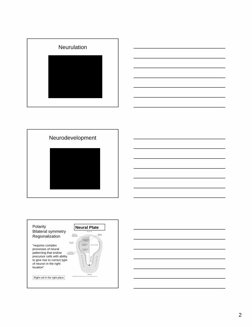
Neurulation
Neurodevelopment
PolarityBilateral symmetryRegionalization
“requires complex processes of neural patterning that endow precursor cells with ability to give rise to correct type of neuron in the right location”
Neural Plate
Right cell in the right place
3
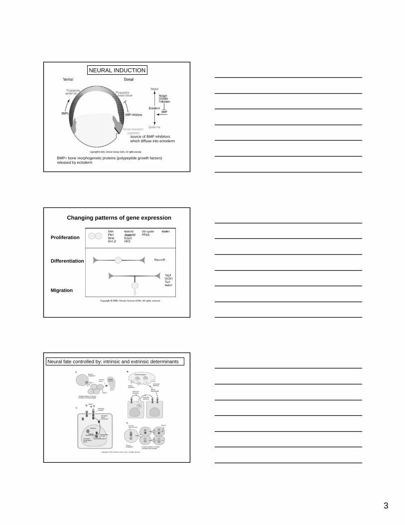
BMP= bone morphogenetic proteins (polypeptide growth factors) released by ectoderm
source of BMP inhibitors which diffuse into ectoderm
NEURAL INDUCTION
Changing patterns of gene expression
Proliferation
Differentiation
Migration
Neural fate controlled by: intrinsic and extrinsic determinants
4
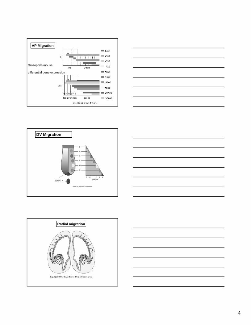
AP Migration
Drosophila-mouse
differential gene expression
DV Migration
SHH
Radial migration
5
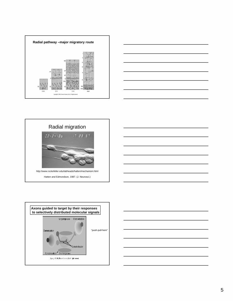
Radial pathway –major migratory route
Radial migration
Hatten and Edmondson, 1987. (J. Neurosci.)
http://www.rockefeller.edu/labheads/hatten/mechanism.html
Axons guided to target by their responsesto selectively distributed molecular signals
“push-pull-hem”
6
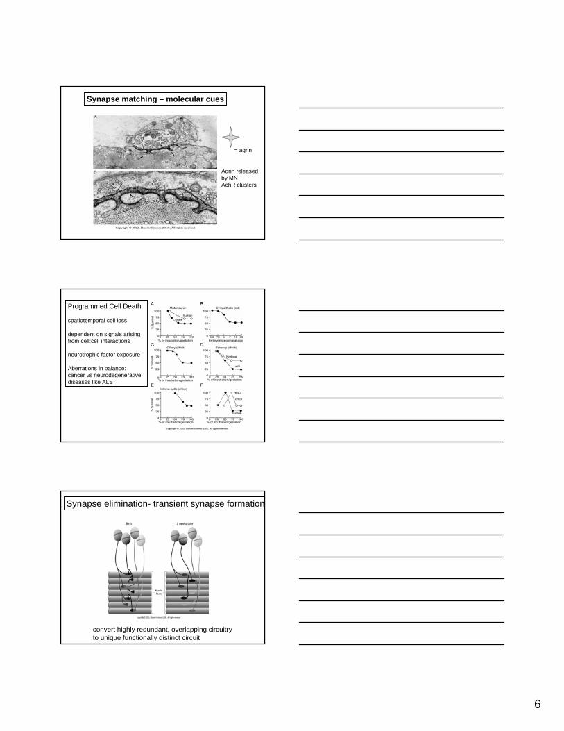
Synapse matching – molecular cues
= agrin
Agrin released by MNAchR clusters
Programmed Cell Death:
spatiotemporal cell loss
dependent on signals arisingfrom cell:cell interactions
neurotrophic factor exposure
Aberrations in balance:cancer vs neurodegenerative diseases like ALS
Synapse elimination- transient synapse formation
convert highly redundant, overlapping circuitry to unique functionally distinct circuit
7
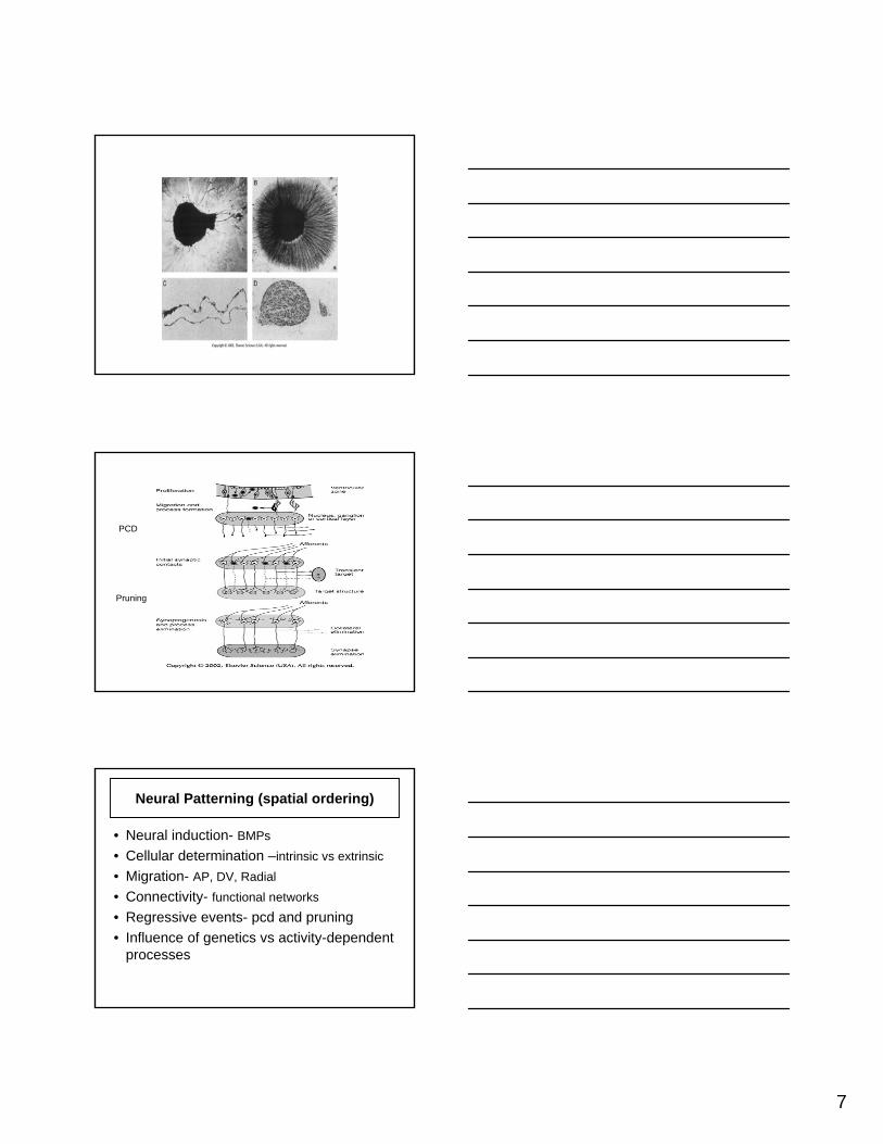
PCD
Pruning
Neural Patterning (spatial ordering)
• Neural induction- BMPs• Cellular determination –intrinsic vs extrinsic• Migration- AP, DV, Radial • Connectivity- functional networks• Regressive events- pcd and pruning• Influence of genetics vs activity-dependent
processes

8
Early experience and critical period
• synaptic connections pass through a period in early life when the capacity to adjust is greater than in adulthood = “critical period”
• neurons are sensitive to modification by experience
• critical period – a pathway awaits specific instructions to develop normally
• Binocular vision, language, social imprinting
Relevance of understanding how the nervous system develops
• regeneration recapitulates development
• application to injury and repair
• intrinsic vs extrinsic
9/29/2009
1
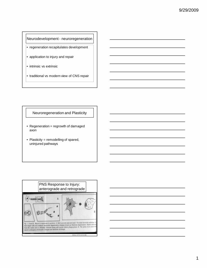
Neurodevelopment - neuroregeneration
• regeneration recapitulates development
• application to injury and repair
• intrinsic vs extrinsic
• traditional vs modern view of CNS repair
Neuroregeneration and Plasticity
• Regeneration = regrowth of damaged axon
• Plasticity = remodelling of spared, uninjured pathways
PNS Response to Injury: anterograde and retrograde
Bishop, J. APTA, Vol. 62, 1982.
9/29/2009
2
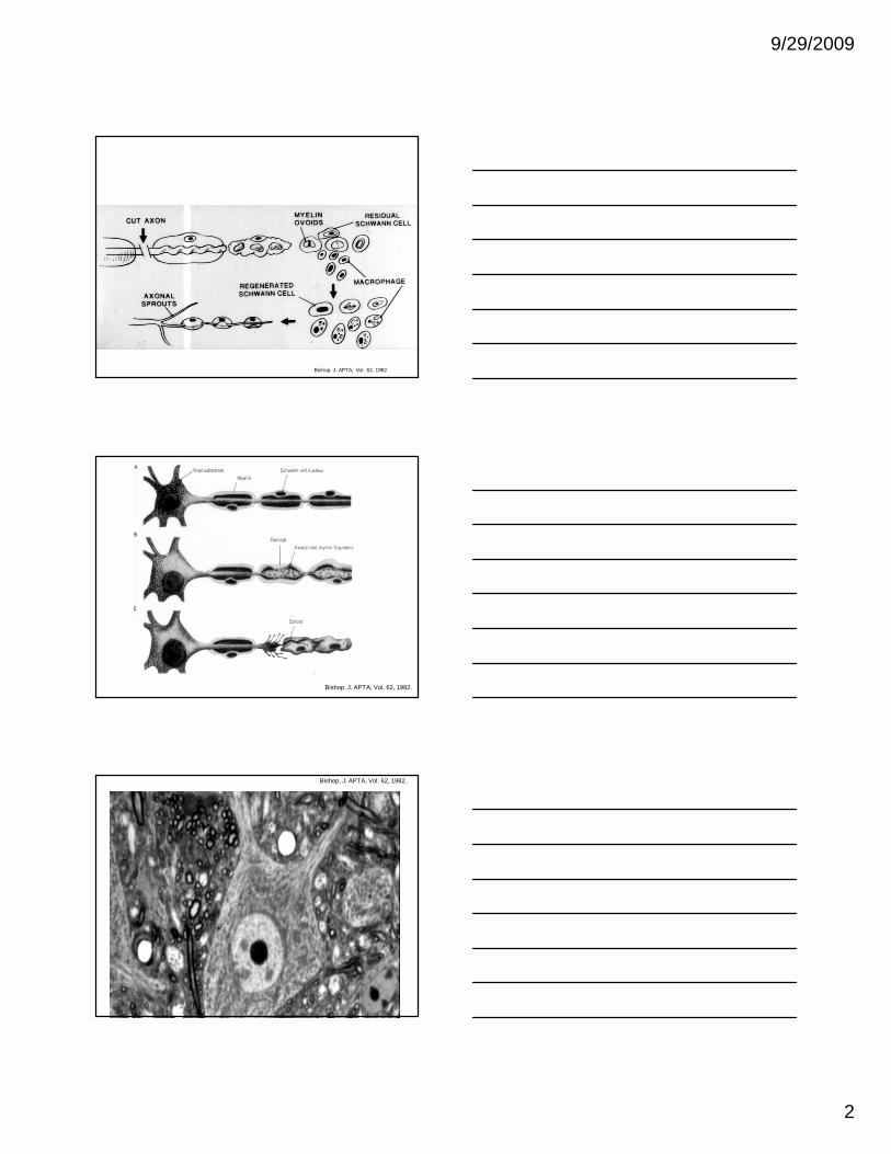
Bishop, J. APTA, Vol. 62, 1982.
Bishop, J. APTA, Vol. 62, 1982.
Bishop, J. APTA, Vol. 62, 1982.
9/29/2009
3
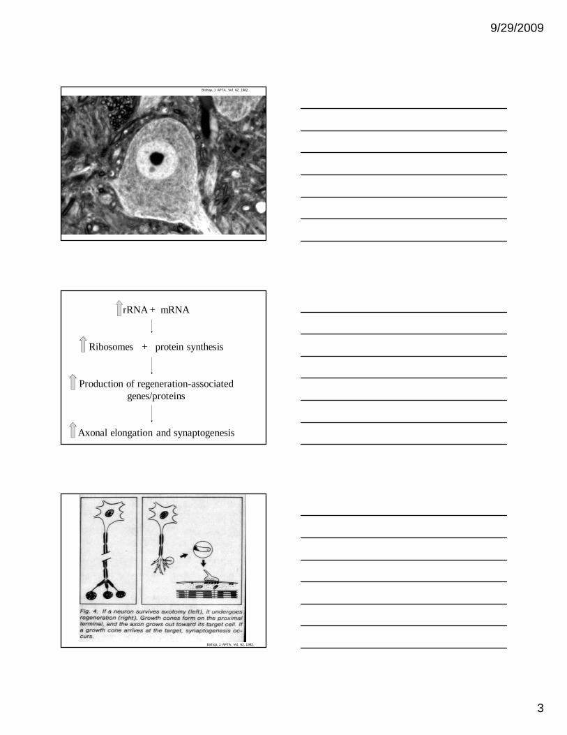
Bishop, J. APTA, Vol. 62, 1982.
rRNA + mRNA
Ribosomes + protein synthesis
Production of regeneration-associated genes/proteins
Axonal elongation and synaptogenesis
Bishop, J. APTA, Vol. 62, 1982.

9/29/2009
4
Extrinsic determinants
+ Schwann cells andperipheral immune cells
Bishop, J. APTA, Vol. 62, 1982.
Astrocytes-can form glial scar

9/29/2009
5
Astrocytes provide metabolic support
growth factorsadhesion moleculesspatial buffering
Protection fromexcitotoxicity
Microglia- mediate immune response
9/29/2009
6
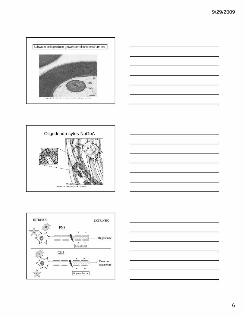
Schwann cells produce growth permissive environment
Oligodendrocytes-NoGoA
PNS
INTRINSIC EXTRINSIC
CNS
Regenerate
Does notregenerate
+
+ +
+
Oligodendrocyte
- -
--
M
A
Schwann cell
M
A
9/29/2009
7
Spinal cord injury
How to make a CNS motoneuron regenerate as effectively as a PNS motoneuron?
Olfactory Ensheathing Cells
• Unique type of macroglia found in the olfactory system
• Cross between PNS Schwann cells and CNS astrocytes
• In contrast to other CNS glia, provide growth permissive environment for regenerating CNS axons
PNS axons grow into CNS and establish synaptic contact with central neurons in
the olfactory bulb
Bishop, J. APTA, Vol. 62, 1982.
9/29/2009
8
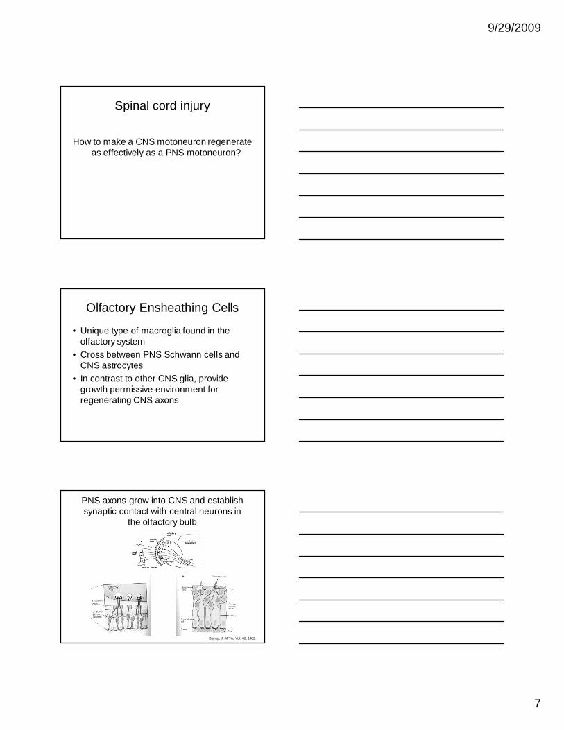
Factors influencing Regeneration
• PNS more likely to regenerate than CNS
• Intrinsic vs. extrinsic
• Age – immature more likely to die
• Site – proximal more severe
What is NEURAL PLASTICITY?
• Flexibility and adaptability of the nervous system
• Experience-related
• Adult CNS capable of plasticity
What LEVEL does NEURAL PLASTICITY occur at?
• Primarily at connectivity points
– (i.e.- formation of synapses)
• Results in alternate pathways for various NS functions
9/29/2009
9
Bishop, J. APTA, Vol. 62, 1982.
PNS: Ia αmn muscle
CNS: Retina LGN visual cortex
Bishop, J. APTA, Vol. 62, 1982.
9/29/2009
10
Bishop, J. APTA, Vol. 62, 1982.
Recovery of function
• Regeneration vs plasticity
• Combinatorial treatment strategies
• Therapeutic staging
• Immune system involvement
1
Edward Hines Jr. VA Hospital/Loyola University Medical CenterChicago, USA
September 29, 2009
Gwendolyn Kartje M.D., Ph.D
Neuroplasticity and Neural Repair
• Defined as the formation of new neuronal connections after CNS injury.
• Commonly seen following injury to the neonatal brain, and not after adult brain injury.
• This neuronal plasticity may be the anatomical substratefor functional recovery more commonly seen after brain injuryin the young.
• Why doesn’t the adult brain show such dramatic plasticity afterinjury…?
Neuronal Plasticity
Taken from Basic Neurochemistry, Ed G.J. Siegel
2
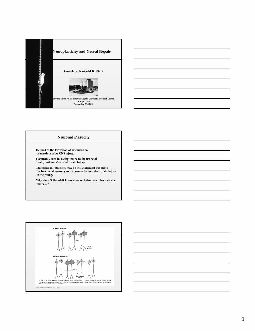
Molecular Mechanisms of Neurite Inhibition
• Three myelin inhibitory proteins have been found, Myelin Associated Glycoprotein (MAG), Oligodendrocyte myelin glycoprotein (OMgp), and Nogo-A.
• One receptor, Ng-66R, binds with all three.
• This receptor interacts with p75 to activate down- stream molecules that cause growth cone collapse.
• Other inhibitory factors are present in glial scars, i.e. proteoglycans.
Adapted from Schwab, Current Opinion in Neurobiology, 2004by AJC
Swiss Alps, Interlaken
me
by G..K.
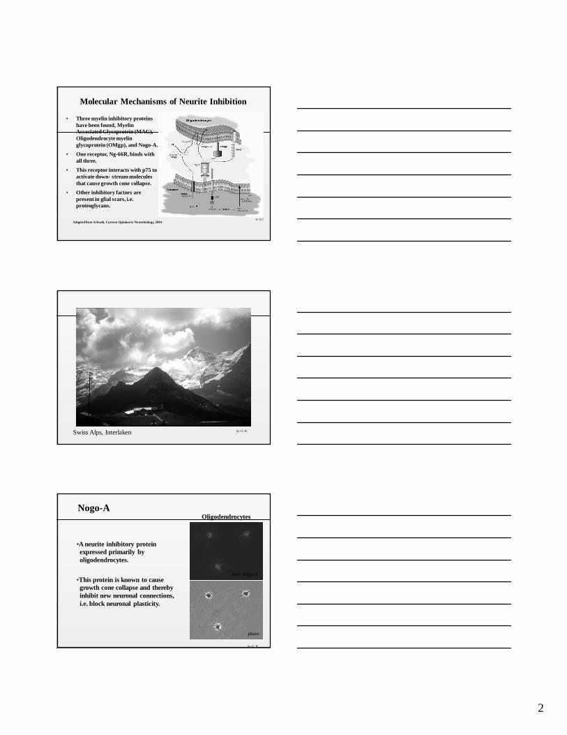
Nogo-AOligodendrocytes
•A neurite inhibitory protein expressed primarily by oligodendrocytes.
•This protein is known to causegrowth cone collapse and thereby inhibit new neuronal connections, i.e. block neuronal plasticity.
phase
Anti- Nogo-A
by G..K.
3
Brittis and Flanagan, Neuron, 2001
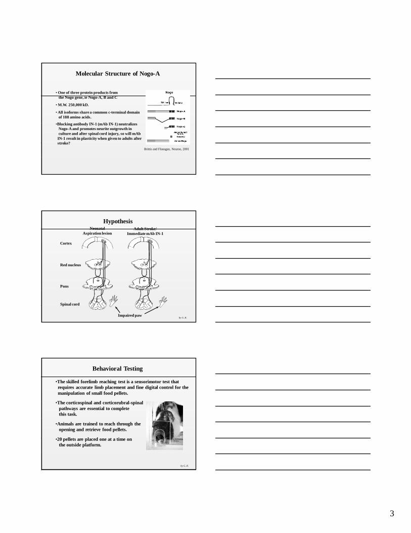
• One of three protein products from the Nogo gene, ie Nogo-A, B and C
• M.W. 250,000 kD.
• All isoforms share a common c-terminal domainof 188 amino acids.
•Blocking antibody IN-1 (mAb IN-1) neutralizes Nogo-A and promotes neurite outgrowth in culture and after spinal cord injury, so will mAb
IN-1 result in plasticity when given to adults after stroke?
Molecular Structure of Nogo-A
Cortex
Red nucleus
Pons
Spinal cord
Impaired paw
HypothesisNeonatal
Aspiration lesionAdult Stroke/
Immediate mAb IN-1
by G..K.
Behavioral Testing
•The skilled forelimb reaching test is a sensorimotor test that requires accurate limb placement and fine digital control for the manipulation of small food pellets.
•The corticospinal and corticorubral-spinalpathways are essential to completethis task.
•Animals are trained to reach through theopening and retrieve food pellets.
•20 pellets are placed one at a time on the outside platform.
by G..K.
4
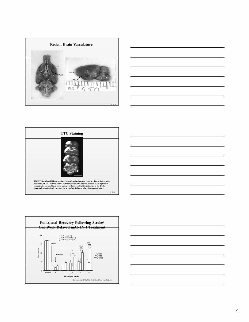
Rodent Brain Vasculature
MCA
MCA
by G..K.
by G..K.
TTC Staining
TTC (2,3,5-triphenyl-2H-tetrazolium chloride)-stained coronal brain sections at 3 days after permanent MCAO demonstrates a representative stroke size and location in the ispilateral sensorimotor cortex. Viable tissue appears red as a result of the reduction of the dye by functional mitochondrial enzymes; the area of the ischemic infarction appears white.
by G..K.
Functional Recovery Following Stroke/One Week Delayed mAb IN-1 Treatment
Weeks post stroke
0
5
10
15
20
Baseline 1 3 5 7 9
Su
cces
s sc
ore
Stroke only (n=7)
Stroke/control Ab (n=4)
Stroke/mAb IN-1 (n=9)
Treatment
Stroke********
**
**
*
* p<0.05** p<0.01
*** p<0.001
(Seymour et al 2005, J Cerebral Blood Flow Metabolism)
5
CCAA
BB
Stroke/mAb IN-lStroke/mAb IN-l
Stroke ControlStroke Control
Stroke control Stroke/mAb IN-1
Co
rtic
oru
bra
l fib
er
rati
o
0
0.20
0.40
0.60
0.80
1.00
*
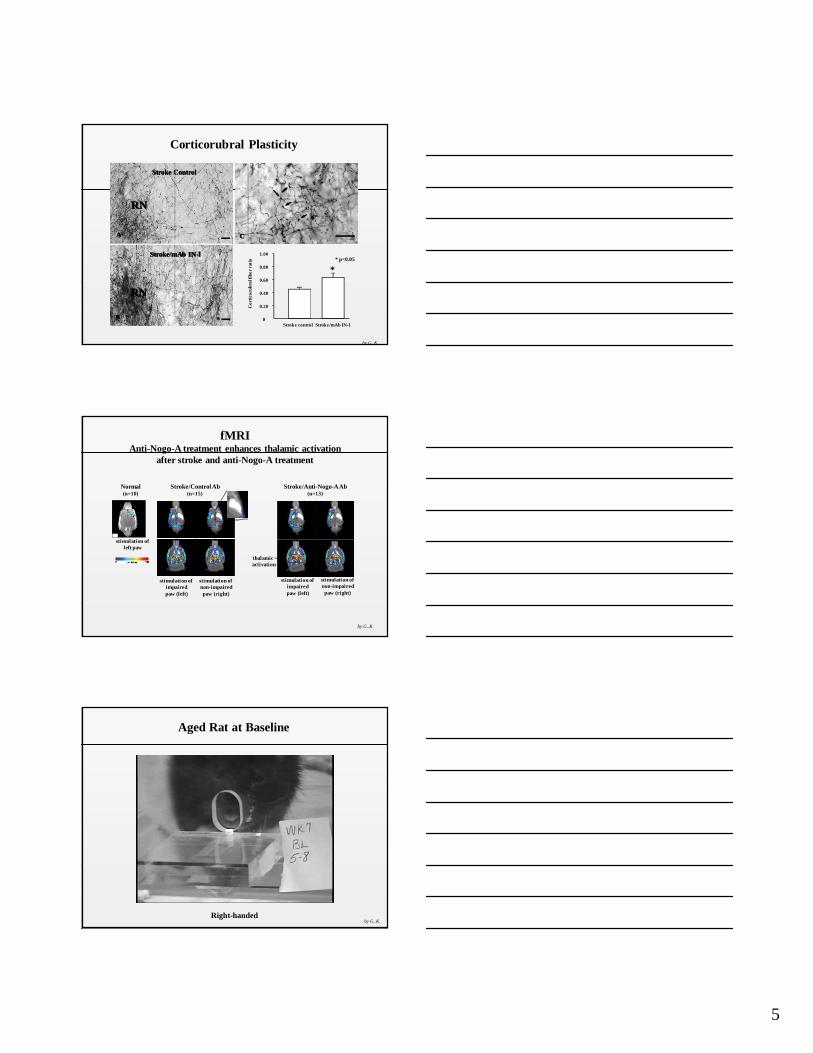
Corticorubral Plasticity
* p<0.05
RNRN
RNRN
by G..K.
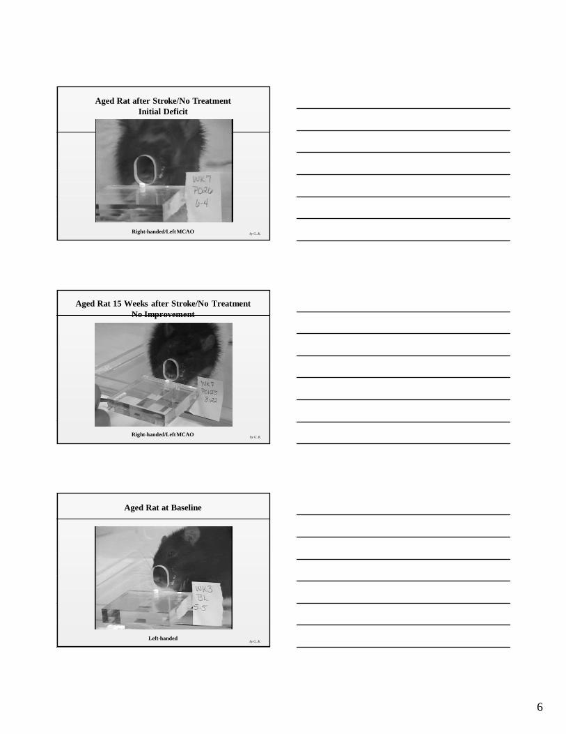
Stroke/Anti-Nogo-A Ab(n=13)
stimulation of impairedpaw (left)
stimulation of non-impairedpaw (right)
thalamic level
cortical level
b’ c’
d’ e’
thalamic activation
Normal(n=10)
stimulation of left paw
a
Stroke/Control Ab(n=15)
stimulation of impairedpaw (left)
stimulation of non-impairedpaw (right)
cortical level
b c
d e
thalamic level
fMRIAnti-Nogo-A treatment enhances thalamic activation
after stroke and anti-Nogo-A treatment
by G..K.
Right-handed
Aged Rat at Baseline
by G..K.
6
Right-handed/Left MCAO
Aged Rat after Stroke/No TreatmentInitial Deficit
by G..K.
Aged Rat 15 Weeks after Stroke/No TreatmentNo Improvement
Right-handed/Left MCAOby G..K.
Aged Rat at Baseline
Left-handedby G..K.
7
Aged Rat after Stroke/Anti-Nogo A TreatmentInitial Deficit
Left-handed/Right MCAO by G..K.
Aged Rat after Stroke/Anti-Nogo A Treatment(15 Weeks Post-Tx): Showing Marked Improvement
Left-handed/Right MCAO by G..K.
Does mAb IN-1 Treatment Enhance Dendritic Arborization In Spared
Neurons After Stroke ?
8
Golgi-Cox Stain of Neuronsin Unlesioned Forelimb Cortex
by G..K.
Dendritic Sprouting Following Stroke and mAb IN-1 Treatment
Golgi Analysis
Stroke only Stroke/mAb IN-1 Stroke/control Ab
(Papadopoulos et al 2006, Cerebral Cortex)
Total Dendritic Length
Mea
n d
end
riti
c le
ngt
h (
µm
)
0
50
100
150
200
250
*****
Golgi Analysis
** p<0.01*** p<0.001
by G..K.
9
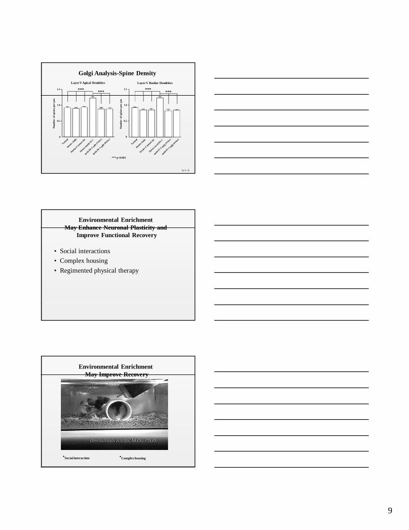
Layer V Basilar Dendrites
Nu
mb
er o
f sp
ines
per
µm
0
0.5
1.0
1.5
Layer V Apical Dendrites
Nu
mb
er o
f sp
ines
per
µm
0
0.5
1.0
1.5
Golgi Analysis-Spine Density
*** ****** ***
*** p<0.001
by G..K.
• Social interactions
• Complex housing
• Regimented physical therapy
Environmental Enrichment May Enhance Neuronal Plasticity and
Improve Functional Recovery
Environmental Enrichment May Improve Recovery
•Social interaction •Complex housing
10
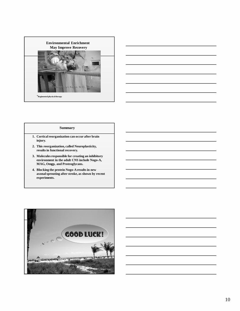
Environmental Enrichment May Improve Recovery
•Regimented physical therapy
Summary
1. Cortical reorganization can occur after brain injury.
2. This reorganization, called Neuroplasticity, results in functional recovery.
3. Molecules responsible for creating an inhibitory environment in the adult CNS include Nogo-A, MAG, Omgp, and Proteoglycans.
4. Blocking the protein Nogo-A results in new axonal sprouting after stroke, as shown by recent experiments.
by G..K.