neurofeedback for autistic spectrum disorder: a review of ......neurofeedback for autistic spectrum...
TRANSCRIPT
Neurofeedback for Autistic Spectrum Disorder: A Reviewof the Literature
Robert Coben • Michael Linden • Thomas E. Myers
! Springer Science+Business Media, LLC 2009
Abstract There is a need for effective interventions toaddress the core symptoms and problems associated with
autistic spectrum disorder (ASD). Behavior therapy
improves communication and behavioral functioning.Additional treatment options include psychopharmacolog-
ical and biomedical interventions. Although these approa-
ches help children with autistic problems, they may beassociated with side effects, risks or require ongoing or
long-term treatment. Neurofeedback is a noninvasive
approach shown to enhance neuroregulation and metabolicfunction in ASD. We present a review of the literature on
the application of Neurofeedback to the multiple problems
associated with ASD. Directions for future research arediscussed.
Keywords Autistic spectrum disorder ! Treatment !Neurofeedback
Introduction
Autistic spectrum disorders (ASD) are a heterogeneous
group of pervasive developmental disorders including
Autistic disorder, Rett disorder, Childhood disintegrativedisorder, Pervasive developmental disorder-not otherwise
specified (PDD-NOS), and Asperger disorder. Children with
ASD demonstrate impairment in the following functions: (1)social interaction, (2) verbal and nonverbal communication,
and (3) behaviors or interests (DSM-IV-TR; APA 2000).
ASD may be comorbid with sensory integration difficulties,mental retardation or seizure disorders. Children with ASD
may have severe sensitivity to sounds, textures, tastes, and
smells. Cognitive deficits are often associated with impairedcommunication skills (National Institute of Mental Health;
NIMH 2006). Repetitive stereotyped behaviors, persevera-
tion, and obsessionality, common in ASD, are associatedwith executive deficits. Executive dysfunction in inhibitory
control, set shifting, and mediating frontostriatal neural
pathways have been attributed to ASD (Schmitz et al. 2006).Seizure disorders may occur in one out of four children with
ASD; frequently beginning in early childhood or adoles-
cence (National Institute of Mental Health; NIMH 2006).Autistic disorder includes the following triad of symp-
toms: (1) impaired social interaction, failure to developpeer relationships, or lack of initiating spontaneous activ-
ities; (2) deficits in communication including delay in or
lack of spoken language, inability to initiate or sustainconversation with others, stereotyped repetitive use of
language or idiosyncratic language; and (3) restricted
repetitive and stereotyped behavior, interests, inflexibleadherence to routines or rituals, and repetitive motor pat-
terns (e.g., hand or finger flapping or twisting) (DSM-IV-
TR; APA 2000).Individuals with Asperger disorder frequently have high
cognitive function, engage in literal pedantic speech,
experience difficulty comprehending implied meaning,exhibit problems with fluid movement, and manifest
inappropriate social interactions. Pervasive developmental
R. Coben (&) ! T. E. MyersNeurorehabilitation & Neuropsychological Services, 1035 ParkBlvd., Suite 2B, Massapequa Park, NY 11762, USAe-mail: [email protected]; [email protected]
M. LindenThe Original ADD Treatment Centers, 30270 Rancho ViejoRoad, Suite C, San Juan Capistrano, CA 92675, USA
T. E. MyersGraduate Center of the City University of New York, ClinicalNeuropsychology Subprogram at Queens College,Queens, NY, USA
123
Appl Psychophysiol Biofeedback
DOI 10.1007/s10484-009-9117-y
disorder-not otherwise specified (PDD-NOS) reflects defi-
cits in language and social skills, which do not meet thecriteria of other disorders. In contrast, childhood disinte-
grative disorder and Rett’s disorder both have normal
periods of early development followed by loss of previ-ously acquired skills. Common features among these con-
ditions include communication and social skill deficits.
There is considerable variability in terms of onset andseverity of symptomatology within the autistic spectrum of
disorders (Attwood 1998; Hamilton 2000; McCandless2005; Sicile-Kira 2004; Siegel 1996).
Research reviewing the epidemiology of autism (Center
for Disease Control and Prevention; CDC 2006) reportedbetween 1 in 500 to 1 in 166 children in the United States
diagnosed with the disorder. In fact, their most recent
report (CDC 2007) suggests a prevalence of 1 in 150 and ashigh as 1 in 92 boys. According to Blaxill (2004), the rates
of ASD were reported to be\3 per 10,000 children in the
1970s and rose to[30 per 10,000 in the 1990s. This rise inthe rate of ASD constituted a ten-fold increase over a
20 year interval in the United States. With increased
prevalence comes a need to design and empirically validateeffective treatments for those impacted by autistic
disorders.
A review of multiple studies reported the rate ofabnormal EEGs in autism ranged from 10 to 83%, while
the mean incidence was 50%. Atypical EEGs often predict
poor outcomes for intelligence, speech, and educationalachievement (Hughes and John 1999). In a more recent
review of research, Rippon et al. (2007) proposed a model
of reduced connectivity between specialized local neuralnetworks and overconnectivity within isolated neural
assemblies in autism. Disordered connectivity may be
associated with an increased ratio of excitation/inhibitionin key neural systems. Anomalies in connectivity may be
linked to abnormalities in information integration. A
common deficit characterizing children with autism isexecutive dysfunction. Executive deficits of planning,
flexibility, and inhibition are associated with dysfunctional
integration of the frontal lobes with other brain regions.Therefore, executive dysfunction impacts upon social,
behavioral, and cognitive function (Hill 2004). In SPECT
(single photon emission computed tomography) scans ofchildren with autism, abnormal regional cerebral blood
flow in the medial prefrontal cortex and anterior cingulate
gyrus was related to impaired communication and socialinteraction. Altered perfusion in the right medial temporal
lobe was associated with the obsessive desire for sameness
(Ohnishi et al. 2000). Functional neuroimaging studieshave linked social cognition dysfunction and language
deficits in autism to neural substrates (Pelphrey et al. 2004;
Welchew et al. 2005). During a sentence comprehensiontest, individuals with autism showed less functional
connectivity between Broca’s area and Wernicke’s area,
suggesting a lower degree of information organization andneural synchronization relative to a control group during
language tasks (Just et al. 2004). A review of neuroimaging
studies has found key brain structures including theamygdala, superior temporal sulcus region, and fusiform
gyrus to function differently in individuals with autism
than in controls (McAlonan et al. 2004). The aforemen-tioned research provides evidence for a neuropathological
basis of ASD.
Treatments for ASD
Green et al. (2006), who surveyed parents on the therapies
they most frequently selected for their children with ASD,found as many as seven different therapies were utilized.
Speech therapy was the most common treatment, being
selected by 70% of parents. Psychopharmacological treat-ment was utilized by 52% of parents. Other treatments
included: visual schedules (43%), sensory integration
(38%), and applied behavior analysis (36%). Special dietswere implemented by 27% of parents and 43% utilized
vitamin supplements.
Behavioral Interventions
The method of treatment with the most empirical support isapplied behavior analysis (ABA), a form of behavior mod-
ification. The goal of this therapy is to improve social
interaction, behavior and communication (Bassett et al.2000). ABA is firmly based on the principles of operant
conditioning and measures small units of behavior to build
more complex and adaptive behaviors through reinforce-ment. Typically, imitation, attention, motivation, and com-
pliance are targeted early (Couper 2004). The first program,
developed in 1970 by Lovaas et al. (1973), which utilizedthis technique was the Young Autism Project (YAP). This
full time program uses an intensive, highly structured
behavioral program which is delivered on a one-to-one basisrequiring several hours a day. An evaluation of the program
for children diagnosed with autism receiving 40 or more
hours per week for two or more years included: increasedcognitive and academic function (47% of the treatment
group versus 2% of controls) (Lovaas 1987); and follow-up
research of children into late childhood and adolescencereported improved cognitive function and education in
regular classrooms (47% of the treatment group versus 0%
of controls) (McEachin et al. 1993). However, these studieshave been criticized based on the use of their outcome
measures (Schopler et al. 1989), which included IQ scores
and school placement. Subjects in the study were not ran-domly assigned, which is believed to have contributed to the
Appl Psychophysiol Biofeedback
123
observed outcome differences. It is also believed that the
individuals used in the study were high functioning autistics,which may account for the high IQ scores (Mundy 1993).
Other studies that measured outcomes of ABA treatment
in autism found less promising results than those of Lovaas(1987). In an attempt to replicate Lovaas’ study, Birnbrauer
and Leach (1993) designed the Murdoch Early Intervention
Program for 24–48 month old children with autism. After24 months of their program, they reported four of the nine
children in the experimental group showed signs ofapproaching normal levels of functioning, compared to
only one of five children in the control group. However,
none of the children achieved completely normal func-tioning and improvements in other symptoms were mini-
mal to moderate. In a retrospective study of a home-based
ABA program of shorter duration than the Lovaas study,Sheinkopf and Siegel (1998) compared 11 children who
received ABA to a control group receiving an unspecified
school based treatment. Children in the experimental grouphad significantly higher posttreatment IQ scores. Smaller
differences in symptom severity were found between the
groups, but the experimental group still met diagnosticcriteria for either autism or PDD. Anderson et al. (1987)
conducted home-based ABA on 14 children for between 15
and 25 h per week. While modest gains were made inmental ages scores and communication skills, the most
impaired children failed to make progress, and none of the
children were able to be integrated into regular classroomsfollowing treatment.
Fenske et al. (1985) examined the influence of age at
intervention on treatment outcome using an ABA protocol.They compared nine children who began treatment before
60 months of age to those who started treatment after
60 months. While four of nine children in the youngergroup were able to be enrolled in a regular classroom after
2 years of treatment, only one of the nine children in the
older group was. Although it appears that age at programentry was an important variable in treatment outcome,
enrollment in a regular classroom was their main outcome
measure and so this conclusion requires further researchsupport. Harris et al. (1991) compared preschool children
who were autistic to those who were not, both before and
after a behavioral intervention during the school year. Thechildren with autism showed a 19 point average IQ gain
and an 8 point average gain in their language quotient. The
more typically developing children showed no significantchange in such measures over the same time period.
Project TEACCH (Treatment and Education of Autistic
and Related Communication Handicapped Children),developed by Eric Schopler and colleagues (Schopler and
Reichler 1971) at the University of North Carolina at
Chapel Hill, differs from ABA, but utilizes behavioralprinciples to maximize the skills of children who are
autistic (Herbert et al. 2002). Structured settings are pro-
vided and teachers use individual workstations wherechildren can practice their skills. For example, because
they process visual information more efficiently than ver-
bal information, visual cues may be provided to compen-sate for auditory processing deficits. Ozonoff and Cathcart
(1998) investigated the effectiveness of a home-based
TEACCH treatment program for children with autism.Parents were taught to work on cognitive, academic, and
prevocational skills, which they provided for 4 months.The treatment group was compared to a control group who
underwent testing with the psychoeducation profile-revised
(PEP-R; Shopler et al. 1990), but did not receive anytreatment. Children who received the TEACCH treatment
from their parents showed significant improvement over
the control group on tests of imitation, fine and grossmotor, nonverbal conceptual skills, and overall PEP-R
scores. Progress was three to four times greater on all
outcome tests in the treatment group as compared to thecontrol group.
Smith et al. (2000) conducted research on behavior
therapy that utilized a matched-pair random assignmentprocedure. One group received intensive behavioral treat-
ment, while the other received parent training. Participants,
who ranged in age from 18 to 42 months at the start oftreatment, were reassessed at follow-up at ages 7–8 years.
Significant differences were noted for the intensive treat-
ment group (30 h of training per week over 2–3 years) incontrast to the parent training group (5 h per week of
training for 3–9 months) for IQ and language function.
Other longitudinal research has also been conductedutilizing intensive behavioral therapy for children with
autism. Sallows and Graupner (2005) randomly assigned
24 children with autism (aged 3 years) to a clinic-directedgroup replicating the behavioral program implemented by
Lovaas (1987) and McEachin et al. (1993), or a parent-
directed group. Both clinic and parent-directed groupsreceived approximately the same treatment of nearly equal
intensity, however. After combining both treatment groups
at the end of the first year of treatment, 11 of 23 (48%)children showed rapid learning (determined by change in
IQ scores), and achieved average scores on cognitive,
language, daily living, and socialization skills measures atpost-treatment. Children in the parent-directed condition
did about as well as the children in the clinic directed
group. Following 4 years of therapy, rapid learners (thosewith a 49 point increase in Full Scale IQ) were successfully
attending regular classrooms. Moderate learners (2.5 point
increase in Full Scale IQ) also showed increases in cog-nitive, adaptive, language, and social skills but continued
to require support services, modified curriculums, or spe-
cial education. The children who had the capacity forimitation, social responsiveness, and language attained the
Appl Psychophysiol Biofeedback
123
best treatment outcomes (Sallows and Graupner 2005).
These authors found an extremely large change in IQ forthe rapid learners. Almost half of the 23 subjects in this
study fell into this group. Some possible confounds include
the fact that some children were originally assessed withthe Bayley Scales of Infant Development (Bayley 1993)
and then assessed using the Wechsler (1989) scales at
follow up. However, the authors found no significant dif-ferences between those assessed with the Bayley scales
twice, and those assessed with the two different measures.Furthermore, although the two groups were matched on IQ,
other variables were not adequately controlled for, such as
imitation.Eikeseth et al. (2002) compared children between 4 and
7 years of age receiving 28 h per week of behavioral
treatment to a comparison group receiving 29 h of eclecticspecial education treatment per week. At a 1 year follow
up evaluation, children in the behavioral treatment group
gained 17 average IQ points, 13 points on a standardizedmeasure of language comprehension, 27 points on a mea-
sure of expressive language, and 11 points on a measure of
adaptive behavior. In contrast, the group receiving theeclectic treatment gained 4 IQ points, 1 point on the lan-
guage measures, and zero points on adaptive behavior. A
follow up study of both groups of children who continuedto receive their respective treatments (i.e., behavioral and
eclectic) 3 years later was recently conducted by Eikeseth
et al. (2007). The behavioral treatment group again showedgreater gains from pretreatment to follow up, though gains
between the first treatment study and this follow up were
only significant on the Vineland Adaptive Behavior Com-posite and Vineland Socialization scales. Although most
gains in IQ score were obtained between pretreatment and
the 1 year follow-up from their first study, the VinelandComposite score increased throughout all years of treat-
ment, and significant changes in the Vineland Socialization
and Daily Living scores occurred only after the first year.Thus, it may be important to continue ABA treatment
beyond 1 year to obtain the greatest benefit to the child.
Butter et al. (2006) recently described eight casereports of children with ASD and mental retardation who
received early intensive behavioral intervention (EIBI), a
behavioral treatment designed to address the core symp-toms of autism. In EIBI, reinforced practice as well as
functional analysis is utilized in a comprehensive and
individualized format. This treatment is most frequentlyconducted for 30–40 h per week, and involves one-on-one
instruction along with small group activities. Following
treatment, none of the children met criteria for eithermental retardation or any PDD. There were large
improvements in IQ (34.6 point increase) and adaptive
behavior (increased 43 standard score points), with bothNonverbal IQ and achievement scores ending in the
average range, although language abilities remained
impaired for seven of the eight children.Ben-Itzchak and Zachor (2007) investigated the effects
of intellectual functioning and severity of autistic symp-
toms on outcome following intensive behavioral interven-tion. Groups were formed based on IQ scores (high[70 vs.
low\70), level of social interaction (high versus low), and
communication deficits (high versus low) using the AutismDiagnostic Observation Schedule (ADOS; Lord et al.
1999), which were assessed prior to treatment. After 1 yearof intervention provided one-on-one by a behavioral ther-
apist for at least 35 h per week, significant improvements
were noted in all domains measured by the ADOS, whichinclude imitation, receptive and expressive language,
nonverbal communication skills, play skills, and stereo-
typed behaviors. The authors found that children withhigher cognitive levels and those with fewer social inter-
action deficits were more apt to acquire developmental
skills post-treatment, particularly in the areas of receptiveand expressive language, and play skills. Progress in
expressive language abilities was more related to good
social abilities, while play skills progress was more relatedto the child’s cognitive level.
In addition to ABA, a new behavioral treatment utilized
with ASD patients is errorless compliance training,developed by Joseph Ducharme (2005). It is a non-coer-
cive, parent-mediated approach, which involves teaching a
child to comply with requests from their parents in a sys-tematic and gradual manner. During an initial observation
period, a hierarchy of compliance probabilities (Levels
1–4) is developed based on a wide range of parentalrequests. Level 1 indicates a high level of child compliance
and level 4 indicates low probability of child compliance.
Several level 1 requests are delivered to the child whileproviding extensive praise and reinforcement, while higher
level requests are slowly faded in to prevent noncompli-
ance. Ducharme et al. (2007) applied errorless compliancetraining to three boys with characteristics of Asperger
syndrome. Observational data from the parents indicated
great improvement in the children’s’ compliance followingtreatment, with generalized and durable effects at 2 months
post treatment. Furthermore, parents reported being satis-
fied with the intervention.In their clinical practice guidelines report, the New York
State Department of Health Early Intervention Program
recommended that ABA and other behavioral interventionsbe included in the treatment of autism. They specify that
intensive behavioral programs should include a minimum
of 20 h of intervention with a therapist per week. Fur-thermore, the guidelines state that parents should be
included in the intervention, and that they be trained in the
use of behavioral techniques to provide additionalinstruction at home, with regular therapist consultation.
Appl Psychophysiol Biofeedback
123
Although promising, intensive behavioral programs are
costly and require extensive time on the part of the thera-pist as well as the family, and debates are ongoing about
who should pay for such services (Couper 2004). The
Human Services Department of Victoria, British Columbiafound that 63% of 262 families with autistic children spend
between $1,000 and $10,000 a year on treatment, while the
median annual income was only $40,000.Although behavior therapy improves social, cognitive
and language skills, a year or more of intensive training hasbeen used in most research studies that have demonstrated
improvement. A strong commitment by parents to com-
plete therapeutic programs is necessary to achieve positiveoutcomes. Although behavioral treatment methods show
the most empirical support, there is still a need for addi-
tional therapies, which may be more easily administeredand used in conjunction with the behavioral methods
described. On the whole, research into this area is prom-
ising. However, there has been great variability betweenstudies in their results, outcome measures have often been
questionable (e.g., IQ scores, returning to regular class-
rooms), and this approach appears to be more effectivewith those who are higher functioning (i.e., higher IQ), thus
leaving out the lower functioning individuals who are
perhaps in greatest need of treatment.
Pharmacological Treatments
Pharmacological and biomedical interventions have also
been utilized to treat individuals with ASD. A study
conducted at the Yale Child Study Center found that 55%of a group of 109 individuals with a PDD were taking
psychotropic medication, with 29.3% taking more than
one medication (Martin et al. 1999). The most commonmedications were antidepressants (32.1%), followed by
stimulants (20.2%) and neuroleptics (16.5%). The objec-
tives of psychopharmacological treatment for autisminclude: decreasing the core symptoms of autism;
decreasing anxiety and overfocus, improving social skills,
reducing aggressive self-injurious behavior; increasing theeffects of other interventions, and improving the quality of
life for the child and their family. There is no single
medication known to be beneficial to all children withASD, nor that has specifically been developed for indi-
viduals with autistic spectrum disorder. Neuroleptics such
as haloperidol and thioridazine have been utilized toreduce dysfunctional behaviors associated with ASD. The
adverse side effects of sedation, irritability, and extrapy-
ramidal dyskinesias limit the use of these medications,however.
A newer class of neuroleptic, referred to as atypical anti-
psychotics, improves social interaction and decreasesaggression, irritability, agitation, and hyperactivity (Barnard
et al. 2002). They have fewer extrapyramidal adverse side
effects than haloperidol and thioridazine. However, mostchildren experience a substantial weight gain within the first
months of treatment (Committee on Children with Disabil-
ities 2001). Risperidone is the only drug that has beenapproved by the FDA to treat the symptoms (irritability) of
autism. A review published in the Cochrane Library exam-
ined three randomized controlled trials (Jesner et al. 2007).Meta-analysis indicated the drug was effective in treating
the symptoms of irritability and aggression. The authorsconcluded that although risperidone may be beneficial, its
use must be weighed against its adverse effects, most nota-
bly weight gain, and that long-term follow up is needed priorto determining its efficacy in clinical practice. However,
researchers have provided some evidence of the drug’s long
term effects. A study conducted by the RUPPS AutismNetwork (Research Units on Pediatric Psychopharmacol-
ogy; RUPP Autism Network 2005a) investigated the long-
term benefit of risperidone in a two part study. Part one wasa 4 month, open label trial, which was followed by an
8 week randomized, double-blind, placebo substitution
study of risperidone withdrawal in those who were consid-ered ‘‘responders’’. Participants whose medication levels
were gradually reduced showed a greater return of aggres-
sion, temper outbursts, and self-injurious behaviors thanthose who continued the medication, for whom over 80%
maintained their improvements and showed ‘‘very good
tolerability’’. Although not an RCT, there is evidence tosuggest that risperidone may be effective for time periods of
up to 1 year (Zuddas et al. 2000). The relapse rate for those
maintained on this medication has ranged from 12.5 to 25%(RUPP-AN, 2005a; Troost et al. 2005).
Recently a similar atypical anti-psychotic medication,
Abilify (Aripiprazole), has been used with patients whoare autistic. Stigler et al. (2004) administered the drug to
5 boys between the ages of 5–18 in a naturalistic, open-label
trial over the course of about 20 weeks. Based on ratingsfrom the Clinical Global Impressions-Improvement scale,
they reported all children responded (CGI-I ratings of
‘‘much improved’’ or ‘‘very much improved’’) with minimalside effects including mild somnolence, and weight gain in
one patient and weight loss in two others. They also
reported significant improvement in aggression, agitation,and self-injurious behavior. This study, however, was
obviously limited by its small sample size, lack of blinding,
absence of a control group, and a failure to use randomi-zation. Santangelo and Tsatsanis (2005) reported that there
are currently no drugs that produce major improvement in
the core social or pragmatic language deficits in autism,although several have limited effects on the behavioral
features of the disorder.
Psychostimulant medications are often used with chil-dren who are autistic due to its success in the treatment of
Appl Psychophysiol Biofeedback
123
ADHD (Jensen et al. 2007). Despite this, stimulant use in
children who are autistic remains controversial and largelyunproven in terms of efficacy. In the RUPP Autism Net-
work Methylphenidate study (2005b), 49% of the sample
was considered positive responders, leaving a significantpercentage as non-responders and an 18% side effect rate
overall.
Repetitive stereotypical and perseverative behaviorshave been shown to be characteristic of both obsessive
compulsive disorder (OCD) and autism (McDougle et al.1995). The overlap between these disorders and the success
of selective serotonin reuptake inhibitors (SSRIs) in treat-
ing OCD (Geller et al. 2001) has led to the use of SSRIs intreating symptoms of autism. One of the first trials of the
SSRI, Prozac (Fluoxetine), found that doses ranging from
20 to 80 mg per day were effective based on ClinicalGlobal Impressions in 15 of 23 individuals with autism
(Cook et al. 1992). However, 6 out of the 23 experienced
significant side effects such as restlessness, hyperactivity,agitation, increased appetite, and insomnia. A more rigor-
ous 20 week placebo controlled crossover study found that
fluoxetine significantly reduced repetitive behaviors com-pared to placebo (Hollander et al. 2005). Although there
were no significant side effects, there also were no signif-
icant improvements in measures of speech or social inter-action. Delong et al. (2002) reported a 69% positive
response rate for fluoxetine in children, aged 2–8, who
were autistic. Treatment parameters were quite variablewith treatment duration ranging from 5 to 76 months and
doses ranging from 4 to 40 mg/day.
A 12 week, double blind, placebo controlled study offluoxetine reported this drug to be efficacious (McDougle
et al. 1996). Eight out of fifteen adult subjects were rated as
‘‘responders’’, with improvements occurring for repetitivethoughts and behaviors, maladaptive behaviors, and
repetitive language use. Side effects were noted to be mild
and included sedation and nausea. However, a more recentstudy by McDougle et al. (2000) with children and ado-
lescents found only 1 of 18 responded to the drug, with
common side effects including insomnia, hyperactivity,agitation, and aggression. Martin et al. (2003) found similar
results in children, reporting only 3 out of 18 subjects to be
responsive to fluvoxamine.At least four other SSRIs have been reported to have at
least some beneficial effect, although none have demon-
strated efficacy through placebo controlled studies.McDougle et al. (1998) found Zoloft (sertraline) to be
effective for aggression and repetitive behavior in 42 adults
with PDD, including adults with autism, Asperger’s, andPDD-NOS, though 3 of the subjects dropped out of the
study due to either agitation or anxiety. They found ser-
traline to be more effective for those with autism and PDD-NOS than for those with Asperger’s disorder. Social
relatedness did not appear to improve. Hellings et al.
(1996) administered sertraline to adults with mental retar-dation, which included adults with autism. Aggression and
self-injurious behavior were decreased in 8 out of 9 sub-
jects. Other evidence of sertaline’s effectiveness comesfrom case studies published by Steingard et al. (1997) who
found improvement in anxiety, irritability, and behavioral
problems associated with change in routine. Very limitedsupport has been reported for the SSRI Paxil (paroxetine).
Case studies have reported reductions in self-injuriousbehavior in a 15 year old male with high functioning aut-
ism (Snead et al. 1994), and improvement in irritability,
temper tantrums, and interfering preoccupations in a 7 yearold boy with autism (Posey et al. 1999).
Two retrospective studies of Celexa (citalopram),
another SSRI, have reported improvement in some of thesymptoms of autism. Couturier and Nicolson (2002) found
improvement in 10 out of 17 children in aggression, anx-
iety, stereotypies, and preoccupations, though not in socialinteractions and communication. In addition, four children
developed adverse side effects such as increased agitation
and insomnia causing their treatment to be stopped.Namerow et al. (2003) found similar improvements in
children and adolescents in repetitive behavior, mood, and
anxiety. Mild side effects were reported in one-third oftheir sample, two of whom discontinued treatment due to
side effects. The fourth SSRI that has been studied to treat
the symptoms of PDD is Lexapro (escitalopram). In anopen label design of children and adolescents with autism,
Asperger’s, or PDD-NOS, Owley et al. (2005) found sig-
nificant improvement in 17 of 28 patients based on ratingsfrom the Aberrant Behavior Checklist Irritability subscale
(Aman et al. 1985). There was wide variability in dose
response, which could not be accounted for by weight orage. Due to the limited research on this drug, more rigor-
ously controlled trials are suggested.
Based on the research cited, the limited benefits ofpsychopharmacology come at the cost of side effects and
rebound of aggressive behavior when medication is dis-
continued. Furthermore, these drugs appear to only betreating certain symptoms, though typically not the core
symptoms of ASD. Many children require multiple
medications to improve their symptoms, and often thebenefits do not outweigh the side effects. In addition to
patients responding to highly variable doses, the majority
of studies reviewed indicate that not all children withASD respond to these various medications, and there is
no good explanation for why some are considered
responders and some are not. However, risperidone hasbeen approved by the FDA, and although more studies are
needed, this and other medications appear to be beneficial
at managing some of the behavioral disturbances seen inautism.
Appl Psychophysiol Biofeedback
123
Diet Treatments
Research has suggested that individuals with autism maynot properly metabolize the proteins in casein (dairy) and
gluten (wheat and related grains) resulting in an opioid
effect on the brain as they enter the bloodstream (Reichelt2001). Autism may be comorbid with metabolic anomalies
including: (1) failure of the digestive tract to fully metab-
olize casein and gluten into amino acids; and (2) leaky gutsyndrome which allows undigested peptides to pass into the
bloodstream (Reichelt 2001). Cade et al. (1999) reported
that following a gluten-casein free diet, children with aut-ism experienced an 81% improvement in symptoms within
3 months based on parent and physician ratings of severity
on a Likert scale. It was also noted, qualitatively, that themothers of four of the children in this study reported sei-
zure frequency had significantly decreased in three children
and had ceased completely in the fourth. Reichelt andKnivsberg (2003) conducted a longitudinal single blind
controlled pair-wise study of children with autism over
4 years to investigate the effects of a gluten-free/casein-free (GFCF) diet. Following the dietary intervention, there
was significant improvement on outcome measures of
cognitive function, language, and social skills. Knivsberget al. (2002) conducted a randomized single blind con-
trolled study of ten children with autism on the GFCF diet.
At 1 year follow up, the experimental group had showedsignificantly greater improvement in autistic behavior,
nonverbal cognitive ability, and motor problems. More
recently, Elder et al. (2006) conducted a rigorous doubleblinded controlled trial of the GFCF diet in autism. Fifteen
(12 boys, 3 girls) children with ASD between the ages of
2–16 were studied over the course of 12 weeks. Theauthors reported no significant differences between groups
on their primary measure, the Childhood Autism Rating
Scale, while parents reported improvement in their chil-dren. The authors noted that the children were quite het-
erogeneous, which may have masked any group
differences, in addition to the relatively small sample size.An obvious limitation to this type of treatment is the
lack of strict control over the diet of these children. When
immunoglobulin A antigliadin and antiendomysium anti-bodies are measured to assess compliance, some studies
indicate that roughly only half strictly follow dietetic pre-
scriptions (Paolo et al. 1998). It may be difficult for parentsto know which foods should be restricted, and children may
respond more slowly than others, requiring greater effects
to be noticed. One of the major problems with the GFCFdiet, however, is that it may lead to reduced bone cortical
thickness (Hediger et al. 2008). Boys, between the ages offour and eight, who were autistic showed a 18.9% devia-
tion in metacarpal bone cortical thickness, which was
nearly twice that of boys on minimally restricted or non-
restricted diets. Furthermore, the GFCF diet may induce
nutritional imbalances by limiting the foods that may beeaten. It has also been shown to increase the risk of
becoming overweight/obese (Paolo et al. 1998).
Vitamin Supplements and Enzymes
An interest in the use of secretin, a gastrointestinal hor-mone, as a treatment for autism began with a report by
Horvath et al. (1998) on three children with ASD. Afterreceiving intravenous administration of secretin for upper
gastrointestinal endoscopy, there was improvement in the
children’s gastrointestinal symptoms. In addition, within5 weeks of the secretin administration the children’s par-
ents noticed behavioral improvements as evidenced by
improved eye contact, alertness, and increased expressivelanguage. The authors suggested that these clinical obser-
vations may indicate an association between GI function-
ing and brain functioning in autism. However, thesebehavioral observations were incidental and not an
expected outcome in the procedure. As a result, there was
no control group utilized and the non-experimental natureof the procedure precludes drawing any firm conclusions
about its use in treating autistic behaviors. However, in
October 1998, Horvath et al.’s (1998) results were reportedon national television on NBC’s Dateline, which likely
sparked the demand and sharp increase in price that fol-
lowed (NIH News Alert 1999). Anecdotal reports followed,with some parents reporting dramatic improvements in
their children, and others reporting no change (NIH News
Alert 1999). The National Institutes of Child Health andHuman Development (NICHD) soon funded a study to
investigate the use of secretin in the treatment of autism
(Sandler et al. 1999). In the double blind, placebo con-trolled study the researchers found no difference on any of
the standardized behavioral measures utilized between the
secretin and placebo groups. Commenting on the results ofthis study, the director of the NICHD, Duane Alexander
stated, ‘‘These findings strongly suggest that secretin
should not be recommended to treat autism until the resultsof our other ongoing studies are known.’’ (NIH News Alert
1999). Similarly, the American Academy of Child and
Adolescent Psychiatry released a policy statement on theuse of secretin in treating autism: ‘‘…the available evi-
dence does not suggest that secretin is a useful treatment
for children with autism’’ (American Academy of Childand Adolescent Psychiatry Policy Statement 1999).
Roberts et al. (2001) investigated the effects of repeated
doses of intravenous secretin on 64 children diagnosed withautism in a randomized, placebo controlled study. Outcome
measures included assessment of cognitive, social, lan-
guage, and gastrointestinal (GI) function. Following treat-ment, receptive and expressive language skill improvement
Appl Psychophysiol Biofeedback
123
occurred to the same extent in the secretin and placebo
groups. However, parents anecdotally reported sleepimprovement, toilet training success shortly after the
injection, and more connectedness. Untoward side effects
of secretin were evident for some of the children; 21% hadgeneralized flushing in the neck, face, or chest following
injection; 6.25% experienced irritability and hyperactivity;
and 4.68% had an increase in aggression. Although nosignificant effects were reported for secretin, parent reports
of improvement suggest there is a small subgroup ofAutistic children with GI symptoms who may benefit from
treatment with secretin (Roberts et al. 2001). However, it is
important to note that repeated use has not been approvedby the FDA, and there is the possibility of an allergic
reaction with multiple doses of secretin (Hirsch 1999), so
extreme caution must be taken when using secretin in thismanner.
A comprehensive review of research studies utilizing
secretin to treat autism was conducted by Esch and Carr(2004). Seventeen quantitative studies were reviewed,
encompassing approximately 600 children, ages 2–15, and
twelve adults with ASD. Only one of the studies reviewedfound a causal relationship between secretin administration
and amelioration of autistic symptoms across various
treatment variables (type of secretin, dosage potency, fre-quency), observation times, and participant characteristics
(e.g., GI status, severity of ASD, age, history of medication
use). Twelve of the thirteen placebo controlled studiesreviewed obtained negative results. Despite the lack of
empirical support for secretin, parents of autistic children
continue to seek out secretin treatment from their physi-cians (Esch and Carr 2004). The reviewers attempted to
explain this by the media attention that secretin received
early on, coupled with the fact that parents of these chil-dren are often desperate to find a treatment for this debil-
itating condition.
In addition to secretin, it has been suggested that theconsumption of omega-3 fatty acids may have a positive
effect on the symptoms of autism (Amminger et al. 2007).
These highly unsaturated fatty acids are essential for nor-mal brain development and functioning (Wainright 2002),
and some studies have found fatty acid deficiencies in
children who are autistic (Bell et al. 2000; Vancassel et al.2001). Amminger and colleagues (2007) recently com-
pleted a double-blind, RCT of omega-3 fatty acid supple-
mentation in children who were autistic. They found thatwith administration of 1.5 g/day, the treatment group
showed no significant change in hyperactive behaviors
including disobedience, distractibility, and impulsivity,relative to the control group. However, this study was
conducted with only 12 subjects, and pre-selection of these
subjects was based on high irritability scores based on theAberrant Behavior Checklist (Aman et al. 1985).
Anecdotal reports that Methyl-B12 (Methylcobalamin)
injections may improve the symptoms of autism have beenplentiful; however, there have been very few controlled
research studies to support the efficacy of this treatment.
The effects of this coenzyme were reportedly discovered,accidentally, by Dr. James Neubrander, in May of 2002. He
reported that following injections of Methyl-B12 in a child
with autism he had been treating, the child’s motherreported dramatic improvements in behavior (Neubrander
2005). He then began using the treatment on his otherpatients, again, anecdotally reporting dramatic improve-
ments by the parents. He has reported that in his practice,
‘‘94% of children have been found to respond to methyl-B12 therapy’’. He has reported that executive functioning
improved in 90% of children, speech and language
improved in 80% of children, and socialization/emotionimproved in 70% of children (Neubrander 2005). Richard
Deth (2004) reported his results on the administration of 75
mcg/kg of methyl-B12, given every 3 days, in 85 children,between the ages of three and eleven, who were autistic. A
‘‘parental questionnaire’’ demonstrated improvements in
speech and language in 71%, cognitive function in 52%,and socialization/emotional stability in 35%. He also
reported that stopping treatment resulted in a worsening of
symptoms, which reversed upon reinstatement of theinjections.
The only published study we were able to find was an
open trial of Methyl-B12 conducted in Japan with 13children with autism, ranging from 2 to 18 years of age
(Nakano et al. 2005). Dosages of 25–30 g/kg/day were
administered for between 6 and 25 months. The authorsfound a significant increase in the intelligence and devel-
opmental quotients, as well as improvement on the child-
hood autism rating scale (Schopler et al. 1980). Even afterthe children were divided into subgroups based on age and
intelligence, these effects did not diminish. However, this
was not a controlled study. In contrast, a preliminary reportof a double-blind crossover study presented at the Ameri-
can Academy of Child and Adolescent Psychiatry confer-
ence revealed no significant benefits in the 14 patients intheir study after 3 months (Deprey et al. 2006). Specifi-
cally, there were no differences between the methyl B12
injections and the placebo on the Clinical Global Impres-sion Scale Improvement, Peabody Picture Vocabulary Test,
or Social Communication Questionnaire verbal results.
Chelation
Although still controversial, studies suggest there has beenan increase in the incidence of ASDs over the past 30 years
(Blaxill 2004). Even more controversial, however, are
theories that the possible increase in autism may be relatedto environmental factors such as exposure to heavy metals
Appl Psychophysiol Biofeedback
123
(Bradstreet et al. 2003), mercury (Hg) in particular. The
medical literature indicates that autism and Hg poisoninghave numerous similarities in their symptom profiles,
including psychiatric disturbances, speech, language, and
hearing difficulties, sensory impairment, and cognitivedifficulties (Bernard et al. 2000). As a result, some health
care providers are performing chelation therapy, which
utilizes Di-mercaptosuccinic-Acid (DMSA) to clear thebody of mercury and other toxic metals. One research study
indicated that children with ASD have significantly higherconcentrations of urinary mercury following oral chelation
with DMSA as compared to normal controls (Bradstreet
et al. 2003). One study has documented the progress ofchildren with autism (n = 85; aged 1–5 years; 6–12 years;
13–17 years; and [18 years.) treated with chelation
(DSMA and lipoic acid) for at least 4 months. In childrenaged 1–5 marked improvement was noted in behavior,
language, and social interaction for 35%, with 39%
revealing moderate improvement. In contrast, 52% ofchildren aged 6–12 and 68% of children aged 13–17 made
only slight improvement, while 75% of individuals over 18
made no improvement (Holmes 2001). The findings ofHolmes suggest that chelation therapy may be effective
only for young children with autism (under age six), with
minimal benefit for older children and adolescents (Kirby2005). Holmes (2001) noted that younger patients excreted
larger quantities of mercury than did older patients, which
may help explain this discrepancy in outcomes.Recently, Adams et al. (2008) reported the results of a
2 phase study intended to determine the efficacy of DMSA/
glutathione in treating children with autism. In phase I,children (n = 82) received nine doses of DMSA over
3 days; levels of metal excretion were measured at baseline
and following the first and ninth dose. Those with ‘‘high’’levels of toxic metal excretion (n = 49) continued to phase
II of the study, a 3 month, double-blind, controlled treat-
ment study, in which children were given DMSA for3 days, followed by 11 days off, repeated six times. Most
of the children (19 of the 26 in the treatment group) con-
tinued excreting lead at a high level following DMSAadministration. Comparisons were made between groups
receiving seven rounds of DMSA with another group
receiving one round of DMSA. On the ATEC, both groupsimproved significantly, even though no significant differ-
ences were evident between the groups with one or seven
rounds of treatment. Across all five measures (ATEC,ADOS, Pervasive Developmental Disorders Behavior
Inventory, Severity of Autism Scale, Parent Global
Impression), 77% reported improvement, 12% reported nochange, and 11% reported worsening (in both seven-round
and one-round groups combined). A regression analysis
revealed that scores on the ATEC, SAS, PDD-BI, andADOS could be partially explained by heavy metal
excretion ‘‘with a very high statistical significance’’. Fur-
thermore, the authors suggested that 22–49% of theseverity of autistic symptoms appeared to be due to toxic
metals, particularly lead, antimony, and mercury. However,
in contrast to Holmes (2001), Adams et al. reported a slightnegative correlation between age and test ratings, sug-
gesting that older children tended to improve slightly more
than younger children.When thimerosal was removed from childhood vaccines
in Denmark in 1992, Madsen et al. (2003) were unable todemonstrate any change in the trajectory of diagnoses of
autism, nor have others who have since examined this
relationship (Verstraeten et al. 2004). Based on availableevidence, the Institute of Medicine has not endorsed any
such association between thimerosal and autism (Stratton
et al. 2001). Furthermore, even when lead is removed fromthe bloodstream, improvement in neurodevelopmental
functioning has not been demonstrated (Dietrich et al.
2004). There have even been reports of death followingchelation therapy in autism (Sinha et al. 2006), making it
one of the more risky forms of intervention.
Hyperbaric Oxygen Therapy (HBOT)
Among other brain abnormalities that have been identified,numerous studies using PET and SPECT have shown
cerebral hypoperfusion in autism (George et al. 1992;
Mountz et al. 1995; Ohnishi et al. 2000; Starkstein et al.2000; Zilbovicius et al. 2000), leading to the hypothesis
that hyperbaric oxygen therapy (HBOT) may be beneficial
in the treatment of autism (Rossignol and Rossignol 2006).HBOT involves the inhalation of 100% oxygen in a pres-
surized chamber, usually above one atmosphere absolute
(ATA). It has been shown that HBOT can lead to improvedfunctioning in various neurological populations that show
cerebral hypoperfusion including stroke (Nighoghossian
et al. 1995), cerebral palsy (Montgomery et al. 1999),chronically brain injured (Golden et al. 2002), and even a
teenage male with Fetal Alcohol Syndrome (Stoller 2005).
Rossignol and Rossignol (2006) have suggested that theincreased oxygen delivered by HBOT could counteract the
hypoxia caused by hypoperfusion, and lead to a reduction
in symptoms of autism. These researchers retrospectivelyreviewed cases of six children with autism children who
had undergone low-pressure HBOT at 1.3 ATA and 28–30%
oxygen over the course of 3 months. Based on ratings fromthe ATEC, they found an average improvement of 22.1%,
though improvement was greater in younger (31.6% in
children under age 5) than in older (8.8% in children overage 5) children. An average improvement of 12.1% was
reported based on the CARS, and a 22.1% improvement on
the SRS, again with greater improvement in younger(28.9%) than in older (13%) children. All children in this
Appl Psychophysiol Biofeedback
123
study, however, continued all other therapies they were
previously receiving, and were also able to initiate newtherapies during the study. Furthermore, the study was
retrospective, parents were not blinded to the treatment,
and there was no control group.Rossignol et al. (2007) treated eighteen children with
autism with 40 sessions of HBOT at either 1.5 atm at 100%
oxygen, or at 1.3 atm and 24% oxygen. They reported atrend toward improvement in C-reactive protein measure-
ments (a marker of inflammation) and no significantincrease in oxidative stress. Parental reports revealed sta-
tistically significant improvements in irritability, social
withdrawal, hyperactivity, motivation, speech, and sensory/cognitive awareness. However, parents were not blinded as
to the type of therapy their children were receiving and
there was no placebo or control group. These results remainpreliminary and further studies are needed with more rig-
orous experimental designs (blinded, placebo controlled,
randomized). This study does suggest that it is a relativelysafe treatment, as no adverse events were reported and all
children were able to complete the 40 treatment sessions.
This review of the autism treatment literature revealsthere are few treatments, except possibly behavior therapy,
that have been well validated or that have exhibited
favorable long term results. In addition, many forms ofintervention include the possibility of adverse effects,
require long-term use, or were not developed specifically
for autistic spectrum disorder. Neurofeedback represents analternative that may have the potential to help on a long
term basis with little risk of harm.
Neurofeedback for ASD
Neurofeedback is designed to train individuals to enhance
poorly regulated brainwave patterns by using sophisticated
computer technology. While there are different forms ofneurofeedback, the most traditional form is known as EEG
Biofeedback. In EEG Biofeedback, information on brain-
wave activity is fed to a computer that converts thisinformation into game-like displays that can be auditory,
visual, or both. During a typical session, EEG electrodes
are placed on the scalp and/or ear lobe(s). These sensorsonly measure a person’s brainwaves; no electrical current
enters the brain. Individuals utilize their brainwaves to
learn to control the feedback they instantly receive aboutthe amplitude and synchronization of their brain activity.
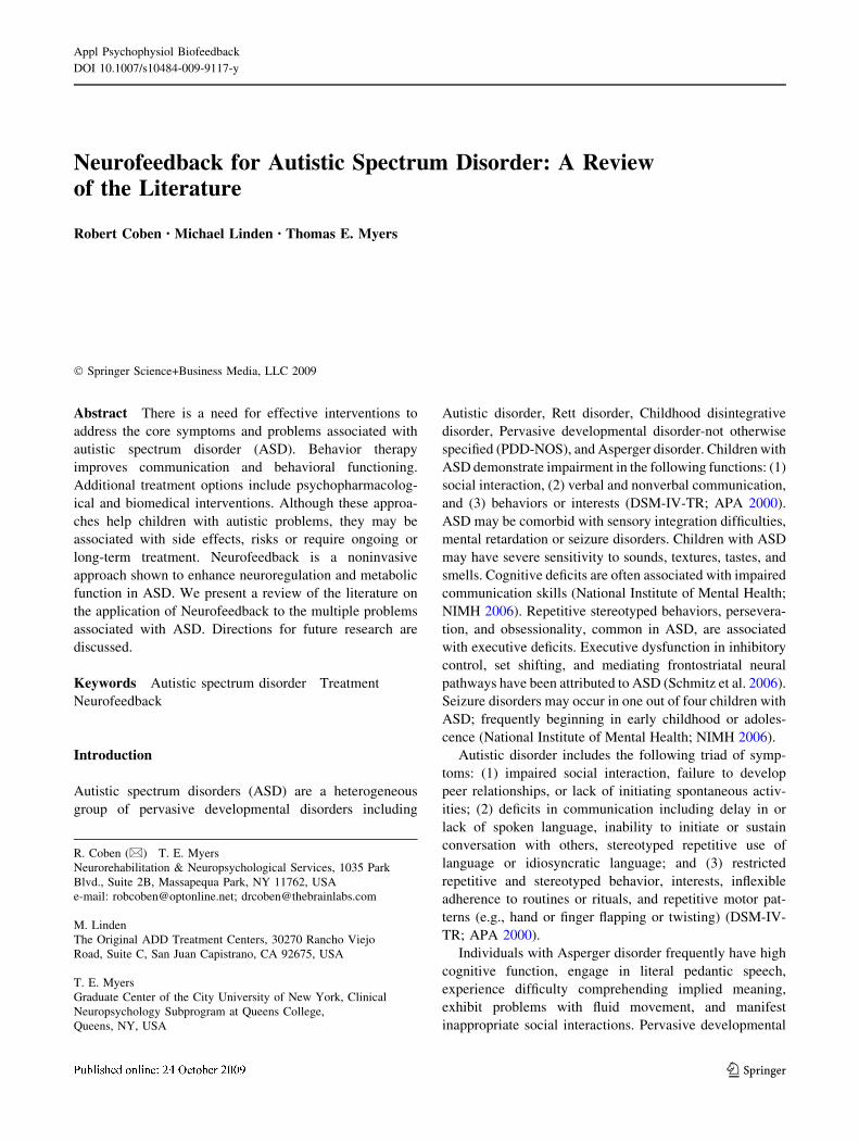
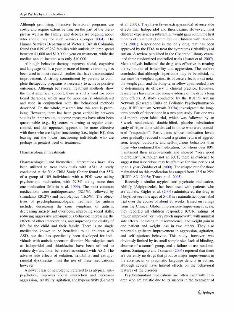
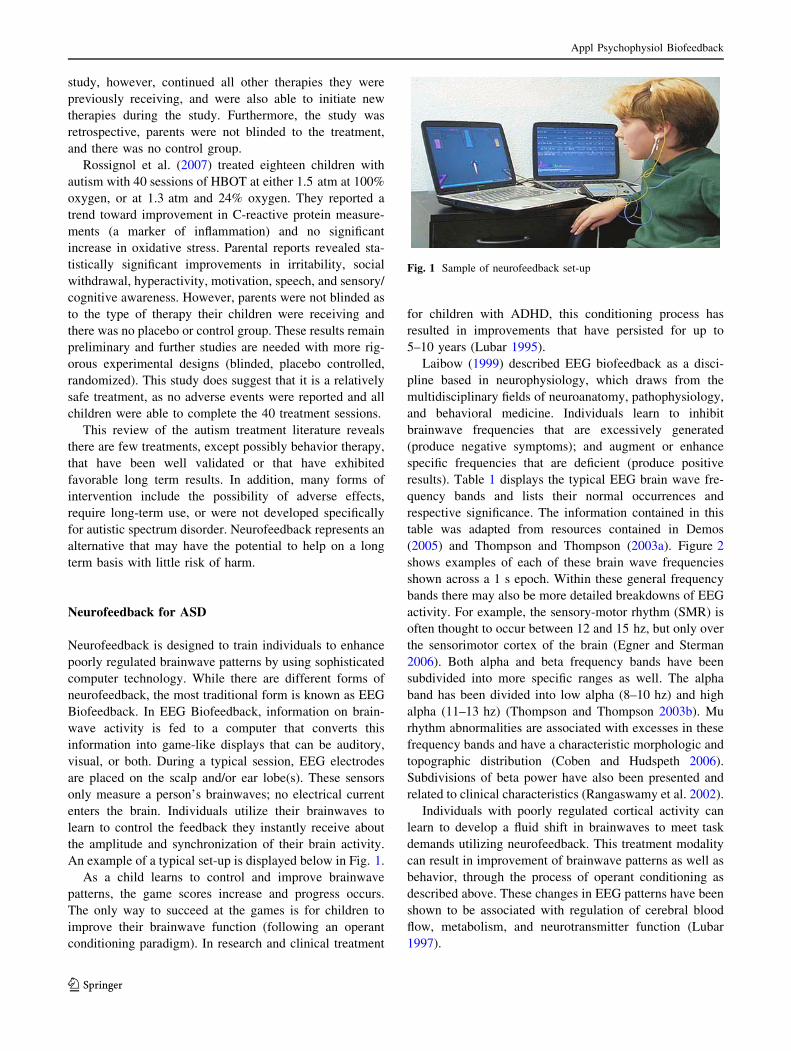
An example of a typical set-up is displayed below in Fig. 1.
As a child learns to control and improve brainwavepatterns, the game scores increase and progress occurs.
The only way to succeed at the games is for children to
improve their brainwave function (following an operantconditioning paradigm). In research and clinical treatment
for children with ADHD, this conditioning process hasresulted in improvements that have persisted for up to
5–10 years (Lubar 1995).
Laibow (1999) described EEG biofeedback as a disci-pline based in neurophysiology, which draws from the
multidisciplinary fields of neuroanatomy, pathophysiology,
and behavioral medicine. Individuals learn to inhibitbrainwave frequencies that are excessively generated
(produce negative symptoms); and augment or enhance
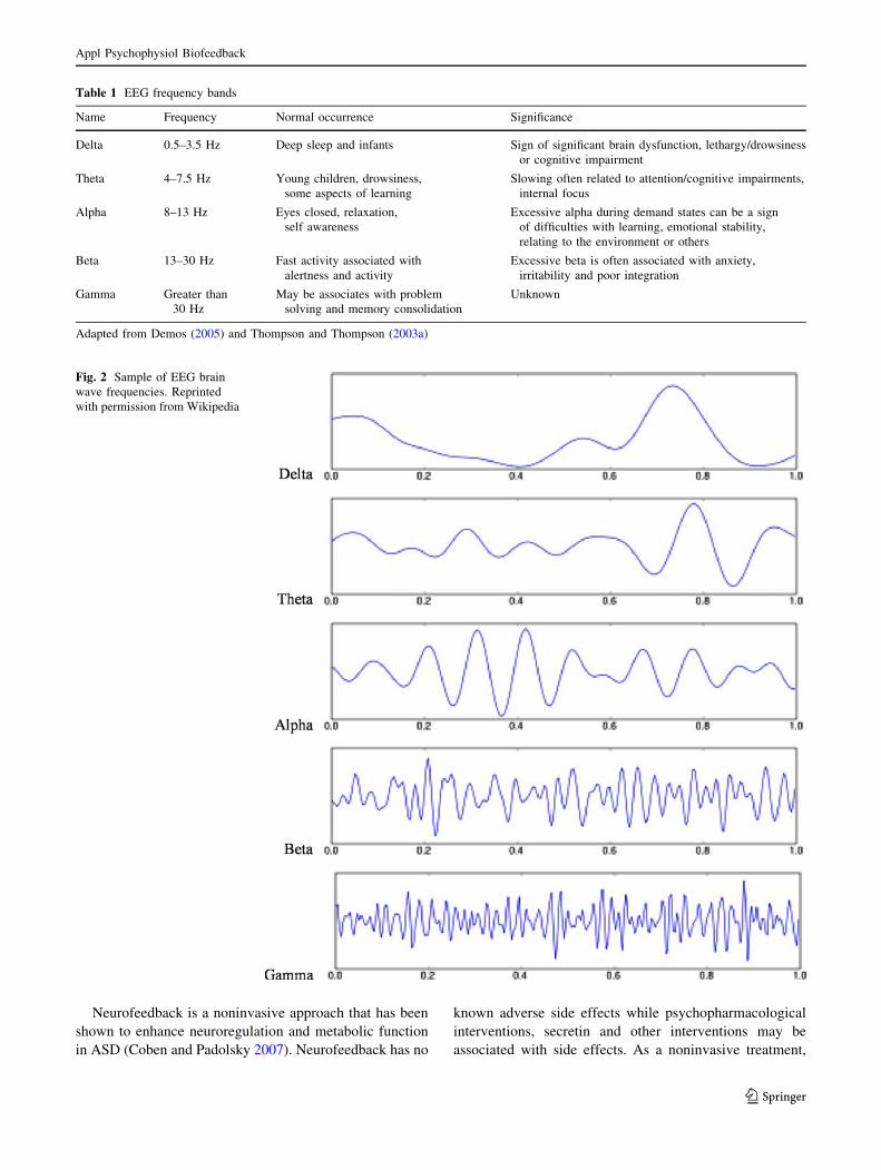
specific frequencies that are deficient (produce positiveresults). Table 1 displays the typical EEG brain wave fre-
quency bands and lists their normal occurrences and
respective significance. The information contained in thistable was adapted from resources contained in Demos
(2005) and Thompson and Thompson (2003a). Figure 2
shows examples of each of these brain wave frequenciesshown across a 1 s epoch. Within these general frequency
bands there may also be more detailed breakdowns of EEG
activity. For example, the sensory-motor rhythm (SMR) isoften thought to occur between 12 and 15 hz, but only over
the sensorimotor cortex of the brain (Egner and Sterman
2006). Both alpha and beta frequency bands have beensubdivided into more specific ranges as well. The alpha
band has been divided into low alpha (8–10 hz) and high
alpha (11–13 hz) (Thompson and Thompson 2003b). Murhythm abnormalities are associated with excesses in these
frequency bands and have a characteristic morphologic and
topographic distribution (Coben and Hudspeth 2006).Subdivisions of beta power have also been presented and
related to clinical characteristics (Rangaswamy et al. 2002).
Individuals with poorly regulated cortical activity canlearn to develop a fluid shift in brainwaves to meet task
demands utilizing neurofeedback. This treatment modality
can result in improvement of brainwave patterns as well asbehavior, through the process of operant conditioning as
described above. These changes in EEG patterns have been
shown to be associated with regulation of cerebral bloodflow, metabolism, and neurotransmitter function (Lubar
1997).
Fig. 1 Sample of neurofeedback set-up
Appl Psychophysiol Biofeedback
123
Neurofeedback is a noninvasive approach that has been
shown to enhance neuroregulation and metabolic function
in ASD (Coben and Padolsky 2007). Neurofeedback has no
known adverse side effects while psychopharmacological
interventions, secretin and other interventions may be
associated with side effects. As a noninvasive treatment,
Table 1 EEG frequency bands
Name Frequency Normal occurrence Significance
Delta 0.5–3.5 Hz Deep sleep and infants Sign of significant brain dysfunction, lethargy/drowsinessor cognitive impairment
Theta 4–7.5 Hz Young children, drowsiness,some aspects of learning
Slowing often related to attention/cognitive impairments,internal focus
Alpha 8–13 Hz Eyes closed, relaxation,self awareness
Excessive alpha during demand states can be a signof difficulties with learning, emotional stability,relating to the environment or others
Beta 13–30 Hz Fast activity associated withalertness and activity
Excessive beta is often associated with anxiety,irritability and poor integration
Gamma Greater than30 Hz
May be associates with problemsolving and memory consolidation
Unknown
Adapted from Demos (2005) and Thompson and Thompson (2003a)
Fig. 2 Sample of EEG brainwave frequencies. Reprintedwith permission from Wikipedia
Appl Psychophysiol Biofeedback
123
there are no external substances introduced internally as
part of neurofeedback interventions. The therapeutictreatment outcomes of neurofeedback training with indi-
viduals with ADHD (increased attention, reduced impul-
sivity and hyperactivity) have been reported to bemaintained over time and not reverse after treatment is
withdrawn (Linden et al. 1996; Lubar et al. 1995; Monastra
et al. 2005; Tansey 1993) as in drug therapy and diettherapy. In contrast to behavior therapy, positive treatment
outcomes that result from neurofeedback training are oftenachieved over the course of several months rather than a
year or more of intensive training.
EEG biofeedback, as a form of internvetion, began withBarry Sterman PhD (Sterman and Friar 1972) at UCLA,
who trained individuals to control their seizures by
increasing their sensorimotor rhythm (SMR) brainwaves.In 1976, Joel Lubar published his first of numerous
research studies using neurofeedback with students diag-
nosed with ADHD (Lubar and Bahler 1976). Studiesindicated that by increasing Beta and decreasing Theta
brainwaves at central scalp locations, improvements in
attention, impulsivity and hyperactivity often occurred(Linden et al. 1996). Lubar’s research in the 1980’s and
1990’s indicated that IQ increases also resulted from
Neurofeedback (Lubar et al. 1995). Lubar (1995) publisheda longitudinal study, indicating that the positive results
from neurofeedback were still significant in 15/16 behav-
iors after 10 years.Linden et al. (1996) published the first controlled, ran-
domized study of neurofeedback with students with
ADHD. Their results supported Lubar’s previous research,and indicated significant improvements in attention and
intelligence compared to a wait list control group. Other
researchers found that the effects of neurofeedback onADHD were similar to the results of stimulant medication
during treatment, but remained after treatment discontin-
ued. For example, Monastra et al. (2002) compared astimulant medication regime to neurofeedback, while pro-
viding parenting training to all parents of the 100 children
with ADHD included in the study. Their results supportedprevious findings of neurofeedback’s significant positive
effects with ADHD children and showed that the effects
were long-lasting, while those of stimulant medicationwere temporary. Fuchs et al. (2003) conducted a similar
comparison of neurofeedback and stimulant medication.
They used QEEG pattern analysis to emphasize more spe-cific NF protocols, including inhibiting high Beta (18–30)
activity. Their neurofeedback approach was shown to rival
the effects of methylphenidate, with similar significanteffects on multiple measures.
Neurofeedback, over a 30 year history of research with
ADHD, has consistently resulted in improvements inattention, impulsivity, hyperactivity, and IQ (see Monastra
et al. 2005, for a review and analysis). This successful
background of literature has been the foundation for theemergence of using neurofeedback with ASD.
QEEG Evaluation and Autistic Spectrum Disorder
Quantitative electroencephalographic (QEEG) evaluationor mapping is an assessment instrument designed to pin-
point anomalies in brain function (Hammond 2005). QEEGMaps, collected using 19 electrodes based on the Interna-
tional 10–20 system (Jasper 1958), are quantitative analy-
ses of EEG characteristics of frequency, amplitude andcoherence during various conditions or tasks. These data
can be statistically compared to an age-matched normative
database to reveal a profile of abnormalities. Such regionsand aspects of dysfunctional neurophysiology may then be
targeted specifically through individualized neurofeedback
protocols. QEEG analyses measure abnormalities, insta-bilities, or lack of proper communications pathways (con-
nectivity) necessary for optimal brain functioning.
QEEG analyses are conducted to assess underlyingneurophysiological patterns related to the symptoms and
challenges of children with ASD. In addition, assessment
of the raw EEG can be used to evaluate neurologicalabnormalities such as seizure disorders, which are common
in children with autism. QEEG data are important for
developing the most individualized, specific and successfulneurofeedback protocols for patients with ASD (Coben and
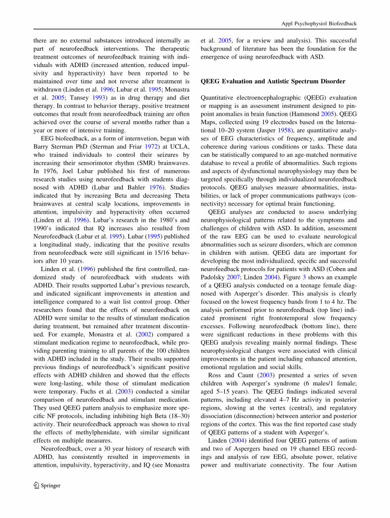
Padolsky 2007; Linden 2004). Figure 3 shows an example
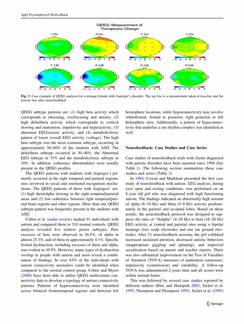
of a QEEG analysis conducted on a teenage female diag-nosed with Asperger’s disorder. This analysis is clearly
focused on the lowest frequency bands from 1 to 4 hz. The
analysis performed prior to neurofeedback (top line) indi-cated prominent right frontotemporal slow frequency
excesses. Following neurofeedback (bottom line), there
were significant reductions in these problems with thisQEEG analysis revealing mainly normal findings. These
neurophysiological changes were associated with clinical
improvements in the patient including enhanced attention,emotional regulation and social skills.
Ross and Caunt (2003) presented a series of seven
children with Asperger’s syndrome (6 males/1 female;aged 5–15 years). The QEEG findings indicated several
patterns, including elevated 4–7 Hz activity in posterior
regions, slowing at the vertex (central), and regulatorydissociation (disconnection) between anterior and posterior
regions of the cortex. This was the first reported case study
of QEEG patterns of a student with Asperger’s.Linden (2004) identified four QEEG patterns of autism
and two of Aspergers based on 19 channel EEG record-
ings and analysis of raw EEG, absolute power, relativepower and multivariate connectivity. The four Autism
Appl Psychophysiol Biofeedback
123
QEEG subtype patterns are: (1) high beta activity whichcorresponds to obsessing, overfocusing and anxiety, (2)
high delta/theta activity which corresponds to cortical
slowing and inattention, impulsivity and hyperactivity, (3)abnormal EEG/seizure activity, and (4) metabolic/toxic
pattern of lower overall EEG activity (voltage). The highbeta subtype was the most common subtype, occurring in
approximately 50–60% of the students with ASD. The
delta/theta subtype occurred in 30–40%, the AbnormalEEG subtype in 33% and the metabolic/toxic subtype in
10%. In addition, coherence abnormalities were usually
present in the QEEG profiles.The QEEG patterns with students with Asperger’s pri-
marily occurred in the right temporal and parietal regions,
sites involved in social and emotional recognition mecha-nisms. The QEEG patterns of those with Aspergers’ are:
(1) high theta/alpha slowing in the right temporal/parietal
areas and (2) low coherence between right temporal/pari-etal brain regions and other regions. More than one QEEG
subtype pattern was frequently present in the students with
ASD.Coben et al. (under review) studied 91 individuals with
autism and compared them to 310 normal controls. QEEG
analysis revealed five relative power subtypes. Pureexcesses of beta were observed in 26.5%, of alpha in
almost 25.3%, and of theta in approximately 4.1%. Specific
frontal dysfunction, including excesses of theta and alpha,was evident in 10.9%. However, many types of dysfunction
overlap in people with autism and most reveal a combi-
nation of findings. In over 83% of the individuals withautism connectivity anomalies could be identified when
compared to the normal control group. Coben and Myers
(2008) have been able to utilize QEEG multivariate con-nectivity data to develop a typology of autism connectivity
patterns. Patterns of hyperconnectivity were identified
across bilateral frontotemporal regions and between left
hemisphere locations, while hypoconnectivity may involveorbitofrontal, frontal to posterior, right posterior or left
hemisphere sites. Additionally, a pattern of hypoconnec-
tivity that underlies a mu rhythm complex was identified aswell.
Neurofeedback: Case Studies and Case Series
Case studies of neurofeedback trials with clients diagnosed
with autistic disorders have been reported since 1994 (See
Table 1). The following section summarizes these casestudies and series (Table 2).
In 1994, Cowan and Markham presented the first case
study of neurofeedback with autism. EEG analysis, duringeyes open and resting conditions, was performed on an
8 year old girl who was diagnosed with high functioning
autism. The findings indicated an abnormally high amountof alpha (8–10 Hz) and theta (4–8 Hz) activity predomi-
nately in the parietal and occipital lobes. Based on these
results, the neurofeedback protocol was designed to sup-press the ratio of ‘‘thalpha’’ (4–10 Hz) to beta (16–20 Hz)
EEG activity at central and parietal sites using a bipolar
montage (two scalp electrodes and one ear ground elec-trode). After 21 neurofeedback sessions, the girl exhibited
increased sustained attention, decreased autistic behaviors
(inappropriate giggling and spinning), and improvedsocialization based on parent and teacher reports. There
was also substantial improvement on the Test of Variables
of Attention (TOVA) measures of inattention (omission),impulsivity (commission) and variability. A follow-up
TOVA was administered 2 years later and all scores were
within normal limits.This was followed by several case studies reported by
different authors (Ibric and Hudspeth 2003; Sichel et al.
1995; Thompson and Thompson 1995). Sichel et al. (1995)
Fig. 3 Case example of QEEG analyses for a teenage female with Asperger’s disorder. The top line is a measurement taken at baseline and thebottom line after neurofeedback
Appl Psychophysiol Biofeedback
123
presented a case of an 8 year old boy with mild autism who
was treated with SMR (12–15 Hz) enhancement and theta
(4–8 Hz) suppression on the sensory-motor strip and pari-etal lobe, based on QEEG findings. Following 31 sessions
of monopolar neurofeedback training, positive changes
were reported across multiple domains related to hissymptoms. Ibric and Hudspeth (2003) presented another
case of an 8 year old boy with autism who was treated with
success using Roshi-assisted neurofeedback. Forty sessionsof training were conducted, based on QEEG data, which
included theta suppression and alpha enhancement. This
led to improvements in sleep, aggressive behavior, obses-sions and involuntary movements.
Multiple cases and case series have been presented
by Thompson and Thompson (1995, 2003a). Initially,Thompson and Thompson (1995) presented three cases of
children with autism and Aspergers disorder whose neu-
rofeedback protocols were primarily SMR enhancementand theta suppression at parietal and temporal scalp loca-
tions (P4-T4). Following neurofeedback, there wereimprovements in behavior and socialization skills. While
these cases were clearly not controlled trials in any way,
they did point to the possible benefits of this technique andencouraged further study.
Thompson and Thompson (2003b) presented a case
series review of neurofeedback in 60 individuals with highfunctioning ASD ranging in age from 5 to 51 years old.
The dependent measures of post-treatment improvement
were EEG assessments (central scalp locations with eyesopen), parent and teacher rating scales, IQ testing, aca-
demic measures and continuous performance tests (CPT).
Neurofeedback training parameters were based on the EEGassessment and the patients’ clinical symptoms. The most
common neurofeedback protocol was suppression of
dominant slow wave activity while enhancing 13–15 Hz
activity with scalp placement at Cz or C4 (central brain
sites) referenced to the right or left ear, respectively. The
number of neurofeedback sessions ranged from approxi-mately 40–100. Their results indicated improved EEG
patterns, with decreased theta/beta ratios and increases in
SMR amplitudes. IQ increases of 10 points were reportedfor the Neurofeedback group. The results of the TOVA
CPT were inconclusive, because many of the patients with
autism were unable to complete the tests. Significantimprovement in social interaction was reported by parents.
The most significant improvements were in those individ-
uals who received greater than 80 sessions. Thompson andThompson continue to collect case series data to date on
hundreds of ASD patients and continue to report successful
treatment outcomes (Thompson and Thompson 2007).Limsila et al. (2004) conducted the largest case series
study of 180 children (aged 3–18) with autism in Thailand.
This included a form of neurofeedback called Hemoen-cephalography (HEG) or cerebral blood flow biofeedback.
Following 40 sessions of near infrared (nir) HEG trainingover frontal sites (Fp1 and Fp2), HEG readings reflecting
brain oxygenation increased by 53%. Eighty-six percent of
the 81 children with the capacity to learn in public schoolincreased their grade point average (GPA) more than
0.5 points, while only 4% decreased their GPA by more
than 0.5 points. However, there was no control or non-treatment group and the study did not assess or control for
IQ or the influence of other treatment interventions. These
findings were suggestive of positive therapeutic outcomesfor HEG neurofeedback as a treatment for children with
autism, but must be viewed cautiously due to the lack of
controls as would be true of any case series.Linden (2004) presented a series of case studies with
15 students (14 males/1 female), aged 5–15 years old,
diagnosed with autism or Aspergers. All subjects received
Table 2 ASD neurofeedback case studies
Author QEEG pattern NF protocol Improvements
Cowan and Markham (1994) High alpha and theta Suppress 4–10, enhance 16–20 Attention, motor behaviors,impulsivity, socialization, TOVA
Sichel et al. (1995) High theta, low Beta Suppress theta, enhance SMR Socialization, self-stim behaviors,speech
Thompson and Thompson (1995) High theta, low SMR Suppress theta, enhance SMRP4-T4
Behaviors, social, academic
Ibric and Hudspeth (2003) High beta, hypocoherence QEEG based Behavior, sleep, movements
Thompson and Thompson (2003a) High theta, low beta/SMR QEEG based; suppress theta,enhance 13–15 C4
EEG patterns, IQ, socialinteractions, alertness
Limsila et al. (2004) Not measured HEG frontally Grades
Linden (2004) High beta, high delta, low voltage,abnormal EEG, hypocoherence
QEEG based Attention, impulsivity,hyperactivity, EEG patterns,communication, socialization
Scolnick (2005) Abnormal patterns EEG based Behaviors
Appl Psychophysiol Biofeedback
123
pre- and post- neurofeedback QEEG evaluations, parent
and teacher ADD and ASD rating scales, and IVA andTOVA Continuous Performance Tests (CPT). IQ was not
measured, however, and there was no control group. All of
the neurofeedback protocols used with the ASD subjectswere QEEG Guided (selected based on the QEEG analysis)
and were selected with the goal of normalizing the QEEG
patterns and improving the clinical symptoms. The subjectsreceived between 20 and 60 (average was 50–60), 45 min
sessions (30 min of actual neurofeedback training time) ofneurofeedback between 2 and 5 times per week. The results
indicated improved CPT scores and deceased inattention
and hyperactivity on Parent and Teacher ADHD behaviorrating scales. In addition, most of the abnormal QEEG
patterns (there were several abnormal EEG patterns for
some students) were improved in all students, includingseveral students whose abnormal raw EEG patterns ‘‘nor-
malized’’. Moreover, many of the students were able to
reduce or eliminate their medications. Improvement wasalso reported on Parent and Teacher Autism (CARS) and
Asperger (OASIS) behavior rating scales for communica-
tion and socialization in all cases. Several of the studentswith ASD were mainstreamed into regular classes without
their classroom aides.
A pilot study of the effects of neurofeedback with As-perger’s syndrome was completed by Scolnick (2005). Five
adolescent males who attended a therapeutic day school
completed 24 sessions of neurofeedback. The results indi-cated a trend to normalize their EEGs, but was not statis-
tically significant. All subjects showed improved focusing,
anxiety and disruptive behavior as rated by parent andteacher rating scales. Again, IQ was not measured and
there was no control group.
In summary, several case studies and case series usingQEEG and neurofeedback with individuals diagnosed with
ASD have been reviewed. Although these studies utilized
different instruments and neurofeedback protocols, and hada varied number of neurofeedback sessions, all reported
significant improvement either on measures of QEEG,
IVA/TOVA CPT tests, or Parent/Teacher behavior ratingscales. In addition, significant clinical symptomatic
improvements were reported for communication, sociali-
zation, anxiety, attention, stereotypic behaviors, and evenmedication reduction/elimination. As noted, however,
these studies must be viewed cautiously, as they are
uncontrolled and cannot demonstrate if the changesobserved are due only to the neurofeedback treatment or
other factors. As many of these referenced case/case series
presentations have not been the subject of extensive peerreview, additional cautions should be exercised in making
generalizable conclusions. Additional controlled studies
with larger sample sizes would be helpful in order tosupport the results of these case studies.
Group Pilot Studies of Neurofeedback for ASD
Two pilot group studies of neurofeedback for ASD have
been conducted. In the first (Jarusiewicz 2002), twelve
children each were assigned to an experimental or a controlgroup. The experimental group received a mean of 36
treatment sessions (range = 20–69). Treatment protocols
were based on the Othmer Assessment (1997) to determineover-, under-, and unstable arousal. The Autism Treatment
Evaluation Checklist (ATEC; Rimland and Edelson 2000)
was used to assess outcome. Children who completedneurofeedback training attained an average 26% reduction
in the total ATEC rated autism symptoms in contrast to 3%
for the control group. Parents reported improvement insocialization, vocalization, anxiety, schoolwork, tantrums,
and sleep while the control group had minimal changes in
these domains. However, the outcome measure used isbased on only parent report with no other objective mea-
sures utilized.
The second pilot study of the effects of neurofeedbackwas conducted by Kouijzer et al. (2009). Fourteen children
with ASD, 7 in the treatment and 7 in the waitlist (no
treatment) control group, were matched for age, gender andintelligence, but were not randomly assigned. The treat-
ment group received 40 sessions of neurofeedback treat-
ment at scalp location C4. Theta activity (4–7 Hz) wasinhibited while SMR activity (12–15 Hz) was rewarded.
Pre and post assessment consisted of EEG learning curves,QEEG analysis, tests of executive functioning and behavior
rating scales (CCC-2, Dutch Autism Scale). The findings
showed that the neurofeedback trained group demonstratedsignificant improvement in attentional control, cognitive
flexibility and goal setting compared to the control group.
Results of parent rating scales also showed improvementsin social interaction and communication skills. These
changes were associated with improvements in EEG
learning curves.Interestingly, this same research group performed a
12-month follow-up of the treated patients with ASD
(Kouijzer et al. 2009b). Both changes in executive func-tioning and behavior were maintained suggesting that
neurofeedback may have long-lasting effects for children
with autism. These pilot studies have shown positiveresults, but caution should be exercized as their sample
sizes were quite small. Nevertheless, the optimism
regarding their findings has led to more controlled researchwith larger sample sizes.
Controlled Group Studies of Neurofeedback for ASD
In the largest published, controlled study to date of neu-rofeedback for autistic disorders, Coben and Padolsky
Appl Psychophysiol Biofeedback
123
(2007) studied 49 children on the autistic spectrum. The
experimental group included 37 children that receivedQEEG connectivity guided neurofeedback (20 sessions
performed twice per week) and the wait-list control group
included 12 children that were matched for age, gender,race, handedness, other treatments, and severity of ASD. A
broad range of assessments were utilized including:
parental judgment of outcome, neuropsychological tests,behavior rating scales, QEEG analysis and infrared imag-
ing. Treatment protocols were assessment-based (includingQEEG power and coherence) and individualized for each
child receiving neurofeedback training with a specific
focus on the remediation of connectivity anomalies. Basedon parental judgment of outcome, there was an 89% suc-
cess rate for neurofeedback. There was an average 40%
reduction in core ASD symptomatology based on parentratings scales. There were also significant improvements,
as compared to the control group, on neuropsychological
measures of attention, executive functioning, visual-perceptual processes and language functions. Reduced
cerebral hyperconnectivity was associated with positive
clinical outcomes in this population. In all cases of reportedimprovement in ASD symptomatology, positive outcomes
were confirmed by neuropsychological and neurophysio-
logical assessment.Another, as yet unpublished, controlled group study was
conducted by Coben (2006). Forty patients with autism
were non-randomly assigned to one of three groups: (1) anear infrared hemoencephalography (HEG) trained group
(n = 10), (2) a passive infrared HEG trained group
(n = 18), or (3) a wait-list control group (n = 12). Nearinfrared HEG (Toomim et al. 2004) measures changes in
cerebral oxygenation at levels below one micron. Optical
diodes detect changes in oxygenated and deoxygenatedblood reflecting changes in regional cerebral blood flow.
The specificity of its measurement is theorized to lead to
highly localized changes. Passive infrared HEG (Carmen2004) measures thermoregulatory output in the frequency
range of 7–14 microns. Its surface areas of measurement
and frequency range are quite larger than nirHEG and itseffects may be more global as a result. The wait list control
group (n = 12) was matched for gender, age, race, hand-
edness, and other treatments. All patients had previouslycompleted 20 sessions of EEG Biofeedback (see Coben and
Padolsky 2007). The next phase of training was assess-
ment-guided HEG, which identified frontal system dys-function in all patients based on neurobehavioral,
neuropsychological testing, infrared imaging, and QEEG
data. Findings indicated an average success rate of 90%based on parental judgment. Parental ratings showed an
average 42% reduction in overall autistic symptoms. Social
interaction deficits decreased by 55%. Communication andsocial communication deficits decreased by 55 and 52%,
respectively. In addition, there were statistically significant
improvements in neurobehavioral and neuropsychologicalfunctioning. These improvements were associated with
enhancement of brain thermal regulation and reduction in
abnormal QEEG findings. Interestingly, there were differ-ences in the effects of each type of HEG with nirHEG
impacting attention more and pirHEG leading to better
emotional control and social skills. There were also dif-ferential effects on the EEG based on which approach was
employed.Two studies have focused on abnormal Mu rhythms (a
sign of mirror neuron dysfunction) (Oberman et al. 2005)
in children with autism and if neurofeedback could lessenthese anomalies. In a series of two experiments, Pineda
et al. (2008) studied 27 children with high functioning
autism. In study 1, 8 high functioning males were randomlyassigned to an experimental (n = 5) or placebo group
(n = 3). One subject dropped out of the experimental
group midway through the training. Neurofeedback train-ing included 30, 30 min sessions with rewards for mu-like
activity (8–13 hz) and inhibits for EMG (30–60 hz). Parent
ratings scale data (Autism Treatment Evaluation Checklist(ATEC); Rimland and Edelson 2000) showed small chan-
ges (9–13%) in two of the four experimental participants.
These data should be considered in line with a pilot studyconsidering the very small sample size. In Study 2, 19
children with high functioning ASD were randomly
assigned to an experimental (n = 9) or placebo (n = 10)group. One very positive addition to this study was the
verification of their diagnoses by administering the Autism
Diagnostic Observation Schedule (ADOS; Lord et al. 1999)and the Autism Diagnostic Interview-Revised (ADI-R;
Rutter et al. 2003). Neurofeedback training was similar to
study one except the reward band was now 10–13 hz.Parent ratings showed a small, but significant reduction in
symptoms (ATEC Total score). However, of concern was
an increase in ratings of Sensory/Cognitive Awareness inexcess of 40% that did not occur in the placebo control
group. This suggests that, according to their parents, par-
ticipants improved in some areas and worsened in others.In another study related to mu-rhythms, Coben and
Hudspeth (2006) studied fourteen children with ASD who
were identified as having significantly high levels of Murhythm (distorted alpha-like) activity and a failure to sup-
press mu during observational activity. They all received
assessment guided neurofeedback, with a strong focus onaspects of mu power and connectivity. The participants
were non-randomly assigned to an interhemispheric bipolar
training (n = 7) or a coherence training (n = 7) groupdesigned to increase connectivity between central regions
and the peripheral frontal cortex. All patients were given
neurobehavioral, neuropsychological testing, and QEEGassessment. Both groups of patients improved significantly
Appl Psychophysiol Biofeedback
123
on neurobehavioral and neuropsychological measures.
However, only in the coherence training treatment groupwas Mu activity significantly reduced. Increased coherence
was associated with diminished mu and improved levels of
social functioning.Lastly, Coben (2007) conducted a controlled neuro-
feedback study focused on intervention for prominent
social skill deficits based on a facial/emotional processingmodel. Fifty individuals with autism were included in these
analyses and all had previously had some neurofeedback.All patients underwent pre- and post neuropsychological,
QEEG and parent rating scale assessments. Twenty-five
individuals were each assigned to an active neurofeedbackand wait list control group, respectively, in a non-
randomized fashion. The two groups were matched for age,
gender, race, handedness, medication usage, autisticsymptom severity, social skill ratings, and visual-percep-
tual impairment levels. Neurofeedback training was QEEG
connectivity guided and included coherence training (alongwith amplitude inhibits) between maximal sights of hypo-
coherence over the right posterior hemisphere. The group
that received the coherence training showed significantchanges in symptoms of autism, social skills and visual-
perceptual abilities such that all improved. Regression
analyses showed that changes in visual-perceptual abilitiessignificantly predicted improvements in social skills. EEG
analyses were also significant, showing improvements in
connectivity and source localization of brain regions(fusiform gyrus, superior temporal sulcus) associated with
enhanced visual/facial/emotional processing.
In the five controlled group studies that have beencompleted, a total of 180 individuals with autism have been
studied with positive results reported in each study. These
findings have included positive changes as evidenced byparental report, neuropsychological findings and changes in
the EEG (Coben 2007). Both Coben and Padolsky (2007)
and Yucha and Montgomery (2008) have viewed these dataas demonstrating a level of efficacy of possibly efficacious
based on the standards put forth by the Association for
Applied Psychophysiology and Biofeedback (AAPB 2006).Added to these initial findings of efficacy is preliminary
evidence that the effects of neurofeedback on the symp-
toms of autism are long-lasting (1–2 years) (Coben 2009;Kouijzer et al. 2009a). While these findings are initially
encouraging, there are many limitations that prevent firm
conclusions to be drawn from the data collected thus far.First, these studies have largely included non-random-
ized samples. It is possible that an unknown selection bias
exists which could have impacted the findings. Second,none of these studies have included participants or thera-
pists/experimenters who were blind to the condition.
Knowledge of group placement could have impacted thefindings such that those in treatment (and their parents)
would be prone to report significant changes. Third, there
has been no attempt to control for placebo effects, attentionfrom a caring professional or expectations of treatment
benefit. A randomized, double-blinded, placebo-controlled
study is clearly needed to further demonstrate efficacy.In terms of generalization of these findings to the larger
population of individuals who are autistic, very young
children and adults have not been well represented in thesegroup studies. Lastly, there is the question of whether
neurofeedback may be applicable to persons who are lowerfunctioning or whom have more severe symptoms associ-
ated with autism. These populations should be the focus of
future investigations.
Discussion
With the possible exception of behavior modification inter-
ventions, there are few interventions for childrenwith autismwith proven efficacy. Pharmacologic interventions, hyper-
baric oxygen and vitamin supplementation have shown some
promise. However, further research is necessary to demon-strate their efficacy. Based on the above review, we consider
neurofeedback to be in a similar position with respect to
efficacy for ASD. While the recent research in this appli-cation is encouraging, further advancements are necessary in
this ongoing research track to demonstrate efficacy accord-
ing to current research standards.The five levels of treatment efficacy that provide guid-
ance for applied psychophysiologic research have been
outlined (LaVaque et al. 2002; Yucha and Montgomery2008) as follows. Level 1 is labeled ‘‘not empirically
supported’’ and is assigned to treatments supported by
evidence from only case studies in non-peer-reviewedjournals and anecdotal reports. Level 2 is entitled ‘‘possibly
efficacious’’ and is given to treatments investigated in at
least one study, where statistical power is sufficient, out-come measures are well-identified, but random assignment
to a control condition is lacking. Level 3, called ‘‘probably
efficacious,’’ is assigned to treatments that demonstratebeneficial effects in multiple observational studies, clinical
studies, wait list controlled studies, or within-subject and
intra-subject replication studies. Level 4, termed ‘‘effica-cious,’’ is reserved for treatments shown to be statistically
superior (with sufficient power) to a control condition or
equivalent to an established treatment, in designs that uti-lize random assignment and clearly identify the population
of interest and the procedures employed, sufficient to
permit replication by others. Additionally, positive treat-ment outcomes need to be confirmed in at least two inde-
pendent research settings. Level 5, labeled ‘‘efficacious and
specific,’’ is assigned to treatments that demonstrate sta-tistically superior results compared to a credible placebo,
Appl Psychophysiol Biofeedback
123
medication, or another established treatment, again repli-
cated in at least two independent research settings.The group studies reviewed above are judged to support
a Level 2 determination, possibly efficacious, for the
application of neurofeedback for autistic disorders (Cobenand Padolsky 2007; Yucha and Montgomery 2008). Further
research is necessary utilizing random assignment, blinding
of participants, procedures to control for placebo effects andreplicated in at least two independent settings to establish
neurofeedback as an efficacious treatment for ASD. Wewould add that such studies should also seek to document
brain-related changes in the participants to further docu-
ment the effects and mechanism of this treatment modality.In addition to questions about the general efficacy of
neurofeedback for ASD, there are additional questions
about the relative efficacy of different types of neuro-feedback. For example, while we have shown that con-
nectivity (QEEG) guided neurofeedback may be effective
(Coben and Padolsky 2007), there is also evidence thatsymptom based approaches are helpful as well (Jarusiewicz
2002). In the only study to compare these approaches,
Coben and Myers (2009) have shown that connectivityguided neurofeedback has demonstrated enhanced efficacy,
but that both approaches can be helpful. More studies of
this type are warranted. Other types of neurofeedback,including power training, coherence training, hemoen-
cephalography, and training of specific QEEG abnormali-
ties (e.g., mu rhythm/mirror neuron dysfunction), may alsoprove to have differential efficacy for autism in general and
particular symptoms specifically.
Future clinical work and research might also focus onpossible synergistic effects between neurofeedback and
other interventions (i.e., HBOT, behavior therapy, etc.)
commonly used for this population. It also may turn outthat such treatments could be used in combination, but in a
sequential fashion. For example, ABA may be indicated
with very young children to be followed by neurofeedbackto ‘‘fine tune’’ treatment effects and address other cognitive
and social aspects.
In sum, we view neurofeedback as an intervention thatmay prove to be efficacious in the treatment of symptoms
of autism. Presently, it should be viewed as possibly effi-
cacious with potential and would then be in the same cat-egory as most interventions used with this challenging
population. There is a great need for carefully and well
designed studies to address the issues discussed above(randomization, blinding, placebo controls, multiple mea-
sures, etc.). Measuring brain-related changes that may
occur as a result of neurofeedback is one way of demon-strating its efficacy and mechanism of action. Additionally,
longer follow-up periods should be included in such studies
to measure the durability of effects.
References
Adams, J., Geis, L., Baral, M., Mitchell, J. I., Hensley, A., Newmark,S., et al. (2008). Results of DMSA treatment study. Presented atthe Autism One Conference, Chicago, Illinois.
Aman, M. G., Singh, N. N., & Stewart, A. W. (1985). The aberrantbehavior checklist: A behavior rating scale for the assessment oftreatment effects. American Journal of Mental Deficiency, 5,485–491.
American Academy of Child & Adolescent Psychiatry. (1999, March3). Secretin in the treatment of autism [Policy statement].Retrieved August 6, 2007 from http://www.aacap.org/cs/root/policy_statements/secretin_in_the_treatment_of_autism.
American Psychiatric Association. (2000). Diagnostic and statisticalmanual of mental disorders, fourth edition-text revision. Wash-ington, DC: American Psychiatric Association.
Amminger, G. P., Berger, G. E., Schafer, M. R., Klier, C., Friedrich,M. H., & Feucht, M. (2007). Omega-3 fatty acids supplemen-tation in children with autism: A double-blind randomized,placebo-controlled study. Biological Psychiatry, 61, 551–553.
Anderson, S. R., Avery, D. L., DiPietro, E. K., Edwards, G. L., &Christian, W. P. (1987). Intensive home-based early interventionwith autistic children. Education and Treatment of Children, 10,352–366.
Association for Applied Psychophysiology & Biofeedback. (2006).Efficacy: How we rate the efficacy of our treatments or how toknow if our treatments actually work. Retrieved February 22, 2006from http://www.aapb.org/i4a/pages/index.cfm?pageid=3336.
Attwood, T. (1998). Asperger’s syndrome: A guide for parents andprofessionals. London, England: Jessica Kingsley Publishers.
Barnard, A. L., Young, A. H., Pearson, A. D. J., Geddes, J., & Obrien,G. (2002). A systematic review of the use of atypical antipsy-chotics in autism. Journal of Psychopharmacology, 16, 93–102.
Bassett, K., Green, C. J., & Kazanjian, A. (2000). Autism and Lovaastreatment: A systematic review of effectiveness evidence. Van-couver, BC: British Columbia Office of Health TechnologyAssessment, The University of British Columbia.
Bayley, N. (1993). Bayley scales of infant development (2nd ed.). SanAntonio: Psychological Corp.
Bell, J. G., Sargent, J. R., Tocher, D. R., & Dick, J. R. (2000). Redblood cell fatty acid compositions in a patient with autisticspectrum disorder: A characteristic abnormality in neurodevel-opmental disorders? Prostaglandins, Leukotienes and EssentialFatty Acids, 63(1–2), 21–25.
Ben-Itzchak, E., & Zachor, D. A. (2007). The effects of intellectualfunctioning and autism severity on outcome of early interventionfor children with autism. Research in Developmental Disabili-ties, 28, 287–303.
Bernard, S., Enayati, A., Binstock, T., Roger, H., Redwood, L., &McGinnis, W. (2000) Autism: A unique type of mercurypoisoning. ARC Research, Available from the Autism ResearchInstitute.
Birnbrauer, J. S., & Leach, D. J. (1993). The Murdoch earlyintervention program after 2 years. Behavior Change, 10, 63–74.
Blaxill, M. F. (2004). What’s going on? The question of time trends inautism. Public Health Reports, 119, 536–551.
Bradstreet, J., Geier, B. A., Kartzinel, J. J., Adams, J. B., & Geier, M.R. (2003). A case–control study of mercury burden in childrenwith autistic spectrum disorders. Journal of American Physiciansand Surgeons, 8(3), 76–79.
Butter, E. M., Mulick, J. A., & Metz, B. (2006). Eight case reports oflearning recovery in children with pervasive developmentaldisorders after early intervention. Behavioral Interventions, 21,227–243.
Appl Psychophysiol Biofeedback
123
Cade, R., Privette, M., Fregley, M., Rowland, N., Sun, Z., Zele, V.,et al. (1999). Autism and schizophrenia: Intestinal disorders.Nutritional Neuroscience, 3, 57–72.
Carmen, J. A. (2004). Passive infared hemoencephalography: Fouryears and 100 migraines. Journal of Neurotherapy, 8(3), 23–51.
Center for Disease Control and Prevention. (2006). How common areautism spectrum disorder (ASD)? Retrieved May 31, 2006 fromhttp://www.cdc.gov/ncbddd/autism/asd_common.htm.
Center for Disease Control and Prevention. (2007). Prevalence of theautism spectrum disorders (ASDs in Multiple Areas of theUnited States, 2000 and 2002). MMWR, 56 (SS 1–2).
Coben, R. (2006, September). Hemoencephalography for autisticspectrum disorder. Presented at the 14th Annual Conference ofthe International Society for Neuronal Regulation, Atlanta,Georgia.
Coben, R. (2007, September). Autistic spectrum disorder: A con-trolled study of EEG coherence training targeting social skilldeficits. Presented at the 15th annual conference of the Interna-tional Society for Neurofeedback and Research, San Diego,California.
Coben, R. (2009). Efficacy of connectivity guided neurofeedback forautistic spectrum disorder: Controlled analysis of 75 cases with a1 to 2 year follow-up. Journal of Neurotherapy, 13(1), 81.
Coben, R., Hirshberg, L. M., & Chabot, R. (Under Review). EEGdiscriminant power and subtypes in autistic spectrum disorder.International Journal of Psychophysiology.
Coben, R., & Hudspeth, W. (2006, September). Mu-like rhythms inautistic spectrum disorder: EEG analyses and neurofeedback.Presented at the 14th Annual Conference of the InternationalSociety for Neuronal Regulation, Atlanta, Georgia.
Coben, R., & Myers, T. E. (2008). Connectivity theory of autism: Useof connectivity measures in assessing and treating autisticdisorders. Journal of Neurotherapy, 12(2–3), 161–179.
Coben, R. & Myers, T. E. (2009). The relative efficacy of con-nectivity guided and symptom based EEG biofeedback forAutistic disorders. Applied Psychophysiology and Biofeedback.doi:10.1007/s10484-009-9102-5.
Coben, R., & Padolsky, I. (2007). Assessment-guided neurofeedbackfor autistic spectrum disorder. Journal of Neurotherapy, 11(3),5–18.
Committee on Children with Disabilities. (2001). Technical report:The pediatrician’s role in the diagnosis and management ofautistic spectrum disorder in children. Pediatrics, 107(5), 1–35.
Cook, E. H., Rowlett, R., & Jaselskis, C. (1992). Fluoxetine treatmentof children and adults with autistic disorder and mentalretardation. Journal of the American Academy of Child andAdolescent Psychiatry, 31, 739–745.
Couper, J. (2004). Who should pay for intensive behaviouralintervention in autism? A parent’s view. Journal of Paediatricsand Child Health, 40(9–10), 559–561.
Couturier, J. L., & Nicolson, R. (2002). A retrospective assessment ofcitalopram in children and adolescents with pervasive develop-mental disorders. Journal of Child and Adolescent Psychophar-macology, 12, 243–248.
Cowan, J., & Markham, L. (1994, March). EEG biofeedback for theattention problems of autism: A case study. Presented at the 25thAnnual Meeting of the Association for Applied Psychophysiol-ogy and Biofeedback, Atlanta, GA.
DeLong, G. R., Ritch, C. R., & Burch, S. (2002). Fluoxetine responsein children with autistic spectrum disorders: Correlation withfamilial major affective disorder and intellectual achievement.Developmental Medicine and Child Neurology, 44, 652–659.
Demos, J. N. (2005). Getting started with neurofeedback. New York:WW Norton & Company.
Deprey, L. J., Brule, N., Widjaja, F., Sepheri, S., Blank, J.,Neubrander, J., et al. (2006, October). Double-blind placebo-
controlled, cross-over trial of subcutaneous methylcobalamin inchildren with autism: Preliminary results. Poster presented at theAnnual Meeting of the American Academy of Child andAdolescent Psychiatry, San Diego, CA.
Deth, R. C. (2004, October) Methylcobalamin for autistic children.Presented at the 34th Annual Meeting of the Society forNeuroscience, San Diego, CA.
Dietrich, K. N., Ware, J. H., Salganik, M., Radcliffe, J., Rogan, W. J.,Rhoads, G. G., et al. (2004). Effect of chelation therapy on theneuropsychological and behavioral development of lead-exposedchildren after school entry. Pediatrics, 114, 19–26.
Ducharme, J. M. (2005). Errorless compliance training. In M. Hersen& J. Rosqvist (Eds.), Encyclopedia of behavior modification andcognitive behavior therapy (Vol. 2: Child Clinical Applications).Thousand Oaks, CA: Sage.
Ducharme, J. M., Sanjuan, E., & Drain, T. (2007). Errorlesscompliance training. Success-focused behavioral treatment ofchildren with Asperger syndrome. Behavior Modification, 31,329–344.
Egner, T., & Sterman, M. B. (2006). Neurofeedback treatment ofepilepsy: From basic rationale to practical application. ExpertReview of Neurotherapeutics, 6, 247–257.
Eikeseth, S., Smith, T., Jahr, E., & Eldevik, S. (2002). Intensivebehavioral treatment at school for 4–7-year-old children withautism: A 1-year comparison controlled study. Behavior Mod-ification, 26, 49–68.
Eikeseth, S., Smith, T., Jahr, E., & Eldevik, S. (2007). Outcome forchildren with autism who began intensive behavioral treatmentbetween ages 4 and 7: A comparison controlled study. BehaviorModification, 31, 264–278.
Elder, J. H., Shankar, M., Shuster, J., Theriaque, D., Burns, S., &Sherrill, L. (2006). The gluten-free, casein-free diet in autism:Results of a preliminary double blind clinical trial. Journal ofAutism and Developmental Disorders, 36, 413–420.
Esch, B. E., & Carr, J. E. (2004). Secretin as a treatment for autism: Areview of the evidence. Journal of Autism and DevelopmentalDisorders, 34, 543–556.
Fenske, E. C., Zalenski, S., Krantz, P. J., & McClannahan, L. E.(1985). Age at intervention and treatment outcome for autisticchildren in a comprehensive intervention program. Analysis andIntervention in Developmental Disabilities, 5, 49–58.
Fuchs, T., Birbaumer, N., Lutzenberger, W., Gruzelier, J. H., & Kaiser,J. (2003). Neurofeedback treatment for attention-deficit/hyper-activity disorder in children: A comparison with methylpheni-date. Applied Psychophysiology and Biofeedback, 28, 1–12.
Geller, D. A., Hoog, S. L., Heiligenstein, J. H., Ricardi, R. K.,Tamura, R., Kluszynski, S., et al. (2001). Fluoxetine treatmentfor obsessive compulsive disorder in children and adolescents:A placebo controlled clinical trial. Journal of the AmericanAcademy of Child and Adolescent Psychiatry, 40(7), 773–779.
George, M. S., Costa, D. C., Kouris, K., Ring, H. A., & Ell, P. J.(1992). Cerebral blood flow abnormalities in adults with infantileautism. Journal of Nervous and Mental Disease, 180, 413–417.
Golden, Z. L., Neubauer, R., Golden, C. J., Greene, L., Marsh, J., &Mleko, A. (2002). Improvement in cerebral metabolism inchronic brain injury after hyperbaric oxygen therapy. Interna-tional Journal of Neuroscience, 112, 119–131.
Green, V. A., Pituch, K. A., Itchon, J., Choi, A., O’Reilly, M., &Sigafoos, J. (2006). Internet survey of treatments used by parentsof children with autism. Research in Developmental Disabilities,27, 70–84.
Hamilton, L. (2000). Facing autism: Giving parents reasons for hopeand guidance for help. Colorado Springs, Colorado: WaterBrook Press.
Hammond, D. C. (2005). What is neurofeedback? Retrieved July 25,2006 from http://www.isnr.org/pubarea/whatisnfb.pdf.
Appl Psychophysiol Biofeedback
123
Harris, S. L., Handleman, J. S., Gordon, R., Kristoff, B., & Fuentes, F.(1991). Changes in cognitive and language functioning ofpreschool children with autism. Journal of Autism and Devel-opmental Disorders, 21, 281–290.
Hediger, M. L., England, L. J., Molloy, C. A., Yu, K. F., Manning-Courtney, P., & Mills, J. L. (2008). Reduced bone corticalthickness in boys with autism or autism spectrum disorder.Journal of Autism and Developmental Disorders, 38, 848–856.
Hellings, J. A., Kelley, L. A., Gabrielli, W. F., Kilgore, E., & Shah, P.(1996). Sertraline response in adults with mental retardation andautistic disorder. Journal of Clinical Psychiatry, 57, 333–336.
Herbert, J. D., Sharp, I. R., & Gaudiano, B. A. (2002). Separating factfrom fiction in the etiology and treatment of autism. A scien-tific review of the evidence. The Scientific Review of MentalHealth Practice, 1(1). Accessed online at: http://www.srmhp.org/0101/autism.html.
Hill, E. L. (2004). Executive dysfunction in autism. Trends inCognitive Sciences, 8(1), 26–32.
Hirsch, D. (1999). Secretin in the treatment of autism. The Excep-tional Parent, 29, 94.
Hollander, E., Phillips, A., Chaplin, W., Zagursky, K., Novotny, S.,Wasserman, S., et al. (2005). A placebo controlled crossover trialof liquid fluoxetine on repetitive behaviors in childhood andadolescent autism. Neuropsychopharmacology, 30, 582–589.
Holmes, A. (2001). Autism treatments: Chelation of mercury.Retrieved November 2, 2005 from http://www.healing-arts.org/children/holmes.htm.
Horvath, K., Stefanatos, G., Sokolski, K. N., Wachtel, R., Nabors, L.,& Tildon, J. T. (1998). Improved social and language skills aftersecretin administration in patients with autistic spectrum disor-ders. Journal of the Association for Academic Minority Physi-cians, 9, 9–15.
Hughes, J. R., & John, E. R. (1999). Conventional and quantitativeelectroencephalography in psychiatry. Journal of Neuropsychi-atry and Clinical Neurosciences, 11, 190–208.
Ibric, V. L., & Hudspeth, W. (2003, February). QEEG and Roshi usein autism post-toxic encephalopathy—a case study. Presented atthe 11th Annual Winter Brain Conference, Palm Springs, CA.
Jarusiewicz, B. (2002). Efficacy of neurofeedback for children in theautistic spectrum: A pilot study. Journal of Neurotherapy, 6(4),39–49.
Jasper, H. (1958). The ten-twenty electrode system of the Interna-tional Federation. Electroencephalography and Clinical Neuro-physiology, 10, 371–375.
Jensen, P. S., et al. (2007). 3-Year follow-up of the NIMH MTAstudy. Journal of the American Academy of Child and Adoles-cent Psychiatry, 46(8), 989–1002.
Jesner, O. S., Aref-Adib, M., & Coren, E. (2007). Risperidone forautism spectrum disorder. Cochrane Database of SystematicReviews, 1(CD005040). Retrieved July 18, 2008, from CochraneDatabase of Systematic Reviews database.
Just, M. A., Cherkassy, V. L., Keller, T. A., & Minshew, N. J. (2004).Cortical activation and synchronization during sentence com-prehension in high-functioning autism: Evidence of undercon-nectivity. Brain, 127, 1811–1821.
Kirby, D. (2005). Evidence of harm: Mercury in vaccines and theautism epidemic: A medical controversy. New York: St. Martin’sPress.
Knivsberg, A. M., Reichelt, K. L., Hoien, T., & Nodland, M. (2002).A randomized, controlled study of dietary intervention in autisticsyndromes. Nutritional Neuroscience, 5, 251–261.
Kouijzer, M. E. J., de Moor, J. M. H., Gerrits, B. J. L., Buitelaar, J. K.,& van Schie, H. T. (2009a). Long-term effects of neurofeedbacktreatment in autism. Research in Autism Spectrum Disorders,3(2), 496–501.
Kouijzer, M. E. J., de Moor, J. M. H., Gerrits, B. J. L., Congedo, M.,& van Schie, H. T. (2009b). Neurofeedback improves executivefunctioning in children with autism spectrum disorders.Research in Autism Spectrum Disorders, 3(1), 145–162.
Laibow, R. (1999). Medical applications of neurofeedback. In J. R.Evans & A. Abarbanel (Eds.), Introduction to quantitative EEGand neurofeedback (pp. 83–102). San Diego, CA: AcademicPress.
LaVaque, T. J., Hammond, D. C., Trudeau, D., Monastra, V., Perry,J., Lehrer, P., et al. (2002). Template for developing guidelinesfor the evaluation of the clinical efficacy of psychophysiologicalevaluations. Applied Psychophysiology and Biofeedback, 27(4),273–281.
Limsila, P., Toomim, H., Kijvithee, J., Bunthong, W., Pookjinda, J.,& Utairatanakit, D. (2004). Hemoencephalography (HEG): Anadditional treatment for autism. Retrieved August 7, 2006 fromhttp://www.biocompresearch.org/Thai_autism_study.htm.
Linden, M. (2004, August). Case studies of QEEG mapping andneurofeedback with autism. Presented at the 12th AnnualConference of the International Society for Neuronal Regulation,Fort Lauderdale, FL.
Linden, M., Habib, T., & Radojevic, V. (1996). A controlled study ofthe effects of EEG biofeedback on cognition and behavior ofchildren with attention deficit disorder and learning disabilities.Biofeedback and Self-Regulation, 21, 35–50.
Lord, C., Rutter, M., DiLavore, P. C., & Risi, S. (1999). Autismdiagnostic observation schedule-WPS (ADOS-WPS). LosAngeles,CA: Western Psychological Services.
Lovaas, O. I. (1987). Behavioral treatment and normal educationaland intellectual functioning in young autistic children. Journal ofConsulting and Clinical Psychology, 55, 3–9.
Lovaas, O. I., Koegel, R., Simmons, J. Q., & Long, J. S. (1973). Somegeneralization and follow-up measures on autistic children inbehavior therapy. Journal of Applied Behavior Analysis, 6(1),131–165.
Lubar, J. F. (1995). Neurofeedback for the management of attention-deficit/hyperactivity disorders. In M. S. Schwartz (Ed.), Bio-feedback: A practitioner’s guide (3rd ed., pp. 493–522). NewYork: Guilford.
Lubar, J. F. (1997). Neurobiological foundation for neurofeedbacktreatment of attention deficit hyperactivity disorder (ADD/HD).Biofeedback, 25(10), 4–23.
Lubar, J. F., & Bahler, W. W. (1976). Behavioral management ofepileptic seizures following EEG biofeedback training ofthe sensorimotor rhythm. Biofeedback and Self-Regulation, 7,77–104.
Lubar, J. F., Swartwood, M. O., Swartwood, J. N., & O’Donnell, P. H.(1995). Evaluation of the effectiveness of EEG training forADHD in a clinical setting as measured by TOVA scores,behavioral ratings, and WISC-R performance. Biofeedback andSelf-Regulation, 20, 83–99.
Madsen, K. M., Lauritsen, M. B., Pedersen, C. B., Thorsen, P.,Plesner, A., Andersen, P. H., et al. (2003). Thimerosal and theoccurrence of autism: Negative ecological evidence from Danishpopulation-based data. Pediatrics, 112, 604–606.
Martin, A., Koenig, K., Anderson, G., & Scahill, L. (2003). Low dosefluvoxamine treatment of children and adolescents with perva-sive developmental disorders: A prospective, open-label study.Journal of Autism and Developmental Disorders, 33(1), 77–85.
Martin, A., Scahill, L., Klin, A., & Volkmar, F. R. (1999). Higherfunctioning pervasive developmental disorders: Rates and pat-terns of psychotropic drug use. Journal of the AmericanAcademy of Child and Adolescent Psychiatry, 38, 923–931.
McAlonan, G. M., Cheung, V., Cheung, C., Suckling, J., Lam, G. Y.,Tai, K. S., et al. (2004). Mapping the brain in autism: A voxel-
Appl Psychophysiol Biofeedback
123
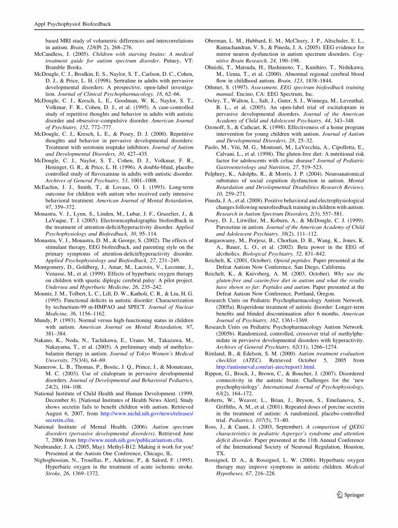
based MRI study of volumetric differences and intercorrelationsin autism. Brain, 128(Pt 2), 268–276.
McCandless, J. (2005). Children with starving brains: A medicaltreatment guide for autism spectrum disorder. Putney, VT:Bramble Books.
McDougle, C. J., Brodkin, E. S., Naylor, S. T., Carlson, D. C., Cohen,D. J., & Price, L. H. (1998). Sertraline in adults with pervasivedevelopmental disorders: A prospective, open-label investiga-tion. Journal of Clinical Psychopharmacology, 18, 62–66.
McDougle, C. J., Kresch, L. E., Goodman, W. K., Naylor, S. T.,Volkmar, F. R., Cohen, D. J., et al. (1995). A case-controlledstudy of repetitive thoughts and behavior in adults with autisticdisorder and obsessive–compulsive disorder. American Journalof Psychiatry, 152, 772–777.
McDougle, C. J., Kresch, L. E., & Posey, D. J. (2000). Repetitivethoughts and behavior in pervasive developmental disorders:Treatment with serotonin reuptake inhibitors. Journal of Autismand Developmental Disorders, 30, 427–435.
McDougle, C. J., Naylor, S. T., Cohen, D. J., Volkmar, F. R.,Heninger, G. R., & Price, L. H. (1996). A double-blind, placebocontrolled study of fluvoxamine in adults with autistic disorder.Archives of General Psychiatry, 53, 1001–1008.
McEachin, J. J., Smith, T., & Lovaas, O. I. (1993). Long-termoutcome for children with autism who received early intensivebehavioral treatment. American Journal of Mental Retardation,97, 359–372.
Monastra, V. J., Lynn, S., Linden, M., Lubar, J. F., Gruzelier, J., &LaVaque, T. J. (2005). Electroencephalographic biofeedback inthe treatment of attention-deficit/hyperactivity disorder. AppliedPsychophysiology and Biofeedback, 30, 95–114.
Monastra, V. J., Monastra, D. M., & George, S. (2002). The effects ofstimulant therapy, EEG biofeedback, and parenting style on theprimary symptoms of attention-deficit/hyperactivity disorder.Applied Psychophysiology and Biofeedback, 27, 231–249.
Montgomery, D., Goldberg, J., Amar, M., Lacroix, V., Lecomte, J.,Venasse, M., et al. (1999). Effects of hyperbaric oxygen therapyon children with spastic diplegic cerebral palsy: A pilot project.Undersea and Hyperbaric Medicine, 26, 235–242.
Mountz, J. M., Tolbert, L. C., Lill, D. W., Katholi, C. R., & Liu, H. G.(1995). Functional deficits in autistic disorder: Characterizationby technetium-99 m-HMPAO and SPECT. Journal of NuclearMedicine, 36, 1156–1162.
Mundy, P. (1993). Normal versus high-functioning status in childrenwith autism. American Journal on Mental Retardation, 97,381–384.
Nakano, K., Noda, N., Tachikawa, E., Urano, M., Takazawa, M.,Nakayama, T., et al. (2005). A preliminary study of methylco-balamin therapy in autism. Journal of Tokyo Women’s MedicalUnversity, 75(3/4), 64–69.
Namerow, L. B., Thomas, P., Bostic, J. Q., Prince, J., & Monuteaux,M. C. (2003). Use of citalopram in pervasive developmentaldisorders. Journal of Developmental and Behavioral Pediatrics,24(2), 104–108.
National Institute of Child Health and Human Development. (1999,December 8). [National Institutes of Health News Alert]. Studyshows secretin fails to benefit children with autism. RetrievedAugust 6, 2007, from http://www.nichd.nih.gov/news/releases/secretin.cfm.
National Institute of Mental Health. (2006). Autism spectrumdisorders (pervasive developmental disorders). Retrieved June7, 2006 from http://www.nimh.nih.gov/publicat/autism.cfm.
Neubrander, J. A. (2005, May). Methyl-B12: Making it work for you!Presented at the Autism One Conference, Chicago, IL.
Nighoghossian, N., Trouillas, P., Adeleine, P., & Salord, F. (1995).Hyperbaric oxygen in the treatment of acute ischemic stroke.Stroke, 26, 1369–1372.
Oberman, L. M., Hubbard, E. M., McCleery, J. P., Altschuler, E. L.,Ramachandran, V. S., & Pineda, J. A. (2005). EEG evidence formirror neuron dysfunction in autism spectrum disorders. Cog-nitive Brain Research, 24, 190–198.
Ohnishi, T., Matsuda, H., Hashimoto, T., Kunihiro, T., Nishikawa,M., Uema, T., et al. (2000). Abnormal regional cerebral bloodflow in childhood autism. Brain, 123, 1838–1844.
Othmer, S. (1997). Assessment. EEG spectrum biofeedback trainingmanual. Encino, CA: EEG Spectrum, Inc.
Owley, T., Walton, L., Salt, J., Guter, S. J., Winnega, M., Leventhal,B. L., et al. (2005). An open-label trial of escitalopram inpervasive developmental disorders. Journal of the AmericanAcademy of Child and Adolescent Psychiatry, 44, 343–348.
Ozonoff, S., & Cathcart, K. (1998). Effectiveness of a home programintervention for young children with autism. Journal of Autismand Developmental Disorders, 28, 25–32.
Paolo, M., Viti, M. G., Montouri, M., LaVecchia, A., Cipolletta, E.,Calvani, L., et al. (1998). The gluten-free diet: A nutritional riskfactor for adolescents with celiac disease? Journal of PediatricGastroenterology and Nutrition, 27, 519–523.
Pelphrey, K., Adolphs, R., & Morris, J. P. (2004). Neuroanatomicalsubstrates of social cognition dysfunction in autism. MentalRetardation and Developmental Disabilities Research Reviews,10, 259–271.
Pineda, J. A., et al. (2008). Positive behavioral and electrophysiologicalchanges following neurofeedback training in childrenwith autism.Research in Autism Spectrum Disorders, 2(3), 557–581.
Posey, D. J., Litwiller, M., Koburn, A., & McDougle, C. J. (1999).Paroxetine in autism. Journal of the American Academy of Childand Adolescent Psychiatry, 38(2), 111–112.
Rangaswamy, M., Porjesz, B., Chorlian, D. B., Wang, K., Jones, K.A., Bauer, L. O., et al. (2002). Beta power in the EEG ofalcoholics. Biological Psychiatry, 52, 831–842.
Reichelt, K. (2001, October). Opioid peptides. Paper presented at theDefeat Autism Now Conference, San Diego, California.
Reichelt, K., & Knivsberg, A. M. (2003, October). Why use thegluten-free and casein-free diet in autism and what the resultshave shown so far: Peptides and autism. Paper presented at theDefeat Autism Now Conference, Portland, Oregon.
Research Units on Pediatric Psychopharmacology Autism Network.(2005a). Risperidone treatment of autistic disorder: Longer-termbenefits and blinded discontinuation after 6 months. AmericanJournal of Psychiatry, 162, 1361–1369.
Research Units on Pediatric Psychopharmacology Autism Network.(2005b). Randomized, controlled, crossover trial of methylphe-nidate in pervasive developmental disorders with hyperactivity.Archives of General Psychiatry, 62(11), 1266–1274.
Rimland, B., & Edelson, S. M. (2000). Autism treatment evaluationchecklist (ATEC). Retrieved October 5, 2005 fromhttp://autismeval.com/ari-atec/report1.html.
Rippon, G., Brock, J., Brown, C., & Boucher, J. (2007). Disorderedconnectivity in the autistic brain: Challenges for the ‘newpsychophysiology’. International Journal of Psychophysiology,63(2), 164–172.
Roberts, W., Weaver, L., Brian, J., Bryson, S., Emelianova, S.,Griffiths, A. M., et al. (2001). Repeated doses of porcine secretinin the treatment of autism: A randomized, placebo-controlledtrial. Pediatrics, 107(5), 71–80.
Ross, J., & Caunt, J. (2003, September). A comparison of QEEGcharacteristics in pediatric Asperger’s syndrome and attentiondeficit disorder. Paper presented at the 11th Annual Conferenceof the International Society of Neuronal Regulation, Houston,TX.
Rossignol, D. A., & Rossignol, L. W. (2006). Hyperbaric oxygentherapy may improve symptoms in autistic children. MedicalHypotheses, 67, 216–228.
Appl Psychophysiol Biofeedback
123
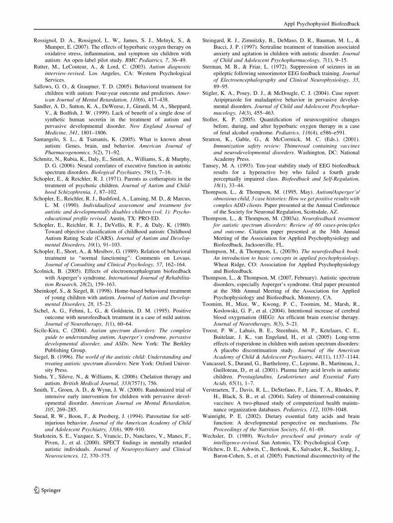
Rossignol, D. A., Rossignol, L. W., James, S. J., Melnyk, S., &Mumper, E. (2007). The effects of hyperbaric oxygen therapy onoxidative stress, inflammation, and symptom sin children withautism: An open-label pilot study. BMC Pediatrics, 7, 36–49.
Rutter, M., LeCouteur, A., & Lord, C. (2003). Autism diagnosticinterview-revised. Los Angeles, CA: Western PsychologicalServices.
Sallows, G. O., & Graupner, T. D. (2005). Behavioral treatment forchildren with autism: Four-year outcome and predictors. Amer-ican Journal of Mental Retardation, 110(6), 417–438.
Sandler, A. D., Sutton, K. A., DeWeese, J., Girardi, M. A., Sheppard,V., & Bodfish, J. W. (1999). Lack of benefit of a single dose ofsynthetic human secretin in the treatment of autism andpervasive developmental disorder. New England Journal ofMedicine, 341, 1801–1806.
Santangelo, S. L., & Tsatsanis, K. (2005). What is known aboutautism: Genes, brain, and behavior. American Journal ofPharmacogenomics, 5(2), 71–92.
Schmitz, N., Rubia, K., Daly, E., Smith, A., Williams, S., & Murphy,D. G. (2006). Neural correlates of executive function in autisticspectrum disorders. Biological Psychiatry, 59(1), 7–16.
Schopler, E., & Reichler, R. J. (1971). Parents as cotherapists in thetreatment of psychotic children. Journal of Autism and Child-hood Schizophrenia, 1, 87–102.
Schopler, E., Reichler, R. J., Bashford, A., Lansing, M. D., & Marcus,L. M. (1990). Individualized assessment and treatment forautistic and developmentally disables children (vol. 1): Psycho-educational profile revised. Austin, TX: PRO-ED.
Schopler, E., Reichler, R. J., DeVellis, R. F., & Daly, K. (1980).Toward objective classification of childhood autism: ChildhoodAutism Rating Scale (CARS). Journal of Autism and Develop-mental Disorders, 10(1), 91–103.
Schopler, E., Short, A., & Mesibov, G. (1989). Relation of behavioraltreatment to ‘‘normal functioning’’: Comments on Lovaas.Journal of Consulting and Clinical Psychology, 57, 162–164.
Scolnick, B. (2005). Effects of electroencephalogram biofeedbackwith Asperger’s syndrome. International Journal of Rehabilita-tion Research, 28(2), 159–163.
Sheinkopf, S., & Siegel, B. (1998). Home-based behavioral treatmentof young children with autism. Journal of Autism and Develop-mental Disorders, 28, 15–23.
Sichel, A. G., Fehmi, L. G., & Goldstein, D. M. (1995). Positiveoutcome with neurofeedback treatment in a case of mild autism.Journal of Neurotherapy, 1(1), 60–64.
Sicile-Kira, C. (2004). Autism spectrum disorders: The completeguide to understanding autism, Asperger’s syndrome, pervasivedevelopmental disorder, and ASDs. New York: The BerkleyPublishing Group.
Siegel, B. (1996). The world of the autistic child: Understanding andtreating autistic spectrum disorders. New York: Oxford Univer-sity Press.
Sinha, Y., Silove, N., & Williams, K. (2006). Chelation therapy andautism. British Medical Journal, 333(7571), 756.
Smith, T., Groen, A. D., & Wynn, J. W. (2000). Randomized trial ofintensive early intervention for children with pervasive devel-opmental disorder. American Journal on Mental Retardation,105, 269–285.
Snead, R. W., Boon, F., & Presberg, J. (1994). Paroxetine for self-injurious behavior. Journal of the American Academy of Childand Adolescent Psychiatry, 33(6), 909–910.
Starkstein, S. E., Vazquez, S., Vrancic, D., Nanclares, V., Manes, F.,Piven, J., et al. (2000). SPECT findings in mentally retardedautistic individuals. Journal of Neuropsychiatry and ClinicalNeurosciences, 12, 370–375.
Steingard, R. J., Zimnitzky, B., DeMaso, D. R., Bauman, M. L., &Bucci, J. P. (1997). Sertraline treatment of transition associatedanxiety and agitation in children with autistic disorder. Journalof Child and Adolescent Psychopharmacology, 7(1), 9–15.
Sterman, M. B., & Friar, L. (1972). Suppression of seizures in anepileptic following sensorimotor EEG feedback training. Journalof Electroencephalography and Clinical Neurophysiology, 33,89–95.
Stigler, K. A., Posey, D. J., & McDougle, C. J. (2004). Case report:Aripiprazole for maladaptive behavior in pervasive develop-mental disorders. Journal of Child and Adolescent Psychophar-macology, 14(3), 455–463.
Stoller, K. P. (2005). Quantification of neurocognitive changesbefore, during, and after hyperbaric oxygen therapy in a caseof fetal alcohol syndrome. Pediatrics, 116(4), e586–e591.
Stratton, K., Gable, G., & McCormick, M. C. (Eds.). (2001).Immunization safety review: Thimerosal containing vaccinesand neurodevelopmental disorders. Washington, DC: NationalAcademy Press.
Tansey, M. A. (1993). Ten-year stability study of EEG biofeedbackresults for a hyperactive boy who failed a fourth gradeperceptually impaired class. Biofeedback and Self-Regulation,18(1), 33–44.
Thompson, L., & Thompson, M. (1995, May). Autism/Asperger’s/obnoxious child, 3 case histories: How we get positive results withcomplex ADD clients. Paper presented at the Annual Conferenceof the Society for Neuronal Regulation, Scottsdale, AZ.
Thompson, L., & Thompson, M. (2003a). Neurofeedback treatmentfor autistic spectrum disorders: Review of 60 cases-principlesand outcome. Citation paper presented at the 34th AnnualMeeting of the Association for Applied Psychophysiology andBiofeedback, Jacksonville, FL.
Thompson, M., & Thompson, L. (2003b). The neurofeedback book:An introduction to basic concepts in applied psychophysiology.Wheat Ridge, CO: Association for Applied Psychophysiologyand Biofeedback.
Thompson, L., & Thompson, M. (2007, February). Autistic spectrumdisorders, especially Asperger’s syndrome. Oral paper presentedat the 38th Annual Meeting of the Association for AppliedPsychophysiology and Biofeedback, Monterey, CA.
Toomim, H., Mize, W., Kwong, P. C., Toomim, M., Marsh, R.,Koslowski, G. P., et al. (2004). Intentional increase of cerebralblood oxygenation (HEG): An efficient brain exercise therapy.Journal of Neurotherapy, 8(3), 5–21.
Troost, P. W., Lahuis, B. E., Steenhuis, M. P., Ketelaars, C. E.,Buitelaar, J. K., van Engeland, H., et al. (2005). Long-termeffects of risperidone in children with autism spectrum disorders:A placebo discontinuation study. Journal of the AmericanAcademy of Child & Adolescent Psychiatry, 44(11), 1137–1144.
Vancassel, S., Durand, G., Barthelemy, C., Lejeune, B., Martineau, J.,Guilloteau, D., et al. (2001). Plasma fatty acid levels in autisticchildren. Prostaglandins, Leukotrienes and Essential FattyAcids, 65(1), 1–7.
Verstraeten, T., Davis, R. L., DeStefano, F., Lieu, T. A., Rhodes, P.H., Black, S. B., et al. (2004). Safety of thimerosal-containingvaccines: A two-phased study of computerized health mainte-nance organization databases. Pediatrics, 112, 1039–1048.
Wainright, P. E. (2002). Dietary essential fatty acids and brainfunction: A developmental perspective on mechanisms. TheProceedings of the Nutrition Society, 61, 61–69.
Wechsler, D. (1989). Wechsler preschool and primary scale ofintelligence-revised. San Antonio, TX: Psychological Corp.
Welchew, D. E., Ashwin, C., Berkouk, K., Salvador, R., Suckling, J.,Baron-Cohen, S., et al. (2005). Functional disconnectivity of the
Appl Psychophysiol Biofeedback
123
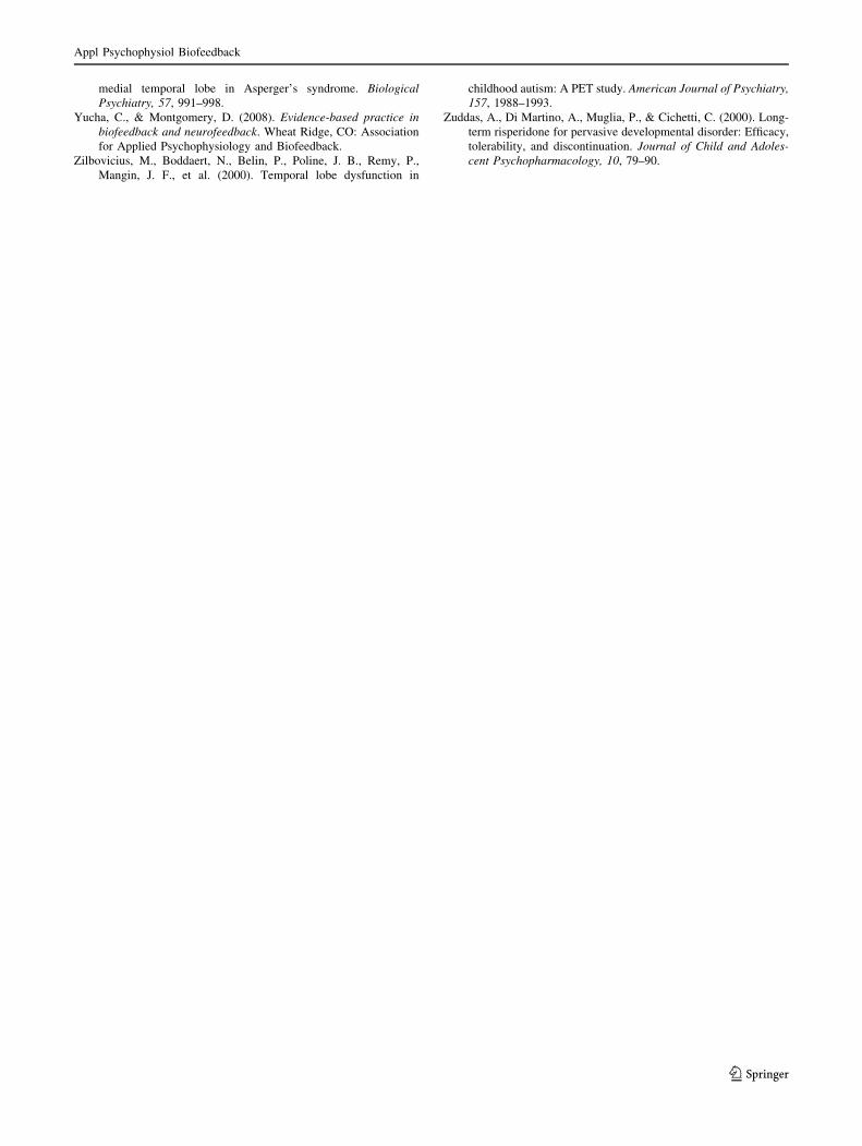
medial temporal lobe in Asperger’s syndrome. BiologicalPsychiatry, 57, 991–998.
Yucha, C., & Montgomery, D. (2008). Evidence-based practice inbiofeedback and neurofeedback. Wheat Ridge, CO: Associationfor Applied Psychophysiology and Biofeedback.
Zilbovicius, M., Boddaert, N., Belin, P., Poline, J. B., Remy, P.,Mangin, J. F., et al. (2000). Temporal lobe dysfunction in
childhood autism: A PET study. American Journal of Psychiatry,157, 1988–1993.
Zuddas, A., Di Martino, A., Muglia, P., & Cichetti, C. (2000). Long-term risperidone for pervasive developmental disorder: Efficacy,tolerability, and discontinuation. Journal of Child and Adoles-cent Psychopharmacology, 10, 79–90.
Appl Psychophysiol Biofeedback
123