neurocognitive late effects in pediatric cancer/media/files/activity files/disease/ncpf...12/20/2001...
TRANSCRIPT
12/20/2001
Neurocognitive Late Effects in Pediatric Cancer
Raymond K. Mulhern, Ph.D.
and
Shawna L. Palmer, Ph.D.
Division of Behavioral Medicine
St. Jude Children’s Research Hospital and
Department of Pediatrics
University of Tennessee College of Medicine
Memphis, Tennessee
Corresponding Author: Raymond K. Mulhern
Division of Behavioral Medicine
St. Jude Children’s Research Hospital
332 North Lauderdale
Memphis, Tennessee 38105-2794
Telephone: (901) 495-3580
Fax: (901) 495-3121
Email: [email protected]
Running Head: Neurocognitive Late Effects
2
Abstract
As survival rates for the most prevalent types of childhood cancer have dramatically improved
over the past three decades, the concept of “cure” has evolved to include optimizing the quality
of life among survivors. Although significant progress has been made in addressing some
adverse late effects of treatment that limit quality of life, such as endocrinopathies, other late
effects remain problematic. This paper will review neurocognitive late effects as defined by
problems with thinking, learning, and remembering among survivors of childhood cancer. After
defining the neurocognitive phenotype that characterizes many such children, we will review the
etiology and risk factors for damage to the central nervous system associated with childhood
cancer and its treatment. We will then discuss methods of pharmacological, behavioral, and
ecological intervention that may be helpful in reducing learning problems among surviving
children. Finally, we will identify areas of future research that will be critical to the elimination of
neurocognitive late effects in childhood cancer survivors and the resources needed to
implement such research.
3
For children diagnosed with cancer in the early 1970’s, the probabilities were
approximately the same as to whether they would be cured or succumb to their illness. For
children diagnosed in the early 1990’s, the overall prognosis for survival had increased to 75%
with some types of cancer exceeding 80% cure rates.1 With improvement in survival, clinicians
became more aware of late occurring adverse effects of treatment for childhood cancer.
Neurocognitive late effects, defined by problems with thinking, learning, and remembering,
have become an expanding area of scientific interest, especially for the two most frequent
types of childhood cancer, acute lymphoblastic leukemia (ALL) and brain tumors.
Although estimates vary according to patient diagnosis and age, aggressiveness of
therapy, and length of follow-up, most researchers would agree that the incidence of
neurocognitive late effects is unacceptably high. Despite recent attempts to modify therapy to
reduce morbidity while maintaining high cure rates, problems in neurocognitive functioning
remain to be experienced by a large majority of survivors. Efforts to eliminate neurocognitive
late effects have been hampered by a deficient understanding of the biological and
developmental mechanisms responsible, as well as a lack of clinical trials directed at treating
the deficits associated with this neurocognitive syndrome.
Several recent reviews of the literature are available which summarize contemporary
findings among empirical studies and which offer methodological recommendations for
improving the level of rigor among future studies.2-6 We shall not provide another
comprehensive review in this paper. Rather, we will attempt to provide an interpretation of
findings within a conceptual framework that can be used to accommodate new studies as well
as to guide future research. Our review of the literature will therefore be more selective,
concentrating on more recent developments that are likely to have a significant impact on future
research priorities.
4
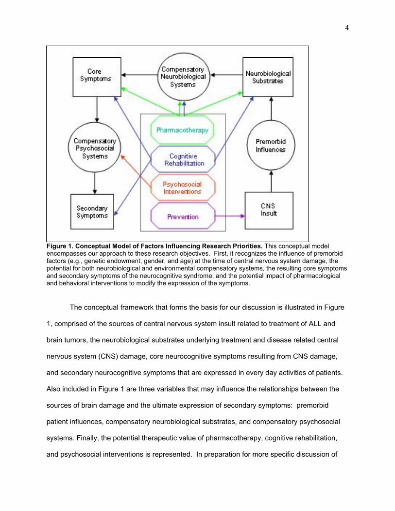
Figure 1. Conceptual Model of Factors Influencing Research Priorities. This conceptual model encompasses our approach to these research objectives. First, it recognizes the influence of premorbid factors (e.g., genetic endowment, gender, and age) at the time of central nervous system damage, the potential for both neurobiological and environmental compensatory systems, the resulting core symptoms and secondary symptoms of the neurocognitive syndrome, and the potential impact of pharmacological and behavioral interventions to modify the expression of the symptoms.
The conceptual framework that forms the basis for our discussion is illustrated in Figure
1, comprised of the sources of central nervous system insult related to treatment of ALL and
brain tumors, the neurobiological substrates underlying treatment and disease related central
nervous system (CNS) damage, core neurocognitive symptoms resulting from CNS damage,
and secondary neurocognitive symptoms that are expressed in every day activities of patients.
Also included in Figure 1 are three variables that may influence the relationships between the
sources of brain damage and the ultimate expression of secondary symptoms: premorbid
patient influences, compensatory neurobiological substrates, and compensatory psychosocial
systems. Finally, the potential therapeutic value of pharmacotherapy, cognitive rehabilitation,
and psychosocial interventions is represented. In preparation for more specific discussion of
5
these factors, we will begin with a brief review of pediatric ALL and brain tumors and associated
sources of CNS insult.
Acute Lymphoblastic Leukemia
Medical Background
Approximately 20,000 children and adolescents under the age of 20 were diagnosed
with cancer in 1999.7 The most commonly diagnosed cancer in this age group is ALL, a
malignant disorder by which lymphoid cells found in the bone marrow migrate to virtually every
organ system, including the CNS, via the circulatory system. ALL accounts for one-fourth of all
childhood cancers and 78% of all cases of childhood leukemia.1 In the United States,
approximately 3000 children are diagnosed with ALL each year with an incidence of 3 to 4
cases per 100,000 white children. ALL is more common among white than black children, and
is also more common among boys than girls with a peak incidence at 4 years of age. Although
genetic, environmental, viral, and immunodeficiency factors have been implicated in the
pathogenesis of ALL, the precise causes of most cases of ALL remain largely unknown.
Presenting symptoms include fever, fatigue, pallor, anorexia, bone pain and bruising.
Because the symptoms of ALL can mimic a number of nonmalignant conditions, definitive
diagnosis, usually made by bone marrow aspiration, is sometimes delayed. The duration of
treatment varies from 30 to 36 months, and, in the modern era is usually restricted to
intervention with combination chemotherapy, reserving cranial radiation therapy (CRT) for
patients who experience a CNS relapse. A better prognosis is associated with female gender,
age at diagnosis between 2 and 10 years, a lower white blood cell count, and an earlier positive
response to treatment. Treatment can be divided into 4 phases: remission induction, CNS
preventative therapy, consolidation, and maintenance. The purpose of the remission induction
phase is to rapidly eradicate leukemia cells from the bone marrow and circulatory system.
Consolidation may be used to intensify therapy following remission induction. Maintenance
therapy is required for a prolonged period because of the presence of undetectable levels of
6
leukemia that, nevertheless, have the capacity to be fatal. After the completion of treatment,
approximately 20% of those children who will eventually relapse will do so in the first year off
therapy with a subsequent risk of relapse in the remaining patients at a rate of 2% to 3% per
year for the next 3 to 4 years.
Sources of CNS Insult
CNS preventative therapy is necessary because the CNS is a sanctuary for occult
leukemia. Traditionally, CNS therapy has included CRT and intrathecal chemotherapy, usually
with methotrexate (MTX) or MTX combined with other drugs. However, because of the risks for
neurocognitive toxicity with CRT, treatment is now usually restricted to intrathecal and systemic
chemotherapy with equivalent success in the prevention of CNS relapses. An overwhelming
majority of studies that have investigated the neurocognitive morbidity of CRT in leukemia
patients have found significant adverse effects,2,4 with the strongest evidence coming from
longitudinally designed studies with internal control or comparison groups.
For example, the group at Children’s Hospital of Los Angeles reported on 24 patients
treated for ALL who had received CNS prophylactic therapy with 18 Gy CRT, intrathecal MTX,
and intravenous MTX.9 Patients were assessed with IQ testing prior to beginning CNS therapy
and at 1 and 4 years later. Although IQ scores remained stable at the 1-year interval, significant
declines in Full-scale, Verbal, and Performance IQ scores were noted at the 4-year follow-up
testing, with a mean loss of 6 to 7 IQ points. Furthermore, 12 of the 24 children had received
special educational services at the final assessment with three of the 12 having repeated a
grade prior to receiving special services. These results were disappointing because of the
expectation that a reduction of the traditional CRT dose from 24 Gy to 18 Gy would minimize
neurocognitive toxicity.
In a unique analysis across two institutional protocols at St. Jude Children’s Research
Hospital, patients who had received 24 Gy CRT and those who had been randomized to receive
18 Gy CRT or no CRT were compared over time with regard to their neurocognitive
7
development.10 All patients also received intrathecal MTX and intravenous MTX therapy. With
a median follow-up of 6.8 to 8.4 years for the groups, only small and nonsignificant changes in
IQ values were noted with no significant differences between groups. However, 22-30% of
patients showed a clinically significant decline of Full-scale IQ over the study interval and scores
on tests of arithmetic declined over time compared to normal expectations of same-age peers.
Interestingly, one explanation for the lack of differences between CNS therapy groups were
differences in parenteral MTX; the 18-Gy group had the lowest total dose, those in the 24 Gy
group had approximately 1.5 times more, and those not receiving CRT received 10.7 times
more MTX.
More recently, serial neurocognitive evaluations were performed on 30 children surviving
ALL to 4-years post-diagnosis.11 Patients had received CNS treatment with chemotherapy only.
Although IQ scores remained stable, arithmetic achievement declined significantly as well as
patients’ verbal fluency and visual-motor skills. These results recapitulated earlier reports that
intrathecal and/or intravenous MTX were not benign to the CNS. For example, one cross-
sectional study of 47 long-term survivors of ALL treated with chemotherapy only found
statistically significant declines in Performance IQ, as well as perceptual organization, and
freedom from distractibility scores but no significant changes in academic achievement.12
It is worth noting that at least one prospective study has failed to find IQ losses among
patients treated without CRT at a 3-year followup.8 Other types of chemotherapy, such as the
use of dexamethasone instead of prednisone, in the treatment of children with ALL may also
confer increased risk for neurocognitive impairment.13
Brain Tumors
Medical Background
Pediatric brain tumors are considerably more heterogeneous than ALL in that they vary
by histology as well as location. Although brain tumors can appear as a second malignancy
following the treatment of ALL with cranial irradiation, the etiology of most pediatric brain tumors
8
is uncertain. Next to ALL, primary CNS tumors are the second most frequently diagnosed
malignancy of childhood and the most common pediatric solid tumor with an annual incidence of
2.7 per 100,000 children under the age of 20 years.1
Among the more common symptoms of a brain tumor are morning headaches, nausea,
and lethargy resulting from tumor obstruction of the ventricles and increased intracranial
pressure. Tumors are oftentimes characterized as being above (supratentorial) or below
(infratentorial) the tentorium, a membrane that separates the cerebellum and brain stem from
the rest of the brain. Problems with balance and cranial nerve findings are more common
among patients with infratentorial tumors whereas seizures are more common among patients
with supratentorial tumors. Computed tomography (CT) and/or magnetic resonance imaging
are critical to the diagnosis of a pediatric brain tumor, and surgical resection or biopsy of tissue
is usually necessary for definitive histological diagnosis. In addition to maximal safe surgical
resection of the tumor, CRT, with or without chemotherapy, is indicated for malignant tumors.
Local target volumes of radiation are applied to tumors that remain confined, with whole brain
fields used for tumors that are mulitfocal. For those tumors with a tendency to spread through
the neuraxis, craniospinal irradiation (CSI) is used. Radiation therapists are increasingly using
3-D techniques where individual beams are altered to conform to the shape of the target,
sparing a greater amount of the normal surrounding tissue.14 The total dose delivered to the
brain depends on disease and patient factors, and can be more than twice that given in the
treatment of ALL.
Prognosis varies with the tumor type. For example, medulloblastoma, the most common
malignant brain tumor in childhood, has a prognosis of approximately 65% long-term survival
whereas children with intrinsic brain stem glioma have a prognosis of less than 10%. Although
this chapter will focus on the neuropsychological toxicity, other potentially serious complications
from irradiation (e.g., hormone deficiencies, growth retardation, second malignancies) as well as
9
hearing loss from treatment with cisplatin chemotherapy, are recognized in the brain tumor
literature.
Sources of CNS Insult
In the treatment of malignant brain tumors CSI has consistently shown a link to
symptoms of neurocognitive deficit. Unfortunately, upon psychological testing, 40-100% of long-
term brain tumor survivors have been found to have some form of cognitive dysfunction15 with
impaired intelligence found in nearly 90% of conventionally treated medulloblastoma patients.16
When compared to brain tumor patients who did not receive radiation therapy, those patients
who did receive radiation therapy consistently scored lower on tests of intellectual functioning.17
Evidence also indicates that the effects of CSI has an immediate impact on intellectual
functioning with a continuing pattern of decline over time.18 These declines in IQ over time
appear more severe for those who receive greater doses of CSI. Children treated for
medulloblastoma with 23.4 Gy performed better on tests of intellectual and academic ability than
those treated with 36 Gy.19,20
Extent of tumor resection is used in staging individual patients, with those who did not
receive total resection of primary tumor tissue generally considered at higher risk. Extent of
surgical resection has been found to have a positive relation with intellectual function21 as well
as no relation at all.22 These equivocal findings could be due to variability in how total resection
was defined and diagnosed, and the corresponding treatment the patient received based on
their resection status.
Despite seizure activity being a common symptom, especially in supratentorial tumors, it
is rarely considered in studies of neurocognitive function within pediatric oncology populations.
Seizures are often successfully treated with anticonvulsant medications. However, these
medications may themselves be linked to behavioral problems.23 Accurate documentation of
seizure activity and anticonvulsant use is therefore an important consideration when interpreting
the cognitive functioning of brain tumor patients.
10
Increased intracranial pressure from the process of hydrocephalus can result when
tumor obstructs the flow of the cerebral spinal fluid. Compression of tissue at both local and
remote sites throughout the brain can result. Although it has been reported that acute
hydrocephalus has had no lasting effects on cognitive functioning,3 these studies have generally
described hydrocephalus as being promptly treated with placement of shunts or surgical
removal of the obstruction. However, there has been at least one study to find brain tumor
patients with hydrocephalus at diagnosis to be at higher risk for intellectual deficits than those
with no hydrocephalus at diagnosis.24 In addition, hydrocephalus in other pediatric populations
has been associated with problems of neurocognitive functioning including memory, attention
and perceptual performance.25 The long-term effects of hydrocephalus within the setting of
childhood cancer requires additional examination.
Core and Secondary Symptoms of the Neurocognitive Phenotype
Within our model, secondary symptoms include broad-spectrum abilities measured by
tests of academic achievement and intellectual functioning. These secondary symptoms can
produce limitations in age-appropriate activities of daily living such as school performance,
employment, independent living, and quality of life among subsets of surviving patients.26-28
These problems were the first to be quantified in the neuro-oncology literature and remain
important endpoints in any pediatric oncology Phase III clinical trial where CNS treatment is
initiated.
Initially, some attributed these deficits to the nonspecific effects of chronic illness or
school absenteeism. However, subsequent studies have objectively measured deficits in
intellectual development and academic achievement relative to healthy age peers as well as
other pediatric patients with cancer treatment not involving the CNS.29,30 More recently, studies
have sought to define the changes in underlying, core mental processes that secondarily result
in changes in IQ and achievement. Many of the core symptoms are defined by the term
“executive functions”, including the ability to allocate attentional resources and to plan and
11
organize behavior, thought to be dependent upon pre-frontal lobe integrity. Other, more widely
distributed functions, such as mental processing speed, the ability to sustain attention, to learn
and retrieve new information efficiently, and to use previously learned information to provide a
context for new learning, also appear to be negatively affected.31-35 The increasing
sophistication of such studies is evidenced by the fact that more recent investigations of
attentional functioning are theoretically-driven, based upon cognitive models of normal
development of attention and learning.31,32
One recent longitudinal study by Palmer and colleagues18 has unequivocally
demonstrated that declining IQ, the single most commonly occurring symptom among affected
patients,2-5 was not due to loss of previously acquired information but instead due to a failure to
acquire new information at a rate commensurate with age peers. Forty-four patients with
pediatric medulloblastoma had completed 150 psychological examinations of intellectual ability.
Changes in patient IQ performance corrected for age (scaled scores) was compared to
uncorrected performance (raw scores). A significant decline in IQ scaled scores was
demonstrated over time since start of radiation treatment. Upon examination of raw scores a
significant increase was found over time since start of radiation treatment. However, this
increase, indicating acquisition of new information and skills across time, was less than
expected in the normal healthy population therefore resulting in a decline in IQ scaled scores.
Memory and attention are critical pre-requisite processes by which knowledge is acquired36 and
in a recent study up to 45% of the variance in IQ was accounted for by working memory ability
and processing speed.37 It is speculated that the inability to acquire new information and skills at
a rate comparable to healthy same-age peers may be due to deficits in underlying core abilities
such as memory and speed of processing.18
Identifying the core mechanisms by which patients experience a loss in academic and
intellectual function is critical for developing efficacious intervention programs. Examples of
standardized tests that are currently available to objectively evaluate the patient’s school failure,
12
intellectual development, and potentially underlying problems with attention, memory, and
processing speed are described in Table 1. An important component in the quest for successful
interventions is to examine the neurobiological substrates associated with cognitive changes.
-------------------------------------
Insert Table 1 about here
-------------------------------------
Neurobiological Substrates
Neuropathological Changes
Studies utilizing neuro-imaging have shown changes in brain tissue following
chemotherapy and radiation treatment. Utilizing computerized tomography (CT) to study
patients with brain malignancies, several CNS abnormalities such as cerebral atrophy,
calcifications, focal and diffuse white matter lesions, and enlarged ventricles, have been
qualitatively defined.38 Late effects of treatment-related CNS injury in ALL survivors include
diffuse and multifocal white matter abnormalities, demyelination, breakdown of the blood-brain
barrier, microvascular occlusion, and calcifications in cortical gray matter and basal ganglia.39
While it has been well documented in the literature that treatment for pediatric cancer is
associated with several CNS abnormalities, the underlying cellular and subcellular mechanisms
of such change require additional research. It is generally accepted however, that damage to
the microvasculature, as well as accompanying injury to glial cells is involved.40 Glial cells serve
a supportive role by providing structure and insulation for the axon of the nerve cells. Within the
white matter of the brain, oligodendrocytes, a type of glial cell, form a fatty sheath called myelin
that surrounds portions of the neuronal axon and acts to dramatically increase the speed of
nerve conduction.41 Myelination normally continues after birth into the third decade of life42 and
exposure to CRT can disrupt this developmental process, ending in demyelination, and
13
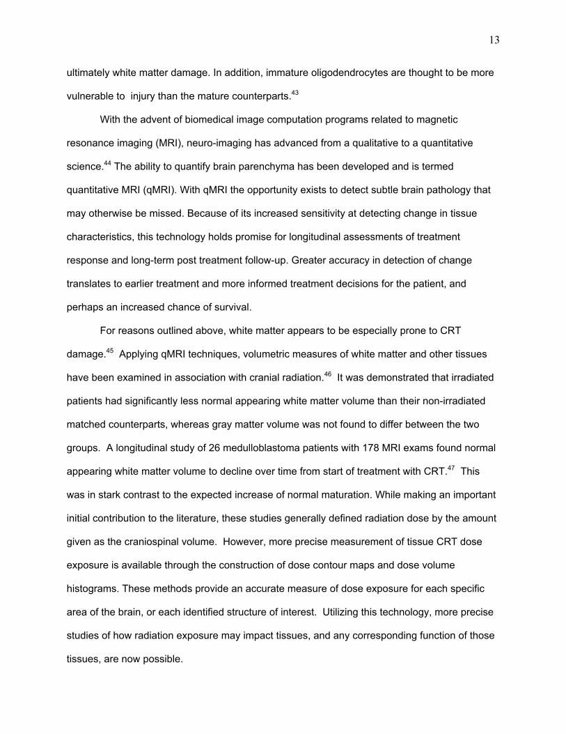
ultimately white matter damage. In addition, immature oligodendrocytes are thought to be more
vulnerable to injury than the mature counterparts.43
With the advent of biomedical image computation programs related to magnetic
resonance imaging (MRI), neuro-imaging has advanced from a qualitative to a quantitative
science.44 The ability to quantify brain parenchyma has been developed and is termed
quantitative MRI (qMRI). With qMRI the opportunity exists to detect subtle brain pathology that
may otherwise be missed. Because of its increased sensitivity at detecting change in tissue
characteristics, this technology holds promise for longitudinal assessments of treatment
response and long-term post treatment follow-up. Greater accuracy in detection of change
translates to earlier treatment and more informed treatment decisions for the patient, and
perhaps an increased chance of survival.
For reasons outlined above, white matter appears to be especially prone to CRT
damage.45 Applying qMRI techniques, volumetric measures of white matter and other tissues
have been examined in association with cranial radiation.46 It was demonstrated that irradiated
patients had significantly less normal appearing white matter volume than their non-irradiated
matched counterparts, whereas gray matter volume was not found to differ between the two
groups. A longitudinal study of 26 medulloblastoma patients with 178 MRI exams found normal
appearing white matter volume to decline over time from start of treatment with CRT.47 This
was in stark contrast to the expected increase of normal maturation. While making an important
initial contribution to the literature, these studies generally defined radiation dose by the amount
given as the craniospinal volume. However, more precise measurement of tissue CRT dose
exposure is available through the construction of dose contour maps and dose volume
histograms. These methods provide an accurate measure of dose exposure for each specific
area of the brain, or each identified structure of interest. Utilizing this technology, more precise
studies of how radiation exposure may impact tissues, and any corresponding function of those
tissues, are now possible.
14
Chemotherapy, especially high dose MTX, is also associated with white matter injury.48
As demyelination occurs, axons near the lesions become swollen without an inflammatory
response.49 These changes are characterized as leukoencephalopathy. The quantity of
leukoencephalopathy, as well as the amount of affected white matter, was found to increase in
proportion to additional courses of high dose methotrexate during treatment.50 The time course
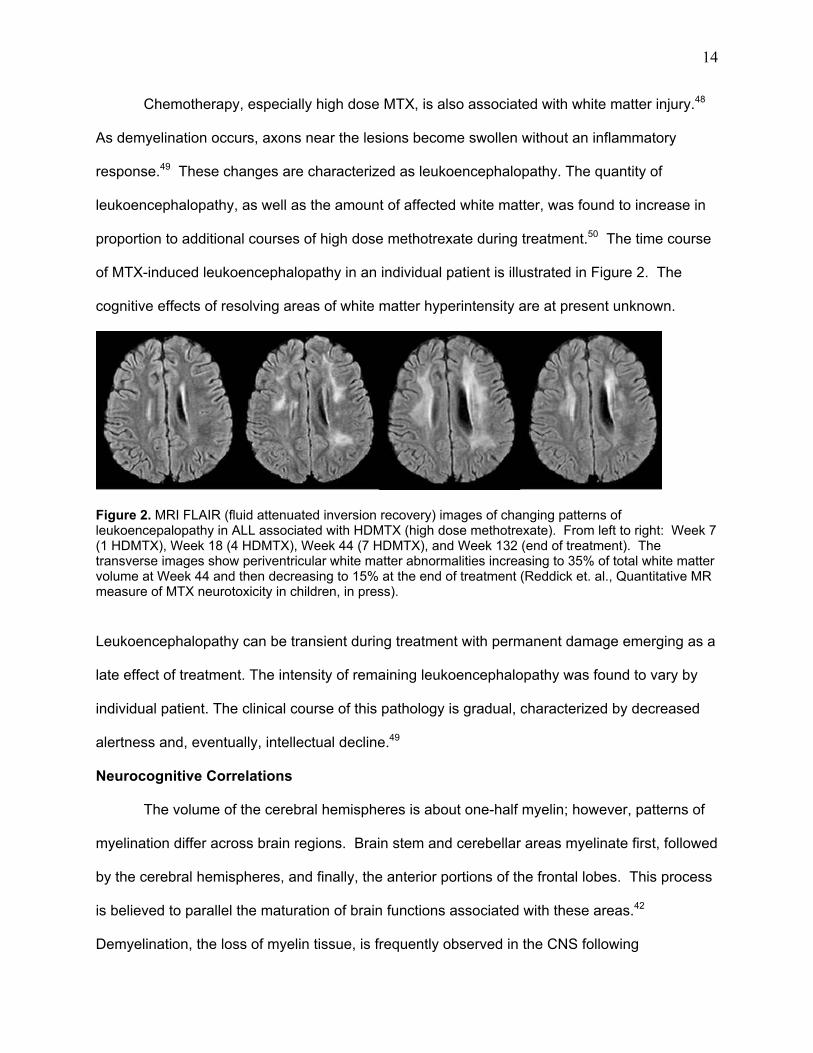
of MTX-induced leukoencephalopathy in an individual patient is illustrated in Figure 2. The
cognitive effects of resolving areas of white matter hyperintensity are at present unknown.
Figure 2. MRI FLAIR (fluid attenuated inversion recovery) images of changing patterns of leukoencepalopathy in ALL associated with HDMTX (high dose methotrexate). From left to right: Week 7 (1 HDMTX), Week 18 (4 HDMTX), Week 44 (7 HDMTX), and Week 132 (end of treatment). The transverse images show periventricular white matter abnormalities increasing to 35% of total white matter volume at Week 44 and then decreasing to 15% at the end of treatment (Reddick et. al., Quantitative MR measure of MTX neurotoxicity in children, in press).
Leukoencephalopathy can be transient during treatment with permanent damage emerging as a
late effect of treatment. The intensity of remaining leukoencephalopathy was found to vary by
individual patient. The clinical course of this pathology is gradual, characterized by decreased
alertness and, eventually, intellectual decline.49
Neurocognitive Correlations
The volume of the cerebral hemispheres is about one-half myelin; however, patterns of
myelination differ across brain regions. Brain stem and cerebellar areas myelinate first, followed
by the cerebral hemispheres, and finally, the anterior portions of the frontal lobes. This process
is believed to parallel the maturation of brain functions associated with these areas.42
Demyelination, the loss of myelin tissue, is frequently observed in the CNS following
15
treatment.45 Frontal lobe white matter has extensive links to more posterior cortical and
subcortical areas of the brain. The right frontal lobe is especially rich in myelin. Therefore,
global white matter pathology might disproportionately affect functions associated with this
region, such as attention and visuospatial ability.51
Considering the role and distribution of myelin in the brain, the neurocognitive impairments of
long-term survivors are somewhat intuitive. For example, the diffuse loss of myelin in survivors might be
expected to be associated with widely distributed functions in the brain such as attention and information
processing speed. Indeed, a recent study demonstrated that deficits in the speed of information
processing were correlated with the volume of white matter loss among older adults.52 Brain volumetry
studies within pediatric cancer have also found secondary functional consequences, such as IQ and
academic achievement declines, of tissue loss17,53 In one study, 18 pediatric patients previously treated
for medulloblastoma, with CSI with or without chemotherapy following surgery, were matched on the
basis of age at the time of evaluation to 18 patients treated for low grade tumors of the posterior fossa
who were treated with surgery alone. Evaluations were conducted with age-appropriate neurocognitive
testing and qMRI of normal appearing white matter. Patients treated for medulloblastoma had
significantly less normal appearing white matter and significantly lower IQ scores than their low grade
controls. In addition, normal appearing white matter has a positive and statistically significant relationship
with IQ. 17 In a more recent study from the St. Jude Group, 53 patients who had completed CRT with or
without chemotherapy for medulloblastoma were evaluated by qMRI and neurocognitive testing. Analysis
of MRI data with an automated segmentation algorithm developed from an artificial neural network
revealed that smaller volumes of normal appearing white matter were predicted by a younger age at
evaluation, higher CRT dose, and increased time from completion of CRT. Furthermore, volumes of
normal appearing white matter were positively correlated with IQ values.53 The investigators have
concluded from these two studies that inhibition of normal white matter development and/or loss of white
matter volume at least partially explains intellectual and academic deficits among children who survive
medulloblastoma. Current efforts are being aimed at identifying the effects of this tissue loss on core
functions of the syndrome (e.g., processing speed, attention).
Premorbid Influences
16
Gender
The most convincing data relating to gender as a risk factor for increased neurotoxicity has come
from the work of the Dana Farber group with patients treated for ALL,54-56 although this effect had been
reported incidentally in other papers.10 When gender effects have been investigated and have had
significant effects on cognitive function, female gender confers a greater risk. Waber’s initial study
utilized a control group with solid tumors who had not received CNS therapy to assess the risks
associated with CNS prophylaxis. Measures of intellectual and academic functioning, including a test of
reading comprehension were obtained for both groups of children. The ALL group performed below
average norms on all measures, while the solid tumor group performed above average on all measures.
Group differences indicated that the ALL group was more cognitively impaired than the solid tumor group,
and females in both groups were more impaired than males. In addition, the correlation between age,
socioeconomic status, and cognitive impairment within the ALL group differed as a function of gender.
Specifically, early age of diagnosis and low SES were associated with more severe cognitive impairment
in females, while these factors did not reliably correlate for males. It was concluded that the major risk
factor for CNS toxicity among children treated for ALL was gender with females at greater risk than males.
Providing further support to the role of gender, these results were confirmed in a more recent
study which also demonstrated clear dose-related, gender-dependent effects of treatment on cognitive
functioning in ALL survivors.55 Both male and female children showed decreased performance on
measures of language-based academic skill and memory for digit strings. Conversely, only girls
demonstrated global decline of cognitive function (i.e., lower IQ). Systemic chemotherapy was
associated with this decline in IQ for girls, indicating that systemic as well as CNS chemotherapy should
be evaluated as a risk factor for cognitive impairment.
Most recently, the same group has published a comparison of patients surviving ALL who had
been treated with intrathecal MTX, conventional dose MTX or high dose MTX with or without 18 Gy CRT.
Females receiving high dose MTX with CRT were more impaired than males receiving the same
combination therapy and more impaired than other females receiving conventional MTX with CRT.56
Researchers speculate that global deficits might be associated with diffuse axonal injury, a neurotoxicity
17
that might be both endocrine-dependent and worsened by large doses of chemotherapy. However, more
research is needed to clarify the relationship between gender, treatment type, and cognitive outcome.
Age at Treatment
A younger age at treatment is often found to be a critical risk factor, with this group showing
significantly greater declines in age-adjusted scaled scores of intellectual functioning over time than their
older counterparts. For example, children who were older (>8.85 years) when treated for
medulloblastoma experienced less neurotoxicity than those who were younger at treatment (<8.85 years)
exhibiting greater performance on tests of intellectual functioning.19 In a longitudinal study of intellectual
development over time since radiation treatment, medulloblastoma patients who were <8.02 years
experienced significantly greater declines in age-adjusted scaled scores of factual knowledge and
nonverbal abstract thinking as well as an overall estimate of full-scale IQ than those who were >8.02
years.18
It has been suggested that age at treatment is a proxy variable for underlying
neurodevelopmental maturity.17,53 While development of cortical gray matter peaks at approximately age
4 years, cortical white matter volume continues to rise until about age 20 years.57 Therefore, those who
are younger at radiation treatment generally have less fully developed white matter. However, since both
younger and older patients have been shown to lose white matter volume at a similar rates,47 the
younger irradiated patients continue to display reduced total white matter volume following radiation
treatment. These deficits in white matter volume among younger patients have also been associated with
increased intellectual morbidity.17,47,53
Compensatory Neurobiological Systems
The concept of "brain reserve capacity" has been proposed to explain individual variation in the
behavioral manifestation of signs and symptoms of brain damage.58 Using a model derived from adult
studies of progressive neurologic diseases that result in dementia (Alzheimer's Disease, Parkinson's
Disease, AIDS), he proposes that each individual has a unique threshold for tolerance of brain damage
before signs and symptoms are noted. Cumulative effects of brain insults remaining below the
threshold allow the individual to remain asymptomatic whereas clinical symptoms become
apparent once the threshold is reached. Patients with greater brain reserve capacity will be
18
more resilient (i.e., have a higher threshold) to the effects of any specific brain insult than those
with lesser reserve capacity. Brain reserve capacity can be estimated neuroanatomically (CT,
MRI) by normal brain volume and functionally by intellectual and educational attainment.
The concept of brain reserve capacity discussed did not make reference to childhood
brain damage or brain tumors. 58 However, it is relevant to the present research discussion in
that it provides a useful framework to explain variability in cognitive function among patients who
have received virtually identical medical treatment. Several hypotheses follow from the reserve
capacity model that have received some support. One hypothesis is that patients who are more
cognitively and neurologically intact prior to a particular form of therapy, such as CRT, will have
a better functional outcome. An analysis of IQ function 4 years following treatment for childhood
brain tumor revealed that postoperative IQ was the best predictor, accounting for 62% of the
variance alone.22
Damage to brain parenchyma during and following treatment gradually develops over
time. A recent study investigated the possibility for reorganization of function among 61 right
handed adult patients who underwent treatment for brain tumors and were experiencing slow
onset of damage.59 When compared to healthy controls, it was found that the brain tumor
patients recruited additional left frontal regions of the brain for language function other than the
traditional language areas. They also recruited frontolateral areas in the non-dominant
hemisphere for language function. This study provides a step toward understanding plasticity of
function among adults and could be used as a foundation for pediatric studies.
Compensatory Psychosocial Systems
Although patients with varying types of CNS treatments may express similar core
neurocognitive symptoms, considerable variability exists in their actual academic performance.
Aside from treatment factors, one possible explanation is that the home, school, and community
environments of patients have differences in their ability to compensate for core deficits and
thus affect the intensity of secondary symptoms such as achievement in school. For example,
19
among healthy samples of children, sociodemographic factors such as ethnicity and parental
education can account for up to 28% of the variance in IQ.60 Among children previously treated
for ALL, several studies have demonstrated that socioeconomic status is correlated with IQ and
other neurocognitive outcomes with patients coming from families with higher socioeconomic
status having better performance.8,32,61
One implication of these relationships is that the patient’s environment may modify the
intensity of the expression of secondary neurocognitive deficits, even among children with
similar core deficits. Can patients who experience enriched environments following treatment
overcome their deficits more easily? One could speculate that the patient who had interactions
with teachers that were taught about the patient’s specific area of deficit, and who were skilled
in assisting the patient to practice these weak areas of function, would be more successful in
learning to overcome and compensate for their deficits. In addition, it is speculated that having
better informed parents with access to extracurricular resources to supplement their child’s
education would increase the child’s eventual outcome. This is an important area of study that
could be implemented with a favorable cost-benefit ratio. However, we are unaware of any
current efforts to examine these possibilities.
Interventions for Neuropsychological Deficits
Although research on the patterns and risks for neuropsychological and educational
deficits among survivors of childhood cancer has been progressing for the past three decades,
the development of empirically validated interventions for these deficits has not been as rapid.
Broadly speaking, interventions can be divided into two approaches: those intended to avoid or
reduce the neuropsychological toxicity of therapy directed toward the CNS, and those intended
to minimize or rehabilitate deficits that cannot be avoided.
A formal plan of prospective surveillance of neuropsychological status should be set
forth for each patient based upon known or suspected developmental risk for problems. This
assumes that a qualified psychologist has been identified as a consultant to the institution. For
20
example, a young adult with a supratentorial low grade glioma treated with surgery alone may
require formal assessment only once or twice during the two year period following diagnosis
with the focus being whether there is evidence of loss of abilities. On the other hand, a young
child with the same tumor and treatment should have a neuropsychological evaluation
scheduled at the completion of therapy and three to five years later, whereas an infant with a
brain tumor should probably be evaluated every six months until the age of three or four years
and then yearly until five years post therapy. Such plans should not depend upon the
presentation of symptoms because presymptomatic assessments oftentimes allow for early
educational interventions that may minimize deficits.
Contemporary treatment protocols for children with for cancer generally show an
enlightened concern for the potential neurotoxicity of therapy, especially among very young
patients. The elimination of CRT, delay of CRT until the patient is older, or CRT dose/volume
reduction to spare more normal brain are frequently considered approaches. Several studies
have documented benefit of CRT dose reduction in terms of IQ and achievement functioning in
survivors of ALL and brain tumors.2,3,20,30 The benefits of more recent technological
improvements, such as the use of 3-dimensional conformal CRT, are not yet known. However,
with ever increasing cure rates, this approach to toxicity reduction is likely to continue to be very
active.
If neuropsychological impairments are unavoidable, one may attempt to minimize the
impact by direct intervention with cognitive rehabilitation or pharmacotherapy, and/or through
more indirect approaches involving manipulations of the patient’s environment. Cognitive
rehabilitation is a term used to describe interventions intended to restore lost cognitive functions
or to teach the patient skills to compensate for cognitive losses that cannot be restored.
Although some evidence for efficacy is available from the child closed head injury literature, we
are aware of only one program in the United States that is attempting to validate a standardized,
21
20-session program of cognitive rehabilitation for survivors of ALL and brain tumors in a seven
institution consortium funded by the National Cancer Institute.62
Pharmacotherapy, especially the use of psychostimulants such as methylphenidate
(Ritalin), has recently received interest. Impressive gains in activity level and quality of life were
shown in one study of adult glioma patients treated with methylphenidate at the University of
Texas/M.D. Anderson Cancer Center.63 A subsequent study at our institution investigated the
acute effects of methylphenidate on the cognitive functioning of pediatric patients treated for
cancer.64 In this double-blind, placebo-controlled study, patients given 0.6 mg/kg
methylphenidate showed significant improvement on measures of attention when compared to
those receiving placebo. Our current study, funded by the National Cancer Institute, expands
upon these findings by conducting a 3-week crossover trial of two doses of methylphenidate and
placebo in the home and school environments to establish the potential for efficacy prior to 12
months of treatment. Parent and teacher ratings of behavior as well as objective testing of the
patients will allow the evaluation of effects on academic achievement and social relations.
Finally, one should not minimize the potential positive impact of optimal communication
and education of the patient’s caregivers.65 Routine communication between the cancer
treatment center and the patient’s school should be the standard of care, especially in cases in
which subtle neurological (e.g., hearing loss in the speech frequencies, visual field cuts) or
neuropsychological deficits (e.g., problems with attention, memory, or processing speed) can
obviously impair the patient’s ability to function in a normal classroom environment. Because all
of the deficits listed above are unobservable to teachers, there may be a tendency to
misinterpret the patient’s behavior in the absence of knowledge of the deficits. For example, we
have experience with children labeled as having attitude problems, or as being daydreamers or
unmotivated to learn when, in fact, the patient had real disabilities that were unknown to the
teacher. Although parents can have an important role in facilitating communication between the
cancer center and the school, we have found that a telephone call or visit from a representative
22
from the cancer center teacher or social worker can have a profound impact on the adaptation
of the classroom environment to meet the patient’s needs.
Priority Areas for Future Research
In November of 2000, a report from the Brain Tumor Progress Review Group, jointly
sponsored by the National Cancer Institute and the National Institute for Neurological Disorders
and Stroke, articulated four priority areas for pediatric research.66 Two of these related to the
neuropsychological functioning of surviving children. In particular, an emphasis was placed on
more accurate assessment of risks for deficits, the development of more precise methods of
assessing functional and structural damage to the developing nervous system, and the
development of methods to prevent, minimize or rehabilitate deficits that cannot be avoided.
These points will be elaborated in the following section.
More fine-grained analyses of cognitive impairments in survivors will continue to expand
upon our understanding of specific cognitive phenotypes and risks for these deficits among our
patients. For example, the objective assessment of “executive functions” and processing speed
may provide us with early harbingers of impending changes in intellectual development and
academic achievement. The addition of recent quantitative neuro-imaging techniques will
facilitate our understanding of the biological substrates underlying neurocognitive deficits.
However, the theoretical frameworks used to conceptualize these more specific neurocognitive
processes differ across studies, as do the tools and methods used to measure cognitive
processes. There is little agreement as to which conceptual model of cognitive processes is
best applied or how to operationally define the cognitive processes delineated by different
models. For this reason, it is critical that future researchers clearly describe their materials,
methods, and the conceptual models they use to select tests and interpret results. This allows
for comparison of cognitive outcomes across studies and cancer diagnoses.
Despite our growing knowledge about neuropsychological outcomes, our understanding
of the neurobiological mechanisms underlying adverse outcomes and our ability to extrapolate
23
interventions from our understanding of these mechanisms is quite rudimentary. Future
research efforts would benefit from the use of animal analogue models and quantitative
neuroimaging (e.g., fMRI, PET) to better characterize biological changes in normal tissue and
their correlation with neuropsychological deficits. Unlike the adult cancer setting, central
nervous system damage in the developing child rarely results in dementia but rather a slowing
in the learning of new skills and information compared to healthy age peers. Furthermore, these
learning problems may not be evident until a particular skill that is normally expected to emerge
does not. It is this dynamic system that makes pediatric neuroscience both more complex and
more exciting in terms of the potential for plasticity. However, many of the most relevant
contemporary research questions require animal models.
Pediatric oncologists and radiation oncologists are generally aware of the risks of brain
tumor therapy for neuropsychological deficits, especially in very young children, and treatment
protocols are now purposefully designed to reduce toxicity as well as to increase survival.
Unfortunately, not all apparent reductions in treatment aggressiveness (e.g., reduction of RT
total dose) result in measurable benefit to the patient and some patients, despite these efforts,
will have unavoidable neuropsychological deficits. Furthermore, it is obvious that the empirical
literature currently gives little guidance in terms of pharmacological, cognitive, or environmental
interventions to rehabilitate neuropsychological deficits in survivors. It is understandable that
correlational and descriptive studies were needed in the past to document the incidence and
prevalence of deficits among different subgroups of survivors. However, future
neuropsychological research should give a higher priority to studies of interventions to prevent,
minimize, or rehabilitate neuropsychological deficits. Intervention research in the context of
clinical trials is expensive, labor-intensive, and time-consuming but these barriers can be
overcome in collaborative, multi-institutional trials.
What, then, are the resources need to remove barriers and facilitate the implementation
of the above recommendations? One issue involves the training of clinical neuroscientists to
24
prepare them to attack these problems. Historically, pediatric psychologists, clinical child
psychologists and, less frequently, pediatric neuropsychologists, have taken the lead in
developing investigations in the area of neurocognitive problems of children surviving cancer.
However, a new model of a hybrid clinical neuroscientist will likely be needed to take the
research to the next level. Investigators trained in human development, developmental
neurobiology, molecular biology, and pediatric neuropsychology who can interface with
investigators from diverse clinical (e.g., diagnostic imaging, hematology/oncology, neurology,)
and basic science (e.g., pharmacology, biochemistry) departments will be best prepared to lead
such an effort. The use of advanced training awards, such as the Cancer Research Training
and Career Development awards from the National Cancer Institute, may provide one route for
such innovative advanced training at the postdoctoral or junior faculty level.
A second stubborn issue that has undeniably limited research on neurocognitive late
effects in childhood cancer is the inconsistent availability of third-party payment for clinical
neuropsychological testing. Despite the fact that routine surveillance testing in high risk groups
of children has long been promoted as a standard of care within the cooperative groups,
medical necessity as defined by managed care panels, prevents most children with cancer from
obtaining such services with coverage, especially in the absence of being able to establish a
mental health diagnosis. A coordinated national approach to this problem, perhaps through the
National Institutes of Health, is seriously needed. One would hope that a legislative approach to
this problem would not be necessary. However, with the growing and significant numbers of
childhood cancer survivors reaching the age of majority, this and other quality of life issues
(e.g., medical and life insurance, employment) of childhood cancer survivorship may eventually
evolve into a political agenda.
Conclusions
This paper has attempted to highlight the most important issues relevant to
neurocognitive late effects in childhood cancer by using a conceptual model newly developed by
25
the authors. Within this model, sources of CNS insult experienced by patients treated for ALL
and brain tumors are discussed as well as the neurobiological factors underlying CNS damage.
Various compensatory mechanisms that can modify the expression of core symptoms (e.g.,
problems with attention, processing speed) and secondary symptoms (e.g., declining IQ,
academic failure) are also described. One goal is that this model may be a useful framework for
the development of future research agendas and to integrate the results of past and future
studies. We have recommended better description of methodologies in future studies, the use
of animal analog models and quantitative neuro-imaging to better elucidate mechanisms of CNS
damage, and an increased emphasis on clinical trials testing interventions for neurocognitive
late effects. Reaching these goals will be expedited by developing novel models of hybrid
neuroscientists and by removing barriers to third party payment for protocol-driven clinical
neuropsychological evaluations of patients. The discovery of effective interventions will facilitate
the achievement of our ultimate goal for survivors of childhood cancer; that is, returning them to
the quality of life that they would have had if they had never had cancer.

26
Acknowledgments
This manuscript was commissioned by the National Cancer Policy Board of the Institute of
Medicine, National Academy of Sciences. Preparation of this manuscript was supported in part
by the American Lebanese Syrian Associated Charities and grants CA 21765 and CA 20180
from the National Cancer Institute.
27
References 1. Ries LAG, Smith MA, Gurney JG et al. Cancer Incidence and Survival Among Children and
Adolescents: United States, SEER Program, 1975-1995. National Institute of Health Pub. No. 99-4649. Bethesda, MD, 1999
2. Roman DH, Sperduto PW. Neuropsychological effects of cranial radiation: Current
knowledge and future directions. International Journal of Radiation Oncology, Biology, Physics 1995;31:983-998.
3. Ris MD, Noll RB. Long-term neurobehavioral outcome in pediatric brain-tumor patients:
Review and methodological critique. Journal of Clinical and Experimental Neuropsychology 1994;16:21-42.
4. Moleski M. Neuropsychological, neuroanatomical, and neurophysiological consequences of
CNS Chemotherapy for acute lymphoblastic leukemia. Archive of clinical Neuropsychology 2000;15:603-630.
5. Maden-Swain A, Brown RT. Cognitive and psychosocial sequelae for children with acute
lymphocytic leukemia and their families. Clinical Psychology Review 1991;11:267-294. 6. Butler RW, Copeland DR. Neuropsychological effects of central nervous system prophylactic
treatment in childhood leukemia: methodological considerations. Journal of Pediatric Psychology 1993;18:319-338.
7. Steen G, Mirro J. Childhood Cancer: A Handbook from St. Jude Children’s Research
Hospital. Perseus Publishing. Cambridge, MA, 2000. 8. Copeland DR, Moore BD, Francis DJ, Jaffe N, Culbert SJ. Neuropsychologic effects of
chemotherapy on children with cancer: a longitudinal study. Journal of Clinical Oncology 1996;14:2826-2835.
9. Rubenstein CL, Varni JW, Katz ER. Cognitive functioning in long-term survivors of childhood
leukemia: a prospective analysis. Developmental and Behavioral Pediatrics 1990;11:301-305.
10. Mulhern RK, Fairclough D, Ochs J. A prospective comparison of neuropsychologic
performance of children surviving leukemia who received 18-Gy, 24-Gy, or no cranial irradiation. Journal of Clinical Oncology 1991;9:1348-1356.
11. Espy KA, Moore IM, Kaufmann PM, Kramer JH, Matthay K, Hutter JJ. Chemotherapeutic
CNS prophylaxis and neuropsychologic change in children with acute lymphoblastic leukemia: a prospective study. Journal of Pediatric Psychology 2001;26:1-9.
12. Brown RT, Madan-Swain A, Walco GA, et al. Cognitive and academic late effects among
children previously treated for acute lymphocytic leukemia receiving chemotherapy as CNS prophylaxis. Journal of Pediatric Psychology 1998;23:333-340.
13. Waber DP, Carpentieri SC, Klar N, et al. Cognitive sequelae in children treated for acute
lymphoblatic leukemia with dexamethasone or prednisone. Journal of Pediatric Hematology Oncology 2000;22:206-213.
28
14. Heideman RL, Packer RJ, Albright LA, Freeman CR, Rorke LB. Tumors of the central
nervous system. In: Pizzo PA, Poplack DG (eds). Principles and Practice of Pediatric Oncology, 2nd edition. J.B. Lippincott. Philadelphia, PA,1997:633-681.
15. Glauser TA, Packer RJ. Cognitive deficits in long-term survivors of childhood brain tumors.
Child’s Nervous System 1991;7:7-12. 16. Dennis M, Spiegler B, Hetherington CR, Greenberg M (1996). Neuropsychological
sequelae of the treatment of children with medulloblastoma. Journal of Neuro-Oncology 1996;29:91-101.
17. Mulhern RK, Reddick WE, Palmer SL, et al. Neurocognitive deficits in medulloblastoma
survivors and white matter loss. Annals of Neurology 1999;46:834-841. 18. Palmer SL, Goloubeva O, Reddick WE, et al. Patterns of intellectual development among
survivors of pediatric medulloblastoma: a longitudinal analysis. Journal of Clinical Oncology 2001;19:2302-2308.
19. Mulhern RK, Kepner JL, Thomas PRM, Armstrong FD, Friedman HS, Kun LE.
Neuropsychological functioning of survivors of childhood medulloblastoma randomized to receive conventional (3,600 cGy/20) or reduced (2,340 cGy/13) dose craniospinal irradiation: a Pediatric Oncology Group study. Journal of Clinical Oncology 1998;16:1723-1728.
20. Silber JH, Radcliffe J, Peckham V, et al. Whole-brain irradiation and decline in intelligence:
the influence of dose and age of IQ score. Journal of Clinical Oncology 1992;10:1390-1396. 21. Packer RJ, Meadows AT, Rorke LB, Goldwein JL, D’Angio G. Long-term sequelae of cancer
treatment on the central nervous system in childhood. Medical and Pediatric Oncology 1987;15:241-253.
22. Ellenberg L, McComb JG, Siegel SE, Stowe S. Factors affecting intellectual outcome in
pediatric brain tumor patients. Neurosurgery 1987;21:638-644. 23. Rankin EJ, Adams RL, Jones HE. Epilepsy and nonepileptic attack disorder. In Adams RL,
Parsons OA, Culbertson JL, Nixon SJ (eds.). Neuropsychology for Clinical Practice: Etiology, Assessment, and Treatment of Common Neurological Disorders. Amercian Psychological Association. Washington, DC, 1996: 131-173.
24. Jannoun L, Bloom HJG. Long-term psychological effects in children treated for intracranial
tumors. International Journal of Radiation, Oncology and Physics 1990;18:747-753. 25. Scott MA, Fletcher JM, Brookshire BL. Memory functions in children with early
hydrocephalus. Neuropsychology 1998;12:578-589. 26. Mostow EN, Byrne J, Connelly RR, et al. Quality of life in long-term survivors of CNS tumors
of childhood and adolescence. Journal of Clinical Oncology 1991;9:592-599.
29
27. Haupt, R, Fears TR, Robison LL, et al. Educational attainment in long-term survivors of acute lymphoblastic leukemia. Journal of American Medical Association 1994;272:1427-1432.
28. Zeltzer LK, Chen E, Weiss R, et al. Comparison of psychologic outcome in adult survivors of
childhood acute lymphoblastic leukemia versus sibling controls: a Cooperative Children’s Cancer Group and National Institutes of Health study. Journal of Clinical Oncology 1997;15:547-556.
29. Said JA, Waters BGH, Cousens P, Stevens MM. Neuropsychological sequelae of central
nervous system prophylaxis in survivors of childhood acute lymphoblastic leukemia. Journal of Consulting and Clinical Psychology 1989;57:251-256.
30. Mulhern RK, Kovnar E, Langston J, et al. Long-term survivors of leukemia treated in infancy:
factors associated with neuropsychologic status. Journal of Clinical Oncology 1992;10:1095-1102.
31. Rodgers J, Horrocks J, Britton PG, et al. Attention ability among survivors of leukemia. Arch
Dis Child 1999;80:318-323. 32. Lockwood KA, Bell TS, Colegrove RW. Long-term effects of cranial radiation therapy on
attention functioning in survivors of childhood leukemia. Journal of Pediatric Psychology 1999;24:55-66.
33. Mulhern RK, Wasserman AL, Fairclough DL, Ochs JO. Memory function of children with
acute lymphocytic leukemia who received central nervous system prophylaxis with or without 1800 cGy cranial irradiation. Journal of Clinical Oncology 1988;6:315-320.
34. Brown RT, Madan-Swain A, Pais R, Lambert RG, Sexson S, Regab A. Chemotherapy for
acute lymphocytic leukemia: cognitive and academic sequelae. Journal of Pediatrics 1992;121:885-889.
35. Cousens P, Ungerer JA, Crawford JA, Stephens MM. Cognitive effects of childhood
leukemia therapy: a case for four specific deficits. Journal of Pediatric Psychology 1991;16:475-488.
36. Dennis M, Hetherington CR, Spiegler B. Memory and attention after childhood brain tumors.
Medical and Pediatric Oncology Supplement 1998;1:25-33. 37. Schatz J, Kramer JH, Albin A, Matthay KK. (2000). Processing speed, working memory,
and IQ: a developmental model of cognitive deficits following cranial radiation therapy. Neuropsychology 2000;14:89-200.
38. Constine LS, Konski A, Ekholm S, McDonald S, Rubin P. Adverse effects of brain irradiation
correlated with MR and CT imaging. International Journal of Radiation Oncology, Biology, Physics 1988;15:319-330.
39. Tsuruda JS, Kortman KE, Bradley WG, Wheeler DC, Van Dalsem W, Bradley TP. Radiation
effects on cerebral white matter MR evaluation. American Journal of Neuroradiology 1987;8:431-437.
30
40. Schultheiss TE, Kun LE, Ang KK, Stephens LC. Radiation response of the central nervous system. International Journal of Radiation Oncology, Biology, Physics 1995; 31: 1093-1112.
41. Barkovich AJ. Concepts of myelin and myelination in neuroradiology. American Journal of
Neuroradiology 2000;21:1099-1109. 42. Sowell ER, Thompson PM, Holmes CJ, Jernigan TL, Toga AW. In vivo evidence for post-
adolescent brain maturation in frontal and striatal regions. Nature Neuroscience 1999;2:859-861.
43. Fern R, Moller T. Rapid ischemic cell death in immature oligodendrocytes: a fatal glutamate
release feedback loop. Journal of Neuroscience 2000;20:34-42. 44. Reddick WE, Glass JO, Cook EN, Elkin TD, Deaton RJ. Automated segmentation and
classification of mulitspectral magnetic resonance images of brain using artificial neural networks. IEEE Transactions on Medical Imaging 1997;16:911-918.
45. Burger PC, Boyko OB. The pathology of central nervous system radiation injury. In Gutin
PH, Leibel SA, Sheline GE (eds). Radiation Injury to the Nervous System. Raven Press Ltd. New York, 1991:3-15.
46. Reddick WE, Mulhern RK, Elkin TD, Glass JO, Merchant TE, Langston JW. A hybrid neural
network analysis of subtle brain volume differences in children surviving brain tumors. Magnetic Resonance Imaging 1998;16:413-421.
47. Reddick WE, Russell JM, Glass JO, et al. Subtle white matter volume differences in
children treated for medulloblastoma with conventional or reduced dose craniospinal irradiation. Magnetic Resonance Imaging 2000;18:787-793.
48. Hudson M. Late complications after leukemia therapy. In Pui CH (ed.). Childhood
Leukemias. Cambridge University Press. Cambridge, MA, 1999:463-481. 49. Lee YY, Nauert C, Glass P. Treatment-related white matter changes in cancer patients.
Cancer 1986;57:1473-1482. 50. Reddick WE, Glass JO, Langston JW, Helton KJ. Quantitative MRI assessment of
leukoencephalopathy. In review. 51. Filley CM. The behavioral neurology of cerebral white matter. Neurology 1998;50:1535-
1540. 52. de Groot JC, de Leeuw FE, Oudkerk M, et al. Cerebral white matter lesions and cognitive
functions: the Rotterdam scan study. Annals of Neurology 2000;47:145-151. 53. Mulhern RK, Palmer SL, Reddick WE, et al. Risks of young age for selected neurocognitive
deficits in medulloblastoma are associated with white matter loss. Journal of Clinical Oncology 2001;19:472-479.
54. Waber DP, Urion DK, Tarbell NJ, Niemeyer C, Gelber R, Sallan SE. Late effects of central
nervous system treatment of acute lymphoblastic leukemia in childhood are sex-dependent. Developmental Medicine and Child Neurology 1990;32:238-248.
31
55. Waber DP. Tarbell NJ, Kahn CM, Gelber RD, Sallan SE. The relationship of sex and
treatment modality to neuropsychologic outcome in childhood acute lymphoblastic leukemia. Journal of Clinical Oncology 1992;10:810-817.
56. Waber DP, Tarbell NJ, Fairclough D, et al. Cognitive sequelae of treatment in childhood
acute lymphoblastic leukemia: cranial radiation requires an accomplice. Journal of Clinical Oncology 1995;13:2490-2496.
57. Pfefferbaum A, Mathalon DH, Sullivan EV, Rawles JM, Zipursky RB, Lim KO. A quantitative
magnetic resonance imaging study of changes in brain morphology from infancy to late adulthood. Archives of Neurology 1994;51:874-887.
58. Satz P. Brain reserve capacity on symptom onset after brain injury: a reformulation and
review of evidence for threshold theory. Neuropsychology 1993;7:273-295. 59. Thiel A, Herholz K, Koyuncu A, et al. Plasticity of language networks in patients with brain
tumors: a positron emission tomography activation study. Annals of Neurology 2001;50:620-629.
60. Vanderploeg RD, Schinka JA, Baum KM.. WISC – III premorbid prediction strategies:
demographic and best performance approaches. Psychological Assessment 1998;10: 277-284.
61. Butler R, Hill JM, Steinherz PG, et al: Neuropsychologic effects of cranial irradiation,
intrathecal methotrexate, and systemic methotrexate in childhood cancer. J Clin Oncol 1994;12:2621-2629.
62. Butler R, Copeland DR. Attentional processes and their remediation in children treated for
cancer: A literature review and the development of a therapeutic approach. Journal of International Neuropsychological Society; in press.
63. Meyers CA, Weitzner MA, Valentine AD, et al: Methylphenidate therapy improves cognition,
mood and function of brain tumor patients. Journal of Clinical Oncology 1998;16:2522-2527. 64. Thompson SJ, Leigh L, Christensen R, et al. Immediate neurocognitive effects of
methylphenidate on learning-impaired survivors of cancer. Journal of Clinical Oncology 2001;19:1802-1808.
65. Armstrong FD, Blumberg MJ, Tolendano SR. Neurobehavioral issues in childhood cancer.
School Psychol Quarterly 1999;28:194-203. 66. Louis DN, Posner J. Report of the Brain Tumor Progress Review Group. National Institute of
Health Pub. No. 01-4902. Bethesda, MD, 2000.
32
Figure Captions
Figure 1. Conceptual framework guiding the discussion of neurocognitive late effects in
childhood cancer with the purpose of integrating existing research and identifying areas of
research priority for the future.
Figure 2. MRI FLAIR (fluid attenuated inversion recovery) images of changing patterns of
leukoencephalopathy in ALL associated with HDMTX (high dose methotrexate). From left to
right: Week 7 (1 HDMTX), Week 18 (4 HDMTX), Week 44 (7 HDMTX), and Week 132 (end of
treatment). The transverse images show periventricular white matter abnormalities increasing
to 35% of total white matter volume at Week 44 and then decreasing to 15% at the end of
treatment (Reddick et. al., Quantitative MR measure of MTX neurotoxicity in children, in press).
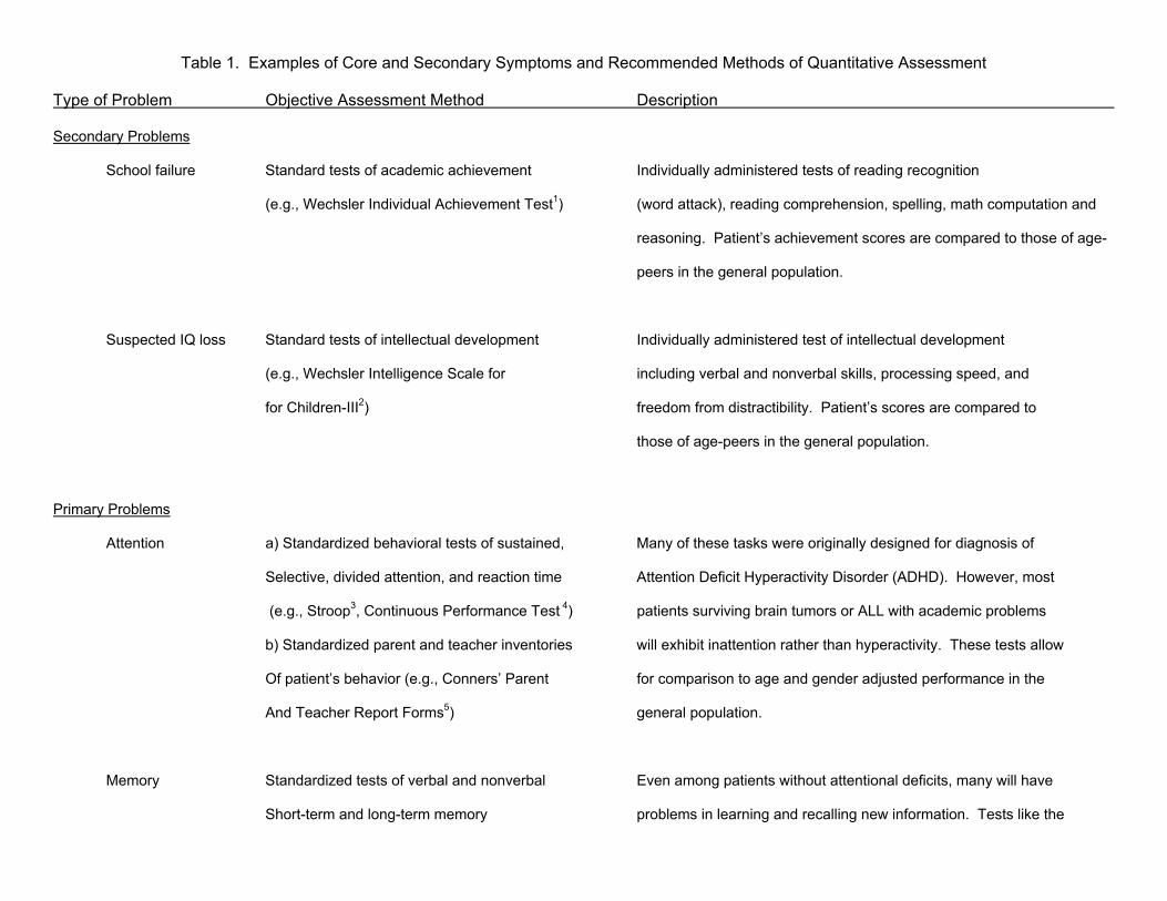
Table 1. Examples of Core and Secondary Symptoms and Recommended Methods of Quantitative Assessment
Type of Problem Objective Assessment Method Description
Secondary Problems
School failure Standard tests of academic achievement Individually administered tests of reading recognition
(e.g., Wechsler Individual Achievement Test1) (word attack), reading comprehension, spelling, math computation and
reasoning. Patient’s achievement scores are compared to those of age-
peers in the general population.
Suspected IQ loss Standard tests of intellectual development Individually administered test of intellectual development
(e.g., Wechsler Intelligence Scale for including verbal and nonverbal skills, processing speed, and
for Children-III2) freedom from distractibility. Patient’s scores are compared to
those of age-peers in the general population.
Primary Problems
Attention a) Standardized behavioral tests of sustained, Many of these tasks were originally designed for diagnosis of
Selective, divided attention, and reaction time Attention Deficit Hyperactivity Disorder (ADHD). However, most
(e.g., Stroop3, Continuous Performance Test 4) patients surviving brain tumors or ALL with academic problems
b) Standardized parent and teacher inventories will exhibit inattention rather than hyperactivity. These tests allow
Of patient’s behavior (e.g., Conners’ Parent for comparison to age and gender adjusted performance in the
And Teacher Report Forms5) general population.
Memory Standardized tests of verbal and nonverbal Even among patients without attentional deficits, many will have
Short-term and long-term memory problems in learning and recalling new information. Tests like the
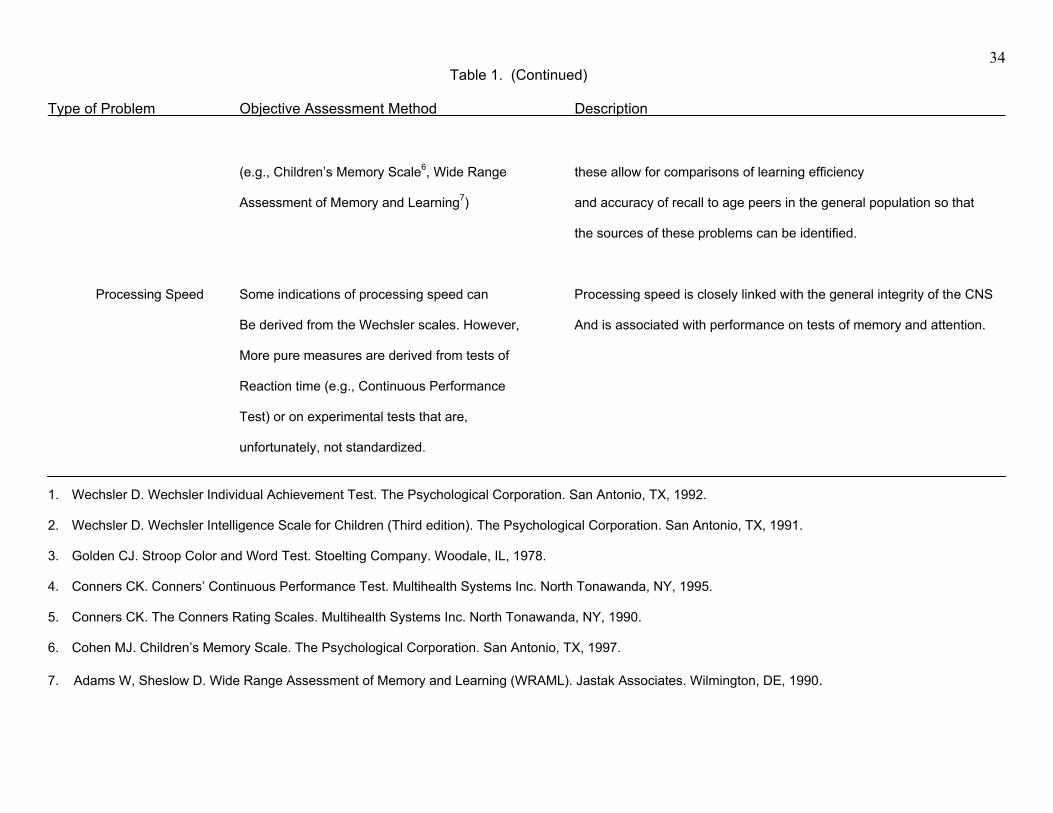
Type of Problem Objective Assessment Method Description
34 Table 1. (Continued)
(e.g., Children’s Memory Scale6, Wide Range these allow for comparisons of learning efficiency
Assessment of Memory and Learning7) and accuracy of recall to age peers in the general population so that
the sources of these problems can be identified.
Processing Speed Some indications of processing speed can Processing speed is closely linked with the general integrity of the CNS
Be derived from the Wechsler scales. However, And is associated with performance on tests of memory and attention.
More pure measures are derived from tests of
Reaction time (e.g., Continuous Performance
Test) or on experimental tests that are,
unfortunately, not standardized.
1. Wechsler D. Wechsler Individual Achievement Test. The Psychological Corporation. San Antonio, TX, 1992. 2. Wechsler D. Wechsler Intelligence Scale for Children (Third edition). The Psychological Corporation. San Antonio, TX, 1991. 3. Golden CJ. Stroop Color and Word Test. Stoelting Company. Woodale, IL, 1978. 4. Conners CK. Conners’ Continuous Performance Test. Multihealth Systems Inc. North Tonawanda, NY, 1995. 5. Conners CK. The Conners Rating Scales. Multihealth Systems Inc. North Tonawanda, NY, 1990. 6. Cohen MJ. Children’s Memory Scale. The Psychological Corporation. San Antonio, TX, 1997. 7. Adams W, Sheslow D. Wide Range Assessment of Memory and Learning (WRAML). Jastak Associates. Wilmington, DE, 1990.�