neurobiology of ocd
TRANSCRIPT
Neurobiology of OCD.Dr. Cijo Alex.
Overview
1) Overview of OCD.
2) Basic Neuroanatomy.
3) Basic neurophysiology.
4) Etiology and pathophysiology of OCD.
- Neurobiology
5) Conclusion
Overview of OCD

Definition and Diagnostic Features
Obsessive–compulsive disorder (OCD) is an intriguingand often debilitating syndrome characterized by the
presence of two distinct phenomena:obsessions and compulsions.
Obsessions are intrusive, recurrent, unwanted ideas,thoughts, or impulses that are difficult to dismiss despite
their disturbing nature.
Compulsions are repetitive behaviors, either observable or mental, that are intended to reduce the anxiety
engendered by obsessions.
OCD usually has its onset during puberty, although it may begin as early as age 2 years and infrequently begins after age 35 years.
Women develop OCD slightly more often than men.
Studies found that the course of OCD is usually chronic, with symptom severity waxing and
waning over time.
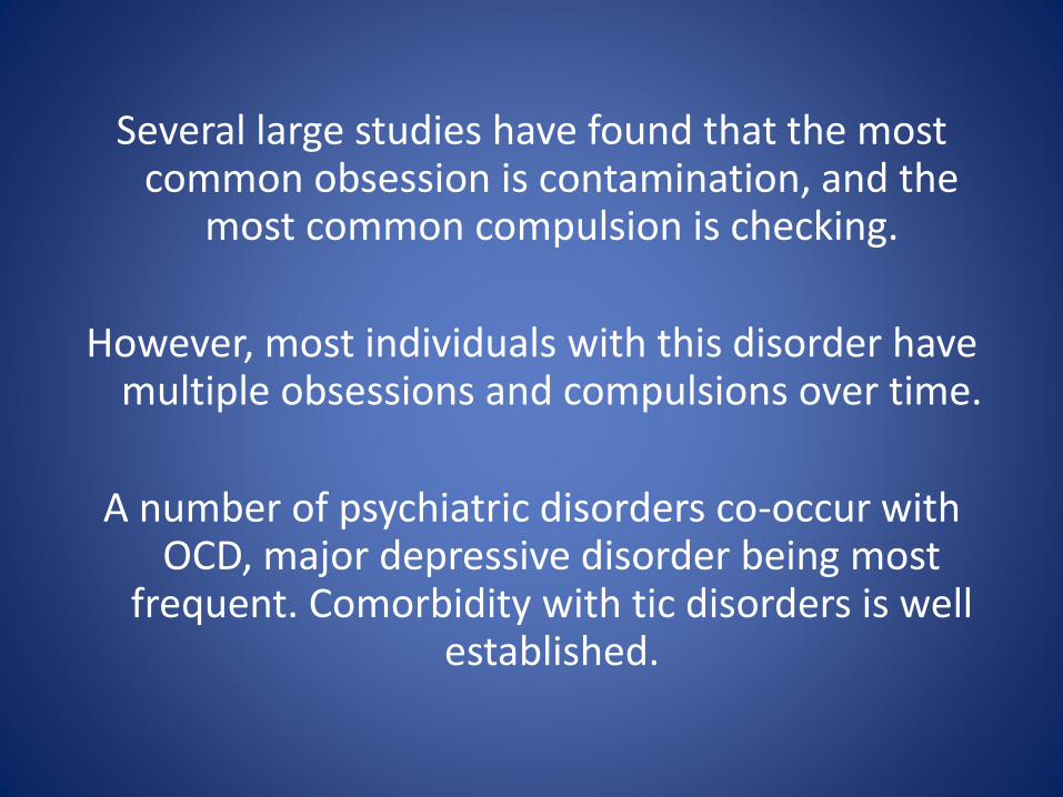
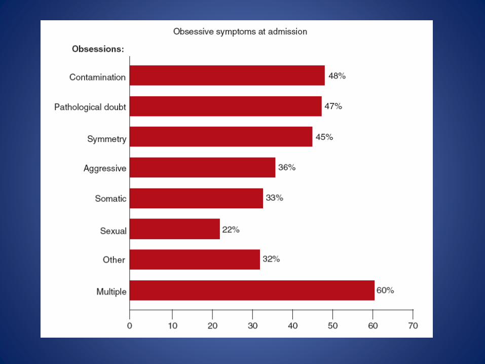
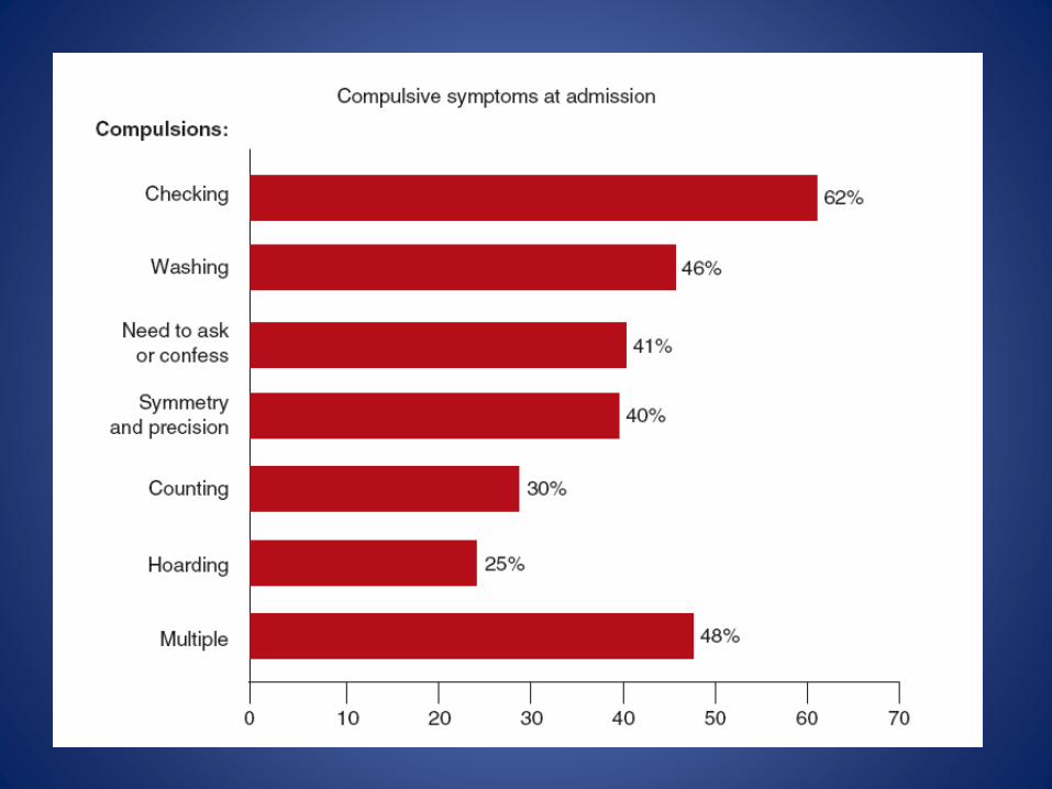
Several large studies have found that the most common obsession is contamination, and the
most common compulsion is checking.
However, most individuals with this disorder have multiple obsessions and compulsions over time.
A number of psychiatric disorders co-occur with OCD, major depressive disorder being most
frequent. Comorbidity with tic disorders is well established.
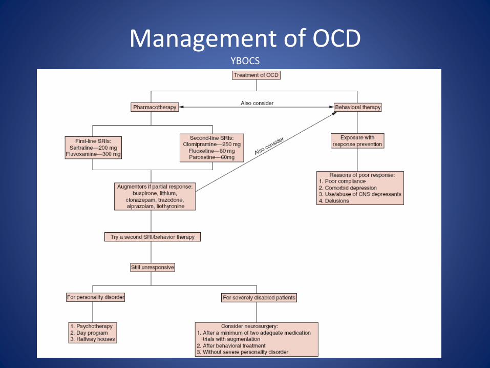
Management of OCDYBOCS
Basic Neuroanatomy
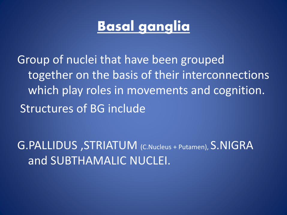
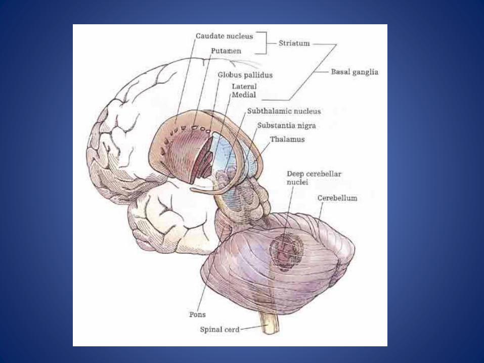
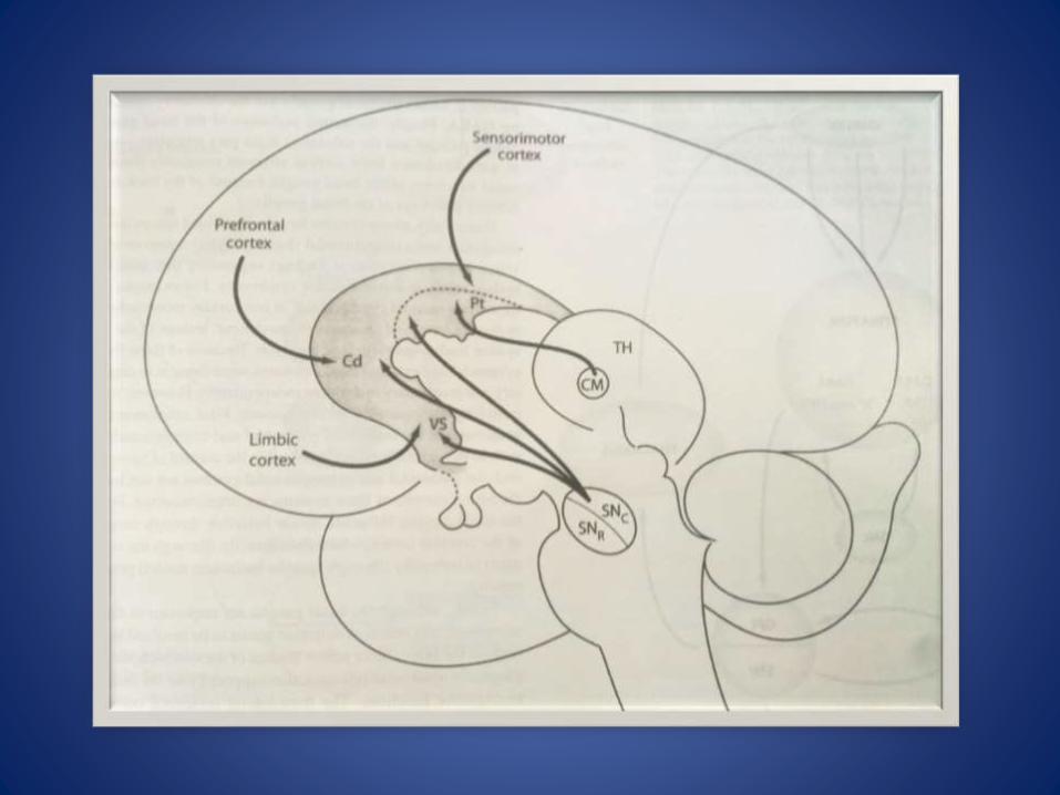
Basal ganglia
Group of nuclei that have been grouped together on the basis of their interconnections which play roles in movements and cognition.
Structures of BG include
G.PALLIDUS ,STRIATUM (C.Nucleus + Putamen), S.NIGRA and SUBTHAMALIC NUCLEI.
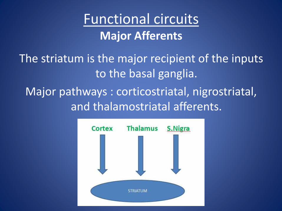
Functional circuitsMajor Afferents
The striatum is the major recipient of the inputs to the basal ganglia.
Major pathways : corticostriatal, nigrostriatal, and thalamostriatal afferents.
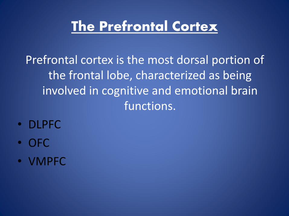
The Prefrontal Cortex
Prefrontal cortex is the most dorsal portion of the frontal lobe, characterized as being
involved in cognitive and emotional brain functions.
• DLPFC
• OFC
• VMPFC
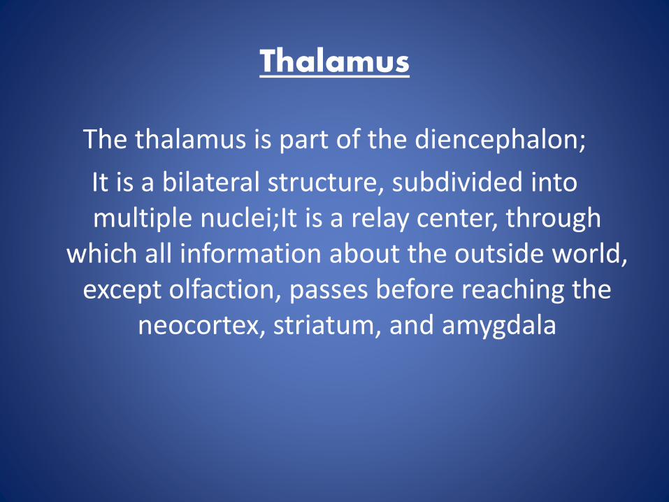
Thalamus
The thalamus is part of the diencephalon;
It is a bilateral structure, subdivided into multiple nuclei;It is a relay center, through
which all information about the outside world, except olfaction, passes before reaching the
neocortex, striatum, and amygdala

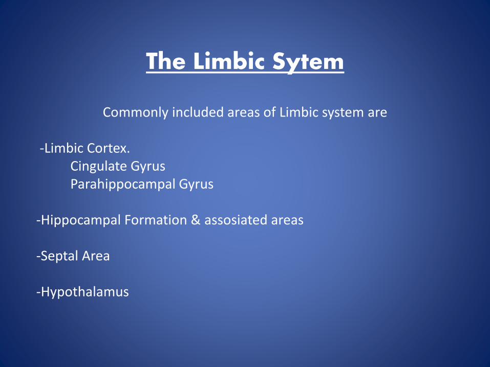
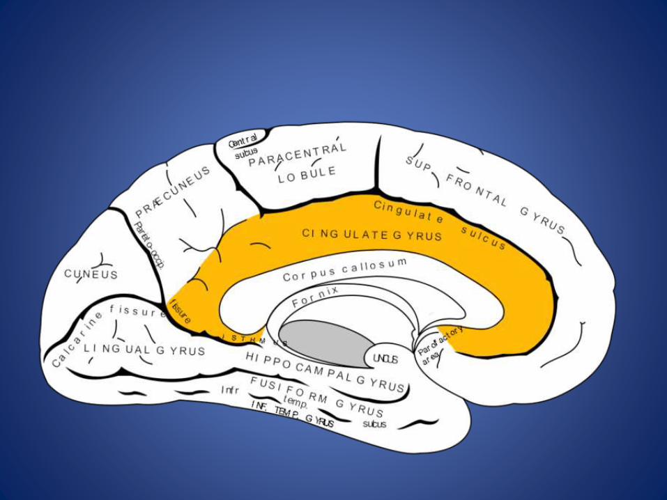
The Limbic Sytem
Commonly included areas of Limbic system are
-Limbic Cortex.Cingulate GyrusParahippocampal Gyrus
-Hippocampal Formation & assosiated areas
-Septal Area
-Hypothalamus
Basic neurophysiology
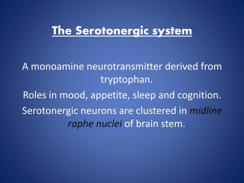
The Serotonergic system
A monoamine neurotransmitter derived from tryptophan.
Roles in mood, appetite, sleep and cognition.
Serotonergic neurons are clustered in midline raphe nuclei of brain stem.
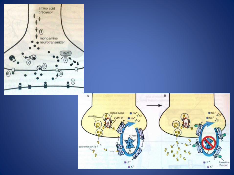
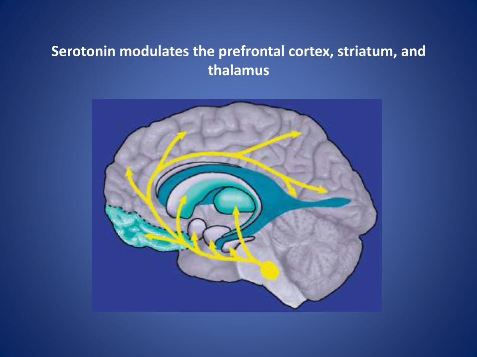
Serotonin modulates the prefrontal cortex, striatum, and thalamus
Etiology and pathophysiology of OCD
Although our understanding of what causes this disorder has continued to grow, there is still
much to learn. It is likely that OCD is caused by a complex interaction of factors rather than a
single defect.
However, for the purpose of clarity, these factors are described separately.
• Genetic Factors
• Psychological and Environmental Factors
• Phylogenetic Model
• Neurobiological Factors
Neurobiological Factors
Neuroanatomical aspects
Neurochemical aspects
Neurogenetical aspects
Neuroimmunology
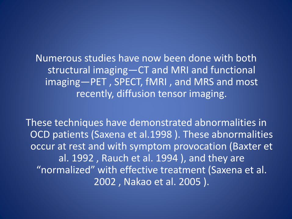
Numerous studies have now been done with both structural imaging—CT and MRI and functional
imaging—PET , SPECT, fMRI , and MRS and most recently, diffusion tensor imaging.
These techniques have demonstrated abnormalities in OCD patients (Saxena et al.1998 ). These abnormalities occur at rest and with symptom provocation (Baxter et
al. 1992 , Rauch et al. 1994 ), and they are “normalized” with effective treatment (Saxena et al.
2002 , Nakao et al. 2005 ).
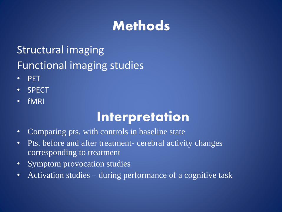
Methods
Structural imaging
Functional imaging studies • PET
• SPECT
• fMRI
Interpretation• Comparing pts. with controls in baseline state
• Pts. before and after treatment- cerebral activity changes corresponding to treatment
• Symptom provocation studies
• Activation studies – during performance of a cognitive task
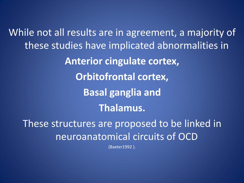
While not all results are in agreement, a majority of these studies have implicated abnormalities in
Anterior cingulate cortex,
Orbitofrontal cortex,
Basal ganglia and
Thalamus.
These structures are proposed to be linked in neuroanatomical circuits of OCD
(Baxter1992 ).
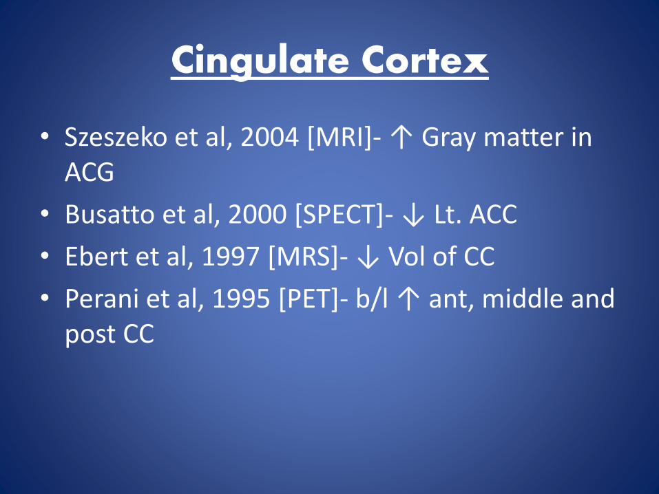
Cingulate Cortex
• Szeszeko et al, 2004 [MRI]- ↑ Gray matter in ACG
• Busatto et al, 2000 [SPECT]- ↓ Lt. ACC
• Ebert et al, 1997 [MRS]- ↓ Vol of CC
• Perani et al, 1995 [PET]- b/l ↑ ant, middle and post CC

Alterations in the anterior cingulate and globus pallidusSzeszko et al., Am J Psychiatry 2004
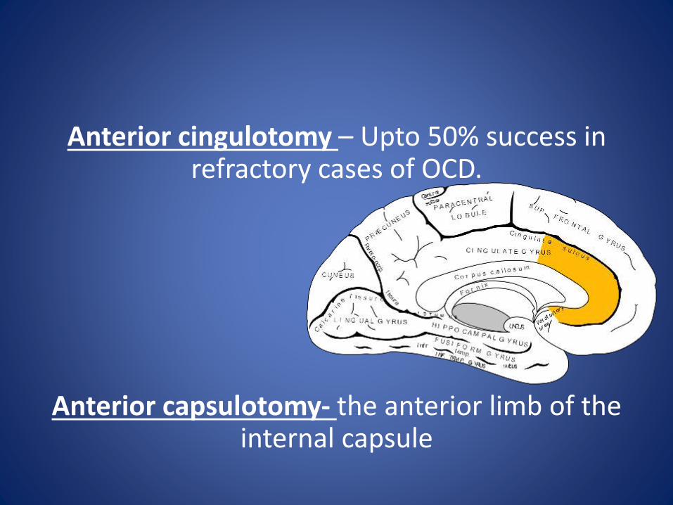
Anterior cingulotomy – Upto 50% success in refractory cases of OCD.
Anterior capsulotomy- the anterior limb of the internal capsule
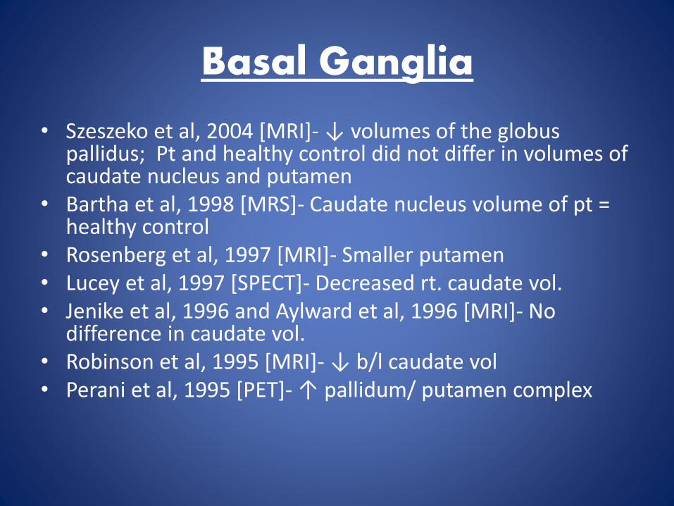
Basal Ganglia
• Szeszeko et al, 2004 [MRI]- ↓ volumes of the globuspallidus; Pt and healthy control did not differ in volumes of caudate nucleus and putamen
• Bartha et al, 1998 [MRS]- Caudate nucleus volume of pt = healthy control
• Rosenberg et al, 1997 [MRI]- Smaller putamen• Lucey et al, 1997 [SPECT]- Decreased rt. caudate vol. • Jenike et al, 1996 and Aylward et al, 1996 [MRI]- No
difference in caudate vol. • Robinson et al, 1995 [MRI]- ↓ b/l caudate vol• Perani et al, 1995 [PET]- ↑ pallidum/ putamen complex
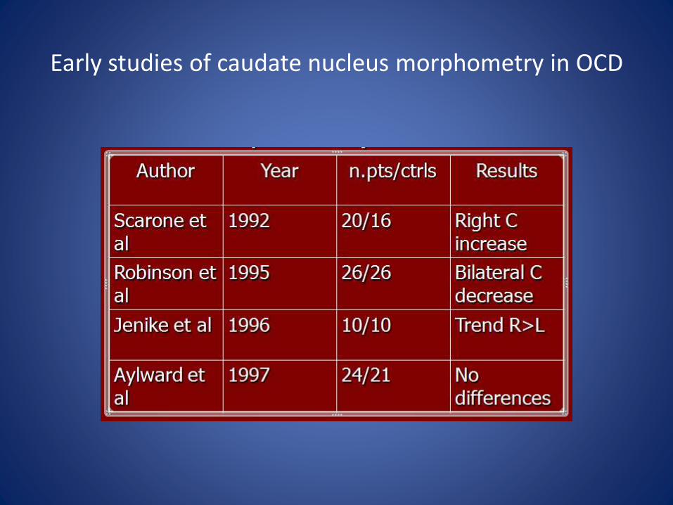
Early studies of caudate nucleus morphometry in OCD
Further indirect evidence implicating a role for basal ganglia dysfunction in OCD lies in the clinical relationship between neurological
insults to the basal ganglia and the subsequent development of obsessions and
compulsions.
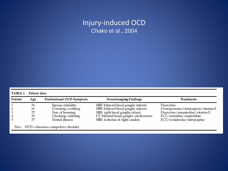
Injury-induced OCDChako et al., 2004
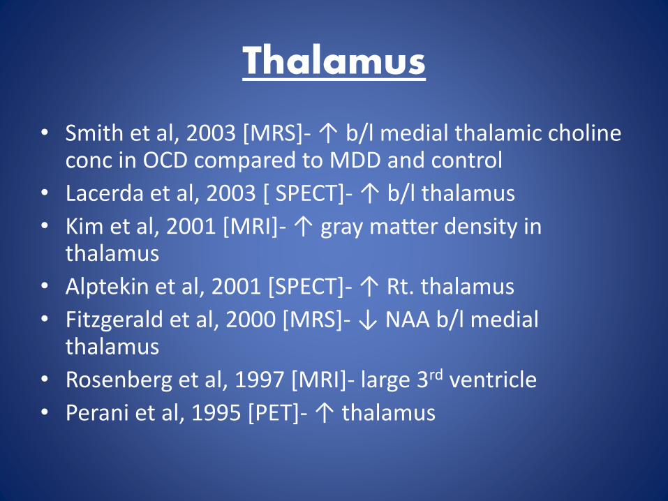
Thalamus
• Smith et al, 2003 [MRS]- ↑ b/l medial thalamic cholineconc in OCD compared to MDD and control
• Lacerda et al, 2003 [ SPECT]- ↑ b/l thalamus
• Kim et al, 2001 [MRI]- ↑ gray matter density in thalamus
• Alptekin et al, 2001 [SPECT]- ↑ Rt. thalamus
• Fitzgerald et al, 2000 [MRS]- ↓ NAA b/l medial thalamus
• Rosenberg et al, 1997 [MRI]- large 3rd ventricle
• Perani et al, 1995 [PET]- ↑ thalamus
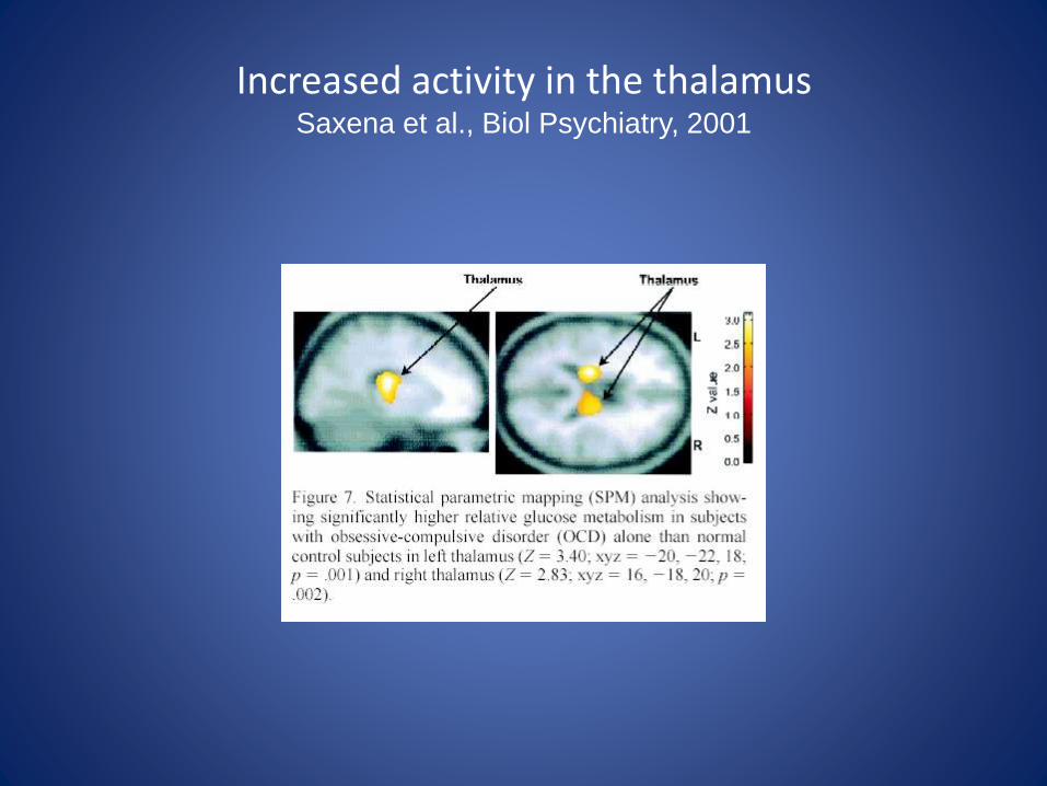
Increased activity in the thalamusSaxena et al., Biol Psychiatry, 2001
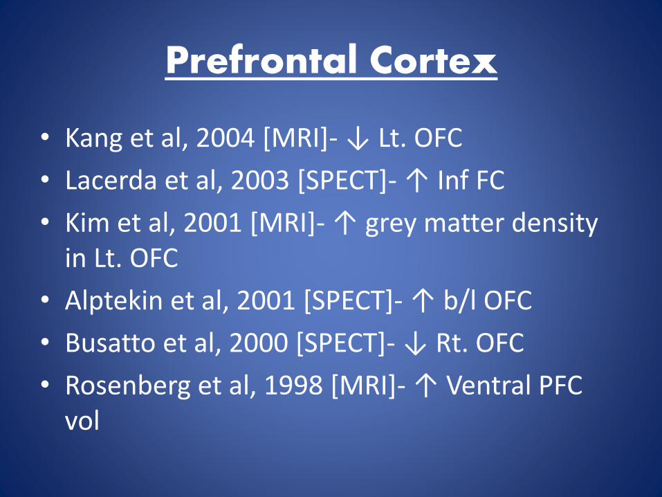
Prefrontal Cortex
• Kang et al, 2004 [MRI]- ↓ Lt. OFC
• Lacerda et al, 2003 [SPECT]- ↑ Inf FC
• Kim et al, 2001 [MRI]- ↑ grey matter density in Lt. OFC
• Alptekin et al, 2001 [SPECT]- ↑ b/l OFC
• Busatto et al, 2000 [SPECT]- ↓ Rt. OFC
• Rosenberg et al, 1998 [MRI]- ↑ Ventral PFC vol
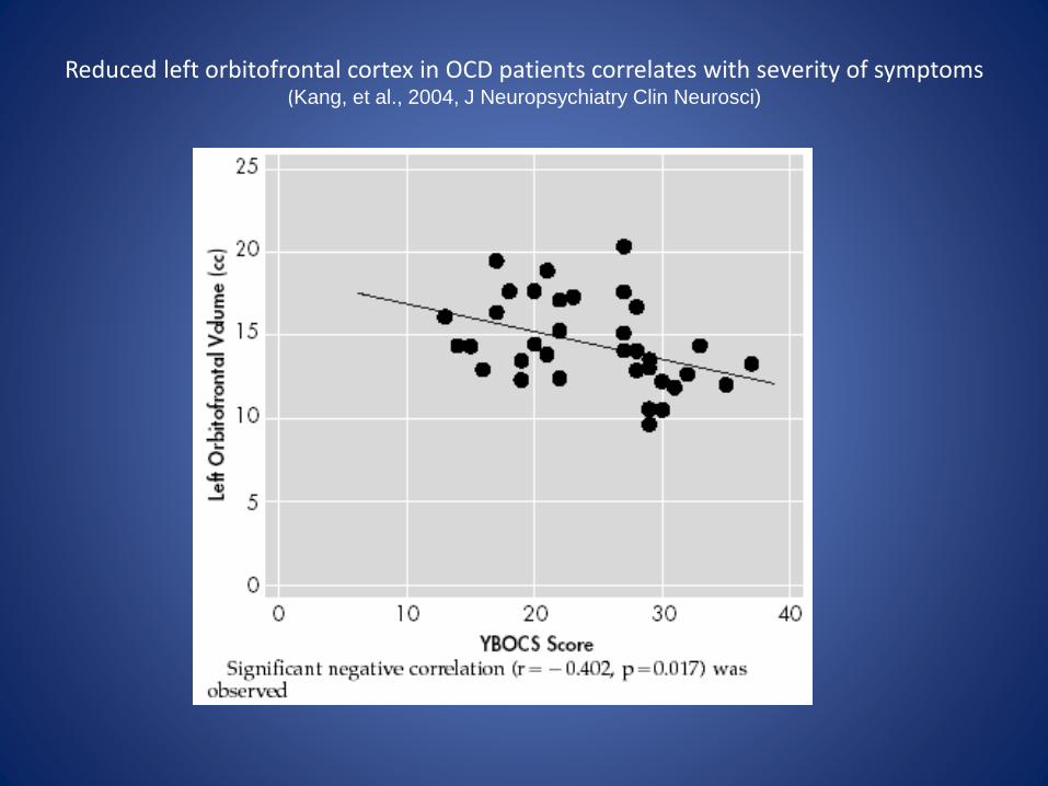
Reduced left orbitofrontal cortex in OCD patients correlates with severity of symptoms(Kang, et al., 2004, J Neuropsychiatry Clin Neurosci)
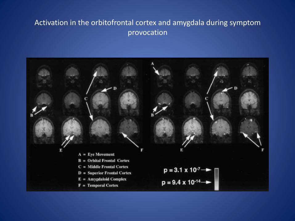
Activation in the orbitofrontal cortex and amygdala during symptom provocation
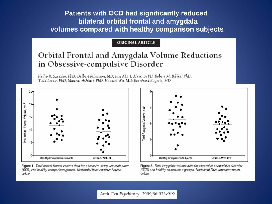
Patients with OCD had significantly reduced
bilateral orbital frontal and amygdalavolumes compared with healthy comparison subjects
Successful treatment of OCD symptoms may lead to normalization of frontal cortical
activation (Saxena et al. 2002 , Nakao et al. 2005 ).
Circuits to explain OCD
(Proposed integration of findings)
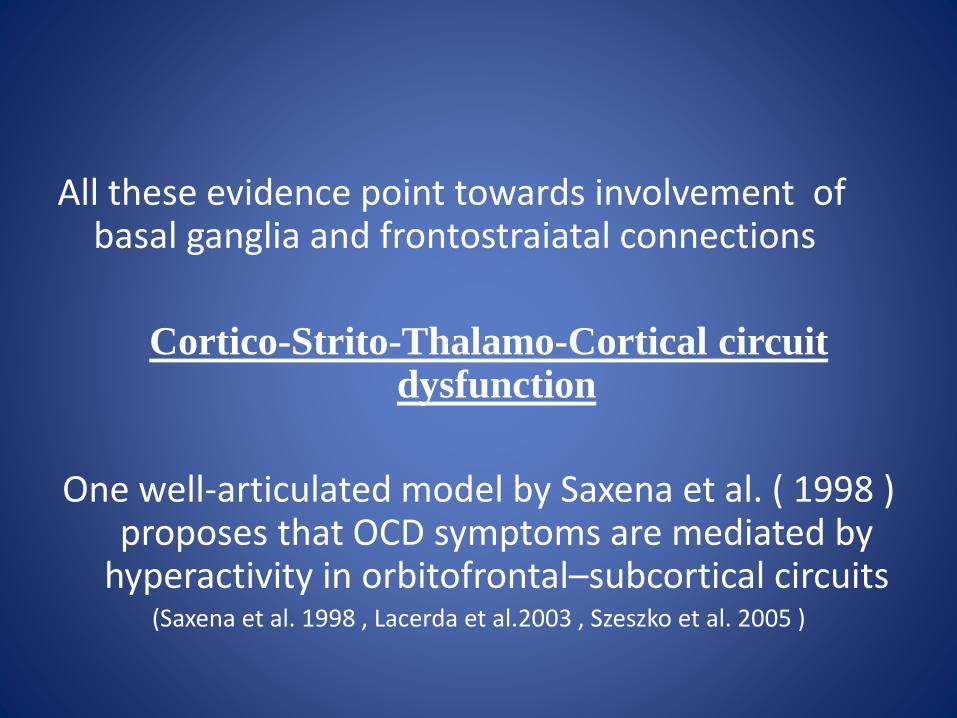
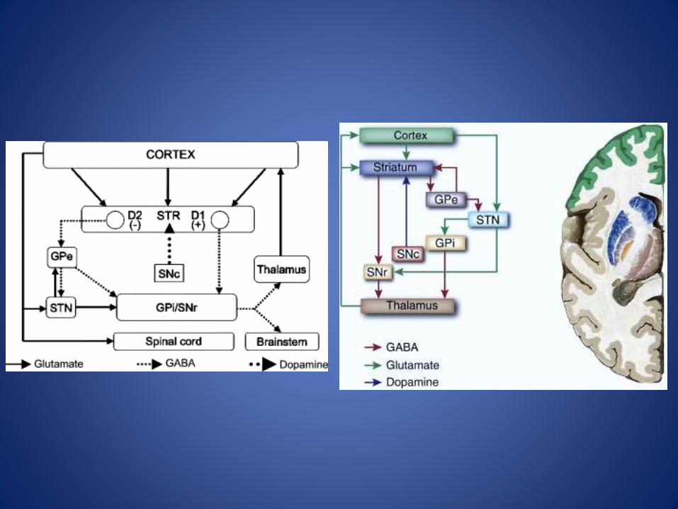
All these evidence point towards involvement of basal ganglia and frontostraiatal connections
Cortico-Strito-Thalamo-Cortical circuit dysfunction
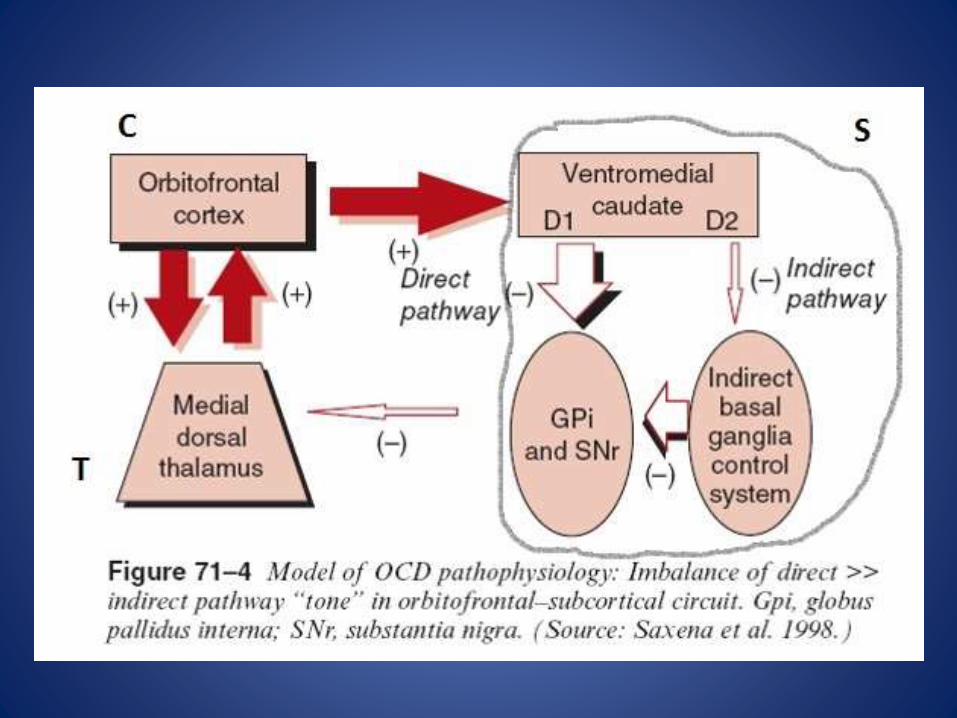
One well-articulated model by Saxena et al. ( 1998 ) proposes that OCD symptoms are mediated by
hyperactivity in orbitofrontal–subcortical circuits(Saxena et al. 1998 , Lacerda et al.2003 , Szeszko et al. 2005 )
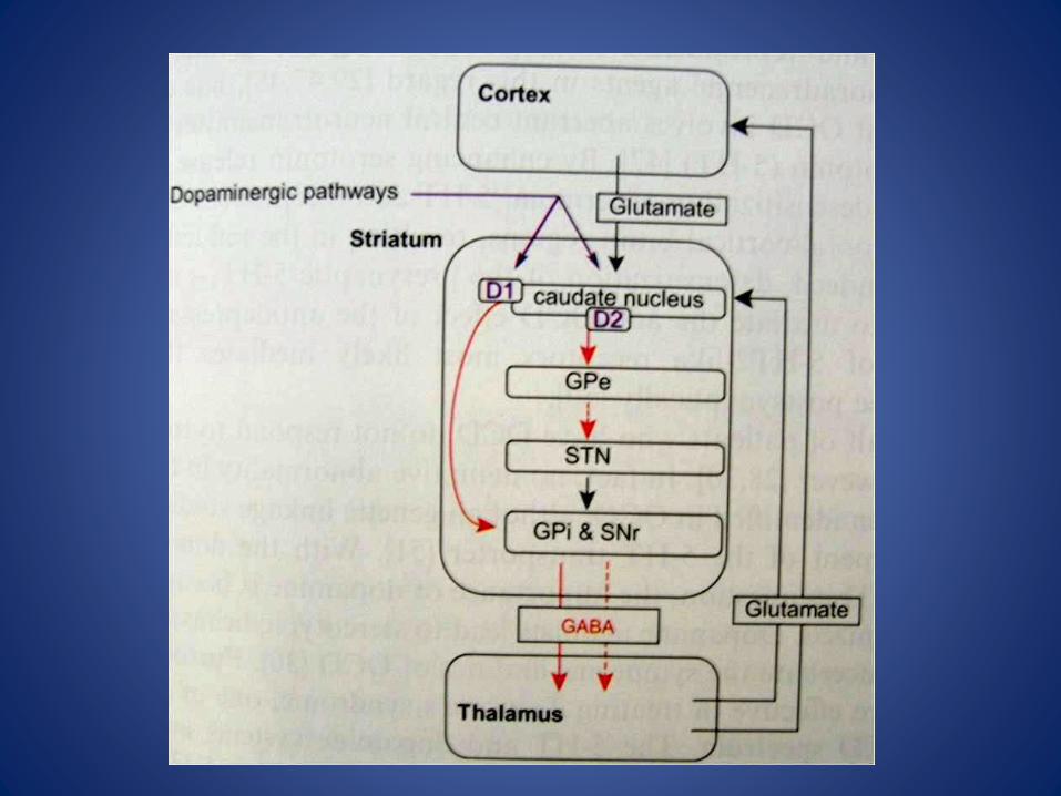
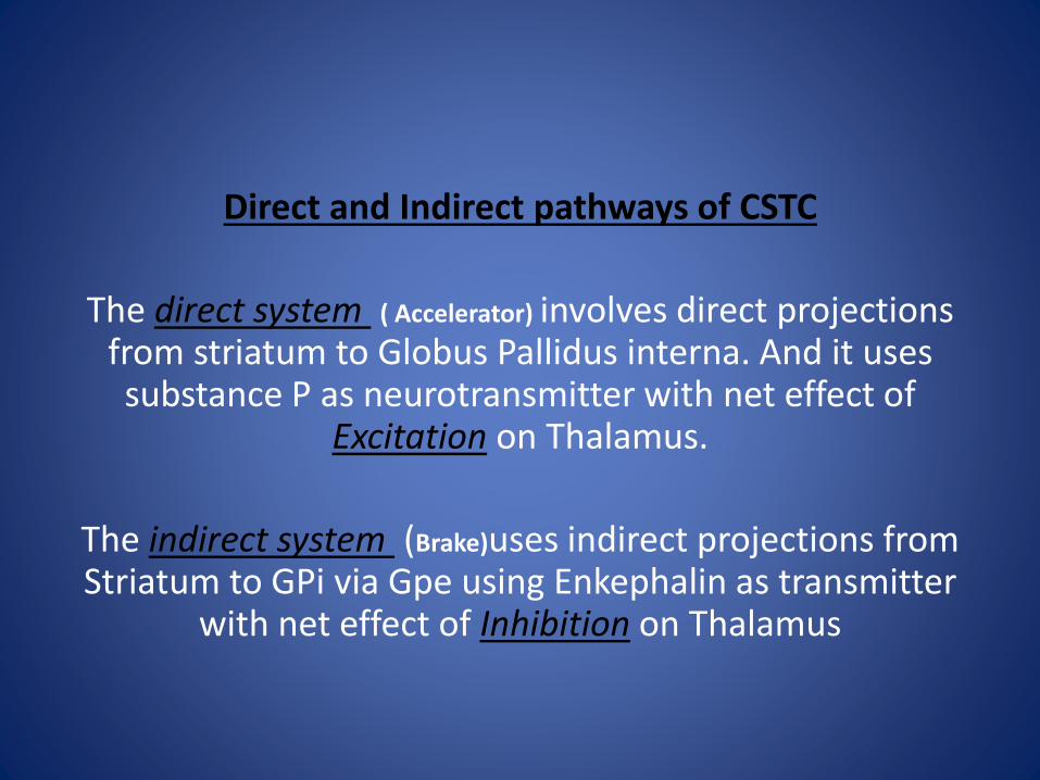
Direct and Indirect pathways of CSTC
The direct system ( Accelerator) involves direct projections from striatum to Globus Pallidus interna. And it uses substance P as neurotransmitter with net effect of
Excitation on Thalamus.
The indirect system (Brake)uses indirect projections from Striatum to GPi via Gpe using Enkephalin as transmitter
with net effect of Inhibition on Thalamus
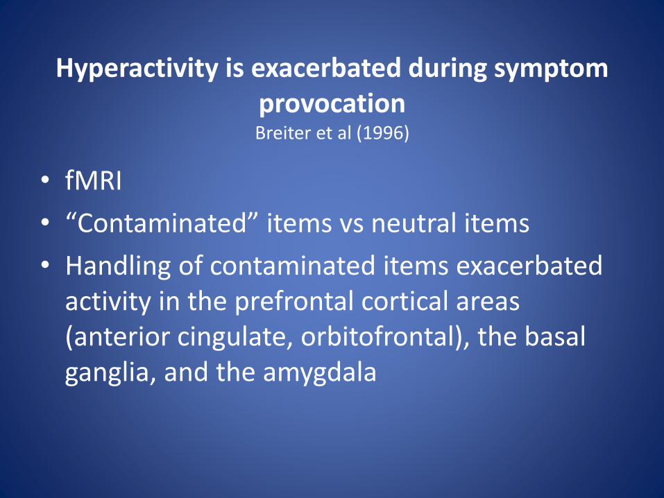
• fMRI
• “Contaminated” items vs neutral items
• Handling of contaminated items exacerbated activity in the prefrontal cortical areas (anterior cingulate, orbitofrontal), the basal ganglia, and the amygdala
Hyperactivity is exacerbated during symptom provocation Breiter et al (1996)
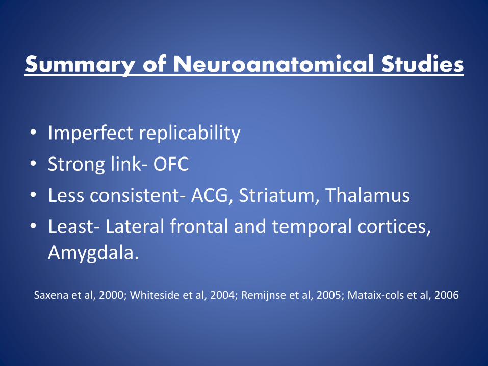
Summary of Neuroanatomical Studies
• Imperfect replicability
• Strong link- OFC
• Less consistent- ACG, Striatum, Thalamus
• Least- Lateral frontal and temporal cortices, Amygdala.
Saxena et al, 2000; Whiteside et al, 2004; Remijnse et al, 2005; Mataix-cols et al, 2006
Neurochemical Aspects
• Role of neurotransmitters in OCD
– Serotonin
– Dopamine
– Glutamate
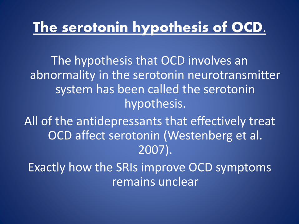
The serotonin hypothesis of OCD.
The hypothesis that OCD involves an abnormality in the serotonin neurotransmitter
system has been called the serotonin hypothesis.
All of the antidepressants that effectively treat OCD affect serotonin (Westenberg et al.
2007).
Exactly how the SRIs improve OCD symptoms remains unclear
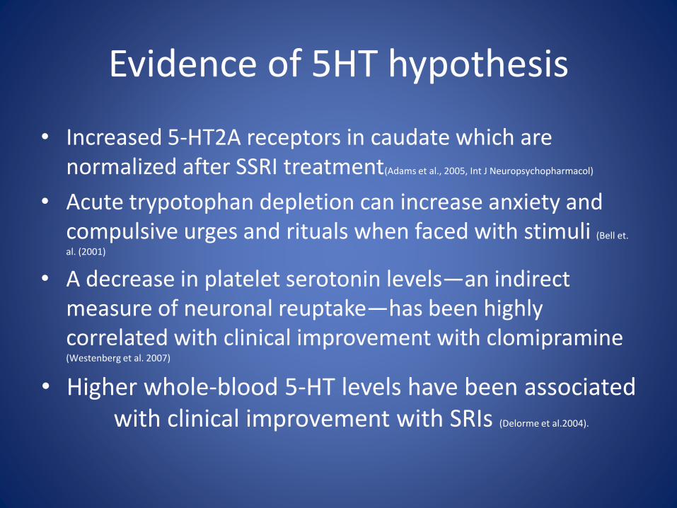
Evidence of 5HT hypothesis
• Increased 5-HT2A receptors in caudate which are normalized after SSRI treatment(Adams et al., 2005, Int J Neuropsychopharmacol)
• Acute trypotophan depletion can increase anxiety and compulsive urges and rituals when faced with stimuli (Bell et.
al. (2001)
• A decrease in platelet serotonin levels—an indirect measure of neuronal reuptake—has been highly correlated with clinical improvement with clomipramine(Westenberg et al. 2007)
• Higher whole-blood 5-HT levels have been associated with clinical improvement with SRIs (Delorme et al.2004).
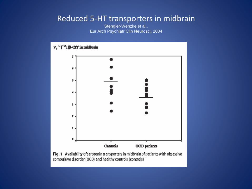
Reduced 5-HT transporters in midbrainStengler-Wenzke et al.,
Eur Arch Psychiatr Clin Neurosci, 2004
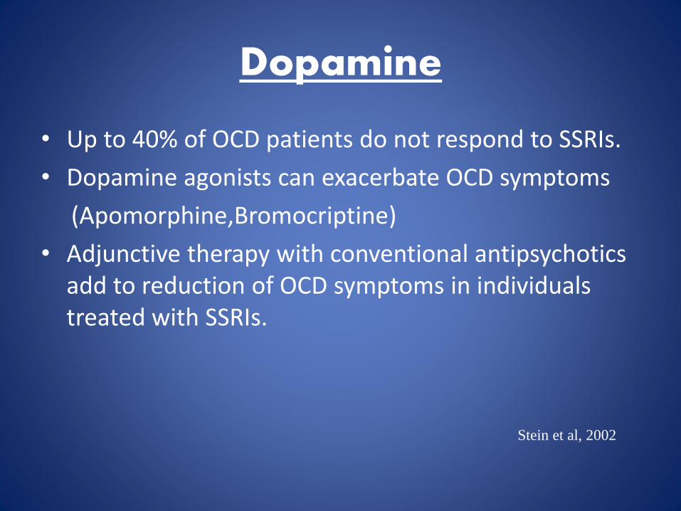
Dopamine
• Up to 40% of OCD patients do not respond to SSRIs.
• Dopamine agonists can exacerbate OCD symptoms
(Apomorphine,Bromocriptine)
• Adjunctive therapy with conventional antipsychotics add to reduction of OCD symptoms in individuals treated with SSRIs.
Stein et al, 2002
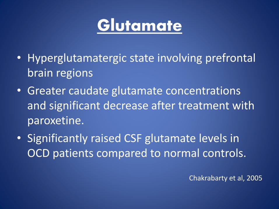
Glutamate
• Hyperglutamatergic state involving prefrontal brain regions
• Greater caudate glutamate concentrations and significant decrease after treatment with paroxetine.
• Significantly raised CSF glutamate levels in OCD patients compared to normal controls.
Chakrabarty et al, 2005
Neurogenetics
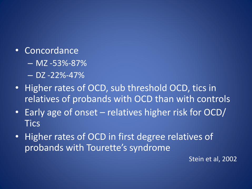
• Concordance– MZ -53%-87%
– DZ -22%-47%
• Higher rates of OCD, sub threshold OCD, tics in relatives of probands with OCD than with controls
• Early age of onset – relatives higher risk for OCD/ Tics
• Higher rates of OCD in first degree relatives of probands with Tourette’s syndrome
Stein et al, 2002
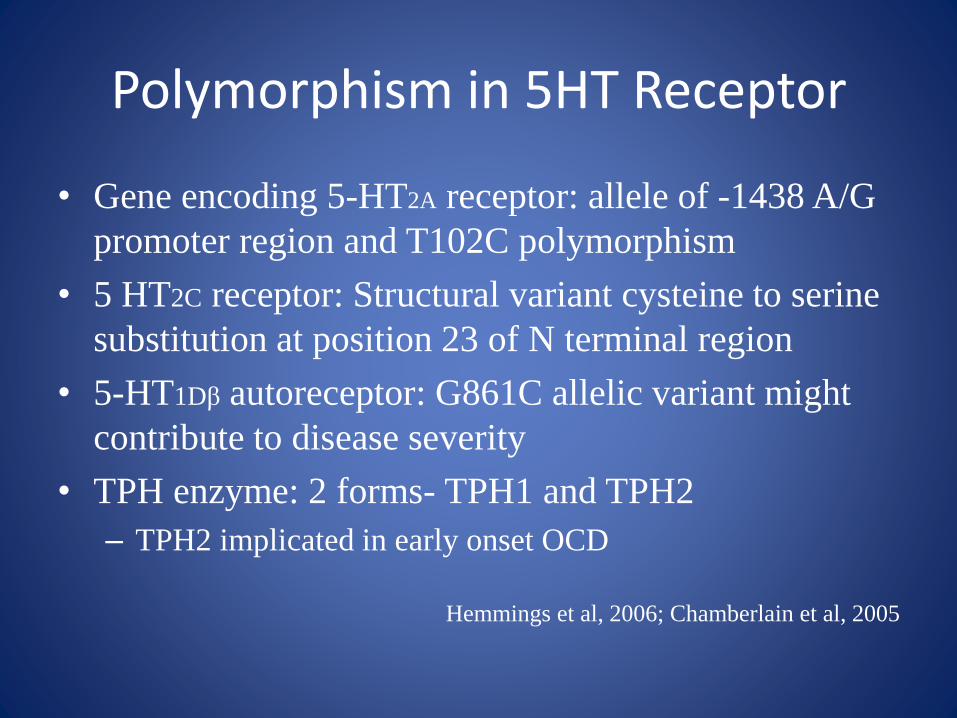
Polymorphism in 5HT Receptor
• Gene encoding 5-HT2A receptor: allele of -1438 A/G
promoter region and T102C polymorphism
• 5 HT2C receptor: Structural variant cysteine to serine
substitution at position 23 of N terminal region
• 5-HT1Dβ autoreceptor: G861C allelic variant might
contribute to disease severity
• TPH enzyme: 2 forms- TPH1 and TPH2
– TPH2 implicated in early onset OCD
Hemmings et al, 2006; Chamberlain et al, 2005
Neuroimmunology
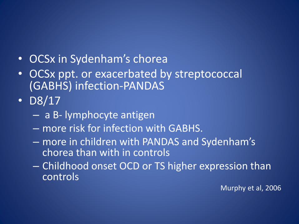
• OCSx in Sydenham’s chorea• OCSx ppt. or exacerbated by streptococcal
(GABHS) infection-PANDAS• D8/17
– a B- lymphocyte antigen – more risk for infection with GABHS.– more in children with PANDAS and Sydenham’s
chorea than with in controls– Childhood onset OCD or TS higher expression than
controlsMurphy et al, 2006
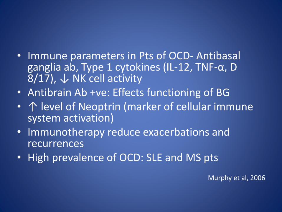
• Immune parameters in Pts of OCD- Antibasalganglia ab, Type 1 cytokines (IL-12, TNF-α, D 8/17), ↓ NK cell activity
• Antibrain Ab +ve: Effects functioning of BG• ↑ level of Neoptrin (marker of cellular immune
system activation)• Immunotherapy reduce exacerbations and
recurrences • High prevalence of OCD: SLE and MS pts
Murphy et al, 2006
Conclusion
No single model can explain OCD as of now.
CSTC loop appears to be the final common pathway and locus of primary pathology
Genetic vulnerability to autoimmune damage to striatum
Serotonergic modulation is important in treatment
Further research is needed to understand the neurobiology of OCD.

Thank You