neuroanatomy of the killer whale orcinus orca) from ... of the killer whale (orcinus orca) from...
TRANSCRIPT
Neuroanatomy of the Killer Whale(Orcinus orca) From Magnetic
Resonance ImagesLORI MARINO,1–3* CHET C. SHERWOOD,4,5 BRADLEY N. DELMAN,6,7
CHEUK Y. TANG,6,7 THOMAS P. NAIDICH,6,7 AND PATRICK R. HOF5,7–9
1Neuroscience and Behavioral Biology Program, Emory University, Atlanta, Georgia2Center for Behavioral Neuroscience, Emory University, Atlanta, Georgia
3Living Links Center for the Advanced Study of Ape and Human Evolution, YerkesRegional Primate Center, Atlanta, Georgia
4Department of Anthropology and School of Biomedical Sciences, Kent StateUniversity, Kent, Ohio
5Foundation for Comparative and Conservation Biology, Needmore, Pennsylvania6Department of Radiology, Mount Sinai School of Medicine, New York, New York
7Advanced Imaging Program, Mount Sinai School of Medicine, New York, New York8Department of Neuroscience, Mount Sinai School of Medicine, New York, New York
9New York Consortium in Evolutionary Primatology, New York, New York
ABSTRACTThis article presents the first series of MRI-based anatomically labeled
sectioned images of the brain of the killer whale (Orcinus orca). Magneticresonance images of the brain of an adult killer whale were acquired in thecoronal and axial planes. The gross morphology of the killer whale brain iscomparable in some respects to that of other odontocete brains, includingthe unusual spatial arrangement of midbrain structures. There are alsointriguing differences. Cerebral hemispheres appear extremely convolutedand, in contrast to smaller cetacean species, the killer whale brain possessesan exceptional degree of cortical elaboration in the insular cortex, temporaloperculum, and the cortical limbic lobe. The functional and evolutionaryimplications of these features are discussed. © 2004 Wiley-Liss, Inc.
Key words: killer whale; Orcinus orca; delphinid, cetacea; brain;MRI
Compared with other mammalian brains, the cetaceanbrain is, in many respects, highly unusual. Morgane et al.(1980: p. 105) state that “the lobular formations in thedolphin brain are organized in a pattern fundamentallydifferent from that seen in the brains of primates or car-nivores.” As there is a 55–60 million year divergencebetween cetaceans and the phylogenetically closest group,the artiodactyls, odontocete brains represent a blend ofearly mammalian and uniquely derived features (Ridg-way, 1986, 1990; Glezer et al., 1988; Manger et al., 1998).Differences between cetacean and other mammalianbrains of similar size have been found in cytoarchitectureand histochemistry (Garey et al., 1985; Garey and Leuba,1986; Glezer and Morgane, 1990; Glezer et al., 1990,1992a, 1992b, 1993, 1998; Hof et al., 1992, 1995, 1999,2000), cortical surface configuration (Jacobs et al., 1979;Morgane et al., 1980; Haug, 1987), and subcortical struc-tural morphology (Tarpley and Ridgway, 1994; Glezer etal., 1995a, 1995b).
The brains of a few cetacean species, particularly the bot-tlenose dolphin (Tursiops truncatus), have been studied rel-atively extensively. This is primarily due to the fact thatbottlenose dolphins are popular in captivity and have beenthe focus of many long-term field studies. Therefore, much isknown about their behavior, cognitive abilities, and socialecology. However, there is little neuroanatomical informa-tion on the brain of the largest Delphinid species, the killerwhale (Orcinus orca), despite the fact that this species has
*Correspondence to: Lori Marino, Neuroscience and BehavioralBiology Program, Emory University, Atlanta, GA 30322. Fax:404-727-0372. E-mail: [email protected]
Received 5 February 2004; Accepted 11 May 2004DOI 10.1002/ar.a.20075Published online 14 October 2004 in Wiley InterScience(www.interscience.wiley.com).
THE ANATOMICAL RECORD PART A 281A:1256–1263 (2004)
© 2004 WILEY-LISS, INC.
also been studied in captivity and in the field quite exten-sively. The lack of information on killer whale brains is likelydue to the difficulties associated with preparing and exam-ining such a large brain (approximately 5,000 g). Yet under-standing killer whale neuroanatomy is important because,like the bottlenose dolphin, killer whales show evidence ofmany complex and unusual social, communicative, and cog-nitive capacities. These include learning-based cooperativeforaging strategies (Baird, 2000), cultural variation andtransmission (Rendell and Whitehead, 2001; Yurk et al.,2002), and possibly mirror self-recognition (Delfour andMarten, 2001). Therefore, if we wish to understand the neu-robiological basis of such abilities, we will need to further ourunderstanding of the brains of killer whales.
A few studies address the size of the killer whale brain(Pilleri and Gihr, 1970; Marino, 1998, 2002) or a specificbrain structure such as the corpus callosum (Tarpley andRidgway, 1994). There are, however, no published descrip-tions of the basic neuroanatomy of the killer whale. In thepresent study, we present the first labeled sequential de-scription of killer whale neuroanatomy. The findings arebased on magnetic resonance imaging (MRI) of a postmor-tem brain. As with previous MRI-based studies of othercetacean species (Marino et al., 2001a, 2001b, 2002,2003a, 2003b), this method offers the opportunity to ob-serve the internal structure of the brain with little or nodistortion and with atlas-level precision.
MATERIALS AND METHODSSpecimen
The specimen is the postmortem brain of an adult malekiller whale (Orcinus orca). The brain was obtained shortlyafter death of natural causes and was immersion-fixed in a
large volume of 10% buffered formalin for an extended pe-riod of time.
Magnetic Resonance ImagingContiguous T2-weighted coronal and axial magnetic
resonance images were acquired with a 1.5 T GE high-gradient MRI scanner equipped with 8.3 software atMount Sinai School of Medicine. Coronal scans were ac-quired using TR � 500 msec and TE � 14.8 msec with anecho train of 2. Axial scans were acquired using TR � 700and TE � 15 msec with an echo train of 2. Images are 2mm thick with a matrix size of 512 � 512 and in-planeresolution of 32 � 32 cm yielding a voxel size of 0.63 �0.63 � 2.0 mm. Data were transferred electronically toeFilm (v1.5.3, eFilm Medical, Toronto, Ontario, Canada)for offline processing.
Anatomical Labeling and NomenclatureAll identifiable anatomical structures of the dolphin
brain were labeled in the coronal and axial plane images.The MR images of the killer whale brain were comparedwith the published photographs and illustrations of thebottlenose dolphin brain from Morgane et al. (1980) aswell as published neuroanatomical atlases based on MRIscans of other adult odontocete brains (Marino et al.,2001a, 2001b, 2002, 2003a, 2003b). The labeling nomen-clature follows that in the above sources.
RESULTSGeneral Morphology
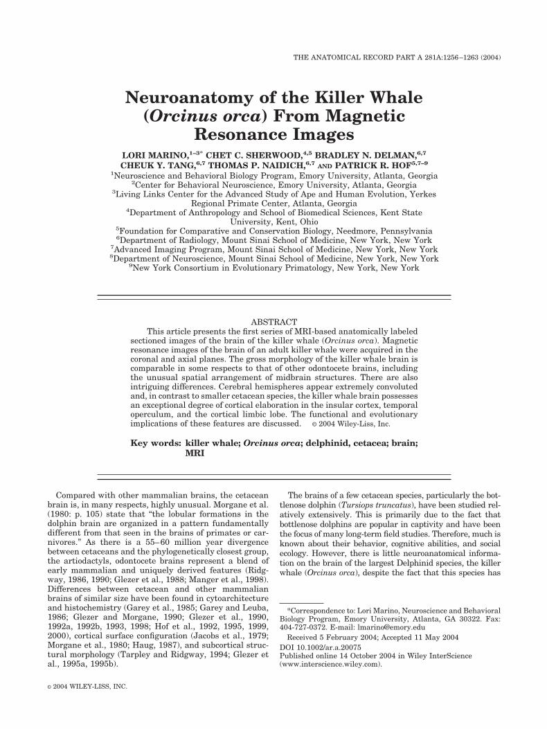
Figures 1–10 display a rostral-to-caudal sequence ofanatomically labeled originally acquired 2 mm thick coro-
Fig. 1. Figures 1–10: Rostral-to-caudal sequence of anatomically labeled 2 mm thick coronal scans of thekiller whale brain at 12 mm intervals. Section 13. L, left; R, right; A (inset), anterior.
Fig. 2. Section 19.
1257KILLER WHALE BRAIN FROM MRI
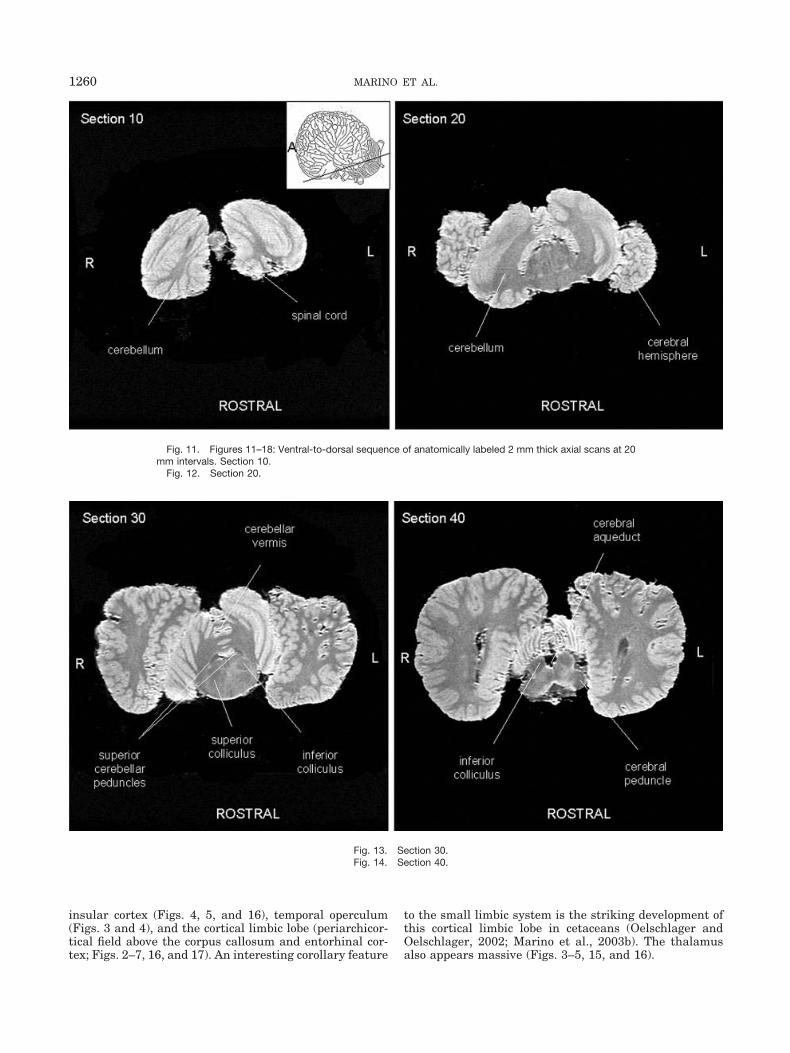
nal scans at 12 mm intervals. Figure 1 also includes aninset diagram of an odontocete brain showing the approx-imate orientation of coronal sections. Figures 11–18 dis-play a ventral-to-dorsal sequence of anatomically labeledoriginally acquired 2 mm thick axial scans at 20 mmintervals. Figure 11 also includes an inset diagram of an
odontocete brain showing the approximate orientation ofhorizontal sections. The figures show that the gross mor-phology of the killer whale brain is generally comparableto that of other odontocete brains (Morgane et al., 1980;Marino et al., 2001a, 2001b, 2002, 2003a, 2003b). Thekiller whale brain is characterized by extreme bitemporal
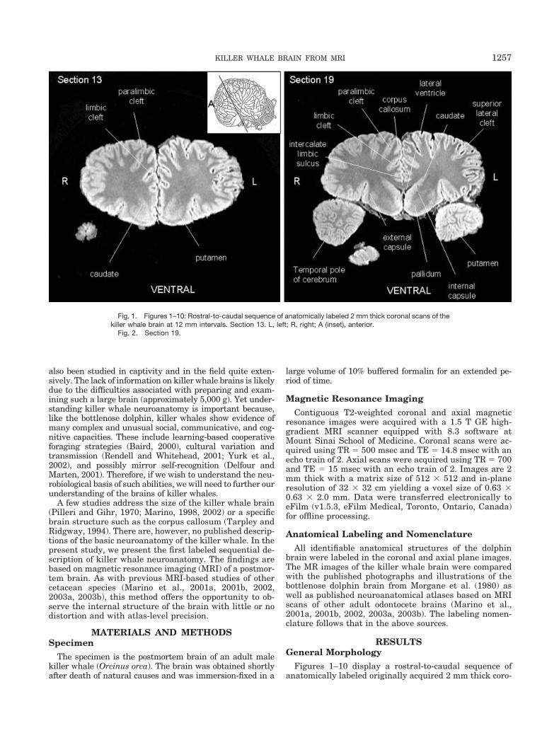
Fig. 3. Section 25.Fig. 4. Section 31.
Fig. 5. Section 37.Fig. 6. Section 43.
1258 MARINO ET AL.
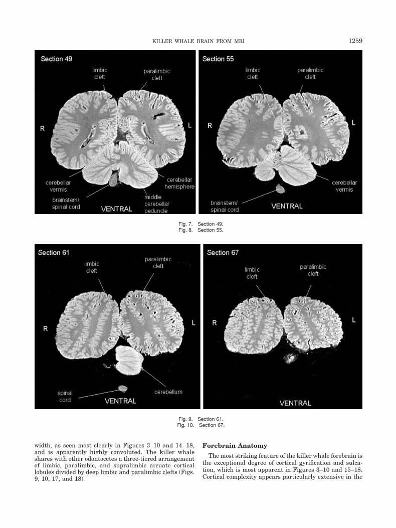
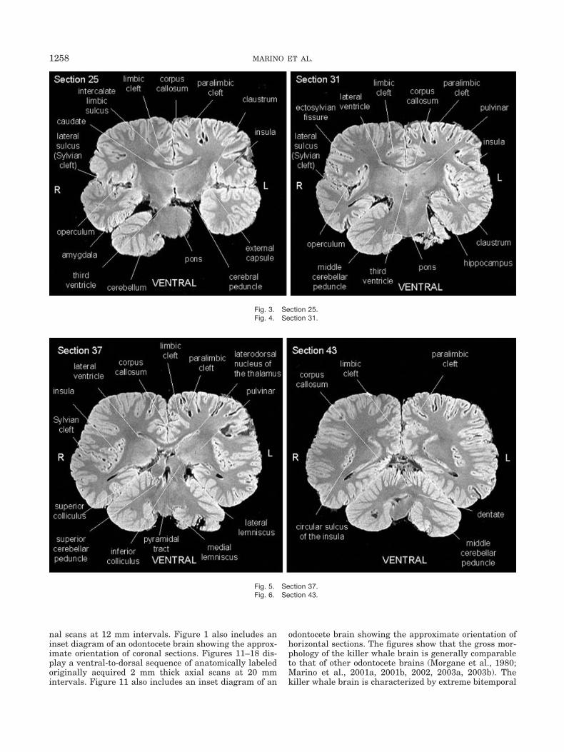
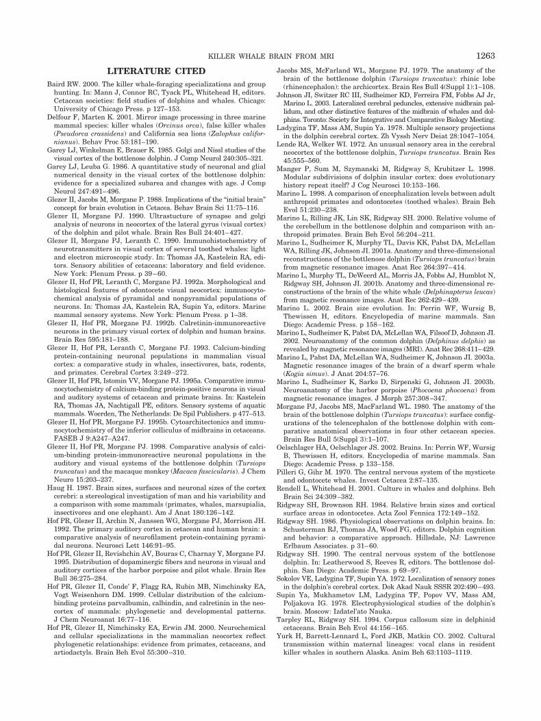
width, as seen most clearly in Figures 3–10 and 14–18,and is apparently highly convoluted. The killer whaleshares with other odontocetes a three-tiered arrangementof limbic, paralimbic, and supralimbic arcuate corticallobules divided by deep limbic and paralimbic clefts (Figs.9, 10, 17, and 18).
Forebrain Anatomy
The most striking feature of the killer whale forebrain isthe exceptional degree of cortical gyrification and sulca-tion, which is most apparent in Figures 3–10 and 15–18.Cortical complexity appears particularly extensive in the
Fig. 7. Section 49.Fig. 8. Section 55.
Fig. 9. Section 61.Fig. 10. Section 67.
1259KILLER WHALE BRAIN FROM MRI
insular cortex (Figs. 4, 5, and 16), temporal operculum(Figs. 3 and 4), and the cortical limbic lobe (periarchicor-tical field above the corpus callosum and entorhinal cor-tex; Figs. 2–7, 16, and 17). An interesting corollary feature
to the small limbic system is the striking development ofthis cortical limbic lobe in cetaceans (Oelschlager andOelschlager, 2002; Marino et al., 2003b). The thalamusalso appears massive (Figs. 3–5, 15, and 16).
Fig. 11. Figures 11–18: Ventral-to-dorsal sequence of anatomically labeled 2 mm thick axial scans at 20mm intervals. Section 10.
Fig. 12. Section 20.
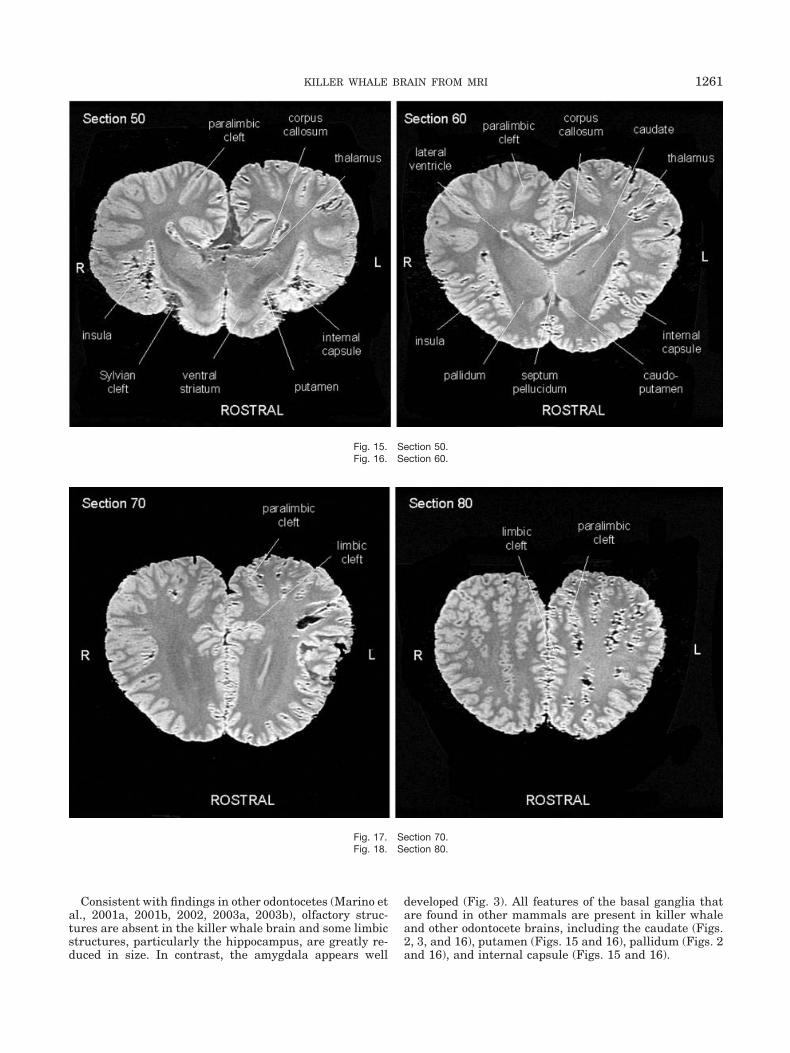
Fig. 13. Section 30.Fig. 14. Section 40.
1260 MARINO ET AL.
Consistent with findings in other odontocetes (Marino etal., 2001a, 2001b, 2002, 2003a, 2003b), olfactory struc-tures are absent in the killer whale brain and some limbicstructures, particularly the hippocampus, are greatly re-duced in size. In contrast, the amygdala appears well
developed (Fig. 3). All features of the basal ganglia thatare found in other mammals are present in killer whaleand other odontocete brains, including the caudate (Figs.2, 3, and 16), putamen (Figs. 15 and 16), pallidum (Figs. 2and 16), and internal capsule (Figs. 15 and 16).
Fig. 15. Section 50.Fig. 16. Section 60.
Fig. 17. Section 70.Fig. 18. Section 80.
1261KILLER WHALE BRAIN FROM MRI
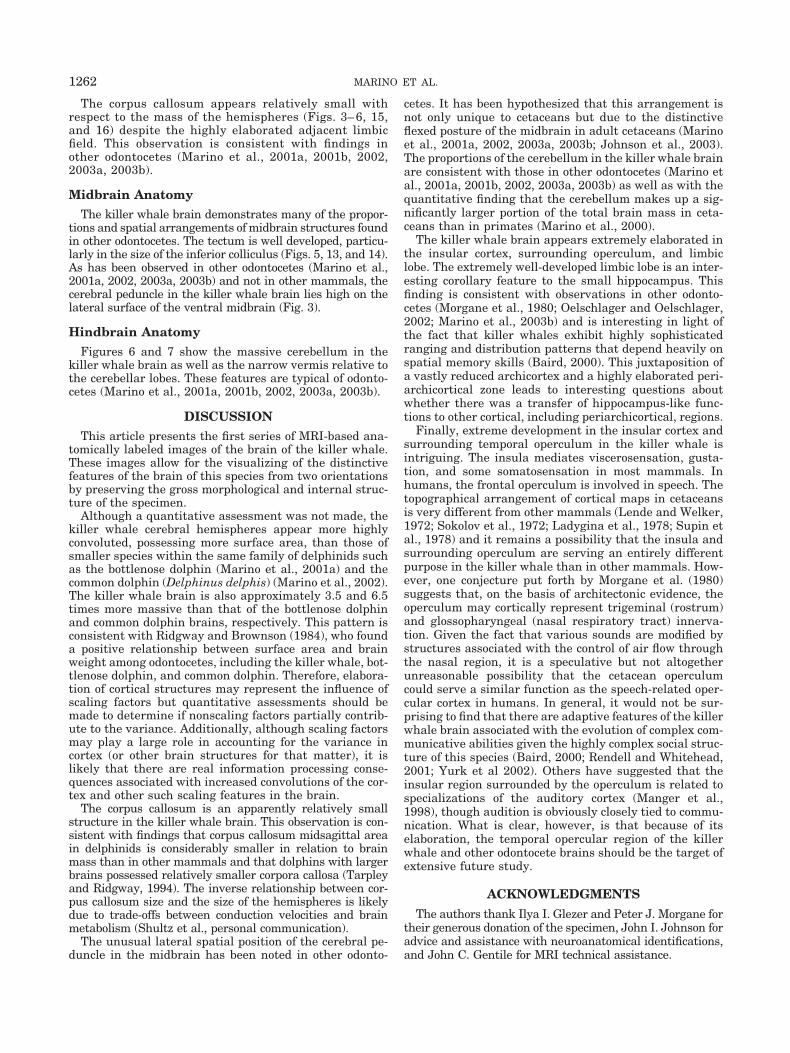
The corpus callosum appears relatively small withrespect to the mass of the hemispheres (Figs. 3– 6, 15,and 16) despite the highly elaborated adjacent limbicfield. This observation is consistent with findings inother odontocetes (Marino et al., 2001a, 2001b, 2002,2003a, 2003b).
Midbrain AnatomyThe killer whale brain demonstrates many of the propor-
tions and spatial arrangements of midbrain structures foundin other odontocetes. The tectum is well developed, particu-larly in the size of the inferior colliculus (Figs. 5, 13, and 14).As has been observed in other odontocetes (Marino et al.,2001a, 2002, 2003a, 2003b) and not in other mammals, thecerebral peduncle in the killer whale brain lies high on thelateral surface of the ventral midbrain (Fig. 3).
Hindbrain AnatomyFigures 6 and 7 show the massive cerebellum in the
killer whale brain as well as the narrow vermis relative tothe cerebellar lobes. These features are typical of odonto-cetes (Marino et al., 2001a, 2001b, 2002, 2003a, 2003b).
DISCUSSIONThis article presents the first series of MRI-based ana-
tomically labeled images of the brain of the killer whale.These images allow for the visualizing of the distinctivefeatures of the brain of this species from two orientationsby preserving the gross morphological and internal struc-ture of the specimen.
Although a quantitative assessment was not made, thekiller whale cerebral hemispheres appear more highlyconvoluted, possessing more surface area, than those ofsmaller species within the same family of delphinids suchas the bottlenose dolphin (Marino et al., 2001a) and thecommon dolphin (Delphinus delphis) (Marino et al., 2002).The killer whale brain is also approximately 3.5 and 6.5times more massive than that of the bottlenose dolphinand common dolphin brains, respectively. This pattern isconsistent with Ridgway and Brownson (1984), who founda positive relationship between surface area and brainweight among odontocetes, including the killer whale, bot-tlenose dolphin, and common dolphin. Therefore, elabora-tion of cortical structures may represent the influence ofscaling factors but quantitative assessments should bemade to determine if nonscaling factors partially contrib-ute to the variance. Additionally, although scaling factorsmay play a large role in accounting for the variance incortex (or other brain structures for that matter), it islikely that there are real information processing conse-quences associated with increased convolutions of the cor-tex and other such scaling features in the brain.
The corpus callosum is an apparently relatively smallstructure in the killer whale brain. This observation is con-sistent with findings that corpus callosum midsagittal areain delphinids is considerably smaller in relation to brainmass than in other mammals and that dolphins with largerbrains possessed relatively smaller corpora callosa (Tarpleyand Ridgway, 1994). The inverse relationship between cor-pus callosum size and the size of the hemispheres is likelydue to trade-offs between conduction velocities and brainmetabolism (Shultz et al., personal communication).
The unusual lateral spatial position of the cerebral pe-duncle in the midbrain has been noted in other odonto-
cetes. It has been hypothesized that this arrangement isnot only unique to cetaceans but due to the distinctiveflexed posture of the midbrain in adult cetaceans (Marinoet al., 2001a, 2002, 2003a, 2003b; Johnson et al., 2003).The proportions of the cerebellum in the killer whale brainare consistent with those in other odontocetes (Marino etal., 2001a, 2001b, 2002, 2003a, 2003b) as well as with thequantitative finding that the cerebellum makes up a sig-nificantly larger portion of the total brain mass in ceta-ceans than in primates (Marino et al., 2000).
The killer whale brain appears extremely elaborated inthe insular cortex, surrounding operculum, and limbiclobe. The extremely well-developed limbic lobe is an inter-esting corollary feature to the small hippocampus. Thisfinding is consistent with observations in other odonto-cetes (Morgane et al., 1980; Oelschlager and Oelschlager,2002; Marino et al., 2003b) and is interesting in light ofthe fact that killer whales exhibit highly sophisticatedranging and distribution patterns that depend heavily onspatial memory skills (Baird, 2000). This juxtaposition ofa vastly reduced archicortex and a highly elaborated peri-archicortical zone leads to interesting questions aboutwhether there was a transfer of hippocampus-like func-tions to other cortical, including periarchicortical, regions.
Finally, extreme development in the insular cortex andsurrounding temporal operculum in the killer whale isintriguing. The insula mediates viscerosensation, gusta-tion, and some somatosensation in most mammals. Inhumans, the frontal operculum is involved in speech. Thetopographical arrangement of cortical maps in cetaceansis very different from other mammals (Lende and Welker,1972; Sokolov et al., 1972; Ladygina et al., 1978; Supin etal., 1978) and it remains a possibility that the insula andsurrounding operculum are serving an entirely differentpurpose in the killer whale than in other mammals. How-ever, one conjecture put forth by Morgane et al. (1980)suggests that, on the basis of architectonic evidence, theoperculum may cortically represent trigeminal (rostrum)and glossopharyngeal (nasal respiratory tract) innerva-tion. Given the fact that various sounds are modified bystructures associated with the control of air flow throughthe nasal region, it is a speculative but not altogetherunreasonable possibility that the cetacean operculumcould serve a similar function as the speech-related oper-cular cortex in humans. In general, it would not be sur-prising to find that there are adaptive features of the killerwhale brain associated with the evolution of complex com-municative abilities given the highly complex social struc-ture of this species (Baird, 2000; Rendell and Whitehead,2001; Yurk et al 2002). Others have suggested that theinsular region surrounded by the operculum is related tospecializations of the auditory cortex (Manger et al.,1998), though audition is obviously closely tied to commu-nication. What is clear, however, is that because of itselaboration, the temporal opercular region of the killerwhale and other odontocete brains should be the target ofextensive future study.
ACKNOWLEDGMENTS
The authors thank Ilya I. Glezer and Peter J. Morgane fortheir generous donation of the specimen, John I. Johnson foradvice and assistance with neuroanatomical identifications,and John C. Gentile for MRI technical assistance.
1262 MARINO ET AL.
LITERATURE CITEDBaird RW. 2000. The killer whale-foraging specializations and group
hunting. In: Mann J, Connor RC, Tyack PL, Whitehead H, editors.Cetacean societies: field studies of dolphins and whales. Chicago:University of Chicago Press. p 127–153.
Delfour F, Marten K. 2001. Mirror image processing in three marinemammal species: killer whales (Orcinus orca), false killer whales(Pseudorca crassidens) and California sea lions (Zalophus califor-nianus). Behav Proc 53:181–190.
Garey LJ, Winkelman E, Brauer K. 1985. Golgi and Nissl studies of thevisual cortex of the bottlenose dolphin. J Comp Neurol 240:305–321.
Garey LJ, Leuba G. 1986. A quantitative study of neuronal and glialnumerical density in the visual cortex of the bottlenose dolphin:evidence for a specialized subarea and changes with age. J CompNeurol 247:491–496.
Glezer II, Jacobs M, Morgane P. 1988. Implications of the “initial brain”concept for brain evolution in Cetacea. Behav Brain Sci 11:75–116.
Glezer II, Morgane PJ. 1990. Ultrastucture of synapse and golgianalysis of neurons in neocortex of the lateral gyrus (visual cortex)of the dolphin and pilot whale. Brain Res Bull 24:401–427.
Glezer II, Morgane PJ, Leranth C. 1990. Immunohistochemistry ofneurotransmitters in visual cortex of several toothed whales: lightand electron microscopic study. In: Thomas JA, Kastelein RA, edi-tors. Sensory abilities of cetaceans: laboratory and field evidence.New York: Plenum Press. p 39–60.
Glezer II, Hof PR, Leranth C, Morgane PJ. 1992a. Morphological andhistological features of odontocete visual neocortex: immunocyto-chemical analysis of pyramidal and nonpyramidal populations ofneurons. In: Thomas JA, Kastelein RA, Supin Ya, editors. Marinemammal sensory systems. New York: Plenum Press. p 1–38.
Glezer II, Hof PR, Morgane PJ. 1992b. Calretinin-immunoreactiveneurons in the primary visual cortex of dolphin and human brains.Brain Res 595:181–188.
Glezer II, Hof PR, Leranth C, Morgane PJ. 1993. Calcium-bindingprotein-containing neuronal populations in mammalian visualcortex: a comparative study in whales, insectivores, bats, rodents,and primates. Cerebral Cortex 3:249–272.
Glezer II, Hof PR, Istomin VV, Morgane PJ. 1995a. Comparative immu-nocytochemistry of calcium-binding protein-positive neurons in visualand auditory systems of cetacean and primate brains. In: KasteleinRA, Thomas JA, Nachtigall PE, editors. Sensory systems of aquaticmammals. Woerden, The Netherlands: De Spil Publishers. p 477–513.
Glezer II, Hof PR, Morgane PJ. 1995b. Cytoarchitectonics and immu-nocytochemistry of the inferior colliculus of midbrains in cetaceans.FASEB J 9:A247–A247.
Glezer II, Hof PR, Morgane PJ. 1998. Comparative analysis of calci-um-binding protein-immunoreactive neuronal populations in theauditory and visual systems of the bottlenose dolphin (Tursiopstruncatus) and the macaque monkey (Macaca fascicularis). J ChemNeuro 15:203–237.
Haug H. 1987. Brain sizes, surfaces and neuronal sizes of the cortexcerebri: a stereological investigation of man and his variability anda comparison with some mammals (primates, whales, marsupialia,insectivores and one elephant). Am J Anat 180:126–142.
Hof PR, Glezer II, Archin N, Janssen WG, Morgane PJ, Morrison JH.1992. The primary auditory cortex in cetacean and human brain: acomparative analysis of neurofilament protein-containing pyrami-dal neurons. Neurosci Lett 146:91–95.
Hof PR, Glezer II, Revishchin AV, Bouras C, Charnay Y, Morgane PJ.1995. Distribution of dopaminergic fibers and neurons in visual andauditory cortices of the harbor porpoise and pilot whale. Brain ResBull 36:275–284.
Hof PR, Glezer II, Conde’ F, Flagg RA, Rubin MB, Nimchinsky EA,Vogt Weisenhorn DM. 1999. Cellular distribution of the calcium-binding proteins parvalbumin, calbindin, and calretinin in the neo-cortex of mammals: phylogenetic and developmental patterns.J Chem Neuroanat 16:77–116.
Hof PR, Glezer II, Nimchinsky EA, Erwin JM. 2000. Neurochemicaland cellular specializations in the mammalian neocortex reflectphylogenetic relationships: evidence from primates, cetaceans, andartiodactyls. Brain Beh Evol 55:300–310.
Jacobs MS, McFarland WL, Morgane PJ. 1979. The anatomy of thebrain of the bottlenose dolphin (Tursiops truncatus): rhinic lobe(rhinencephalon): the archicortex. Brain Res Bull 4(Suppl 1):1–108.
Johnson JI, Switzer RC III, Sudheimer KD, Ferreira FM, Fobbs AJ Jr,Marino L. 2003. Lateralized cerebral peduncles, extensive midbrain pal-lidum, and other distinctive features of the midbrain of whales and dol-phins. Toronto: Society for Integrative and Comparative Biology Meeting.
Ladygina TF, Mass AM, Supin Ya. 1978. Multiple sensory projectionsin the dolphin cerebral cortex. Zh Vyssh Nerv Deiat 28:1047–1054.
Lende RA, Welker WI. 1972. An unusual sensory area in the cerebralneocortex of the bottlenose dolphin, Tursiops truncatus. Brain Res45:555–560.
Manger P, Sum M, Szymanski M, Ridgway S, Krubitzer L. 1998.Modular subdivisions of dolphin insular cortex: does evolutionaryhistory repeat itself? J Cog Neurosci 10:153–166.
Marino L. 1998. A comparison of encephalization levels between adultanthropoid primates and odontocetes (toothed whales). Brain BehEvol 51:230–238.
Marino L, Rilling JK, Lin SK, Ridgway SH. 2000. Relative volume ofthe cerebellum in the bottlenose dolphin and comparison with an-thropoid primates. Brain Beh Evol 56:204–211.
Marino L, Sudheimer K, Murphy TL, Davis KK, Pabst DA, McLellanWA, Rilling JK, Johnson JI. 2001a. Anatomy and three-dimensionalreconstructions of the bottlenose dolphin (Tursiops truncatus) brainfrom magnetic resonance images. Anat Rec 264:397–414.
Marino L, Murphy TL, DeWeerd AL, Morris JA, Fobbs AJ, Humblot N,Ridgway SH, Johnson JI. 2001b. Anatomy and three-dimensional re-constructions of the brain of the white whale (Delphinapterus leucas)from magnetic resonance images. Anat Rec 262:429–439.
Marino L. 2002. Brain size evolution. In: Perrin WF, Wursig B,Thewissen H, editors. Encyclopedia of marine mammals. SanDiego: Academic Press. p 158–162.
Marino L, Sudheimer K, Pabst DA, McLellan WA, Filsoof D, Johnson JI.2002. Neuroanatomy of the common dolphin (Delphinus delphis) asrevealed by magnetic resonance images (MRI). Anat Rec 268:411–429.
Marino L, Pabst DA, McLellan WA, Sudheimer K, Johnson JI. 2003a.Magnetic resonance images of the brain of a dwarf sperm whale(Kogia simus). J Anat 204:57–76.
Marino L, Sudheimer K, Sarko D, Sirpenski G, Johnson JI. 2003b.Neuroanatomy of the harbor porpoise (Phocoena phocoena) frommagnetic resonance images. J Morph 257:308–347.
Morgane PJ, Jacobs MS, MacFarland WL. 1980. The anatomy of thebrain of the bottlenose dolphin (Tursiops truncatus): surface config-urations of the telencephalon of the bottlenose dolphin with com-parative anatomical observations in four other cetacean species.Brain Res Bull 5(Suppl 3):1–107.
Oelschlager HA, Oelschlager JS. 2002. Brains. In: Perrin WF, WursigB, Thewissen H, editors. Encyclopedia of marine mammals. SanDiego: Academic Press. p 133–158.
Pilleri G, Gihr M. 1970. The central nervous system of the mysticeteand odontocete whales. Invest Cetacea 2:87–135.
Rendell L, Whitehead H. 2001. Culture in whales and dolphins. BehBrain Sci 24:309–382.
Ridgway SH, Brownson RH. 1984. Relative brain sizes and corticalsurface areas in odontocetes. Acta Zool Fennica 172:149–152.
Ridgway SH. 1986. Physiological observations on dolphin brains. In:Schusterman RJ, Thomas JA, Wood FG, editors. Dolphin cognitionand behavior: a comparative approach. Hillsdale, NJ: LawrenceErlbaum Associates. p 31–60.
Ridgway SH. 1990. The central nervous system of the bottlenosedolphin. In: Leatherwood S, Reeves R, editors. The bottlenose dol-phin. San Diego: Academic Press. p 69–97.
Sokolov VE, Ladygina TF, Supin YA. 1972. Localization of sensory zonesin the dolphin’s cerebral cortex. Dok Akad Nauk SSSR 202:490–493.
Supin Ya, Mukhametov LM, Ladygina TF, Popov VV, Mass AM,Poljakova IG. 1978. Electrophysiological studies of the dolphin’sbrain. Moscow: Izdatel’ato Nauka.
Tarpley RL, Ridgway SH. 1994. Corpus callosum size in delphinidcetaceans. Brain Beh Evol 44:156–165.
Yurk H, Barrett-Lennard L, Ford JKB, Matkin CO. 2002. Culturaltransmission within maternal lineages: vocal clans in residentkiller whales in southern Alaska. Anim Beh 63:1103–1119.
1263KILLER WHALE BRAIN FROM MRI