neuro-ophthalmology curriculum standard · neuro-ophthalmology curriculum standard. purpose . the...
TRANSCRIPT

Neuro-ophthalmologyCurriculum Standard
September 2014
This standard has been prepared by The Royal Australian and New Zealand College of Ophthalmologists (RANZCO) and is copyright.
Please acknowledge authorship when using or quoting from material contained in this document.Except as permitted under applicable legislation, no part of this document may be adapted, modified or reproduced by any process (electronic or otherwise) without the specific written permission of the copyright owner. Permission may be refused at the copyright owner’s absolute discretion.

Neuro-Ophthalmology Curriculum Standard
Table of Contents
Purpose ............................................................................................................. 1
References ......................................................................................................... 1
Level of Mastery ................................................................................................ 2
Learning outcomes and performance criteria .................................................... 3
NO1 GENERAL MEDICAL AND OCULAR HISTORY RELEVANT TO NEURO-OPHTHALMIC CONDITIONS ......................................................................3
NO2 PERFORM EYE EXAMINATIONS FOR NEURO-OPHTHALMIC CONDITIONS .... 4
NO3 SPECIAL NEURO-OPHTHALMIC TESTING ................................................... 6
NO4 IMPLEMENT A NEURO-OPHTHALMIC MANAGEMENT PLAN ......................... 9
Context ............................................................................................................ 11
i

Neuro-ophthalmology Curriculum Standard
Purpose The Neuro-ophthalmology Clinical Performance Standard covers the knowledge, process, skills and competencies required for the diagnosis and management of neuro-ophthalmic conditions. Neuro-ophthalmology involves visual problems that are related to the nervous system. Neuro-ophthalmic disorders may be life threatening or sight threatening and require careful recognition and prompt management. A thorough knowledge of the diagnosis, examination, investigation and management of neuro-ophthalmic disorders is an essential skill for the ophthalmic trainee to acquire. References Neuro-ophthalmology Reading In addition to the core texts, the following references are recommended: • Pane, A., Burdon, M. & Miller, N.R. 2007, The neuro-ophthalmology survival guide,
Mosby/Elsevier, Edinburgh/New York, NY.
• Biousse, V. & Newman, N.J. 2009, Neuro-ophthalmology illustrated, Thieme Medical Publishers, NY.
• Gerstenblith, A.T. & Rabinowits, M.P. 2012, The Wills eye manual: office and
emergency room diagnosis and treatment of eye disease, 6th edn, Wolters Kluwer/Lippincott Williams & Wilkins, Philadelphia, PA.
• Savino, P.J., Danesh-Meyer, H.V. & Wills Eye Hospital 2012, Neuro-ophthalmology
(Color atlas & synopsis of clinical ophthalmology series), 2nd edn, Wolters Kluwer/Lippincott Williams & Wilkins Health, Philadelphia, PA.
Key Neuro-ophthalmology Randomised Clinical Trials • Keltner, J.L., Johnson, C.A., Cello, K.E., Dontchev, M., Gal, R.L. & Beck, R.W. (Optic
Neuritis Study Group) 2010, ‘Visual field profile of optic neuritis: a final follow-up report from the optic neuritis treatment trial from baseline through 15 years’, Archives of Ophthalmology, vol. 128, no. 3, pp. 330-7.
• Beck, R.W. & Gal, R.L. 2008, ‘Treatment of acute optic neuritis: a summary of findings
from the optic neuritis treatment trial’, Archives of Ophthalmology, vol. 126, no. 7, pp. 994-5.
• Optic Neuritis Study Group 2008, ‘Multiple sclerosis risk after optic neuritis: final optic
neuritis treatment trial follow-up’, Archives of Neurology, vol. 65, no. 6, pp. 727-32. • Beck, R.W., Smith, C.H., Gal, R.L., Xing, D., Bhatti, M.T., Brodsky, M.C., Buckley,
E.G., Chrousos, G.A., Corbett, J., Eggenberger, E., Goodwin, J.A., Katz, B., Kaufman, D.I., Keltner, J.L., Kupersmith, M.J., Miller, N.R., Moke, P.S., Nazarian, S., Orengo-Nania, S., Savino, P.J., Shults, W.T., Trobe, J.D. & Wall, M. (Optic Neuritis Study Group) 2004, ‘Neurologic impairment 10 years after optic neuritis’, Archives of Neurology, vol. 61, no. 9, pp.1386-9.
© 2014 Royal Australian and New Zealand College of Ophthalmologists Page 1
Neuro-ophthalmology Curriculum Standard
Additional Reading • Miller, N.R., Hoyt, W.F. & Walsh, F.B. et al. (eds) 2008, Walsh and Hoyt's Clinical
neuro-ophthalmology: the essentials, 2nd edn, Wolters Kluwer/Lippincott Williams & Wilkins, Philadelphia, PA.
• Albert, D.M., Miller, J.W., Azar, D.T. & Jakobiec, F.A, et al. 2008, Albert & Jakobiec's
Principles and practice of ophthalmology, 3rd edn, Saunders/Elsevier, Philadelphia, PA.
It is recommended that reading be supplemented with appropriate articles from current and relevant peer-reviewed journals. Level of Mastery For each learning outcome, the level of mastery to be attained by the trainee at the end of training is indicated as follows:
*** Core knowledge of which trainees must be able to demonstrate understanding Skills and procedures that trainees must be able to perform autonomously
** Knowledge of which trainees must have a good practical understanding Skills and procedures with which trainees should have assisted, and of which have good practical knowledge
* Knowledge, skills and procedures of which trainees must have some understanding
© 2014 Royal Australian and New Zealand College of Ophthalmologists Page 2

Neuro-ophthalmology Curriculum Standard
Learning outcomes and performance criteria NO1 GENERAL MEDICAL AND OCULAR HISTORY RELEVANT TO
NEURO-OPHTHALMIC CONDITIONS This element covers the processes for observing, prompting and recording a general medical and ocular history in preparation for diagnosis and treatment of neuro-ophthalmic conditions. The trainee is expected to have obtained and recorded a general medical and ocular history (including family history) as outlined in the Ophthalmic Basic Competencies and Knowledge (OBCK) standard.
LEARNING OUTCOMES LEVEL OF MASTERY PERFORMANCE CRITERIA
1.1 Take a thorough history
of presenting symptoms, past medical history, ocular history and family history from a patient presenting with a neuro-ophthalmic condition
***
Identify and record features that have relevance to neuro-ophthalmic conditions in a patient who may present with the following symptoms: 1.1.1 Visual loss 1.1.2 Disorders of higher visual function
including visual hallucinations 1.1.3 Unexplained loss of vision 1.1.4 Diplopia 1.1.5 Proptosis or enophthalmos 1.1.6 Ptosis 1.1.7 Transient visual loss 1.1.8 Ansicoria 1.1.9 Facial weakness or spasm 1.1.10 Oribital pain or headache 1.1.11 Nystagmus
© 2014 Royal Australian and New Zealand College of Ophthalmologists Page 3

Neuro-ophthalmology Curriculum Standard
NO2 PERFORM EYE EXAMINATIONS FOR NEURO-OPHTHALMIC CONDITIONS
This element covers the performance and interpretation of a range of eye examinations associated with neuro-ophthalmic conditions, as well as the demonstration of judgement in selecting the appropriate examinations for particular patients. The trainee is expected to have performed preliminary eye examinations as outlined in the Ophthalmic Basic Competencies and Knowledge (OBCK) standard. The trainee is also expected to have obtained and recorded a neuro-ophthalmic medical history.
LEARNING OUTCOMES LEVEL OF MASTERY PERFORMANCE CRITERIA
2.1 Undertake relevant neuro-
ophthalmic examination based on presenting complaint
***
2.1.1 Perform, measure, record and interpret
the results of the following examinations and note the relevance to the diagnosis of neuro-ophthalmic disease: • visual acuity (best corrected) • colour vision testing using Ishihara
colour plates, HRR, or other office based techniques.
• lid examination (identification, if present, of ptosis, retraction)
• pupil examination – including pupil size in light and dark, shape, reaction, relative afferent pupillary defect and near response
• ocular movements quantified with prisms
• ascertain where there is deviation, whether it is comitant or incomitant
• tests of muscle restriction, fatigability, saccadic, pursuit and nystagmoid movement including optokinetic nystagmus (OKN)
• contrast sensitivity testing • photostress test • ocular motility with respect to specific
nerve palsies – including patterns of lid abnormality
• detect and quantify exophthalmos or enophthalmos
• ascertain horizontal and vertical fusion range
© 2014 Royal Australian and New Zealand College of Ophthalmologists Page 4
Neuro-ophthalmology Curriculum Standard
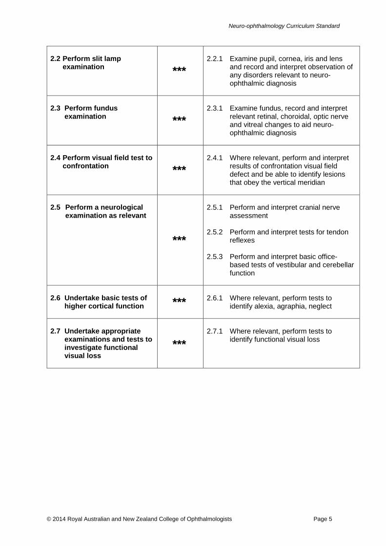
2.2 Perform slit lamp
examination ***
2.2.1 Examine pupil, cornea, iris and lens
and record and interpret observation of any disorders relevant to neuro-ophthalmic diagnosis
2.3 Perform fundus
examination ***
2.3.1 Examine fundus, record and interpret
relevant retinal, choroidal, optic nerve and vitreal changes to aid neuro-ophthalmic diagnosis
2.4 Perform visual field test to
confrontation ***
2.4.1 Where relevant, perform and interpret
results of confrontation visual field defect and be able to identify lesions that obey the vertical meridian
2.5 Perform a neurological
examination as relevant
***
2.5.1 Perform and interpret cranial nerve
assessment 2.5.2 Perform and interpret tests for tendon
reflexes 2.5.3 Perform and interpret basic office-
based tests of vestibular and cerebellar function
2.6 Undertake basic tests of
higher cortical function ***
2.6.1 Where relevant, perform tests to
identify alexia, agraphia, neglect
2.7 Undertake appropriate
examinations and tests to investigate functional visual loss
***
2.7.1 Where relevant, perform tests to
identify functional visual loss
© 2014 Royal Australian and New Zealand College of Ophthalmologists Page 5

Neuro-ophthalmology Curriculum Standard
NO3 SPECIAL NEURO-OPHTHALMIC TESTING This element covers the performance and interpretation of a range of special neuro-ophthalmic tests associated with neuro-ophthalmic conditions. The trainee is required to demonstrate judgement in selecting the appropriate tests for particular patients.
LEARNING OUTCOMES LEVEL OF MASTERY PERFORMANCE CRITERIA
3.1 Perform a range of
specific tests to confirm diagnosis
***
3.1.1 Perform, record and / or interpret the
results of the following examinations and note the relevance to the diagnosis of neuro-ophthalmic disease: • automated threshold perimetry
Goldmann perimetry • Hess Screens testing • Ice and Rest tests • ocular ultra-sonography • optic disc morphology and nerve fibre
layer abnormalities using contemporary investigative techniques, e.g. optical coherence tomography (OCT)
3.1.2 Understand the performance and
interpretation of the Tensilon test and: • recognise the parameters that are
relevant to the diagnosis • identify the indications and contra-
indications for the test • be aware that necessary
resuscitation equipment needs to be within easy reach
3.1.3 Perform and interpret pharmacological
pupil tests: • determine which pharmacological
tests may be appropriate for investigation of anisocoria to identify the underlying cause
3.1.4 Identify the indications for a fluorescein
angiogram and interpret results 3.1.5 Identify the indications for a temporal
artery biopsy, perform surgery, use techniques that avoid possible complications and interpret results
3.1.6 Identify optic nerve head drusen using
ultrasound and/or autofluroescence
© 2014 Royal Australian and New Zealand College of Ophthalmologists Page 6

Neuro-ophthalmology Curriculum Standard
3.2 Use blood testing to
confirm diagnosis
***
3.2.1 Identify the indications for and interpret
results from the following types of tests: • haematology • biochemistry • microbiology • immunology
3.3 Use genetic testing to
confirm diagnosis
***
3.3.1 Identify the indications for and interpret
results from genetic testing 3.3.2 Seek appropriate informed consent
from the patient for testing 3.3.3 Provide or arrange genetic counselling
3.4 Use radiologic testing to
confirm diagnosis
**
3.4.1 Identify the indications for and interpret
results of: • x-rays • computed tomography (CT) scans • magnetic resonance imaging (MRI)
scans • magnetic resonance arteriography
(MRA) • magnetic resonance venography
(MRV) • digital subtraction angiography
3.4.2 Understand which is the most relevant
imaging modality for diagnosis or exclusion of a condition
3.5 Use ultrasonography
testing of carotid and vertebral vasculature to confirm diagnosis
**
3.5.1 Identify the indications for and
interpretation of results of Doppler studies
3.6 Use the results of lumbar
puncture testing to confirm diagnosis **
3.6.1 Identify the indications for, the
principles of conducting and interpretation of results from a lumbar puncture
© 2014 Royal Australian and New Zealand College of Ophthalmologists Page 7

Neuro-ophthalmology Curriculum Standard
3.7 Understand the rationale
behind and results of electrophysiological testing
**
3.7.1 Identify the indications for conducting
the following testing of retinal and visual pathways and interpret the results thereof: • electro-oculogram (EOG) • electro-retinogram (ERG) • electro-myogram (EMG) • multifocal ERG • pattern ERG • visual evoked potential (VEP)
3.8 Use muscle biopsy
testing to confirm diagnosis *
3.8.1 Identify the indications for and
interpretation of fibre patterns of muscle biopsies for neuro-ophthalmology
3.9 Use auto-immune testing
to confirm diagnosis *
3.9.1 Identify the indications for skin and
buccal mucous membrane biopsies, the patterns of staining and significance
© 2014 Royal Australian and New Zealand College of Ophthalmologists Page 8
Neuro-ophthalmology Curriculum Standard
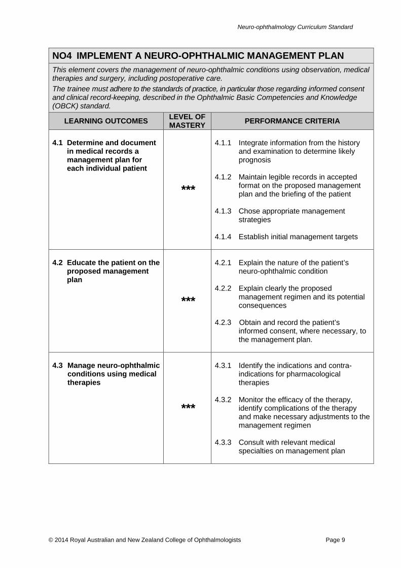
NO4 IMPLEMENT A NEURO-OPHTHALMIC MANAGEMENT PLAN This element covers the management of neuro-ophthalmic conditions using observation, medical therapies and surgery, including postoperative care. The trainee must adhere to the standards of practice, in particular those regarding informed consent and clinical record-keeping, described in the Ophthalmic Basic Competencies and Knowledge (OBCK) standard.
LEARNING OUTCOMES LEVEL OF MASTERY PERFORMANCE CRITERIA
4.1 Determine and document in medical records a management plan for each individual patient
***
4.1.1 Integrate information from the history and examination to determine likely prognosis
4.1.2 Maintain legible records in accepted format on the proposed management plan and the briefing of the patient
4.1.3 Chose appropriate management strategies
4.1.4 Establish initial management targets
4.2 Educate the patient on the proposed management plan
***
4.2.1 Explain the nature of the patient’s neuro-ophthalmic condition
4.2.2 Explain clearly the proposed management regimen and its potential consequences
4.2.3 Obtain and record the patient’s informed consent, where necessary, to the management plan.
4.3 Manage neuro-ophthalmic conditions using medical therapies
***
4.3.1 Identify the indications and contra-indications for pharmacological therapies
4.3.2 Monitor the efficacy of the therapy, identify complications of the therapy and make necessary adjustments to the management regimen
4.3.3 Consult with relevant medical specialties on management plan
© 2014 Royal Australian and New Zealand College of Ophthalmologists Page 9

Neuro-ophthalmology Curriculum Standard
4.4 Understand the
indications for the use of systemic steroids
***
4.4.1 Clearly outline:
• indications, dosage and management regimen of the use of systemic steroids in neuro-ophthalmic conditions
• potential complications • appropriate management strategies
to address complications (for example, bone-sparing treatments, treatment for potential gastric ulceration etc.)
4.5 Manage neuro-ophthalmic
conditions using surgery
***
4.5.1 Perform temporal artery biopsy:
• counsel the patient on surgery, potential outcomes and potential complications
• understand the details of the surgical procedure and management of intraoperative complications
• provide follow-up and continuing care
* 4.5.2 Design a surgical plan for botulinum
toxin injection
4.6 Describe the rationale
behind the management of neuro-ophthalmic conditions using radio-therapy
*
4.6.1 Identify methods of delivery of radiation
therapy for eye, neck and visual pathways
4.6.2 Be familiar with the selected technique
including effects and complications
4.7 Provide psychological
support for patient
**
4.7.1 Counsel patient on condition, likely
prognosis and progression 4.7.2 Provide genetic counselling 4.7.3 Provide emotional support 4.7.4 Refer patient to relevant support
services
© 2014 Royal Australian and New Zealand College of Ophthalmologists Page 10
Neuro-ophthalmology Curriculum Standard
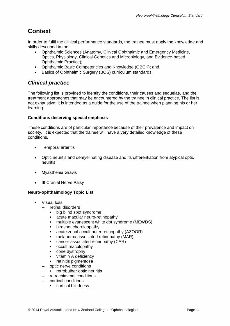
Context In order to fulfil the clinical performance standards, the trainee must apply the knowledge and skills described in the:
• Ophthalmic Sciences (Anatomy, Clinical Ophthalmic and Emergency Medicine, Optics, Physiology, Clinical Genetics and Microbiology, and Evidence-based Ophthalmic Practice);
• Ophthalmic Basic Competencies and Knowledge (OBCK); and, • Basics of Ophthalmic Surgery (BOS) curriculum standards.
Clinical practice The following list is provided to identify the conditions, their causes and sequelae, and the treatment approaches that may be encountered by the trainee in clinical practice. The list is not exhaustive; it is intended as a guide for the use of the trainee when planning his or her learning. Conditions deserving special emphasis These conditions are of particular importance because of their prevalence and impact on society. It is expected that the trainee will have a very detailed knowledge of these conditions.
• Temporal arteritis • Optic neuritis and demyelinating disease and its differentiation from atypical optic
neuritis • Myasthenia Gravis • III Cranial Nerve Palsy
Neuro-ophthalmology Topic List
• Visual loss – retinal disorders
• big blind spot syndrome • acute macular neuro-retinopathy • multiple evanescent white dot syndrome (MEWDS) • birdshot choroidopathy • acute zonal occult outer retinopathy (AZOOR) • melanoma associated retinopathy (MAR) • cancer associated retinopathy (CAR) • occult maculopathy • cone dystrophy • vitamin A deficiency • retinitis pigmentosa
– optic nerve conditions • retrobulbar optic neuritis
– retrochiasmal conditions – cortical conditions
• cortical blindness
© 2014 Royal Australian and New Zealand College of Ophthalmologists Page 11

Neuro-ophthalmology Curriculum Standard
• Conditions which present with sudden loss of vision
• Conditions which present with progressive loss of vision
• Unilateral swollen optic nerve
• Bilateral optic nerve swelling
• Pseudodisc swelling
• Non-metastatic manifestations of cancer – carcinoma associated – melanoma associated – optic neuropathy
• Phakomatoses
– neurofibromatosis Type I and II – racemose haemangioma – tuberous sclerosis – Von-Hippel-Lindau – Kippel-Trelauny-Weber
• Retinal vascular – retinal vein and artery occlusion – branch, hemi and central – haematological disorders: hypertension, anaemia, subacute bacterial
endocarditis (SBE) – Vogt-Koyanagi-Harada disease – migraine – neurological disease with retinovascular manifestations e.g. Susac syndrome – retinal vasculitidies
• Sclera
– anterior scleritis – posterior scleritis
• Pupil
– anatomy – normal – testing – abnormal – testing – describe clinical setting and relevance of:
• Horner syndrome • light-near dissociation • aberrant regeneration • Adie pupil • afferent pupil defect • mid-brain / Parinaud pupil • pharmacological blockade / tests • Raede paratrigeminal neuralgia • episodic dysfunction • traumatic pupillary abnormalities
• Orbit (orbital disease relevant to neuro- ophthalmology)
– thyroid eye disease • discuss the pathogenesis and clinical manifestation
© 2014 Royal Australian and New Zealand College of Ophthalmologists Page 12

Neuro-ophthalmology Curriculum Standard
• describe the investigations and diagnosis (differential radiological findings) • explain the immune function and its clinical relevance • understand the principles of management:
– systemic — explain the basis of treatment of thyrotoxicosis (medical, surgical and radiological treatments) and its effects on the eye
– ophthalmic — describe the principles, order and timing of treatment • Tumours
– describe the modes of clinical presentation, treatments and complications of: • meningiomas
– optic nerve sheath – spheroidal wing – optic disc swelling associated with meningiomas and optico-ciliary shunt
vessels, differential diagnosis of shunt vessels – olfactory nerve – understand treatment options including modality of radiotherapy,
dosage and complications of radiotherapy • lymphoma – relevant systemic and ophthalmic investigations from a
suspected diagnosis of lymphoma • secondary tumours of the orbit – relevant systemic and ophthalmic
investigations from a suspected diagnosis of a secondary tumour – vascular fistulas – clinical manifestations, the carotico-cavernous fistula, dural
fistula – inflammation
• non-infective: idiopathic orbital inflammation syndrome (Tolosa-Hunt) • infective: viral, bacterial and fungal infection or orbit and adnexal (including
mucormycosis, sinusitis, orbital cellulitis and adnexal infection) • Optic nerve
– congenital abnormalities – true swelling versus pseudo-swelling − understand conditions mimicking optic
disc swelling: optic nerve head drusen, hypermetropic discs – swollen disc:
• hereditary • infiltrative • papilloedema (elevated intra-cranial pressure, idiopathic intra-cranial
hypertension) • optic papillitis • granulomas on optic nerve – e.g. sarcoid, toxoplasmosis • infective – cat scratch • anterior ischaemic optic neuropathy: non-arteritic and arteritic
– optic neuropathies • toxic • infiltrative • hereditary • radiation • ischaemic • compressive • traumatic • inflammatory
– conditions mimicking optic disc swelling: optic nerve head drusen
© 2014 Royal Australian and New Zealand College of Ophthalmologists Page 13
Neuro-ophthalmology Curriculum Standard
• Ocular myasthenia – discuss the aetiology, clinical presentation and association with general
myasthenia – describe clinical findings and specific clinical tests including ice test, Tensilon
test, fatiguing, Cogan lid twitch, single muscle fibre electromyography (EMG) – discuss relevant systemic investigations and their significance – understand the principles of management of myasthenia gravis with appropriate
referral to a neurologist for systemic disease management
• Mitochondrial diseases – describe acute and chronic presentations and their investigations, epidemiology,
characteristic fundus findings – Leber hereditary optic neuropathy (LHON) common mutation – chronic progressive external ophthalmoplegia
• Myotonic dystrophy
• Eye movement disorders
– III nerve • clinical presentation including signs of aberrant regeneration • develop appropriate differentials of the cause of the III nerve palsy based on
clinical presentation and age e.g. aneurysm, microvascular, other causes • perform appropriate investigation(s)
– IV nerve • recognise clinical presentation of IV nerve palsy. • determine the differentials (e.g. congenital versus acquired) based on the
clinical findings • perform appropriate investigation and discuss appropriate management
options. – V nerve
• recognise clinical presentations of V nerve dysfunction, e.g. trigeminal neuralgia, orbital apex syndromes, cavernous sinus lesions
• manage clinical complications of V nerve dysfunction, e.g. neusotrophic keratitis
– VI nerve • recognise clinical presentations of VI nerve palsy. • able to generate appropriate differential e.g. trauma, tumour, microvascular • aware of conditions that may present with VI nerve palsy as a false localising
sign – VII nerve
• upper motor neuron versus lower motor neuron • blepharospasm • hemifacial spasm • botulinum toxin treatment including pharmacology and treatment effect • meiges syndrome
– combined cranial neuropathies • recognise the clinical features of combined cranial neuropathies • localization of pathology and discuss differentials of the cause
• Abnormal vertical dissociation and deviation
• Supranuclear gaze disorders
– progressive supranuclear palsy: Steele-Richardson syndrome – Parkinsonism
© 2014 Royal Australian and New Zealand College of Ophthalmologists Page 14

Neuro-ophthalmology Curriculum Standard
– traumatic head injury – dorsal midbrain lesions
• Nystagmus – methods for diagnosing, clinical types and localising types
– upbeat – downbeat – horizontal – rotary – convergence / retraction – opsoclonus – periodic alternating – latent – congenital – pendular – monocular – seesaw
• Optic chiasm
– pituitary tumour • describe the generalised clinical manifestation (e.g. acromegaly, Cushing,
Addison) • explain the spectrum of visual field and central visual loss • recognise urgency and presentation of pituitary apoplexy • impact of modality of treatment on visual function • understand indications of neuro-surgery and endocrine treatments
– craniopharyngiomas (particularly in children) – meningiomas (including those occurring more posteriorly impinging on visual
pathways and eye movement) • Optic tract and radiation
– describe the visual field defects – congruous vs. non-congruous, macula sparing vs. macular involving
– anatomical characterisation and localization on the visual pathway • Lateral geniculate nucleus related visual field defects
• Higher disorders of visual function
– visual hallucination (pathological, non-pathological) – differential diagnosis of formed and unformed visual hallucinations and their clinical significance
– ocular manifestations of dementia • Transient visual loss
– embolic – amaurosis fugax: clinical manifestations and association with vascular disease;
appropriate investigations and treatment – ocular ischaemic syndrome – hypoperfusion – transient obscuration associated with disc swelling – migraine – ocular surface disorders – in association with optic nerve tumour – in association with glaucoma
• Functional visual loss
© 2014 Royal Australian and New Zealand College of Ophthalmologists Page 15
Neuro-ophthalmology Curriculum Standard
– paediatric – including strategies for detection – adult
• Headaches
– sinus – neck disease – migraine
• ophthalmoplegic migraine • retinal migraine • common migraine • classic migraine • variants – cluster headache • acephalgic migraine
– occipital stroke – carotid artery and vertebral artery dissection – tumours – tension – cluster – aesthenopic
• Idiopathic intra-cranial hypertension / pseudotumour cerebri
– discuss the clinical manifestations – outline the investigations and differential diagnosis (including venous sinus
thrombosis) – discuss management options
• Other systemic disease with neuro-ophthalmic manifestations
– infective – include human immunodeficiency virus (HIV), tuberculosis (TB), syphilis, Lyme disease
– non-infective – include Behçet disease – functional visual loss
• Combined cranial neuropathies
– recognise the clinical features of combined cranial neuropathies – localisation of pathology and discuss differentials of the cause – superior orbital fissure syndromes – cavernous sinus disease
• Orbit (orbital disease relevant to neuro- ophthalmology)
– thyroid eye disease – discuss the pathogenesis and clinical manifestation – describe the investigations and diagnosis (differential radiological findings) – explain the immune function and its clinical relevance – understand the principles of management – systemic - explain the basis of treatment of thyrotoxicosis (medical, surgical and
radiological treatments) and its effects on the eye – ophthalmic - describe the principles, order and timing of treatment
• Orbital apex syndromes
© 2014 Royal Australian and New Zealand College of Ophthalmologists Page 16