neural precursor cells in the ischemic brain .... hermann et... · neural precursor cells in the...
TRANSCRIPT
REVIEW ARTICLEpublished: 16 September 2014doi: 10.3389/fncel.2014.00291
Neural precursor cells in the ischemic brain – integration,cellular crosstalk, and consequences for stroke recoveryDirk M. Hermann1*, Luca Peruzzotti-Jametti 2 , Jana Schlechter1, Joshua D. Bernstock 2 ,
Thorsten R. Doeppner1 and Stefano Pluchino 2*
1 Chair of Vascular Neurology, Dementia and Cognitive Health of the Elderly, Department of Neurology, University Hospital Essen, Essen, Germany2 John van Geest Centre for Brain Repair, Department of Clinical Neurosciences, NIHR Biomedical Research Centre, and Wellcome Trust-Medical Research
Council Stem Cell Institute, University of Cambridge, Cambridge, UK
Edited by:
Lawrence Rajendran, University ofZurich, Switzerland
Reviewed by:
Mazahir T. Hasan,Charité-Universitätsmedizin-Berlin,GermanyMaria Vittoria Podda, UniversitàCattolica, Medical School, Italy
*Correspondence:
Dirk M. Hermann, Chair of VascularNeurology, Dementia and CognitiveHealth of the Elderly, Department ofNeurology, University Hospital Essen,Hufelandstraße 55, D-45122 Essen,Germanye-mail: [email protected];Stefano Pluchino, John van GeestCentre for Brain Repair, Departmentof Clinical Neurosciences, NIHRBiomedical Research Centre, andWellcome Trust-Medical ResearchCouncil Stem Cell Institute, Universityof Cambridge, Robinson Way,CB2 0PY Cambridge, UKe-mail: [email protected]
After an ischemic stroke, neural precursor cells (NPCs) proliferate within major germinalniches of the brain. Endogenous NPCs subsequently migrate toward the ischemic lesionwhere they promote tissue remodeling and neural repair. Unfortunately, this restorativeprocess is generally insufficient and thus unable to support a full recovery of lost neurolog-ical functions. Supported by solid experimental and preclinical data, the transplantation ofexogenous NPCs has emerged as a potential tool for stroke treatment.Transplanted NPCsare thought to act mainly via trophic and immune modulatory effects, thereby complement-ing the restorative responses initially executed by the endogenous NPC population. Recentstudies have attempted to elucidate how the therapeutic properties of transplanted NPCsvary depending on the route of transplantation. Systemic NPC delivery leads to potentimmune modulatory actions, which prevent secondary neuronal degeneration, reducesglial scar formation, diminishes oxidative stress and stabilizes blood–brain barrier integrity.On the contrary, local stem cell delivery allows for the accumulation of large numbers oftransplanted NPCs in the brain, thus achieving high levels of locally available tissue trophicfactors, which may better induce a strong endogenous NPC proliferative response. Hereinwe describe the diverse capabilities of exogenous (systemically vs. locally transplanted)NPCs in enhancing the endogenous neurogenic response after stroke, and how the routeof transplantation may affect migration, survival, bystander effects and integration of thecellular graft. It is the authors’ claim that understanding these aspects will be of pivotalimportance in discerning how transplanted NPCs exert their therapeutic effects in stroke.
Keywords: cell therapy, neurogenesis, stroke, blood–brain barrier, brain plasticity, neuroprotection
INTRODUCTIONIschemic stroke represents the most common cause of seriousmorbidity and the second most common cause of mortality inindustrialized countries (Donnan et al., 2008). While a certaindegree of spontaneous recovery of lost functions takes place insome stroke patients, the majority never regain full functionalindependence and ultimately suffer from a reduced quality of life(Lloyd-Jones et al., 2009). Clearly, this health burden representsa major unmet clinical need that may in part be fulfilled via adetailed understanding of the mechanisms driving neurologicalrecovery after stroke.
In animal models, the mobilization and recruitment of NPCsfrom the major stem cell niches within the central nervous system
Abbreviations: BDNF, brain-derived neurotrophic factor; BMP, bone morpho-genetic protein; CCL, CC chemokine ligands; CSPGs, chondroitin sulfate proteogly-cans; CNTF, ciliary neurotrophic factor; GDNF, glial cell line-derived neurotrophicfactor; HO-1, heme oxygenase 1; IFN-γ, interferon-γ; IGF1, insulin-like growthfactor-1; IL, interleukin; LIF, leukemia inhibitory factor; MMPs, matrix metallo-proteinases; NGF, nerve growth factor; NO, nitric oxide; NPCs, neural precursorcells; NT-3, neurotrophin-3; PGE, prostaglandin E; SDF-1α, stromal cell-derivedfactor-1α; SHH, sonic hedgehog homolog; SVZ, subventricular zone; TIMPs, tis-sue inhibitors of metalloproteinases; TN-C, tenascin-C; TNF-α, tumor necrosisfactor-α; TSP, thrombospondin; VEGF, vascular endothelial growth factor.
[CNS; i.e., the sub-ventricular zone (SVZ) of the lateral ventri-cles and the sub-granular zone of the dentate gyrus (DG)] areessential compensatory responses after an ischemic insult (Arvids-son et al., 2002). However, while it is known that endogenousNPCs do positively enhance the brain’s own restorative poten-tial via trophic influences on the ischemic microenvironment,the overall neurogenic response after stroke is insufficient for anumber of reasons, which include the limited survival of NPCs,their transient mobilization from the neurogenic niches and theirincomplete integration within damaged brain circuitries (Thoredet al., 2007).
The observation that NPCs can be harvested from the adultbrain and used therapeutically in animal models of stroke arguesin favor of the potential utility of cell-based therapies in ischemicstroke. We have shown that the injection of somatic mouseNPCs ameliorates the clinicopathological features of stroke inrelevant murine models by reducing secondary neurodegener-ation, decreasing glial scar formation, promoting endogenousneurogenesis and stabilizing blood–brain barrier (BBB) integrity(Bacigaluppi et al., 2009; Doeppner et al., 2012). Grafted NPCsadapt to the ischemic microenvironment and facilitate homeosta-sis via the secretion of numerous tissue trophic factors that have
Frontiers in Cellular Neuroscience www.frontiersin.org September 2014 | Volume 8 | Article 291 | 1
Hermann et al. Neural precursors in ischemic brain
beneficial effects on endogenous brain cells, as well as modulatoryactions on both innate and adaptive immune responses (Pluchinoand Cossetti, 2013). This concept has now come to be knownas functional stem cell multipotency, and it is one of the coretenants behind the use of NPC grafts in attempt to boost therecovery potential of the ischemic brain (Martino and Pluchino,2006).
However, there remains an enormous need to understand howthe complex interactions of stem cell grafts with the ischemicbrain may be affected by the route and timing of cell delivery. Inparticular, we still have to define how NPCs should best be admin-istered in order to enhance endogenous restorative responses thatdepend on (i) the homing, survival and integration of trans-planted NPCs, (ii) the proliferation of the host’s endogenousNPCs, (iii) the modification of the cerebral microenvironment,and (iv) the remodeling of ischemic tissue via actions that includethe modification of glial responses and the promotion of neuronalplasticity.
Herein, we summarize current knowledge with regard to howtransplanted NPCs interact with host tissues, aiming to iden-tify how exogenously delivered NPCs may eventually be usedto promote neurological recovery in mouse models of ischemicstroke.
REGULATION OF ENDOGENOUS NEUROGENESIS AFTERSTROKEThe SVZ is situated within the lateral walls of the lateral ventriclesand is composed of four main cell types: ciliated ependymal cells(type E), slowly proliferating stem cells (type B), transient ampli-fying progenitors (type C) and proliferating neuroblasts (type A;Mirzadeh et al., 2008). After an ischemic stroke that involves thestriatum, the number of type A and C cells in the SVZ is per-sistently increased, while type B and E cells undergo a periodof transient proliferation (Zhang et al., 2004, 2007). Increases inmitotic activity within the SVZ appear to peak between 7 and10 days, subsequently decrease during weeks 3–5 post-stroke, andthereafter continues at lower levels over the course of the followingyear (Arvidsson et al., 2002; Parent et al., 2002; Thored et al., 2006).This suggests that the SVZ may serve as a constant reservoir of newneurons after stroke even in the chronic phases of recovery, andthereby offers an extended window of opportunity for therapeuticintervention.
Signals that stimulate the stroke-induced neurogenic responsehave yet to be fully elucidated, but likely involve the interplay ofmorphogens, growth factors, and inflammatory mediators. Sev-eral groups have found that the notch pathway stimulates SVZcell proliferation and neurogenesis after stroke (Androutsellis-Theotokis et al., 2006; Wang et al., 2009). Other signaling pathwaysthat appear to be important for stroke-induced neurogenesisinclude retinoic acid (RA), sonic hedgehog (SHH), and bone mor-phogenic protein (BMP; Chou et al., 2006; Plane et al., 2008; Simset al., 2009; Kernie and Parent, 2010). Soluble growth factors,such as basic fibroblast growth factor (bFGF), BDNF, epider-mal growth factor (EGF), glial cell-derived neurotrophic factor(GDNF), erythropoietin (EPO), CNTF, transforming growth fac-tor (TGF)-α, VEGF, and granulocyte-colony stimulating factor(G-CSF), have also been inextricably linked to stroke-induced
neurogenesis (Kokaia et al., 1995; Planas et al., 1998; Kitagawaet al., 1999; Sun et al., 2003; Schneider et al., 2005; Tureyenet al., 2005; Leker et al., 2009; Kang et al., 2012). Inflammatorymediators have been shown to have variable effects on NPC pro-liferation, migration, survival, and incorporation within injuredCNS circuitries (Peruzzotti-Jametti et al., 2014). Some studieshave indeed reported that activated microglial cells can reduceNPC viability through the secretion of soluble molecules suchas IFN-γ, IL-1β, IL-6, and tumour necrosis factor (TNF)-α orby direct cell-to-cell contact (Ben-Hur et al., 2003; Cacci et al.,2008). Other studies suggest instead that microglial cells canincrease neurogenesis via TNF-α/ TNF-R2 interaction or insulin-like growth factor (IGF)-1 secretion (Heldmann et al., 2005;Thored et al., 2009). These differential effects of inflammation,which appear to either support or impair the adult neurogenicresponse, most likely depend on the phenotype of the inflam-matory cells (and their cytokine production profile; Ekdahl et al.,2009).
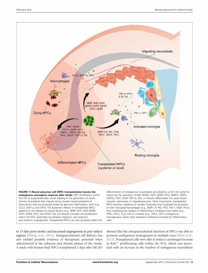
Since NPCs can secrete many of the factors that regulateneurogenesis (Drago et al., 2013), and are also able to ben-eficially modulate inflammatory responses after CNS damage(Bacigaluppi et al., 2009; Cusimano et al., 2012), the possi-bility of exploiting NPC transplantation in an effort to aug-ment endogenous neurogenesis and the brain’s spontaneousreparative processes (e.g., plasticity) after stroke is readilyapparent. In this review, cellular and molecular interac-tions of NPCs with the brain environment have been illus-trated in Figure 1. Effects of exogenously delivered NPCsin experimental models of stroke have been summarized inTable 1.
EFFECTS OF TRANSPLANTED NPCs ON ENDOGENOUSNEUROGENESISIn view of their intrinsic actions, the possibility of exploiting NPCsin an effort to augment endogenous neurogenesis has been thefocus of intensive research efforts. Among the different routes ofNPC delivery, there are several studies suggesting that the local(i.e., intracerebral or intracerebroventricular) administration ofNPCs has the most relevant effects on endogenous neurogenesis.The local route of cell delivery indeed allows for a large num-ber of cells to be administered, which facilitates the secretion ofhigh concentrations of growth factors that ultimately promote theendogenous neurogenic response (Hao et al., 2014). In line withthe need for an efficient accumulation of NPCs within the ischemicparenchyma, the intravenous administration of NPCs may be infe-rior with regard to the stimulation of neurogenesis. As such, whenmouse embryonic stem (ES)-derived NPCs were transplanted 24 hafter photothrombotic stroke in adult immunosuppressed rats, noeffect on post-stroke neurogenesis of the SVZ was shown, anda decrease in newly generated neurons in the DG was observed(Minnerup et al., 2011).
The early transplantation of human NPCs has instead beenproven highly effective in stimulating endogenous neurogene-sis in rats when cells were delivered directly into the ischemicbrain parenchyma. Human fetal NPCs, injected in the corticalperi-infarct tissue 24 h after permanent middle cerebral arteryocclusion (MCAO) promoted cell proliferation in the SVZ (up
Frontiers in Cellular Neuroscience www.frontiersin.org September 2014 | Volume 8 | Article 291 | 2
Hermann et al. Neural precursors in ischemic brain
FIGURE 1 | Neural precursor cell (NPC) transplantation boosts the
endogenous neurogenic response after stroke. NPC proliferation withinthe SVZ is augmented after stroke leading to the generation of newlyformed neuroblasts that migrate along vessels toward gradients ofchemokines that are produced locally by glial and inflammatory cells (e.g.,CCL2, SDF1-α, and EPO). The bystander effects of transplanted NPCsdepend on the release of several factors (e.g., BMP, SHH, NGF, BDNF,CNTF, GDNF, NT-3, and VEGF) that can directly increase cell proliferationwithin the SVZ, potentiate neuroblasts migration, and augmentperi-ischemic angiogenesis. Transplanted NPCs can also positively affect the
differentiation of endogenous neuroblasts and plasticity within the ischemictissue (via the secretion of NGF, BDNF, CNTF, GDNF, NT-3, MMPs, TIMPs,CSPGs, TN-C, VEGF, TSP1-2, Slit), or directly differentiate into post-mitoticneurons, astrocytes, or oligodendrocytes. Most importantly, transplantedNPCs secrete a plethora of soluble molecules that modulate the activationof host microglia/macrophages (e.g., BMP, LIF, NO, PGE, HO-1, VEGF, TN-C),thus modifying the release of inflammatory mediators that inhibit (e.g.,TNF-α, IFN-γ, IL1β, IL6) or increase (e.g., TNF-α, IGF1) endogenousneurogenesis. Green dots represent mediators secreted by inflammatorycells.
to 15 days post-stroke) and increased angiogenesis in peri-infarctregions (Zhang et al., 2011). Intraparenchymal cell delivery hasalso yielded possible evidence of therapeutic potential whenadministered in the subacute and chronic phases of the stroke.A study with human fetal NPCs transplanted 2 days after MCAO
showed that the intraparenchymal injection of NPCs was able topromote endogenous neurogenesis in multiple ways (Mine et al.,2013). Transplanted cells were able to induce a prolonged increasein Ki67+ proliferating cells within the SVZ, which was associ-ated with an increase in the number of endogenous neuroblasts
Frontiers in Cellular Neuroscience www.frontiersin.org September 2014 | Volume 8 | Article 291 | 3
Hermann et al. Neural precursors in ischemic brain
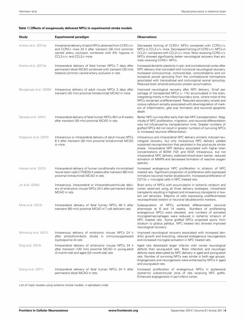
Table 1 | Effects of exogenously delivered NPCs in experimental stroke models.
Study Experimental paradigm Observations
Andres et al. (2011a) Intraarterial delivery of adult NPCs obtained from CCR2+/+and CCR2-/- mice 24 h after transient (30 min) commoncarotid artery occlusion combined with 8% hypoxia inCCL2+/+ and CCL2-/- mice
Decreased homing of CCR2-/- NPCs compared with CCR2+/+NPCs in CCL2+/+ mice. Decreased homing of CCR2+/+ NPCs inCCL2-/- compared with CCL2+/+ mice. Mice receiving CCR2+/+NPCs showed significantly better neurological recovery than ani-mals receiving CCR2-/- NPCs.
Andres et al. (2011b) Intracerebral delivery of fetal human NPCs 7 days afterpermanent distal MCAO combined with transient (30 min)bilateral common carotid artery occlusion in rats
Increased dendritic plasticity in ipsi- and contralesional cortex afterNPC delivery that coincided with functional neurological recovery.Increased corticocortical, corticostriatal, corticothalamic and cor-ticospinal axonal sprouting from the contralesional hemisphereassociated with transcallosal and corticospinal axonal sprouting.Reduced brain amyloid precursor protein accumulation.
Bacigaluppi et al. (2009) Intravenous delivery of adult mouse NPCs 3 days aftertransient (45 min) proximal (intraluminal) MCAO in mice
Improved neurological recovery after NPC delivery. Small per-centage of transplanted NPCs (< 1%) accumulated in the brain,integrating mainly in the infarct boundary zone, where most of theNPCs remained undifferentiated. Reduced secondary striatal andcorpus callosum atrophy associated with downregulation of mark-ers of inflammation, glial scar formation and neuronal apoptoticdeath.
Darsalia et al. (2011) Intracerebral delivery of fetal human NPCs 48 h or 6 weeksafter transient (30 min) proximal MCAO in rats
Better NPC survival after early than late NPC transplantation. Mag-nitude of NPC proliferation, migration, and neuronal differentiationwas not influenced by transplantation time. Greater numbers ofgrafted NPCs did not result in greater numbers of surviving NPCsor increased neuronal differentiation.
Doeppner et al. (2012) Intravenous or intracerebral delivery of adult mouse NPCs6 h after transient (30 min) proximal (intraluminal) MCAOin mice
Intravenous and intracerebral NPC delivery similarly induced neu-rological recovery, but only intravenous NPC delivery yieldedsustained neuroprotection that persisted in the post-acute strokephase. Intracerebral NPF delivery associated with higher brainconcentrations of BDNF, FGF, and VEGF. Intravenous, but notintracerebral NPC delivery stabilized blood–brain barrier, reducedactivation of MMP9 and decreased formation of reactive oxygenspecies.
Hassani et al. (2012) Intracerebral delivery of human conditionally immortalizedneural stem cells CTX0E03 4 weeks after transient (60 min)proximal (intraluminal) MCAO in rats
Increased endogenous NPC proliferation in striatum of NPCtreated rats. Significant proportion of proliferative cells expressedimmature neuronal marker doublecortin. Increased proliferation ofCD11b + microglial cells in NPC treated rats.
Jin et al. (2005) Intravenous, intracerebral or intracerebroventricular deliv-ery of embryonic mouse NPCs 24 h after permanent distalMCAO in rats
Brain entry of NPCs with accumulation in ischemic striatum andcortex observed using all three delivery strategies, intrastriataltransplants resulting in highest and intravenous transplants in low-est cell densities. Majority of cells expressing undifferentiatedneuroepithelial (nestin) or neuronal (doublecortin) markers.
Mine et al. (2013) Intracerebral delivery of fetal human NPCs 48 h aftertransient (60 min) proximal MCAO in T cell deficient rats
Subpopulation of NPCs exhibited differentiated neuronalphenotype at 6 and 14 weeks. Numbers of proliferatingendogenous NPCs were elevated, and numbers of activatedmicroglia/macrophages were reduced in ischemic striatum ofNPC treated rats. Some grafted NPCs projected axons fromstriatum to globus pallidus. NPC treated rats showed improvedneurological recovery.
Minnerup et al. (2011) Intravenous delivery of embryonic mouse NPCs 24 hafter photothrombotic stroke in immunosuppressed(cyclosporine A) rats
Improved neurological recovery associated with increased den-dritic growth and branching, reduced endogenous neurogenesisand increased microglial activation in NPC treated rats.
Tang et al. (2014) Intracerebral delivery of embryonic mouse NPCs 24 hafter transient (120 min) proximal MCAO in young-adult(3 month-old) and aged (24 month-old) rats
Aged rats developed larger infarcts with worse neurologicaldeficits than young-adult rats. Brain infarction and neurologicdeficits were attenuated by NPC delivery in aged and young-adultrats. Number of surviving NPCs was similar in both age groups.Angiogenesis and neurogenesis were enhanced by NPCs in agedand young-adult rats.
Zhang et al. (2011) Intracerebral delivery of fetal human NPCs 24 h afterpermanent distal MCAO in rats
Increased proliferation of endogenous NPCs in ipsilesional(ischemic) subventricular zone of rats receiving NPC grafts.Enhanced angiogenesis in peri-infarct cortex.
List of major studies using ischemic stroke models, in alphabetic order.
Frontiers in Cellular Neuroscience www.frontiersin.org September 2014 | Volume 8 | Article 291 | 4
Hermann et al. Neural precursors in ischemic brain
survival, migration and maturation in the striatum. This pro-longed endogenous neurogenic response, which persisted upto 14 weeks, was accompanied by the long-term suppressionof microglia/macrophage driven inflammatory responses (Mineet al., 2013). Similarly, when human conditionally immortalizedNPCs (cell line CTX0E03) were transplanted in immunosup-pressed rats 4 weeks after MCAO, the number of proliferat-ing doublecortin (DCX)+ neuroblasts considerably increased inthe ischemic striatum (Hassani et al., 2012). Interestingly, theauthors attributed this finding to an increase (rather than adecrease) of proliferating microglia in the striatum (Hassani et al.,2012).
Despite these data, which highlight the promising effects ofintraparenchymal NPC delivery, comparative studies of differ-ent routes and times of transplantation (without the use ofconfounding immunosuppressive regimens) must be performedin order to determine the optimal spatiotemporal settings thatwould allow for the ideal stimulation of endogenous neurogen-esis in stroke. Additionally, given that the neurogenic potentialof adult NPCs declines with age (Conover and Shook, 2011), theeffect of NPC transplantation in aged brains also warrants inves-tigation. Interestingly, a recent study has shown that the localNPC transplantation (24 h post-ischemia) is capable of simi-lar increases in neurogenesis and angiogenesis in the ischemicstriatum of both young and aged mice (Tang et al., 2014). Theaforesaid, coupled with recent data showing increased neuroge-nesis in the SVZ of both young and aged animals following thelocal administration of human ES-derived NPCs, suggests thatthe ischemic environment may effectively be modified in order torestore deficient neurogenic responses in the aged brain as well(Jin et al., 2011).
HOMING AND SURVIVAL OF TRANSPLANTED NPCsIn a comparative study evaluating intrastriatal, intracerebroven-tricular and intravenous NPC delivery (24 h after MCAO), theintrastriatal transplant of NPCs yielded the highest numbers ofgrafted cells within the ischemic brain (Jin et al., 2005). Intracere-broventricular transplantation into the lateral ventricle led to thesurvival of less cells, yet more cells were found when comparedto intravenous NPC delivery (Jin et al., 2005). The reasons behindthese observations are manifold in nature and may be linked toboth the differential homing of grafted cells and their distinctsurvival profiles within the ischemic brain.
The mechanisms that regulate the homing of transplanted cellsto the ischemic lesion are extremely similar to those that regulatethe migration from within the endogenous NPC compartment(Jin et al., 2003; Zhang et al., 2009). Upon ischemia, EPO acti-vates endothelial cells, which promote endogenous neuroblastsmigration by secreting MMPs that degrade extracellular matrix(ECM) components (Wang et al., 2006). Migration of neurob-lasts along the vessels is then modulated by the interaction ofchemokine receptors on NPCs [e.g., C-X-C motif chemokinereceptor (CXCR)-4 and C-C motif chemokine receptor (CCR2)]with molecules secreted by activated neuronal and glial cells withinthe ischemic lesion [e.g., stromal-derived factor (SDF)-1α and C-Cchemokine ligand (CCL)-2, respectively; Robin et al., 2006; Yanet al., 2007].
When exogenous NPCs are administered intravenously, thecrossing of the BBB involves a further degree of complexity. TheCCL2/CCR2 interaction has been demonstrated to be critical fortransendothelial recruitment of intraarterially delivered NPCs inresponse to ischemic injury (Andres et al., 2011a). Stem cells incirculation directly enter the injured brain through endothelialrolling and adhering on cadherins (VCAM-1) and integrins thatbecome selectively up-regulated within the zone of ischemic dam-age (Mueller et al., 2006). This phenomenon, coupled with thedisturbance of BBB integrity that occurs as a consequence ofischemia, is responsible for the pathotropism of transplanted NPCsfor ischemic tissue that occurs after systemic delivery. Interestingly,intravenous transplantation of NPCs has the unique advantageof stabilizing the BBB, via mechanisms that involve a reduc-tion of MMP9 expression and reactive oxygen species (ROS), aswe recently showed after the transplantation of adult NPCs at6 h after transient MCAO (Doeppner et al., 2012). Despite thefact that systemic NPC transplantation yields fewer cells in theischemic brain parenchyma, current evidence suggests that intra-venous injection is clearly able to promote neuronal/glial survivalat delayed time-points (Bacigaluppi et al., 2009; Doeppner et al.,2012). As such, while intracerebrally transplanted adult mouseNPCs transiently improved motor coordination during the first1–2 weeks after MCAO in mice, intravenous NPC transplan-tation resulted in persistent motor coordination improvementsthat persisted over at least 8 weeks post-stroke (Doeppner et al.,2012).
We have previously shown that upon the intravenous trans-plantation of adult mouse NPCs in the subacute stroke phase(72 h post-ischemia) only a small minority of transplanted cells(approximately 0.3% based on systematic counts) accumulate inthe brain (Bacigaluppi et al., 2009). Notably, within the first 72 hpost transplantation, intravenously injected NPCs were foundboth in the ischemic and contralesional non-ischemic hemisphere(Bacigaluppi et al., 2009). In the subsequent 7 days the NPCs inthe contralesional hemisphere disappeared, whereas those in theischemic hemisphere steadily increased in number in a narrowrim around the lesion border. Given that a significant proportion(25%) of transplanted NPCs in the mouse brain expressed the pro-liferation marker Ki67 at 3 days post-transplantation, this selectiveaccumulation may be explained in part by the local proliferationof transplanted cells.
Beyond the proliferation of grafted NPCs, their emergencefrom peripheral organs (where they may have previously homed),such as the lungs, liver and spleen (Lappalainen et al., 2008),may also contribute to the delayed accumulation of systemicallyinjected cells around the stroke lesion. While the localization ofcells of a neural lineage within peripheral organs may carry healthrisks related to malignant transformation, the idea that NPCshave the capacity to exert their therapeutic efficacy via periph-eral actions is intriguing. When fetal human NPCs were deliveredintravenously in rats submitted to collagenase-induced intracere-bral haemorrhage (ICH), these cells were identified primarily insecondary lymphoid organs where they induced a reduction inthe levels of inflammatory mediators and activated macrophagenumbers (Lee et al., 2008). This effect was found to be therapeu-tically relevant as splenectomy, performed before ICH, abolished
Frontiers in Cellular Neuroscience www.frontiersin.org September 2014 | Volume 8 | Article 291 | 5
Hermann et al. Neural precursors in ischemic brain
the effects of NPC transplantation on both brain oedema andinflammatory infiltrates (Lee et al., 2008). This finding, com-bined with observations that transplanted NPCs can hamperthe activation of myeloid dendritic cells (DCs) and restrain theexpansion of antigen-specific T cells in inflammatory CNS con-ditions (Pluchino et al., 2005, 2009), supports the putative abilityof systemically delivered NPCs in modulating important aspectsof the stroke-induced activation of innate and adaptive immuneresponses.
It is becoming clear that the therapeutic effect of intravenouscell delivery is independent of the amount of NPCs that isachieved within the brain, while local intracerebral NPC trans-plantation outcomes strictly depend on the amount of NPCs thatreside within the lesion site. Consequently, intracerebral trans-plantation of NPCs is successful only if grafted NPCs within theischemic brain survive in adequate quantity. The major deter-minant of the survival of locally transplanted cells within theischemic brain is the timing of delivery. In a recent study ithas been shown that while NPC proliferation, migration, andneuronal differentiation did not differ when cells were intras-triatally transplanted in the subacute (48 h) or chronic phase(6 weeks) after stroke, NPC survival was strikingly reduced fol-lowing delayed cell delivery (Darsalia et al., 2011). The reasonsunderlying these findings are largely unknown. It is known thatthe majority (approximately 80%) of adult-born neurons aris-ing from the endogenous neurogenic niche after stroke die beforeintegrating in the ischemic tissue (Arvidsson et al., 2002), andthat the inflammatory milieu plays a pivotal role in this phe-nomenon. As such, the treatment with anti-inflammatory agents(e.g., indomethacin or minocycline) that suppress microglialactivation result in the preservation of these newly formedneurons (Hoehn et al., 2005; Liu et al., 2007). Similarly, the sur-vival of intrastriatal grafts may be strictly dependent on thelocal inflammatory milieu and, as such, intraparenchymal trans-plantation should take place before microglial cells are fullyactivated.
FUNCTIONAL INTEGRATION OF TRANSPLANTED NPCsThe route of NPC administration has little effect on the phenotypeof cells that accumulate inside the ischemic brain, as transplantedNPCs via local or systemic routes retain a profile of characteristicsthat is similar to those observed in vitro prior to transplanta-tion (Jin et al., 2005). This may either be interpreted as evidencethat NPCs are able to retain their character, despite the manip-ulations brought about by transplantation, or that the ischemicmicroenvironment arrests the capacity of the transplanted NPCsto differentiate into mature neurons or glia. It has indeed beenshown that when neuroinflammation predominates, transplantedcells retain an undifferentiated phenotype as a result of the releaseof soluble mediators (e.g., noggin) by blood-borne inflamma-tory cells, activated endothelial cells, and astrocytes (Pluchinoet al., 2005; Martino and Pluchino, 2006). It is therefore reason-able to speculate that when inflammatory signals begin to fade,transplanted cells are subsequently enabled to differentiate intopost-mitotic CNS cells.
We have shown that at 3 and 10 days post transplanta-tion the majority of intravenously transplanted NPCs exhibit
an undifferentiated phenotype, lacking lineage-specific markerssuch as microtubule-associated protein (MAP)-2, DCX, glial fib-rillary protein (GFAP), and the oligodendroglial transcriptionfactor (Olig)-2 (Bacigaluppi et al., 2009). Interestingly at 30 dayspost transplantation, when inflammation was down regulated,the number of transplanted NPCs expressing Olig2 and DCXincreased (albeit to only 4.4 and 0.8%, respectively, of the total),whereas the majority of the transplanted cells still exhibited anundifferentiated morphology in the brain tissue. Understandingthe mechanisms which foster graft differentiation and reduce thequantity of NPCs restricted to an undifferentiated state (oncetheir therapeutic bystander effects have been fully exploited) isof pivotal importance for future cell-replacement therapies instroke.
Valuable insights into the abovementioned may eventu-ally come from the observation of the spontaneous dif-ferentiation of endogenous neuroblasts after ischemic stroke(Kernie and Parent, 2010). It has been reported that the major-ity of neuroblasts after ischemia give rise to striatal mediumspiny neurons (Parent et al., 2002). This response can be mod-ulated by the addition of growth factors (such as angiopoietin orEGF), which increase the number of differentiated neurons and/ordrive the fating of specific neuronal subtypes (i.e., parvalbumin-expressing interneurons; Teramoto et al., 2003; Liu et al., 2009).The potential of pushing NPCs via growth factors toward spe-cific neuronal subtypes has been exploited by a recent study oninduced human pluripotent stem (iPS) cells, which were fatedbefore local transplantation to obtain functional cortical neu-rons in vivo (Tornero et al., 2013). Despite major methodologicaladvances associated with this approach, definitive proof regardingthe additional value of cell fating on behavioral recovery, whencompared to non-fated cellular grafts, is still lacking (Pluchinoand Peruzzotti-Jametti, 2013).
BYSTANDER EFFECTS OF TRANSPLANTED NPCsFunctional recovery of stroke-induced deficits has similarly beenreported after intracerebral, intracerebroventricular, or intra-venous NPC delivery (Bliss et al., 2007; Doeppner et al., 2012).As already noted, the common finding to all routes of cell deliveryis the undifferentiated state in which the majority of transplantedcells are found within the brain parenchyma (Jin et al., 2005). Asa matter of fact, the therapeutic potential of NPCs seems to beinitially independent of cell differentiation and rather relies onthe multiple bystander mechanisms exerted by adult NPCs, whichserve to boost restorative responses in the brain and modulate theinjured microenvironment (Martino et al., 2011).
We have shown that adult mouse NPCs reduced inflamma-tory responses in the ischemic brain, thereby preventing delayedneuronal degeneration and brain atrophy, even when trans-planted intravenously as late as 3 days post MCAO (Bacigaluppiet al., 2009). Iba-1+/MHC class II+ microglial activation wasreduced upon transplantation of adult NPCs, as was GFAP+astroglial scar formation in the infarct rim (Bacigaluppi et al.,2009). On the histochemical level, dopamine-2 receptor+, andcAMP regulated phosphoprotein (DARPP)-32+ medium spinyneurons were protected against delayed degeneration in thestriatum, which is particularly sensitive to intraluminal MCAO
Frontiers in Cellular Neuroscience www.frontiersin.org September 2014 | Volume 8 | Article 291 | 6
Hermann et al. Neural precursors in ischemic brain
(Bacigaluppi et al., 2009). Diminished delayed neuronal degen-eration was noticed not only inside the ischemic lesion but alsoat distance to it, as the corpus callosum (CC) of mice receiv-ing transplants of adult mouse NPCs was significantly thickerthan those of control mice at 30 days post transplantation(Bacigaluppi et al., 2009).
Further, transplanted NPCs were identified in close contactwith von Willebrand factor+ endothelial cells, CD45+ leuco-cytes and F4/80+ macrophages (Bacigaluppi et al., 2009). On themolecular level, pronounced down-regulation of messenger tran-scripts of inflammatory signals (IFN-γ, TNF-α, IL-1β), regulatorsof glial proliferation and reactivity (bFGF, vimentin) and neu-ronal death and plasticity (caspase-3, growth associated protein-43,versican) was observed in the brains of ischemic mice receivingintravenous transplantation of adult mouse NPCs while a sin-gle transcript was up-regulated by adult mouse NPCs, which wasthe spiny neuron marker DARPP-32 (Bacigaluppi et al., 2009).These data clearly suggest that NPCs modulate their microen-vironment via inhibition rather than activation of transcriptionalprocesses.
Reduced delayed neuronal degeneration and CC atrophy werealso noticed with the grafting of human fetal NPCs into the ipsile-sional cortex of rats at 7 days following distal MCAO (Andreset al., 2011b). Dendritic branching, as evaluated by Golgi staining,was enhanced by human fetal NPC transplantation, as was con-tralesional corticospinal axonal sprouting (Andres et al., 2011b).Accumulation of amyloid precursor protein was reduced byhuman fetal NPC transplantation, pointing toward the restorationof axonal transport processes (Andres et al., 2011b).
Although both systemic and intracerebral transplantationimprove neurological recovery, the restorative effects of each routeof transplantation exhibit important differences with regard tothe potential to influence injured tissue via bystander effects. Thelocal intracerebral grafting of adult mouse NPCs in the brainparenchyma is associated with elevated brain concentrations ofBDNF, FGF, and VEGF in the subacute stroke phase, i.e., at4 days after MCAO in mice (Doeppner et al., 2012). Notably,such elevated growth factor levels could not be observed in theischemic brain after systemic intravenous NPC delivery, but werestill present for as late as 2 months after intracerebral transplanta-tion of NPCs transduced with heat shock protein (Doeppner et al.,2012).
CONCLUSIONConsidering the lack of therapeutic options that promote brainremodeling and neurological recovery after stroke, there is a clearneed to reevaluate therapeutic strategies and treatment modal-ities within the stroke field. Accumulating evidence suggeststhat beyond the recanalization of blood vessels (by means ofthrombolytic therapies), it will not be possible to promote neuro-logical recovery post-ischemia via the modulation of single targets(Hermann and Chopp, 2012).
NPCs possess unique characteristics that differ drastically fromconventional therapies (namely pharmacological small moleculecompounds, recombinant growth factors and/or antibody-basedtherapeutics). Transplanted stem cells can promote tissue regen-eration by sensing diverse signals in the brain microenvironment,
migrating to specific sites of damage, integrating inputs andexecuting complex response behaviors all aimed at the remodel-ing/protection of injured ischemic tissue (Fischbach et al., 2013).
While the local delivery of NPCs is able to achieve high levels ofprotective mediators (e.g., growth factors) in the ischemic braintissue, questions remain about the feasibility of such surgicallyinvasive procedures in the clinical setting. One might thereforeconsider systemic NPC transplantation, which is minimally inva-sive. In view that systemically administered NPCs do possesspotent anti-inflammatory effects, promote brain remodeling andinduce functional neurological recovery in rodents, this optioncould be indeed of clinical value.
Future studies will have to clearly define the safety and efficacyof NPC transplantation after both systemic and local delivery. Suchknowledge will increase our understanding of cellular therapiesand in turn guide future translational strategies that are urgentlyneeded to promote brain remodeling and repair in stroke patients.
ACKNOWLEDGMENTSThis work was supported by the German Research Foundation(HE3173/2-1, HE3173/2-2, and HE3173/3-1; to Dirk M. Her-mann), Heinz Nixdorf Foundation (to Dirk M. Hermann) andWerner Jackstädt Foundation (to Dirk M. Hermann); as well asby grants from the National Multiple Sclerosis Society (NMSS;RG-4001-A1 to Stefano Pluchino), the Italian Multiple Sclero-sis Foundation (FISM; RG 2010/R/31; to Stefano Pluchino), theItalian Ministry of Health (GR08/7; to Stefano Pluchino) the Euro-pean Research Council (ERC) 2010-StG (RG 260511-SEM_SEM;to Stefano Pluchino), the European Community (EC) 7th Frame-work Program (FP7/2007-2013; RG 280772-iONE; to StefanoPluchino), The Evelyn Trust (RG69865; to Stefano Pluchino), TheBascule Charitable Trust (RG 75149; to Stefano Pluchino), andThe Great Britain Sakakawa Foundation (to Stefano Pluchino).
REFERENCESAndres, R. H., Choi, R., Pendharkar, A. V., Gaeta, X., Wang, N., Nathan, J. K., et al.
(2011a). The CCR2/CCL2 interaction mediates the transendothelial recruitmentof intravascularly delivered neural stem cells to the ischemic brain. Stroke 42,2923–2931. doi: 10.1161/STROKEAHA.110.606368
Andres, R. H., Horie, N., Slikker, W., Keren-Gill, H., Zhan, K., Sun, G., et al. (2011b).Human neural stem cells enhance structural plasticity and axonal transport inthe ischaemic brain. Brain 134, 1777–1789. doi: 10.1093/brain/awr094
Androutsellis-Theotokis, A., Leker, R. R., Soldner, F., Hoeppner, D. J., Ravin, R.,Poser, S. W., et al. (2006). Notch signalling regulates stem cell numbers in vitroand in vivo. Nature 442, 823–826. doi: 10.1038/nature04940
Arvidsson, A., Collin, T., Kirik, D., Kokaia, Z., and Lindvall, O. (2002). Neuronalreplacement from endogenous precursors in the adult brain after stroke. Nat.Med. 8, 963–970. doi: 10.1038/nm747
Bacigaluppi, M., Pluchino, S., Peruzzotti-Jametti, L., Kilic, E., Kilic, U., Salani, G.,et al. (2009). Delayed post-ischaemic neuroprotection following systemic neuralstem cell transplantation involves multiple mechanisms. Brain 132, 2239–2251.doi: 10.1093/brain/awp174
Ben-Hur, T., Ben-Menachem, O., Furer, V., Einstein, O., Mizrachi-Kol, R., andGrigoriadis, N. (2003). Effects of proinflammatory cytokines on the growth, fate,and motility of multipotential neural precursor cells. Mol. Cell. Neurosci. 24,623–631. doi: 10.1016/S1044-7431(03)00218-5
Bliss, T., Guzman, R., Daadi, M., and Steinberg, G. K. (2007). Cell transplantationtherapy for stroke. Stroke 38, 817–826. doi: 10.1161/01.STR.0000247888.25985.62
Cacci, E., Ajmone-Cat, M. A., Anelli, T., Biagioni, S., and Minghetti, L. (2008). Invitro neuronal and glial differentiation from embryonic or adult neural precursorcells are differently affected by chronic or acute activation of microglia. Glia 56,412–425. doi: 10.1002/glia.20616
Frontiers in Cellular Neuroscience www.frontiersin.org September 2014 | Volume 8 | Article 291 | 7
Hermann et al. Neural precursors in ischemic brain
Chou, J., Harvey, B. K., Chang, C. F., Shen, H., Morales, M., and Wang, Y. (2006).Neuroregenerative effects of BMP7 after stroke in rats. J. Neurol. Sci. 240, 21–29.doi: 10.1016/j.jns.2005.08.015
Conover, J. C., and Shook, B. A. (2011). Aging of the subventricular zone neuralstem cell niche. Aging Dis. 2, 149–163.
Cusimano, M., Biziato, D., Brambilla, E., Donega, M., Alfaro-Cervello, C., Snider,S., et al. (2012). Transplanted neural stem/precursor cells instruct phagocytes andreduce secondary tissue damage in the injured spinal cord. Brain 135, 447–460.doi: 10.1093/brain/awr339
Darsalia, V., Allison, S. J., Cusulin, C., Monni, E., Kuzdas, D., Kallur, T., et al. (2011).Cell number and timing of transplantation determine survival of human neuralstem cell grafts in stroke-damaged rat brain. J. Cereb. Blood Flow Metab. 31,235–242. doi: 10.1038/jcbfm.2010.81
Doeppner, T. R., Ewert, T. A., Tonges, L., Herz, J., Zechariah, A., ElAli, A., et al.(2012). Transduction of neural precursor cells with TAT-heat shock protein 70chaperone: therapeutic potential against ischemic stroke after intrastriatal andsystemic transplantation. Stem Cells 30, 1297–1310. doi: 10.1002/stem.1098
Donnan, G. A., Fisher, M., Macleod, M., and Davis, S. M. (2008). Stroke. Lancet371, 1612–1623. doi: 10.1016/S0140-6736(08)60694-7
Drago, D., Cossetti, C., Iraci, N., Gaude, E., Musco, G., Bachi, A., et al. (2013). Thestem cell secretome and its role in brain repair. Biochimie 95, 2271–2285. doi:10.1016/j.biochi.2013.06.020
Ekdahl, C. T., Kokaia, Z., and Lindvall, O. (2009). Brain inflammation and adultneurogenesis: the dual role of microglia. Neuroscience 158, 1021–1029. doi:10.1016/j.neuroscience.2008.06.052
Fischbach, M. A., Bluestone, J. A., and Lim, W. A. (2013). Cell-based ther-apeutics: the next pillar of medicine. Sci. Transl. Med. 5, 1797. doi:10.1126/scitranslmed.3005568
Hao, L., Zou, Z., Tian, H., Zhang, Y., Zhou, H., and Liu, L. (2014). Stemcell-based therapies for ischemic stroke. Biomed Res. Int. 2014, 468748. doi:10.1155/2014/468748
Hassani, Z., O’Reilly, J., Pearse, Y., Stroemer, P., Tang, E., Sinden, J., et al.(2012). Human neural progenitor cell engraftment increases neurogenesis andmicroglial recruitment in the brain of rats with stroke. PLoS ONE 7:e50444. doi:10.1371/journal.pone.0050444
Heldmann, U., Thored, P., Claasen, J. H., Arvidsson, A., Kokaia, Z., andLindvall, O. (2005). TNF-alpha antibody infusion impairs survival of stroke-generated neuroblasts in adult rat brain. Exp. Neurol. 196, 204–208. doi:10.1016/j.expneurol.2005.07.024
Hermann, D. M., and Chopp, M. (2012). Promoting brain remodelling andplasticity for stroke recovery: therapeutic promise and potential pitfalls ofclinical translation. Lancet Neurol. 11, 369–380. doi: 10.1016/S1474-4422(12)70039-X
Hoehn, B. D., Palmer, T. D., and Steinberg, G. K. (2005). Neurogenesis in rats afterfocal cerebral ischemia is enhanced by indomethacin. Stroke 36, 2718–2724. doi:10.1161/01.STR.0000190020.30282.cc
Jin, K., Sun, Y., Xie, L., Mao, X. O., Childs, J., Peel, A., et al. (2005). Comparison ofischemia-directed migration of neural precursor cells after intrastriatal, intraven-tricular, or intravenous transplantation in the rat. Neurobiol. Dis. 18, 366–374.doi: 10.1016/j.nbd.2004.10.010
Jin, K., Sun, Y., Xie, L., Peel, A., Mao, X. O., Batteur, S., et al. (2003).Directed migration of neuronal precursors into the ischemic cerebral cortexand striatum. Mol. Cell. Neurosci. 24, 171–189. doi: 10.1016/S1044-7431(03)00159-3
Jin, K., Xie, L., Mao, X., Greenberg, M. B., Moore, A., Peng, B., et al. (2011).Effect of human neural precursor cell transplantation on endogenous neuro-genesis after focal cerebral ischemia in the rat. Brain Res. 1374, 56–62. doi:10.1016/j.brainres.2010.12.037
Kang, S. S., Keasey, M. P., Arnold, S. A., Reid, R., Geralds, J., and Hagg, T. (2012).Endogenous CNTF mediates stroke-induced adult CNS neurogenesis in mice.Neurobiol. Dis. 49C, 68–78. doi: 10.1016/j.nbd.2012.08.020
Kernie, S. G., and Parent, J. M. (2010). Forebrain neurogenesis after focalIschemic and traumatic brain injury. Neurobiol. Dis. 37, 267–274. doi:10.1016/j.nbd.2009.11.002
Kitagawa, H., Sasaki, C., Zhang, W. R., Sakai, K., Shiro, Y., Warita, H., et al. (1999).Induction of glial cell line-derived neurotrophic factor receptor proteins in cere-bral cortex and striatum after permanent middle cerebral artery occlusion in rats.Brain Res. 834, 190–195. doi: 10.1016/S0006-8993(99)01563-2
Kokaia, Z., Zhao, Q., Kokaia, M., Elmer, E., Metsis, M., Smith, M. L., et al. (1995).Regulation of brain-derived neurotrophic factor gene expression after transientmiddle cerebral artery occlusion with and without brain damage. Exp. Neurol.136, 73–88. doi: 10.1006/exnr.1995.1085
Lappalainen, R. S., Narkilahti, S., Huhtala, T., Liimatainen, T., Suuronen, T.,Narvanen, A., et al. (2008). The SPECT imaging shows the accumulationof neural progenitor cells into internal organs after systemic administrationin middle cerebral artery occlusion rats. Neurosci. Lett. 440, 246–250. doi:10.1016/j.neulet.2008.05.090
Lee, S. T., Chu, K., Jung, K. H., Kim, S. J., Kim, D. H., Kang, K. M., et al. (2008).Anti-inflammatory mechanism of intravascular neural stem cell transplantationin haemorrhagic stroke. Brain 131, 616–629. doi: 10.1093/brain/awm306
Leker, R. R., Toth, Z. E., Shahar, T., Cassiani-Ingoni, R., Szalayova, I.,Key, S., et al. (2009). Transforming growth factor alpha induces angio-genesis and neurogenesis following stroke. Neuroscience 163, 233–243. doi:10.1016/j.neuroscience.2009.05.050
Liu, X. S., Chopp, M., Zhang, R. L., Hozeska-Solgot, A., Gregg, S. C., Buller,B., et al. (2009). Angiopoietin 2 mediates the differentiation and migration ofneural progenitor cells in the subventricular zone after stroke. J. Biol. Chem. 284,22680–22689. doi: 10.1074/jbc.M109.006551
Liu, Z., Fan, Y., Won, S. J., Neumann, M., Hu, D., Zhou, L., et al. (2007).Chronic treatment with minocycline preserves adult new neurons and reducesfunctional impairment after focal cerebral ischemia. Stroke 38, 146–152. doi:10.1161/01.STR.0000251791.64910.cd
Lloyd-Jones, D., Adams, R., Carnethon, M., De Simone, G., Ferguson, T. B., Flegal,K., et al. (2009). Heart disease and stroke statistics–2009 update: a report from theAmerican Heart Association Statistics Committee and Stroke Statistics Subcom-mittee. Circulation 119, e21–e181. doi: 10.1161/CIRCULATIONAHA.108.191261
Martino, G., Bacigaluppi, M., and Peruzzotti-Jametti, L. (2011). Therapeuticstem cell plasticity orchestrates tissue plasticity. Brain 134, 1585–1587. doi:10.1093/brain/awr115
Martino, G., and Pluchino, S. (2006). The therapeutic potential of neural stem cells.Nat. Rev. 7, 395–406. doi: 10.1038/nrn1908
Mine, Y., Tatarishvili, J., Oki, K., Monni, E., Kokaia, Z., and Lindvall, O. (2013).Grafted human neural stem cells enhance several steps of endogenous neuroge-nesis and improve behavioral recovery after middle cerebral artery occlusion inrats. Neurobiol. Dis. 52, 191–203. doi: 10.1016/j.nbd.2012.12.006
Minnerup, J., Kim, J. B., Schmidt, A., Diederich, K., Bauer, H., Schilling, M.,et al. (2011). Effects of neural progenitor cells on sensorimotor recovery andendogenous repair mechanisms after photothrombotic stroke. Stroke 42, 1757–1763. doi: 10.1161/STROKEAHA.110.599282
Mirzadeh, Z., Merkle, F. T., Soriano-Navarro, M., Garcia-Verdugo, J. M., andAlvarez-Buylla, A. (2008). Neural stem cells confer unique pinwheel architec-ture to the ventricular surface in neurogenic regions of the adult brain. Cell StemCell 3, 265–278. doi: 10.1016/j.stem.2008.07.004
Mueller, F. J., Serobyan, N., Schraufstatter, I. U., DiScipio, R., Wakeman, D., Loring,J. F., et al. (2006). Adhesive interactions between human neural stem cells andinflamed human vascular endothelium are mediated by integrins. Stem Cells 24,2367–2372. doi: 10.1634/stemcells.2005-0568
Parent, J. M., Vexler, Z. S., Gong, C., Derugin, N., and Ferriero, D. M. (2002). Ratforebrain neurogenesis and striatal neuron replacement after focal stroke. Ann.Neurol. 52, 802–813. doi: 10.1002/ana.10393
Peruzzotti-Jametti, L., Donega, M., Giusto, E., Mallucci, G., Marchetti, B., andPluchino, S. (2014). The role of the immune system in central nervous systemplasticity after acute injury. Neuroscience doi: 10.1016/j.neuroscience.2014.04.036[Epub ahead of print].
Planas, A. M., Justicia, C., Soriano, M. A., and Ferrer, I. (1998). Epider-mal growth factor receptor in proliferating reactive glia following transientfocal ischemia in the rat brain. Glia 23, 120–129. doi: 10.1002/(SICI)1098-1136(199806)23:2<120::AID-GLIA3>3.0.CO;2-A
Plane, J. M., Whitney, J. T., Schallert, T., and Parent, J. M. (2008). Retinoic acid andenvironmental enrichment alter subventricular zone and striatal neurogenesisafter stroke. Exp. Neurol. 214, 125–134. doi: 10.1016/j.expneurol.2008.08.006
Pluchino, S., and Cossetti, C. (2013). How stem cells speak with host immune cellsin inflammatory brain diseases. Glia 61, 1379–1401. doi: 10.1002/glia.22500
Pluchino, S., and Peruzzotti-Jametti, L. (2013). Rewiring the ischaemic brain withhuman-induced pluripotent stem cell-derived cortical neurons. Brain 136, 3525–3527. doi: 10.1093/brain/awt330
Frontiers in Cellular Neuroscience www.frontiersin.org September 2014 | Volume 8 | Article 291 | 8
Hermann et al. Neural precursors in ischemic brain
Pluchino, S., Zanotti, L., Brambilla, E., Rovere-Querini, P., Capobianco, A.,Alfaro-Cervello, C., et al. (2009). Immune regulatory neural stem/precursorcells protect from central nervous system autoimmunity by restrainingdendritic cell function. PLoS ONE 4:e5959. doi: 10.1371/journal.pone.0005959
Pluchino, S., Zanotti, L., Rossi, B., Brambilla, E., Ottoboni, L., Salani, G.,et al. (2005). Neurosphere-derived multipotent precursors promote neuropro-tection by an immunomodulatory mechanism. Nature 436, 266–271. doi:10.1038/nature03889
Robin, A. M., Zhang, Z. G., Wang, L., Zhang, R. L., Katakowski, M., Zhang, L.,et al. (2006). Stromal cell-derived factor 1alpha mediates neural progenitor cellmotility after focal cerebral ischemia. J. Cereb. Blood Flow Metab. 26, 125–134.doi: 10.1038/sj.jcbfm.9600172
Schneider, A., Kruger, C., Steigleder, T., Weber, D., Pitzer, C., Laage, R., et al.(2005). The hematopoietic factor G-CSF is a neuronal ligand that counteractsprogrammed cell death and drives neurogenesis. J. Clin. Invest. 115, 2083–2098.doi: 10.1172/JCI23559
Sims, J. R., Lee, S. W., Topalkara, K., Qiu, J., Xu, J., Zhou, Z., et al. (2009). Sonichedgehog regulates ischemia/hypoxia-induced neural progenitor proliferation.Stroke 40, 3618–3626. doi: 10.1161/STROKEAHA.109.561951
Sun, Y., Jin, K., Xie, L., Childs, J., Mao, X. O., Logvinova, A., et al. (2003). VEGF-induced neuroprotection, neurogenesis, and angiogenesis after focal cerebralischemia. J. Clin. Invest. 111, 1843–1851. doi: 10.1172/JCI200317977
Tang, Y., Wang, J., Lin, X., Wang, L., Shao, B., Jin, K., et al. (2014). Neuralstem cell protects aged rat brain from ischemia-reperfusion injury through neu-rogenesis and angiogenesis. J. Cereb. Blood Flow Metab. 34, 1138–1147. doi:10.1038/jcbfm.2014.61
Teramoto, T., Qiu, J., Plumier, J. C., and Moskowitz, M. A. (2003). EGF amplifiesthe replacement of parvalbumin-expressing striatal interneurons after ischemia.J. Clin. Invest. 111, 1125–1132. doi: 10.1172/JCI200317170
Thored, P., Arvidsson, A., Cacci, E., Ahlenius, H., Kallur, T., Darsalia, V., et al. (2006).Persistent production of neurons from adult brain stem cells during recovery afterstroke. Stem Cells 24, 739–747. doi: 10.1634/stemcells.2005-0281
Thored, P., Heldmann, U., Gomes-Leal, W., Gisler, R., Darsalia, V., Taneera, J.,et al. (2009). Long-term accumulation of microglia with proneurogenic pheno-type concomitant with persistent neurogenesis in adult subventricular zone afterstroke. Glia 57, 835–849. doi: 10.1002/glia.20810
Thored, P., Wood, J., Arvidsson, A., Cammenga, J., Kokaia, Z., and Lindvall, O.(2007). Long-term neuroblast migration along blood vessels in an area withtransient angiogenesis and increased vascularization after stroke. Stroke 38, 3032–3039. doi: 10.1161/STROKEAHA.107.488445
Tornero, D., Wattananit, S., Gronning Madsen, M., Koch, P., Wood, J., Tatarishvili,J., et al. (2013). Human induced pluripotent stem cell-derived cortical neuronsintegrate in stroke-injured cortex and improve functional recovery. Brain 136,3561–3577. doi: 10.1093/brain/awt278
Tureyen, K., Vemuganti, R., Bowen, K. K., Sailor, K. A., and Dempsey, R.J. (2005). EGF and FGF-2 infusion increases post-ischemic neural progeni-tor cell proliferation in the adult rat brain. Neurosurgery 57, 1254–1263. doi:10.1227/01.NEU.0000186040.96929.8A
Wang, L., Zhang, Z. G., Zhang, R. L., Gregg, S. R., Hozeska-Solgot, A.,LeTourneau, Y., et al. (2006). Matrix metalloproteinase 2 (MMP2) and MMP9secreted by erythropoietin-activated endothelial cells promote neural progeni-tor cell migration. J. Neurosci. 26, 5996–6003. doi: 10.1523/JNEUROSCI.5380-05.2006
Wang, X., Mao, X., Xie, L., Greenberg, D. A., and Jin, K. (2009). Involvementof Notch1 signaling in neurogenesis in the subventricular zone of normal andischemic rat brain in vivo. J. Cereb. Blood Flow Metab. 29, 1644–1654. doi:10.1038/jcbfm.2009.83
Yan, Y. P., Sailor, K. A., Lang, B. T., Park, S. W., Vemuganti, R., and Dempsey, R.J. (2007). Monocyte chemoattractant protein-1 plays a critical role in neuroblastmigration after focal cerebral ischemia. J. Cereb. Blood Flow Metab. 27, 1213–1224.doi: 10.1038/sj.jcbfm.9600432
Zhang, P., Li, J., Liu, Y., Chen, X., Lu, H., Kang, Q., et al. (2011).Human embryonic neural stem cell transplantation increases subventricularzone cell proliferation and promotes peri-infarct angiogenesis after focal cere-bral ischemia. Neuropathology 31, 384–391. doi: 10.1111/j.1440-1789.2010.01182.x
Zhang, R. L., Chopp, M., Gregg, S. R., Toh, Y., Roberts, C., Letourneau, Y., et al.(2009). Patterns and dynamics of subventricular zone neuroblast migration in theischemic striatum of the adult mouse. J. Cereb. Blood Flow Metab. 29, 1240–1250.doi: 10.1038/jcbfm.2009.55
Zhang, R. L., Zhang, Z. G., Wang, Y., LeTourneau, Y., Liu, X. S., Zhang, X., et al.(2007). Stroke induces ependymal cell transformation into radial glia in thesubventricular zone of the adult rodent brain. J. Cereb. Blood Flow Metab. 27,1201–1212. doi: 10.1038/sj.jcbfm.9600430
Zhang, R., Zhang, Z., Zhang, C., Zhang, L., Robin, A., Wang, Y., et al. (2004).Stroke transiently increases subventricular zone cell division from asymmetric tosymmetric and increases neuronal differentiation in the adult rat. J. Neurosci. 24,5810–5815. doi: 10.1523/JNEUROSCI.1109-04.2004
Conflict of Interest Statement: The authors declare that the research was conductedin the absence of any commercial or financial relationships that could be construedas a potential conflict of interest.
Received: 15 June 2014; accepted: 01 September 2014; published online: 16 September2014.Citation: Hermann DM, Peruzzotti-Jametti L, Schlechter J, Bernstock JD, DoeppnerTR and Pluchino S (2014) Neural precursor cells in the ischemic brain – integration,cellular crosstalk, and consequences for stroke recovery. Front. Cell. Neurosci. 8:291.doi: 10.3389/fncel.2014.00291This article was submitted to the journal Frontiers in Cellular Neuroscience.Copyright © 2014 Hermann, Peruzzotti-Jametti, Schlechter, Bernstock, Doeppner andPluchino. This is an open-access article distributed under the terms of the CreativeCommons Attribution License (CC BY). The use, distribution or reproduction in otherforums is permitted, provided the original author(s) or licensor are credited and thatthe original publication in this journal is cited, in accordance with accepted academicpractice. No use, distribution or reproduction is permitted which does not comply withthese terms.
Frontiers in Cellular Neuroscience www.frontiersin.org September 2014 | Volume 8 | Article 291 | 9