negative-pressure wound therapy in the management of ... · negative-pressure wound therapy in the...
TRANSCRIPT
Negative-pressure wound therapy in themanagement of diabetic Charcot footand ankle wounds
Crystal L. Ramanujam, DPM, MSc1*, John J. Stapleton,DPM, FACFAS2 and Thomas Zgonis, DPM, FACFAS1
1University of Texas Health Science Center at San Antonio, San Antonio, TX, USA; 2VSAS Orthopaedics,Allentown, PA, USA
As the prevalence of diabetes mellitus continues to rise, innovative medical and surgical treatment options
have increased dramatically to address diabetic-related foot and ankle complications. Among the most
challenging clinical case scenarios is Charcot neuroarthropathy associated with soft tissue loss and/or
osteomyelitis. In this review article, the authors present a review of the most common utilizations of negative-
pressure wound therapy as an adjunctive therapy or combined with plastic surgery as it relates to the surgical
management of diabetic Charcot foot and ankle wounds.
Keywords: negative-pressure wound therapy; Charcot foot; diabetes mellitus; neuropathy; plastic surgery; external fixation
Received: 17 March 2013; Revised: 10 July 2013; Accepted: 19 August 2013; Published: 23 September 2013
Negative-pressure wound therapy (NPWT), also
known as topical negative-pressure therapy or
vacuum-assisted closure, has revolutionized the
world of wound care. Morykwas et al. first published
on this modality using an open-cell foam dressing with
application of a controlled subatmospheric pressure for
treatment of acute and chronic wounds in 1997 (1�3).
Since then multiple studies have demonstrated the useful-
ness of NPWT in diabetic foot wounds (4�6). In the
diabetic patient, the combination of Charcot neuroar-
thropathy (CN) and an open wound with or without
osteomyelitis further challenges treatment protocols
since each of these clinical manifestations carries with
it an increased risk for infection and amputation (7).
Expedited wound closure is a priority and may decrease
morbidity and mortality rates in this population (8).
Although NPWT’s exact mechanism of action has yet
to be elucidated, its success as an adjunct for wound
healing has well been established in the surgeon’s
armamentarium.
Much of the existing evidence supports that the success
of NPWT in wound healing is likely multifactorial.
A study by Zoch on 10 patients found an increase in
blood flow to diabetic foot wounds treated with NPWT
measured by IC-view perfusography (9). Kamolz et al.
showed a clinically apparent reduction in edema with
NPWT used on burn wounds in the hand as a result
of fluid removal by the device (10). Even in the
early studies by Morykwas et al. using an animal
model, he noted an increase in the formation of granula-
tion tissue at wounds through NPWT (2). In a rando-
mized controlled trial of 30 patients with diabetic foot
ulcers, Kopp et al. demonstrated an increase of growth
factors within the wound fluid of those treated with
NPWT compared to the control group treated with a
hydrocolloid (11).
As the conventional option for care of diabetic foot
wounds has been moist wound dressings, randomized
controlled trials comparing NPWT to moist wound
healing have populated the literature. Eginton et al.
compared the rates of wound healing between NPWT
and moist dressings, clearly showing improved healing
rates in the diabetic foot wounds treated with NPWT (5).
In regard to quicker achievement of wound closure
and formation of granulation tissue, a study by Blume
et al. also found NPWT superior to standard wound
dressings (12). Although this was a randomized con-
trolled trial, the results described by the study should
be considered carefully since industry-sponsored re-
search can be biased in reporting mostly positive results
when compared to non-industry-sponsored studies.
A study by Paola et al. further demonstrated that NPWT
(page number not for citation purpose)
�REVIEW ARTICLE
Diabetic Foot & Ankle 2013. # 2013 Crystal L. Ramanujam et al. This is an Open Access article distributed under the terms of the Creative Commons Attribution-Noncommercial 3.0 Unported License (http://creativecommons.org/licenses/by-nc/3.0/), permitting all non-commercial use, distribution, and reproduction inany medium, provided the original work is properly cited.
1
Citation: Diabetic Foot & Ankle 2013, 4: 20878 - http://dx.doi.org/10.3402/dfa.v4i0.20878
reduced the need for subsequent amputations at a 6-month
follow-up period when compared to the control group (13).
These studies also suggested no differences between the
treatment-related complications of NPWT and conven-
tional moist wound dressings (14). Studies investigating
the role of NPWT on bacterial clearance and infection
control showed inconclusive results (15�17). Furthermore,
questions still remain regarding the optimal values for
duration of NPWT use and levels of pressure, as well
as specific materials used, and their discussion is beyond
the scope of this article.
Clinical case scenariosNeuropathic wounds in the diabetic CN foot and ankle
can manifest through a variety of different pathways
and mechanisms. Most common clinical case scenarios
that can lead into a CN deformity from a pre-existing
neuropathic ulceration(s), osteomyelitis and/or surgical
intervention may include but are not limited to the
following two clinical presentations: (1) diabetic neuro-
pathic submetatarsal ulcerations that are being treated
conservatively with off-loading can potentially alter the
biomechanics of the foot and increase trauma to adja-
cent joints that could initiate the CN process and (2)
diabetic neuropathic partial pedal (e.g. toe, partial ray,
transmetatarsal) amputations with a recent history of
surgical intervention that are permitted to an early
weight-bearing status. In the above clinical presentations,
the diabetic patient presents with a history of a pre-
existing ulceration and/or infection that may have con-
tributed to the development of CN in a different joint(s)
with or without a new ulceration. However, it is not
yet known whether the incidence of CN is caused by
changes in biomechanical function following partial
foot amputation, elicited by surgery itself, or triggered
by osteomyelitis (18�20).
For any other patient with diabetic CN, identification
of the stage of CN is important in formulating a
treatment strategy. In the acute stage of CN, inflamma-
tion may produce severe edema that when combined
with ambulation in the insensate patient can cause
significant skin breakdown and/or compromise a pre-
existing wound. NPWT can be carefully applied during
this stage if a wound is present but must be combined
with a period of immobilization in order to halt the
inflammatory process, limit peri-wound edema, and
maintain the foot and/or ankle architecture to prevent
further deformity (21). Immobilization can be in the
form of removable splints, casts, prefabricated braces,
and external fixation devices that are designed to allow
for appropriate access to the wound for regular NPWT
dressing changes, while enforcing a non-weight-bearing
status through the utilization of a gait assistive device(s)
or wheelchair.
As CN can compromise the osseous architecture and
stability of the foot and ankle, the chronic form of the
condition typically presents with progressive deformity
resulting from a cumulative effect of repeated injury that
goes undetected or neglected during the acute process.
Failure to properly accommodate and off-load braceable
deformities or to recognize unstable and/or unbraceable
deformities which require surgical intervention may
potentially lead to an ulceration and/or infection. In
this clinical scenario, patients often present to a medical
facility because they have noticed a wound and possibly
a misshapen foot, yet cannot recall any of the signs or
symptoms of acute CN. For these patients in the absence
of infection, the foot and ankle specialist should identify
whether the deformity is stable or unstable in order to
determine whether surgical intervention may be indicated
for correction of an unstable deformity. In a wound with
stable chronic CN, sharp debridement of the wound and
use of NPWT can facilitate secondary healing, while
re-ulceration can be avoided through accommodative
shoe gear and/or bracing combined with frequent clinical
examinations.
Delayed or ineffective treatment of CN foot and
ankle wounds in the diabetic population can potentially
lead to soft tissue infection and osteomyelitis. Appropriate
surgical debridement of infected soft tissue and bone
often leaves large defects that may be amenable to
treatment with non-biodegradable cemented antibiotic
beads/spacers. In these surgical case scenarios, NPWT
may only be considered for surgical wounds with eradi-
cated bone or soft tissue infection, possess adequate
perfusion, and are in need for frequent dressing changes.
For exposed tendon structures, a non-adherent dressing
can be applied first directly over the tendon prior to
placement of the NPWT foam. Furthermore, commer-
cially available open-cell foams of different compositions
(white foam or silver-impregnated foam) are designed for
placement over bone or in tunneling wounds; however,
the efficacy of these materials is yet to be determined.
Although NPWT is beneficial as an adjunct of treating
these complex case scenarios, a thorough surgical debri-
dement and appropriate antibiotic therapy are necessary
to a successful wound healing in the diabetic CN foot
or ankle (Fig. 1).
NPWT is also indicated as an adjunct to surgical wound
closure utilizing plastic surgical techniques and osseous
reconstruction for the unstable and/or unbraceable CN
deformity. Due to the effects of NPWT mentioned earlier,
this modality can be useful in wound bed preparation
prior to definitive wound closure techniques such as skin
grafts or flaps. NPWT can also be considered to promote
wound healing at flap donor sites. When used cautiously,
observed frequently and typically at lower pressure
levels, NPWT can also be beneficial directly over skin
Crystal L. Ramanujam et al.
2(page number not for citation purpose)
Citation: Diabetic Foot & Ankle 2013, 4: 20878 - http://dx.doi.org/10.3402/dfa.v4i0.20878
grafts or flaps during early incorporation. In a clinical trial
comparing NPWT with standard non-adherent gauze
dressings for skin-grafted diabetic foot wounds, Paola
et al. found that the percentage of subjects with complete
skin graft healing was increased in the NPWT group
(13). Reduction of flap edema, increase in blood flow
to the flap, and bolstering of the flap to the recipient
wound are all effects which support the use of NPWT in
this setting (22). Similarly, rather than using standard
bolster-type dressings, NPWT can also be used in combi-
nation with orthobiologics for dermal replacement.
This technique maintains contact between the biologi-
cal layer and the wound, reducing micro-motion while
also preventing excessive fluid accumulation. In cases
of surgical wound dehiscence or partial flap necrosis,
NPWT can also be used to facilitate delayed primary
closure or healing by secondary intention after adequate
debridement.
Similarly, NPWT has also found a role in stabilizing
closed incisions which is useful in extensive CN foot and
ankle reconstructions where there is a high risk for wound
dehiscence. Arthrodesis procedures often used for recon-
struction of unstable CN foot or ankle fracture disloca-
tions can produce tenuous incisions and may lead to
significant post-operative edema. A study by Kilpadi
et al. demonstrated that the use of NPWT over closed
incisions can reduce and normalize tissue stresses and
bolster appositional forces at the incision site (23). In
CN reconstructive cases where primary wound closure is
not feasible, NPWT systems can be used in conjunction
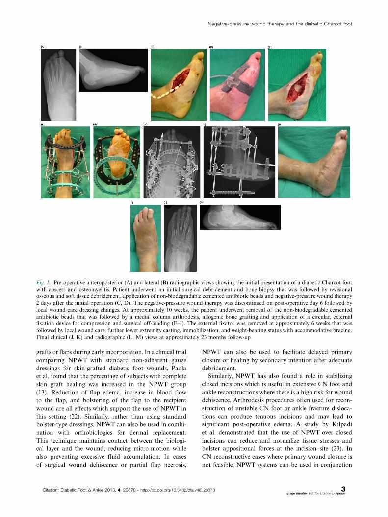
Fig. 1. Pre-operative anteroposterior (A) and lateral (B) radiographic views showing the initial presentation of a diabetic Charcot foot
with abscess and osteomyelitis. Patient underwent an initial surgical debridement and bone biopsy that was followed by revisional
osseous and soft tissue debridement, application of non-biodegradable cemented antibiotic beads and negative-pressure wound therapy
2 days after the initial operation (C, D). The negative-pressure wound therapy was discontinued on post-operative day 6 followed by
local wound care dressing changes. At approximately 10 weeks, the patient underwent removal of the non-biodegradable cemented
antibiotic beads that was followed by a medial column arthrodesis, allogenic bone grafting and application of a circular, external
fixation device for compression and surgical off-loading (E�I). The external fixator was removed at approximately 6 weeks that was
followed by local wound care, further lower extremity casting, immobilization, and weight-bearing status with accommodative bracing.
Final clinical (J, K) and radiographic (L, M) views at approximately 23 months follow-up.
Negative-pressure wound therapy and the diabetic Charcot foot
Citation: Diabetic Foot & Ankle 2013, 4: 20878 - http://dx.doi.org/10.3402/dfa.v4i0.20878 3(page number not for citation purpose)
with off-loading external fixation to encourage wound
healing. External fixation provides for proper stabiliza-
tion of the corrected deformity while simultaneously
allowing regular access to the wound(s) for continued
care. For diabetic CN patients with chronic foot and
ankle wounds who are not optimal candidates for
reconstructive procedures, NPWT can stabilize wounds
until their comorbidities are controlled and defini-
tive wound closure with corrective osseous procedures
can then be safely accomplished.
Prior to application of NPWT in any patient, one
should consider the existing contraindications to this
modality (24). Absolute contraindications include but
are not limited to untreated osteomyelitis or sepsis within
the area of the wound, inadequately debrided wounds
or those with necrotic tissue and/or eschar, presence
of untreated coagulopathies, malignancy at the wound
site, and allergy to any materials related to the device.
Relative contraindications to NPWT include but are
not limited to wounds with active bleeding, patients
with poor nutritional status, and patients on active
anticoagulant therapy.
ConclusionSince its initial development, NPWT has gained wide-
spread acceptance for a broad range of wound indications,
including those found among diabetic CN. NPWT can
be used to treat CN wounds produced as a result of
neuropathy and deformity, following debridement of
infection or amputation, and in reconstructive soft
tissue and osseous procedures. Several studies emphasize
that treatment outcomes are often based on the specific
techniques and materials used for NPWT application,
therefore additional suitably powered, high-quality clin-
ical trials are needed to fully determine efficacy. Careful
patient and procedure selection along with appropriate
technique is imperative for successful use of NPWT in the
diabetic CN foot and ankle.
Conflict of interest and fundingThe authors have received no funding or benefits from
industry to conduct this literature review.
References
1. Morykwas MJ, Argenta LC. Nonsurgical modalities to enhance
healing and care of soft tissue wounds. J South Orthop Assoc
1997; 6: 279�88.
2. Morykwas MJ, Argenta LC, Shelton-Brown EI, McGuirt W.
Vacuum-assisted closure: a new method for wound control and
treatment: animal studies and basic foundation. Ann Plast Surg
1997; 38: 553�62.
3. Argenta LC, Morykwas MJ. Vacuum-assisted closure: a new
method for wound control and treatment: clinical experience.
Ann Plast Surg 1997; 38: 563�77.
4. McCallon SK, Knight CA, Valiulus JP, Cunningham MW,
McCulloch JM, Farinas LP. Vacuum-assisted closure versus
saline-moistened gauze in the healing of postoperative diabetic
foot wounds. Ostomy Wound Manage 2000; 46: 28�32.
5. Eginton MT, Brown KR, Seabrook GR, Towne JB, Cambria
RA. A prospective randomized evaluation of negative-pressure
wound dressings for diabetic foot wounds. Ann Vasc Surg 2003;
17: 645�9.
6. Armstrong DG, Lavery LA, Diabetic Foot Study Consortium.
Negative pressure wound therapy after partial diabetic foot
amputation: a multicenter, randomized controlled trial. Lancet
2005; 366: 1704�10.
7. Sohn MW, Stuck RM, Pinzur M, Lee TA, Budiman-Mak E.
Lower-extremity amputation risk after Charcot arthropathy and
diabetic foot ulcer. Diabetes Care 2010; 33: 98�100.
8. Schaper NC, Apelqvist J, Bakker K. Reducing lower leg
amputations in diabetes: a challenge for patients, healthcare
providers and the healthcare system. Diabetologia 2012; 55:
1869�72.
9. Zoch G. [V.A.C.-therapy and laser-induced fluorescence of
indocyanine-green (IC-view), an assessment of wound perfusion
in diabetic foot syndrome]. [Article in German]. Zentralbl Chir
2004; 129(Suppl 1): S80�1.
10. Kamolz LP, Andel H, Haslik W, Winter W, Meissl G, Frey M.
Use of subatmospheric pressure therapy to prevent burn wound
progression in human: first experiences. Burns 2004; 30: 253�8.
11. Kopp J, Hoff C, Rosenberg B, von Buelow S, Kilpadi DV,
Schroeder W, et al. Application of VAC-therapy upregulates
growth factor levels in neuropathic diabetic foot ulcers. Wound
Repair Reg 2003; 11: 0.007.
12. Blume PA, Walters J, Payne W, Ayala J, Lantis J. Comparison of
negative pressure wound therapy using vacuum-assisted closure
with advanced moist wound therapy in the treatment of diabetic
foot ulcers: a multicenter randomized controlled trial. Diabetes
Care 2008; 31: 631�6.
13. Paola LD, Carone A, Ricci S, Russo A, Ceccacci T, Ninkovic S.
Use of vacuum assisted closure therapy in the treatment of
diabetic foot wounds. J Diabet Foot Complications 2010; 2:
33�44.
14. Yarwood-Ross L, Dignon AM. NPWT and moist wound
dressings in the treatment of the diabetic foot. Br J Nurs
2012; 21: S26, S28, S30�2.
15. Weed T, Ratliff C, Drake DB. Quantifying bacterial bioburden
during negative pressure wound therapy: does the wound VAC
enhance bacterial clearance? Ann Plast Surg 2004; 52: 276�80.
16. Moues CM, Vos MC, van den Bemd GJ, Stijnen T, Hovius SE.
Bacterial load in relation to vacuum-assisted closure wound
therapy: a prospective randomized trial. Wound Repair Regen
2004; 12: 11�7.
17. Birke-Sorensen H, Malmsjo M, Rome P, Hudson D, Krug E,
Berg L, et al. Evidence-based recommendations for negative
pressure wound therapy: treatment variables (pressure levels,
wound filler and contact layer)*steps towards an international
consensus. J Plast Reconstr Aesthet Surg 2011; 64(Suppl):
S1�16.
18. Zgonis T, Stapleton JJ, Shibuya N, Roukis TS. Surgically
induced Charcot neuroarthropathy following partial forefoot
amputation in diabetes. J Wound Care 2007; 16: 57�9.
19. Aragon-Sanchez J, Lazaro-Martınez JL, Hernandez-Herrero
MJ. Triggering mechanisms of neuroarthropathy following
conservative surgery for osteomyelitis. Diabet Med 2010; 27:
844�7.
20. Ndip A, Jude EB, Whitehouse R, Prescott M, Boulton AJ.
Charcot neuroarthropathy triggered by osteomyelitis and/or
surgery. Diabet Med 2008; 25: 1469�72.
Crystal L. Ramanujam et al.
4(page number not for citation purpose)
Citation: Diabetic Foot & Ankle 2013, 4: 20878 - http://dx.doi.org/10.3402/dfa.v4i0.20878
21. Molines L, Darmon P, Raccah D. Charcot’s foot: newest
findings on its pathophysiology, diagnosis and treatment.
Diabetes Metab 2010; 36: 251�5.
22. Krug E, Berg L, Lee C, Hudson D, Birke-Sorensen H,
Depoorter M, et al. Evidence-based recommendations for the
use of negative pressure wound therapy in traumatic wounds
and reconstructive surgery: steps towards an international
consensus. Injury 2011; 42(Suppl 1): S1�12.
23. Kilpadi DV, Cunningham MR. Evaluation of closed incision
management with negative pressure wound therapy (CIM):
hematoma/seroma and involvement of the lymphatic system.
Wound Repair Regen 2011; 19: 588�96.
24. Capobianco CM, Zgonis T. An overview of negative pressure
wound therapy for the lower extremity. Clin Podiatr Med Surg
2009; 26: 619�31.
*Crystal L. RamanujamUniversity of Texas Health Science Center at San AntonioSan Antonio, TXUSAEmail: [email protected]
Negative-pressure wound therapy and the diabetic Charcot foot
Citation: Diabetic Foot & Ankle 2013, 4: 20878 - http://dx.doi.org/10.3402/dfa.v4i0.20878 5(page number not for citation purpose)