necroptosis and ferroptosis are alternative cell death … · 2017-05-31 · necroptosis and...
TRANSCRIPT
ORIGINAL ARITCLE
Necroptosis and ferroptosis are alternative cell death pathwaysthat operate in acute kidney failure
Tammo Muller1• Christin Dewitz1
• Jessica Schmitz2• Anna Sophia Schroder1
• Jan Hinrich Brasen2•
Brent R. Stockwell3 • James M. Murphy4,5• Ulrich Kunzendorf1
• Stefan Krautwald1
Received: 7 February 2017 / Revised: 23 May 2017 / Accepted: 24 May 2017
� The Author(s) 2017. This article is an open access publication
Abstract Ferroptosis is a recently recognized caspase-in-
dependent form of regulated cell death that is characterized
by the accumulation of lethal lipid ROS produced through
iron-dependent lipid peroxidation. Considering that regu-
lation of fatty acid metabolism is responsible for the
membrane-resident pool of oxidizable fatty acids that
undergo lipid peroxidation in ferroptotic processes, we
examined the contribution of the key fatty acid metabolism
enzyme, acyl-CoA synthetase long-chain family member 4
(ACSL4), in regulating ferroptosis. By using CRISPR/Cas9
technology, we found that knockout of Acsl4 in ferroptosis-
sensitive murine and human cells conferred protection
from erastin- and RSL3-induced cell death. In the same cell
types, deletion of mixed lineage kinase domain-like (Mlkl)
blocked susceptibility to necroptosis, as expected. Sur-
prisingly, these studies also revealed ferroptosis and
necroptosis are alternative, in that resistance to one path-
way sensitized cells to death via the other pathway. These
data suggest a mechanism by which one regulated necrosis
pathway compensates for another when either ferroptosis
or necroptosis is compromised. We verified the synergistic
contributions of ferroptosis and necroptosis to tissue
damage during acute organ failure in vivo. Interestingly, in
the course of pathophysiological acute ischemic kidney
injury, ACSL4 was initially upregulated and its expression
level correlated with the severity of tissue damage. Toge-
ther, our findings reveal ACSL4 to be a reliable biomarker
of the emerging cell death modality of ferroptosis, which
may also serve as a novel therapeutic target in preventing
pathological cell death processes.
Keywords Ferroptosis � ACSL4 � Necroptosis �MLKL � Ischemia-reperfusion injury
Introduction
Apoptosis, a form of cell death triggered by proteases of
the caspase family, had long been considered synonymous
with regulated cell death (RCD), whereas necrosis was
thought to occur predominantly in an accidental manner.
However, this paradigm has been challenged by numerous
recent studies, which demonstrated that necrotic signaling
pathways can also occur in a highly regulated and geneti-
cally controlled manner [1]. To date, necroptosis—
originally defined as being dependent on the receptor
interacting protein kinase 1 (RIPK1)—is the most thor-
oughly examined form of regulated necrosis, executed by
RIPK3 and its substrate, the pseudokinase mixed lineage
Tammo Muller and Christin Dewitz contributed equally to this study.
Electronic supplementary material The online version of thisarticle (doi:10.1007/s00018-017-2547-4) contains supplementarymaterial, which is available to authorized users.
& Stefan Krautwald
http://www.nephrologie-uni-kiel.de
1 Department of Nephrology and Hypertension,
University Hospital Schleswig–Holstein, Campus Kiel,
Georges-Kohler-Haus, Fleckenstr. 4, 24105 Kiel, Germany
2 Department of Pathology, University of Hannover, 30625
Hannover, Germany
3 Department of Biological Sciences and Department of
Chemistry, Columbia University of New York, New York,
NY 10027, USA
4 The Walter and Eliza Hall Institute of Medical Research,
Parkville, VIC 3052, Australia
5 Department of Medical Biology, University of Melbourne,
Parkville, VIC 3052, Australia
Cell. Mol. Life Sci.
DOI 10.1007/s00018-017-2547-4 Cellular and Molecular Life Sciences
123
kinase domain-like protein (MLKL). The precise mecha-
nism by which MLKL induces membrane rupture and
ultimately causes cell death remains to be definitively
established [2–4]. However, RIPK3-mediated phosphory-
lation of the activation loop in MLKL has been suggested
to trigger a molecular switch to induce necroptotic cell
death [5]. Consistent with an immune-activating role for
this form of cell death, necroptosis is characterized by
cellular swelling, rapid membrane permeabilization and
concomitant release of damage-associated molecular pat-
terns (DAMPs) into the extracellular space. Therefore,
necroptosis has been implicated in the development of a
range of autoimmune, neurodegenerative and inflammatory
diseases, such as acute pancreatitis and ischemic injury,
among others [6].
In contrast, ferroptosis is a recently recognized iron- and
reactive oxygen species (ROS)-dependent form of RCD
that is remarkably distinct from necroptosis and other
forms of RCD at genetic, biochemical and morphological
levels [7]. Ferroptotic cell death is characterized by lipid
peroxidation: a process is negatively regulated by the
cystine-glutamate antiporter system Xc-, which provides the
cysteine used for glutathione synthesis. Glutathione is
crucial for the activity of glutathione peroxidase 4 (GPX4),
an enzyme that protects cells against lipid oxidation.
Accordingly, in vivo studies in mice confirmed that the
activity of GPX4 is essential to prevent ferroptosis [8]. An
additional requirement for cells to undergo ferroptosis is
the presence of polyunsaturated fatty acids (PUFAs),
including arachidonic acid (AA, 20:4), which are suscep-
tible to peroxidation, leading to the formation of lipid
hydroperoxides [9]. Recently, it was shown that dysregu-
lation of lipid metabolism is associated with ferroptosis,
but it has remained unclear which genes precisely confer
resistance to ferroptosis. Using insertional mutagenesis of
haploid KBM7 cells, a recent study revealed that the
deletion of two genes, lysophosphatidylcholine acyltrans-
ferase 3 (Lpcat3) and acyl-CoA synthetase long-chain
family member 4 (Acsl4), suppress ferroptosis by limiting
the membrane-resident pool of oxidation-sensitive fatty
acids [10]. Mechanistically, it is expected that the execu-
tion of ferroptosis can only proceed when highly oxidizable
PUFAs such as AA are present at sufficient concentration
in target cell membranes. Indeed, during the finalization of
this manuscript, an important role for Acls4 in modifying
the plasma membrane lipidome in ferroptosis was reported
[11]. However, mechanistic details of the events in fer-
roptotic cell death that occur downstream of PUFA
peroxidation are yet to be elucidated.
RCD is either immunologically silent or immunogenic.
Immunogenic cell death by regulated necrosis causes
extensive tissue damage in a wide variety of diseases,
including sepsis, stroke, myocardial infarction, ischemia-
reperfusion injury (IRI) and solid organ transplantation.
We have found previously that necroptosis and ferroptosis
represent two different modes of regulated necrosis that
mediate IRI in mice [12]. Nevertheless, it remains of
interest whether inter-pathway cross-talk contributes to
regulation of these two cell death pathways, their asso-
ciated pathologies in common clinical models, and
whether dual targeting of both pathways by combination
therapies may be necessary for effective clinical
intervention.
In the present study we found that the deletion of
Acsl4, an essential gene of lipid metabolism, confers
resistance to ferroptosis in both murine and human cells,
suggesting that ACSL4 expression and mutation might
serve as a biomarker to aid in predicting sensitivity to
ferroptosis. Interestingly, genetic and pharmacological
suppression of ferroptosis in these cells leads to a time-
and concentration-dependent hypersensitization to
necroptosis. Reciprocally, we confirmed the coordinated
regulation of these two different pathways of regulated
necrosis by deletion of Mlkl, an essential mediator of
RIPK3-initiated necroptosis, which led to necroptosis-in-
sensitive cells that were more susceptible to ferroptosis.
Again, this scenario was time- and concentration-depen-
dent and was recapitulated by pharmacological inhibition
of necroptosis in the genetically unmodified parental cell
line. Subsequently, we verified these in vitro data in a
functionally relevant in vivo model of IRI. Mlkl-knockout
animals exhibited significantly increased ACSL4 protein
expression within the first 24 h after reperfusion com-
pared to the wildtype counterparts, indicating that
defective necroptotic signaling in this model switches the
etiopathology at the onset towards enhanced ferroptosis.
Further, we observed an increased expression of ACSL4
in human kidney biopsies from patients with acute tubular
injury (ATI) following kidney transplantation and severe
thrombotic microangiopathy of native kidney, suggesting
that ACSL4 abundance might also serve as a pharmaco-
dynamic marker of ferroptosis execution. Overall, these
data support the ideas that the interplay between the
ferroptosis and necroptosis cell death pathways is crucial
to pathophysiological acute kidney failure and that both
pathways contribute to the total organ damage. In parallel
and independent of our study, other groups have drawn
similar conclusions, consistent with our findings, that
ACSL4 is a potential biomarker and contributor to fer-
roptosis [11, 13].
Here, we have found that ACSL4 is both a predictive
biomarker and pharmacodynamic marker of the regulated
cell death modality of ferroptosis in vivo in acute kidney
failure, thereby providing an important platform for clini-
cal monitoring and diagnosis of ferroptosis-mediated
pathologies.
T. Muller et al.
123
Materials and methods
Cell culture
NIH3T3, HT-29, HT-1080 and L929 cells were originally
obtained from ATCC and were cultured in Dulbecco’s
Modified Eagle’s Medium (DMEM) (Gibco/Thermo Fisher
Scientific, Darmstadt, Germany) supplemented with 10%
(vol/vol) FCS, 100 U/mL penicillin, and 100 lg/mL
streptomycin. HT-1080 medium was additionally supple-
mented with non-essential amino acids (1009 solution
purchased from Thermo Fisher Scientific). All cell lines
were cultured in a humidified 5% CO2 atmosphere.
Reagents and antibodies
Recombinant purified TNFa, the annexin V-FITC antibody
and the 7-AAD antibody were purchased from BioLegend
(London, United Kingdom). Necrostatin-1s (Nec-1s) was
obtained from BioVision, Inc. (Milpitas, CA, USA). The
zVAD-fmk (herein referred to as zVAD) was purchased
from Bachem (Weil, Germany). Erastin (era), GSK’872,
Necrosulfonamide (NSA) and the anti-MLKL antibody
(clone 3H1) were obtained from Calbiochem (Merck Mil-
lipore, Darmstadt, Germany). Synthesis of the ferrostatin
derivative 16-86 and the ferroptosis inducer RSL3 were
described previously [12, 14]. Ferrostatin-1 (Fer-1) was
purchased from Xcess Biosciences Inc. (San Diego, CA,
USA). Dabrafenib was purchased from Selleckchem (Ab-
source Diagnostics, Munich, Germany). GW806742X was
purchased from Biomol (Hamburg, Germany). The mon-
oclonal anti-ACSL4, anti-GPX4, the anti-human and anti-
mouse monoclonal phospho-MLKL antibodies were all
obtained from Abcam (Cambridge, United Kingdom). The
b-actin antibody was purchased from Cell Signaling
(Frankfurt, Germany).
Generating stable CRISPR/Cas9 knockout cell lines
All sgRNAs for the selected targets (Acsl4 and Mlkl) were
designed in silico via the CRISPR Design Tool (http://
tools.genome-engineering.org). The single-stranded
sgRNA oligos were annealed and ligated into an expression
plasmid bearing both sgRNA scaffold backbone and Cas9
(pX330-U6-Chimeric_BB-CBh-hSpCas9, Addgene Plas-
mid No. 42230). The resulting plasmid (annotated as
pSpCas9_sgRNA) was then co-transfected with the
pcDNA3.1(?) vector (Invitrogen, No. V790-20) containing
a geneticin resistance gene into target cells. After single-
cell cloning by serial dilution in 96-well plates (media was
supplemented with 1 mg/ml G418) the clones were assayed
and selected for their target gene-knockout by Western blot
analysis to identify stable CRISPR/Cas9-ko cell lines as
illustrated in Fig. 1a, b and Supplementary Material,
Fig. S1a, S1b. To exclude feasible off-target effects or
clonal variations within the cell population we generated
and analyzed three different guide RNAs for each target
gene and species (Acsl4 and Mlkl, respectively and
observed congruent outcomes in each case. Nevertheless,
to avoiding confusion we have presented data obtained
using single cell clones, if not indicated otherwise. The
chosen sequences for these different synthesized sgRNAs
are listed subsequently (m = targeting gene of interest in
murine NIH3T3 and L929 cells, h = targeting gene of
interest in human HT-1080 and HT-29 cells):
mMlkl_1.1: GCACACGGTTTCCTAGACGCTGG
mMlkl_1.2: GACTTCATCAAAACGGCCCAGGG
mMlkl_3.1: AGGAACATCTTGGACCTCCGTGG
hMlkl_1.1: AAGAAACAGTGCCGGCGCCTGGG
hMlkl_1.2: CACACCGTTTGTGGATGACCTGG
hMlkl_1.3: GGAGCTCTCGCTGTTACTTCAGG
mAcsl4_1.1: ACAGAGCGATATGGACTTCCAGG
mAcsl4_1.2: CTAGCTGTCATAGACATCCCTGG
mAcsl4_3.1: GATTACTAGTGTTGAGCTTCTGG
hAcsl4_1.1: CTAGCTGTAATAGACATCCCTGG
hAcsl4_2.1: TGCAATCATCCATTCGGCCCTGG
hAcsl4_3.1: GATTACCAGTGTTGAACTTCTGG
Detection of knockout clones and analysis of cell
death by Western blotting
For immunoblotting, cells were lysed in ice-cold 10 mM
Tris–HCl, pH7.5, 50 mM NaCl, 1% Triton X-100, 30 mM
sodium pyrophosphate, 50 mM NaF, 100 lM Na3VO4,
2 lM ZnCl2, and 1 mM phenylmethylsulfonyl fluoride
(modified Frackelton buffer). Insoluble material was
removed by centrifugation (14,0009g, 10 min, 4 �C), andprotein concentration was determined using a commercial
Bradford assay kit according to the manufacturer’s
instructions (Bio-Rad, Munich, Germany). Equal amounts
of protein (20 lg per lane) were resolved by reducing SDS/PAGE and transferred to a nitrocellulose membrane (GE
Healthcare Life Sciences, Freiburg, Germany). Western
blots were performed using specified primary antibodies
and corresponding secondary horseradish peroxidase-
linked polyclonal anti-rabbit antibody from abcam (Berlin,
Germany). Immune complexes were visualized by
enhanced chemiluminescence (ECL) from GE Healthcare
Life Sciences.
Assessment of cell death in vitro
Phosphatidylserine exposure to the outer cell membrane of
apoptotic cells or at the inner plasma membrane of necrotic
cells and incorporation of 7-AAD into necrotic cells was
Necroptosis and ferroptosis are alternative cell death pathways that operate in acute kidney…
123

quantified by fluorescence-activated cell sorting (FACS)
analysis. Stainings were performed according to the man-
ufacturer’s instructions (BioLegend). Fluorescence was
analyzed using an FC-500 (Beckman Coulter, Krefeld,
Germany) flow cytometer.
To determine the release of lactate dehydrogenase
(LDH) from damaged cells the CytoTox 96 Non-Ra-
dioactive Cytotoxicity Assay was used according to the
manufacturer’s instructions (Promega, Mannheim, Ger-
many). The LDH activity was measured by colorimetry at
an absorbance of 490 nm on a SunriseTM plate reader
(Tecan, Crailsheim, Germany) and the percent LDH
release [LDH release (%) = 100 9 experimental LDH
release (OD490)/maximum LDH release (OD490)] was
calculated from the average of three identically treated
wells.
Analysis of reactive oxygen species (ROS)
production
Parental and Mlkl-edited NIH3T3 cells were treated with
10 lM Erastin for the indicated times and harvested by
trypsinization. Thereafter, the cells were suspended in
500 ll PBS containing 2 lM BODIPY� (581/591) C11
(Gibco/Thermo Fisher Scientific, Darmstadt, Germany).
Afterwards, cells were incubated for 10 min at 37 �C in a
tissue culture incubator, resuspended in 700 ll of fresh
PBS, strained through a 40 lM cell strainer (BD
Fig. 1 Acsl4- and Mlkl-knockout NIH3T3 cells are protected from
ferroptosis and necroptosis, respectively. a Five different Acsl4-
silenced clones, referred to as NIH-A1 to NIH-A5, and b five different
Mlkl-silenced clones, referred to as NIH-M1 to NIH-M5, were
analyzed by Western blotting for the ablation of the indicated target
genes. Lysates of mock-transfected (non-edited) control cells were
plotted in each case on the left lane. Both blots were redeveloped with
an antibody against b-actin as a loading control. For all assays shown
subsequently we used the NIH3T3 clones NIH-A1 (for Acsl4-ko) and
NIH-M1 (for Mlkl-ko), respectively. FACS analysis for the necrotic
marker 7-AAD and phosphatidylserine exposure (annexin V-FITC
positivity) in c mock-transfected (non-edited), d Acsl4-edited, and
e Mlkl-edited NIH3T3 cells which were treated for 16 h at 37 �C with
DMSO (vehicle), 100 ng/ml TNFa ? 25 lM zVAD (TZ), 100 ng/ml
TNFa ? 25 lM zVAD ? 50 lM necrostatin-1s (TZ ? Nec-1s),
10 lM erastin (era), and 10 lM erastin ? 1 lM ferrostatin-1
(era ? Fer-1) as indicated. Necroptosis was blocked by addition of
Nec-1 s, and ferroptosis blocked by Fer-1. c–e FACS dot plots of one
representative experiment are shown, with adjacent box plots
presenting the mean and standard deviation of four independent
experiments
T. Muller et al.
123

Biosciences, Heidelberg, Germany), and analyzed using a
flow cytometer (FC-500 from Beckman Coulter, Krefeld,
Germany) equipped with 488 nm laser for excitation. Data
were collected from the FL1. A minimum of 10,000 cells
were analyzed per condition.
Mice
All male mice reported in this study were on C57BL/6
background and carefully matched for age, sex and weight.
For our analyses animals were used in an age of 8 weeks.
The Mlkl-knockout mice have been described previously
[15] and were bred and co-housed in Kiel. All mice were
kept on a standard diet and a 12 h day night rhythm. All
in vivo experiments were performed according to the
Protection of Animals Act after approval of the German
local authorities.
Ischemia-reperfusion injury (IRI)
Induction of murine kidney IRI was performed via a mid-
line abdominal incision and a bilateral renal pedicle
clamping for 35 min using microaneurysm clamps (Aes-
culab, Inc. Center Valley, PA, USA). Throughout the
surgical procedure, the body temperature was maintained
between 36 and 37 �C by continuous monitoring using a
temperature-controlled self-regulated heating system (Fine
Science Tools). After removal of the clamps, reperfusion of
the kidneys was confirmed visually. The abdomen was
closed in two layers using standard 6-0 sutures. To maintain
fluid balance, all of the mice were supplemented with 1 ml
of prewarmed phosphate buffered saline (PBS) adminis-
tered intraperitoneally directly after surgery. Mice were
sacrificed after different reperfusion time points as indi-
cated in the text. All ischemia-reperfusion experiments
were performed in a double-blinded manner. Where indi-
cated, 16–86 (c = 2.0 mg/kg body weight) was applied
intraperitoneally 15 min before the onset of ischemia and
additionally every 3 h for 24 h in a final volume of 300 lL,respectively. In those experiments, control mice received
300 lL of vehicle (1.5% DMSO in PBS) at each time point.
Histology, immunohistochemistry,
and morphological assessment
Kidney biopsies were fixed in 4% neutral buffered
formaldehyde and embedded in paraffin. 3 lm sections
were dewaxed, rehydrated and stained by periodic acid-
Schiff (PAS) according to routine protocols. Tubular
damage was graded (no, mild, moderate, severe damage) in
blinded samples by an experienced renal pathologist.
Antigen retrieval for phospho-MLKL and ACSL4 was
performed in 0.01 M sodium citrate buffer (pH 6.0), for
MLKL Tris–EDTA buffer (pH 9.0) (Zytomed Systems,
Berlin, Germany) for 30 min at 98 �C was used followed
by 10 min 3% H2O2 to block endogenous peroxidase
activity. Mouse sections were incubated 1:50 with mouse-
specific monoclonal rabbit phospho-MLKL (phospho
S345) antibody (clone EPR9515; abcam), monoclonal rat
MLKL (clone 3H1; Merck) 1:100 and monoclonal rabbit
anti-ACSL4 antibody (clone EPR8640; abcam) 1:500 for
60 min. For MLKL stains HRP-conjugated polyclonal
donkey anti-rat immunoglobulin (dianova, Hamburg, Ger-
many) and for both ACSL4 and phospho-MLKL stains
HRP-conjugated polyclonal goat anti-rabbit immunoglob-
ulin (Jackson ImmunoResearch Laboratories, Inc., West
Baltimore Pike, PA, USA) were used 1:100 as secondary
antibodies for 30 min followed by 3,30-diaminobenzidine
detection. In human tissues human-specific monoclonal
rabbit to phospho-MLKL (phospho S358) (clone EPR9514;
abcam) was used 1:50 for 60 min. The detection system for
phospho-MLKL and ACSL4 in human samples consisted
of ZytoChem Plus HRP Polymer System (Mouse/Rabbit)
and 3,30-diaminobenzidine (Zytomed Systems) according
to the manufacturers recommendations. For negative con-
trols, primary antibodies were omitted. Subsequently
sections were mildly counterstained with hemalum. Sec-
tions were evaluated using an Olympus U-DO3
microscope. Representative photomicrographs were taken
using a Zeiss system (Axioplan microscope with MRT
digital camera and Axiovision Software; Zeiss, Oberko-
chen, Germany).
Statistical methods and analyses
For all experiments, differences of datasets were consid-
ered statistically significant when p values were lower than
0.05, if not otherwise specified. Statistical comparisons
were performed using the two-tailed Student’s t test.
Asterisks are used in the figures to specify statistical sig-
nificance (*p\ 0.05; **p\ 0.02; ***p\ 0.001).
Statistics are indicated as SD unless otherwise specified.
Sample collection of human kidney biopsies
Ethical approval has been obtained from the local author-
ities, AZ: D415/14. All patients gave informed written
consent.
Necroptosis and ferroptosis are alternative cell death pathways that operate in acute kidney…
123

Results
Constitutive deletion of Acsl4 and Mlkl protects
these cells from ferroptosis and necroptosis,
respectively, in different murine and human cell
lines
In investigations of the role of regulated cell death sce-
narios in renal diseases, we have found previously that
cyclophilin D-mediated mitochondrial permeability tran-
sition (MPT) and receptor-interacting protein kinase
(RIPK)-1 and RIPK3-mediated necroptosis mediate com-
mon ischemia-reperfusion injury in vivo [16]. The role of
ferroptosis in this context was subsequently illustrated by
different studies (reviewed in [17] and our own data sug-
gesting that this mode of cell death is additionally involved
in IR-mediated organ damage [12]. It remains of interest to
establish the extent to which these two pathways intersect,
especially because an interconnection between these dif-
ferent pathways may represent a common target for
therapeutic intervention. Earlier mutagenesis studies indi-
cated that Acsl4 is an essential gene for the execution of
ferroptosis induced by inhibition or degradation of glu-
tathione peroxidase 4 (GPX4), an enzyme that uses
glutathione to detoxify lipid peroxides [10]. Therefore, we
disrupted this gene using CRISPR (clustered regularly
interspaced short palindromic repeat)/Cas9-based genome-
editing technology [18]. Knockout of Acsl4 in the other-
wise ferroptosis-sensitive murine NIH3T3 and human HT-
1080 cells was verified by loss of protein expression
(Fig. 1a and Supplementary Material, Fig. S1a). To further
assess the relative roles of ferroptosis and necroptosis, we
disruptedMlkl by the same technique in murine NIH3T3 as
well as human HT-29 cells (Fig. 1b and Supplementary
Material, Fig. S1b). The latter cell lines were selected
partly because HT-1080 cells do not express RIPK3 and
therefore are per se not sensitive for RIPK3-MLKL-de-
pendent necroptosis. Like the murine fibroblastoma L929
cells, HT-29 cells express wildtype, rather than oncogenic
HRAS, which renders them less sensitive to erastin-in-
duced and RSL3-induced ferroptotic cell death [19].
However, NIH3T3 cells are susceptible to death-receptor-
ligand-mediated necroptosis involving MLKL as well as
erastin-induced and RSL3-induced ferroptosis (Fig. 1c and
Supplementary Material, Fig. S1c). Therefore, we focused
our subsequent in vitro studies on this cell line, in which
we could examine both necroptosis and ferroptosis. As
expected, Acsl4-knockout NIH3T3 cells were protected
from erastin-induced as well as RSL3-induced ferroptosis,
but underwent necroptosis after treatment with a combi-
nation of the death receptor ligand TNFa and the pan-
caspase inhibitor zVAD-fmk (TZ) (Fig. 1d and
Supplementary Material, Fig. S1d). Reciprocally, Mlkl-
knockout NIH3T3 cells were protected from TZ-induced
necroptosis, but underwent ferroptotic cell death after
incubation with the small molecules, erastin or RSL3
(Fig. 1e and Supplementary Material, Fig. S1e). To best
illustrate these findings, we have presented a representative
FACS dot plot from each experiment in Fig. 1c–e, in
addition to summary data representing the mean and
standard deviation of four independent experiments. Fur-
thermore, results obtained by flow cytometry quantification
of cell death were verified by measuring loss of plasma
membrane integrity using LDH release assays (Supple-
mentary Material, Fig. S1f). We verified expression of
ACSL4 in genetically unmodified parental HT-29 and
L929 cells by Western blotting, thus ruling out the possi-
bility that loss of ACSL4 expression might be a protective
mechanism of cancer cells to avoid their own demise by
ferroptosis (Supplementary Material, Fig. S1g). The fact
that Acsl4-edited cells still died by necroptosis and MLKL-
edited cells by ferroptosis indicated that both signaling
pathways operate independently from one another,
although they can contribute in a combined fashion to
mediate regulated cell death processes [12].
Necroptosis and ferroptosis, two pathways
of regulated necrosis, are alternative
The data above suggest that Acsl4 deletion, mutation or
expression may aid in predicting sensitivity to ferroptotic
cell death, thereby serving as a predictive biomarker. This
finding led us to hypothesize that ACSL4 levels could
potentially also serve as a pharmacodynamic marker of the
execution of ferroptosis and thus overcome the dearth of
suitable markers in this field for monitoring pathway acti-
vation.We examined regulation of ACSL4 at protein level in
genetically unmodified NIH3T3 undergoing ferroptosis,
revealing that ACSL4 levels decreased in a time-dependent
manner. Notably, ACSL4 protein was no longer
detectable 14 h post induction of ferroptosis (Fig. 2a). Pre-
treatment of these cells with ferrostatin-1 (Fer-1), an
arylalkylamine that was identified as one of the first chemical
inhibitors of ferroptosis, which has been suggested to act by
preventing oxidative damage to membrane lipids [7],
blocked ACSL4 degradation completely (Fig. 2a). These
data validate the integral role ofACSL4 in ferroptosis, and its
suitability as a marker of this regulated cell death modality.
While much remains to be learned, recent studies have
identified ferroptosis regulators and the molecular mecha-
nism underlying this mode of cell death [20]. One key
component of the ferroptosis pathway is GPX4, which can
inhibit ferroptosis by reducing lipid peroxidation [14]: a
process negated by its degradation following erastin
T. Muller et al.
123

treatment [21]. Therefore, we examined a possible link
between ACSL4 and GPX4 depletion in our cellular
models by Western blot analysis (Fig. 2a). These studies
revealed simultaneous degradation of both ACSL4 and
GPX4 after stimulation with erastin, suggesting a common
feedback pathway affecting both proteins. Unexpectedly,
we detected a significant acceleration in degradation of
ACSL4 in necroptosis-incompetent Mlkl-edited NIH3T3
cells relative to the parental cells, with detectable protein
levels observed only up to 6 h after stimulation rather than
12 h in the parental counterparts (Fig. 2a), Such an accel-
erated ferroptotic program was echoed in the degradation
of GPX4 in these necroptosis-incompetent cells (Fig. 2a),
in spite of identical test conditions between parental and
Mlkl-edited NIH3T3 cells.
Interestingly, we observed the reciprocal phenomenon in
the Acsl4-edited NIH3T3 for activated MLKL (pMLKL),
where phosphorylated MLKL was maximally detected 6 h
after necroptotic stimulation compared to 12 h in the
unedited counterparts (Fig. 2b). Correspondingly, the Ac-
sl4-edited NIH3T3 cells were more susceptible to
necroptosis compared to the unmodified NIH3T3 cells. It
should be noted that different stimulation periods are
shown (as indicated in the Figure legend) to most clearly
illustrate the differences observed between parental and
edited cells (Fig. 2).
To exclude the possibility that non-specific effects of
stimuli used to induce ferroptosis or necroptosis may
contribute to our observations, we examined ACSL4 and
GPX4 expression levels in unmodified NIH3T3 cells in a
bFig. 2 Loss of ferroptosis or necroptosis signaling sensitizes cells to
the alternate pathway. a Genetically unmodified NIH3T3 and Mlkl-
edited NIH3T3 cells were left untreated or were stimulated for
different time points with 10 lM erastin (era) in the presence or
absence of 1 lM ferrostatin (Fer-1), as indicated. Equal amounts of
protein (20 lg/lane) were resolved by SDS/PAGE and expression of
ACSL4 (Mr = 79.0 kDa) was detected by Western blotting. Notably,
different stimulation periods are shown to most clearly illustrate the
differences observed between parental and edited cells (see axis
labeling). The blot was stripped and re-probed first with an antibody
against GPX4 (Mr = 17.0 kDa, predicted molecular
weight = 22.0 kDa) and thereafter with an antibody against b-actinas loading control. b Genetically unmodified NIH3T3 and Acsl4-
edited NIH3T3 cells were left untreated or were stimulated for
different time points with 100 ng/ml TNFa ? 25 lM zVAD (TZ) in
the presence or absence of 50 lM necrostatin-1s (Nec-1s), as
indicated. Equal amounts of protein (20 lg/lane) were resolved by
SDS/PAGE and expression of phospho-MLKL (Mr = 54.0 kDa) was
detected by Western blotting. Notably, different stimulation periods
are shown to most clearly illustrate the differences observed between
parental and edited cells (see axis labeling). The blot was stripped and
re-probed first with an antibody against whole MLKL (Mr = 54.0 -
kDa) and thereafter with an antibody against b-actin as loading
control. c Genetically unmodified NIH3T3 cells were left untreated or
were stimulated for different time points with 100 ng/ml
TNFa ? 25 lM zVAD (TZ), as indicated. Equal amounts of protein
(20 lg/lane) were resolved by SDS/PAGE and expression of ACSL4
was detected by Western blotting. The blot was stripped and re-
probed first with an antibody against GPX4 (Mr = 17.0 kDa,
predicted molecular weight = 22.0 kDa) and thereafter with an
antibody against b-actin as loading control. d Genetically unmodified
NIH3T3 cells were left untreated or were stimulated for different time
points with 10 lM erastin (era), as indicated. Equal amounts of
protein (20 lg/lane) were resolved by SDS/PAGE and phosphory-
lated MLKL was detected by Western blotting. The blot was stripped
and re-probed first with an antibody against whole MLKL and
thereafter with an antibody against b-actin as loading control
Necroptosis and ferroptosis are alternative cell death pathways that operate in acute kidney…
123

time-dependent manner after induction of TZ-mediated
necroptosis (Fig. 2c) and, complementarily, MLKL
expression in unmodified NIH3T3 cells after induction of
ferroptosis (Fig. 2d). These time courses (up to 16 h)
indicate that induction of necroptosis (confirmed simulta-
neously by FACS analysis, data not shown) does not alter
ACSL4 and GPX4 expression in unmodified NIH3T3 cells.
Analogously, induction of ferroptosis by erastin-stimula-
tion in non-edited NIH3T3 cells (confirmed simultaneously
by FACS analysis, data not shown) did not alter MLKL
expression, nor induce MLKL phosphorylation, in these
cells over the entire time course (Fig. 2d). These data
eliminate the possibility that degradation of these core
effector molecules following treatment with necroptotic or
ferroptotic stimuli might account for the observed alter-
native between death modalities.
These observations prompted us to investigate the
interdependency of necroptotic and ferroptotic pathways in
real time at the cellular level in a time- and concentration-
dependent manner. By initially using a single TZ-concen-
tration, we detected an increased time-dependent
sensitivity to necroptosis in the Acsl4-knockout cells that
was not observed in unmodified NIH3T3 cells (Fig. 3a).
The hypersensitization of the Acsl4-knockout cells to
necroptotic cell death was also clear at a 16 h time point
across a range of TNFa concentrations (25 - 100 ng/ml,
Fig. 3b). These data support that loss of Acsl4 predisposed
cells to necroptosis, and correlated with elevated MLKL
activation. We substantiated this hypothesis by pretreat-
ment of the parental NIH3T3 cells with the ferroptosis
inhibitor, ferrostatin-1 (Fer-1). As expected, Fer-1 pre-
treated fibroblasts were protected from ferroptotic stimuli,
but notably were sensitized to TZ-induced necroptosis
relative to non-Fer-1 pretreated NIH3T3 cells (Fig. 3e).
The interdependence of necroptosis and ferroptosis was
clearly demonstrated using the Mlkl-knockout NIH3T3 cell
line, where loss of MLKL enhanced sensitivity to ferrop-
tosis stimuli (Fig. 3c, d, f). Initially, we induced ferroptosis
in Mlkl-knockout cells with 10 lM erastin, leading to clear
time-dependent increases in ferroptosis (Fig. 3c) that was
also evident at erastin concentrations as low as 1 lM(Fig. 3d). Further support for the idea that RCD signaling
pathways are intertwined and interact with each other arose
from studies of the parental NIH3T3 cells pretreated with
the small molecule GW806742X [22] which is described as
an inhibitor of the protein kinase VEGFR2, but also
functions as a potent binder of murine MLKL [5] and
RIPK1 [23]. GW806742X, also known as ‘‘Compound 1’’
was recently described to target the pseudokinase domain
of MLKL, whereas inhibition of VEGFR2 does not play a
part in the inhibition of necroptosis [5]. Therefore,
GW806742X is currently the best available drug for
inhibition of murine MLKL, because another compound,
necrosulfonamide, only targets the human ortholog.
Consistent with the outcomes of genetic knockout
experiments, NIH3T3 cells pretreated with GW806742X to
block MLKL-dependent necroptosis exhibited increased
sensitivity to erastin-induced ferroptosis (Fig. 3f; FACS
dot plot shown for a representative experiment alongside
mean and standard deviation of three independent experi-
ments). Interestingly, we did not observe this increased
sensitivity to erastin-mediated ferroptosis following pre-
treatment of the parental NIH3T3 cells with a stable analog
of the necroptosis inhibitor necrostatin-1 (Nec-1s), which
targets RIPK1 [24], and the RIPK3 inhibitors, GSK’872
and dabrafenib [25, 26] (Supplementary Material,
Fig. S2a), which suggests that the mechanism of cross-
pathway alternative is downstream of RIPK3. To further
examine the manner by which ferroptosis is activated, we
also assessed the lipid ROS production over time by flow
cytometry using BODIPY� (581/591) C11. Once again,
our findings with Mlkl-knockout NIH3T3 cells support the
notion that loss of Mlkl sensitizes cells to ferroptotic death
(Supplementary Material, Fig. S2b).
Necroptosis and ferroptosis act independently
in numerous pathologies driven by cell death,
but are intertwined in murine renal IRI
Our previous in vivo studies demonstrated that two inde-
pendent regulated necrosis (RN) pathways—necroptosis
and ferroptosis—contribute to the same IRI process
[12, 16], but it remains of outstanding interest whether
these individual RN pathways are functionally intercon-
nected. Because the in vitro data described above using the
Acsl4- and Mlkl-edited NIH3T3 cells provide support for
inter-pathway alternative, it was thus of interest to examine
whether this impacts pathogenesis in relevant in vivo dis-
ease models. Due to the partial preweaning lethality of
constitutive Acsl4-knockout mice (only females are for-
mally homozygous, since the gene of interest is located on
X-chromosome), we subjected Mlkl-knockout mice main-
tained on a C57BL/6 background, and C57BL/6 wildtype
counterparts, to acute ischemic reperfusion injury (IRI). As
noted previously [15], the Mlkl-knockout mice did not
show any overt phenotypic abnormalities under non-chal-
lenged conditions, were fertile and bred as homozygotes.
To track the expression level of ACSL4 in a time course of
cell death, mice underwent 35 min of bilateral pedicle
clamping followed by different times (up to 72 h) of
reperfusion. Wildtype mice exhibited elevated serum con-
centrations of creatinine (Fig. 4a) and urea (Fig. 4b) up to
24 and 48 h of reperfusion, respectively, indicative of
compromised kidney function. By contrast, the Mlkl-
T. Muller et al.
123

knockout mice exhibited slight protection in this model,
which was most evident, relative to identically-handled
wildtype C57BL/6 counterparts, beyond the 24 h time
point. Interestingly, this slight protection of Mlkl-knockout
mice correlates with elevated expression of ACSL4 within
the first 24 h of reperfusion, whereas ACSL4 expression
was only markedly elevated beyond 24 h in wildtype mice
(Fig. 4c top and repeated in Supplementary Material,
Fig. S3a). In contrast, treatment of animals with the
ferrostatin-derivative, 16–86 [12] (Fig. 4c bottom),
reduced expression of ACSL4 in Mlkl-deficient mice to a
level comparable to those observed in wildtype counter-
parts post-reperfusion with similar expression kinetics. The
reduction of ACSL4 expression to wildtype levels by
concomitant treatment with 16–86, and the aforementioned
in vitro data (Figs. 2, 3), provide support for the idea that
elevated ACSL4 expression in mice will be accompanied
with an increased ratio of ferroptotic-mediated cell death in
Fig. 3 Alternative of necroptotic and ferroptotic pathways was seen
across a range of time points and stimuli concentrations. a, b,e Detection of necroptosis in parental and Acsl4-edited NIH3T3 cells,
respectively. a The cells were treated at the time indicated with
100 ng/ml TNFa ? 25 lM zVAD (TZ) or b for 16 h with constant
25 lM zVAD and different (indicated) concentration of TNFa.Mean ± SD is shown for three independent experiments. e FACS
analysis of parental NIH3T3 cells that were pretreated as indicated for
30 min with 1 lM ferrostatin (Fer-1). Necroptosis was induced
thereafter for 12 h by the addition of 100 ng/ml TNFa ? 25 lMzVAD (TZ). Depicted is one of three independent experiments. c, d,
f Detection of ferroptosis in parental and Mlkl-edited NIH3T3 cells,
respectively. c Cells were treated for indicated time at 37 �C with
10 lM erastin (era) or d for 16 h with different (indicated)
concentration of erastin. Mean ± SD is shown for three independent
experiments. f FACS analysis of parental NIH3T3 cells that were
pretreated as indicated for 30 min with 2.5 lM GW806742X.
Ferroptosis was induced thereafter for 12 h by the addition of
10 lM erastin (era). e, f FACS dot plots of one representative
experiment are shown, with adjacent box plots presenting the mean
and standard deviation of three independent experiments
Necroptosis and ferroptosis are alternative cell death pathways that operate in acute kidney…
123

Fig. 4 Necroptosis and ferroptosis are intertwined in murine IRI.
Male C57BL/6 wildtype mice and Mlkl-knockout mice underwent
35 min of bilateral pedicle clamping (ischemia) followed by different
times of reperfusion. a Serum creatinine and b serum urea concen-
trations were determined, respectively, after sacrificing the mice at the
indicated reperfusion time points. c left Expression levels of ACSL4
(79.0 kDa) in whole-kidney lysates taken from wildtype or Mlkl-
knockout mice during the time course of IRI. A concomitant
treatment of the animals for the first 24 h after the onset of
reperfusion with the ferrostatin-derivative 16-86 in this setting is
illustrated in the lower part. c right Thereafter, the blots were stripped
and re-probed with an antibody against b-actin as loading control.
d Representative histological sections of murine kidneys (contralat-
eral to c) after staining with periodic acid-Schiff (PAS) show less
severe tubular damage in Mlkl-knockout mice compared to C57BL/6
wildtype mice. Depicted are representative enlarged sections of the
untreated groups and 24 h IRI values, respectively (n = 4 animals for
each group and time point, scale bars 50 lm)
T. Muller et al.
123
this model. Notwithstanding this, the pharmacologically-
induced reduction—albeit not elimination—of ACSL4
expression in Mlkl-knockout mice at the outset of reper-
fusion did not correspond to an improved outcome
following IRI at the 72 h post-reperfusion time point
compared to vehicle-treated Mlkl-knockout mice alone,
most likely reflecting the in vivo lability of 16–86. In
contrast, in wildtype mice, we observed a time-dependent
elevation in MLKL protein expression post-reperfusion,
which is consistent with necroptosis contributing to the
pathology of IRI (Supplementary Material, Fig. S3b). We
also examined MLKL phosphorylation at S345, an event
known to promote MLKL-mediated cell death [15], as a
candidate biomarker for necroptotic cell death in primary
cells and tissues. In contrast to lysates obtained from
transformed cell lines treated with necroptotic stimuli (e.g.
by addition of TZ; Supplementary Material, Fig. S3e), and
other reported in vivo studies [27, 28], we were not able to
detect authentic phosphorylated MLKL (pMLKL) in any
kind of primary mammalian material, either by performing
Western blot analysis (Supplementary Material, Fig. S3c)
nor immunostaining (Supplementary Material, Fig. S4c).
While we do not have a simple explanation for the dif-
ferences in our findings and those reported previously, it is
possible that variations in head-to-head controls (wildtype
vs. Mlkl-knockouts) (Supplementary Material, Fig. S3, S4)
and loading controls (Supplementary Material, Fig. S3d),
as well differences in experimental protocols, may have led
to different results or interpretations. As described above,
our data show a strong correlation between increased
ACSL4 expression at the onset of reperfusion in whole cell
lysates of the kidneys from Mlkl-knockout mice, as
detected by Western blotting (Fig. 4c top), and the corre-
sponding histology from the contralateral kidney
(Supplementary Material, Fig. S4a). Similarly, we
observed a time-dependent increase in MLKL protein
expression in wildtype mice following the onset of reper-
fusion (Supplementary Material, Fig. S3b), which was
reflected in the analogous histopathological staining (Sup-
plementary Material, Fig. S4b). Notably, reliable pMLKL
detection in wildtype mice in this setting was not possible
by immunostaining using the aforementioned commercial
antibodies, with only non-specific staining evident from
parallel, head-to-head examination of Mlkl-knockout con-
trols (Supplementary Material, Fig. S4c). Although we
tested a variety of different conditions for tissue prepara-
tion, antigen retrieval or protein blotting, in our hands,
none of these conditions proved to be suitable for the
detection of endogenous pMLKL in primary tissues.
However, we cannot exclude the possibility that phospho-
rylation of MLKL at sites other than the activation loop
[29], which is the target of the pMLKL antibody, con-
tribute to this mode of cell death. Further studies will be
required to address this in detail. Although MLKL is
thought to be the principal substrate of RIPK3 in necrop-
tosis signaling, the molecular determinants acting
downstream of MLKL remain unclear. As a result, it
remains of ongoing interest whether activated MLKL is the
terminal executioner of necroptotic cell death, whether
additional proteins are required to augment or activate its
killer function, and/or whether other proteins act down-
stream of MLKL to induce cell death. It is therefore
foreseeable that as we learn more about the necroptosis
pathway, future studies may uncover additional biomarkers
downstream of activated MLKL.
While commercially-available reagents were not suit-
able for pMLKL detection, histological analysis over the
entire observation period revealed diminished kidney
damage after renal IRI in Mlkl-knockout (Mlkl-ko) mice,
but not wildtype counterparts (wt), beyond 24 h post-is-
chemia. At 24 h reperfusion, wt mice showed extensive
necrosis of tubular epithelia with detachment, in addition to
loss of brush border and polarity, following IR (Fig. 4d,
lower left). These changes are only focal and pronounced
in the Mlkl-ko mice after IR (Fig. 4d, lower right). The
tubular damage is potentially reversible, but would initiate
an inflammatory response and negatively influence long
term kidney function. Focal interstitial leukocyte infiltrates
can be seen in the wt animals. No tubular damage was
observed in each of the untreated wt and Mlkl-ko animals.
Increased expression of ACSL4 in human kidney
transplants indicates a role for ferroptotic cell death
in vivo
Expression and/or activation of RIPK1, RIPK3 and MLKL
have been established as RCD markers in patient biopsies
(summarized in [30]) and possible therapeutic targets in
necroptosis-driven pathologies. Despite its pathophysio-
logical importance, similar types of markers have not been
identified so far for ferroptotic cell death. Consequently,
we explored whether increased expression of ACSL4 could
serve as a bona fide RCD marker in human kidney biopsies
from patients with acute tubular injury (ATI), and in so-
doing, verify a role of ferroptosis in this complex pathology
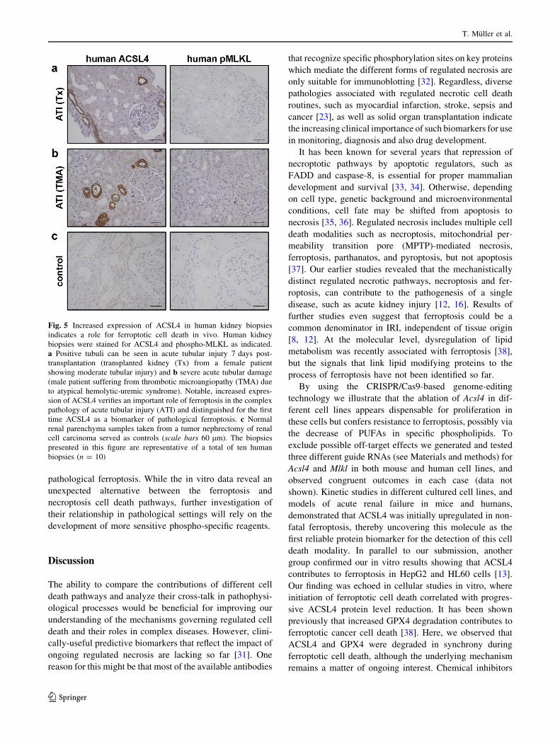
for the first time. As demonstrated in Fig. 5, we detected
robust immunostaining of ACSL4 in human ATI seven
days post-transplantation (Fig. 5a), as well as in severe
thrombotic microangiopathy of native kidney (Fig. 5b).
Normal renal parenchyma samples taken from a tumor
nephrectomy of renal cell carcinoma served as controls
(Fig. 5c). Although we suggest that necroptosis can occur
in parallel in the same transplanted organ sections, avail-
able reagents did not permit us to detect phosphorylated
MLKL in these biopsies. Nevertheless, these results high-
light for the first time ACSL4 as a biomarker of
Necroptosis and ferroptosis are alternative cell death pathways that operate in acute kidney…
123
pathological ferroptosis. While the in vitro data reveal an
unexpected alternative between the ferroptosis and
necroptosis cell death pathways, further investigation of
their relationship in pathological settings will rely on the
development of more sensitive phospho-specific reagents.
Discussion
The ability to compare the contributions of different cell
death pathways and analyze their cross-talk in pathophysi-
ological processes would be beneficial for improving our
understanding of the mechanisms governing regulated cell
death and their roles in complex diseases. However, clini-
cally-useful predictive biomarkers that reflect the impact of
ongoing regulated necrosis are lacking so far [31]. One
reason for this might be that most of the available antibodies
that recognize specific phosphorylation sites on key proteins
which mediate the different forms of regulated necrosis are
only suitable for immunoblotting [32]. Regardless, diverse
pathologies associated with regulated necrotic cell death
routines, such as myocardial infarction, stroke, sepsis and
cancer [23], as well as solid organ transplantation indicate
the increasing clinical importance of such biomarkers for use
in monitoring, diagnosis and also drug development.
It has been known for several years that repression of
necroptotic pathways by apoptotic regulators, such as
FADD and caspase-8, is essential for proper mammalian
development and survival [33, 34]. Otherwise, depending
on cell type, genetic background and microenvironmental
conditions, cell fate may be shifted from apoptosis to
necrosis [35, 36]. Regulated necrosis includes multiple cell
death modalities such as necroptosis, mitochondrial per-
meability transition pore (MPTP)-mediated necrosis,
ferroptosis, parthanatos, and pyroptosis, but not apoptosis
[37]. Our earlier studies revealed that the mechanistically
distinct regulated necrotic pathways, necroptosis and fer-
roptosis, can contribute to the pathogenesis of a single
disease, such as acute kidney injury [12, 16]. Results of
further studies even suggest that ferroptosis could be a
common denominator in IRI, independent of tissue origin
[8, 12]. At the molecular level, dysregulation of lipid
metabolism was recently associated with ferroptosis [38],
but the signals that link lipid modifying proteins to the
process of ferroptosis have not been identified so far.
By using the CRISPR/Cas9-based genome-editing
technology we illustrate that the ablation of Acsl4 in dif-
ferent cell lines appears dispensable for proliferation in
these cells but confers resistance to ferroptosis, possibly via
the decrease of PUFAs in specific phospholipids. To
exclude possible off-target effects we generated and tested
three different guide RNAs (see Materials and methods) for
Acsl4 and Mlkl in both mouse and human cell lines, and
observed congruent outcomes in each case (data not
shown). Kinetic studies in different cultured cell lines, and
models of acute renal failure in mice and humans,
demonstrated that ACSL4 was initially upregulated in non-
fatal ferroptosis, thereby uncovering this molecule as the
first reliable protein biomarker for the detection of this cell
death modality. In parallel to our submission, another
group confirmed our in vitro results showing that ACSL4
contributes to ferroptosis in HepG2 and HL60 cells [13].
Our finding was echoed in cellular studies in vitro, where
initiation of ferroptotic cell death correlated with progres-
sive ACSL4 protein level reduction. It has been shown
previously that increased GPX4 degradation contributes to
ferroptotic cancer cell death [38]. Here, we observed that
ACSL4 and GPX4 were degraded in synchrony during
ferroptotic cell death, although the underlying mechanism
remains a matter of ongoing interest. Chemical inhibitors
Fig. 5 Increased expression of ACSL4 in human kidney biopsies
indicates a role for ferroptotic cell death in vivo. Human kidney
biopsies were stained for ACSL4 and phospho-MLKL as indicated.
a Positive tubuli can be seen in acute tubular injury 7 days post-
transplantation (transplanted kidney (Tx) from a female patient
showing moderate tubular injury) and b severe acute tubular damage
(male patient suffering from thrombotic microangiopathy (TMA) due
to atypical hemolytic-uremic syndrome). Notable, increased expres-
sion of ACSL4 verifies an important role of ferroptosis in the complex
pathology of acute tubular injury (ATI) and distinguished for the first
time ACSL4 as a biomarker of pathological ferroptosis. c Normal
renal parenchyma samples taken from a tumor nephrectomy of renal
cell carcinoma served as controls (scale bars 60 lm). The biopsies
presented in this figure are representative of a total of ten human
biopsies (n = 10)
T. Muller et al.
123

of ferroptosis, like Fer-1, suppressed these protein
expression kinetics (Fig. 2a). These data are consistent
with the idea that ACSL4 expression level correlates with
the induction and progress of ferroptotic cell death, and
monitoring ACSL4 protein level would be suitable for
diagnostic purposes.
Additionally, comparative studies with Mlkl-knockout
cells and mice demonstrated for the first time that the
presumed mechanistically-distinct and independent modes
of regulated necrosis, necroptosis and ferroptosis, are not
only interconnected, but also are alternative to one another
in a setting where both contribute to cell death and, one has
been compromised. The mechanism underlying this phe-
nomenon remains a matter to be addressed in the future,
but it is possible that membrane lipid composition repre-
sents a point of alternative between ferroptosis and
necroptosis, where MLKL drives basal resistance to fer-
roptosis through depleting PUFAs, and ACSL4 drives basal
resistance to necroptosis by making the membrane less
amenable to MLKL-driven membrane permeabilization.
Our data support the crosstalk occurring at the level of
MLKL or via as-yet-unidentified downstream effectors,
because pharmacological inhibition of the necrosome
component kinases, RIPK1 and RIPK3, did not impact
ferroptosis in our cellular studies (see Supplementary
Material, Fig. S2a). Nevertheless, further studies are nee-
ded to confirm whether unchanged ACSL4 expression
contributes to ferroptosis resistance in other tumors and
diseases. Interestingly, interpathway crosstalk has emerged
as an important regulatory mechanism in cell death. For
example, the autophagy machinery can serve as a scaffold
to control the switch between necroptosis and apoptosis in
the context of Map3k7 loss [39], and MLKL was recently
shown to activate the inflammasome protein, NLRP3 [40].
Activation of ACSL4 in the course of ferroptosis rep-
resents an important but poorly understood phenomenon,
as do the details of how loss of Acsl4 confers protection
against ferroptosis. In cells undergoing ferroptosis, PUFAs
such as arachidonic acid (AA) are significantly depleted
[9, 41]. Acsl4 encode an enzyme that is involved in the
insertion of AA into membrane phospholipids. These
PUFAs might change the biophysical properties of cell
membranes, such as lipid rafts. Lipids are emerging as key
components of several non-apoptotic RCD pathways [42].
Therefore, it is conceivable that the initiation and execution
of ferroptosis can only proceed when highly oxidizable
PUFAs such as AA are present in the membrane. The
identification of ACSL4 loss as a key mediator of resis-
tance to ferroptosis supports this mechanism.
In a similar vein, the data presented here demonstrate
that alternative signal transduction pathways of regulated
necrosis act in opposition in pathophysiological processes
of acute kidney failure and each of them can contribute to
the total organ damage. These findings need to be taken
into consideration in the conception and development of
pharmacological strategies for therapeutic intervention
where therapeutic targeting of both ferroptosis and
necroptosis may be necessary to negate complex diseases
like AKI. To explore this idea further, we attempted to
model dual inhibition of ferroptosis and necroptosis in
murine IRI by administration of the ferroptosis inhibitor,
16–86, in necroptosis-insensitive Mlkl-knockout mice.
Surprisingly, although 16–86 treatment reduced ACSL4
levels in Mlkl-knockout mice, this was not reflected in
protection from IRI. This finding is consistent with the
inherent instability of 16–86 in vivo over the time frame
required for the IRI model, which limits its effectiveness in
in vivo settings at this stage. Additionally, because we
focused on the 24 h reperfusion time point, when kidney
damage had begun and was accompanied by a clear win-
dow between ACSL4 levels in Mlkl-knockout and wt mice,
we stopped the therapy with 16–86 24 h after the onset of
reperfusion. Termination of the experiment at this time
point could explain why these mice did not exhibit sig-
nificant protection relative to vehicle-treated Mlkl-
knockout mice. Furthermore, it is worth mentioning that
detection and diagnosis of AKI relies usually on changes in
serum creatinine and urea. Individually, they remain the
longstanding parameters used to test renal function, even
though are known to be imperfect because they do not
reflect genuine injury or real-time changes in kidney
function [43]. Accumulation and concomitant increase of
serum creatinine concentration lags far behind renal injury,
which causes deterioration of glomerular filtration rate.
Thus, substantial rises in serum creatinine are often not
witnessed until 24–72 h after the initial insult to the kid-
ney. Meanwhile, it is presumed that de novo synthesized
proteins are more suitable for early detection of AKI.
ACSL4 is one such protein, because it is expressed early
after injury and therefore has value in predicting AKI in
clinical settings, such as after kidney transplant.
More detailed analyses of the therapeutic potential of
pharmacological targeting of both necroptosis and ferrop-
tosis in complex disease models will depend on future
development of less labile ferroptosis inhibitors. The data
presented herein suggest that combination therapy using
anti-necroptosis and anti-ferroptosis compounds, or small
molecules (e.g. chimera) that simultaneously inhibit both
pathways, with concomitant enhancements in in vivo effi-
cacy, may be beneficial for the prevention of IRI; of course,
it may be necessary to additionally disable other pathways
at the same time. While these ideas represent an exciting
therapeutic possibility, clinical translation of these findings
will rely on future development of novel inhibitors of
regulated necrosis with improved potency and plasma
stability suitable for efficient application in diseases.
Necroptosis and ferroptosis are alternative cell death pathways that operate in acute kidney…
123
Acknowledgements We thank Janina Kahl, Maike Berger and Katja
Bruch for excellent technical assistance. This work is funded by the
Medical Faculty of Kiel University (to CD and SK), Dr. Werner
Jackstadt-Stiftung (to CD and SK), and Fresenius Medical Care
Germany (to UK and SK). BRS is supported by NIH grant
R01CA09706. JMM acknowledges funding from the National Health
and Medical Research Council of Australia (1057905, 1067289,
1105754, 1124735 and IRIISS 9000220) and Victorian Government
Operational Infrastructure Support.
Compliance with ethical standards
Conflict of interest The authors declare no competing or financial
interests.
Open Access This article is distributed under the terms of the
Creative Commons Attribution 4.0 International License (http://
creativecommons.org/licenses/by/4.0/), which permits unrestricted
use, distribution, and reproduction in any medium, provided you give
appropriate credit to the original author(s) and the source, provide a
link to the Creative Commons license, and indicate if changes were
made.
References
1. Linkermann A, Stockwell BR, Krautwald S et al (2014) Regu-
lated cell death and inflammation: an auto-amplification loop
causes organ failure. Nat Rev Immunol 14:759–767
2. Cai Z, Jitkaew S, Zhao J et al (2014) Plasma membrane
translocation of trimerized MLKL protein is required for TNF-
induced necroptosis. Nat Cell Biol 16:55–65
3. Dondelinger Y, Declercq W, Montessuit S et al (2014) MLKL
compromises plasma membrane integrity by binding to phos-
phatidylinositol phosphates. Cell Rep 7:971–981
4. Xia B, Fang S, Chen X et al (2016) MLKL forms cation channels.
Cell Res 26:517–528
5. Hildebrand JM, Tanzer MC, Lucet IS et al (2014) Activation of
the pseudokinase MLKL unleashes the four-helix bundle domain
to induce membrane localization and necroptotic cell death. Proc
Natl Acad Sci USA 111:15072–15077
6. Pasparakis M, Vandenabeele P (2015) Necroptosis and its role in
inflammation. Nature 517:311–320
7. Dixon SJ, Lemberg KM, Lamprecht MR et al (2012) Ferroptosis:
an iron-dependent form of nonapoptotic cell death. Cell
149:1060–1072
8. Friedmann Angeli JP, Schneider M, Proneth B et al (2014)
Inactivation of the ferroptosis regulator Gpx4 triggers acute renal
failure in mice. Nat Cell Biol 16:1180–1191
9. Skouta R, Dixon SJ, Wang J et al (2014) Ferrostatins inhibit
oxidative lipid damage and cell death in diverse disease models.
J Am Chem Soc 136:4551–4556
10. Dixon SJ, Winter GE, Musavi LS et al (2015) Human haploid cell
genetics reveals roles for lipid metabolism genes in nonapoptotic
cell death. ACS Chem Biol 10:1604–1609
11. Doll S, Proneth B, Tyurina YY et al (2017) ACSL4 dictates
ferroptosis sensitivity by shaping cellular lipid composition. Nat
Chem Biol 13:91–98
12. Linkermann A, Skouta R, Himmerkus N et al (2014) Synchro-
nized renal tubular cell death involves ferroptosis. Proc Natl Acad
Sci USA 111:16836–16841
13. Yuan H, Li X, Zhang X et al (2016) Identification of ACSL4 as a
biomarker and contributor of ferroptosis. Biochem Biophys Res
Commun 478:1338–1343
14. Yang WS, Sriramaratnam R, Welsch ME et al (2014) Regulation
of ferroptotic cancer cell death by GPX4. Cell 156:317–331
15. Murphy JM, Czabotar PE, Hildebrand JM et al (2013) The
pseudokinase MLKL mediates necroptosis via a molecular switch
mechanism. Immunity 39:443–453
16. Linkermann A, Brasen JH, Darding M et al (2013) Two inde-
pendent pathways of regulated necrosis mediate ischemia-
reperfusion injury. Proc Natl Acad Sci USA 110:12024–12029
17. Cao JY, Dixon SJ (2016) Mechanisms of ferroptosis. Cell Mol
Life Sci 73:2195–2209
18. Doudna JA, Charpentier E (2014) Genome editing The new
frontier of genome engineering with CRISPR-Cas9. Science
346:1258096
19. Yang WS, Stockwell BR (2008) Synthetic lethal screening
identifies compounds activating iron-dependent, nonapoptotic
cell death in oncogenic-RAS-harboring cancer cells. Chem Biol
15:234–245
20. Xie Y, Hou W, Song X et al (2016) Ferroptosis: process and
function. Cell Death Differ 23:369–379
21. Zhu S, Zhang Q, Sun X, et al (2017) HSPA5 Regulates Ferrop-
totic Cell Death in Cancer Cells. Cancer Res 77:2064–2077
22. Gonzalez-Juarbe N, Gilley RP, Hinojosa CA et al (2015) Pore-
forming toxins induce macrophage necroptosis during acute
bacterial pneumonia. PLoS Pathog 11:e1005337
23. Conrad M, Angeli JP, Vandenabeele P et al (2016) Regulated
necrosis: disease relevance and therapeutic opportunities. Nat
Rev Drug Discov 15:348–366
24. Takahashi N, Duprez L, Grootjans S et al (2012) Necrostatin-1
analogues: critical issues on the specificity, activity and in vivo
use in experimental disease models. Cell Death Dis 3:e437
25. Kaiser WJ, Sridharan H, Huang C et al (2013) Toll-like Receptor
3-mediated necrosis via TRIF, RIP3 and MLKL. J Biol Chem
288:31268–31279
26. Li JX, Feng JM, Wang Y et al (2014) The B-Raf(V600E) inhi-
bitor dabrafenib selectively inhibits RIP3 and alleviates
acetaminophen-induced liver injury. Cell Death Dis 5:e1278
27. Ofengeim D, Ito Y, Najafov A et al (2015) Activation of
necroptosis in multiple sclerosis. Cell Rep 10:1836–1849
28. Wang H, Sun L, Su L et al (2014) Mixed lineage kinase domain-
like protein MLKL causes necrotic membrane disruption upon
phosphorylation by RIP3. Mol Cell 54:133–146
29. Tanzer MC, Tripaydonis A, Webb AI et al (2015) Necroptosis
signalling is tuned by phosphorylation of MLKL residues outside
the pseudokinase domain activation loop. Biochem J
471:255–265
30. Vanden Berghe T, Hassannia B, Vandenabeele P (2016) An
outline of necrosome triggers. Cell Mol Life Sci 73:2137–2152
31. Krautwald S, Dewitz C, Fandrich F et al (2016) Inhibition of
regulated cell death by cell-penetrating peptides. Cell Mol Life
Sci 73:2269–2284
32. He S, Huang S, Shen Z (2016) Biomarkers for the detection of
necroptosis. Cell Mol Life Sci 73:2177–2181
33. Kaiser WJ, Upton JW, Long AB et al (2011) RIP3 mediates the
embryonic lethality of caspase-8-deficient mice. Nature
471:368–372
34. Oberst A, Dillon CP, Weinlich R et al (2011) Catalytic activity of
the caspase-8-FLIP(L) complex inhibits RIPK3-dependent
necrosis. Nature 471:363–367
35. Duprez L, Bertrand MJ, Vanden Berghe T et al (2012) Interme-
diate domain of receptor-interacting protein kinase 1 (RIPK1)
determines switch between necroptosis and RIPK1 kinase-de-
pendent apoptosis. J Biol Chem 287:14863–14872
36. Morioka S, Broglie P, Omori E et al (2014) TAK1 kinase
switches cell fate from apoptosis to necrosis following TNF
stimulation. J Cell Biol 204:607–623
T. Muller et al.
123

37. Galluzzi L, Kepp O, Krautwald S et al (2014) Molecular mech-
anisms of regulated necrosis. Semin Cell Dev Biol 35:24–32
38. Shimada K, Skouta R, Kaplan A et al (2016) Global survey of
cell death mechanisms reveals metabolic regulation of ferropto-
sis. Nat Chem Biol 12:497–503
39. Goodall ML, Fitzwalter BE, Zahedi S et al (2016) The autophagy
machinery controls cell death switching between apoptosis and
necroptosis. Dev Cell 37:337–349
40. Conos SA, Chen KW, De ND et al (2017) Active MLKL triggers
the NLRP3 inflammasome in a cell-intrinsic manner. Proc Natl
Acad Sci USA 114:E961–E969
41. Yang WS, Kim KJ, Gaschler MM et al (2016) Peroxidation of
polyunsaturated fatty acids by lipoxygenases drives ferroptosis.
Proc Natl Acad Sci USA 113:E4966–E4975
42. Dixon SJ, Stockwell BR (2014) The role of iron and reactive
oxygen species in cell death. Nat Chem Biol 10:9–17
43. Moran SM, Myers BD (1985) Course of acute renal failure
studied by a model of creatinine kinetics. Kidney Int 27:928–937
Necroptosis and ferroptosis are alternative cell death pathways that operate in acute kidney…
123