nccn clinical practice guidelines in oncology (nccn guidelines...
TRANSCRIPT
NCCN Clinical Practice Guidelines in Oncology (NCCN Guidelines®)
Neuroendocrine TumorsVersion 3.2017 — August 2, 2017
Continue
NCCN.org
NCCN Evidence Blocks™
Version 3.2017, 08/02/17 © National Comprehensive Cancer Network, Inc. 2017, All rights reserved. The NCCN Evidence BlocksTM, NCCN Guidelines® and this illustration may not be reproduced in any form without the express written permission of NCCN®.
NCCN Guidelines IndexTable of Contents
Discussion
NCCN Guidelines Version 3.2017 Panel MembersNeuroendocrine TumorsNCCN Evidence BlocksTM
Matthew H. Kulke, MD/Chair †Dana-Farber/Brigham and Women’s Cancer Center
Manisha H. Shah, MD/Vice Chair †The Ohio State University Comprehensive Cancer Center - James Cancer Hospital and Solove Research Institute
Al B. Benson, III, MD †Robert H. Lurie Comprehensive Cancer Center of Northwestern University
Emily Bergsland, MD †UCSF Helen Diller Family Comprehensive Cancer Center
Jordan D. Berlin, MD †Vanderbilt-Ingram Cancer Center
Stephen A. Besh, MD †St. Jude Children`s Research Hospital/The University of Tennessee Health Science Center
Lawrence S. Blaszkowsky, MD †Massachusetts General Hospital Cancer Center
Jennifer Eads, MD † ÞCase Comprehensive Cancer Center/University Hospitals Seidman Cancer Center and Cleveland Clinic Taussig Cancer Institute
Lyska Emerson, MD ≠Huntsman Cancer Institute at the University of Utah
Paul F. Engstrom, MD †Fox Chase Cancer Center
Paul Fanta, MD †UC San Diego Moores Cancer CenterThomas Giordano, MD, PhD ≠University of MichiganComprehensive Cancer Center Whitney S. Goldner, MD ðFred & Pamela Buffett Cancer Center Thorvardur R. Halfdanarson, MD Þ †Mayo Clinic Cancer CenterMartin J. Heslin, MD ¶University of Alabama at Birmingham Comprehensive Cancer CenterGregory P. Kalemkerian, MD/Liaison †University of MichiganComprehensive Cancer CenterFouad Kandeel, MD, PhD ðCity of Hope Comprehensive Cancer CenterWajih Zaheer Kidwai, MD †Yale Cancer Center/Smilow Cancer HospitalPamela L. Kunz, MD †Stanford Cancer InstituteBoris W. Kuvshinoff, II, MD, MBA ¶Roswell Park Cancer InstituteChristopher Lieu, MD †University of Colorado Cancer Center
Jeffrey F. Moley, MD ¶ Siteman Cancer Center at Barnes-Jewish Hospital and Washington University School of Medicine
Venu G. Pillarisetty, MD ¶Fred Hutchinson Cancer Research Center/Seattle Cancer Care Alliance
Leonard Saltz, MD † ÞMemorial Sloan Kettering Cancer Center
Julie Ann Sosa, MD ¶ ðDuke Cancer Institute
Jonathan R. Strosberg, MD †Moffitt Cancer Center
Craig A. Sussman, MD ÞVanderbilt-Ingram Cancer Center
Nataliya A. Uboha, MD, PhD †University of Wisconsin Carbone Cancer Center
Christopher Wolfgang, MD, PhD ¶The Sidney Kimmel Comprehensive Cancer Center at Johns Hopkins
James C. Yao, MD †The University of Texas MD Anderson Cancer Center
NCCNJennifer Burns Deborah Freedman-Cass, PhD Ndiya Ogba, PhD
NCCN Guidelines Panel Disclosures
¶ Surgery/Surgical oncology† Medical oncologyð Endocrinology≠ PathologyÞ Internal medicine* Discussion Section Writing Committee
*
Continue
Version 3.2017, 08/02/17 © National Comprehensive Cancer Network, Inc. 2017, All rights reserved. The NCCN Evidence BlocksTM, NCCN Guidelines® and this illustration may not be reproduced in any form without the express written permission of NCCN®.
Printed by Philip Jax on 8/9/2017 2:44:55 PM. For personal use only. Not approved for distribution. Copyright © 2017 National Comprehensive Cancer Network, Inc., All Rights Reserved.
Clinical Trials: NCCN believes that the best management for any patient with cancer is in a clinical trial. Participation in clinical trials is especially encouraged. To find clinical trials online at NCCN Member Institutions, click here: nccn.org/clinical_trials/physician.html.NCCN Categories of Evidence and Consensus: All recommendations are category 2A unless otherwise specified. See NCCN Categories of Evidence and Consensus.
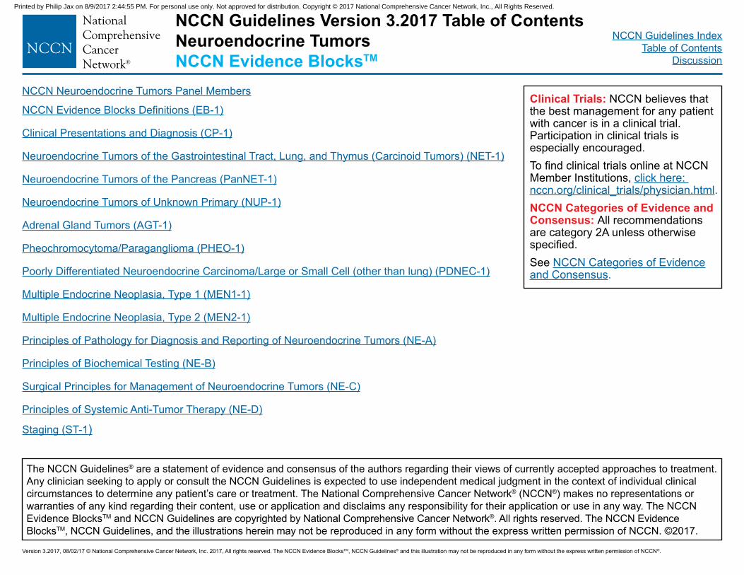
NCCN Neuroendocrine Tumors Panel Members
NCCN Evidence Blocks Definitions (EB-1)
Clinical Presentations and Diagnosis (CP-1)
Neuroendocrine Tumors of the Gastrointestinal Tract, Lung, and Thymus (Carcinoid Tumors) (NET-1)
Neuroendocrine Tumors of the Pancreas (PanNET-1)
Neuroendocrine Tumors of Unknown Primary (NUP-1)
Adrenal Gland Tumors (AGT-1)
Pheochromocytoma/Paraganglioma (PHEO-1)
Poorly Differentiated Neuroendocrine Carcinoma/Large or Small Cell (other than lung) (PDNEC-1)
Multiple Endocrine Neoplasia, Type 1 (MEN1-1)
Multiple Endocrine Neoplasia, Type 2 (MEN2-1)
Principles of Pathology for Diagnosis and Reporting of Neuroendocrine Tumors (NE-A)
Principles of Biochemical Testing (NE-B)
Surgical Principles for Management of Neuroendocrine Tumors (NE-C)
Principles of Systemic Anti-Tumor Therapy (NE-D)
Staging (ST-1)
The NCCN Guidelines® are a statement of evidence and consensus of the authors regarding their views of currently accepted approaches to treatment. Any clinician seeking to apply or consult the NCCN Guidelines is expected to use independent medical judgment in the context of individual clinical circumstances to determine any patient’s care or treatment. The National Comprehensive Cancer Network® (NCCN®) makes no representations or warranties of any kind regarding their content, use or application and disclaims any responsibility for their application or use in any way. The NCCN Evidence BlocksTM and NCCN Guidelines are copyrighted by National Comprehensive Cancer Network®. All rights reserved. The NCCN Evidence BlocksTM, NCCN Guidelines, and the illustrations herein may not be reproduced in any form without the express written permission of NCCN. ©2017.
NCCN Guidelines Version 3.2017 Table of ContentsNeuroendocrine TumorsNCCN Evidence BlocksTM
NCCN Guidelines IndexTable of Contents
Discussion
Version 3.2017, 08/02/17 © National Comprehensive Cancer Network, Inc. 2017, All rights reserved. The NCCN Evidence BlocksTM, NCCN Guidelines® and this illustration may not be reproduced in any form without the express written permission of NCCN®.
Printed by Philip Jax on 8/9/2017 2:44:55 PM. For personal use only. Not approved for distribution. Copyright © 2017 National Comprehensive Cancer Network, Inc., All Rights Reserved.
NCCN Guidelines IndexTable of Contents
Discussion
NCCN Guidelines Version 3.2017Neuroendocrine TumorsNCCN Evidence BlocksTM
EB-1
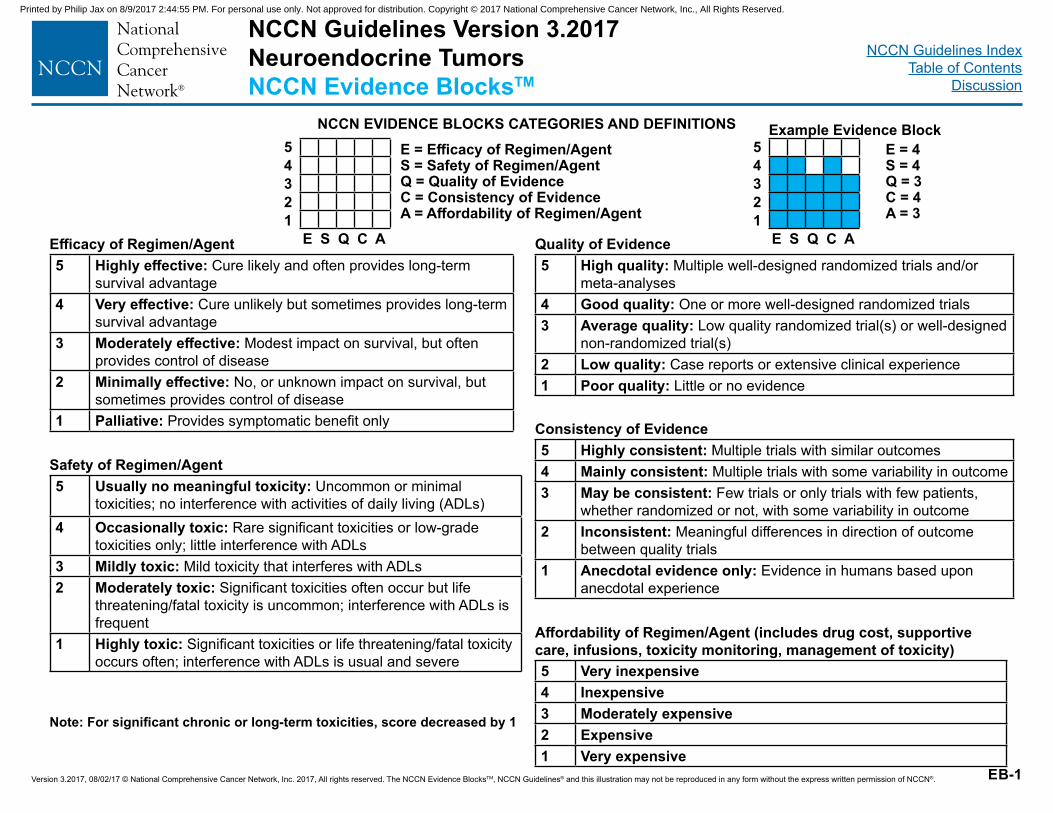
NCCN EVIDENCE BLOCKS CATEGORIES AND DEFINITIONSE = Efficacy of Regimen/AgentS = Safety of Regimen/AgentQ = Quality of EvidenceC = Consistency of EvidenceA = Affordability of Regimen/Agent
Efficacy of Regimen/Agent5 Highly effective: Cure likely and often provides long-term
survival advantage4 Very effective: Cure unlikely but sometimes provides long-term
survival advantage3 Moderately effective: Modest impact on survival, but often
provides control of disease2 Minimally effective: No, or unknown impact on survival, but
sometimes provides control of disease1 Palliative: Provides symptomatic benefit only
Safety of Regimen/Agent5 Usually no meaningful toxicity: Uncommon or minimal
toxicities; no interference with activities of daily living (ADLs)4 Occasionally toxic: Rare significant toxicities or low-grade
toxicities only; little interference with ADLs3 Mildly toxic: Mild toxicity that interferes with ADLs2 Moderately toxic: Significant toxicities often occur but life
threatening/fatal toxicity is uncommon; interference with ADLs is frequent
1 Highly toxic: Significant toxicities or life threatening/fatal toxicity occurs often; interference with ADLs is usual and severe
5 High quality: Multiple well-designed randomized trials and/or meta-analyses
4 Good quality: One or more well-designed randomized trials3 Average quality: Low quality randomized trial(s) or well-designed
non-randomized trial(s)2 Low quality: Case reports or extensive clinical experience1 Poor quality: Little or no evidence
Quality of Evidence
5 Highly consistent: Multiple trials with similar outcomes4 Mainly consistent: Multiple trials with some variability in outcome3 May be consistent: Few trials or only trials with few patients,
whether randomized or not, with some variability in outcome2 Inconsistent: Meaningful differences in direction of outcome
between quality trials1 Anecdotal evidence only: Evidence in humans based upon
anecdotal experience
Consistency of Evidence
5 Very inexpensive4 Inexpensive3 Moderately expensive2 Expensive1 Very expensive
Affordability of Regimen/Agent (includes drug cost, supportive care, infusions, toxicity monitoring, management of toxicity)
E S Q C A
54321
Example Evidence BlockE = 4S = 4Q = 3C = 4A = 3
E S Q C A
54321
Note: For significant chronic or long-term toxicities, score decreased by 1
Version 3.2017, 08/02/17 © National Comprehensive Cancer Network, Inc. 2017, All rights reserved. The NCCN Evidence BlocksTM, NCCN Guidelines® and this illustration may not be reproduced in any form without the express written permission of NCCN®.
Printed by Philip Jax on 8/9/2017 2:44:55 PM. For personal use only. Not approved for distribution. Copyright © 2017 National Comprehensive Cancer Network, Inc., All Rights Reserved.
NCCN Guidelines IndexTable of Contents
Discussion
Version 3.2017, 08/02/17 © National Comprehensive Cancer Network, Inc. 2017, All rights reserved. The NCCN Evidence BlocksTM, NCCN Guidelines® and this illustration may not be reproduced in any form without the express written permission of NCCN®.
NCCN Guidelines Version 3.2017 Neuroendocrine TumorsNCCN Evidence BlocksTM
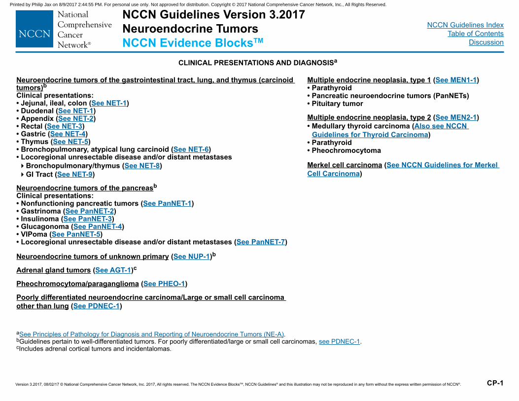
Neuroendocrine tumors of the gastrointestinal tract, lung, and thymus (carcinoid tumors)bClinical presentations:• Jejunal, ileal, colon (See NET-1)• Duodenal (See NET-1)• Appendix (See NET-2) • Rectal (See NET-3)• Gastric (See NET-4)• Thymus (See NET-5)• Bronchopulmonary, atypical lung carcinoid (See NET-6)• Locoregional unresectable disease and/or distant metastases �Bronchopulmonary/thymus (See NET-8)�GI Tract (See NET-9)
Neuroendocrine tumors of the pancreasbClinical presentations:• Nonfunctioning pancreatic tumors (See PanNET-1)• Gastrinoma (See PanNET-2)• Insulinoma (See PanNET-3) • Glucagonoma (See PanNET-4)• VIPoma (See PanNET-5)• Locoregional unresectable disease and/or distant metastases (See PanNET-7)
Neuroendocrine tumors of unknown primary (See NUP-1)b
Adrenal gland tumors (See AGT-1)c
Pheochromocytoma/paraganglioma (See PHEO-1)
Poorly differentiated neuroendocrine carcinoma/Large or small cell carcinoma other than lung (See PDNEC-1)
CP-1
aSee Principles of Pathology for Diagnosis and Reporting of Neuroendocrine Tumors (NE-A).bGuidelines pertain to well-differentiated tumors. For poorly differentiated/large or small cell carcinomas, see PDNEC-1.cIncludes adrenal cortical tumors and incidentalomas.
CLINICAL PRESENTATIONS AND DIAGNOSISa
Multiple endocrine neoplasia, type 1 (See MEN1-1)• Parathyroid• Pancreatic neuroendocrine tumors (PanNETs) • Pituitary tumor
Multiple endocrine neoplasia, type 2 (See MEN2-1)• Medullary thyroid carcinoma (Also see NCCN
Guidelines for Thyroid Carcinoma)• Parathyroid• Pheochromocytoma
Merkel cell carcinoma (See NCCN Guidelines for Merkel Cell Carcinoma)
Printed by Philip Jax on 8/9/2017 2:44:55 PM. For personal use only. Not approved for distribution. Copyright © 2017 National Comprehensive Cancer Network, Inc., All Rights Reserved.
NCCN Guidelines IndexTable of Contents
Discussion
Version 3.2017, 08/02/17 © National Comprehensive Cancer Network, Inc. 2017, All rights reserved. The NCCN Evidence BlocksTM, NCCN Guidelines® and this illustration may not be reproduced in any form without the express written permission of NCCN®.
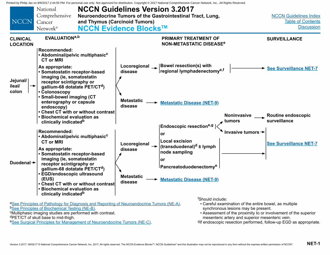
NCCN Guidelines Version 3.2017Neuroendocrine Tumors of the Gastrointestinal Tract, Lung, and Thymus (Carcinoid Tumors)NCCN Evidence BlocksTM
NET-1
f Should include:• Careful examination of the entire bowel, as multiple
synchronous lesions may be present.• Assessment of the proximity to or involvement of the superior
mesenteric artery and superior mesenteric vein.gIf endoscopic resection performed, follow-up EGD as appropriate.
a See Principles of Pathology for Diagnosis and Reporting of Neuroendocrine Tumors (NE-A).b See Principles of Biochemical Testing (NE-B).cMultiphasic imaging studies are performed with contrast. dPET/CT of skull base to mid-thigh.eSee Surgical Principles for Management of Neuroendocrine Tumors (NE-C).
CLINICAL LOCATION
EVALUATIONa,b PRIMARY TREATMENT OF NON-METASTATIC DISEASEe
SURVEILLANCE
Jejunal/ileal/colon
Duodenal
Recommended:• Abdominal/pelvic multiphasicc
CT or MRIAs appropriate:• Somatostatin receptor-based
imaging (ie, somatostatin receptor scintigraphy or gallium-68 dotatate PET/CTd)
• Colonoscopy• Small-bowel imaging (CT
enterography or capsule endoscopy)
• Chest CT with or without contrast• Biochemical evaluation as
clinically indicatedb
Recommended:• Abdominal/pelvic multiphasicc
CT or MRIAs appropriate:• Somatostatin receptor-based
imaging (ie, somatostatin receptor scintigraphy or gallium-68 dotatate PET/CTd)
• EGD/endoscopic ultrasound (EUS)
• Chest CT with or without contrast• Biochemical evaluation as
clinically indicatedb
Locoregional disease
Metastatic disease
Locoregional disease
Metastatic disease
Bowel resection(s) with regional lymphadenectomye,f
Metastatic Disease (NET-9)
Endoscopic resectione,g
orLocal excision (transduodenal)d ± lymph node samplingorPancreatoduodenectomye
Metastatic Disease (NET-9)
Noninvasive tumors
Invasive tumors
Routine endoscopic surveillance
See Surveillance NET-7
See Surveillance NET-7
Printed by Philip Jax on 8/9/2017 2:44:55 PM. For personal use only. Not approved for distribution. Copyright © 2017 National Comprehensive Cancer Network, Inc., All Rights Reserved.
NCCN Guidelines IndexTable of Contents
Discussion
Version 3.2017, 08/02/17 © National Comprehensive Cancer Network, Inc. 2017, All rights reserved. The NCCN Evidence BlocksTM, NCCN Guidelines® and this illustration may not be reproduced in any form without the express written permission of NCCN®. NET-2
CLINICAL LOCATION
EVALUATIONa,b PRIMARY TREATMENT OF NON-METASTATIC DISEASEe
SURVEILLANCE
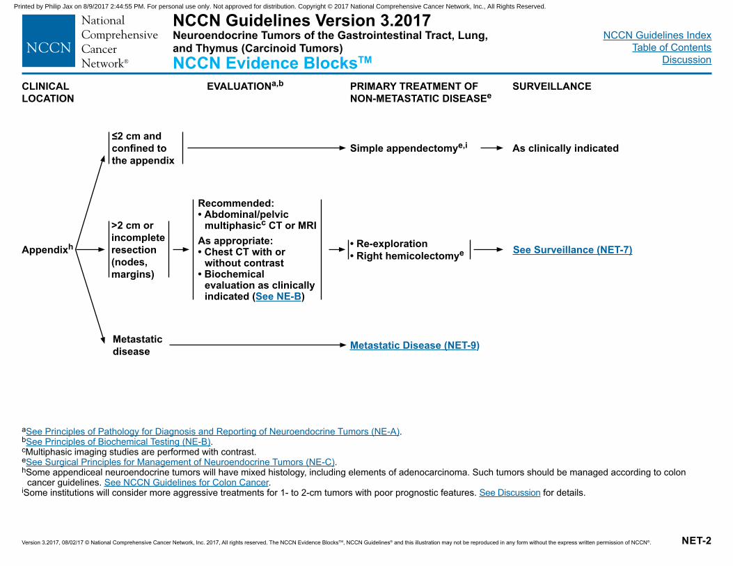
a See Principles of Pathology for Diagnosis and Reporting of Neuroendocrine Tumors (NE-A).bSee Principles of Biochemical Testing (NE-B).cMultiphasic imaging studies are performed with contrast.eSee Surgical Principles for Management of Neuroendocrine Tumors (NE-C).h Some appendiceal neuroendocrine tumors will have mixed histology, including elements of adenocarcinoma. Such tumors should be managed according to colon
cancer guidelines. See NCCN Guidelines for Colon Cancer.i Some institutions will consider more aggressive treatments for 1- to 2-cm tumors with poor prognostic features. See Discussion for details.
Appendixh
Metastatic disease
>2 cm or incomplete resection (nodes, margins)
≤2 cm and confined to the appendix
Recommended:• Abdominal/pelvic
multiphasicc CT or MRIAs appropriate:• Chest CT with or
without contrast• Biochemical
evaluation as clinically indicated (See NE-B)
Simple appendectomye,i
• Re-exploration• Right hemicolectomye
As clinically indicated
Metastatic Disease (NET-9)
See Surveillance (NET-7)
NCCN Guidelines Version 3.2017Neuroendocrine Tumors of the Gastrointestinal Tract, Lung, and Thymus (Carcinoid Tumors)NCCN Evidence BlocksTM
Printed by Philip Jax on 8/9/2017 2:44:55 PM. For personal use only. Not approved for distribution. Copyright © 2017 National Comprehensive Cancer Network, Inc., All Rights Reserved.
NCCN Guidelines IndexTable of Contents
Discussion
Version 3.2017, 08/02/17 © National Comprehensive Cancer Network, Inc. 2017, All rights reserved. The NCCN Evidence BlocksTM, NCCN Guidelines® and this illustration may not be reproduced in any form without the express written permission of NCCN®. NET-3
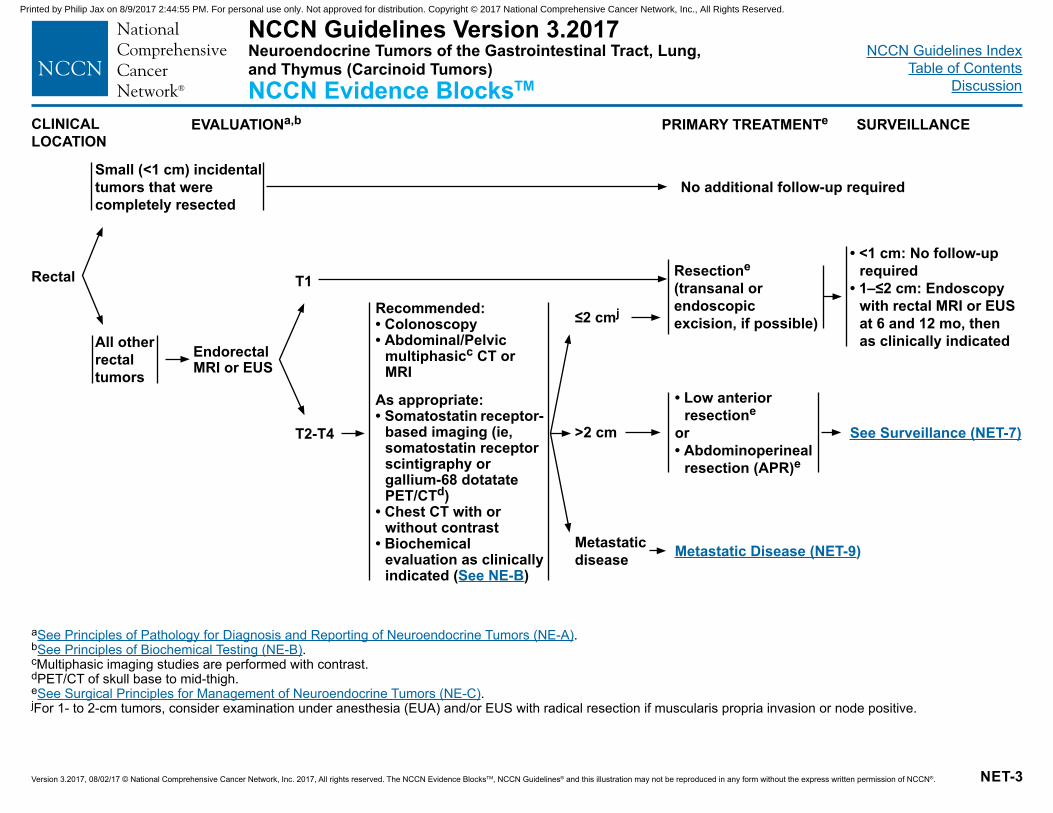
CLINICAL LOCATION
EVALUATIONa,b PRIMARY TREATMENTe SURVEILLANCE
Rectal
Metastatic disease
>2 cm
≤2 cmj
T1
T2-T4
Resectione (transanal or endoscopic excision, if possible)
• Low anterior resectione
or• Abdominoperineal
resection (APR)e
• <1 cm: No follow-up required
• 1–≤2 cm: Endoscopy with rectal MRI or EUS at 6 and 12 mo, then as clinically indicated
Metastatic Disease (NET-9)
Recommended:• Colonoscopy• Abdominal/Pelvic
multiphasicc CT or MRI
As appropriate:• Somatostatin receptor-
based imaging (ie, somatostatin receptor scintigraphy or gallium-68 dotatate PET/CTd)
• Chest CT with or without contrast
• Biochemical evaluation as clinically indicated (See NE-B)
a See Principles of Pathology for Diagnosis and Reporting of Neuroendocrine Tumors (NE-A).bSee Principles of Biochemical Testing (NE-B).cMultiphasic imaging studies are performed with contrast.dPET/CT of skull base to mid-thigh.eSee Surgical Principles for Management of Neuroendocrine Tumors (NE-C).jFor 1- to 2-cm tumors, consider examination under anesthesia (EUA) and/or EUS with radical resection if muscularis propria invasion or node positive.
Endorectal MRI or EUS
See Surveillance (NET-7)
Small (<1 cm) incidental tumors that were completely resected
All other rectal tumors
No additional follow-up required
NCCN Guidelines Version 3.2017Neuroendocrine Tumors of the Gastrointestinal Tract, Lung, and Thymus (Carcinoid Tumors)NCCN Evidence BlocksTM
Printed by Philip Jax on 8/9/2017 2:44:55 PM. For personal use only. Not approved for distribution. Copyright © 2017 National Comprehensive Cancer Network, Inc., All Rights Reserved.
NCCN Guidelines IndexTable of Contents
Discussion
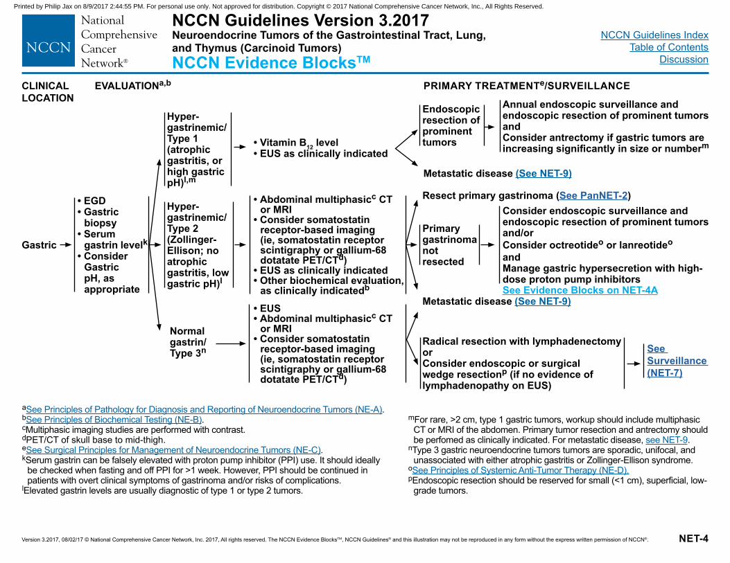
Version 3.2017, 08/02/17 © National Comprehensive Cancer Network, Inc. 2017, All rights reserved. The NCCN Evidence BlocksTM, NCCN Guidelines® and this illustration may not be reproduced in any form without the express written permission of NCCN®. NET-4
CLINICAL LOCATION
EVALUATIONa,b PRIMARY TREATMENTe/SURVEILLANCE
Gastric
• EGD • Gastric
biopsy• Serum
gastrin levelk • Consider
Gastric pH, as appropriate
Hyper-gastrinemic/ Type 2 (Zollinger-Ellison; no atrophic gastritis, low gastric pH)l
Hyper-gastrinemic/Type 1 (atrophic gastritis, or high gastric pH)l,m
Normal gastrin/Type 3n
• Vitamin B12 level• EUS as clinically indicated
Annual endoscopic surveillance and endoscopic resection of prominent tumors andConsider antrectomy if gastric tumors are increasing significantly in size or numberm
• Abdominal multiphasicc CT or MRI
• Consider somatostatin receptor-based imaging (ie, somatostatin receptor scintigraphy or gallium-68 dotatate PET/CTd)
• EUS as clinically indicated• Other biochemical evaluation,
as clinically indicatedb
• EUS• Abdominal multiphasicc CT
or MRI• Consider somatostatin
receptor-based imaging (ie, somatostatin receptor scintigraphy or gallium-68 dotatate PET/CTd)
Consider endoscopic surveillance and endoscopic resection of prominent tumorsand/orConsider octreotideo or lanreotideo andManage gastric hypersecretion with high-dose proton pump inhibitors
Radical resection with lymphadenectomyorConsider endoscopic or surgical wedge resectionp (if no evidence of lymphadenopathy on EUS)
a See Principles of Pathology for Diagnosis and Reporting of Neuroendocrine Tumors (NE-A).b See Principles of Biochemical Testing (NE-B).cMultiphasic imaging studies are performed with contrast.dPET/CT of skull base to mid-thigh.eSee Surgical Principles for Management of Neuroendocrine Tumors (NE-C).kSerum gastrin can be falsely elevated with proton pump inhibitor (PPI) use. It should ideally
be checked when fasting and off PPI for >1 week. However, PPI should be continued in patients with overt clinical symptoms of gastrinoma and/or risks of complications.
lElevated gastrin levels are usually diagnostic of type 1 or type 2 tumors.
Metastatic disease (See NET-9)
Metastatic disease (See NET-9)
mFor rare, >2 cm, type 1 gastric tumors, workup should include multiphasic CT or MRI of the abdomen. Primary tumor resection and antrectomy should be perfomed as clinically indicated. For metastatic disease, see NET-9.
nType 3 gastric neuroendocrine tumors tumors are sporadic, unifocal, and unassociated with either atrophic gastritis or Zollinger-Ellison syndrome.
o See Principles of Systemic Anti-Tumor Therapy (NE-D).pEndoscopic resection should be reserved for small (<1 cm), superficial, low-
grade tumors.
Resect primary gastrinoma (See PanNET-2)
Primary gastrinoma not resected
See Surveillance (NET-7)
Endoscopic resection of prominent tumors
NCCN Guidelines Version 3.2017Neuroendocrine Tumors of the Gastrointestinal Tract, Lung, and Thymus (Carcinoid Tumors)NCCN Evidence BlocksTM
See Evidence Blocks on NET-4A
Printed by Philip Jax on 8/9/2017 2:44:55 PM. For personal use only. Not approved for distribution. Copyright © 2017 National Comprehensive Cancer Network, Inc., All Rights Reserved.
NCCN Guidelines IndexTable of Contents
Discussion
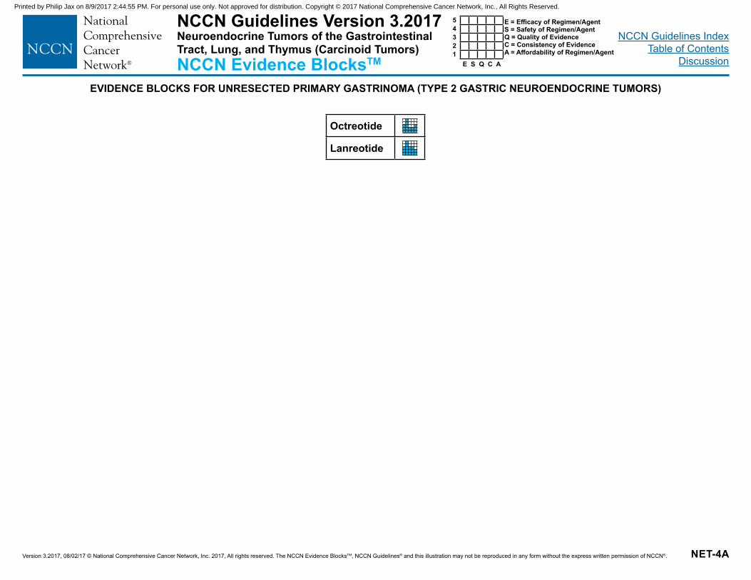
Version 3.2017, 08/02/17 © National Comprehensive Cancer Network, Inc. 2017, All rights reserved. The NCCN Evidence BlocksTM, NCCN Guidelines® and this illustration may not be reproduced in any form without the express written permission of NCCN®. NET-4A
NCCN Guidelines Version 3.2017Neuroendocrine Tumors of the Gastrointestinal Tract, Lung, and Thymus (Carcinoid Tumors)NCCN Evidence BlocksTM E S Q C A
54321
E = Efficacy of Regimen/AgentS = Safety of Regimen/AgentQ = Quality of EvidenceC = Consistency of EvidenceA = Affordability of Regimen/Agent
EVIDENCE BLOCKS FOR UNRESECTED PRIMARY GASTRINOMA (TYPE 2 GASTRIC NEUROENDOCRINE TUMORS)
Octreotide
Lanreotide
Printed by Philip Jax on 8/9/2017 2:44:55 PM. For personal use only. Not approved for distribution. Copyright © 2017 National Comprehensive Cancer Network, Inc., All Rights Reserved.
NCCN Guidelines IndexTable of Contents
Discussion
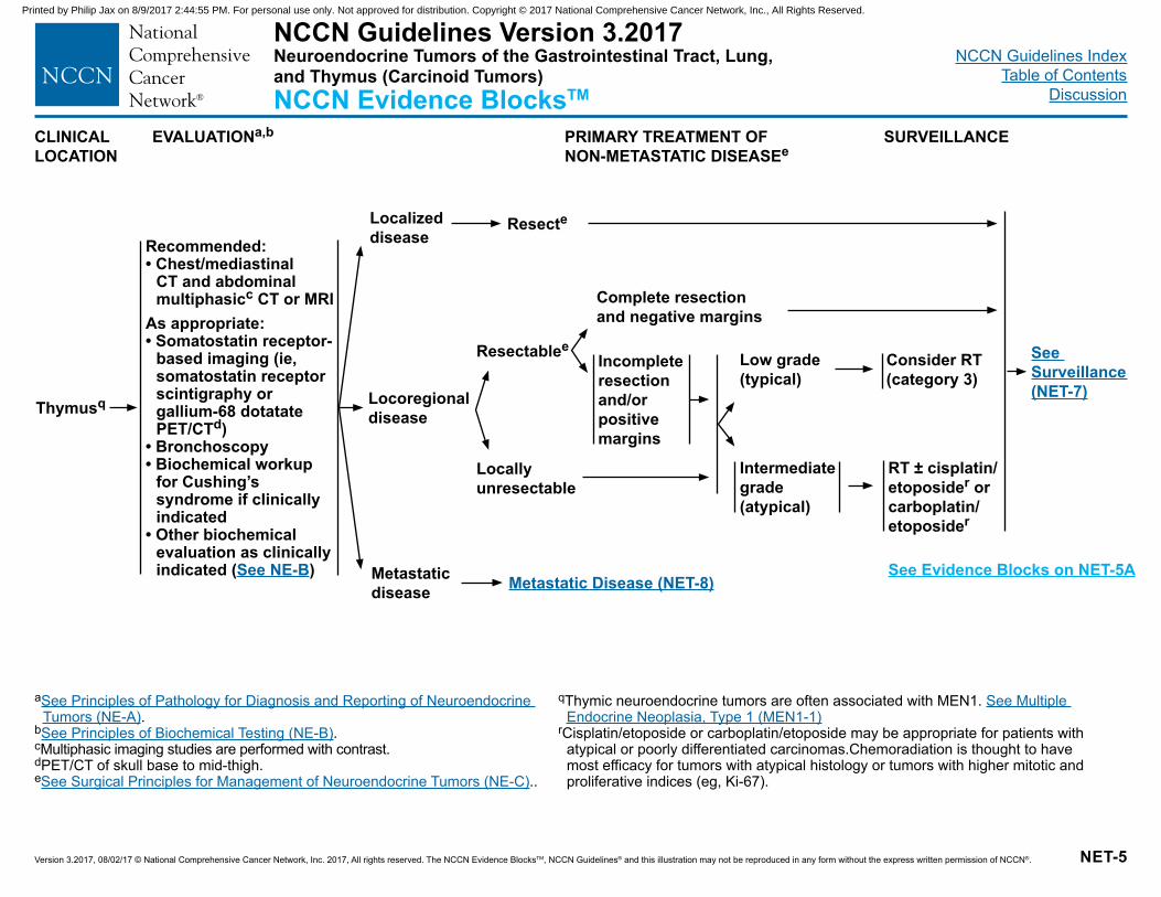
Version 3.2017, 08/02/17 © National Comprehensive Cancer Network, Inc. 2017, All rights reserved. The NCCN Evidence BlocksTM, NCCN Guidelines® and this illustration may not be reproduced in any form without the express written permission of NCCN®. NET-5
a See Principles of Pathology for Diagnosis and Reporting of Neuroendocrine Tumors (NE-A).
bSee Principles of Biochemical Testing (NE-B).cMultiphasic imaging studies are performed with contrast.dPET/CT of skull base to mid-thigh. eSee Surgical Principles for Management of Neuroendocrine Tumors (NE-C)..
CLINICAL LOCATION
EVALUATIONa,b PRIMARY TREATMENT OF NON-METASTATIC DISEASEe
SURVEILLANCE
Thymusq
Recommended:• Chest/mediastinal
CT and abdominal multiphasicc CT or MRI
As appropriate:• Somatostatin receptor-
based imaging (ie, somatostatin receptor scintigraphy or gallium-68 dotatate PET/CTd)
• Bronchoscopy• Biochemical workup
for Cushing’s syndrome if clinically indicated
• Other biochemical evaluation as clinically indicated (See NE-B)
Metastatic Disease (NET-8)
Consider RT (category 3)
RT ± cisplatin/etoposider or carboplatin/etoposider
Locoregional disease
Metastatic disease
Localized disease
Resecte
Complete resection and negative margins
Resectablee
Locally unresectable
q Thymic neuroendocrine tumors are often associated with MEN1. See Multiple Endocrine Neoplasia, Type 1 (MEN1-1)
rCisplatin/etoposide or carboplatin/etoposide may be appropriate for patients with atypical or poorly differentiated carcinomas.Chemoradiation is thought to have most efficacy for tumors with atypical histology or tumors with higher mitotic and proliferative indices (eg, Ki-67).
See Surveillance (NET-7)
Low grade (typical)
Intermediate grade (atypical)
Incomplete resection and/or positive margins
NCCN Guidelines Version 3.2017Neuroendocrine Tumors of the Gastrointestinal Tract, Lung, and Thymus (Carcinoid Tumors)NCCN Evidence BlocksTM
See Evidence Blocks on NET-5A
Printed by Philip Jax on 8/9/2017 2:44:55 PM. For personal use only. Not approved for distribution. Copyright © 2017 National Comprehensive Cancer Network, Inc., All Rights Reserved.
NCCN Guidelines IndexTable of Contents
Discussion
Version 3.2017, 08/02/17 © National Comprehensive Cancer Network, Inc. 2017, All rights reserved. The NCCN Evidence BlocksTM, NCCN Guidelines® and this illustration may not be reproduced in any form without the express written permission of NCCN®. NET-5A
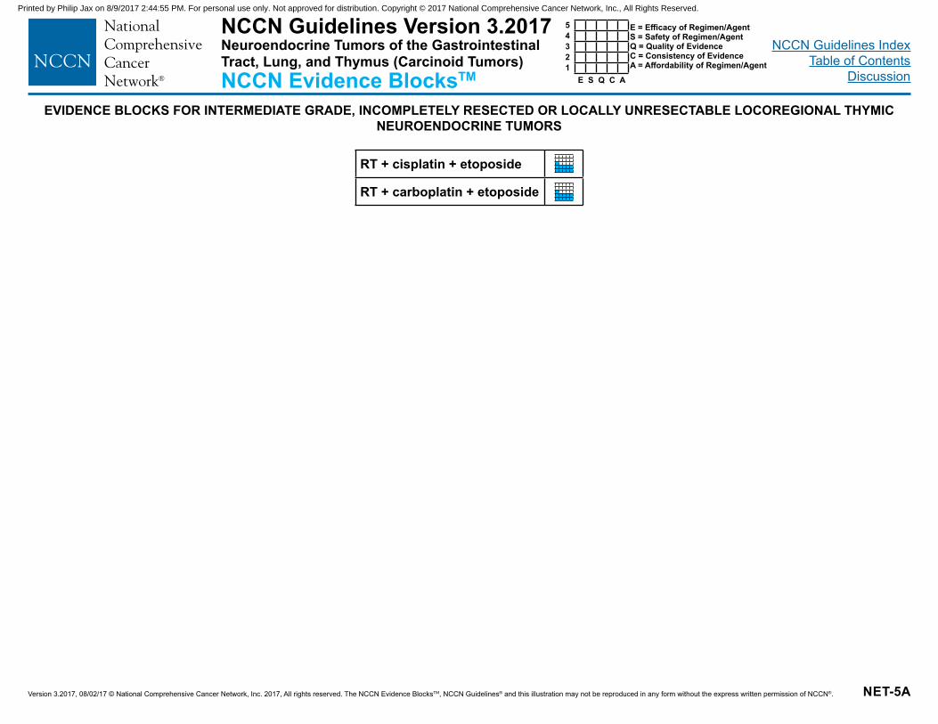
NCCN Guidelines Version 3.2017Neuroendocrine Tumors of the Gastrointestinal Tract, Lung, and Thymus (Carcinoid Tumors)NCCN Evidence BlocksTM E S Q C A
54321
E = Efficacy of Regimen/AgentS = Safety of Regimen/AgentQ = Quality of EvidenceC = Consistency of EvidenceA = Affordability of Regimen/Agent
EVIDENCE BLOCKS FOR INTERMEDIATE GRADE, INCOMPLETELY RESECTED OR LOCALLY UNRESECTABLE LOCOREGIONAL THYMIC NEUROENDOCRINE TUMORS
RT + cisplatin + etoposide
RT + carboplatin + etoposide
Printed by Philip Jax on 8/9/2017 2:44:55 PM. For personal use only. Not approved for distribution. Copyright © 2017 National Comprehensive Cancer Network, Inc., All Rights Reserved.
NCCN Guidelines IndexTable of Contents
Discussion
Version 3.2017, 08/02/17 © National Comprehensive Cancer Network, Inc. 2017, All rights reserved. The NCCN Evidence BlocksTM, NCCN Guidelines® and this illustration may not be reproduced in any form without the express written permission of NCCN®. NET-6
a See Principles of Pathology for Diagnosis and Reporting of Neuroendocrine Tumors (NE-A).
bSee Principles of Biochemical Testing (NE-B).cMultiphasic imaging studies are performed with contrast.dPET/CT of skull base to mid-thigh.eSee Surgical Principles for Management of Neuroendocrine Tumors (NE-C).oSee Principles of Systemic Anti-Tumor Therapy (NE-D).
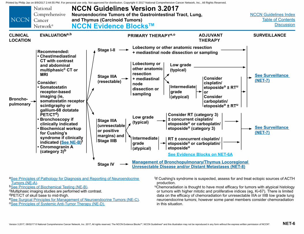
CLINICAL LOCATION
EVALUATIONa,b PRIMARY THERAPYe,o
Broncho-pulmonary
Recommended:• Chest/mediastinal
CT with contrast and abdominal multiphasicc CT or MRI
Consider:• Somatostatin
receptor-based imaging (ie, somatostatin receptor scintigraphy or gallium-68 dotatate PET/CTd)
• Bronchoscopy if clinically indicated
• Biochemical workup for Cushing’s syndrome if clinically indicated (See NE-B)t
• Chromogranin A (category 3)b
Stage I-II
Stage IIIA (resectable)
Stage IIIA (unresectable or positive margins) and Stage IIIB
Stage IV
Lobectomy or other anatomic resection + mediastinal node dissection or sampling
Lobectomy or other anatomic resection + mediastinal node dissection or sampling
Management of Bronchopulmonary/Thymus Locoregional Unresectable Disease and/or Distant Metastases (NET-8)
Consider cisplatin/etoposideo ± RTuor Consider carboplatin/etoposideo ± RTu
Low grade (typical)
Intermediate grade (atypical)
RT ± concurrent cisplatin/etoposideu or carboplatin/etoposideu
Consider RT (category 3) ± concurrent cisplatin/etoposideu or carboplatin/etoposideu (category 3)
SURVEILLANCEADJUVANT THERAPY
Low grade (typical)
Intermediate grade (atypical)
See Surveillance (NET-7)
See Surveillance (NET-7)
tIf Cushing's syndrome is suspected, assess for and treat ectopic sources of ACTH production.
uChemoradiation is thought to have most efficacy for tumors with atypical histology or tumors with higher mitotic and proliferative indices (eg, Ki-67). There is limited data on the efficacy of chemoradiation for unresectable IIIA or IIIB low grade lung neuroendocrine tumors; however some panel members consider chemoradiation in this situation.
NCCN Guidelines Version 3.2017Neuroendocrine Tumors of the Gastrointestinal Tract, Lung, and Thymus (Carcinoid Tumors)NCCN Evidence BlocksTM
See Evidence Blocks on NET-6A
Printed by Philip Jax on 8/9/2017 2:44:55 PM. For personal use only. Not approved for distribution. Copyright © 2017 National Comprehensive Cancer Network, Inc., All Rights Reserved.
NCCN Guidelines IndexTable of Contents
Discussion
Version 3.2017, 08/02/17 © National Comprehensive Cancer Network, Inc. 2017, All rights reserved. The NCCN Evidence BlocksTM, NCCN Guidelines® and this illustration may not be reproduced in any form without the express written permission of NCCN®. NET-6A
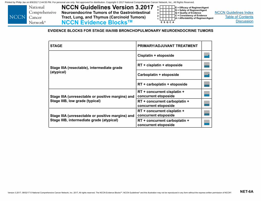
NCCN Guidelines Version 3.2017Neuroendocrine Tumors of the Gastrointestinal Tract, Lung, and Thymus (Carcinoid Tumors)NCCN Evidence BlocksTM E S Q C A
54321
E = Efficacy of Regimen/AgentS = Safety of Regimen/AgentQ = Quality of EvidenceC = Consistency of EvidenceA = Affordability of Regimen/Agent
EVIDENCE BLOCKS FOR STAGE IIIA/IIIB BRONCHOPULMONARY NEUROENDOCRINE TUMORS
STAGE PRIMARY/ADJUVANT TREATMENT
Stage IIIA (resectable), intermediate grade (atypical)
Cisplatin + etoposide
RT + cisplatin + etoposide
Carboplatin + etoposide
RT + carboplatin + etoposide
Stage IIIA (unresectable or positive margins) and Stage IIIB, low grade (typical)
RT + concurrent cisplatin + concurrent etoposideRT + concurrent carboplatin + concurrent etoposide
Stage IIIA (unresectable or positive margins) and Stage IIIB, intermediate grade (atypical)
RT + concurrent cisplatin + concurrent etoposideRT + concurrent carboplatin + concurrent etoposide
Printed by Philip Jax on 8/9/2017 2:44:55 PM. For personal use only. Not approved for distribution. Copyright © 2017 National Comprehensive Cancer Network, Inc., All Rights Reserved.
NCCN Guidelines IndexTable of Contents
Discussion
Version 3.2017, 08/02/17 © National Comprehensive Cancer Network, Inc. 2017, All rights reserved. The NCCN Evidence BlocksTM, NCCN Guidelines® and this illustration may not be reproduced in any form without the express written permission of NCCN®. NET-7
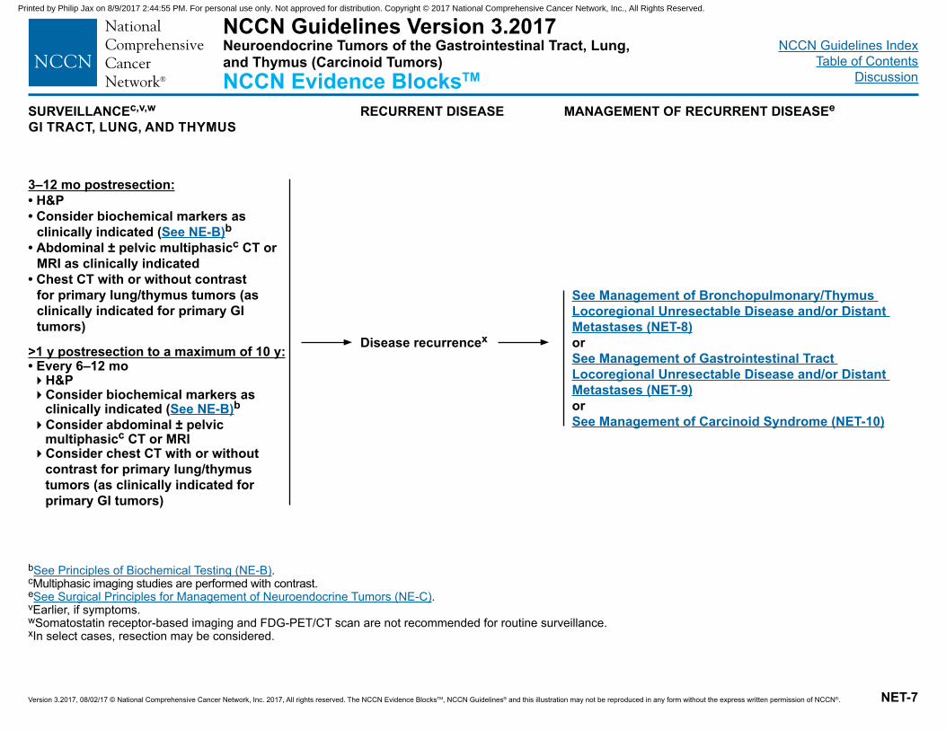
SURVEILLANCEc,v,w
GI TRACT, LUNG, AND THYMUSRECURRENT DISEASE MANAGEMENT OF RECURRENT DISEASEe
3–12 mo postresection:• H&P • Consider biochemical markers as
clinically indicated (See NE-B)b• Abdominal ± pelvic multiphasicc CT or
MRI as clinically indicated • Chest CT with or without contrast
for primary lung/thymus tumors (as clinically indicated for primary GI tumors)
>1 y postresection to a maximum of 10 y:• Every 6–12 mo�H&P�Consider biochemical markers as
clinically indicated (See NE-B)b �Consider abdominal ± pelvic
multiphasicc CT or MRI�Consider chest CT with or without
contrast for primary lung/thymus tumors (as clinically indicated for primary GI tumors)
bSee Principles of Biochemical Testing (NE-B).cMultiphasic imaging studies are performed with contrast.eSee Surgical Principles for Management of Neuroendocrine Tumors (NE-C).vEarlier, if symptoms.wSomatostatin receptor-based imaging and FDG-PET/CT scan are not recommended for routine surveillance.xIn select cases, resection may be considered.
See Management of Bronchopulmonary/Thymus Locoregional Unresectable Disease and/or Distant Metastases (NET-8)orSee Management of Gastrointestinal Tract Locoregional Unresectable Disease and/or Distant Metastases (NET-9)orSee Management of Carcinoid Syndrome (NET-10)
Disease recurrencex
NCCN Guidelines Version 3.2017Neuroendocrine Tumors of the Gastrointestinal Tract, Lung, and Thymus (Carcinoid Tumors)NCCN Evidence BlocksTM
Printed by Philip Jax on 8/9/2017 2:44:55 PM. For personal use only. Not approved for distribution. Copyright © 2017 National Comprehensive Cancer Network, Inc., All Rights Reserved.
NCCN Guidelines IndexTable of Contents
Discussion
Version 3.2017, 08/02/17 © National Comprehensive Cancer Network, Inc. 2017, All rights reserved. The NCCN Evidence BlocksTM, NCCN Guidelines® and this illustration may not be reproduced in any form without the express written permission of NCCN®. NET-8
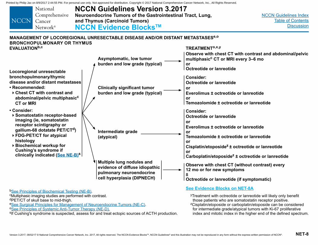
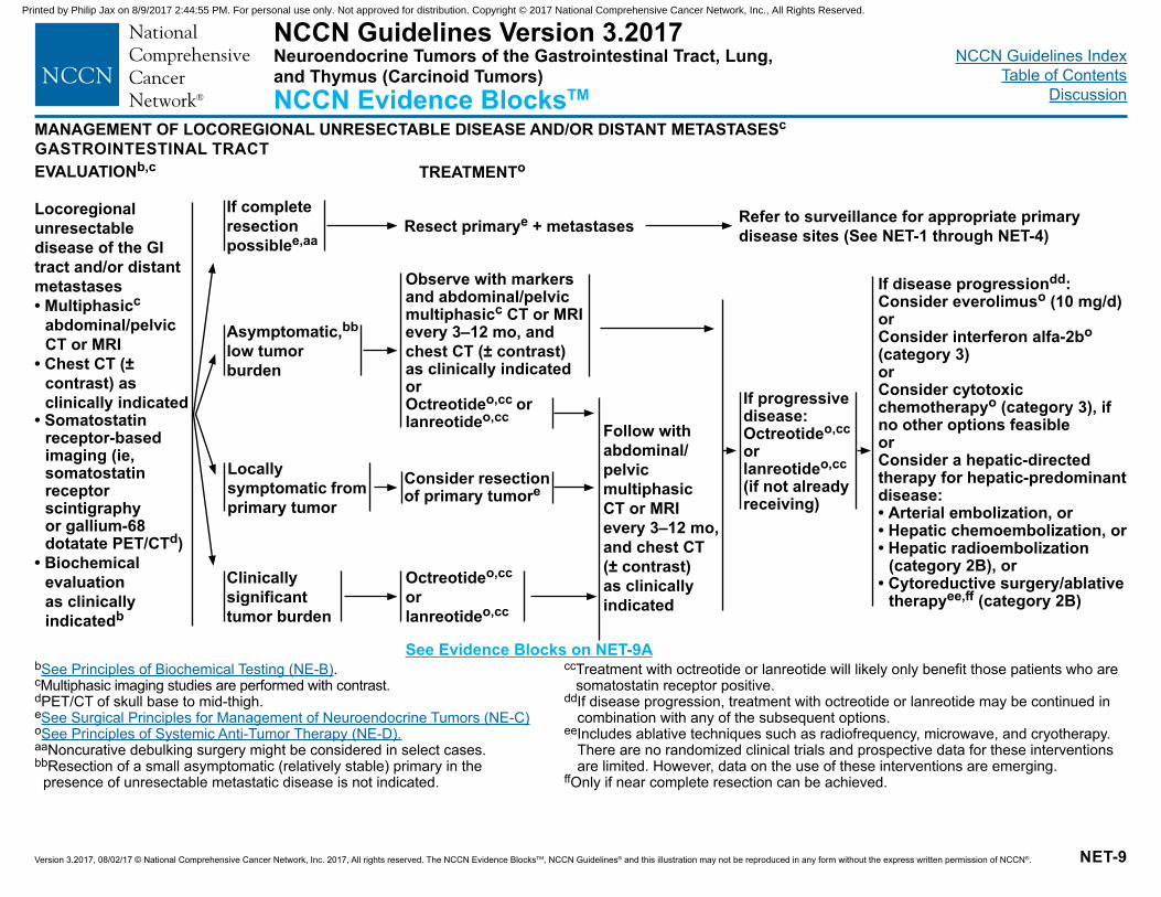
MANAGEMENT OF LOCOREGIONAL UNRESECTABLE DISEASE AND/OR DISTANT METASTASESe,o
BRONCHOPULMONARY OR THYMUS
Locoregional unresectable bronchopulmonary/thymic disease and/or distant metastases• Recommended: �Chest CT with contrast and
abdominal/pelvic multiphasicc CT or MRI
• Consider:�Somatostatin receptor-based
imaging (ie, somatostatin receptor scintigraphy or gallium-68 dotatate PET/CTd)�FDG-PET/CT for atypical
histology�Biochemical workup for
Cushing’s syndrome if clinically indicated (See NE-B)s
bSee Principles of Biochemical Testing (NE-B).cMultiphasic imaging studies are performed with contrast.dPET/CT of skull base to mid-thigh.eSee Surgical Principles for Management of Neuroendocrine Tumors (NE-C).oSee Principles of Systemic Anti-Tumor Therapy (NE-D).sIf Cushing's syndrome is suspected, assess for and treat ectopic sources of ACTH production.
Asymptomatic, low tumor burden and low grade (typical)
Observe with chest CT with contrast and abdominal/pelvic multiphasicc CT or MRI every 3–6 mo or Octreotide or lanreotide
Consider:Octreotide or lanreotideorEverolimus ± octreotide or lanreotideor Temozolomide ± octreotide or lanreotide
Consider:Octreotide or lanreotideorEverolimus ± octreotide or lanreotideor Temozolomide ± octreotide or lanreotideor Cisplatin/etoposidez ± octreotide or lanreotideor Carboplatin/etoposidez ± octreotide or lanreotide
Clinically significant tumor burden and low grade (typical)
Intermediate grade (atypical)
EVALUATIONb,c TREATMENTo,x,y
Multiple lung nodules and evidence of diffuse idiopathic pulmonary neuroendocrine cell hyperplasia (DIPNECH)
Observe with chest CT (without contrast) every 12 mo or for new symptoms ± Octreotide or lanreotide (if symptomatic)
yTreatment with octreotide or lanreotide will likely only benefit those patients who are somatostatin receptor positive.
zCisplatin/etoposide or carboplatin/etoposide can be considered for intermediate grade/atypical tumors with Ki-67 proliferative index and mitotic index in the higher end of the defined spectrum.
NCCN Guidelines Version 3.2017Neuroendocrine Tumors of the Gastrointestinal Tract, Lung, and Thymus (Carcinoid Tumors)NCCN Evidence BlocksTM
See Evidence Blocks on NET-8A
Printed by Philip Jax on 8/9/2017 2:44:55 PM. For personal use only. Not approved for distribution. Copyright © 2017 National Comprehensive Cancer Network, Inc., All Rights Reserved.
NCCN Guidelines IndexTable of Contents
Discussion
Version 3.2017, 08/02/17 © National Comprehensive Cancer Network, Inc. 2017, All rights reserved. The NCCN Evidence BlocksTM, NCCN Guidelines® and this illustration may not be reproduced in any form without the express written permission of NCCN®. NET-8A
NCCN Guidelines Version 3.2017Neuroendocrine Tumors of the Gastrointestinal Tract, Lung, and Thymus (Carcinoid Tumors)NCCN Evidence BlocksTM E S Q C A
54321
E = Efficacy of Regimen/AgentS = Safety of Regimen/AgentQ = Quality of EvidenceC = Consistency of EvidenceA = Affordability of Regimen/Agent
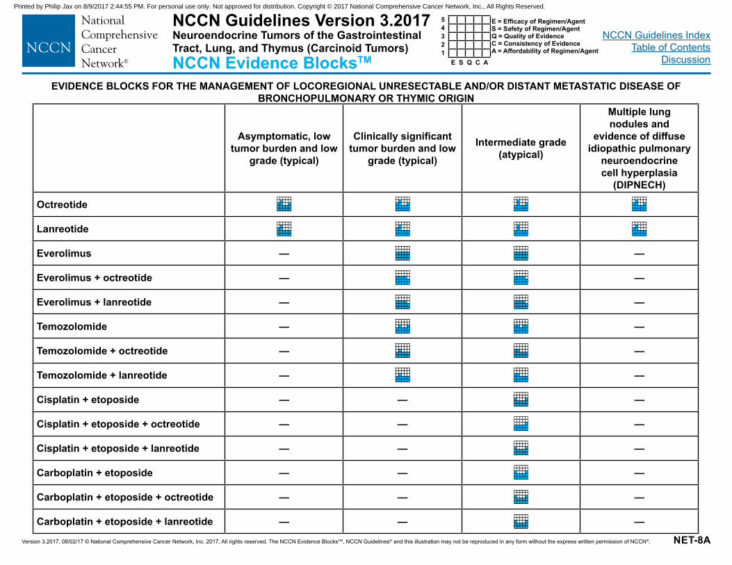
EVIDENCE BLOCKS FOR THE MANAGEMENT OF LOCOREGIONAL UNRESECTABLE AND/OR DISTANT METASTATIC DISEASE OF BRONCHOPULMONARY OR THYMIC ORIGIN
Asymptomatic, low tumor burden and low
grade (typical)
Clinically significant tumor burden and low
grade (typical)
Intermediate grade (atypical)
Multiple lung nodules and
evidence of diffuse idiopathic pulmonary
neuroendocrine cell hyperplasia
(DIPNECH)
Octreotide
Lanreotide
Everolimus — —
Everolimus + octreotide — —
Everolimus + lanreotide — —
Temozolomide — —
Temozolomide + octreotide — —
Temozolomide + lanreotide — —
Cisplatin + etoposide — — —
Cisplatin + etoposide + octreotide — — —
Cisplatin + etoposide + lanreotide — — —
Carboplatin + etoposide — — —
Carboplatin + etoposide + octreotide — — —
Carboplatin + etoposide + lanreotide — — —
Printed by Philip Jax on 8/9/2017 2:44:55 PM. For personal use only. Not approved for distribution. Copyright © 2017 National Comprehensive Cancer Network, Inc., All Rights Reserved.
NCCN Guidelines IndexTable of Contents
Discussion
Version 3.2017, 08/02/17 © National Comprehensive Cancer Network, Inc. 2017, All rights reserved. The NCCN Evidence BlocksTM, NCCN Guidelines® and this illustration may not be reproduced in any form without the express written permission of NCCN®. NET-9
MANAGEMENT OF LOCOREGIONAL UNRESECTABLE DISEASE AND/OR DISTANT METASTASESc
GASTROINTESTINAL TRACT
If complete resection possiblee,aa
Observe with markers and abdominal/pelvic multiphasicc CT or MRI every 3–12 mo, and chest CT (± contrast) as clinically indicatedor Octreotideo,cc or lanreotideo,cc
Octreotideo,cc or lanreotideo,cc
If disease progressiondd:Consider everolimuso (10 mg/d) orConsider interferon alfa-2bo (category 3) or Consider cytotoxic chemotherapyo (category 3), if no other options feasibleorConsider a hepatic-directed therapy for hepatic-predominant disease:• Arterial embolization, or• Hepatic chemoembolization, or• Hepatic radioembolization
(category 2B), or• Cytoreductive surgery/ablative
therapyee,ff (category 2B)
Locoregional unresectable disease of the GI tract and/or distant metastases• Multiphasicc
abdominal/pelvic CT or MRI
• Chest CT (± contrast) as clinically indicated
• Somatostatin receptor-based imaging (ie, somatostatin receptor scintigraphy or gallium-68 dotatate PET/CTd)
• Biochemical evaluation as clinically indicatedb
bSee Principles of Biochemical Testing (NE-B).cMultiphasic imaging studies are performed with contrast.dPET/CT of skull base to mid-thigh.eSee Surgical Principles for Management of Neuroendocrine Tumors (NE-C) oSee Principles of Systemic Anti-Tumor Therapy (NE-D).aaNoncurative debulking surgery might be considered in select cases.bbResection of a small asymptomatic (relatively stable) primary in the
presence of unresectable metastatic disease is not indicated.
cc Treatment with octreotide or lanreotide will likely only benefit those patients who are somatostatin receptor positive.
dd If disease progression, treatment with octreotide or lanreotide may be continued in combination with any of the subsequent options.
ee Includes ablative techniques such as radiofrequency, microwave, and cryotherapy. There are no randomized clinical trials and prospective data for these interventions are limited. However, data on the use of these interventions are emerging.
ffOnly if near complete resection can be achieved.
Asymptomatic,bb
low tumor burden
Locally symptomatic from primary tumor
Clinically significant tumor burden
Resect primarye + metastases
Follow with abdominal/pelvic multiphasic CT or MRI every 3–12 mo, and chest CT (± contrast) as clinically indicated
Consider resection of primary tumore
Refer to surveillance for appropriate primary disease sites (See NET-1 through NET-4)
If progressive disease: Octreotideo,cc or lanreotideo,cc (if not already receiving)
EVALUATIONb,c TREATMENTo
NCCN Guidelines Version 3.2017Neuroendocrine Tumors of the Gastrointestinal Tract, Lung, and Thymus (Carcinoid Tumors)NCCN Evidence BlocksTM
See Evidence Blocks on NET-9A
Printed by Philip Jax on 8/9/2017 2:44:55 PM. For personal use only. Not approved for distribution. Copyright © 2017 National Comprehensive Cancer Network, Inc., All Rights Reserved.
NCCN Guidelines IndexTable of Contents
Discussion
Version 3.2017, 08/02/17 © National Comprehensive Cancer Network, Inc. 2017, All rights reserved. The NCCN Evidence BlocksTM, NCCN Guidelines® and this illustration may not be reproduced in any form without the express written permission of NCCN®. NET-9A
NCCN Guidelines Version 3.2017Neuroendocrine Tumors of the Gastrointestinal Tract, Lung, and Thymus (Carcinoid Tumors)NCCN Evidence BlocksTM E S Q C A
54321
E = Efficacy of Regimen/AgentS = Safety of Regimen/AgentQ = Quality of EvidenceC = Consistency of EvidenceA = Affordability of Regimen/Agent
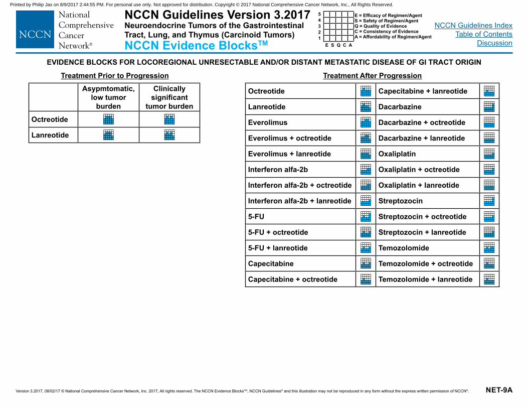
EVIDENCE BLOCKS FOR LOCOREGIONAL UNRESECTABLE AND/OR DISTANT METASTATIC DISEASE OF GI TRACT ORIGIN
Treatment Prior to Progression Treatment After ProgressionAsypmtomatic,
low tumor burden
Clinically significant
tumor burden
Octreotide
Lanreotide
Octreotide Capecitabine + lanreotide
Lanreotide Dacarbazine
Everolimus Dacarbazine + octreotide
Everolimus + octreotide Dacarbazine + lanreotide
Everolimus + lanreotide Oxaliplatin
Interferon alfa-2b Oxaliplatin + octreotide
Interferon alfa-2b + octreotide Oxaliplatin + lanreotide
Interferon alfa-2b + lanreotide Streptozocin
5-FU Streptozocin + octreotide
5-FU + octreotide Streptozocin + lanreotide
5-FU + lanreotide Temozolomide
Capecitabine Temozolomide + octreotide
Capecitabine + octreotide Temozolomide + lanreotide
Printed by Philip Jax on 8/9/2017 2:44:55 PM. For personal use only. Not approved for distribution. Copyright © 2017 National Comprehensive Cancer Network, Inc., All Rights Reserved.
NCCN Guidelines IndexTable of Contents
Discussion
Version 3.2017, 08/02/17 © National Comprehensive Cancer Network, Inc. 2017, All rights reserved. The NCCN Evidence BlocksTM, NCCN Guidelines® and this illustration may not be reproduced in any form without the express written permission of NCCN®. NET-10
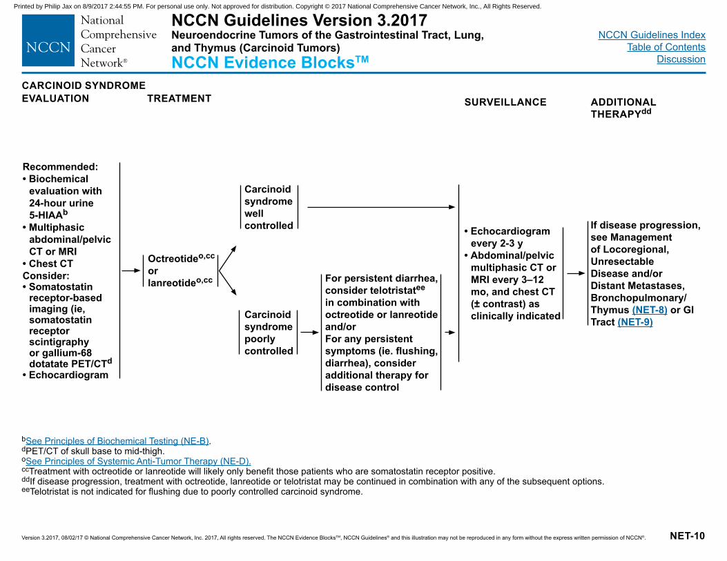
CARCINOID SYNDROMEEVALUATION SURVEILLANCE ADDITIONAL
THERAPYddTREATMENT
Octreotideo,cc or lanreotideo,cc For persistent diarrhea,
consider telotristatee
in combination with octreotide or lanreotideand/orFor any persistent symptoms (ie. flushing, diarrhea), consider additional therapy for disease control
If disease progression, see Management of Locoregional, Unresectable Disease and/or Distant Metastases, Bronchopulmonary/Thymus (NET-8) or GI Tract (NET-9)
bSee Principles of Biochemical Testing (NE-B).dPET/CT of skull base to mid-thigh. oSee Principles of Systemic Anti-Tumor Therapy (NE-D).cc Treatment with octreotide or lanreotide will likely only benefit those patients who are somatostatin receptor positive.dd If disease progression, treatment with octreotide, lanreotide or telotristat may be continued in combination with any of the subsequent options.eeTelotristat is not indicated for flushing due to poorly controlled carcinoid syndrome.
Recommended:• Biochemical
evaluation with 24-hour urine 5-HIAAb
• Multiphasic abdominal/pelvic CT or MRI
• Chest CTConsider:• Somatostatin
receptor-based imaging (ie, somatostatin receptor scintigraphy or gallium-68 dotatate PET/CTd
• Echocardiogram
Carcinoid syndrome well controlled
Carcinoid syndrome poorly controlled
• Echocardiogram every 2-3 y
• Abdominal/pelvic multiphasic CT or MRI every 3–12 mo, and chest CT (± contrast) as clinically indicated
NCCN Guidelines Version 3.2017Neuroendocrine Tumors of the Gastrointestinal Tract, Lung, and Thymus (Carcinoid Tumors)NCCN Evidence BlocksTM
Printed by Philip Jax on 8/9/2017 2:44:55 PM. For personal use only. Not approved for distribution. Copyright © 2017 National Comprehensive Cancer Network, Inc., All Rights Reserved.
NCCN Guidelines IndexTable of Contents
Discussion
Version 3.2017, 08/02/17 © National Comprehensive Cancer Network, Inc. 2017, All rights reserved. The NCCN Evidence BlocksTM, NCCN Guidelines® and this illustration may not be reproduced in any form without the express written permission of NCCN®.
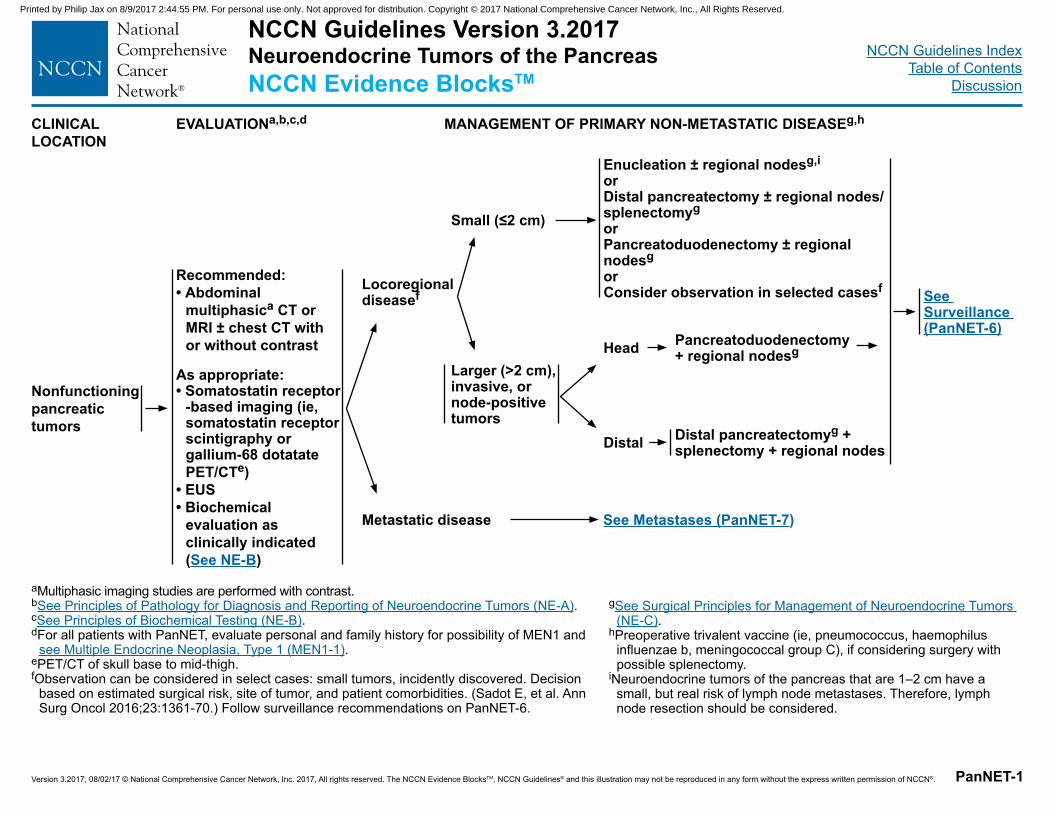
NCCN Guidelines Version 3.2017 Neuroendocrine Tumors of the PancreasNCCN Evidence BlocksTM
PanNET-1
CLINICAL LOCATION
EVALUATIONa,b,c,d MANAGEMENT OF PRIMARY NON-METASTATIC DISEASEg,h
Nonfunctioning pancreatic tumors
Recommended:• Abdominal
multiphasica CT or MRI ± chest CT with or without contrast
As appropriate:• Somatostatin receptor
-based imaging (ie, somatostatin receptor scintigraphy or gallium-68 dotatate PET/CTe)
• EUS• Biochemical
evaluation as clinically indicated (See NE-B)
aMultiphasic imaging studies are performed with contrast.bSee Principles of Pathology for Diagnosis and Reporting of Neuroendocrine Tumors (NE-A).cSee Principles of Biochemical Testing (NE-B).dFor all patients with PanNET, evaluate personal and family history for possibility of MEN1 and
see Multiple Endocrine Neoplasia, Type 1 (MEN1-1).ePET/CT of skull base to mid-thigh.fObservation can be considered in select cases: small tumors, incidently discovered. Decision
based on estimated surgical risk, site of tumor, and patient comorbidities. (Sadot E, et al. Ann Surg Oncol 2016;23:1361-70.) Follow surveillance recommendations on PanNET-6.
Locoregional diseasef
Metastatic disease
Small (≤2 cm)
Larger (>2 cm), invasive, or node-positive tumors
Distal
Enucleation ± regional nodesg,i orDistal pancreatectomy ± regional nodes/splenectomygorPancreatoduodenectomy ± regional nodesgorConsider observation in selected casesf See
Surveillance (PanNET-6)
Head
See Metastases (PanNET-7)
Pancreatoduodenectomy + regional nodesg
Distal pancreatectomyg + splenectomy + regional nodes
gSee Surgical Principles for Management of Neuroendocrine Tumors (NE-C).
hPreoperative trivalent vaccine (ie, pneumococcus, haemophilus influenzae b, meningococcal group C), if considering surgery with possible splenectomy.
iNeuroendocrine tumors of the pancreas that are 1–2 cm have a small, but real risk of lymph node metastases. Therefore, lymph node resection should be considered.
Printed by Philip Jax on 8/9/2017 2:44:55 PM. For personal use only. Not approved for distribution. Copyright © 2017 National Comprehensive Cancer Network, Inc., All Rights Reserved.
NCCN Guidelines IndexTable of Contents
Discussion
Version 3.2017, 08/02/17 © National Comprehensive Cancer Network, Inc. 2017, All rights reserved. The NCCN Evidence BlocksTM, NCCN Guidelines® and this illustration may not be reproduced in any form without the express written permission of NCCN®. PanNET-2
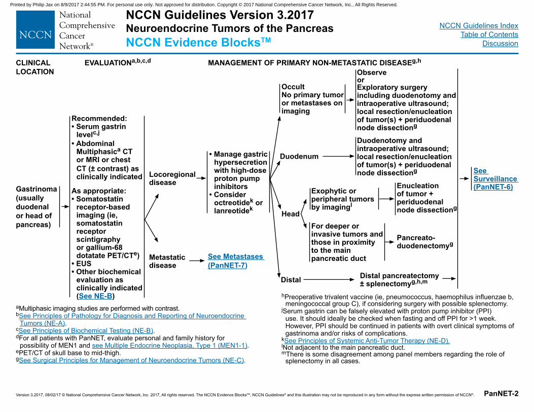
CLINICAL LOCATION
EVALUATIONa,b,c,d MANAGEMENT OF PRIMARY NON-METASTATIC DISEASEg,h
Gastrinoma (usually duodenal or head of pancreas)
Recommended:• Serum gastrin
levelc,j
• Abdominal Multiphasica CT or MRI or chest CT (± contrast) as clinically indicated
As appropriate:• Somatostatin
receptor-based imaging (ie, somatostatin receptor scintigraphy or gallium-68 dotatate PET/CTe)
• EUS • Other biochemical
evaluation as clinically indicated (See NE-B)
aMultiphasic imaging studies are performed with contrast.bSee Principles of Pathology for Diagnosis and Reporting of Neuroendocrine
Tumors (NE-A).cSee Principles of Biochemical Testing (NE-B).dFor all patients with PanNET, evaluate personal and family history for
possibility of MEN1 and see Multiple Endocrine Neoplasia, Type 1 (MEN1-1).ePET/CT of skull base to mid-thigh.gSee Surgical Principles for Management of Neuroendocrine Tumors (NE-C).
Locoregional disease
Metastatic disease
Distal
ObserveorExploratory surgery including duodenotomy and intraoperative ultrasound; local resection/enucleation of tumor(s) + periduodenal node dissectiong
See Surveillance (PanNET-6)
Head
See Metastases (PanNET-7)
Exophytic or peripheral tumors by imagingl
For deeper or invasive tumors and those in proximity to the main pancreatic duct
• Manage gastric hypersecretion with high-dose proton pump inhibitors
• Consider octreotidek or lanreotidek
Duodenum
Occult No primary tumor or metastases on imaging
Distal pancreatectomy ± splenectomyg,h,m
Pancreato-duodenectomyg
Enucleation of tumor + periduodenal node dissectiong
Duodenotomy and intraoperative ultrasound; local resection/enucleation of tumor(s) + periduodenal node dissectiong
hPreoperative trivalent vaccine (ie, pneumococcus, haemophilus influenzae b, meningococcal group C), if considering surgery with possible splenectomy.
jSerum gastrin can be falsely elevated with proton pump inhibitor (PPI) use. It should ideally be checked when fasting and off PPI for >1 week. However, PPI should be continued in patients with overt clinical symptoms of gastrinoma and/or risks of complications.
kSee Principles of Systemic Anti-Tumor Therapy (NE-D).lNot adjacent to the main pancreatic duct.mThere is some disagreement among panel members regarding the role of
splenectomy in all cases.
NCCN Guidelines Version 3.2017 Neuroendocrine Tumors of the PancreasNCCN Evidence BlocksTM
Printed by Philip Jax on 8/9/2017 2:44:55 PM. For personal use only. Not approved for distribution. Copyright © 2017 National Comprehensive Cancer Network, Inc., All Rights Reserved.
NCCN Guidelines IndexTable of Contents
Discussion
Version 3.2017, 08/02/17 © National Comprehensive Cancer Network, Inc. 2017, All rights reserved. The NCCN Evidence BlocksTM, NCCN Guidelines® and this illustration may not be reproduced in any form without the express written permission of NCCN®. PanNET-3
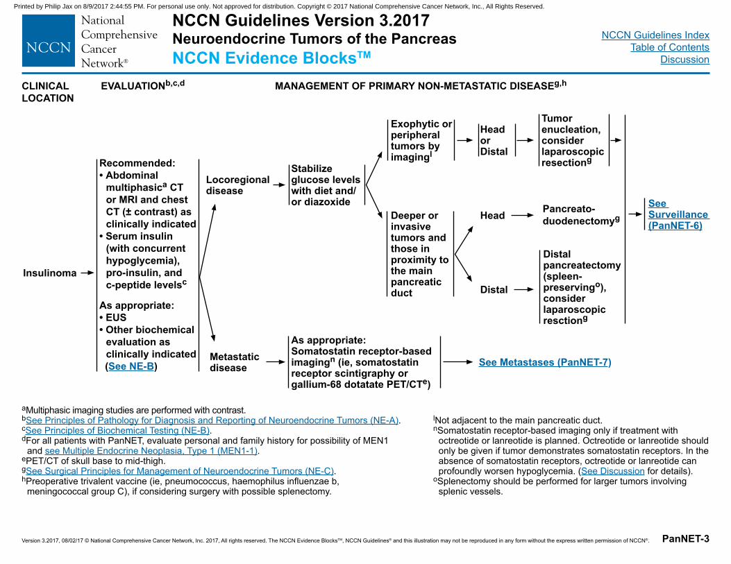
CLINICAL LOCATION
EVALUATIONb,c,d MANAGEMENT OF PRIMARY NON-METASTATIC DISEASEg,h
Insulinoma
Recommended:• Abdominal
multiphasica CT or MRI and chest CT (± contrast) as clinically indicated
• Serum insulin (with concurrent hypoglycemia), pro-insulin, and c-peptide levelsc
As appropriate: • EUS• Other biochemical
evaluation as clinically indicated
(See NE-B)
aMultiphasic imaging studies are performed with contrast.bSee Principles of Pathology for Diagnosis and Reporting of Neuroendocrine Tumors (NE-A).cSee Principles of Biochemical Testing (NE-B).dFor all patients with PanNET, evaluate personal and family history for possibility of MEN1
and see Multiple Endocrine Neoplasia, Type 1 (MEN1-1).ePET/CT of skull base to mid-thigh.gSee Surgical Principles for Management of Neuroendocrine Tumors (NE-C).hPreoperative trivalent vaccine (ie, pneumococcus, haemophilus influenzae b,
meningococcal group C), if considering surgery with possible splenectomy.
Locoregional disease
Metastatic disease
Distal
Tumor enucleation, consider laparoscopic resectiong
See Surveillance (PanNET-6)
Head
As appropriate:Somatostatin receptor-based imagingn (ie, somatostatin receptor scintigraphy or gallium-68 dotatate PET/CTe)
Pancreato-duodenectomyg
Distal pancreatectomy (spleen-preservingo), consider laparoscopic resctiong
lNot adjacent to the main pancreatic duct.nSomatostatin receptor-based imaging only if treatment with
octreotide or lanreotide is planned. Octreotide or lanreotide should only be given if tumor demonstrates somatostatin receptors. In the absence of somatostatin receptors, octreotide or lanreotide can profoundly worsen hypoglycemia. (See Discussion for details).
oSplenectomy should be performed for larger tumors involving splenic vessels.
See Metastases (PanNET-7)
Exophytic or peripheral tumors by imagingl
Stabilize glucose levels with diet and/or diazoxide
Deeper or invasive tumors and those in proximity to the main pancreatic duct
Head or Distal
NCCN Guidelines Version 3.2017 Neuroendocrine Tumors of the PancreasNCCN Evidence BlocksTM
Printed by Philip Jax on 8/9/2017 2:44:55 PM. For personal use only. Not approved for distribution. Copyright © 2017 National Comprehensive Cancer Network, Inc., All Rights Reserved.
NCCN Guidelines IndexTable of Contents
Discussion
Version 3.2017, 08/02/17 © National Comprehensive Cancer Network, Inc. 2017, All rights reserved. The NCCN Evidence BlocksTM, NCCN Guidelines® and this illustration may not be reproduced in any form without the express written permission of NCCN®. PanNET-4
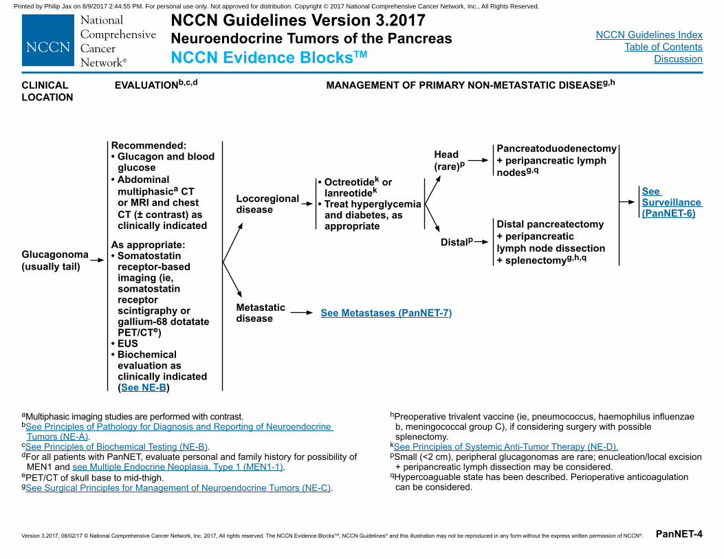
CLINICAL LOCATION
EVALUATIONb,c,d MANAGEMENT OF PRIMARY NON-METASTATIC DISEASEg,h
Glucagonoma(usually tail)
Recommended:• Glucagon and blood
glucose• Abdominal
multiphasica CT or MRI and chest CT (± contrast) as clinically indicated
As appropriate:• Somatostatin
receptor-based imaging (ie, somatostatin receptor scintigraphy or gallium-68 dotatate PET/CTe)
• EUS• Biochemical
evaluation as clinically indicated (See NE-B)
aMultiphasic imaging studies are performed with contrast.bSee Principles of Pathology for Diagnosis and Reporting of Neuroendocrine
Tumors (NE-A).cSee Principles of Biochemical Testing (NE-B).dFor all patients with PanNET, evaluate personal and family history for possibility of
MEN1 and see Multiple Endocrine Neoplasia, Type 1 (MEN1-1).ePET/CT of skull base to mid-thigh.gSee Surgical Principles for Management of Neuroendocrine Tumors (NE-C).
Locoregional disease
Metastatic disease
Distalp
See Surveillance (PanNET-6)
Head (rare)p
Pancreatoduodenectomy + peripancreatic lymph nodesg,q
Distal pancreatectomy
+ peripancreatic lymph node dissection + splenectomyg,h,q
See Metastases (PanNET-7)
• Octreotidek or lanreotidek
• Treat hyperglycemia and diabetes, as appropriate
hPreoperative trivalent vaccine (ie, pneumococcus, haemophilus influenzae b, meningococcal group C), if considering surgery with possible splenectomy.
kSee Principles of Systemic Anti-Tumor Therapy (NE-D).pSmall (<2 cm), peripheral glucagonomas are rare; enucleation/local excision
+ peripancreatic lymph dissection may be considered.qHypercoaguable state has been described. Perioperative anticoagulation
can be considered.
NCCN Guidelines Version 3.2017 Neuroendocrine Tumors of the PancreasNCCN Evidence BlocksTM
Printed by Philip Jax on 8/9/2017 2:44:55 PM. For personal use only. Not approved for distribution. Copyright © 2017 National Comprehensive Cancer Network, Inc., All Rights Reserved.
NCCN Guidelines IndexTable of Contents
Discussion
Version 3.2017, 08/02/17 © National Comprehensive Cancer Network, Inc. 2017, All rights reserved. The NCCN Evidence BlocksTM, NCCN Guidelines® and this illustration may not be reproduced in any form without the express written permission of NCCN®. PanNET-5
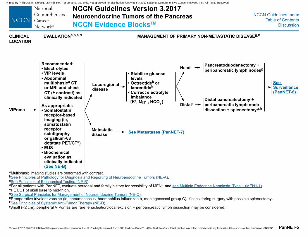
CLINICAL LOCATION
EVALUATIONa,b,c,d MANAGEMENT OF PRIMARY NON-METASTATIC DISEASEg,h
VIPoma
Recommended:• Electrolytes• VIP levels• Abdominal
multiphasica CT or MRI and chest CT (± contrast) as clinically indicated
As appropriate:• Somatostatin
receptor-based imaging (ie, somatostatin receptor scintigraphy or gallium-68 dotatate PET/CTe)
• EUS• Biochemical
evaluation as clinically indicated (See NE-B)
aMultiphasic imaging studies are performed with contrast.bSee Principles of Pathology for Diagnosis and Reporting of Neuroendocrine Tumors (NE-A).cSee Principles of Biochemical Testing (NE-B).dFor all patients with PanNET, evaluate personal and family history for possibility of MEN1 and see Multiple Endocrine Neoplasia, Type 1 (MEN1-1).ePET/CT of skull base to mid-thigh.gSee Surgical Principles for Management of Neuroendocrine Tumors (NE-C). hPreoperative trivalent vaccine (ie, pneumococcus, haemophilus influenzae b, meningococcal group C), if considering surgery with possible splenectomy.kSee Principles of Systemic Anti-Tumor Therapy (NE-D).rSmall (<2 cm), peripheral VIPomas are rare; enucleation/local excision + peripancreatic lymph dissection may be considered.
Locoregional disease
Metastatic disease
Distalr
See Surveillance (PanNET-6)
Headr Pancreatoduodenectomy + peripancreatic lymph nodesg
Distal pancreatectomy + peripancreatic lymph node dissection + splenectomyg,h
See Metastases (PanNET-7)
• Stabilize glucose levels
• Octreotidek or lanreotidek
• Correct electrolyte imbalance
(K+, Mg2+, HCO3-)
NCCN Guidelines Version 3.2017 Neuroendocrine Tumors of the PancreasNCCN Evidence BlocksTM
Printed by Philip Jax on 8/9/2017 2:44:55 PM. For personal use only. Not approved for distribution. Copyright © 2017 National Comprehensive Cancer Network, Inc., All Rights Reserved.
NCCN Guidelines IndexTable of Contents
Discussion
Version 3.2017, 08/02/17 © National Comprehensive Cancer Network, Inc. 2017, All rights reserved. The NCCN Evidence BlocksTM, NCCN Guidelines® and this illustration may not be reproduced in any form without the express written permission of NCCN®. PanNET-6
SURVEILLANCEs,t,u RECURRENT DISEASE MANAGEMENT OF RECURRENT DISEASEg
3–12 mo postresection:• H&P • Consider biochemical markers as
clinically indicatedc
• Abdominal multiphasica CT or MRI and chest CT (± contrast) as clinically indicated
>1 y postresection to a maximum of 10 y:• Every 6–12 mo�H&P�Consider biochemical markers as
clinically indicatedc �Consider abdominal multiphasica CT
or MRI and chest CT (± contrast) as clinically indicated
aMultiphasic imaging studies are performed with contrast.cSee Principles of Biochemical Testing (NE-B).gSee Surgical Principles for Management of Neuroendocrine Tumors (NE-C). sEarlier, if symptoms.tSomatostatin receptor-based imaging and FDG-PET/CT scan are not recommended for routine surveillance.uSurveillance recommendations also apply to cases where observation has been chosen.vIn select cases, resection may be considered.
See Management of Locoregional Unresectable Disease and/or Distant Metastases (PanNET-7) Disease recurrencev
NCCN Guidelines Version 3.2017 Neuroendocrine Tumors of the PancreasNCCN Evidence BlocksTM
Printed by Philip Jax on 8/9/2017 2:44:55 PM. For personal use only. Not approved for distribution. Copyright © 2017 National Comprehensive Cancer Network, Inc., All Rights Reserved.
NCCN Guidelines IndexTable of Contents
Discussion
Version 3.2017, 08/02/17 © National Comprehensive Cancer Network, Inc. 2017, All rights reserved. The NCCN Evidence BlocksTM, NCCN Guidelines® and this illustration may not be reproduced in any form without the express written permission of NCCN®. PanNET-7
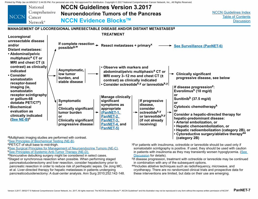
MANAGEMENT OF LOCOREGIONAL UNRESECTABLE DISEASE AND/OR DISTANT METASTASESg
Locoregional unresectable disease and/or Distant metastases:• Abdominal/pelvic
multiphasica CT or MRI and chest CT (± contrast) as clinically indicated
• Consider somatostatin receptor-based imaging (ie, somatostatin receptor scintigraphy or gallium-68 dotatate PET/CTe)
• Biochemical evaluation as clinically indicated (See NE-B)c
aMultiphasic imaging studies are performed with contrast.cSee Principles of Biochemical Testing (NE-B).ePET/CT of skull base to mid-thigh.gSee Surgical Principles for Management of Neuroendocrine Tumors (NE-C).kSee Principles of Systemic Anti-Tumor Therapy (NE-D). wNoncurative debulking surgery might be considered in select cases.xStaged or synchronous resection when possible. When performing staged
pancreatoduodenectomy and liver resection, consider hepatectomy prior to pancreatic resection in order to reduce risk of perihepatic sepsis. De Jong MC, et al. Liver-directed therapy for hepatic metastases in patients undergoing pancreaticoduodenectomy: A dual-center analysis. Ann Surg 2010;252:142-148.
• Observe with markers and abdominal/pelvic multiphasica CT or MRI every 3–12 mo and chest CT (± contrast) as clinically indicated
• Consider octreotidek,y or lanreotidek,y
Resect metastases + primaryx See Surveillance (PanNET-6)
Clinically significant progressive disease, see below
Manage clinically significant symptoms as appropriate (PanNET-1, PanNET-2, PanNET-3, PanNET-4, and PanNET-5)
If complete resection possibleg,w
Asymptomatic, low tumor burden, and stable disease
Symptomatic or Clinically significant tumor burden or Clinically significant progressive disease
If disease progressionz:Everolimusk (10 mg/d)orSunitinibk (37.5 mg/d)orCytotoxic chemotherapyk orConsider a hepatic-directed therapy for hepatic-predominant disease:• Arterial embolization, or • Hepatic chemoembolization, or• Hepatic radioembolization (category 2B), or• Cytoreductive surgery/ablative therapyaa
(category 2B)yFor patients with insulinoma, octreotide or lanreotide should be used only if
somatostatin scintigraphy is positive. If used, they should be used with caution in patients with insulinoma as they may transiently worsen hypoglycemia. (See Discussion for details).
zIf disease progression, treatment with octreotide or lanreotide may be continued in combination with any of the subsequent options.
aaIncludes ablative techniques such as radiofrequency, microwave, and cryotherapy. There are no randomized clinical trials and prospective data for these interventions are limited, but data on their use are emerging.
If progressive disease, consider octreotidek,y or lanreotidek,y (if not already receiving)
TREATMENT
NCCN Guidelines Version 3.2017 Neuroendocrine Tumors of the PancreasNCCN Evidence BlocksTM
Printed by Philip Jax on 8/9/2017 2:44:55 PM. For personal use only. Not approved for distribution. Copyright © 2017 National Comprehensive Cancer Network, Inc., All Rights Reserved.
NCCN Guidelines IndexTable of Contents
Discussion
Version 3.2017, 08/02/17 © National Comprehensive Cancer Network, Inc. 2017, All rights reserved. The NCCN Evidence BlocksTM, NCCN Guidelines® and this illustration may not be reproduced in any form without the express written permission of NCCN®.
NCCN Guidelines Version 3.2017 Neuroendocrine Tumors of Unknown PrimaryNCCN Evidence BlocksTM
NUP-1
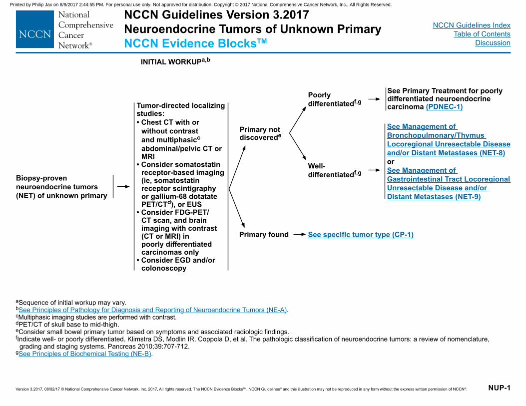
INITIAL WORKUPa,b
Tumor-directed localizing studies:• Chest CT with or
without contrast and multiphasicc abdominal/pelvic CT or MRI
• Consider somatostatin receptor-based imaging (ie, somatostatin receptor scintigraphy or gallium-68 dotatate PET/CTd), or EUS
• Consider FDG-PET/CT scan, and brain imaging with contrast (CT or MRI) in poorly differentiated carcinomas only
• Consider EGD and/or colonoscopy
aSequence of initial workup may vary.bSee Principles of Pathology for Diagnosis and Reporting of Neuroendocrine Tumors (NE-A).cMultiphasic imaging studies are performed with contrast.dPET/CT of skull base to mid-thigh.eConsider small bowel primary tumor based on symptoms and associated radiologic findings. fIndicate well- or poorly differentiated. Klimstra DS, Modlin IR, Coppola D, et al. The pathologic classification of neuroendocrine tumors: a review of nomenclature,
grading and staging systems. Pancreas 2010;39:707-712.gSee Principles of Biochemical Testing (NE-B).
Primary not discoverede
Primary found
Well- differentiatedf,g
Poorly differentiatedf,g
See Primary Treatment for poorly differentiated neuroendocrine carcinoma (PDNEC-1)
See Management of Bronchopulmonary/Thymus Locoregional Unresectable Disease and/or Distant Metastases (NET-8)orSee Management of Gastrointestinal Tract Locoregional Unresectable Disease and/or Distant Metastases (NET-9)
See specific tumor type (CP-1)
Biopsy-proven neuroendocrine tumors (NET) of unknown primary
Printed by Philip Jax on 8/9/2017 2:44:55 PM. For personal use only. Not approved for distribution. Copyright © 2017 National Comprehensive Cancer Network, Inc., All Rights Reserved.
NCCN Guidelines IndexTable of Contents
Discussion
Version 3.2017, 08/02/17 © National Comprehensive Cancer Network, Inc. 2017, All rights reserved. The NCCN Evidence BlocksTM, NCCN Guidelines® and this illustration may not be reproduced in any form without the express written permission of NCCN®.
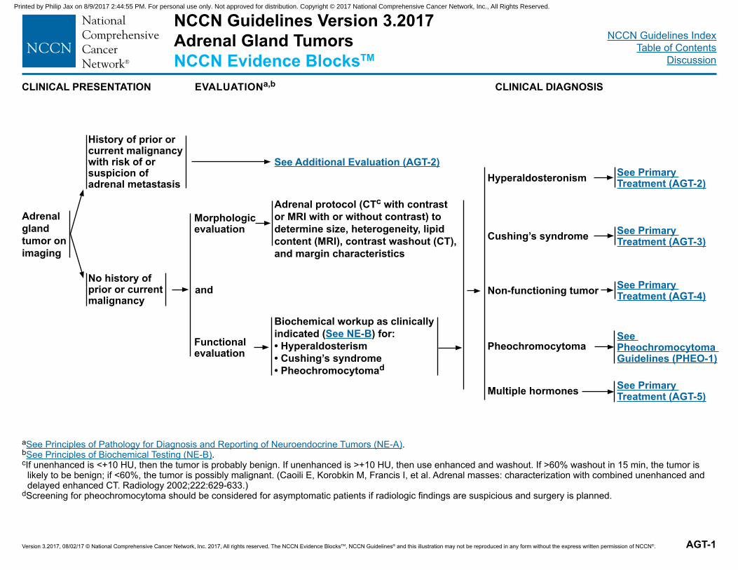
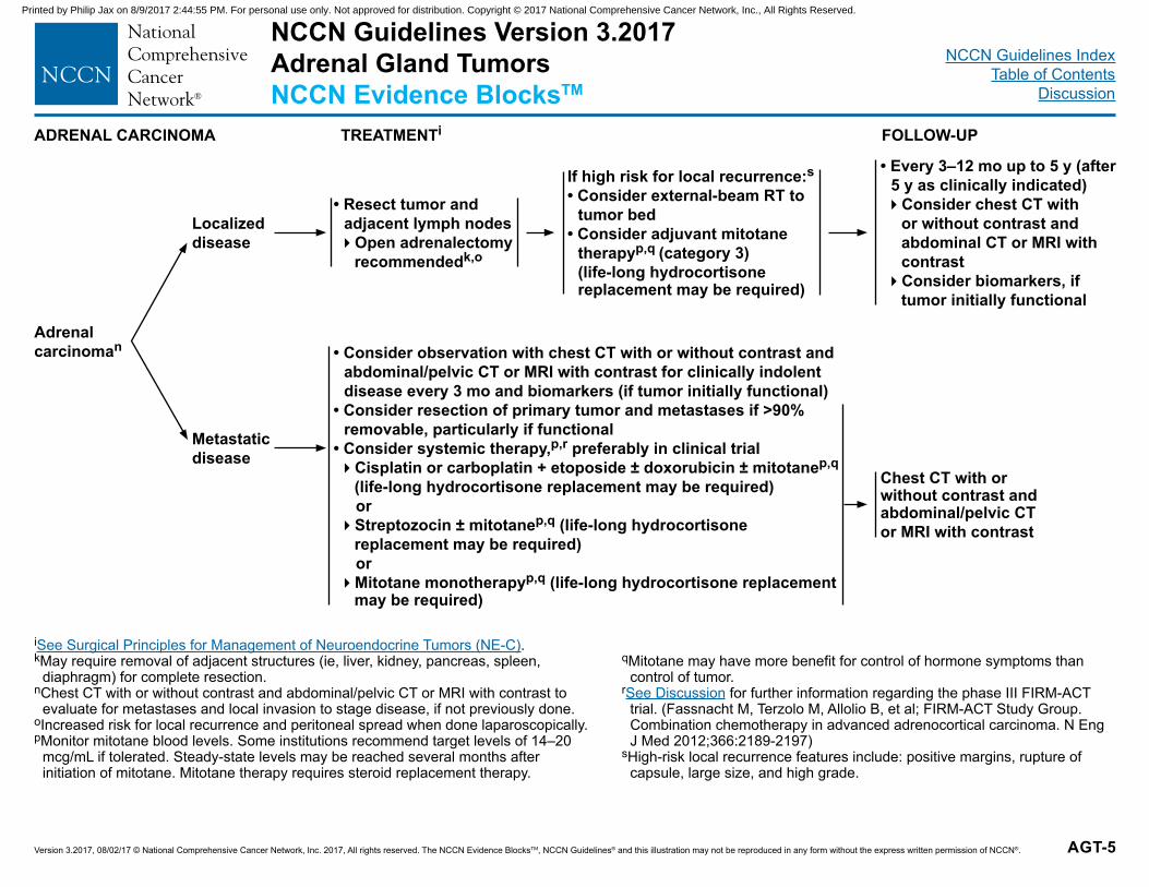
NCCN Guidelines Version 3.2017 Adrenal Gland TumorsNCCN Evidence BlocksTM
AGT-1
EVALUATIONa,b
Adrenal gland tumor on imaging
aSee Principles of Pathology for Diagnosis and Reporting of Neuroendocrine Tumors (NE-A).bSee Principles of Biochemical Testing (NE-B).cIf unenhanced is <+10 HU, then the tumor is probably benign. If unenhanced is >+10 HU, then use enhanced and washout. If >60% washout in 15 min, the tumor is
likely to be benign; if <60%, the tumor is possibly malignant. (Caoili E, Korobkin M, Francis I, et al. Adrenal masses: characterization with combined unenhanced and delayed enhanced CT. Radiology 2002;222:629-633.)
dScreening for pheochromocytoma should be considered for asymptomatic patients if radiologic findings are suspicious and surgery is planned.
Morphologic evaluation
See Additional Evaluation (AGT-2)Hyperaldosteronism
Cushing’s syndrome
Biochemical workup as clinically indicated (See NE-B) for:• Hyperaldosterism• Cushing’s syndrome• Pheochromocytomad
History of prior or current malignancy with risk of or suspicion of adrenal metastasis
No history of prior or current malignancy
Functional evaluation Pheochromocytoma
Multiple hormones
and
Adrenal protocol (CTc with contrast or MRI with or without contrast) to determine size, heterogeneity, lipid content (MRI), contrast washout (CT), and margin characteristics
Non-functioning tumor
See Primary Treatment (AGT-2)
See Pheochromocytoma Guidelines (PHEO-1)
See Primary Treatment (AGT-5)
See Primary Treatment (AGT-4)
See Primary Treatment (AGT-3)
CLINICAL PRESENTATION CLINICAL DIAGNOSIS
Printed by Philip Jax on 8/9/2017 2:44:55 PM. For personal use only. Not approved for distribution. Copyright © 2017 National Comprehensive Cancer Network, Inc., All Rights Reserved.
NCCN Guidelines IndexTable of Contents
Discussion
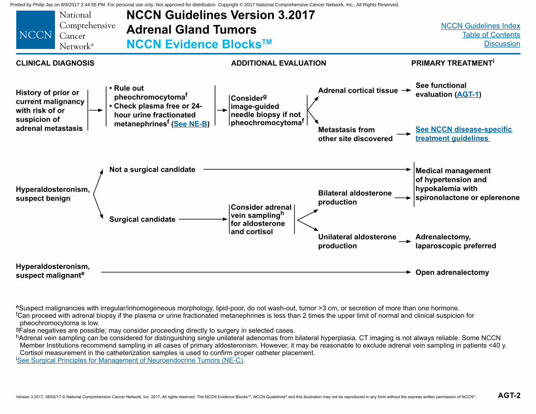
Version 3.2017, 08/02/17 © National Comprehensive Cancer Network, Inc. 2017, All rights reserved. The NCCN Evidence BlocksTM, NCCN Guidelines® and this illustration may not be reproduced in any form without the express written permission of NCCN®. AGT-2
PRIMARY TREATMENTi
• Rule out pheochromocytomaf
• Check plasma free or 24-hour urine fractionated metanephrinesf (See NE-B)
eSuspect malignancies with irregular/inhomogeneous morphology, lipid-poor, do not wash-out, tumor >3 cm, or secretion of more than one hormone. fCan proceed with adrenal biopsy if the plasma or urine fractionated metanephrines is less than 2 times the upper limit of normal and clinical suspicion for
pheochromocytoma is low.gFalse negatives are possible; may consider proceeding directly to surgery in selected cases. hAdrenal vein sampling can be considered for distinguishing single unilateral adenomas from bilateral hyperplasia. CT imaging is not always reliable. Some NCCN
Member Institutions recommend sampling in all cases of primary aldosteronism. However, it may be reasonable to exclude adrenal vein sampling in patients <40 y. Cortisol measurement in the catheterization samples is used to confirm proper catheter placement.
iSee Surgical Principles for Management of Neuroendocrine Tumors (NE-C).
Considergimage-guided needle biopsy if not pheochromocytomaf
Metastasis from other site discovered
Adrenal cortical tissue See functional evaluation (AGT-1)
See NCCN disease-specific treatment guidelines
History of prior or current malignancy with risk of or suspicion of adrenal metastasis
CLINICAL DIAGNOSIS ADDITIONAL EVALUATION
Hyperaldosteronism, suspect benign
Surgical candidateConsider adrenal vein samplingh for aldosterone and cortisol Unilateral aldosterone
production
Bilateral aldosterone production
Not a surgical candidate Medical management of hypertension and hypokalemia with spironolactone or eplerenone
Adrenalectomy, laparoscopic preferred
Open adrenalectomyHyperaldosteronism, suspect malignante
NCCN Guidelines Version 3.2017 Adrenal Gland TumorsNCCN Evidence BlocksTM
Printed by Philip Jax on 8/9/2017 2:44:55 PM. For personal use only. Not approved for distribution. Copyright © 2017 National Comprehensive Cancer Network, Inc., All Rights Reserved.
NCCN Guidelines IndexTable of Contents
Discussion
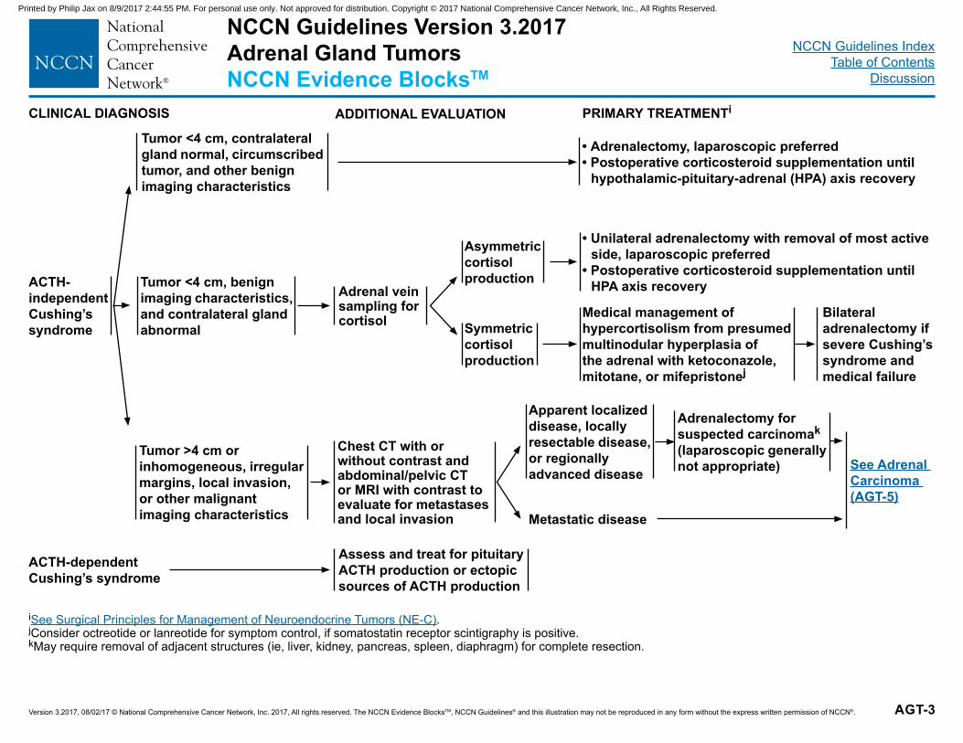
Version 3.2017, 08/02/17 © National Comprehensive Cancer Network, Inc. 2017, All rights reserved. The NCCN Evidence BlocksTM, NCCN Guidelines® and this illustration may not be reproduced in any form without the express written permission of NCCN®. AGT-3
PRIMARY TREATMENTi
Tumor <4 cm, contralateral gland normal, circumscribed tumor, and other benign imaging characteristics
iSee Surgical Principles for Management of Neuroendocrine Tumors (NE-C).jConsider octreotide or lanreotide for symptom control, if somatostatin receptor scintigraphy is positive.kMay require removal of adjacent structures (ie, liver, kidney, pancreas, spleen, diaphragm) for complete resection.
Adrenal vein sampling for cortisol Symmetric
cortisol production
Asymmetric cortisol production
See Adrenal Carcinoma (AGT-5)
CLINICAL DIAGNOSIS ADDITIONAL EVALUATION
ACTH- independent Cushing’s syndrome
Tumor >4 cm or inhomogeneous, irregular margins, local invasion, or other malignant imaging characteristics
Chest CT with or without contrast and abdominal/pelvic CT or MRI with contrast to evaluate for metastases and local invasion Metastatic disease
Apparent localized disease, locally resectable disease, or regionally advanced disease
Tumor <4 cm, benign imaging characteristics, and contralateral gland abnormal
• Adrenalectomy, laparoscopic preferred• Postoperative corticosteroid supplementation until
hypothalamic-pituitary-adrenal (HPA) axis recovery
Adrenalectomy for suspected carcinomak
(laparoscopic generally not appropriate)
Assess and treat for pituitary ACTH production or ectopic sources of ACTH production
ACTH-dependent Cushing’s syndrome
• Unilateral adrenalectomy with removal of most active side, laparoscopic preferred
• Postoperative corticosteroid supplementation until HPA axis recovery
Bilateral adrenalectomy if severe Cushing’s syndrome and medical failure
Medical management of hypercortisolism from presumed multinodular hyperplasia of the adrenal with ketoconazole, mitotane, or mifepristonej
NCCN Guidelines Version 3.2017 Adrenal Gland TumorsNCCN Evidence BlocksTM
Printed by Philip Jax on 8/9/2017 2:44:55 PM. For personal use only. Not approved for distribution. Copyright © 2017 National Comprehensive Cancer Network, Inc., All Rights Reserved.
NCCN Guidelines IndexTable of Contents
Discussion
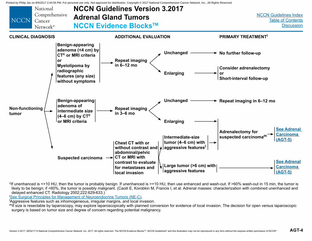
Version 3.2017, 08/02/17 © National Comprehensive Cancer Network, Inc. 2017, All rights reserved. The NCCN Evidence BlocksTM, NCCN Guidelines® and this illustration may not be reproduced in any form without the express written permission of NCCN®. AGT-4
PRIMARY TREATMENTi
Benign-appearing adenoma (<4 cm) by CTc or MRI criteria or Myelolipoma by radiographic features (any size) without symptoms
cIf unenhanced is <+10 HU, then the tumor is probably benign. If unenhanced is >+10 HU, then use enhanced and wash-out. If >60% wash-out in 15 min, the tumor is likely to be benign; if <60%, the tumor is possibly malignant. (Caoili E, Korobkin M, Francis I, et al. Adrenal masses: characterization with combined unenhanced and delayed enhanced CT. Radiology 2002;222:629-633.)
iSee Surgical Principles for Management of Neuroendocrine Tumors (NE-C).lAggressive features such as inhomogeneous, irregular margins, and local invasion.mIf size is resectable by laparoscopy, may explore laparoscopically with planned conversion for evidence of local invasion. The decision for open versus laparoscopic
surgery is based on tumor size and degree of concern regarding potential malignancy.
Repeat imaging in 3–6 mo
Enlarging
Unchanged
CLINICAL DIAGNOSIS ADDITIONAL EVALUATION
Non-functioning tumor
Suspected carcinoma
Benign-appearing adenoma of intermediate size (4–6 cm) by CTc or MRI criteria
Repeat imaging in 6–12 mo
See Adrenal Carcinoma (AGT-5)
Adrenalectomy for suspected carcinomam
See Adrenal Carcinoma (AGT-5)
Repeat imaging in 6–12 mo
Enlarging
Unchanged No further follow-up
Consider adrenalectomy orShort-interval follow-up
Large tumor (>6 cm) with aggressive features
Intermediate-size tumor (4–6 cm) with aggressive featuresl
Chest CT with or without contrast and abdominal/pelvic CT or MRI with contrast to evaluate for metastases and local invasion
NCCN Guidelines Version 3.2017 Adrenal Gland TumorsNCCN Evidence BlocksTM
Printed by Philip Jax on 8/9/2017 2:44:55 PM. For personal use only. Not approved for distribution. Copyright © 2017 National Comprehensive Cancer Network, Inc., All Rights Reserved.
NCCN Guidelines IndexTable of Contents
Discussion
Version 3.2017, 08/02/17 © National Comprehensive Cancer Network, Inc. 2017, All rights reserved. The NCCN Evidence BlocksTM, NCCN Guidelines® and this illustration may not be reproduced in any form without the express written permission of NCCN®. AGT-5
TREATMENTi
iSee Surgical Principles for Management of Neuroendocrine Tumors (NE-C).kMay require removal of adjacent structures (ie, liver, kidney, pancreas, spleen,
diaphragm) for complete resection.nChest CT with or without contrast and abdominal/pelvic CT or MRI with contrast to
evaluate for metastases and local invasion to stage disease, if not previously done.oIncreased risk for local recurrence and peritoneal spread when done laparoscopically. pMonitor mitotane blood levels. Some institutions recommend target levels of 14–20
mcg/mL if tolerated. Steady-state levels may be reached several months after initiation of mitotane. Mitotane therapy requires steroid replacement therapy.
ADRENAL CARCINOMA FOLLOW-UP
Adrenal carcinoman
Metastatic disease
• Consider observation with chest CT with or without contrast and abdominal/pelvic CT or MRI with contrast for clinically indolent disease every 3 mo and biomarkers (if tumor initially functional)
• Consider resection of primary tumor and metastases if >90% removable, particularly if functional
• Consider systemic therapy,p,r preferably in clinical trial�Cisplatin or carboplatin + etoposide ± doxorubicin ± mitotanep,q
(life-long hydrocortisone replacement may be required) or�Streptozocin ± mitotanep,q (life-long hydrocortisone
replacement may be required) or�Mitotane monotherapyp,q (life-long hydrocortisone replacement
may be required)
If high risk for local recurrence:s
• Consider external-beam RT to tumor bed
• Consider adjuvant mitotane therapyp,q (category 3) (life-long hydrocortisone replacement may be required)
• Resect tumor and adjacent lymph nodes�Open adrenalectomy
recommendedk,o
Localized disease
• Every 3–12 mo up to 5 y (after 5 y as clinically indicated)�Consider chest CT with
or without contrast and abdominal CT or MRI with contrast�Consider biomarkers, if
tumor initially functional
qMitotane may have more benefit for control of hormone symptoms than control of tumor.
rSee Discussion for further information regarding the phase III FIRM-ACT trial. (Fassnacht M, Terzolo M, Allolio B, et al; FIRM-ACT Study Group. Combination chemotherapy in advanced adrenocortical carcinoma. N Eng J Med 2012;366:2189-2197)
sHigh-risk local recurrence features include: positive margins, rupture of capsule, large size, and high grade.
Chest CT with or without contrast and abdominal/pelvic CT or MRI with contrast
NCCN Guidelines Version 3.2017 Adrenal Gland TumorsNCCN Evidence BlocksTM
Printed by Philip Jax on 8/9/2017 2:44:55 PM. For personal use only. Not approved for distribution. Copyright © 2017 National Comprehensive Cancer Network, Inc., All Rights Reserved.
NCCN Guidelines IndexTable of Contents
Discussion
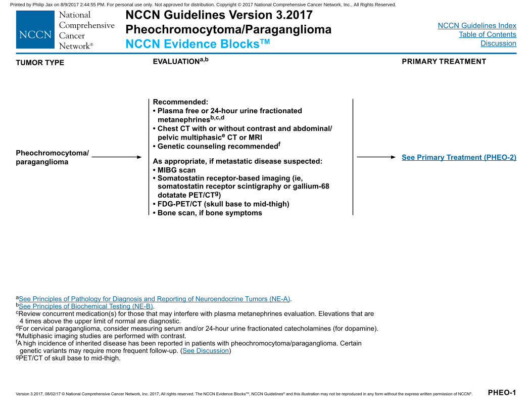
Version 3.2017, 08/02/17 © National Comprehensive Cancer Network, Inc. 2017, All rights reserved. The NCCN Evidence BlocksTM, NCCN Guidelines® and this illustration may not be reproduced in any form without the express written permission of NCCN®. PHEO-1
NCCN Guidelines Version 3.2017 Pheochromocytoma/ParagangliomaNCCN Evidence BlocksTM
PRIMARY TREATMENTEVALUATIONa,bTUMOR TYPE
Pheochromocytoma/paraganglioma
Recommended:• Plasma free or 24-hour urine fractionated
metanephrinesb,c,d
• Chest CT with or without contrast and abdominal/pelvic multiphasice CT or MRI
• Genetic counseling recommendedf
As appropriate, if metastatic disease suspected:• MIBG scan • Somatostatin receptor-based imaging (ie,
somatostatin receptor scintigraphy or gallium-68 dotatate PET/CTg)
• FDG-PET/CT (skull base to mid-thigh)• Bone scan, if bone symptoms
See Primary Treatment (PHEO-2)
aSee Principles of Pathology for Diagnosis and Reporting of Neuroendocrine Tumors (NE-A).bSee Principles of Biochemical Testing (NE-B).cReview concurrent medication(s) for those that may interfere with plasma metanephrines evaluation. Elevations that are
4 times above the upper limit of normal are diagnostic.dFor cervical paraganglioma, consider measuring serum and/or 24-hour urine fractionated catecholamines (for dopamine).eMultiphasic imaging studies are performed with contrast. fA high incidence of inherited disease has been reported in patients with pheochromocytoma/paraganglioma. Certain
genetic variants may require more frequent follow-up. (See Discussion)gPET/CT of skull base to mid-thigh.
Printed by Philip Jax on 8/9/2017 2:44:55 PM. For personal use only. Not approved for distribution. Copyright © 2017 National Comprehensive Cancer Network, Inc., All Rights Reserved.
NCCN Guidelines IndexTable of Contents
Discussion
Version 3.2017, 08/02/17 © National Comprehensive Cancer Network, Inc. 2017, All rights reserved. The NCCN Evidence BlocksTM, NCCN Guidelines® and this illustration may not be reproduced in any form without the express written permission of NCCN®. PHEO-2
Resectable
3–12 mo postresection:• H&P, blood pressure, and markersb• Consider chest CT ± contrast and abdominal/
pelvic CT or MRI with contrast or FDG-PET/CT >1 y postresection up to 10 y:• H&P, blood pressure, and markersb�Years 1–3: every 6–12 mo�Years 4+ up to 10 y: annually
• Consider chest CT ± contrast and abdominal/pelvic CT or MRI with contrast or FDG-PET/CT
bSee Principles of Biochemical Testing (NE-B).fA high incidence of inherited disease has been reported in patients with
pheochromocytoma/paraganglioma. Certain genetic variants may require more frequent follow-up. (See Discussion)
hSee Surgical Principles for Management of Neuroendocrine Tumors (NE-C).iEarlier, if symptoms.jAlpha 1 selective receptor blockers include terazosin, doxazosin, and
prazosin, and non-selective receptors include phenoxybenzamine. Therapy for 7–14 days is recommended prior to surgical therapy. Nonselective alpha blockade phentolamine (IV) can be used intraoperatively.
SURVEILLANCEf,iPRIMARY TREATMENTh
Locallyunresectable
Distantmetastases
Every 3–12 mo:• H&P, blood pressure,
and markersb• Consider imaging: Chest/abdominal/pelvic
CT with contrast or Chest CT (± contrast)
and abdominal/pelvic MRI without contrast (if risk for hypertensive episode)
or FDG-PET/CT
Alpha blockadej
with volume repletion and high salt diet for 7–14 days or until stable
Resection (laparoscopic preferred when safe and feasible)h
• Continue medical therapy for secreting tumors and consider referral to multidisciplinary center
• RT ± cytoreductive (R2) resection, when possiblej or 131I-MIBG (requires prior positive MIBG scan
with dosimetry)
Consider adding:k• Dihydropyridine
calcium channel blockade
• Beta blockade• Methyltyrosine • Continue medical therapy for secreting tumors
and• Cytoreductive (R2) resection, when possiblej or Clinical trial or Systemic chemotherapy (eg, CVDl, or temozolomide) or 131I-MIBG (requires prior positive MIBG scan with dosimetry) or Palliative RT for bone metastases
kAlpha blockade is first-line therapy for all hormonally secreting pheochromocytomas and paragangliomas. After alpha blockade, if additional blood pressure (bp) support is needed, the addition of dihydropyridine calcium channel blockers can be used. This is not recommended as monotherapy unless the patient cannot tolerate alpha blockade. Methyltyrosine can also be used in addition to alpha blockade to stabilize bp. Beta blockade can be added to alpha blockade for tachycardia. B1 selective blockers or nonselective beta blockers can be used. Combination beta/alpha blockers are not recommended.
lCVD = cyclophosphamide, vincristine, and dacarbazine
NCCN Guidelines Version 3.2017 Pheochromocytoma/ParagangliomaNCCN Evidence BlocksTM
Printed by Philip Jax on 8/9/2017 2:44:55 PM. For personal use only. Not approved for distribution. Copyright © 2017 National Comprehensive Cancer Network, Inc., All Rights Reserved.
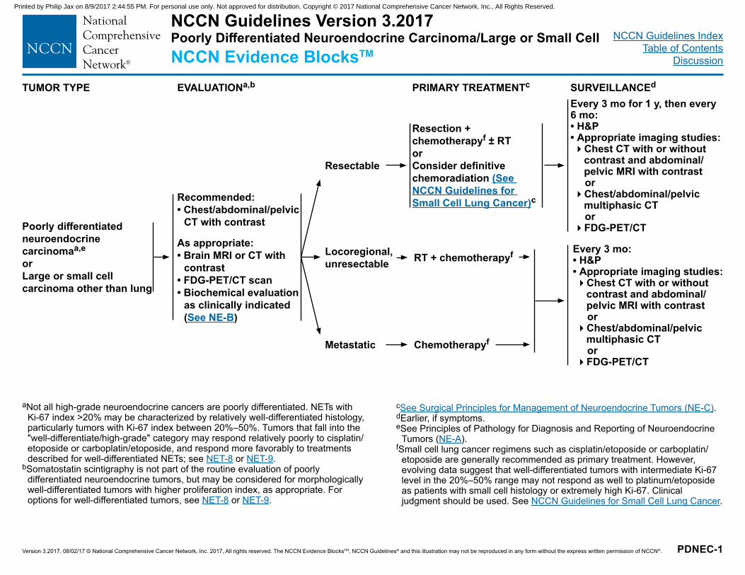
NCCN Guidelines IndexTable of Contents
Discussion
Version 3.2017, 08/02/17 © National Comprehensive Cancer Network, Inc. 2017, All rights reserved. The NCCN Evidence BlocksTM, NCCN Guidelines® and this illustration may not be reproduced in any form without the express written permission of NCCN®.
NCCN Guidelines Version 3.2017 Poorly Differentiated Neuroendocrine Carcinoma/Large or Small CellNCCN Evidence BlocksTM
PDNEC-1
TUMOR TYPE EVALUATIONa,b PRIMARY TREATMENTc SURVEILLANCEd
Poorly differentiated neuroendocrine carcinomaa,e
orLarge or small cell carcinoma other than lung
Metastatic
Locoregional, unresectable
Resectable
Resection + chemotherapyf ± RTor Consider definitive chemoradiation (See NCCN Guidelines for Small Cell Lung Cancer)c
RT + chemotherapyf
Every 3 mo for 1 y, then every 6 mo:• H&P• Appropriate imaging studies: �Chest CT with or without
contrast and abdominal/pelvic MRI with contrast
or �Chest/abdominal/pelvic
multiphasic CT or �FDG-PET/CT
Every 3 mo:• H&P• Appropriate imaging studies: �Chest CT with or without
contrast and abdominal/pelvic MRI with contrast
or �Chest/abdominal/pelvic
multiphasic CT or �FDG-PET/CT
Chemotherapyf
Recommended: • Chest/abdominal/pelvic
CT with contrast
As appropriate:• Brain MRI or CT with
contrast• FDG-PET/CT scan• Biochemical evaluation
as clinically indicated (See NE-B)
aNot all high-grade neuroendocrine cancers are poorly differentiated. NETs with Ki-67 index >20% may be characterized by relatively well-differentiated histology, particularly tumors with Ki-67 index between 20%–50%. Tumors that fall into the "well-differentiate/high-grade" category may respond relatively poorly to cisplatin/etoposide or carboplatin/etoposide, and respond more favorably to treatments described for well-differentiated NETs; see NET-8 or NET-9.
bSomatostatin scintigraphy is not part of the routine evaluation of poorly differentiated neuroendocrine tumors, but may be considered for morphologically well-differentiated tumors with higher proliferation index, as appropriate. For options for well-differentiated tumors, see NET-8 or NET-9.
cSee Surgical Principles for Management of Neuroendocrine Tumors (NE-C).dEarlier, if symptoms.eSee Principles of Pathology for Diagnosis and Reporting of Neuroendocrine
Tumors (NE-A).fSmall cell lung cancer regimens such as cisplatin/etoposide or carboplatin/
etoposide are generally recommended as primary treatment. However, evolving data suggest that well-differentiated tumors with intermediate Ki-67 level in the 20%–50% range may not respond as well to platinum/etoposide as patients with small cell histology or extremely high Ki-67. Clinical judgment should be used. See NCCN Guidelines for Small Cell Lung Cancer.
Printed by Philip Jax on 8/9/2017 2:44:55 PM. For personal use only. Not approved for distribution. Copyright © 2017 National Comprehensive Cancer Network, Inc., All Rights Reserved.
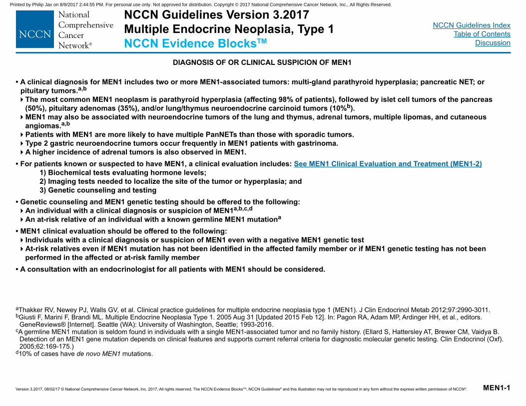
NCCN Guidelines IndexTable of Contents
Discussion
Version 3.2017, 08/02/17 © National Comprehensive Cancer Network, Inc. 2017, All rights reserved. The NCCN Evidence BlocksTM, NCCN Guidelines® and this illustration may not be reproduced in any form without the express written permission of NCCN®.
• A clinical diagnosis for MEN1 includes two or more MEN1-associated tumors: multi-gland parathyroid hyperplasia; pancreatic NET; or pituitary tumors.a,b
�The most common MEN1 neoplasm is parathyroid hyperplasia (affecting 98% of patients), followed by islet cell tumors of the pancreas (50%), pituitary adenomas (35%), and/or lung/thymus neuroendocrine carcinoid tumors (10%b). �MEN1 may also be associated with neuroendocrine tumors of the lung and thymus, adrenal tumors, multiple lipomas, and cutaneous
angiomas.a,b
�Patients with MEN1 are more likely to have multiple PanNETs than those with sporadic tumors. �Type 2 gastric neuroendocrine tumors occur frequently in MEN1 patients with gastrinoma. �A higher incidence of adrenal tumors is also observed in MEN1.
• For patients known or suspected to have MEN1, a clinical evaluation includes: See MEN1 Clinical Evaluation and Treatment (MEN1-2) 1) Biochemical tests evaluating hormone levels; 2) Imaging tests needed to localize the site of the tumor or hyperplasia; and 3) Genetic counseling and testing • Genetic counseling and MEN1 genetic testing should be offered to the following:�An individual with a clinical diagnosis or suspicion of MEN1a,b,c,d
�An at-risk relative of an individual with a known germline MEN1 mutationa
• MEN1 clinical evaluation should be offered to the following:�Individuals with a clinical diagnosis or suspicion of MEN1 even with a negative MEN1 genetic test�At-risk relatives even if MEN1 mutation has not been identified in the affected family member or if MEN1 genetic testing has not been
performed in the affected or at-risk family member• A consultation with an endocrinologist for all patients with MEN1 should be considered.
MEN1-1
NCCN Guidelines Version 3.2017 Multiple Endocrine Neoplasia, Type 1NCCN Evidence BlocksTM
DIAGNOSIS OF OR CLINICAL SUSPICION OF MEN1
aThakker RV, Newey PJ, Walls GV, et al. Clinical practice guidelines for multiple endocrine neoplasia type 1 (MEN1). J Clin Endocrinol Metab 2012;97:2990-3011.bGiusti F, Marini F, Brandi ML. Multiple Endocrine Neoplasia Type 1. 2005 Aug 31 [Updated 2015 Feb 12]. In: Pagon RA, Adam MP, Ardinger HH, et al., editors.
GeneReviews® [Internet]. Seattle (WA): University of Washington, Seattle; 1993-2016.cA germline MEN1 mutation is seldom found in individuals with a single MEN1-associated tumor and no family history. (Ellard S, Hattersley AT, Brewer CM, Vaidya B.
Detection of an MEN1 gene mutation depends on clinical features and supports current referral criteria for diagnostic molecular genetic testing. Clin Endocrinol (Oxf). 2005;62:169-175.)
d10% of cases have de novo MEN1 mutations.
Printed by Philip Jax on 8/9/2017 2:44:55 PM. For personal use only. Not approved for distribution. Copyright © 2017 National Comprehensive Cancer Network, Inc., All Rights Reserved.
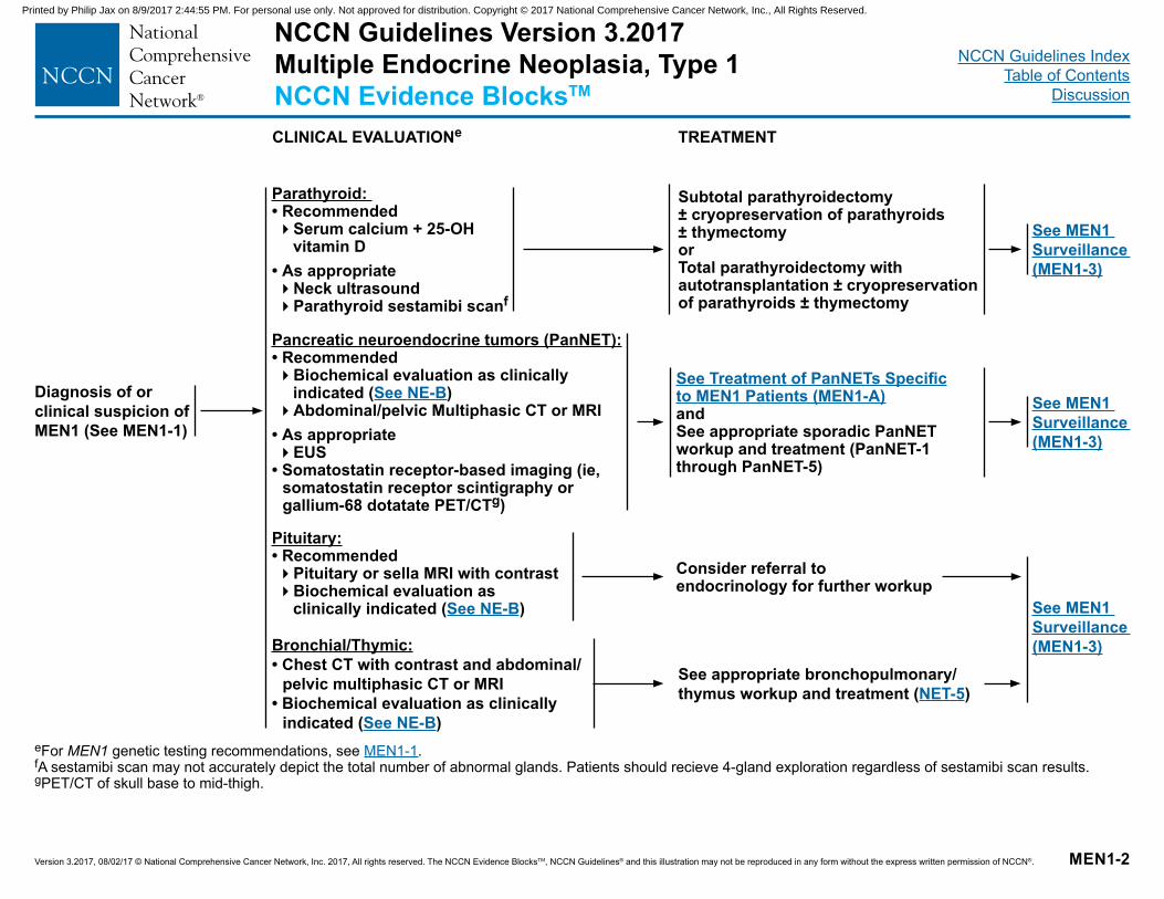
NCCN Guidelines IndexTable of Contents
Discussion
Version 3.2017, 08/02/17 © National Comprehensive Cancer Network, Inc. 2017, All rights reserved. The NCCN Evidence BlocksTM, NCCN Guidelines® and this illustration may not be reproduced in any form without the express written permission of NCCN®.
Diagnosis of or clinical suspicion of MEN1 (See MEN1-1)
MEN1-2
Parathyroid: • Recommended�Serum calcium + 25-OH
vitamin D • As appropriate�Neck ultrasound �Parathyroid sestamibi scanf
See MEN1 Surveillance (MEN1-3)
Subtotal parathyroidectomy± cryopreservation of parathyroids ± thymectomyorTotal parathyroidectomy with autotransplantation ± cryopreservation of parathyroids ± thymectomy
eFor MEN1 genetic testing recommendations, see MEN1-1.fA sestamibi scan may not accurately depict the total number of abnormal glands. Patients should recieve 4-gland exploration regardless of sestamibi scan results.gPET/CT of skull base to mid-thigh.
TREATMENTCLINICAL EVALUATIONe
Pancreatic neuroendocrine tumors (PanNET): • Recommended�Biochemical evaluation as clinically
indicated (See NE-B)�Abdominal/pelvic Multiphasic CT or MRI
• As appropriate�EUS
• Somatostatin receptor-based imaging (ie, somatostatin receptor scintigraphy or gallium-68 dotatate PET/CTg)
Pituitary:• Recommended�Pituitary or sella MRI with contrast�Biochemical evaluation as
clinically indicated (See NE-B)
Bronchial/Thymic:• Chest CT with contrast and abdominal/
pelvic multiphasic CT or MRI• Biochemical evaluation as clinically
indicated (See NE-B)
Consider referral to endocrinology for further workup
See Treatment of PanNETs Specific to MEN1 Patients (MEN1-A)andSee appropriate sporadic PanNET workup and treatment (PanNET-1 through PanNET-5)
See MEN1 Surveillance (MEN1-3)
See MEN1 Surveillance (MEN1-3)
See appropriate bronchopulmonary/thymus workup and treatment (NET-5)
NCCN Guidelines Version 3.2017 Multiple Endocrine Neoplasia, Type 1NCCN Evidence BlocksTM
Printed by Philip Jax on 8/9/2017 2:44:55 PM. For personal use only. Not approved for distribution. Copyright © 2017 National Comprehensive Cancer Network, Inc., All Rights Reserved.
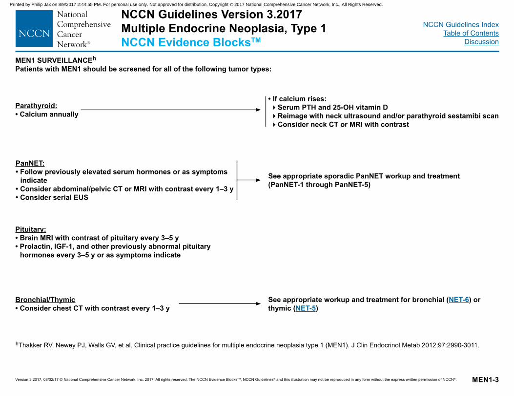
NCCN Guidelines IndexTable of Contents
Discussion
Version 3.2017, 08/02/17 © National Comprehensive Cancer Network, Inc. 2017, All rights reserved. The NCCN Evidence BlocksTM, NCCN Guidelines® and this illustration may not be reproduced in any form without the express written permission of NCCN®.
Parathyroid: • Calcium annually
MEN1-3
• If calcium rises:�Serum PTH and 25-OH vitamin D �Reimage with neck ultrasound and/or parathyroid sestamibi scan�Consider neck CT or MRI with contrast
hThakker RV, Newey PJ, Walls GV, et al. Clinical practice guidelines for multiple endocrine neoplasia type 1 (MEN1). J Clin Endocrinol Metab 2012;97:2990-3011.
MEN1 SURVEILLANCEh
Patients with MEN1 should be screened for all of the following tumor types:
PanNET:• Follow previously elevated serum hormones or as symptoms
indicate• Consider abdominal/pelvic CT or MRI with contrast every 1–3 y• Consider serial EUS
See appropriate sporadic PanNET workup and treatment (PanNET-1 through PanNET-5)
See appropriate workup and treatment for bronchial (NET-6) or thymic (NET-5)
Pituitary: • Brain MRI with contrast of pituitary every 3–5 y• Prolactin, IGF-1, and other previously abnormal pituitary
hormones every 3–5 y or as symptoms indicate
Bronchial/Thymic• Consider chest CT with contrast every 1–3 y
NCCN Guidelines Version 3.2017 Multiple Endocrine Neoplasia, Type 1NCCN Evidence BlocksTM
Printed by Philip Jax on 8/9/2017 2:44:55 PM. For personal use only. Not approved for distribution. Copyright © 2017 National Comprehensive Cancer Network, Inc., All Rights Reserved.
NCCN Guidelines IndexTable of Contents
Discussion
Version 3.2017, 08/02/17 © National Comprehensive Cancer Network, Inc. 2017, All rights reserved. The NCCN Evidence BlocksTM, NCCN Guidelines® and this illustration may not be reproduced in any form without the express written permission of NCCN®.
• In general, surgical management of patients with MEN1 is similar to those with sporadic tumors. Refer to the relevant site-specific recommendations earlier in these guidelines. (See PanNET-1 through PanNET-5)
• However, one notable exception is the multi-focality of pancreaticoduodenal NETs in patients with MEN1. The role of surgery remains controversial in patients with multifocal tumors.
• Decision to resect a pancreatic or duodenal NET in the setting of multifocal disease is complex. If surgery is performed to resect hormonally functional tumor(s), attempts should be made to preoperatively localize the site of the functional tumor. Surgical resection can be considered in the following scenarios:�Symptomatic functional tumors refractory to medical management�Tumor larger than 1–2 cm in size�Tumor with relatively rapid rate of growth over 6–12 months
• Endoscopy with EUS is recommended prior to pancreatic surgery in order to preoperatively assess and localize tumors.• MEN1-associated metastatic pancreatic NETs are often slower growing than metastatic sporadic tumors. Observation can be considered for
non-functioning indolent tumors.
MEN1-A
TREATMENT OF PanNETs SPECIFIC TO MEN1 PATIENTS1
1Yates CJ, Newey PJ, Thakker RV. Challenges and controversies in management of pancreatic neuroendocrine tumours in patients with MEN1. Lancet Diabetes Endocrinol 2015;3:895-905.
NCCN Guidelines Version 3.2017 Multiple Endocrine Neoplasia, Type 1NCCN Evidence BlocksTM
Printed by Philip Jax on 8/9/2017 2:44:55 PM. For personal use only. Not approved for distribution. Copyright © 2017 National Comprehensive Cancer Network, Inc., All Rights Reserved.
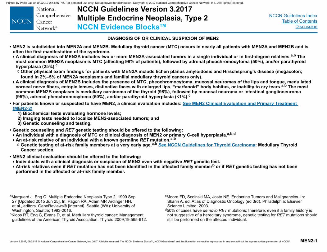
NCCN Guidelines IndexTable of Contents
Discussion
Version 3.2017, 08/02/17 © National Comprehensive Cancer Network, Inc. 2017, All rights reserved. The NCCN Evidence BlocksTM, NCCN Guidelines® and this illustration may not be reproduced in any form without the express written permission of NCCN®.
• MEN2 is subdivided into MEN2A and MEN2B. Medullary thyroid cancer (MTC) occurs in nearly all patients with MEN2A and MEN2B and is often the first manifestation of the syndrome. �A clinical diagnosis of MEN2A includes two or more MEN2A-associated tumors in a single individual or in first-degree relatives.a,b The
most common MEN2A neoplasm is MTC (affecting 98% of patients), followed by adrenal pheochromocytoma (50%), and/or parathyroid hyperplasia (25%).c
◊ Other physical exam findings for patients with MEN2A include lichen planus amyloidosis and Hirschsprung's disease (megacolon; found in 2%–5% of MEN2A neoplasms and familial medullary thryroid cancers only).
�A clinical diagnosis of MEN2B includes the presence of MTC, pheochromocytoma, mucosal neuromas of the lips and tongue, medullated corneal nerve fibers, ectopic lenses, distinctive faces with enlarged lips, “marfanoid” body habitus, or inability to cry tears.a,b The most common MEN2B neoplasm is medullary carcinoma of the thyroid (98%), followed by mucosal neuroma or intestinal ganglioneuroma (95%), adrenal pheochromocytoma (50%), and/or parathyroid hyperplasia (<1%).c
• For patients known or suspected to have MEN2, a clinical evaluation includes: See MEN2 Clinical Evaluation and Primary Treatment (MEN2-2)
1) Biochemical tests evaluating hormone levels; 2) Imaging tests needed to localize MEN2-associated tumors; and 3) Genetic counseling and testing.
• Genetic counseling and RET genetic testing should be offered to the following:�An individual with a diagnosis of MTC or clinical diagnosis of MEN2 or primary C-cell hyperplasia.a,b,d�An at-risk relative of an individual with a known germline RET mutation.a,b
◊ Genetic testing of at-risk family members at a very early age.a,b See NCCN Guidelines for Thyroid Carcinoma: Medullary Thyroid Cancer section.
• MEN2 clinical evaluation should be offered to the following:�Individuals with a clinical diagnosis or suspicion of MEN2 even with negative RET genetic test.�At-risk relatives even if RET mutation has not been identified in the affected family memberb or if RET genetic testing has not been
performed in the affected or at-risk family member.
MEN2-1
NCCN Guidelines Version 3.2017 Multiple Endocrine Neoplasia, Type 2NCCN Evidence BlocksTM
DIAGNOSIS OF OR CLINICAL SUSPICION OF MEN2
aMarquard J, Eng C. Multiple Endocrine Neoplasia Type 2. 1999 Sep 27 [Updated 2015 Jun 25]. In: Pagon RA, Adam MP, Ardinger HH, et al., editors. GeneReviews® [Internet]. Seattle (WA): University of Washington, Seattle; 1993-2016.
bKloos RT, Eng C, Evans D, et al. Medullary thyroid cancer: Management guidelines of the American Thyroid Association. Thyroid 2009;19:565-612.
cMoore FD, Scoinski MA, Joste NE. Endocrine Tumors and Malignancies. In: Skarin A, ed. Atlas of Diagnostic Oncology (ed 3rd). Philadelphia: Elsevier Science Limited; 2003.
d50% of cases have de novo RET mutations; therefore, even if a family history is not suggestive of a hereditary syndrome, genetic testing for RET mutations should still be performed on the affected individual.
Printed by Philip Jax on 8/9/2017 2:44:55 PM. For personal use only. Not approved for distribution. Copyright © 2017 National Comprehensive Cancer Network, Inc., All Rights Reserved.
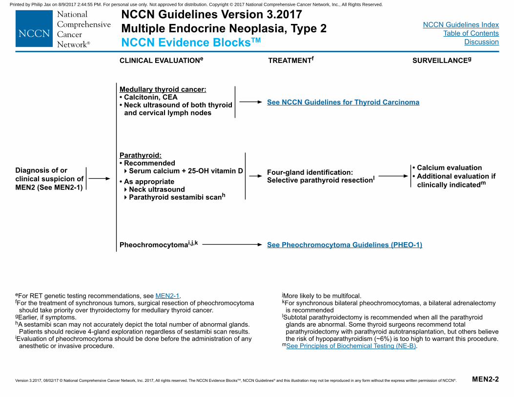
NCCN Guidelines IndexTable of Contents
Discussion
Version 3.2017, 08/02/17 © National Comprehensive Cancer Network, Inc. 2017, All rights reserved. The NCCN Evidence BlocksTM, NCCN Guidelines® and this illustration may not be reproduced in any form without the express written permission of NCCN®.
Diagnosis of or clinical suspicion of MEN2 (See MEN2-1)
MEN2-2
Medullary thyroid cancer:• Calcitonin, CEA • Neck ultrasound of both thyroid
and cervical lymph nodes
• Calcium evaluation• Additional evaluation if
clinically indicatedm
eFor RET genetic testing recommendations, see MEN2-1.fFor the treatment of synchronous tumors, surgical resection of pheochromocytoma
should take priority over thyroidectomy for medullary thyroid cancer.gEarlier, if symptoms.hA sestamibi scan may not accurately depict the total number of abnormal glands.
Patients should recieve 4-gland exploration regardless of sestamibi scan results.iEvaluation of pheochromocytoma should be done before the administration of any
anesthetic or invasive procedure.
TREATMENTfCLINICAL EVALUATIONe
Parathyroid: • Recommended�Serum calcium + 25-OH vitamin D
• As appropriate�Neck ultrasound �Parathyroid sestamibi scanh
Pheochromocytomai,j,k
Four-gland identification:Selective parathyroid resectionl
See NCCN Guidelines for Thyroid Carcinoma
See Pheochromocytoma Guidelines (PHEO-1)
SURVEILLANCEg
jMore likely to be multifocal.kFor synchronous bilateral pheochromocytomas, a bilateral adrenalectomy
is recommendedlSubtotal parathyroidectomy is recommended when all the parathyroid
glands are abnormal. Some thyroid surgeons recommend total parathyroidectomy with parathyroid autotransplantation, but others believe the risk of hypoparathyroidism (~6%) is too high to warrant this procedure.
mSee Principles of Biochemical Testing (NE-B).
NCCN Guidelines Version 3.2017 Multiple Endocrine Neoplasia, Type 2NCCN Evidence BlocksTM
Printed by Philip Jax on 8/9/2017 2:44:55 PM. For personal use only. Not approved for distribution. Copyright © 2017 National Comprehensive Cancer Network, Inc., All Rights Reserved.
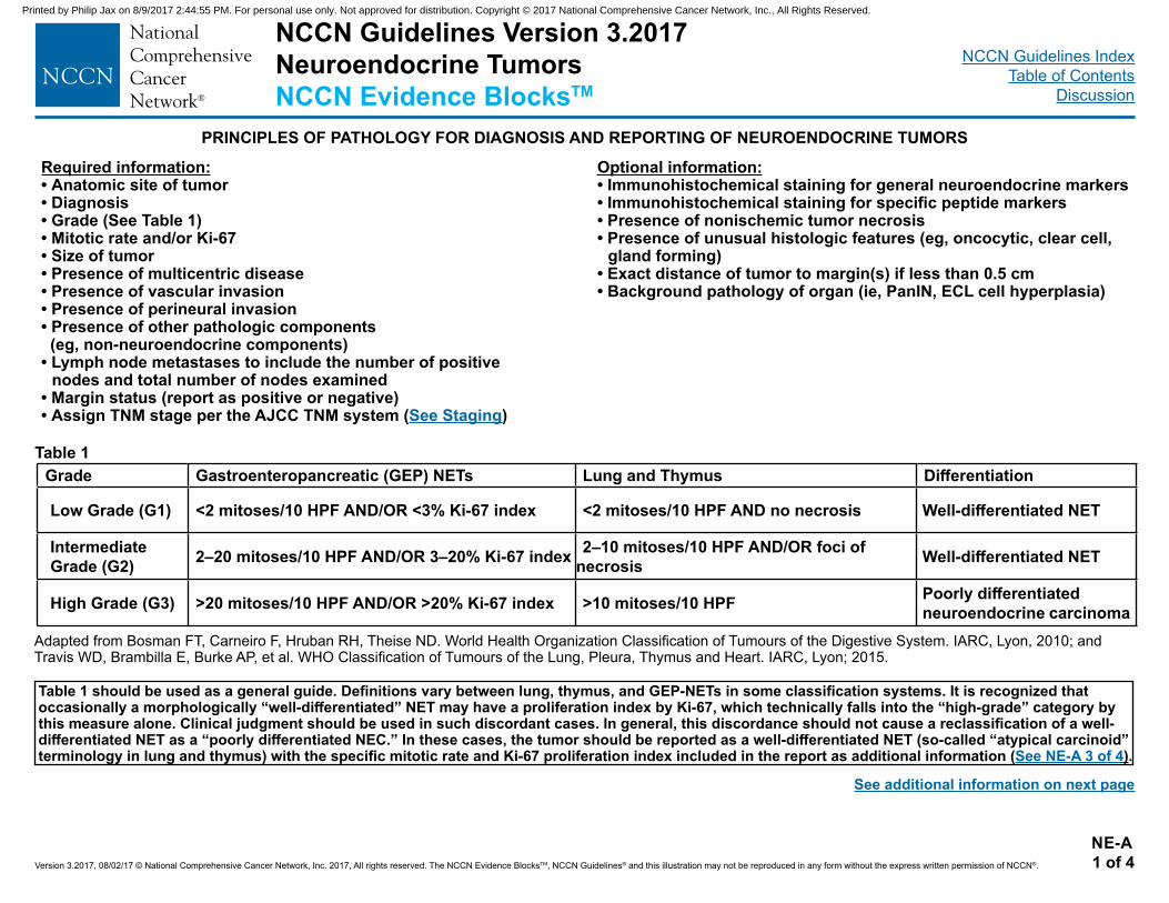
NCCN Guidelines Version 3.2017Neuroendocrine TumorsNCCN Evidence BlocksTM
NCCN Guidelines IndexTable of Contents
Discussion
Version 3.2017, 08/02/17 © National Comprehensive Cancer Network, Inc. 2017, All rights reserved. The NCCN Evidence BlocksTM, NCCN Guidelines® and this illustration may not be reproduced in any form without the express written permission of NCCN®.
Required information:• Anatomic site of tumor• Diagnosis• Grade (See Table 1)• Mitotic rate and/or Ki-67• Size of tumor• Presence of multicentric disease• Presence of vascular invasion• Presence of perineural invasion• Presence of other pathologic components (eg, non-neuroendocrine components)• Lymph node metastases to include the number of positive
nodes and total number of nodes examined• Margin status (report as positive or negative)• Assign TNM stage per the AJCC TNM system (See Staging)
NE-A 1 of 4
PRINCIPLES OF PATHOLOGY FOR DIAGNOSIS AND REPORTING OF NEUROENDOCRINE TUMORS
Optional information:• Immunohistochemical staining for general neuroendocrine markers• Immunohistochemical staining for specific peptide markers • Presence of nonischemic tumor necrosis• Presence of unusual histologic features (eg, oncocytic, clear cell,
gland forming)• Exact distance of tumor to margin(s) if less than 0.5 cm• Background pathology of organ (ie, PanIN, ECL cell hyperplasia)
Grade Gastroenteropancreatic (GEP) NETs Lung and Thymus Differentiation
Low Grade (G1) <2 mitoses/10 HPF AND/OR <3% Ki-67 index <2 mitoses/10 HPF AND no necrosis Well-differentiated NET
Intermediate Grade (G2) 2–20 mitoses/10 HPF AND/OR 3–20% Ki-67 index 2–10 mitoses/10 HPF AND/OR foci of
necrosis Well-differentiated NET
High Grade (G3) >20 mitoses/10 HPF AND/OR >20% Ki-67 index >10 mitoses/10 HPF Poorly differentiated neuroendocrine carcinoma
Table 1
Table 1 should be used as a general guide. Definitions vary between lung, thymus, and GEP-NETs in some classification systems. It is recognized that occasionally a morphologically “well-differentiated” NET may have a proliferation index by Ki-67, which technically falls into the “high-grade” category by this measure alone. Clinical judgment should be used in such discordant cases. In general, this discordance should not cause a reclassification of a well-differentiated NET as a “poorly differentiated NEC.” In these cases, the tumor should be reported as a well-differentiated NET (so-called “atypical carcinoid” terminology in lung and thymus) with the specific mitotic rate and Ki-67 proliferation index included in the report as additional information (See NE-A 3 of 4).
Adapted from Bosman FT, Carneiro F, Hruban RH, Theise ND. World Health Organization Classification of Tumours of the Digestive System. IARC, Lyon, 2010; and Travis WD, Brambilla E, Burke AP, et al. WHO Classification of Tumours of the Lung, Pleura, Thymus and Heart. IARC, Lyon; 2015.
See additional information on next page
Printed by Philip Jax on 8/9/2017 2:44:55 PM. For personal use only. Not approved for distribution. Copyright © 2017 National Comprehensive Cancer Network, Inc., All Rights Reserved.
NCCN Guidelines IndexTable of Contents
Discussion
Version 3.2017, 08/02/17 © National Comprehensive Cancer Network, Inc. 2017, All rights reserved. The NCCN Evidence BlocksTM, NCCN Guidelines® and this illustration may not be reproduced in any form without the express written permission of NCCN®.
Functional status• Functioning NETs should have the same pathologic diagnosis as the non-functioning NETs at the same anatomic site, since the functional
status is based upon clinical symptoms and should not alter the pathologic diagnosis. However, if a specific clinical situation suggests that correlation with histologic evidence of peptide hormone may be helpful, then histochemical or immunohistochemical studies may be performed and included in the report.
Immunohistochemistry and other ancillary techniques• Immunohistochemistry and other ancillary techniques may not be required to diagnose well-differentiated NETs when sufficient tumor
material is available for histologic review. • Specific markers that may be used to establish neuroendocrine differentiation include chromogranin A, synaptophysin, and CD56, although
CD56 has recently proven to be less specific. In less well-differentiated tumors or tumors of unknown origin, it may be helpful (or required in the case of poorly differentiated neuroendocrine carcinomas) to utilize immunohistochemistry panels.
• Although not entirely specific, lung origin is favored by thyroid transcription factor 1 (TTF-1); intestinal or pancreatic origin by CDX2; and pancreatic and rectal NETs by Isl1 and PAX8.1,2
Classification and grade• Many classification schemes have been proposed for NETs.3-9 The most recent WHO classification system is suggested for GEP NETs and
represents an attempt to unify European and American approaches.8 Multiple site-specific grading systems also exist.• Therefore, the specific classification and grading scheme being utilized should be reported in parentheses after the diagnosis to avoid
confusion with overlapping terminology and criteria used in other systems. • The raw data used to derive the grade should be reported. • Regardless of the system used, it is most important to realize that the term “neuroendocrine tumor” or “neuroendocrine carcinoma” without
any further qualification as to grade is inadequate for prognostication and therapy and is inappropriate for pathology reporting.1,10
NE-A 2 of 4
PRINCIPLES OF PATHOLOGY FOR DIAGNOSIS AND REPORTING OF NEUROENDOCRINE TUMORS
See References on NE-A 4 of 4
Continued on next page
NCCN Guidelines Version 3.2017 Neuroendocrine TumorsNCCN Evidence BlocksTM
Printed by Philip Jax on 8/9/2017 2:44:55 PM. For personal use only. Not approved for distribution. Copyright © 2017 National Comprehensive Cancer Network, Inc., All Rights Reserved.
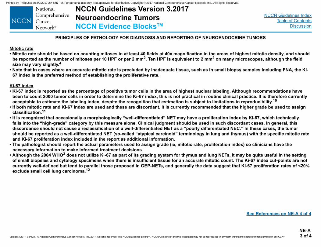
NCCN Guidelines IndexTable of Contents
Discussion
Version 3.2017, 08/02/17 © National Comprehensive Cancer Network, Inc. 2017, All rights reserved. The NCCN Evidence BlocksTM, NCCN Guidelines® and this illustration may not be reproduced in any form without the express written permission of NCCN®.
Mitotic rate• Mitotic rate should be based on counting mitoses in at least 40 fields at 40x magnification in the areas of highest mitotic density, and should
be reported as the number of mitoses per 10 HPF or per 2 mm2. Ten HPF is equivalent to 2 mm2 on many microscopes, although the field size may vary slightly.4
• Note that in cases where an accurate mitotic rate is precluded by inadequate tissue, such as in small biopsy samples including FNA, the Ki-67 index is the preferred method of establishing the proliferative rate.
Ki-67 index• Ki-67 index is reported as the percentage of positive tumor cells in the area of highest nuclear labeling. Although recommendations have
been to count 2000 tumor cells in order to determine the Ki-67 index, this is not practical in routine clinical practice. It is therefore currently acceptable to estimate the labeling index, despite the recognition that estimation is subject to limitations in reproducibility.10
• If both mitotic rate and Ki-67 index are used and these are discordant, it is currently recommended that the higher grade be used to assign classification.11
• It is recognized that occasionally a morphologically “well-differentiated” NET may have a proliferation index by Ki-67, which technically falls into the “high-grade” category by this measure alone. Clinical judgment should be used in such discordant cases. In general, this discordance should not cause a reclassification of a well-differentiated NET as a “poorly differentiated NEC.” In these cases, the tumor should be reported as a well-differentiated NET (so-called “atypical carcinoid” terminology in lung and thymus) with the specific mitotic rate and Ki-67 proliferation index included in the report as additional information.
• The pathologist should report the actual parameters used to assign grade (ie, mitotic rate, proliferation index) so clinicians have the necessary information to make informed treatment decisions.
• Although the 2004 WHO3 does not utilize Ki-67 as part of its grading system for thymus and lung NETs, it may be quite useful in the setting of small biopsies and cytology specimens when there is insufficient tissue for an accurate mitotic count. The Ki-67 index cut-points are not currently well-defined but tend to parallel those proposed in GEP-NETs, and generally the data suggest that Ki-67 proliferation rates of <20% exclude small cell lung carcinoma.12
NE-A 3 of 4
PRINCIPLES OF PATHOLOGY FOR DIAGNOSIS AND REPORTING OF NEUROENDOCRINE TUMORS
See References on NE-A 4 of 4
NCCN Guidelines Version 3.2017 Neuroendocrine TumorsNCCN Evidence BlocksTM
Printed by Philip Jax on 8/9/2017 2:44:55 PM. For personal use only. Not approved for distribution. Copyright © 2017 National Comprehensive Cancer Network, Inc., All Rights Reserved.
NCCN Guidelines IndexTable of Contents
Discussion
Version 3.2017, 08/02/17 © National Comprehensive Cancer Network, Inc. 2017, All rights reserved. The NCCN Evidence BlocksTM, NCCN Guidelines® and this illustration may not be reproduced in any form without the express written permission of NCCN®.
1Klimstra DS, Modlin IR, Coppola D, et al. The pathologic classification of neuroendocrine tumors: a review of nomenclature, grading and staging systems. Pancreas 2010;39:707-712.
2Koo J, Mertens RB, Mirocha JM, et al. Value of Islet 1 and PAX8 in identifying metastatic neuroendocrine tumors of pancreatic origin. Modern Pathology 2012; 25:893-901.
3Travis WD, Brambilla E, Burke AP, et al. WHO Classification of Tumours of the Lung, Pleura, Thymus and Heart. IARC, Lyon; 2015.4Rindi G, Kloppel G, Alhman H, et al. TNM staging of foregut (neuro)endocrine tumors: a consensus proposal including a grading system. Virchows Arch
2006;449:395-401.5Strosberg JR, Coppola D, Klimstra DS et al. The NANETS consensus guidelines for the diagnosis and management of poorly differentiated (high-grade)
extrapulmonary neuroendocrine carcinomas. Pancreas 2010;39,799-800.6Boudreaux JP, Klimstra DS, Hassan MM et al. The NANETS consensus guideline for the diagnosis and management of neuroendocrine tumors: well-differentiated
neuroendocrine tumors of the jejunum, ileum, appendix, and cecum. Pancreas 2010;39,753-766.7Anthony LB, Strosberg JR, Klimstra DS et al. The NANETS consensus guidelines for the diagnosis and management of gastrointestinal neuroendocrine tumors
(NETs): well-differentiated NETs of the distal colon and rectum. Pancreas 2010;39,767-774.8Bosman F, Carneiro F, Hruban R, and Theise ND. WHO Classification of tumours of the digestive system. Lyon, France: IARC Press; 2010.9Oberg K and Castellano D. Current knowledge on diagnosis and staging of neuroendocrine tumors. Cancer Metastasis Rev 2011;30S,S3-S7.10Klimstra DS, Modlin IR, Adsay NV, et al. Pathology reporting of neuroendocrine tumors: application of the Delphic consensus process to the development of a
minimum pathology data set. Am J Surg Pathol 2010;34:300-313.11Rindi G, Bordi C, La Rosa S, et al. Gastroenteropancreatic (neuro)endocrine neoplasms: The histology report. Digestive and Liver Disease 2011;43S;S356-S360. 12Rekhtman N. Neuroendocrine tumors of the lung. An Update. Arch Pathol Lab Med 2010;134:1628-1638.
NE-A 4 of 4
PRINCIPLES OF PATHOLOGY FOR DIAGNOSIS AND REPORTING OF NEUROENDOCRINE TUMORSREFERENCES
NCCN Guidelines Version 3.2017 Neuroendocrine TumorsNCCN Evidence BlocksTM
Printed by Philip Jax on 8/9/2017 2:44:55 PM. For personal use only. Not approved for distribution. Copyright © 2017 National Comprehensive Cancer Network, Inc., All Rights Reserved.
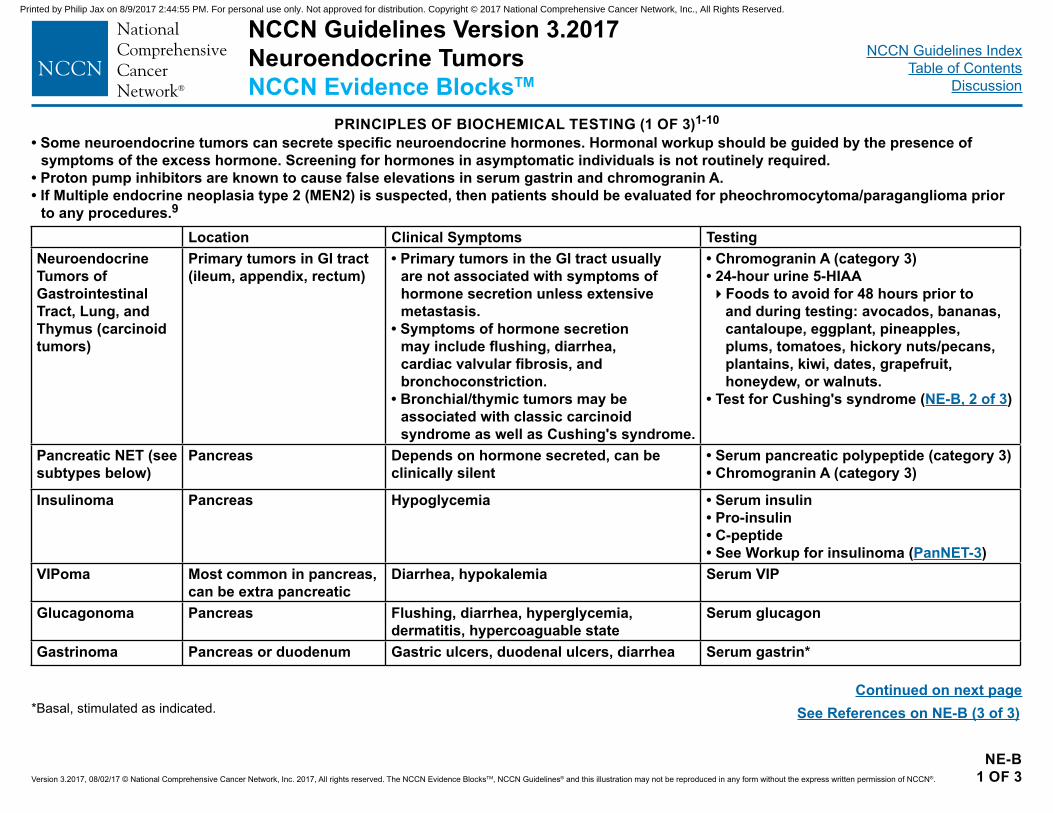
NCCN Guidelines Version 3.2017Neuroendocrine TumorsNCCN Evidence BlocksTM
NCCN Guidelines IndexTable of Contents
Discussion
Version 3.2017, 08/02/17 © National Comprehensive Cancer Network, Inc. 2017, All rights reserved. The NCCN Evidence BlocksTM, NCCN Guidelines® and this illustration may not be reproduced in any form without the express written permission of NCCN®.
NE-B 1 OF 3
PRINCIPLES OF BIOCHEMICAL TESTING (1 OF 3)1-10
• Some neuroendocrine tumors can secrete specific neuroendocrine hormones. Hormonal workup should be guided by the presence of symptoms of the excess hormone. Screening for hormones in asymptomatic individuals is not routinely required.
• Proton pump inhibitors are known to cause false elevations in serum gastrin and chromogranin A.• If Multiple endocrine neoplasia type 2 (MEN2) is suspected, then patients should be evaluated for pheochromocytoma/paraganglioma prior
to any procedures.9
Location Clinical Symptoms TestingNeuroendocrine Tumors of Gastrointestinal Tract, Lung, and Thymus (carcinoid tumors)
Primary tumors in GI tract (ileum, appendix, rectum)
• Primary tumors in the GI tract usually are not associated with symptoms of hormone secretion unless extensive metastasis.
• Symptoms of hormone secretion may include flushing, diarrhea, cardiac valvular fibrosis, and bronchoconstriction.
• Bronchial/thymic tumors may be associated with classic carcinoid syndrome as well as Cushing's syndrome.
• Chromogranin A (category 3)• 24-hour urine 5-HIAA�Foods to avoid for 48 hours prior to
and during testing: avocados, bananas, cantaloupe, eggplant, pineapples, plums, tomatoes, hickory nuts/pecans, plantains, kiwi, dates, grapefruit, honeydew, or walnuts.
• Test for Cushing's syndrome (NE-B, 2 of 3)
Pancreatic NET (see subtypes below)
Pancreas Depends on hormone secreted, can be clinically silent
• Serum pancreatic polypeptide (category 3)• Chromogranin A (category 3)
Insulinoma Pancreas Hypoglycemia • Serum insulin• Pro-insulin• C-peptide• See Workup for insulinoma (PanNET-3)
VIPoma Most common in pancreas, can be extra pancreatic
Diarrhea, hypokalemia Serum VIP
Glucagonoma Pancreas Flushing, diarrhea, hyperglycemia, dermatitis, hypercoaguable state
Serum glucagon
Gastrinoma Pancreas or duodenum Gastric ulcers, duodenal ulcers, diarrhea Serum gastrin*
Continued on next pageSee References on NE-B (3 of 3)*Basal, stimulated as indicated.
Printed by Philip Jax on 8/9/2017 2:44:55 PM. For personal use only. Not approved for distribution. Copyright © 2017 National Comprehensive Cancer Network, Inc., All Rights Reserved.
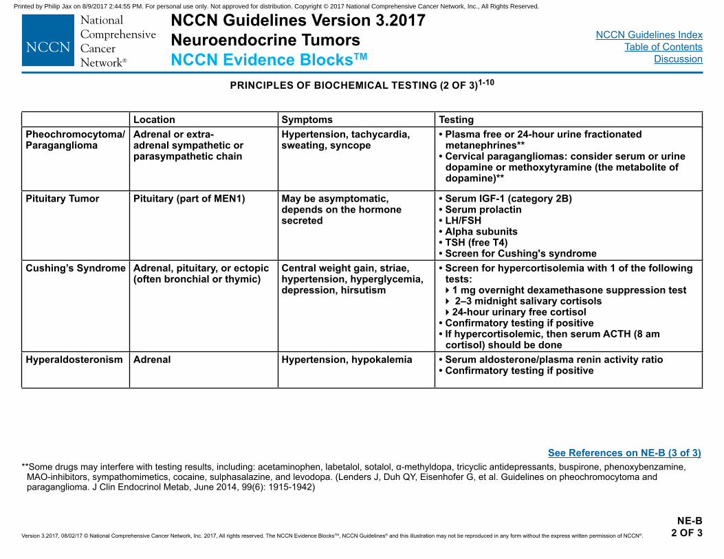
NCCN Guidelines Version 3.2017Neuroendocrine TumorsNCCN Evidence BlocksTM
NCCN Guidelines IndexTable of Contents
Discussion
Version 3.2017, 08/02/17 © National Comprehensive Cancer Network, Inc. 2017, All rights reserved. The NCCN Evidence BlocksTM, NCCN Guidelines® and this illustration may not be reproduced in any form without the express written permission of NCCN®.
NE-B2 OF 3
Location Symptoms TestingPheochromocytoma/Paraganglioma
Adrenal or extra-adrenal sympathetic or parasympathetic chain
Hypertension, tachycardia, sweating, syncope
• Plasma free or 24-hour urine fractionated metanephrines**
• Cervical paragangliomas: consider serum or urine dopamine or methoxytyramine (the metabolite of dopamine)**
Pituitary Tumor Pituitary (part of MEN1) May be asymptomatic, depends on the hormone secreted
• Serum IGF-1 (category 2B)• Serum prolactin• LH/FSH• Alpha subunits• TSH (free T4)• Screen for Cushing's syndrome
Cushing’s Syndrome Adrenal, pituitary, or ectopic (often bronchial or thymic)
Central weight gain, striae, hypertension, hyperglycemia, depression, hirsutism
• Screen for hypercortisolemia with 1 of the following tests:�1 mg overnight dexamethasone suppression test� 2–3 midnight salivary cortisols�24-hour urinary free cortisol
• Confirmatory testing if positive• If hypercortisolemic, then serum ACTH (8 am
cortisol) should be doneHyperaldosteronism Adrenal Hypertension, hypokalemia • Serum aldosterone/plasma renin activity ratio
• Confirmatory testing if positive
PRINCIPLES OF BIOCHEMICAL TESTING (2 OF 3)1-10
**Some drugs may interfere with testing results, including: acetaminophen, labetalol, sotalol, α-methyldopa, tricyclic antidepressants, buspirone, phenoxybenzamine, MAO-inhibitors, sympathomimetics, cocaine, sulphasalazine, and levodopa. (Lenders J, Duh QY, Eisenhofer G, et al. Guidelines on pheochromocytoma and paraganglioma. J Clin Endocrinol Metab, June 2014, 99(6): 1915-1942)
See References on NE-B (3 of 3)
Printed by Philip Jax on 8/9/2017 2:44:55 PM. For personal use only. Not approved for distribution. Copyright © 2017 National Comprehensive Cancer Network, Inc., All Rights Reserved.
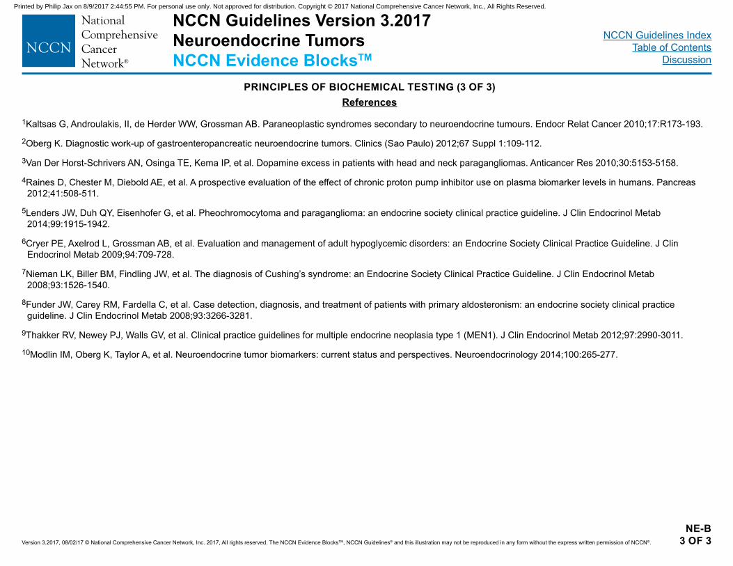
NCCN Guidelines Version 3.2017Neuroendocrine TumorsNCCN Evidence BlocksTM
NCCN Guidelines IndexTable of Contents
Discussion
Version 3.2017, 08/02/17 © National Comprehensive Cancer Network, Inc. 2017, All rights reserved. The NCCN Evidence BlocksTM, NCCN Guidelines® and this illustration may not be reproduced in any form without the express written permission of NCCN®.
NE-B3 OF 3
PRINCIPLES OF BIOCHEMICAL TESTING (3 OF 3)References
1Kaltsas G, Androulakis, II, de Herder WW, Grossman AB. Paraneoplastic syndromes secondary to neuroendocrine tumours. Endocr Relat Cancer 2010;17:R173-193.
2Oberg K. Diagnostic work-up of gastroenteropancreatic neuroendocrine tumors. Clinics (Sao Paulo) 2012;67 Suppl 1:109-112.
3Van Der Horst-Schrivers AN, Osinga TE, Kema IP, et al. Dopamine excess in patients with head and neck paragangliomas. Anticancer Res 2010;30:5153-5158.
4Raines D, Chester M, Diebold AE, et al. A prospective evaluation of the effect of chronic proton pump inhibitor use on plasma biomarker levels in humans. Pancreas 2012;41:508-511.
5Lenders JW, Duh QY, Eisenhofer G, et al. Pheochromocytoma and paraganglioma: an endocrine society clinical practice guideline. J Clin Endocrinol Metab 2014;99:1915-1942.
6Cryer PE, Axelrod L, Grossman AB, et al. Evaluation and management of adult hypoglycemic disorders: an Endocrine Society Clinical Practice Guideline. J Clin Endocrinol Metab 2009;94:709-728.
7Nieman LK, Biller BM, Findling JW, et al. The diagnosis of Cushing’s syndrome: an Endocrine Society Clinical Practice Guideline. J Clin Endocrinol Metab 2008;93:1526-1540.
8Funder JW, Carey RM, Fardella C, et al. Case detection, diagnosis, and treatment of patients with primary aldosteronism: an endocrine society clinical practice guideline. J Clin Endocrinol Metab 2008;93:3266-3281.
9Thakker RV, Newey PJ, Walls GV, et al. Clinical practice guidelines for multiple endocrine neoplasia type 1 (MEN1). J Clin Endocrinol Metab 2012;97:2990-3011. 10Modlin IM, Oberg K, Taylor A, et al. Neuroendocrine tumor biomarkers: current status and perspectives. Neuroendocrinology 2014;100:265-277.
Printed by Philip Jax on 8/9/2017 2:44:55 PM. For personal use only. Not approved for distribution. Copyright © 2017 National Comprehensive Cancer Network, Inc., All Rights Reserved.
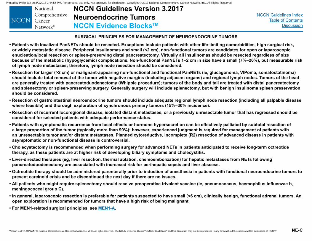
NCCN Guidelines Version 3.2017Neuroendocrine TumorsNCCN Evidence BlocksTM
NCCN Guidelines IndexTable of Contents
Discussion
Version 3.2017, 08/02/17 © National Comprehensive Cancer Network, Inc. 2017, All rights reserved. The NCCN Evidence BlocksTM, NCCN Guidelines® and this illustration may not be reproduced in any form without the express written permission of NCCN®.
• Patients with localized PanNETs should be resected. Exceptions include patients with other life-limiting comorbidities, high surgical risk, or widely metastatic disease. Peripheral insulinomas and small (<2 cm), non-functional tumors are candidates for open or laparoscopic enucleation/local resection or spleen-preserving distal pancreatectomy. Virtually all insulinomas should be resected regardless of size because of the metabolic (hypoglycemic) complications. Non-functional PanNETs 1–2 cm in size have a small (7%–26%), but measurable risk of lymph node metastases; therefore, lymph node resection should be considered.
• Resection for larger (>2 cm) or malignant-appearing non-functional and functional PanNETs (ie, glucagonoma, VIPoma, somatostatinoma) should include total removal of the tumor with negative margins (including adjacent organs) and regional lymph nodes. Tumors of the head are generally treated with pancreatoduodenectomy (Whipple procedure); tumors of the body and tail are treated with distal pancreatectomy and splenectomy or spleen-preserving surgery. Generally surgery will include splenectomy, but with benign insulinoma spleen preservation should be considered.
• Resection of gastrointestinal neuroendocrine tumors should include adequate regional lymph node resection (including all palpable disease where feasible) and thorough exploration of synchronous primary tumors (15%–30% incidence).
• Resection of recurrent locoregional disease, isolated distant metastases, or a previously unresectable tumor that has regressed should be considered for selected patients with adequate performance status.
• Patients with symptomatic recurrence from local effects or hormone hypersecretion can be effectively palliated by subtotal resection of a large proportion of the tumor (typically more than 90%); however, experienced judgment is required for management of patients with an unresectable tumor and/or distant metastases. Planned cytoreductive, incomplete (R2) resection of advanced disease in patients with asymptomatic or non-functional disease is controversial.
• Cholecystectomy is recommended when performing surgery for advanced NETs in patients anticipated to receive long-term octreotide therapy, as these patients are at higher risk of developing biliary symptoms and cholecystitis.
• Liver-directed therapies (eg, liver resection, thermal ablation, chemoembolization) for hepatic metastases from NETs following pancreatoduodenectomy are associated with increased risk for perihepatic sepsis and liver abscess.
• Octreotide therapy should be administered parenterally prior to induction of anesthesia in patients with functional neuroendocrine tumors to prevent carcinoid crisis and be discontinued the next day if there are no issues.
• All patients who might require splenectomy should receive preoperative trivalent vaccine (ie, pneumococcus, haemophilus influenzae b, meningococcal group C).
• In general, laparoscopic resection is preferable for patients suspected to have small (<6 cm), clinically benign, functional adrenal tumors. An open exploration is recommended for tumors that have a high risk of being malignant.
• For MEN1-related surgical principles, see MEN1-A.
NE-C
SURGICAL PRINCIPLES FOR MANAGEMENT OF NEUROENDOCRINE TUMORS
Printed by Philip Jax on 8/9/2017 2:44:55 PM. For personal use only. Not approved for distribution. Copyright © 2017 National Comprehensive Cancer Network, Inc., All Rights Reserved.
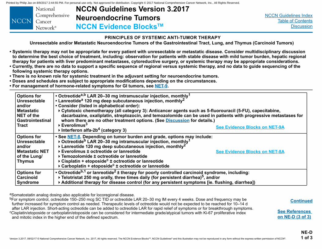
NCCN Guidelines Version 3.2017Neuroendocrine TumorsNCCN Evidence BlocksTM
NCCN Guidelines IndexTable of Contents
Discussion
Version 3.2017, 08/02/17 © National Comprehensive Cancer Network, Inc. 2017, All rights reserved. The NCCN Evidence BlocksTM, NCCN Guidelines® and this illustration may not be reproduced in any form without the express written permission of NCCN®.
NE-D1 of 3
PRINCIPLES OF SYSTEMIC ANTI-TUMOR THERAPYUnresectable and/or Metastatic Neuroendocrine Tumors of the Gastrointestinal Tract, Lung, and Thymus (Carcinoid Tumors)
aSomatostatin analog dosing also applicable for locoregional disease.bFor symptom control, octreotide 150–250 mcg SC TID or octreotide LAR 20–30 mg IM every 4 weeks. Dose and frequency may be
further increased for symptom control as needed. Therapeutic levels of octreotide would not be expected to be reached for 10–14 d after LAR injection. Short-acting octreotide can be added to octreotide LAR for rapid relief of symptoms or for breakthrough symptoms.
cCisplatin/etoposide or carboplatin/etoposide can be considered for intermediate grade/atypical tumors with Ki-67 proliferative index and mitotic index in the higher end of the defined spectrum.
• Systemic therapy may not be appropriate for every patient with unresectable or metastatic disease. Consider multidisciplinary discussion to determine the best choice of treatment, including: observation for patients with stable disease with mild tumor burden, hepatic regional therapy for patients with liver predominant metastases, cytoreductive surgery, or systemic therapy may be appropriate considerations.
• Currently, there are no data to support a specific sequence of regional versus systemic therapy, and no data to guide sequencing of the following systemic therapy options.
• There is no known role for systemic treatment in the adjuvant setting for neuroendocrine tumors.• Doses and schedules are subject to appropriate modifications depending on the circumstances.• For management of hormone-related symptoms for GI tumors, see NET-9.
Continued
See References on NE-D (3 of 3)
Options for Unresectable and/or Metastatic NET of the Gastrointestinal Tract
• Octreotidea,b LAR 20–30 mg intramuscular injection, monthly1• Lanreotidea 120 mg deep subcutaneous injection, monthly2• Consider (listed in alphabetical order):�Cytotoxic chemotherapy (all category 3): Anticancer agents such as 5-fluorouracil (5-FU), capecitabine,
dacarbazine, oxaliplatin, streptozocin, and temozolomide can be used in patients with progressive metastases for whom there are no other treatment options. (See Discussion for details.)�Everolimus3�Interferon alfa-2b4 (category 3)
Options for Unresectable and/or Metastatic NET of the Lung/Thymus
• See NET-8. Depending on tumor burden and grade, options may include:�Octreotideb LAR 20–30 mg intramuscular injection, monthly1�Lanreotide 120 mg deep subcutaneous injection, monthly2�Everolimus ± octreotide or lanreotide�Temozolomide ± octreotide or lanreotide�Cisplatin + etoposidec ± octreotide or lanreotide�Carboplatin + etoposidec ± octreotide or lanreotide
Options for Carcinoid Syndrome
• Octreotideb,1 or lanreotide2 ± therapy for poorly controlled carcinoid syndrome, including:�Telotristat 250 mg orally, three times daily (for persistent diarrhea)5, and/or�Additional therapy for disease control (for any persistent symptoms [ie. flushing, diarrhea])
See Evidence Blocks on NET-8A
See Evidence Blocks on NET-9A
Printed by Philip Jax on 8/9/2017 2:44:55 PM. For personal use only. Not approved for distribution. Copyright © 2017 National Comprehensive Cancer Network, Inc., All Rights Reserved.
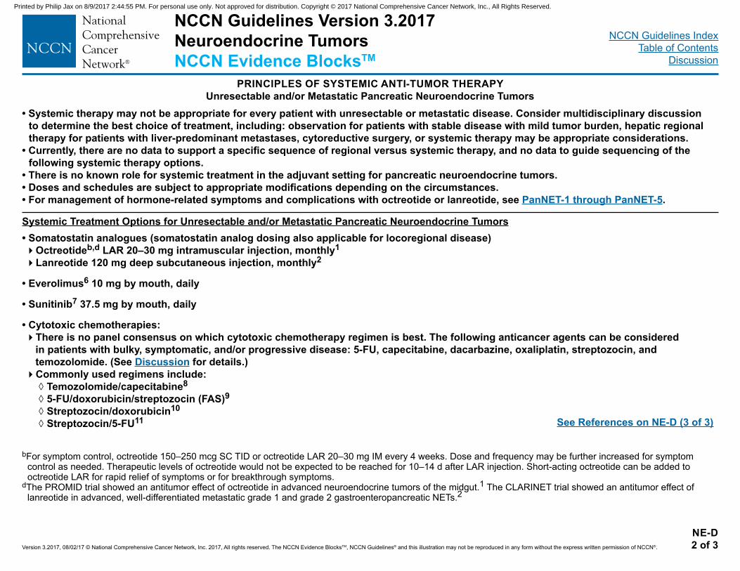
NCCN Guidelines Version 3.2017Neuroendocrine TumorsNCCN Evidence BlocksTM
NCCN Guidelines IndexTable of Contents
Discussion
Version 3.2017, 08/02/17 © National Comprehensive Cancer Network, Inc. 2017, All rights reserved. The NCCN Evidence BlocksTM, NCCN Guidelines® and this illustration may not be reproduced in any form without the express written permission of NCCN®.
NE-D2 of 3
PRINCIPLES OF SYSTEMIC ANTI-TUMOR THERAPYUnresectable and/or Metastatic Pancreatic Neuroendocrine Tumors
Systemic Treatment Options for Unresectable and/or Metastatic Pancreatic Neuroendocrine Tumors• Somatostatin analogues (somatostatin analog dosing also applicable for locoregional disease)�Octreotideb,d LAR 20–30 mg intramuscular injection, monthly1
�Lanreotide 120 mg deep subcutaneous injection, monthly2
• Everolimus6 10 mg by mouth, daily
• Sunitinib7 37.5 mg by mouth, daily
• Cytotoxic chemotherapies: �There is no panel consensus on which cytotoxic chemotherapy regimen is best. The following anticancer agents can be considered
in patients with bulky, symptomatic, and/or progressive disease: 5-FU, capecitabine, dacarbazine, oxaliplatin, streptozocin, and temozolomide. (See Discussion for details.)�Commonly used regimens include:
◊ Temozolomide/capecitabine8
◊ 5-FU/doxorubicin/streptozocin (FAS)9 ◊ Streptozocin/doxorubicin10
◊ Streptozocin/5-FU11
bFor symptom control, octreotide 150–250 mcg SC TID or octreotide LAR 20–30 mg IM every 4 weeks. Dose and frequency may be further increased for symptom control as needed. Therapeutic levels of octreotide would not be expected to be reached for 10–14 d after LAR injection. Short-acting octreotide can be added to octreotide LAR for rapid relief of symptoms or for breakthrough symptoms.
dThe PROMID trial showed an antitumor effect of octreotide in advanced neuroendocrine tumors of the midgut.1 The CLARINET trial showed an antitumor effect of lanreotide in advanced, well-differentiated metastatic grade 1 and grade 2 gastroenteropancreatic NETs.2
• Systemic therapy may not be appropriate for every patient with unresectable or metastatic disease. Consider multidisciplinary discussion to determine the best choice of treatment, including: observation for patients with stable disease with mild tumor burden, hepatic regional therapy for patients with liver-predominant metastases, cytoreductive surgery, or systemic therapy may be appropriate considerations.
• Currently, there are no data to support a specific sequence of regional versus systemic therapy, and no data to guide sequencing of the following systemic therapy options.
• There is no known role for systemic treatment in the adjuvant setting for pancreatic neuroendocrine tumors.• Doses and schedules are subject to appropriate modifications depending on the circumstances.• For management of hormone-related symptoms and complications with octreotide or lanreotide, see PanNET-1 through PanNET-5.
See References on NE-D (3 of 3)
Printed by Philip Jax on 8/9/2017 2:44:55 PM. For personal use only. Not approved for distribution. Copyright © 2017 National Comprehensive Cancer Network, Inc., All Rights Reserved.
NCCN Guidelines Version 3.2017Neuroendocrine TumorsNCCN Evidence BlocksTM
NCCN Guidelines IndexTable of Contents
Discussion
Version 3.2017, 08/02/17 © National Comprehensive Cancer Network, Inc. 2017, All rights reserved. The NCCN Evidence BlocksTM, NCCN Guidelines® and this illustration may not be reproduced in any form without the express written permission of NCCN®.
NE-D3 of 3
PRINCIPLES OF SYSTEMIC ANTI-TUMOR THERAPYREFERENCES
1 Rinke A, Muller HH, Schade-Brittinger C, et al. Placebo-controlled, double-blind, prospective, randomized study on the effect of octreotide LAR in the control of tumor growth in patients with metastatic neuroendocrine midgut tumors: a report from the PROMID Study Group. J Clin Oncol 2009;27:4656-63.
2 Caplin ME, Pavel M, Cwikla JB, et al. Lanreotide in metastatic enteropancreatic neuroendocrine tumors. N Engl J Med 2014;371:224-33. 3 Pavel ME, Hainsworth JD, Baudin E, et al. Everolimus plus octreotide long-acting repeatable for the treatment of advanced neuroendocrine tumours associated
with carcinoid syndrome (RADIANT-2): a randomised, placebo-controlled, phase 3 study. Lancet 2011;378:2005-12. 4 Moertel CG, Rubin J, Kvols LK. Therapy of metastatic carcinoid tumor and the malignant carcinoid syndrome with recombinant leukocyte A interferon. J Clin Oncol
1989;7:865-8. 5 Kulke MH, Hörsch D, Caplin ME, et al. Telotristat Ethyl, a Tryptophan Hydroxylase Inhibitor for the Treatment of Carcinoid Syndrome. J Clin Oncol. 2017; 35(1):14-
23. 6Yao JC, Shah MH, Ito T, et al. Everolimus for advanced pancreatic neuroendocrine tumors. N Engl J Med 2011;364:514-23. 7 Raymond E, Dahan L, Raoul JL, et al. Sunitinib malate for the treatment of pancreatic neuroendocrine tumors. N Engl J Med 2011;364:501-13. 8 Strosberg JR, Fine RL, Choi J, et al. First-line chemotherapy with capecitabine and temozolomide in patients with metastatic pancreatic endocrine carcinomas.
Cancer 2011;117:268-275. 9 Kouvaraki MA, Ajani JA, Hoff P, et al. Fluorouracil, doxorubicin, and streptozocin in the treatment of patients with locally advanced and metastatic pancreatic
endocrine carcinomas. J Clin Oncol 2004;22:4762-71.10 Moertel CG, Lefkopoulo M, Lipsitz S, Hahn RG, Klaassen D. Streptozocin-doxorubicin, streptozocin-fluorouracil or chlorozotocin in the treatment of advanced
islet-cell carcinoma. N Engl J Med 1992;326:519-23.11 Sun W, Lipsitz S, Catalano P, Mailliard JA, Haller DG. Phase II/III study of doxorubicin with fluorouracil compared with streptozocin with fluorouracil or dacarbazine
in the treatment of advanced carcinoid tumors: Eastern Cooperative Oncology Group Study E1281. J Clin Oncol 2005;23:4897-904.
Printed by Philip Jax on 8/9/2017 2:44:55 PM. For personal use only. Not approved for distribution. Copyright © 2017 National Comprehensive Cancer Network, Inc., All Rights Reserved.
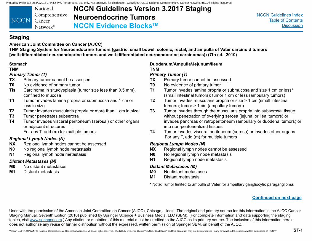
NCCN Guidelines IndexTable of Contents
Discussion
Version 3.2017, 08/02/17 © National Comprehensive Cancer Network, Inc. 2017, All rights reserved. The NCCN Evidence BlocksTM, NCCN Guidelines® and this illustration may not be reproduced in any form without the express written permission of NCCN®.
American Joint Committee on Cancer (AJCC)TNM Staging System for Neuroendocrine Tumors (gastric, small bowel, colonic, rectal, and ampulla of Vater carcinoid tumors [well-differentiated neuroendocrine tumors and well-differentiated neuroendocrine carcinomas]) (7th ed., 2010)
ST-1
NCCN Guidelines Version 3.2017 StagingNeuroendocrine TumorsNCCN Evidence BlocksTM
Staging
StomachTNMPrimary Tumor (T)TX Primary tumor cannot be assessedT0 No evidence of primary tumorTis Carcinoma in situ/dysplasia (tumor size less than 0.5 mm),
confined to mucosaT1 Tumor invades lamina propria or submucosa and 1 cm or
less in sizeT2 Tumor invades muscularis propria or more than 1 cm in sizeT3 Tumor penetrates subserosaT4 Tumor invades visceral peritoneum (serosal) or other organs
or adjacent structures For any T, add (m) for multiple tumorsRegional Lymph Nodes (N)NX Regional lymph nodes cannot be assessedN0 No regional lymph node metastasisN1 Regional lymph node metastasisDistant Metastases (M)M0 No distant metastasesM1 Distant metastasis
Duodenum/Ampulla/Jejunum/IleumTNMPrimary Tumor (T)TX Primary tumor cannot be assessedT0 No evidence of primary tumorT1 Tumor invades lamina propria or submucosa and size 1 cm or less*
(small intestinal tumors); tumor 1 cm or less (ampullary tumors)T2 Tumor invades muscularis propria or size > 1 cm (small intestinal
tumors); tumor > 1 cm (ampullary tumors)T3 Tumor invades through the muscularis propria into subserosal tissue
without penetration of overlying serosa (jejunal or ileal tumors) or invades pancreas or retroperitoneum (ampullary or duodenal tumors) or into non-peritonealized tissues
T4 Tumor invades visceral peritoneum (serosa) or invades other organs For any T, add (m) for multiple tumors
Regional Lymph Nodes (N)NX Regional lymph nodes cannot be assessedN0 No regional lymph node metastasisN1 Regional lymph node metastasisDistant Metastases (M)M0 No distant metastasesM1 Distant metastasis
Continued on next page
Used with the permission of the American Joint Committee on Cancer (AJCC), Chicago, Illinois. The original and primary source for this information is the AJCC Cancer Staging Manual, Seventh Edition (2010) published by Springer Science + Business Media, LLC (SBM). (For complete information and data supporting the staging tables, visit www.springer.com.) Any citation or quotation of this material must be credited to the AJCC as its primary source. The inclusion of this information herein does not authorize any reuse or further distribution without the expressed, written permission of Springer SBM, on behalf of the AJCC.
* Note: Tumor limited to ampulla of Vater for ampullary gangliocytic paraganglioma.
Printed by Philip Jax on 8/9/2017 2:44:55 PM. For personal use only. Not approved for distribution. Copyright © 2017 National Comprehensive Cancer Network, Inc., All Rights Reserved.
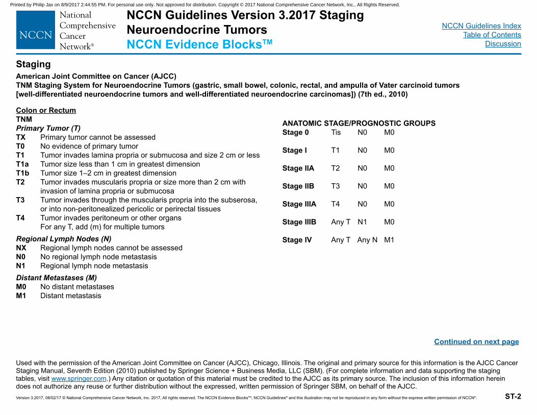
NCCN Guidelines IndexTable of Contents
Discussion
Version 3.2017, 08/02/17 © National Comprehensive Cancer Network, Inc. 2017, All rights reserved. The NCCN Evidence BlocksTM, NCCN Guidelines® and this illustration may not be reproduced in any form without the express written permission of NCCN®.
American Joint Committee on Cancer (AJCC)TNM Staging System for Neuroendocrine Tumors (gastric, small bowel, colonic, rectal, and ampulla of Vater carcinoid tumors [well-differentiated neuroendocrine tumors and well-differentiated neuroendocrine carcinomas]) (7th ed., 2010)
ST-2
Staging
Colon or RectumTNMPrimary Tumor (T)TX Primary tumor cannot be assessedT0 No evidence of primary tumorT1 Tumor invades lamina propria or submucosa and size 2 cm or lessT1a Tumor size less than 1 cm in greatest dimensionT1b Tumor size 1–2 cm in greatest dimensionT2 Tumor invades muscularis propria or size more than 2 cm with
invasion of lamina propria or submucosaT3 Tumor invades through the muscularis propria into the subserosa,
or into non-peritonealized pericolic or perirectal tissuesT4 Tumor invades peritoneum or other organs
For any T, add (m) for multiple tumorsRegional Lymph Nodes (N)NX Regional lymph nodes cannot be assessedN0 No regional lymph node metastasisN1 Regional lymph node metastasisDistant Metastases (M)M0 No distant metastasesM1 Distant metastasis
ANATOMIC STAGE/PROGNOSTIC GROUPSStage 0 Tis N0 M0
Stage I T1 N0 M0
Stage IIA T2 N0 M0
Stage IIB T3 N0 M0
Stage IIIA T4 N0 M0
Stage IIIB Any T N1 M0
Stage IV Any T Any N M1
Used with the permission of the American Joint Committee on Cancer (AJCC), Chicago, Illinois. The original and primary source for this information is the AJCC Cancer Staging Manual, Seventh Edition (2010) published by Springer Science + Business Media, LLC (SBM). (For complete information and data supporting the staging tables, visit www.springer.com.) Any citation or quotation of this material must be credited to the AJCC as its primary source. The inclusion of this information herein does not authorize any reuse or further distribution without the expressed, written permission of Springer SBM, on behalf of the AJCC.
Continued on next page
NCCN Guidelines Version 3.2017 StagingNeuroendocrine TumorsNCCN Evidence BlocksTM
Printed by Philip Jax on 8/9/2017 2:44:55 PM. For personal use only. Not approved for distribution. Copyright © 2017 National Comprehensive Cancer Network, Inc., All Rights Reserved.
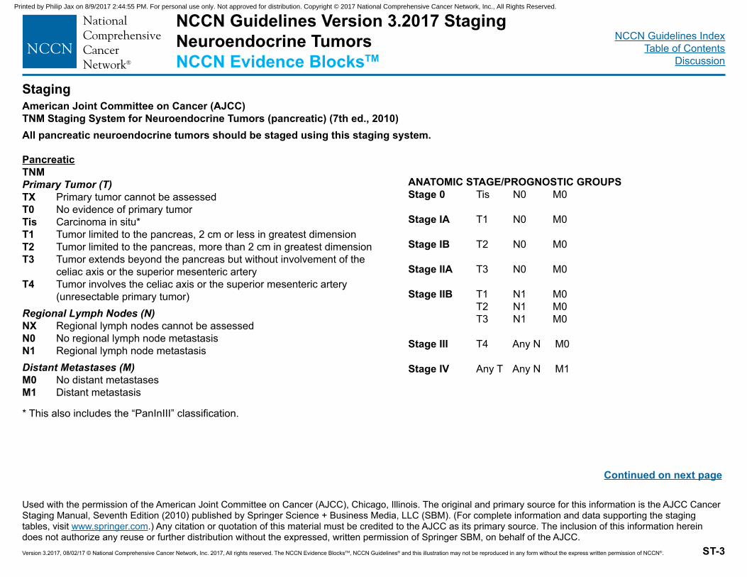
NCCN Guidelines IndexTable of Contents
Discussion
Version 3.2017, 08/02/17 © National Comprehensive Cancer Network, Inc. 2017, All rights reserved. The NCCN Evidence BlocksTM, NCCN Guidelines® and this illustration may not be reproduced in any form without the express written permission of NCCN®.
American Joint Committee on Cancer (AJCC)TNM Staging System for Neuroendocrine Tumors (pancreatic) (7th ed., 2010)All pancreatic neuroendocrine tumors should be staged using this staging system.
ST-3
Staging
PancreaticTNMPrimary Tumor (T)TX Primary tumor cannot be assessedT0 No evidence of primary tumorTis Carcinoma in situ*T1 Tumor limited to the pancreas, 2 cm or less in greatest dimensionT2 Tumor limited to the pancreas, more than 2 cm in greatest dimensionT3 Tumor extends beyond the pancreas but without involvement of the
celiac axis or the superior mesenteric arteryT4 Tumor involves the celiac axis or the superior mesenteric artery
(unresectable primary tumor)Regional Lymph Nodes (N)NX Regional lymph nodes cannot be assessedN0 No regional lymph node metastasisN1 Regional lymph node metastasisDistant Metastases (M)M0 No distant metastasesM1 Distant metastasis
* This also includes the “PanInIII” classification.
ANATOMIC STAGE/PROGNOSTIC GROUPSStage 0 Tis N0 M0
Stage IA T1 N0 M0
Stage IB T2 N0 M0 Stage IIA T3 N0 M0
Stage IIB T1 N1 M0 T2 N1 M0 T3 N1 M0
Stage III T4 Any N M0
Stage IV Any T Any N M1
Used with the permission of the American Joint Committee on Cancer (AJCC), Chicago, Illinois. The original and primary source for this information is the AJCC Cancer Staging Manual, Seventh Edition (2010) published by Springer Science + Business Media, LLC (SBM). (For complete information and data supporting the staging tables, visit www.springer.com.) Any citation or quotation of this material must be credited to the AJCC as its primary source. The inclusion of this information herein does not authorize any reuse or further distribution without the expressed, written permission of Springer SBM, on behalf of the AJCC.
Continued on next page
NCCN Guidelines Version 3.2017 StagingNeuroendocrine TumorsNCCN Evidence BlocksTM
Printed by Philip Jax on 8/9/2017 2:44:55 PM. For personal use only. Not approved for distribution. Copyright © 2017 National Comprehensive Cancer Network, Inc., All Rights Reserved.
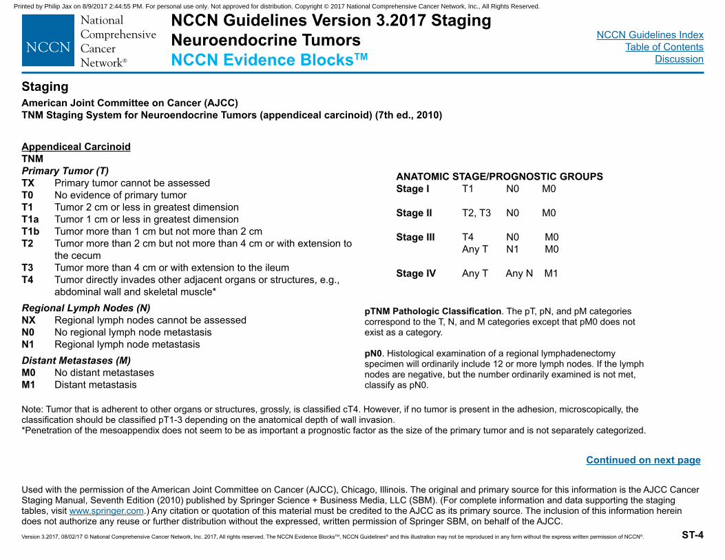
NCCN Guidelines IndexTable of Contents
Discussion
Version 3.2017, 08/02/17 © National Comprehensive Cancer Network, Inc. 2017, All rights reserved. The NCCN Evidence BlocksTM, NCCN Guidelines® and this illustration may not be reproduced in any form without the express written permission of NCCN®.
American Joint Committee on Cancer (AJCC)TNM Staging System for Neuroendocrine Tumors (appendiceal carcinoid) (7th ed., 2010)
ST-4
Staging
Appendiceal CarcinoidTNMPrimary Tumor (T)TX Primary tumor cannot be assessedT0 No evidence of primary tumorT1 Tumor 2 cm or less in greatest dimensionT1a Tumor 1 cm or less in greatest dimensionT1b Tumor more than 1 cm but not more than 2 cmT2 Tumor more than 2 cm but not more than 4 cm or with extension to
the cecumT3 Tumor more than 4 cm or with extension to the ileumT4 Tumor directly invades other adjacent organs or structures, e.g.,
abdominal wall and skeletal muscle*Regional Lymph Nodes (N)NX Regional lymph nodes cannot be assessedN0 No regional lymph node metastasisN1 Regional lymph node metastasisDistant Metastases (M)M0 No distant metastasesM1 Distant metastasis
ANATOMIC STAGE/PROGNOSTIC GROUPSStage I T1 N0 M0
Stage II T2, T3 N0 M0
Stage III T4 N0 M0 Any T N1 M0
Stage IV Any T Any N M1
Used with the permission of the American Joint Committee on Cancer (AJCC), Chicago, Illinois. The original and primary source for this information is the AJCC Cancer Staging Manual, Seventh Edition (2010) published by Springer Science + Business Media, LLC (SBM). (For complete information and data supporting the staging tables, visit www.springer.com.) Any citation or quotation of this material must be credited to the AJCC as its primary source. The inclusion of this information herein does not authorize any reuse or further distribution without the expressed, written permission of Springer SBM, on behalf of the AJCC.
pTNM Pathologic Classification. The pT, pN, and pM categories correspond to the T, N, and M categories except that pM0 does not exist as a category.
pN0. Histological examination of a regional lymphadenectomyspecimen will ordinarily include 12 or more lymph nodes. If the lymph nodes are negative, but the number ordinarily examined is not met, classify as pN0.
Note: Tumor that is adherent to other organs or structures, grossly, is classified cT4. However, if no tumor is present in the adhesion, microscopically, the classification should be classified pT1-3 depending on the anatomical depth of wall invasion.*Penetration of the mesoappendix does not seem to be as important a prognostic factor as the size of the primary tumor and is not separately categorized.
Continued on next page
NCCN Guidelines Version 3.2017 StagingNeuroendocrine TumorsNCCN Evidence BlocksTM
Printed by Philip Jax on 8/9/2017 2:44:55 PM. For personal use only. Not approved for distribution. Copyright © 2017 National Comprehensive Cancer Network, Inc., All Rights Reserved.
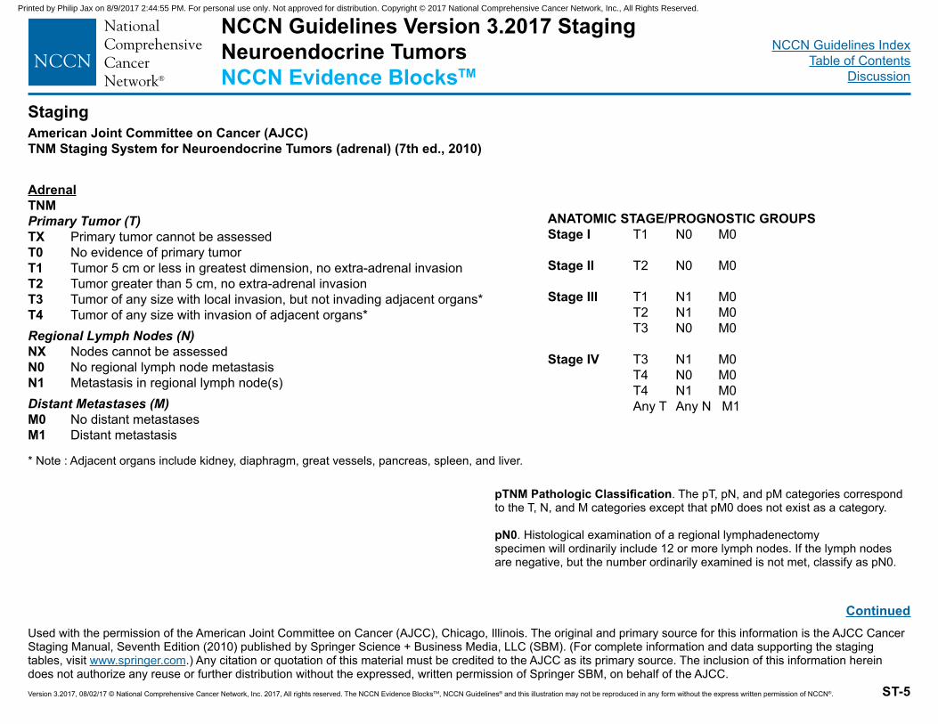
NCCN Guidelines IndexTable of Contents
Discussion
Version 3.2017, 08/02/17 © National Comprehensive Cancer Network, Inc. 2017, All rights reserved. The NCCN Evidence BlocksTM, NCCN Guidelines® and this illustration may not be reproduced in any form without the express written permission of NCCN®.
American Joint Committee on Cancer (AJCC)TNM Staging System for Neuroendocrine Tumors (adrenal) (7th ed., 2010)
ST-5
Staging
AdrenalTNMPrimary Tumor (T)TX Primary tumor cannot be assessedT0 No evidence of primary tumorT1 Tumor 5 cm or less in greatest dimension, no extra-adrenal invasionT2 Tumor greater than 5 cm, no extra-adrenal invasion T3 Tumor of any size with local invasion, but not invading adjacent organs*T4 Tumor of any size with invasion of adjacent organs*Regional Lymph Nodes (N)NX Nodes cannot be assessedN0 No regional lymph node metastasisN1 Metastasis in regional lymph node(s)Distant Metastases (M)M0 No distant metastasesM1 Distant metastasis
ANATOMIC STAGE/PROGNOSTIC GROUPSStage I T1 N0 M0
Stage II T2 N0 M0
Stage III T1 N1 M0 T2 N1 M0 T3 N0 M0
Stage IV T3 N1 M0 T4 N0 M0 T4 N1 M0 Any T Any N M1
Used with the permission of the American Joint Committee on Cancer (AJCC), Chicago, Illinois. The original and primary source for this information is the AJCC Cancer Staging Manual, Seventh Edition (2010) published by Springer Science + Business Media, LLC (SBM). (For complete information and data supporting the staging tables, visit www.springer.com.) Any citation or quotation of this material must be credited to the AJCC as its primary source. The inclusion of this information herein does not authorize any reuse or further distribution without the expressed, written permission of Springer SBM, on behalf of the AJCC.
* Note : Adjacent organs include kidney, diaphragm, great vessels, pancreas, spleen, and liver.
pTNM Pathologic Classification. The pT, pN, and pM categories correspond to the T, N, and M categories except that pM0 does not exist as a category.
pN0. Histological examination of a regional lymphadenectomyspecimen will ordinarily include 12 or more lymph nodes. If the lymph nodes are negative, but the number ordinarily examined is not met, classify as pN0.
Continued
NCCN Guidelines Version 3.2017 StagingNeuroendocrine TumorsNCCN Evidence BlocksTM
Printed by Philip Jax on 8/9/2017 2:44:55 PM. For personal use only. Not approved for distribution. Copyright © 2017 National Comprehensive Cancer Network, Inc., All Rights Reserved.
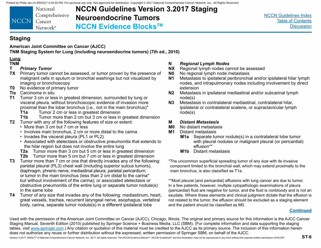
NCCN Guidelines IndexTable of Contents
Discussion
Version 3.2017, 08/02/17 © National Comprehensive Cancer Network, Inc. 2017, All rights reserved. The NCCN Evidence BlocksTM, NCCN Guidelines® and this illustration may not be reproduced in any form without the express written permission of NCCN®.
American Joint Committee on Cancer (AJCC)TNM Staging System for Lung (including neuroendocrine tumors) (7th ed., 2010)
ST-6
Staging
Used with the permission of the American Joint Committee on Cancer (AJCC), Chicago, Illinois. The original and primary source for this information is the AJCC Cancer Staging Manual, Seventh Edition (2010) published by Springer Science + Business Media, LLC (SBM). (For complete information and data supporting the staging tables, visit www.springer.com.) Any citation or quotation of this material must be credited to the AJCC as its primary source. The inclusion of this information herein does not authorize any reuse or further distribution without the expressed, written permission of Springer SBM, on behalf of the AJCC.
LungTNMT Primary Tumor TX Primary tumor cannot be assessed, or tumor proven by the presence of
malignant cells in sputum or bronchial washings but not visualized by imaging or bronchoscopy
T0 No evidence of primary tumorTis Carcinoma in situT1 Tumor 3 cm or less in greatest dimension, surrounded by lung or
visceral pleura, without bronchoscopic evidence of invasion more proximal than the lobar bronchus (i.e., not in the main bronchus)*
T1a Tumor 2 cm or less in greatest dimension T1b Tumor more than 2 cm but 3 cm or less in greatest dimensionT2 Tumor with any of the following features of size or extent: • More than 3 cm but 7 cm or less • Involves main bronchus, 2 cm or more distal to the carina • Invades the visceral pleura (PL1 or PL2) • Associated with atelectasis or obstructive pneumonitis that extends to
the hilar region but does not involve the entire lung T2a Tumor more than 3 cm but 5 cm or less in greatest dimension T2b Tumor more than 5 cm but 7 cm or less in greatest dimensionT3 Tumor more than 7 cm or one that directly invades any of the following:
parietal pleural (PL3) chest wall (including superior sulcus tumors), diaphragm, phrenic nerve, mediastinal pleura, parietal pericardium; or tumor in the main bronchus (less than 2 cm distal to the carina* but without involvement of the carina); or associated atelectasis or obstructive pneumonitis of the entire lung or separate tumor nodule(s) in the same lobe
T4 Tumor of any size that invades any of the following: mediastinum, heart, great vessels, trachea, recurrent laryngeal nerve, esophagus, vertebral body, carina, separate tumor nodule(s) in a different ipsilateral lobe
N Regional Lymph Nodes NX Regional lymph nodes cannot be assessedN0 No regional lymph node metastasisN1 Metastasis to ipsilateral peribronchial and/or ipsilateral hilar lymph
nodes, and intrapulmonary nodes including involvement by direct extension
N2 Metastasis in ipsilateral mediastinal and/or subcarinal lymph node(s)
N3 Metastasis in contralateral mediastinal, contralateral hilar, ipsilateral or contralateral scalene, or supraclavicular lymph node(s)
M Distant Metastasis M0 No distant metastasisM1 Distant metastasis M1a Separate tumor nodule(s) in a contralateral lobe tumor
with pleural nodules or malignant pleural (or pericardial) effusion**
M1b Distant metastasis
* The uncommon superficial spreading tumor of any size with its invasive component limited to the bronchial wall, which may extend proximally to the main bronchus, is also classified as T1a.
**Most pleural (and pericardial) effusions with lung cancer are due to tumor. In a few patients, however, multiple cytopathologic examinations of pleura (pericardial) fluid are negative for tumor, and the fluid is nonbloody and is not an exudate. Where these elements and clinical judgment dictate that the effusion is not related to the tumor, the effusion should be excluded as a staging element and the patient should be classified as M0.
Continued
NCCN Guidelines Version 3.2017 StagingNeuroendocrine TumorsNCCN Evidence BlocksTM
Printed by Philip Jax on 8/9/2017 2:44:55 PM. For personal use only. Not approved for distribution. Copyright © 2017 National Comprehensive Cancer Network, Inc., All Rights Reserved.
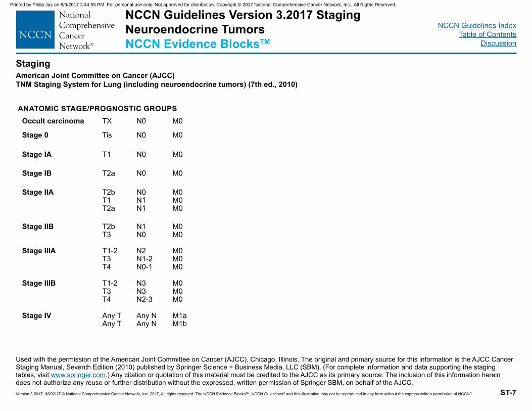
NCCN Guidelines IndexTable of Contents
Discussion
Version 3.2017, 08/02/17 © National Comprehensive Cancer Network, Inc. 2017, All rights reserved. The NCCN Evidence BlocksTM, NCCN Guidelines® and this illustration may not be reproduced in any form without the express written permission of NCCN®.
American Joint Committee on Cancer (AJCC)TNM Staging System for Lung (including neuroendocrine tumors) (7th ed., 2010)
ST-7
Staging
Used with the permission of the American Joint Committee on Cancer (AJCC), Chicago, Illinois. The original and primary source for this information is the AJCC Cancer Staging Manual, Seventh Edition (2010) published by Springer Science + Business Media, LLC (SBM). (For complete information and data supporting the staging tables, visit www.springer.com.) Any citation or quotation of this material must be credited to the AJCC as its primary source. The inclusion of this information herein does not authorize any reuse or further distribution without the expressed, written permission of Springer SBM, on behalf of the AJCC.
Occult carcinoma TX N0 M0
Stage 0 Tis N0 M0
Stage IA T1 N0 M0
Stage IB T2a N0 M0
Stage IIA T2bT1T2a
N0N1N1
M0M0M0
Stage IIB T2bT3
N1N0
M0M0
Stage IIIA T1-2T3T4
N2N1-2N0-1
M0M0M0
Stage IIIB T1-2T3T4
N3N3N2-3
M0M0M0
Stage IV Any TAny T
Any NAny N
M1aM1b
ANATOMIC STAGE/PROGNOSTIC GROUPS
NCCN Guidelines Version 3.2017 StagingNeuroendocrine TumorsNCCN Evidence BlocksTM
Printed by Philip Jax on 8/9/2017 2:44:55 PM. For personal use only. Not approved for distribution. Copyright © 2017 National Comprehensive Cancer Network, Inc., All Rights Reserved.
Version 3.2017, 08/02/17 © National Comprehensive Cancer Network, Inc. 2017, All rights reserved. The NCCN Evidence BlocksTM, NCCN Guidelines® and this illustration may not be reproduced in any form without the express written permission of NCCN®. MS-1
NCCN Guidelines Index Table of Contents
Discussion
NCCN Guidelines Version 3.2017 Neuroendocrine Tumors
Discussion NCCN Categories of Evidence and Consensus
Category 1: Based upon high-level evidence, there is uniform NCCN consensus that the intervention is appropriate.
Category 2A: Based upon lower-level evidence, there is uniform NCCN consensus that the intervention is appropriate.
Category 2B: Based upon lower-level evidence, there is NCCN consensus that the intervention is appropriate.
Category 3: Based upon any level of evidence, there is major NCCN disagreement that the intervention is appropriate.
All recommendations are category 2A unless otherwise noted.
Table of Contents Overview ............................................................................................. MS-2 Literature Search Criteria and Guidelines Update Methodology ......... MS-2 Histologic Classification and Staging of Neuroendocrine Tumors ...... MS-3 Sporadic Neuroendocrine Tumors ...................................................... MS-5
Neuroendocrine Tumors of the Gastrointestinal Tract, Lung, and Thymus (Carcinoid Tumors) ............................................................ MS-5
Evaluation of Neuroendocrine Tumors of the Gastrointestinal Tract, Lung, and Thymus ....................................................................... MS-6 Management of Locoregional Disease ........................................ MS-7 Surveillance of Resected Neuroendocrine Tumors of the Gastrointestinal Tract, Lung, and Thymus ................................... MS-9 Evaluation of Locoregional Unresectable and/or Distant Metastatic Gastrointestinal Tract, Bronchopulmonary and Thymic Neuroendocrine Tumors ............................................................ MS-11 Management of Locoregional Unresectable and/or Distant Metastatic Gastrointestinal Tract Neuroendocrine Tumors ......................... MS-11 Management of Locoregional Unresectable and/or Distant Metastatic Bronchopulmonary or Thymic Neuroendocrine Tumors ............ MS-15
Neuroendocrine Tumors of the Pancreas ..................................... MS-16 Evaluation of Neuroendocrine Tumors of the Pancreas ............ MS-17
Primary Treatment of Locoregional Resectable Neuroendocrine Tumors of the Pancreas ............................................................ MS-19 Surveillance of Resected Pancreatic Neuroendocrine Tumors . MS-21 Management of Locoregional Unresectable and/or Metastatic Neuroendocrine Tumors of the Pancreas .................................. MS-21
Neuroendocrine Tumors of Unknown Primary .............................. MS-25 Evaluation of Neuroendocrine Tumors of Unknown Primary ..... MS-25 Primary Treatment of Neuroendocrine Tumors of Unknown Primary .................................................................................................. MS-25
Adrenal Gland Tumors .................................................................. MS-26 Evaluation and Treatment of Adrenal Gland Tumors ................ MS-26
Pheochromocytomas/Paragangliomas .......................................... MS-30 Evaluation for Pheochromocytomas/Paragangliomas ............... MS-31 Genetic Counseling/Testing in Pheochromocytomas/Paragangliomas .................................................................................................. MS-31 Primary Treatment of Pheochromocytomas/Paragangliomas ... MS-32 Surveillance of Pheochromocytomas/Paragangliomas.............. MS-33
High Grade or Poorly Differentiated Neuroendocrine Carcinomas/Large or Small Cell Carcinomas .............................................................. MS-33
Evaluation of High Grade or Poorly Differentiated/Large or Small Cell Carcinomas ............................................................................... MS-33 Primary Treatment of Extrapulmonary Poorly Differentiated/Large or Small Cell Neuroendocrine Carcinomas .................................... MS-33 Surveillance of Poorly Differentiated/Large or Small Cell Carcinomas .................................................................................................. MS-34
Multiple Endocrine Neoplasia ........................................................... MS-34 MEN1 ............................................................................................ MS-34
Evaluation of MEN1 Syndromes ................................................ MS-35 Genetic Counseling/Testing in MEN1 ........................................ MS-36 Primary Treatment of MEN1 Syndromes ................................... MS-36 MEN1 Surveillance .................................................................... MS-37
MEN2 and Familial MTC ............................................................... MS-38 Evaluation of MEN2A, MEN2B, and Familial MTC .................... MS-39 Genetic Counseling/Testing in MEN2 ........................................ MS-39 Primary Treatment of MEN2A, MEN2B, and Familial MTC ....... MS-39 MEN2 Surveillance .................................................................... MS-40
Future Trial Design ........................................................................... MS-40 References ....................................................................................... MS-42
Printed by Philip Jax on 8/9/2017 2:44:55 PM. For personal use only. Not approved for distribution. Copyright © 2017 National Comprehensive Cancer Network, Inc., All Rights Reserved.
Version 3.2017, 08/02/17 © National Comprehensive Cancer Network, Inc. 2017, All rights reserved. The NCCN Evidence BlocksTM, NCCN Guidelines® and this illustration may not be reproduced in any form without the express written permission of NCCN®. MS-2
NCCN Guidelines Index Table of Contents
Discussion
NCCN Guidelines Version 3.2017 Neuroendocrine Tumors
Overview
Neuroendocrine tumors are thought to arise from cells throughout the diffuse endocrine system. They comprise a broad family of tumors, the most common of which are carcinoid tumors (most commonly arising in the lungs and bronchi [so-called bronchopulmonary], small intestine, appendix, rectum, and thymus) and pancreatic neuroendocrine tumors. Other less common neuroendocrine tumors include those arising in the parathyroid, thyroid, adrenal, and pituitary glands.
An analysis of the SEER database estimated that the incidence of neuroendocrine tumors in the United States was 5.25 cases per 100,000 people in the year 2004.1 This analysis suggested that the incidence of neuroendocrine tumors is increasing, and that the prevalence of individuals with neuroendocrine tumors in the United States may exceed 100,000.1 Other independent analyses of the SEER database also found that the incidence of gastrointestinal (GI) neuroendocrine tumors increased from 1975 to 2008.2,3 The reasons for this increase are unclear, although it seems likely that improved diagnosis and classification is one factor.4
Most neuroendocrine tumors seem to be sporadic, and risk factors for sporadic neuroendocrine tumors are poorly understood. Neuroendocrine tumors may also arise in the context of inherited genetic syndromes, including multiple endocrine neoplasia (MEN) types 1 and 2. Multiple endocrine neoplasia type 1 (MEN1), associated with mutations in the menin gene, is characterized by multiple tumors of the parathyroid, pituitary, and pancreatic glands.5 Multiple endocrine neoplasia type 2 (MEN2), associated with mutations in the RET proto-oncogene, is characterized by the development of medullary thyroid cancer, pheochromocytoma (often bilateral), and hyperparathyroidism.6
Neuroendocrine tumors have also been associated with von Hippel-Lindau disease, tuberous sclerosis complex, and neurofibromatosis.7,8
Patients with neuroendocrine tumors may or may not have symptoms attributable to hormonal hypersecretion. These symptoms include intermittent flushing and diarrhea in patients with carcinoid syndrome,9 hypertension in patients with pheochromocytoma,10 and symptoms attributable to secretion of insulin, glucagon, gastrin, and other peptides in patients with pancreatic neuroendocrine tumors.11 Patients with hormonal symptoms are considered to have “functional” tumors, and those without symptoms are considered to have “nonfunctional” tumors.
Appropriate diagnosis and treatment of neuroendocrine tumors often involves collaboration between specialists in multiple disciplines, using specific biochemical, radiologic, and surgical methods. Specialists include pathologists, endocrinologists, radiologists (including nuclear medicine specialists), and medical, radiation, and surgical oncologists.
These guidelines discuss the diagnosis and management of both sporadic and hereditary neuroendocrine tumors and are intended to assist with clinical decision-making. Most of the guideline sections pertain to well-differentiated, low- to intermediate-grade tumors, although poorly differentiated/high-grade/large or small cell carcinomas are also addressed (see Poorly Differentiated Neuroendocrine Carcinomas/Large or Small Cell Carcinomas, below). Medical practitioners should note that unusual patient scenarios (presenting in <5% of patients) are not specifically discussed in these guidelines.
Literature Search Criteria and Guidelines Update Methodology Prior to the update of this version of the NCCN Guidelines for Neuroendocrine Tumors, an electronic search of the PubMed database
Printed by Philip Jax on 8/9/2017 2:44:55 PM. For personal use only. Not approved for distribution. Copyright © 2017 National Comprehensive Cancer Network, Inc., All Rights Reserved.
Version 3.2017, 08/02/17 © National Comprehensive Cancer Network, Inc. 2017, All rights reserved. The NCCN Evidence BlocksTM, NCCN Guidelines® and this illustration may not be reproduced in any form without the express written permission of NCCN®. MS-3
NCCN Guidelines Index Table of Contents
Discussion
NCCN Guidelines Version 3.2017 Neuroendocrine Tumors
was performed to obtain key literature in the field published between May 1, 2015 and May 1, 2016, using the following search terms: (neuroendocrine tumor) OR (adrenal cancer) OR (carcinoid) OR (pheochromocytoma) OR (paraganglioma) OR (Multiple Endocrine Neoplasia). The PubMed database was chosen because it remains the most widely used resource for medical literature and indexes only peer-reviewed biomedical literature.12
The search results were narrowed by selecting studies in humans published in English. Results were confined to the following article types: Clinical Trial, Phase II; Clinical Trial, Phase III; Practice Guideline; Randomized Controlled Trial; Meta-Analysis; Systematic Reviews; and Validation Studies.
The PubMed search resulted in 138 citations, and their potential relevance was examined. The data from key PubMed articles and articles from additional sources deemed as relevant to these guidelines and discussed by the panel have been included in this version of the Discussion section (eg, e-publications ahead of print, meeting abstracts). Recommendations for which high-level evidence is lacking are based on the panel’s review of lower-level evidence and expert opinion.
The complete details of the Development and Update of the NCCN Guidelines are available on the NCCN website (www.NCCN.org).
Histologic Classification and Staging of Neuroendocrine Tumors Neuroendocrine tumors are generally subclassified by site of origin, stage, and histologic characteristics.
Histologic Classification
Neuroendocrine tumors are classified histologically based on tumor differentiation (well or poorly differentiated) and tumor grade (grades 1–3). Most neuroendocrine tumors fall into 3 broad histologic categories: well-differentiated, low-grade (G1); well-differentiated, intermediate-grade (G2); and poorly differentiated, high-grade (G3).
Tumor differentiation and tumor grade often correlate with mitotic count and Ki-67 proliferation index. In fact, most commonly used histologic classification schemes, including both the European Neuroendocrine Tumor Society and WHO systems, incorporate mitotic rate and Ki-67 index.11,13,14 Numerous studies have confirmed that increased mitotic rate and high Ki-67 index are associated with a more aggressive clinical course and worse prognosis.15-18 In most cases, well-differentiated, low-grade tumors have a mitotic count of less than 2/10 high-power field (HPF) and/or a Ki-67 index of less than 3%. Well-differentiated, intermediate-grade tumors usually have a mitotic count of 2 to 20/10 HPF and/or a Ki-67 index of 3% to 20%. In high-grade tumors, the mitotic count usually exceeds 20/10 HPF and/or the Ki-67 index exceeds 20%.
Grade is generally defined by mitotic count and/or Ki-67 index, whichever is higher. In some cases, however, tumors may not fall clearly into one category. For example, a morphologically well-differentiated neuroendocrine tumor with a low mitotic index may have a Ki-67 proliferation index that falls into the high-grade category.19,20 While technically classified as a high-grade tumor, clinical judgment should be used in making treatment decisions for such cases. A key recommendation is that tumor differentiation, mitotic rate, and Ki-67 index should all be included in the pathology report. Doing so allows the
Printed by Philip Jax on 8/9/2017 2:44:55 PM. For personal use only. Not approved for distribution. Copyright © 2017 National Comprehensive Cancer Network, Inc., All Rights Reserved.
Version 3.2017, 08/02/17 © National Comprehensive Cancer Network, Inc. 2017, All rights reserved. The NCCN Evidence BlocksTM, NCCN Guidelines® and this illustration may not be reproduced in any form without the express written permission of NCCN®. MS-4
NCCN Guidelines Index Table of Contents
Discussion
NCCN Guidelines Version 3.2017 Neuroendocrine Tumors
treating physician to factor these data into the clinical picture to make appropriate treatment decisions.
The classification of lung and thymus carcinoids varies from that of gastroenteropancreatic neuroendocrine tumors in some classification systems, and in particular does not include Ki-67 and includes the assessment of necrosis. Well-differentiated neuroendocrine tumors of the lung and thymus are either considered typical (low-grade, <2 mitoses/10 HPF and no necrosis) or atypical (2–10 mitosis/10 HPF and/or foci of necrosis).
Poorly differentiated neuroendocrine carcinomas are of either small cell or large cell cytology, with greater than 10 mitoses/10 HPF.21,22
Considerable debate remains as to the most appropriate Ki-67 proliferative threshold for the determination of tumor grade and consequent treatment decisions.23,24 A retrospective database review of 252 patients with high-grade GI neuroendocrine carcinoma suggested that platinum-based chemotherapy is most active in those with a Ki-67 index of greater than or equal to 55%.25 These results suggest that a higher Ki-67 cutoff than is currently recommended may be more appropriate to classify tumors as high-grade. Conversely, for low-grade tumors, some studies have suggested that the currently accepted cutoff may be too low. An analysis of data from 274 patients with pancreatic neuroendocrine tumors found that a 5% Ki-67 cutoff (rather than 2%) was the optimal prognostic indicator.26 A comparable analysis based on 691 patients with jejunal-ileocecal neuroendocrine tumors similarly found that a threshold of 5 mitoses/10 HPF provided better prognostic information than one of 2 mitoses/10 HPF.27 The panel recommends that the current histologic grading system be used more as a general guide, in conjunction with clinical judgment, when treatment decisions are made.
Staging
Neuroendocrine tumors are staged according to the AJCC tumor (T), node (N), metastasis (M) staging system. The AJCC introduced its first TNM staging system for the classification of neuroendocrine tumors in its 7th edition of the AJCC Cancer Staging Manual.28 Carcinoids of the stomach, duodenum/ampulla/jejunum/ileum, colon/rectum, and appendix have separate staging systems. The association of tumor stage with prognosis has been confirmed in analyses of the SEER database and the National Cancer Data Base.29-34 A recent analysis of 691 patients with jejunal-ileocecal neuroendocrine tumors treated at the Moffitt Cancer Center between 2000 and 2010 revealed 5-year survival rates of 100%, 100%, 91%, and 72% for stages I through IV, respectively, further validating the TNM staging system.27 Of note, however, this analysis also suggested that, unlike other malignancies, primary tumor size and depth of invasion had little bearing on survival in early-stage disease.24 Similar results were reported in a recent analysis of 6792 small intestine neuroendocrine tumors in the SEER database, which found that outcomes were similar for patients with T1 and T2 tumors.35 These results have been supported in additional analyses, confirming that the presence of lymph node and distant metastases have the strongest effect on survival.36,37
Carcinoids of the lungs and bronchi are staged in the same manner as more common lung carcinomas. As in lung carcinoma, more advanced tumor stage for carcinoid tumors of the lungs and bronchi is associated with worse prognosis.28
The TNM staging system for the classification of pancreatic neuroendocrine tumors in the 7th edition of the AJCC Cancer Staging Manual is the same as for exocrine pancreatic carcinoma.28 The primary tumor (T) is differentiated based on size and involvement of major
Printed by Philip Jax on 8/9/2017 2:44:55 PM. For personal use only. Not approved for distribution. Copyright © 2017 National Comprehensive Cancer Network, Inc., All Rights Reserved.
Version 3.2017, 08/02/17 © National Comprehensive Cancer Network, Inc. 2017, All rights reserved. The NCCN Evidence BlocksTM, NCCN Guidelines® and this illustration may not be reproduced in any form without the express written permission of NCCN®. MS-5
NCCN Guidelines Index Table of Contents
Discussion
NCCN Guidelines Version 3.2017 Neuroendocrine Tumors
vessels or other organs (see Staging in the guidelines). A retrospective analysis of 425 patients with pancreatic neuroendocrine tumors treated at the Moffitt Cancer Center between 1999 and 2010 validated this system, with 5-year overall survival rates of 92%, 84%, 81%, and 57% for stages I through IV, respectively (P < .001).38 Although the trends of this analysis are consistent with population-based studies, the survival rates from this analysis were significantly higher than those seen in population-based studies.39,40 For example, in the SEER database analysis of pancreatic neuroendocrine tumors, the 5-year survival rate for patients with metastatic disease was only 19.5%.40
Pathologic Reporting
In addition to information on histologic classification and stage, the margin status (positive or negative) and the presence of vascular or perineural invasion should be included in the pathology report; some studies have suggested that these factors may also have prognostic significance.41,42
Whether or not tumors are associated with symptoms of hormone hypersecretion (”functioning” or “non-functioning”) is, in general, a part of the clinical rather than histologic diagnosis. Thus, functional status is usually not included in the pathology report. However, if a specific clinical situation suggests that correlation with histologic evidence of peptide hormone may be helpful, then histochemical or immunohistochemical studies may be performed and included in the report.
Other Potential Prognostic Markers
Chromogranin A is a secreted protein that may be elevated in patients with neuroendocrine tumors; elevated levels have been associated with poorer prognosis. The molecular basis of neuroendocrine tumors
remains poorly understood, and additional molecular predictors of outcome remain investigational. A recent study found that overexpression of mammalian target of rapamycin (mTOR) or its downstream targets was associated with shorter overall survival in 195 neuroendocrine tissue samples (15% were located in the pancreas; 85% were GI carcinoids).43 Small bowel carcinoid tumors have been found to have recurrent mutations in the cyclin-dependent kinase inhibitor, CDKN1B (p27),44 and loss of CDKN1B expression has been reported to be an adverse prognostic factor in gastroenteropancreatic neuroendocrine tumors.45 Circulating tumor cells (CTCs) have also been studied as possible prognostic markers, based on the idea that tumor cells in the blood would be indicative of more disseminated disease. A recent study found that the presence of greater than or equal to 1 CTC in 7.5 mL of blood was independently associated with worse progression-free survival (PFS) and overall survival in patients with varyingly pre-treated metastatic neuroendocrine tumors from various primary sites.46
More research is required, however, before these and other new molecular assays are routinely used in the clinic. A multinational consensus meeting of experts concluded that, to date, no single currently available biomarker is sufficient as a diagnostic, prognostic, or predictive marker in patients with neuroendocrine tumors.47
Sporadic Neuroendocrine Tumors Neuroendocrine Tumors of the Gastrointestinal Tract, Lung, and Thymus (Carcinoid Tumors) Approximately one-third of carcinoid tumors arise in the lungs or thymus, and two-thirds arise in the GI tract. Sites of origin within the GI tract include the stomach, small intestine, appendix, and rectum.1 The prognosis for patients with carcinoid tumors varies according to the
Printed by Philip Jax on 8/9/2017 2:44:55 PM. For personal use only. Not approved for distribution. Copyright © 2017 National Comprehensive Cancer Network, Inc., All Rights Reserved.
Version 3.2017, 08/02/17 © National Comprehensive Cancer Network, Inc. 2017, All rights reserved. The NCCN Evidence BlocksTM, NCCN Guidelines® and this illustration may not be reproduced in any form without the express written permission of NCCN®. MS-6
NCCN Guidelines Index Table of Contents
Discussion
NCCN Guidelines Version 3.2017 Neuroendocrine Tumors
stage at diagnosis, histologic classification, and primary site of the tumor (see Histologic Classification and Staging of Neuroendocrine Tumors, above).
Neuroendocrine tumors of the GI tract, lung, or thymus may secrete various hormones and vasoactive peptides. Bronchial and thymic neuroendocrine tumors have been associated with adrenocorticotropic hormone (ACTH) production and are a cause of Cushing’s syndrome.48,49 Neuroendocrine tumors arising in the small intestine or appendix are more commonly associated with carcinoid syndrome, related to the secretion of serotonin, histamine, or tachykinins into the systemic circulation causing episodic flushing and diarrhea.50 Approximately 50% to 66% of patients with carcinoid syndrome develop valvular cardiac complications consisting of tricuspid regurgitation and/or pulmonary stenosis.51
The metabolic products released by intestinal neuroendocrine tumors are rapidly destroyed by liver enzymes in the portal circulation. Thus, the classical syndrome, occurring in approximately 8% to 28% of patients with neuroendocrine tumors,52,53 is not usually observed unless liver metastases or rarely retroperitoneal disease have occurred, in which case hepatic metastases release metabolic products directly into the systemic circulation via the hepatic veins.
These guidelines address 7 major subtypes of carcinoid tumors: 1) jejunal/ileal/colon, 2) duodenal, 3) appendix, 4) rectal, 5) gastric, 6) bronchopulmonary, and 7) thymus.
Evaluation of Neuroendocrine Tumors of the Gastrointestinal Tract, Lung, and Thymus Patients who present with suspected carcinoid tumors should be evaluated with imaging studies to assess disease burden and possible primary location. Commonly used techniques include CT and MRI.
Neuroendocrine tumors of the GI tract, lung, and thymus are highly vascular and can appear isodense with liver on conventional CT scan, depending on contrast phase. Multiphase CT or MRI scans with contrast should therefore be used for evaluation of liver metastasis. Chest CT scans with or without contrast are also recommended as appropriate to assess for lung metastases.
Because most neuroendocrine tumors express high-affinity receptors for somatostatin,50,54 somatostatin receptor-based imaging may be considered in the initial evaluation of patients with neuroendocrine tumors. Such imaging can provide useful information on overall tumor burden and location; additionally, positive imaging confirms the presence of somatostatin receptors, which can have therapeutic implications. Scintigraphy using 111Indium-diethylenetriaminepentaacetic acid (111In-DPTA)-octreotide is considered to be one standard imaging technique.55,56 Several studies have also shown diagnostic utility, as well as high sensitivity, of PET/CT imaging using the radiolabeled somatostatin analog gallium-68 (68Ga) dotatate.57-59 Unless otherwise indicated, somatostatin receptor-based imaging in this discussion includes imaging with either somatostatin receptor scintigraphy or 68Ga-dotatate PET/CT. Additional recommendations vary by disease site and include colonoscopy and small bowel imaging with CT enterography or capsule endoscopy as appropriate for jejunal, ileal, and colonic neuroendocrine tumors; endoscopic ultrasound (EUS) and/or esophagogastroduodenoscopy (EGD) as appropriate for duodenal and gastric neuroendocrine tumors; proctoscopic examination for rectal neuroendocrine tumors; and bronchoscopy as appropriate for bronchopulmonary and thymic neuroendocrine tumors.
Biochemical evaluation can also be helpful in the initial diagnostic evaluation, particularly in patients who have clinical symptoms that are suggestive of hormone hypersecretion. Evaluation of serotonin
Printed by Philip Jax on 8/9/2017 2:44:55 PM. For personal use only. Not approved for distribution. Copyright © 2017 National Comprehensive Cancer Network, Inc., All Rights Reserved.
Version 3.2017, 08/02/17 © National Comprehensive Cancer Network, Inc. 2017, All rights reserved. The NCCN Evidence BlocksTM, NCCN Guidelines® and this illustration may not be reproduced in any form without the express written permission of NCCN®. MS-7
NCCN Guidelines Index Table of Contents
Discussion
NCCN Guidelines Version 3.2017 Neuroendocrine Tumors
secretion, using a 24-hour urine collection for 5-HIAA, is generally recommended in patients with metastatic lung or GI carcinoid tumors, particularly if carcinoid syndrome, manifested by symptoms of flushing and diarrhea, is suspected. Screening for hormones in asymptomatic individuals is not routinely recommended. Chromogranin A is sometimes used as a biochemical marker in non-functioning tumors (category 3). Whereas one meta-analysis calculated the sensitivity and specificity of chromogranin A to be 73% and 95%, respectively, for diagnosis of neuroendocrine tumors,60 others have questioned its value. Chromogranin A is elevated in patients with renal or hepatic impairment and in patients receiving proton pump inhibitors (PPIs), and in general should not be relied upon in isolation as a diagnostic test. A workup for Cushing’s syndrome (discussed in Evaluation and Treatment of Cushing’s Syndrome, below) may also be indicated in cases of bronchopulmonary or thymic neuroendocrine tumors if signs and symptoms of hypercortisolemia are suspected. Details of the evaluation and diagnosis of a patient with Cushing’s syndrome from a bronchial neuroendocrine tumor have recently been published.61
Management of Locoregional Disease The management of locoregional neuroendocrine tumors of the GI tract, lung, and thymus depends on tumor size, primary site, and the general condition of the patient. Resection is the primary treatment approach for most localized carcinoid tumors. Although symptoms of hormone hypersecretion are more common in patients with metastatic disease, for patients with locoregional disease and symptoms of hormone hypersecretion, symptom control with octreotide or lanreotide is paramount (see Management of Locoregional Unresectable and/or Metastatic Neuroendocrine Tumors of the Gastrointestinal Tract, Lung, and Thymus, below). Specific recommendations for management of neuroendocrine tumor subtypes are described herein.
Gastric Neuroendocrine Tumors Three types of gastric neuroendocrine tumors are recognized: type 1 (associated with chronic atrophic gastritis or high gastric pH); type 2 (associated with antrum-sparing type A Zollinger-Ellison syndrome); and type 3 (sporadic, unifocal, unassociated with either atrophic gastritis or Zollinger-Ellison syndrome).62 Types 1 and 2 gastric neuroendocrine tumors are both associated with hypergastrinemia; the major difference between them is that patients with type 1 gastric neuroendocrine tumors generally have antrum-sparing atrophic gastritis with a loss of the usual negative feedback loop on the gastrin-producing cells of the antrum by acid, resulting in hypergastrinemia and excess stimulation of the endocrine cells of the fundus, and patients with type 2 gastric neuroendocrine tumors have evidence of acid hypersecretion secondary to gastrinoma (Zollinger-Ellison syndrome).62 Type 1 gastric neuroendocrine tumors pursue an indolent course, with a rate of metastases of <5%. Evidence suggestive of type 1 disease includes a histologic diagnosis of atrophic gastritis on gastric biopsy, elevated gastric pH, vitamin B12 deficiency, and positive anti-intrinsic factor antibodies (not all tests need to be done to make a diagnosis). For rare type 1 tumors that are >2 cm, the workup should include multiphasic CT or MRI of the abdomen performed with contrast. Type 2 tumors are rare and occur in the setting of gastrinoma in which elevated gastrin levels produce gastric neuroendocrine hyperplasia and multifocal gastric neuroendocrine tumors.
Annual endoscopic surveillance and endoscopic resection of prominent tumors is recommended for patients with locoregional type 1 gastric neuroendocrine tumors. Antrectomy can be considered if gastric tumors are increasing significantly in size or number. For locoregional type 2 gastric neuroendocrine tumors, the primary gastrinoma should, in general, be resected. If the primary tumor is not resected, endoscopic
Printed by Philip Jax on 8/9/2017 2:44:55 PM. For personal use only. Not approved for distribution. Copyright © 2017 National Comprehensive Cancer Network, Inc., All Rights Reserved.
Version 3.2017, 08/02/17 © National Comprehensive Cancer Network, Inc. 2017, All rights reserved. The NCCN Evidence BlocksTM, NCCN Guidelines® and this illustration may not be reproduced in any form without the express written permission of NCCN®. MS-8
NCCN Guidelines Index Table of Contents
Discussion
NCCN Guidelines Version 3.2017 Neuroendocrine Tumors
surveillance and endoscopic resection of prominent gastric carcinoid tumors should be considered and/or octreotide or lanreotide can be given. Gastric acid hypersecretion should be managed with high-dose PPIs. Patients with nonmetastatic gastric neuroendocrine tumors and normal gastrin levels (type 3) often have more aggressive tumors and are usually treated with radical resection of the tumor and regional lymphadenectomy. For early-stage, smaller tumors, endoscopic or wedge resection can be considered if there is no evidence of lymphadenopathy on EUS.63 Endoscopic resection should be reserved for small (<1 cm), superficial, low-grade tumors.
Thymic Neuroendocrine Tumors Localized and locoregional neuroendocrine tumors in the thymus are generally treated with surgical resection without adjuvant therapy if they have been completely resected with negative margins. There is limited data on the utility of radiation with or without chemotherapy in patients with unresectable disease or in the setting of incomplete resection or positive margins. Radiation therapy (RT) is considered by some panel members to be an option for low-grade (typical) tumors (category 3). If tumors are intermediate grade (atypical), treatment with RT with or without cisplatin or carboplatin and etoposide is more generally recommended given evidence that radiation and chemotherapy appear to have greater efficacy in tumors with higher mitotic and proliferative indices.
Bronchopulmonary Neuroendocrine Tumors Surgery, including lobectomy or other anatomic resection and mediastinal node dissection or sampling, is recommended for patients with stage I, II, and IIIA bronchopulmonary tumors. If surgery is feasible and the disease is in stage I, II, or low grade IIIA, patients may be monitored under surveillance procedures as described (see Surveillance of Resected Neuroendocrine Tumors of the
Gastrointestinal Tract, Lung, and Thymus, below). If the stage IIIA disease is intermediate grade, adjuvant therapy using cisplatin or carboplatin with etoposide in the presence or absence of radiotherapy may be considered.
There are limited data on the efficacy of chemoradiation for unresectable IIIA or IIIB lung neuroendocrine tumors. If surgical resection is not medically feasible or if there are positive margins following resection for patients with low-grade, stage IIIA disease, or if patients have low-grade, stage IIIB disease, then RT (category 3) with or without chemotherapy using cisplatin or carboplatin and etoposide (category 3) is considered by some panel members. If the stage IIIA and IIIB disease in this setting are intermediate grade, RT in the presence or absence of concurrent cisplatin or carboplatin and etoposide is generally recommended. Chemoradiation is thought to have the most efficacy for tumors with atypical histology or tumors with higher mitotic and proliferative indices.64,65
Neuroendocrine Tumors of the Duodenum, Small Intestine, and Colon For localized lesions arising in the duodenum, endoscopic resection is recommended if feasible. Transduodenal local excision with or without lymph node sampling and pancreatoduodenectomy are other options for primary treatment of nonmetastatic duodenal neuroendocrine tumors. If endoscopic resection was performed, follow-up upper endoscopy (EGD) should be performed as appropriate.
For patients presenting with tumors in the jejunum, ileum, or colon, surgical resection(s) of the bowel with regional lymphadenectomy is recommended. The surgical procedure should include careful examination of the entire bowel, because multiple synchronous lesions may be present. In addition, the proximity to or involvement of the
Printed by Philip Jax on 8/9/2017 2:44:55 PM. For personal use only. Not approved for distribution. Copyright © 2017 National Comprehensive Cancer Network, Inc., All Rights Reserved.
Version 3.2017, 08/02/17 © National Comprehensive Cancer Network, Inc. 2017, All rights reserved. The NCCN Evidence BlocksTM, NCCN Guidelines® and this illustration may not be reproduced in any form without the express written permission of NCCN®. MS-9
NCCN Guidelines Index Table of Contents
Discussion
NCCN Guidelines Version 3.2017 Neuroendocrine Tumors
superior mesenteric artery and superior mesenteric vein should be assessed during surgery.
Appendiceal Neuroendocrine Tumors Most appendiceal neuroendocrine tumors are identified incidentally, during appendectomy performed for appendicitis. Most appendiceal neuroendocrine tumors have well-differentiated histology, and for most appendiceal tumors 2 cm or smaller and confined to the appendix, simple appendectomy is sufficient because metastases are uncommon.66,67
However, some controversy exists regarding the management of appendiceal neuroendocrine tumors measuring less than 2 cm with more aggressive histologic features. A population-based study analyzing the SEER database found evidence that lymph node metastases can develop in some patients with appendiceal neuroendocrine tumors 2 cm or smaller.68 Some NCCN Member Institutions thus consider more aggressive treatment for 1- to 2-cm tumors with poor prognostic features, such as lymphovascular or mesoappendiceal invasion or atypical histologic features. In a retrospective case series that included 79 patients with appendiceal carcinoid tumors, small-vessel invasion was a risk factor for metastases in patients with tumors <2 cm.69
Patients with an incomplete resection or tumors larger than 2 cm are at risk for locoregional or distant metastases. These patients should be staged with abdominal/pelvic CT or MRI scans with contrast. Chest CT scans with contrast and biochemical evaluations may be performed as appropriate or as clinically indicated. If no distant disease is identified, they should undergo reexploration with a right hemicolectomy. Additionally, a small proportion of appendiceal neuroendocrine tumors may also contain evidence of adenocarcinoma (ie, “adenocarcinoid” or
“goblet cell carcinoid”). These tumors should be managed according to the NCCN Guidelines® for Colon Cancer (available at www.NCCN.org).
Neuroendocrine Tumors of the Rectum The treatment of rectal lesions is based on the size of the primary tumor. For small (<1 cm) and incidental lesions, complete endoscopic resection may be sufficient. All other rectal lesions should be staged using MRI or EUS. If the lesion is ≤2 cm and minimally invasive (T1), endoscopic or transanal excision is recommended. Given the higher risk of invasion with larger tumors, examination under anesthesia and/or EUS before the procedure should be considered for tumors 1 to 2 cm in size. A recent retrospective review found that metastases were present in 66% of 87 patients with well-differentiated rectal neuroendocrine tumors of 11 to 19 mm.70
Tumors larger than 2 cm, those with invasion of the muscularis propria (T2-T4), or those associated with lymph node metastases should be treated with low anterior resection or, in rare cases, an abdominoperineal resection.71
Surveillance of Resected Neuroendocrine Tumors of the Gastrointestinal Tract, Lung, and Thymus Surveillance of bronchopulmonary and GI neuroendocrine tumors should include complete patient history and physical examination (H&P) and consideration of a multiphasic CT or an MRI scan with contrast (usually abdominal and/or pelvic). For patients with primary lung and thymic tumors, chest CT scans with or without contrast are recommended. Surveillance imaging of the chest may also be considered if clinically indicated in patients with primary GI tumors. Most patients with neuroendocrine tumors of the jejunum/ileum/colon, duodenum, rectum, and thymus, and type 3 gastric neuroendocrine tumors with normal gastrin levels should be reevaluated 3 to 12 months
Printed by Philip Jax on 8/9/2017 2:44:55 PM. For personal use only. Not approved for distribution. Copyright © 2017 National Comprehensive Cancer Network, Inc., All Rights Reserved.
Version 3.2017, 08/02/17 © National Comprehensive Cancer Network, Inc. 2017, All rights reserved. The NCCN Evidence BlocksTM, NCCN Guidelines® and this illustration may not be reproduced in any form without the express written permission of NCCN®. MS-10
NCCN Guidelines Index Table of Contents
Discussion
NCCN Guidelines Version 3.2017 Neuroendocrine Tumors
after resection (earlier if the patient is symptomatic) and then every 6 to 12 months for up to 10 years.
Relevant biochemical evaluations can also be performed based on pre-resection findings. Chromogranin A may be used as a tumor marker (category 3); although not diagnostic, elevated levels have been associated with recurrence.72,73 In addition, an analysis of a large prospective database showed that chromogranin A levels elevated twice the normal limit or higher were associated with shorter survival times for patients with metastatic neuroendocrine tumors (HR, 2.8; 95% CI, 1.9–4.0; P < .001).74 Chromogranin A levels can be elevated in several concurrent medical conditions, including renal or hepatic insufficiency, and are also commonly elevated in the setting of concurrent PPIs. Several panelists therefore caution that rising chromogranin A levels in an asymptomatic patient with a tumor that looks stable on imaging does not necessarily indicate that a patient should be initiated on a new therapy.
5-Hydroxyindoleacetic acid (5-HIAA), a metabolite of serotonin, in a 24-hour urine sample may also be considered as a biochemical marker in some cases, particularly in patients with metastatic small-intestinal neuroendocrine tumors. During monitoring of patients after treatment of a carcinoid tumor, decreasing levels of 5-HIAA indicate a response to treatment, whereas increasing or excessive concentration indicates that the treatment has not been successful. However, a patient with symptoms may still have a neuroendocrine tumor even if the concentration of 5-HIAA is normal. Diet and a variety of drugs can affect the 5-HIAA test. Therefore, patients should be advised not to eat avocados, bananas, cantaloupe, eggplant, pineapples, plums, tomatoes, hickory nuts/pecans, plantains, kiwi, dates, grapefruit, honeydew, or walnuts for 48 hours before the start of and during urine collection. Medications that can increase 5-HIAA include
acetaminophen, ephedrine, diazepam, nicotine, glyceryl guaiacolate (an ingredient found in some cough medicines), and phenobarbital.
Somatostatin receptor-based imaging or FDG-PET/CT scans (for high-grade tumors) are not routinely recommended for surveillance after definitive resection, but may be indicated to assess disease location and disease burden for comparison in cases of subsequent possible recurrence.
In specific cases, follow-up recommendations for patients with resected GI neuroendocrine tumors differ from the above general recommendations. For rectal tumors smaller than 1 cm, prognosis is excellent and no follow-up is usually required. Follow-up endoscopies with rectal MRI or EUS are recommended for rectal tumors that are between 1 and 2 cm, 6 and 12 months after primary therapy, and then as clinically indicated.
For appendiceal tumors 2 cm or smaller without aggressive features, follow-up examinations are done as clinically indicated. Patients with small, well-differentiated appendiceal neuroendocrine tumors are at very low risk for recurrence,75-77 and some institutions recommend no follow-up in these patients. Other institutions recommend a follow-up examination 1 year after simple appendectomy and then with decreasing frequency. However, because recurrences have rarely been reported even after resection of small appendiceal tumors, any patients with symptoms of hormone hypersecretion should be more fully evaluated.
Follow-up recommendations also differ to some extent for hypergastrinemic patients with type 1 or 2 gastric neuroendocrine tumors. For these patients, follow-up endoscopies are recommended every 6 to 12 months for the first 3 years and annually thereafter if no
Printed by Philip Jax on 8/9/2017 2:44:55 PM. For personal use only. Not approved for distribution. Copyright © 2017 National Comprehensive Cancer Network, Inc., All Rights Reserved.
Version 3.2017, 08/02/17 © National Comprehensive Cancer Network, Inc. 2017, All rights reserved. The NCCN Evidence BlocksTM, NCCN Guidelines® and this illustration may not be reproduced in any form without the express written permission of NCCN®. MS-11
NCCN Guidelines Index Table of Contents
Discussion
NCCN Guidelines Version 3.2017 Neuroendocrine Tumors
evidence of progression is seen. If clinically indicated, imaging studies should also be performed. Because gastrin levels remain persistently high in patients with atrophic gastritis, gastrin levels are generally uninformative in patients with type 1 gastric neuroendocrine tumors. Antrectomy to remove the source of gastrin production can be considered in patients with type 1 gastric neuroendocrine tumors if new lesions or increasing tumor burden is observed.
Evaluation of Locoregional Unresectable and/or Distant Metastatic Gastrointestinal Tract, Bronchopulmonary and Thymic Neuroendocrine Tumors Baseline imaging recommendations for patients suspected to have distant metastatic disease include multiphase technique CT or MRI.78,79 The most common sites of metastases from intestinal neuroendocrine tumors include regional/mesenteric lymph nodes, liver, and bones. When evaluating locoregional unresectable and/or metastatic neuroendocrine tumors of the GI tract, lung, and thymus, abdominal/pelvic multiphasic CT or MRI scans with contrast are recommended. Chest CT scans with contrast are recommended for initial evaluation of locoregional, unresectable, and/or metastatic disease in patients with lung or thymic primary tumors and if carcinoid syndrome is suspected. Chest CT scans may be performed with or without contrast when evaluating for metastases from primary tumors in other sites.
Somatostatin receptor-based imaging is recommended to assess the somatostatin receptor status of locoregional unresectable and/or metastatic neuroendocrine tumors of the GI tract, lung, or thymus, if treatment with octreotide or lanreotide is being considered. Poorly differentiated bronchopulmonary or thymic tumors may have less avidity for 68Ga-dotatate PET/CT;80 therefore, 18F-fluorodeoxyglucose (FDG)-PET/CT may be considered for neuroendocrine tumors that are poorly
differentiated or have atypical histology. If carcinoid syndrome is suspected, somatostatin receptor-based imaging may be considered to assess the somatostatin receptor status of neuroendocrine tumors, and a cardiology consultation and echocardiogram may also be considered to assess whether the patient has carcinoid heart disease.81
Baseline levels of chromogranin A (category 3) or 24-hour urine 5-HIAA may also be considered, and then repeated over time to monitor subsequent disease progression. As mentioned previously, if carcinoid syndrome is suspected, evaluation of serotonin secretion, using a 24-hour urine collection for 5-HIAA, is recommended. Bronchial and thymic tumors may also be associated with hypersecretion of ACTH that causes the development of Cushing’s syndrome;82 therefore, if clinically indicated, patients should be screened for hypercortisolemia. If Cushing’s syndrome is suspected, see discussion below (see Evaluation and Treatment of Cushing’s Syndrome, below).
Management of Locoregional Unresectable and/or Distant Metastatic Gastrointestinal Tract Neuroendocrine Tumors
Somatostatin Analogs for Control of Symptoms and Tumor Growth Patients who have metastatic neuroendocrine tumors and carcinoid syndrome should be treated with octreotide or lanreotide.83 The long-acting release (LAR) formulation of octreotide is commonly used for the chronic management of symptoms in patients with carcinoid syndrome. Standard doses of octreotide LAR are 20 to 30 mg intramuscularly every 4 weeks. Dose and frequency may be further increased for symptom control as needed. Therapeutic levels are not achieved for 10 to 14 days after LAR injection. Short-acting octreotide (usually 150–250 mcg subcutaneously 3 times daily) can be added to octreotide LAR for rapid relief of symptoms or for breakthrough symptoms.84-86
Printed by Philip Jax on 8/9/2017 2:44:55 PM. For personal use only. Not approved for distribution. Copyright © 2017 National Comprehensive Cancer Network, Inc., All Rights Reserved.
Version 3.2017, 08/02/17 © National Comprehensive Cancer Network, Inc. 2017, All rights reserved. The NCCN Evidence BlocksTM, NCCN Guidelines® and this illustration may not be reproduced in any form without the express written permission of NCCN®. MS-12
NCCN Guidelines Index Table of Contents
Discussion
NCCN Guidelines Version 3.2017 Neuroendocrine Tumors
Lanreotide has a similar mechanism of action as octreotide, but is administered as a deep subcutaneous injection. Several studies have shown it to be effective at controlling symptoms of hormone secretion in patients with carcinoid tumors, gastrinomas, or tumors secreting vasoactive intestinal peptide (VIPomas).87-91 The multinational phase III ELECT trial randomized 115 patients with carcinoid syndrome who were either naïve to or responsive to octreotide to receive 120 mg of lanreotide or placebo and evaluated the number of days patients required use of rescue octreotide.92 Patients in the lanreotide arm required less frequent rescue octreotide than those in the placebo arm (33.7% vs. 48.5%; P = .017), supporting the use of lanreotide for symptom control.
If carcinoid syndrome is poorly controlled, telotristat, in combination with octreotide or lanreotide, should be considered for persistent diarrhea. Telotristat or telotristat ethyl is a novel, small-molecule tryptophan hydroxylase (TPH) inhibitor, which decreases urinary 5-HIAA levels and the frequency of bowel movements (BMs) in patients with carcinoid syndrome.93,94 It was approved by the FDA in February 2017 and the recommendation to use telotristat for persistent diarrhea in this context, is based on the results of the TELESTAR study, a multicenter, randomized, double-blind, placebo-controlled phase III trial of 135 patients with metastatic neuroendocrine tumors and a documented history of carcinoid syndrome, who were experiencing an average of ≥4 BMs a day while receiving stable-dose somatostatin analog therapy for at least 3 months prior to enrollment in the study.95 Patients were randomized to receive placebo, telotristat ethyl (250 mg) or telotristat ethyl (500 mg), in a 1:1:1 ratio three times per day orally for 12 weeks during a double-blind treatment period. From baseline to week 12, mean BM frequency reductions per day for placebo, telotristat ethyl (250 mg), and telotristat ethyl (500 mg) were -0.9, -1.7, and -2.1,
respectively. In addition, both telotristat dosages significantly decreased mean urinary 5-HIAA compared to placebo at week 12 (P < .001).95 Compared to placebo, treatment with telotristat at either dosage, did not result in a statistically significant change in the number of observed flushing episodes,95 therefore, additional options may be considered to manage other symptoms associated with carcinoid syndrome.
During treatment for carcinoid syndrome, a cardiology consultation and echocardiogram to assess whether the patient has carcinoid heart disease should also be performed every 2 to 3 years.83 Cardiac heart disease is frequent in patients with carcinoid syndrome; in one study, 59% of patients with carcinoid syndrome were diagnosed with tricuspid regurgitation.96,97 A study of 250 patients with carcinoid syndrome showed that patients with 5-HIAA levels of 300 μmol or greater (57 mg) over 24 hours and with 3 or more flushing episodes per day were more likely to have carcinoid heart disease.98 To monitor disease control and/or progression, surveillance imaging of the abdomen and pelvis using multiphasic CT or MRI every 3–12 months and chest CT scans with or without contrast should be considered.
In patients with GI tract primary tumors who have clinically significant tumor burden or progressive disease, initiation of either octreotide or lanreotide is recommended to potentially control tumor growth if they are not already receiving it. The recommendation to consider octreotide in these patients is based on the results of the PROMID study, a placebo-controlled phase III trial of 85 patients with metastatic midgut neuroendocrine tumors, which showed median times to tumor progression of 14.3 and 6 months in the octreotide LAR and placebo groups, respectively (P = .000072).99 After 6 months of treatment, stable disease was observed in 66.7% of patients in the octreotide LAR group and in 37.2% of patients in the placebo group. Results of long-term survival of patients in the PROMID study were recently reported.100 After
Printed by Philip Jax on 8/9/2017 2:44:55 PM. For personal use only. Not approved for distribution. Copyright © 2017 National Comprehensive Cancer Network, Inc., All Rights Reserved.
Version 3.2017, 08/02/17 © National Comprehensive Cancer Network, Inc. 2017, All rights reserved. The NCCN Evidence BlocksTM, NCCN Guidelines® and this illustration may not be reproduced in any form without the express written permission of NCCN®. MS-13
NCCN Guidelines Index Table of Contents
Discussion
NCCN Guidelines Version 3.2017 Neuroendocrine Tumors
long-term follow-up, median OS was not significantly different between the arms (83.7 months in the placebo arm and 84.7 months in the octreotide arm; HR, 0.83; 95% CI, 0.44–1.46; P = .51).101 However, post-study treatment included octreotide in 38 of 43 patients in the placebo arm, possibly confounding interpretation of long-term survival results.
The recommendation that lanreotide be considered for control of tumor growth in patients with clinically significant tumor burden or progressive disease is based on results of the CLARINET study. The CLARINET study randomized 204 patients with locally advanced or metastatic nonfunctioning pancreatic or intestinal neuroendocrine tumors to receive either lanreotide or placebo and followed patients for PFS. Results from this trial showed that treatment with lanreotide for 2 years resulted in an improvement in PFS over placebo (PFS, not reached vs. 18 months; HR, 0.47; 95% CI, 0.30–0.73; P < .001).102
Patients with clinically significant progression of metastatic bronchopulmonary and GI neuroendocrine tumors can pursue several other options, as discussed below.
Resection of Metastatic Disease In some cases, patients with limited hepatic metastases or other sites of disease can undergo complete resection of the primary tumor and metastases with curative intent. One study of 172 patients who underwent hepatic resection of metastatic neuroendocrine tumors showed that long-term survival can be achieved in selected cases: the reported 10-year overall survival rate was 50.4%.103 A recent meta-analysis reported 5-year overall survival rates ranging from 41% to 100% in patients undergoing hepatic resection.104 Most patients with resected metastatic disease, however, will eventually experience recurrence.105,106 Noncurative debulking surgery can also be considered
in select cases, especially if the patient is symptomatic either from tumor bulk or hormone production.
Resection of the primary site in the setting of unresectable metastases is generally not indicated if the primary site remains asymptomatic and is relatively stable.104 However, it is not uncommon for patients with small bowel primary tumors to experience symptoms of intermittent abdominal pain from episodic bowel obstruction or bowel ischemia related to the primary tumor and surrounding fibrosis. Palliative small bowel resection is recommended in these patients.
If resection is performed and future treatment with octreotide or lanreotide is anticipated, a prophylactic cholecystectomy can be considered given the association between long-term treatment with somatostatin analogs and the development of biliary symptoms and gallstones.83
Hepatic-Directed Therapies for Metastatic Neuroendocrine Tumors of the Gastrointestinal Tract For patients with unresectable, hepatic-predominant, progressive disease, hepatic-directed therapies may be considered, mainly with the palliative goals of extending life and relieving hormonal symptoms.107-110
Cytoreductive surgery or ablative therapies such as radiofrequency ablation (RFA) or cryoablation may be considered if near-complete treatment of tumor burden can be achieved (category 2B).111-115 For unresectable liver metastases, hepatic regional therapy (arterial embolization,116 chemoembolization,117-119 or radioembolization [category 2B])119-126 is recommended.
Printed by Philip Jax on 8/9/2017 2:44:55 PM. For personal use only. Not approved for distribution. Copyright © 2017 National Comprehensive Cancer Network, Inc., All Rights Reserved.
Version 3.2017, 08/02/17 © National Comprehensive Cancer Network, Inc. 2017, All rights reserved. The NCCN Evidence BlocksTM, NCCN Guidelines® and this illustration may not be reproduced in any form without the express written permission of NCCN®. MS-14
NCCN Guidelines Index Table of Contents
Discussion
NCCN Guidelines Version 3.2017 Neuroendocrine Tumors
Everolimus for Advanced Neuroendocrine Tumors of the Gastrointestinal Tract, Lung, and Thymus For patients with progressive metastatic GI tract carcinoid tumors, everolimus can be considered. Everolimus is an inhibitor of mTOR and was well tolerated and showed evidence of antitumor effect in patients with advanced carcinoid tumors when given with octreotide LAR in a phase II trial.127 In the randomized phase III RADIANT-2 trial, 429 patients with advanced neuroendocrine tumors and carcinoid syndrome were randomized to receive octreotide LAR with everolimus or placebo.128 Based on central review, patients receiving octreotide plus everolimus had a median PFS of 16.4 months, compared with 11.3 months for patients receiving octreotide alone (P = .026). This difference in the primary endpoint of PFS did not, however, meet the predefined threshold for statistical significance. Adverse events associated with everolimus included stomatitis, rash, fatigue, and diarrhea.128 Other side effects have also been described.129-131
A subsequent trial, RADIANT-4, was an international, double-blind, placebo-controlled, phase 3 trial that randomized 302 patients with progressive, non-functional, lung or GI neuroendocrine tumors 2:1 to receive everolimus or placebo.132 In contrast to RADIANT 2, patients in RADIANT 4 were not receiving a somatostatin analog at the time of study enrollment and concurrent somatostatin analog was not a study requirement. Median PFS was 11.0 months (95% CI, 9.2–13.3) in the everolimus arm and 3.9 months (95% CI, 3.6–7.4) in the placebo arm. The hazard ratio for progression or death was 0.48 (95% CI, 0.35–0.67; P < .001). Drug-related grade 3/4 adverse events included stomatitis (9% vs. 0%), infections (7% vs. 0%), diarrhea (7% vs. 2%), anemia (4% vs. 1%), fatigue (3% vs. 1%), and hyperglycemia (3% vs. 0%). A recent report highlights the outcomes of 169 pre-treated patients with advanced neuroendocrine tumors of the pancreas (n = 85) or other sites (n = 84) who received everolimus through a compassionate use
program.133 An increased risk of adverse events in patients who had received previous radiolabeled peptide therapy or chemotherapy was noted.
Systemic Therapy for Advanced Neuroendocrine Tumors of the Gastrointestinal Tract, Lung, and Thymus Cytotoxic chemotherapy: The benefits associated with cytotoxic chemotherapy in patients with advanced carcinoid tumors appear, at best, to be modest. Tumor response rates are generally low, and no PFS benefit has been clearly demonstrated.134
Capecitabine was tested in patients with metastatic carcinoid tumors in a phase II trial; no objective responses were reported although 13 of 19 patients were reported to have experienced stable disease.135 The combination of capecitabine and oxaliplatin was assessed in a phase II study, with response rates of 23% in patients with poorly differentiated neuroendocrine tumors and 30% in well-differentiated disease.136 5-FU was assessed in the phase III E1281 trial in combination with streptozocin or doxorubicin.137 Response rates in both arms were around 16%. Dacarbazine was given following progression, with a response rate of 8%. Responses to temozolomide in advanced carcinoid are rare.138
A phase II trial assessed bevacizumab plus capecitabine and included 49 patients with GI neuroendocrine tumors.139 A PFS of 23.4 months was reported, with 18% of patients achieving a partial response and 70% achieving stable disease.
The panel lists cytotoxic chemotherapy for neuroendocrine tumors of the GI tract as a category 3 recommendation. While some panelists believe the toxicity of systemic therapy does not warrant its wide-spread use in this population, others believe that it is an important alternative
Printed by Philip Jax on 8/9/2017 2:44:55 PM. For personal use only. Not approved for distribution. Copyright © 2017 National Comprehensive Cancer Network, Inc., All Rights Reserved.
Version 3.2017, 08/02/17 © National Comprehensive Cancer Network, Inc. 2017, All rights reserved. The NCCN Evidence BlocksTM, NCCN Guidelines® and this illustration may not be reproduced in any form without the express written permission of NCCN®. MS-15
NCCN Guidelines Index Table of Contents
Discussion
NCCN Guidelines Version 3.2017 Neuroendocrine Tumors
for patients without other options for treatment. For patients with clinically significant bronchopulmonary or thymic tumor burden that is low or intermediate grade, temozolomide either administered alone or in combination with octreotide or lanreotide is an option to manage tumor burden and any associated symptoms.64,140
Alpha Interferon: Use of interferon in the setting of advanced GI tract carcinoid tumors is a category 3 recommendation. Interferon alpha has been shown in several large, non-randomized series to be associated with an antitumor effect in patients with advanced carcinoid tumors.85,141-
144 In a recent, large randomized study led by the Southwest Oncology Group, treatment with alpha interferon (5 million units 3 d/wk) was compared to treatment with bevacizumab (15 mg/kg administered every 21 days) in more than 400 patients with progressive neuroendocrine tumors.145 Treatment with octreotide was included in both arms of this study. In a preliminary report of the results, no significant difference in PFS was observed; however, the long PFS durations in both arms of the study (15.4 and 16.6 months for interferon and bevacizumab, respectively) suggest both drugs may be active in this setting.145 Because of its potential side effects, interferon is usually not initiated until failure of somatostatin analog treatment.134
Radiolabeled Somatostatin Analogs for Advanced Neuroendocrine Tumors of the Gastrointestinal Tract Several early studies initially reported that treatment with radiolabeled somatostatin analogs was associated with tumor responses in patients with advanced carcinoid tumors.146-150 A prospective phase II study of radiopeptide therapy in 90 patients with metastatic carcinoid tumors refractory to octreotide showed that treatment was associated with improvement in symptoms; radiographic regression, however, was relatively uncommon.151 Numerous large, non-randomized cohort
analyses have also reported encouraging survival rates with this approach.152-154
A recent prospective study randomized more than 200 patients with advanced midgut neuroendocrine tumors to receive treatment with either 177Lu-DOTATATE or high-dose octreotide. Results of this study showed that treatment with 177Lu-DOTATATE was associated with a significant improvement in PFS (not reached vs. 8.4 months; P < .0001).155 Objective tumor responses were observed in 18% of patients who received 177Lu-DOTATATE versus 3% in the control group (P < .001).155
Liver Transplantation for Liver Metastases of Neuroendocrine Tumors of the Gastrointestinal Tract Several series have now reported the results of liver transplantation patients with carcinoid tumors whose metastases are confined to the liver.156-161 Results from a multicenter database of 85 patients at 28 centers who underwent liver transplantation for neuroendocrine tumors were also reported.162 A recent meta-analysis showed that, while 5-year survival rates are encouraging, the majority of patients undergoing liver transplantation ultimately develop recurrence.163 The panel acknowledged the considerable associated risks and deemed liver transplantation to be investigational and not part of routine care at this time.
Management of Locoregional Unresectable and/or Distant Metastatic Bronchopulmonary or Thymic Neuroendocrine Tumors Asymptomatic patients with low tumor burden may be observed with markers and abdominal or pelvic multiphasic CT or MRI scans every 3 to 12 months. A chest CT scan with or without contrast may be performed if clinically indicated. Alternatively, such patients may be initiated on treatment with octreotide or lanreotide. No clear consensus
Printed by Philip Jax on 8/9/2017 2:44:55 PM. For personal use only. Not approved for distribution. Copyright © 2017 National Comprehensive Cancer Network, Inc., All Rights Reserved.
Version 3.2017, 08/02/17 © National Comprehensive Cancer Network, Inc. 2017, All rights reserved. The NCCN Evidence BlocksTM, NCCN Guidelines® and this illustration may not be reproduced in any form without the express written permission of NCCN®. MS-16
NCCN Guidelines Index Table of Contents
Discussion
NCCN Guidelines Version 3.2017 Neuroendocrine Tumors
exists on the timing of octreotide or lanreotide initiation in asymptomatic patients with metastatic neuroendocrine tumors and low tumor burden. Although initiation of octreotide or lanreotide can be considered in these patients, deferring initiation until evidence of tumor progression is seen may also be appropriate in selected patients.
Lung neuroendocrine tumors include a spectrum from low-grade typical carcinoids to intermediate-grade atypical carcinoids.164 If patients present with asymptomatic, low tumor burden that is low grade, they can be observed with chest CT scans with contrast and abdominal/pelvic multiphasic CT or MRI scans every 3 to 6 months. Alternatively, these patients can be treated with octreotide and lanreotide. As with GI primary tumors above, there is no clear consensus on the timing of initiation of octreotide or lanreotide in such patients and either approach may be appropriate in selected patients.
If patients with advanced low-grade lung or thymic neuroendocrine tumors present with clinically significant tumor burden, initiation of octreotide and lanreotide may be considered. Additional options for the management of advanced low-grade tumors include initiation of everolimus or temozolomide. Both treatments may be given with or without octreotide or lanreotide.
Patients with advanced intermediate-grade lung or thymic neuroendocrine tumors should generally be initiated on systemic treatment. Options include initiation of octreotide or lanreotide. Additional options include initiation of everolimus (based on the results of the RADIANT 4 study, described above). Temozolomide represents another option; temozolomide monotherapy was associated with partial responses in 14% of patients with progressive metastatic bronchial neuroendocrine tumors in a retrospective study of 31 patients140, or initiation of treatment with carboplatin or cisplatin and etoposide.
Carboplatin or cisplatin and etoposide is generally considered for tumors on the higher end of the atypical category with respect to Ki-67 and grade.64 These treatments may be given with or without octreotide or lanreotide.
Although rare, some patients may present with multiple lung nodules and widespread peripheral airway neuroendocrine cell hyperplasia. In this case, a diagnosis of diffuse idiopathic pulmonary neuroendocrine cell hyperplasia (DIPNECH) can be made.164 This condition is generally indolent, and patients can be observed with chest CT scans without contrast every 12 months or for new symptoms. If patients are symptomatic, treatment with octreotide or lanreotide is recommended.
Neuroendocrine Tumors of the Pancreas According to a population-based study, malignant pancreatic neuroendocrine tumors account for approximately 1% of pancreatic cancers by incidence and 10% of pancreatic cancers by prevalence.165 Although the peak incidence of occurrence is between ages 40 and 69 years, a significant number of patients diagnosed with pancreatic neuroendocrine tumors are younger than 35 years.165,166 Based on an analysis of pancreatic neuroendocrine tumors in the SEER database from 1973 to 2000, the annual incidence per 1 million was 1.8 in women and 2.6 in men.40 An estimated 40% to 91% of pancreatic neuroendocrine tumors are nonfunctional. The remainder manifest with clinically evident hormonal symptoms.11,40 Consistent with these numbers, analysis of the NCCN Neuroendocrine Tumors Outcomes Database found that 22% of patients with pancreatic neuroendocrine tumors had a hormonal syndrome.52 Of these functioning tumors, up to 70% are insulinomas, and only 10% are associated with metastases. Approximately 15% are glucagonomas. Gastrinomas and somatostatinomas account for another 10%; gastrinomas and
Printed by Philip Jax on 8/9/2017 2:44:55 PM. For personal use only. Not approved for distribution. Copyright © 2017 National Comprehensive Cancer Network, Inc., All Rights Reserved.

Version 3.2017, 08/02/17 © National Comprehensive Cancer Network, Inc. 2017, All rights reserved. The NCCN Evidence BlocksTM, NCCN Guidelines® and this illustration may not be reproduced in any form without the express written permission of NCCN®. MS-17
NCCN Guidelines Index Table of Contents
Discussion
NCCN Guidelines Version 3.2017 Neuroendocrine Tumors
somatostatinomas (80%–90%) are associated with a relatively high risk for metastases.166 The remaining rare pancreatic neuroendocrine tumors include VIPoma, and the recently described cholecystokininoma (CCKoma).167
Pancreatic neuroendocrine tumors occurring in patients with MEN1 are typically multiple and require different treatment strategies from those used for patients with sporadic pancreatic neuroendocrine tumors, which are usually solitary (see MEN1, below). Gastrinoma and insulinoma are the most common pancreatic neuroendocrine tumors in patients with MEN1.168
Evaluation of Neuroendocrine Tumors of the Pancreas Personal and family history should be evaluated for the possibility of MEN1 (see Multiple Endocrine Neoplasia, below). The recommended evaluation also includes an abdominal multiphasic CT or MRI scan with contrast and/or a chest CT scan with or without contrast, if clinically indicated. For evaluation of nonfunctioning pancreatic tumors, the chest CT scan may be omitted. Hormone-secreting tumors may result in significant clinical symptoms even when very small, and lesion identification can be difficult.169 Somatostatin receptor-based imaging and EUS can also be considered as appropriate.170
Biochemical evaluation is also often considered in patients with pancreatic neuroendocrine tumors because many pancreatic neuroendocrine tumors secrete specific hormones.166 Biochemical evaluation is generally guided by the presence of symptoms that might indicate the presence of excess hormone. Screening for hormones in asymptomatic individuals is not routinely recommended. The range of symptoms associated with hormonal secretion is diverse. Classic syndromes include those associated with insulinomas, which secrete insulin, resulting in fasting or nocturnal hypoglycemia. Gastrinomas
secrete gastrin, and patients often present with recurrent peptide ulcers. Glucagonomas are associated with the development of diabetes mellitus and/or migratory necrolytic erythema. Patients with somatostatinomas may also present with diabetes mellitus and/or diarrhea/steatorrhea from secretion of somatostatin. VIPomas are characterized by watery diarrhea, hypokalemia, and achlorhydria (WDHA syndrome) from secretion of vasoactive intestinal polypeptide (VIP). The guidelines describe appropriate tests for each of these situations. For nonfunctioning tumors, pancreatic polypeptide (PP; category 3) and chromogranin A (category 3) may also be tested as appropriate.
Chromogranin A levels are elevated in 60% or more of patients with either functioning or nonfunctioning pancreatic endocrine tumors.171-173 In addition, analysis of a large prospective database found that chromogranin A levels elevated twice the normal limit or higher were associated with shorter survival times for patients with metastatic neuroendocrine tumors (HR, 2.8; 95% CI, 1.9–4.0; P < .001).74 Chromogranin A was also found to be a prognostic factor in a prospective study of patients treated with everolimus.174 Care should be taken in measuring chromogranin A and interpreting the results, because spuriously elevated levels of chromogranin A have been reported in patients using PPIs, those with renal or liver failure, those with hypertension, and those with chronic gastritis.
Evaluation of Gastrinomas Gastrinoma should be suspected in patients with severe and refractory gastroduodenal ulcers or symptoms such as dyspepsia, usually accompanied by diarrhea. Evaluation of a patient with suspected gastrinoma includes measurement of basal and stimulated serum gastrin levels.175 Diagnosis of gastrinoma can be confounded by the concurrent use of PPIs, which will elevate serum gastrin levels.
Printed by Philip Jax on 8/9/2017 2:44:55 PM. For personal use only. Not approved for distribution. Copyright © 2017 National Comprehensive Cancer Network, Inc., All Rights Reserved.
Version 3.2017, 08/02/17 © National Comprehensive Cancer Network, Inc. 2017, All rights reserved. The NCCN Evidence BlocksTM, NCCN Guidelines® and this illustration may not be reproduced in any form without the express written permission of NCCN®. MS-18
NCCN Guidelines Index Table of Contents
Discussion
NCCN Guidelines Version 3.2017 Neuroendocrine Tumors
Importantly, most patients who are found to have an elevated level of serum gastrin do not have a gastrinoma but have achlorhydria or are receiving PPIs or antacids. To be useful for diagnosis, gastrin levels (basal or stimulated) must be measured after the patient is off PPI therapy for at least 1 week. After excluding retained gastric antrum by history, a combination of fasting serum gastrin level greater than 10 times the elevated and a gastric pH less than 2 is diagnostic of a gastrinoma. Patients who have clinical manifestations suspicious for a gastrinoma and a gastric pH less than 2 but with less than 10 times the elevation of serum gastrin levels require further testing.176
In addition, imaging studies (abdominal multiphasic CT/MRI scan with contrast or chest CT scan with or without contrast) often aid not only in localizing the tumor but also in confirming the diagnosis. Other tests, such as somatostatin receptor-based imaging, EUS, and chromogranin A levels (category 3), may be performed as appropriate. Approximately 70% of patients with MEN1 and gastrinoma have tumors situated in the duodenum.
The New England Journal of Medicine recently published a case report outlining the diagnosis of gastrinoma in a patient presenting with severe, recurrent diarrhea.177
Evaluation of Insulinomas Insulinomas are generally small tumors that are best localized with EUS, which has been shown to localize approximately 82% of pancreatic endocrine tumors.178 Insulinomas can also be localized by injecting calcium into selective pancreatic arteries and measuring the insulin levels in the right (usually) or left hepatic vein (Imamura-Doppman procedure).179 Most experts recommend this test only for patients with persistent or recurrent insulinoma or when other localization tests are equivocal or negative.
Serum insulin (with concurrent hypoglycemia), pro-insulin, and C-peptide should be tested.180 An insulin level greater than 3 mcIU/mL (usually >6 mcIU/mL), C-peptide concentrations of at least 0.6 ng/mL, and proinsulin levels of greater than or equal to 5 pmol/L when fasting blood glucose is less than 55 mg/dL indicated the presence of these tumors.180
To rule out metastatic disease, abdominal multiphasic CT or MRI scans with contrast should be performed accompanied by chest CT scans with or without contrast if clinically indicated. Ninety percent of insulinomas pursue an indolent course and can be cured surgically. Insulinomas are less consistently octreotide-avid than other pancreatic neuroendocrine tumors, and somatostatin receptor-based imaging may consequently be less useful as an imaging technique in insulinomas than in other tumor subtypes. Somatostatin receptor-based imaging should be performed if octreotide or lanreotide is being considered as a treatment for metastatic disease. Octreotide or lanreotide should only be administered to patients whose tumors are somatostatin-receptor positive, and patients with insulinoma should be carefully monitored when receiving octreotide or lanreotide because in some cases these drugs can profoundly worsen hypoglycemia (see Preoperative Management, below).181
The New England Journal of Medicine published a case report describing the diagnosis of insulinoma in a lactating patient presenting with periodic numbness and prolonged episodes of confusion and lethargy.182
Evaluation of Glucagonomas and VIPomas For patients with recent-onset diabetes, cachexia, and/or a necrolytic erythematous skin rash, the panel recommends a blood test for glucagon and blood glucose. For both glucagonomas and VIPomas,
Printed by Philip Jax on 8/9/2017 2:44:55 PM. For personal use only. Not approved for distribution. Copyright © 2017 National Comprehensive Cancer Network, Inc., All Rights Reserved.
Version 3.2017, 08/02/17 © National Comprehensive Cancer Network, Inc. 2017, All rights reserved. The NCCN Evidence BlocksTM, NCCN Guidelines® and this illustration may not be reproduced in any form without the express written permission of NCCN®. MS-19
NCCN Guidelines Index Table of Contents
Discussion
NCCN Guidelines Version 3.2017 Neuroendocrine Tumors
abdominal multiphase contrast-enhanced CT or MRI scans with contrast may be useful for identifying large tumors or metastatic disease. Chest CT scans with or without contrast can be performed as clinically indicated. Somatostatin receptor-based imaging and EUS can be performed as appropriate.
For suspected VIPomas with characteristic watery diarrhea, testing for VIP and electrolytes is recommended. A recent case report describes the diagnosis and treatment of a patient with VIPoma.183
Primary Treatment of Locoregional Resectable Neuroendocrine Tumors of the Pancreas Resection is the primary treatment approach for localized pancreatic neuroendocrine tumors when possible, and can result in excellent outcomes. Exceptions to surgery include patients with other life-limiting comorbidities or high surgical risk, particularly if tumors are small and indolent.
Preoperative Management Surgical resection is the optimal treatment for locoregional pancreatic endocrine tumors. Before excision, however, any symptoms of hormonal excess must be treated. Octreotide or lanreotide can be considered for symptom control in most pancreatic neuroendocrine tumor subtypes.83 Octreotide or lanreotide should be used with caution in patients with insulinoma, because they can also suppress counterregulatory hormones such as growth hormone (GH), glucagon, and catecholamines. In this situation, octreotide and lanreotide can precipitously worsen hypoglycemia, and can result in fatal complications.181 Octreotide and lanreotide should not be used in patients with insulinoma in patients who have a negative result by somatostatin receptor-based imaging.
In addition, specific measures are often recommended based on symptoms. For insulinomas, the panel advises stabilizing glucose levels with diet and/or diazoxide. Everolimus can also be considered in this scenario.184 For gastrinomas, gastrin hypersecretion may be treated with high-dose PPIs. For patients with glucagonoma, appropriate measures should be taken to treat hyperglycemia and diabetes, including the use of IV fluids. All patients who might require splenectomy should receive preoperative trivalent vaccine (ie, pneumococcus, haemophilus influenzae b, meningococcus group c).
Surgical Management of Nonfunctioning Pancreatic Neuroendocrine Tumors Most patients with localized pancreatic neuroendocrine tumors should undergo surgical resection, absent any contraindications. Exceptions include patients with other life-limiting comorbidities, high surgical risk, or widely metastatic disease. Additionally, several studies have suggested that patients with incidentally discovered tumors <1 cm in size may be safely followed in some cases, depending on the site of the tumor.185,186 Other studies, including an analysis of the SEER database, suggest that some small tumors (measuring <2 cm in size in these studies) can pursue a more aggressive course.187-189 Other retrospective studies suggest nonoperative management can be safe for nonfunctioning pancreatic neuroendocrine tumors that are <1.7 cm or <3 cm.190,191 Based on these limited data, the panel includes observation alone as an option for selected cases of incidentally discovered small pancreatic neuroendocrine tumors, but recommends surgical resection for larger tumors absent contraindications.
Resection for larger (>2 cm), node-positive, or malignant-appearing nonfunctional tumors should include total removal of the tumor with negative margins (including adjacent organs) and regional lymph nodes.
Printed by Philip Jax on 8/9/2017 2:44:55 PM. For personal use only. Not approved for distribution. Copyright © 2017 National Comprehensive Cancer Network, Inc., All Rights Reserved.
Version 3.2017, 08/02/17 © National Comprehensive Cancer Network, Inc. 2017, All rights reserved. The NCCN Evidence BlocksTM, NCCN Guidelines® and this illustration may not be reproduced in any form without the express written permission of NCCN®. MS-20
NCCN Guidelines Index Table of Contents
Discussion
NCCN Guidelines Version 3.2017 Neuroendocrine Tumors
Lymph node resection should also be considered for tumors of 1 to 2 cm, because of the small but real risk of lymph node metastases.192,193
Surgical Management of Gastrinomas The treatment approach for gastrinoma usually depends on the results of preoperative localization studies and on findings during exploratory laparotomy. In patients with occult gastrinoma (ie, no primary tumor or metastasis is seen on imaging), the panel recommends either observation or exploratory surgery, including duodenotomy and intraoperative ultrasound with enucleation or local resection of tumors if identified at operation, and removal of periduodenal nodes.
Gastrinomas in the duodenum are treated with duodenotomy and intraoperative ultrasound with local resection or enucleation of tumors and periduodenal node dissection.
Gastrinomas in the head of the pancreas that are exophytic or peripheral as determined by imaging and are not immediately adjacent to the pancreatic duct should be enucleated. The periduodenal nodes should also be removed. Gastrinomas in the pancreatic head that are deeper or invasive and those with proximity to the main pancreatic duct should be managed with pancreatoduodenectomy.
Gastrinomas in the distal pancreas are treated with distal pancreatectomy. The role of routine splenectomy in such cases is debated. Gastrinomas in some cases may be associated with lymph node metastases,194 which are removed with splenectomy. However, no firm data support splenectomy in all cases. A third alternative is the “Warshaw technique,” which, with resection of splenic vessels but preservation of the spleen,195 can achieve lymph node retrieval comparable to distal pancreatectomy with en-bloc splenectomy.
Surgical Management of Insulinomas The primary treatment for exophytic or peripheral insulinomas, because they are primarily benign, is enucleation. This procedure can be performed laparoscopically for localized solitary tumors within the body and tail of the pancreas. Sporadic tumors are usually solitary, whereas familial tumors are multiple. If enucleation is not possible because of invasion or the location of the tumor within the pancreas, then pancreatoduodenectomy for tumors in the head of the pancreas or distal pancreatectomy with preservation of the spleen for smaller tumors not involving splenic vessels may be considered. Distal pancreatectomy can be performed laparoscopically, and a recent meta-analysis reported that laparoscopic procedures are safe for patients with insulinomas and may be associated with shorter hospital stays.196
Surgical Management of Glucagonomas Most glucagonomas are malignant and calcified and located in the tail of the pancreas, with regional node involvement. The recommended treatment is distal pancreatectomy with splenectomy and resection of the peripancreatic lymph nodes. For tumors in the pancreatic head, pancreatoduodenectomy with resection of the peripancreatic lymph nodes is recommended. Small (<2 cm) peripheral glucagonomas are rare; enucleation or local excision with peripancreatic lymph dissection may be considered for small peripheral tumors of the head or distal pancreas. A hypercoagulable state has been reported in 10% to 33% of patients with glucagonoma.197,198 Therefore, perioperative anticoagulation can be considered because of the increased risk of pulmonary emboli.
Surgical Management of VIPomas Distal VIPomas are treated with distal pancreatectomy with resection of peripancreatic lymph nodes and spleen. Pancreatoduodenectomy with dissection of peripancreatic nodes is recommended for tumors in the
Printed by Philip Jax on 8/9/2017 2:44:55 PM. For personal use only. Not approved for distribution. Copyright © 2017 National Comprehensive Cancer Network, Inc., All Rights Reserved.
Version 3.2017, 08/02/17 © National Comprehensive Cancer Network, Inc. 2017, All rights reserved. The NCCN Evidence BlocksTM, NCCN Guidelines® and this illustration may not be reproduced in any form without the express written permission of NCCN®. MS-21
NCCN Guidelines Index Table of Contents
Discussion
NCCN Guidelines Version 3.2017 Neuroendocrine Tumors
head of the pancreas. Small (<2 cm) peripheral VIPomas are rare; enucleation or local excision with peripancreatic lymph dissection may be considered for small peripheral tumors of the head or distal pancreas.
Surgical Management of Other Pancreatic Neuroendocrine Tumors The treatment recommendations for tumors secreting hormones such as somatostatinoma, ACTH, PTHrP, and PP are similar to those for nonfunctioning tumors. Tumors that are small (<2 cm) and peripheral can be enucleated with or without removal of regional nodes, or distal pancreatectomy can be performed with or without removal of regional nodes and with or without splenectomy. Deeper, larger (>2 cm), or invasive tumors are treated with pancreatoduodenectomy if they are located in the head of the pancreas, and with distal pancreatectomy and splenectomy if they are distally localized. Resection for larger (>2 cm) or malignant-appearing tumors should include total removal of the tumor with negative margins (including adjacent organs) and regional lymph nodes.
Surveillance of Resected Pancreatic Neuroendocrine Tumors Disease recurrence has been observed in 21% to 42% of patients with pancreatic neuroendocrine tumors and can occur after many years.199-201 Higher lymph node ratio and Ki-67 status may indicate a higher chance of recurrence.199 Patients should undergo follow-up 3 to 12 months after resection, or earlier if the patient presents with symptoms, and then every 6 to 12 months for a maximum of 10 years with an H&P and appropriate biochemical markers. Abdominal multiphasic CT or MRI with contrast and chest CT scans as clinically indicated can also be considered. These surveillance recommendations may also apply to cases where observation of patients with metastatic disease has been chosen. Less frequent surveillance may be appropriate for low-risk tumors such as well-differentiated stage I pancreatic neuroendocrine
tumors. Somatostatin receptor-based imaging or FDG-PET/CT scans are not recommended for routine surveillance.
The optimal duration of surveillance is unknown. In one study of 123 patients with resected sporadic pancreatic neuroendocrine tumors, most recurrences occurred within 5 years of resection, and all recurrences occurred within 10 years.202 In select cases, including resectable locoregional or oligometastatic recurrence, surgical resection may be considered.
Management of Locoregional Unresectable and/or Metastatic Neuroendocrine Tumors of the Pancreas To evaluate the extent of locoregional unresectable disease and/or distant metastases, multiphasic CT or MRI scans with contrast of the abdomen and pelvis should be performed. Somatostatin receptor-based imaging may also be considered. A chest CT scan with or without contrast and appropriate biochemical evaluation may be carried out if clinically indicated. Metastases in patients with neuroendocrine tumors of the pancreas, when they develop, often occur first in the liver. In patients with limited hepatic disease, surgical excision of both the primary tumor and liver metastases should be considered with curative intent when possible and can be performed in a staged or synchronous fashion. A recent meta-analysis reported that 5-year OS ranges from 41% to 100% in this population of patients.104 Noncurative debulking surgery can also be considered in select cases. When performing staged pancreatoduodenectomy and liver resection, hepatectomy should be considered before pancreatic resection to reduce the risk of perihepatic sepsis from the contaminated biliary tree.203 Although resection may provide clinical benefit, most patients with metastatic disease will experience recurrence.105,106 Additional resection or ablation may be possible. A study of 172 patients who had liver resection of metastatic neuroendocrine tumors (55 with the primary tumor in the
Printed by Philip Jax on 8/9/2017 2:44:55 PM. For personal use only. Not approved for distribution. Copyright © 2017 National Comprehensive Cancer Network, Inc., All Rights Reserved.
Version 3.2017, 08/02/17 © National Comprehensive Cancer Network, Inc. 2017, All rights reserved. The NCCN Evidence BlocksTM, NCCN Guidelines® and this illustration may not be reproduced in any form without the express written permission of NCCN®. MS-22
NCCN Guidelines Index Table of Contents
Discussion
NCCN Guidelines Version 3.2017 Neuroendocrine Tumors
pancreas) showed that significant long-term survival can be achieved after recurrence in many patients, with a 10-year overall survival rate of 50.4%.103
If resection is performed and future treatment with octreotide or lanreotide is anticipated, a prophylactic cholecystectomy can be considered given the association between long-term treatment with somatostatin analogs and the development of biliary symptoms and gallstones.83
Unfortunately, most patients who present with advanced pancreatic neuroendocrine tumors have unresectable disease. For selected patients with unresectable disease who are asymptomatic and have low tumor burden and stable disease, observation can be considered, with marker assessment and abdominal and pelvic multiphasic CT or MRI scans every 3 to 12 months until clinically significant disease progression occurs. Chest CT scans with or without contrast may also be performed if clinically indicated. In addition, however, treatment with lanreotide or octreotide can be considered (see discussion below). The optimal time to begin therapy in this patient population is not known.
For symptomatic patients with unresectable disease, those who initially present with clinically significant tumor burden, or those with clinically significant disease progression, octreotide or lanreotide should be considered if patients are not already receiving treatment with these options. Several different options can be considered if the disease continues to progress. Systemic options include treatment with biologically targeted agents (everolimus or sunitinib, category 2A) or treatment with cytotoxic chemotherapy (category 2A). These options, as well as hepatic-directed therapies, are discussed in more detail in the following sections.
Somatostatin Analogs Patients with pancreatic neuroendocrine tumors and symptoms of hormone secretion should, in most cases, receive treatment with either lanreotide or octreotide and/or other medication to manage their symptoms as previously described. Patients without hormone-related symptoms who have uptake with somatostatin scintigraphy can also be considered for treatment with octreotide or lanreotide. Results from the CLARINET study, in which 204 patients with gastroenteropancreatic neuroendocrine tumors (including both carcinoid and pancreatic neuroendocrine tumors) were randomized to receive treatment with either lanreotide or placebo, showed that treatment with lanreotide was associated with in an improvement in PFS (PFS, not reached vs. 18 months; HR, 0.47; 95% CI, 0.30–0.73; P < .001).102 Although no randomized studies to date have directly shown an antitumor effect of octreotide in pancreatic neuroendocrine tumors, the PROMID trial showed an improvement in its primary endpoint of time to tumor progression (14.3 vs. 6 months; P = .000072) in carcinoid tumors of the midgut.99 Lanreotide and octreotide share the same mechanism of action, and the panel believes that either lanreotide or octreotide are appropriate options for tumor control in this setting.
Additional therapies can be given in place of or in addition to octreotide or lanreotide, as discussed below.
Molecularly Targeted Therapies The molecularly targeted agents everolimus and sunitinib have been confirmed to have antitumor activity and to improve PFS in patients with advanced pancreatic neuroendocrine tumors.
Everolimus, administered orally at a dose of 10 mg once daily, was evaluated in a multicenter study (RADIANT-3) enrolling 410 patients with advanced, progressive, pancreatic neuroendocrine tumors.204 In
Printed by Philip Jax on 8/9/2017 2:44:55 PM. For personal use only. Not approved for distribution. Copyright © 2017 National Comprehensive Cancer Network, Inc., All Rights Reserved.
Version 3.2017, 08/02/17 © National Comprehensive Cancer Network, Inc. 2017, All rights reserved. The NCCN Evidence BlocksTM, NCCN Guidelines® and this illustration may not be reproduced in any form without the express written permission of NCCN®. MS-23
NCCN Guidelines Index Table of Contents
Discussion
NCCN Guidelines Version 3.2017 Neuroendocrine Tumors
this study, the median PFS duration for patients randomized to everolimus was 11.0 months, compared with 4.6 months for patients receiving placebo (P < .001). Subset analyses of RADIANT-3 suggested that the PFS benefit associated with everolimus is independent of prior or concurrent somatostatin analog therapy or prior chemotherapy.205-207 Adverse events associated with everolimus include stomatitis, hyperglycemia, and, in rare cases, pneumonitis.204 Other side effects have also been described.129-131 A recent report highlights the outcomes of 169 pre-treated patients with advanced neuroendocrine tumors of the pancreas (n = 85) or other sites (n = 84) who received everolimus through a compassionate use program.133 A higher risk of adverse events was noted in patients with previous radiolabeled peptide therapy and chemotherapy.
Sunitinib, administered orally at a dose of 37.5 mg once daily, was compared with placebo in a multicenter randomized study of patients with advanced, progressive, metastatic pancreatic neuroendocrine tumors.208 The study was designed to enroll 340 patients but was discontinued after enrollment of 171 patients, before the predefined efficacy analysis. At discontinuation, patients who received sunitinib had a median PFS duration of 11.4 months, compared with 5.5 months for patients receiving placebo (P < .001). The objective response rate seen with sunitinib was 9.3%.208 A large proportion of patients on the placebo arm subsequently received sunitinib at progression, and no significant difference in overall survival was observed between the arms.209 Adverse events associated with sunitinib include fatigue and, in rare cases, congestive heart failure.210 Other side effects have also been described.211,212
Cytotoxic Chemotherapy for Advanced Pancreatic Neuroendocrine Tumors Cytotoxic chemotherapy is another option for patients with unresectable or metastatic pancreatic neuroendocrine tumors (category 2A). While a number of regimens have been associated with antitumor activity in this setting, there is no panel consensus on which cytotoxic chemotherapy regimen is best. The alkylating agents streptozocin and temozolomide appear to have the most antitumor activity in pancreatic neuroendocrine tumors.
Streptozocin is FDA-approved for use in patients with advanced pancreatic neuroendocrine tumors. The combination of doxorubicin and streptozocin was initially reported to be associated with an overall response rate of 69% and a survival benefit in a relatively small randomized study of patients with advanced pancreatic neuroendocrine tumors.213 A retrospective review from MD Anderson Cancer Center reported an objective response rate of 39% with the combination of 5-FU, doxorubicin, and streptozocin.214 A phase II trial assessed bevacizumab combined with 5-FU and streptozocin.215 A PFS of 23.7 months was reported, with 56% of patients achieving a partial response and 44% achieving stable disease.
More recently, oral temozolomide-based therapy has been used in patients with advanced pancreatic neuroendocrine tumors. Temozolomide has been administered using different schedules, either alone or in combination with other agents.138,216-219 A retrospective series reported that the combination of temozolomide with capecitabine was associated with an objective radiographic response rate of 70% and a median PFS of 18 months.219 Another retrospective review of the temozolomide and capecitabine combination reported a 61% response rate in 18 patients, with 1 surgically proven complete pathologic
Printed by Philip Jax on 8/9/2017 2:44:55 PM. For personal use only. Not approved for distribution. Copyright © 2017 National Comprehensive Cancer Network, Inc., All Rights Reserved.
Version 3.2017, 08/02/17 © National Comprehensive Cancer Network, Inc. 2017, All rights reserved. The NCCN Evidence BlocksTM, NCCN Guidelines® and this illustration may not be reproduced in any form without the express written permission of NCCN®. MS-24
NCCN Guidelines Index Table of Contents
Discussion
NCCN Guidelines Version 3.2017 Neuroendocrine Tumors
response.220 A small recent retrospective study (7 patients) reported a response rate of 43%.221
Temozolomide-based combination regimens have also been formally evaluated in prospective, phase II studies. One such study assessed the safety and efficacy of temozolomide administered with bevacizumab, a monoclonal antibody targeted against vascular endothelial growth factor (VEGF).216 Five of the 15 patients (33%) with pancreatic neuroendocrine tumors had a radiographic response (with no responses in the 19 patients with carcinoid tumors), and the toxicity was acceptable. The combination of temozolomide with everolimus has also been studied and found to be safe, with partial responses observed in 40% of patients with pancreatic neuroendocrine tumors.222
These results suggest that the activity of temozolomide in pancreatic neuroendocrine tumors is at least comparable to that of streptozocin, and support its use in pancreatic neuroendocrine tumors. The combination of temozolomide with everolimus has also been studied. There is no current consensus, however, on the optimal temozolomide dosing regimen or whether temozolomide should be administered alone or in combination with other agents.
Other cytotoxic agents appear to be less active than streptozocin or temozolomide in pancreatic neuroendocrine tumors. 5-FU was assessed in the phase II/III E1281 trial in combination with streptozocin or doxorubicin in patients with neuroendocrine tumors of various locations, including the pancreas.137 Response rates in both arms were around 16%. Dacarbazine was given following progression, with a response rate of 8%. Other studies have also shown the combination of 5-FU and streptozocin to be effective in this setting.223,224 The combination of capecitabine and oxaliplatin was assessed in a phase II
study, with response rates of 23% in patients with poorly differentiated neuroendocrine tumors and 30% in well-differentiated disease.136
Radiolabeled Somatostatin Analogs for Advanced Pancreatic Neuroendocrine Tumors Treatment with radiolabeled somatostatin analogs has been reported to result in tumor responses in patients with advanced pancreatic neuroendocrine tumors.146-150 Numerous large, non-randomized cohort analyses have also reported encouraging survival rates with this approach.153,154 In general, these studies have enrolled only patients with evidence of high tumoral somatostatin receptor expression. A randomized study of high-dose octreotide vs. 177Lu-DOTATATE has been reported in patients with advanced midgut neuroendocrine tumors, and results from this study suggest this approach is both safe and associated with improved PFS in this setting.225 Prospective, randomized studies of radiolabeled somatostatin analogs have not yet been completed in patients with advanced pancreatic NET. At this time, treatment with radiolabeled somatostatin analogs remains investigational in patients with pancreatic neuroendocrine tumors.226
Hepatic-Directed Therapies Hepatic-directed therapies may be considered in patients with progressive hepatic-predominant metastatic disease, to reduce tumor bulk and relieve symptoms of hormone hypersecretion.109 The panel lists cytoreductive surgery or ablative therapy (ie, RFA115, cryotherapy, microwave112,114) as category 2B recommendations for these patients. Although some groups report that the risks of cytoreductive surgery outweigh its benefits,227 others have reported good outcomes.228,229
Additional options include hepatic regional therapies including bland hepatic arterial embolization,116 radioembolization (category 2B),120-126 and chemoembolization.230 Whereas embolization in general is
Printed by Philip Jax on 8/9/2017 2:44:55 PM. For personal use only. Not approved for distribution. Copyright © 2017 National Comprehensive Cancer Network, Inc., All Rights Reserved.
Version 3.2017, 08/02/17 © National Comprehensive Cancer Network, Inc. 2017, All rights reserved. The NCCN Evidence BlocksTM, NCCN Guidelines® and this illustration may not be reproduced in any form without the express written permission of NCCN®. MS-25
NCCN Guidelines Index Table of Contents
Discussion
NCCN Guidelines Version 3.2017 Neuroendocrine Tumors
considered an effective approach in patients with hepatic-predominant disease,107,108,110 only limited data compare the various embolization techniques, and the optimal embolization approach remains uncertain.
Liver Transplantation Several series have now reported the results of liver transplantation in patients with pancreatic neuroendocrine tumors whose metastases are confined to the liver.156-161,231 A recent meta-analysis showed that, while 5-year survival rates are encouraging, the majority of patients undergoing liver transplantation ultimately develop recurrence.163 The panel acknowledged the considerable associated risks and deemed liver transplantation to be investigational and not part of routine care at this time.
Neuroendocrine Tumors of Unknown Primary In a SEER database analysis, a primary tumor site could not be found in as many as 4,752 (13%) of 35,618 neuroendocrine tumors.1 When a neuroendocrine tumor of unknown primary is diagnosed, attempts are usually first made to identify the origin of the neoplasm to help guide treatment decisions. If the primary tumor cannot be identified, treatment decisions are generally guided by tumor histology (see Histologic Classification and Staging of Neuroendocrine Tumors, above). Many of these tumors are poorly differentiated and aggressive.232
Evaluation of Neuroendocrine Tumors of Unknown Primary The initial evaluation of a patient with biopsy-proven neuroendocrine tumors of unknown primary includes family history, clinical manifestations, laboratory studies, imaging studies, and/or immunohistochemical studies. Family history is particularly relevant as it may identify affected relatives and patients who are at increased risk for multiple endocrine tumors, such as patients with MEN1 or MEN2.
Given the differences in systemic treatment approaches for carcinoid and pancreatic neuroendocrine tumors, establishing whether or not a patient has a primary pancreatic neuroendocrine tumor can have important treatment implications. Potential primary sites may be investigated with imaging studies, such as chest CT scans with or without contrast, and multiphasic abdominal and pelvic CT or MRI scans. Many neuroendocrine tumors express specific receptors for amines or peptides (eg, somatostatin receptors), and somatostatin receptor-based imaging may be helpful in localizing primary neuroendocrine tumors.58,233 Ultrasound or EUS of the pancreas is useful for patients with possible insulinomas or other neuroendocrine tumors of the pancreas. In addition, radionucleotide bone imaging (bone scan) is recommended to evaluate patients suspected of having metastatic bone disease. An FDG-PET/CT scan and brain imaging with contrast (CT or MRI) can occasionally be useful in finding a primary tumor, but are less sensitive in well-differentiated neuroendocrine tumors and should only be considered in cases of poorly differentiated tumors.
Colonoscopy can also be considered, especially in cases of well-differentiated liver metastases, to identify possible primary tumors in the small intestine or colon.234 It is not uncommon for small bowel carcinoid tumors to be small and difficult to visualize, although in some cases imaging may demonstrate an associated mesenteric mass. Exploratory surgery is generally not recommended for purely diagnostic purposes. However, if a small bowel primary tumor is suggested by symptoms and radiologic findings and if metastases are completely resectable, surgery can be considered.234
Primary Treatment of Neuroendocrine Tumors of Unknown Primary If the primary tumor is not identified, poorly differentiated neuroendocrine tumors should be treated as described for Poorly
Printed by Philip Jax on 8/9/2017 2:44:55 PM. For personal use only. Not approved for distribution. Copyright © 2017 National Comprehensive Cancer Network, Inc., All Rights Reserved.
Version 3.2017, 08/02/17 © National Comprehensive Cancer Network, Inc. 2017, All rights reserved. The NCCN Evidence BlocksTM, NCCN Guidelines® and this illustration may not be reproduced in any form without the express written permission of NCCN®. MS-26
NCCN Guidelines Index Table of Contents
Discussion
NCCN Guidelines Version 3.2017 Neuroendocrine Tumors
Differentiated Neuroendocrine Carcinomas/Large or Small Cell Carcinomas, below. In the absence of a primary tumor identified in the pancreas, well-differentiated tumors should be treated similarly to typical carcinoid tumors, as described above.
Adrenal Gland Tumors Adrenocortical carcinomas (ACCs) are rare (incidence, 1–2 per million).235-237 ACC has a bimodal age distribution, with peak incidences in early childhood and the fourth to fifth decades of life. The female-to-male ratio is approximately 1.5 to 1.238,239 Most cases are sporadic; however, ACCs have been observed in association with several hereditary syndromes, including Li-Fraumeni syndrome, Beckwith-Wiedemann syndrome, and MEN1.5,240-243 The underlying mechanisms of carcinogenesis in sporadic ACCs have not been fully elucidated; however, inactivating somatic mutations of the p53 tumor suppressor gene (chromosome 17p13244,245) and alterations at the 11p15 locus (site of the IGF-2 gene246,247) seem to occur frequently.
Approximately 60% of patients present with evidence of adrenal steroid hormone excess, with or without virilization.235,248-250 Signs and symptoms associated with hypersecretion of cortisol, called Cushing’s syndrome, include weight gain, weakness (primarily in proximal muscles), hypertension, psychiatric disturbances, hirsutism, centripetal obesity, purple striae, buffalo hump, supraclavicular fat pad enlargement, hyperglycemia, and hypokalemia. Aldosterone-secreting tumors may present with hypertension, weakness, and hypokalemia. Androgen-secreting tumors in women may induce hirsutism, deepening of the voice, and oligo/amenorrhea.248 In men, estrogen-secreting tumors may induce gynecomastia and testicular atrophy. Hormonally inactive ACCs typically produce symptoms related to tumor burden, including abdominal pain, back pain, early satiety, and weight loss.248,251
Evaluation and Treatment of Adrenal Gland Tumors Evaluation of patients with adrenal gland tumors should take into account whether patients have a history of prior malignancy. Such a history raises suspicion that the tumor represents a metastatic site rather than a primary site. In these patients, an image-guided needle biopsy can be considered. Usually, a functioning adrenal neoplasm (in particular pheochromocytoma) should be ruled out before biopsy with plasma or 24-hour urine fractionated metanephrines. Such screening for pheochromocytoma should be considered even for asymptomatic patients if radiologic findings are suspicious and surgery is planned. If the clinical suspicion for pheochromocytoma is low and plasma or urine fractionated metanephrines are less than 2 times the upper limit of normal, it is reasonable to proceed with an adrenal biopsy. False-negative biopsies are possible; therefore, proceeding directly to surgery can also be considered in selected cases. If the tumor is determined to be a metastasis from another site, treatment should be according to the appropriate NCCN disease-specific treatment guideline (to see the NCCN Guidelines Table of Contents, go to www.NCCN.org). If biopsy reveals adrenal cortical tissue, then morphologic and functional evaluation should proceed as described here.
The morphologic evaluation should include an adrenal protocol CT with contrast or MRI with or without contrast to determine the size, heterogeneity, lipid content (MRI), contrast washout (CT), and margin characteristics. Functional evaluation should include evaluation for hyperaldosteronism, Cushing’s syndrome, and pheochromocytoma, as described here and below. Most adrenal cortical carcinomas express multiple hormones. Therefore, when the evaluation shows that several hormones are expressed, adrenal cortical carcinomas are likely.
Printed by Philip Jax on 8/9/2017 2:44:55 PM. For personal use only. Not approved for distribution. Copyright © 2017 National Comprehensive Cancer Network, Inc., All Rights Reserved.
Version 3.2017, 08/02/17 © National Comprehensive Cancer Network, Inc. 2017, All rights reserved. The NCCN Evidence BlocksTM, NCCN Guidelines® and this illustration may not be reproduced in any form without the express written permission of NCCN®. MS-27
NCCN Guidelines Index Table of Contents
Discussion
NCCN Guidelines Version 3.2017 Neuroendocrine Tumors
Evaluation and Treatment of Hyperaldosteronism When hyperaldosteronism (also called primary aldosteronism) is suspected, plasma aldosterone and renin activity should be assessed. Patients with primary aldosteronism have elevated plasma levels of aldosterone and low levels of renin activity. The plasma aldosterone-to-renin ratio in patients with primary hyperaldosteronism is usually greater than 30.252 Confirmatory testing is indicated for positive results, because false positives can occur. Electrolytes should also be measured, because excessive aldosterone production causes both retention of sodium and excretion of potassium. The Endocrine Society has developed detailed guidelines for the detection, diagnosis, and treatment of primary aldosteronism.253
Hyperaldosteronism is rarely associated with malignancy, but malignancy should be suspected if the tumor has an irregular morphology, is lipid-poor, does not wash out on contrast-enhanced CT, is larger than 3 cm, or is secreting more than one hormone. When malignant hyperaldosteronism is suspected, an open adrenalectomy is recommended, because these tumors are prone to rupture.254,255
Benign hyperaldosteronism is much more common and can be caused by a unilateral adrenal adenoma or bilateral adrenal hyperplasia. Adrenal vein sampling for aldosterone and cortisol can be considered for distinguishing these 2 causes of benign hyperaldosteronism and should be considered if the patient is a surgical candidate, because CT imaging is not always reliable. It may be reasonable, however, to exclude adrenal vein sampling in patients younger than 40 years when imaging only shows one affected gland, because bilateral hyperplasia is rare in this population. Laparoscopic adrenalectomy is recommended for adenoma, whereas medical management with spironolactone or eplerenone for hypertension and hypokalemia is recommended for
patients with bilateral adrenal hyperplasia and for nonsurgical candidates.
Evaluation and Treatment of Cushing’s Syndrome Patients who present with symptoms of Cushing’s syndrome should be screened for evidence of hypercortisolemia with 1 of the following tests: 1) overnight 1-mg dexamethasone suppression test with 8 AM plasma cortisol; 2) 2 to 3 midnight salivary cortisols; or 3) free cortisol in a 24-hour urine sample.256,257 Confirmatory testing should be performed if positive. Elevated levels of cortisol are indicative of Cushing’s syndrome. In addition to treatment of the underlying hypercortisolemia, patients who experience symptoms secondary to increased adrenocortical steroid levels often require aggressive treatment of associated conditions such as hypertension, hyperglycemia, and hypokalemia.
Patients who are hypercortisolemic should have levels of serum ACTH assessed by an 8 AM cortisol measurement. Elevated levels of ACTH indicate that excessive cortisol secretion is not coming from the adrenal gland. Pituitary tumors, which are usually benign, or neuroendocrine tumors in the lung, thyroid, pancreas, or bowel are possible sources. These patients should be assessed and treated for pituitary or ectopic sources of ACTH production. A case report from the Massachusetts General Hospital provides an example of the evaluation, diagnosis, and treatment of a patient with Cushing’s syndrome resulting from a bronchial carcinoid.61
Cushing’s syndrome can also be associated with either benign adrenal tumors (adrenal adenoma) or malignant adrenal tumors. Malignancy should be suspected if the tumor is larger than 4 cm or is inhomogeneous with irregular margins and/or local invasion and other malignant imaging characteristics. Chest CT scans with or without
Printed by Philip Jax on 8/9/2017 2:44:55 PM. For personal use only. Not approved for distribution. Copyright © 2017 National Comprehensive Cancer Network, Inc., All Rights Reserved.
Version 3.2017, 08/02/17 © National Comprehensive Cancer Network, Inc. 2017, All rights reserved. The NCCN Evidence BlocksTM, NCCN Guidelines® and this illustration may not be reproduced in any form without the express written permission of NCCN®. MS-28
NCCN Guidelines Index Table of Contents
Discussion
NCCN Guidelines Version 3.2017 Neuroendocrine Tumors
contrast and CT or MRI scans with contrast of the abdomen and pelvis is required to evaluate for metastases and local invasion. Benign adrenal tumors (ie, <4 cm, contralateral gland normal, circumscribed tumor, other benign imaging characteristics) are generally resected with a laparoscopic adrenalectomy, when feasible. Postoperative corticosteroid supplementation is required until recovery of the hypothalamic-pituitary-adrenal (HPA) axis.
ACTH-independent Cushing’s syndrome can also rarely be caused by bilateral multinodular hyperplasia. When the tumor appears benign but the contralateral gland appears abnormal, adrenal vein sampling of cortisol production determines treatment. If cortisol production is asymmetric, laparoscopic unilateral adrenalectomy with removal of the most active side is recommended, again with postoperative corticosteroid supplementation. If cortisol production is symmetric, medical management is indicated.
Medical management of hypercortisolism is achieved with adrenostatic agents, including ketoconazole, mitotane, and/or mifepristone. Ketoconazole is most commonly used (at doses of 400–1200 mg/d) because of its easy availability and relatively tolerable toxicity profile. The data supporting use of other individual drugs for the management of Cushing’s disease are limited.258 Octreotide or lanreotide can also be considered for ectopic Cushing’s syndrome if the tumor is somatostatin scintography-positive, although it may be less effective in controlling ectopic ACTH secretion than it is in other contexts. Bilateral adrenalectomy is generally recommended when medical management of ectopic Cushing’s syndrome fails.
Treatment of Nonfunctioning, Benign Adrenal Tumors Adrenal tumors that do not secrete hormones are often discovered incidentally during scans for unrelated reasons (incidentalomas). Most
nonfunctioning tumors are benign and can be left untreated. Masses showing radiographic features of myelolipoma are considered benign. In addition, tumors smaller than 4 cm that are homogenous, with smooth margins, and that appear lipid-rich according to CT or MRI criteria are also usually benign. If no change in size is noted on repeat imaging in 6 to 12 months, no further follow-up is required. Adrenalectomy can be considered if the mass is enlarging. Alternatively, these masses can be observed with short-interval follow-up. Larger tumors (4–6 cm) with benign-appearing features can also be left untreated, but repeat imaging is recommended sooner (3–6 months). Without evidence of growth, repeat imaging can be performed in 6 to 12 months. If these larger tumors continue to grow, however, malignancy should be suspected and adrenalectomy is recommended. This procedure can be performed laparoscopically if the tumor and the concern for malignancy are small, with a planned conversion to an open procedure if evidence of local invasion is observed during surgery.
Evaluation of Adrenal Carcinoma ACC should be strongly suspected in nonfunctioning tumors larger than 4 cm with irregular margins or that are internally heterogenous.259 On CT scans with intravenous contrast, adjacent lymph nodes or liver metastases may be present. On unenhanced CTs, the Hounsfield unit (HU) number is typically higher in carcinomas than in adenomas, and a threshold value of 10 HU has been proposed as a means of distinguishing benign from malignant adrenal tumors.260 If the HU attenuation value is greater than 10 on unenhanced CT, then enhanced CT and washout at 15 minutes is recommended. If the enhancement washout value is greater than 60% at 15 minutes, the tumor is likely benign.259 MRIs more clearly document local invasion and involvement of the inferior vena cava than CT scans.261,262 Whether CT or MRI scans are performed, they should be performed using an adrenal protocol to
Printed by Philip Jax on 8/9/2017 2:44:55 PM. For personal use only. Not approved for distribution. Copyright © 2017 National Comprehensive Cancer Network, Inc., All Rights Reserved.
Version 3.2017, 08/02/17 © National Comprehensive Cancer Network, Inc. 2017, All rights reserved. The NCCN Evidence BlocksTM, NCCN Guidelines® and this illustration may not be reproduced in any form without the express written permission of NCCN®. MS-29
NCCN Guidelines Index Table of Contents
Discussion
NCCN Guidelines Version 3.2017 Neuroendocrine Tumors
determine size, heterogeneity, lipid content (MRI), contrast washout (CT), and margin characteristics.
Chest CT scans with or without contrast and CT or MRI scans with contrast of the abdomen and pelvis is also recommended to evaluate for metastatic disease and local invasion when the primary tumor is larger than 4 cm and carcinoma is suspected.
A recent analysis found that approximately 3% of patients with ACC have Lynch syndrome, leading the authors to recommend that patients with ACC and a personal or family history of Lynch syndrome-associated tumors undergo genetic counseling.263
Treatment and Surveillance of Nonmetastatic Adrenal Carcinoma Surgical resection of the tumor with removal of adjacent lymph nodes is recommended in patients with localized adrenal carcinoma, and may require removal of adjacent structures such as the liver, kidney, pancreas, spleen, and/or diaphragm for complete resection. Open adrenalectomy is preferred in tumors with a high risk of being malignant because of increased risk for local recurrence and peritoneal spread when performed laparoscopically.254,255
Because of the rarity of ACCs, no randomized, prospective trials of adjuvant therapy have been published. Most retrospective reports have examined the use of adjuvant mitotane, an oral adrenocorticolytic agent.264-266 The largest study retrospectively analyzed 177 patients with resected ACC (stages I-III) treated in Italy and Germany.267 In the Italian cohort, nearly half of the patients received adjuvant mitotane (47/102 patients) at doses ranging from 1 to 5 g/d, whereas none of the 75 German patients received adjuvant mitotane. The median duration of treatment was 29 months. In follow-up, disease-free and overall survivals were significantly longer in those treated with mitotane versus
the controls, suggesting that adjuvant mitotane may be an effective postoperative strategy. The randomized phase III ADIUVO trial is currently underway to assess the efficacy of adjuvant mitotane in patients with ACCs considered to be at low to intermediate risk for progression (ClinicalTrials.gov identifier: NCT00777244). Disease-free survival is the primary endpoint.
Based on the available data, adjuvant therapy can be considered if the patient is at high risk for local recurrence based on positive margins, ruptured capsule, large size, or high grade. Adjuvant RT to the tumor bed can be considered in these cases, particularly if concern exists regarding tumor spillage or close margins after surgery. Adjuvant mitotane therapy can also be considered after resection of adrenal carcinoma, although its use in this setting is controversial (category 3). Because of the adrenolytic effects of mitotane, replacement doses of corticosteroids (hydrocortisone or prednisone) should be prescribed to prevent adrenal insufficiency if it is used; corticosteroids may be required for the rest of the patient’s life. Because of the potential risks and uncertain benefits of adjuvant mitotane, several NCCN Member Institutions do not advocate its use in the adjuvant treatment of patients with resected adrenal carcinomas.
Follow-up CT or MRI and biomarkers (for functioning tumors) should be performed every 3 to 12 months for up to 5 years, and then as clinically indicated. Recurrences after 5 years are thought to be very rare.
Management of Metastatic Adrenal Carcinoma Resection may be considered if greater than 90% of the tumor and metastases can be removed. Otherwise, systemic therapy should be initiated. Observation with chest CT scans with or without contrast, abdominal/pelvic CT or MRI scans, and relevant biomarkers every 3
Printed by Philip Jax on 8/9/2017 2:44:55 PM. For personal use only. Not approved for distribution. Copyright © 2017 National Comprehensive Cancer Network, Inc., All Rights Reserved.
Version 3.2017, 08/02/17 © National Comprehensive Cancer Network, Inc. 2017, All rights reserved. The NCCN Evidence BlocksTM, NCCN Guidelines® and this illustration may not be reproduced in any form without the express written permission of NCCN®. MS-30
NCCN Guidelines Index Table of Contents
Discussion
NCCN Guidelines Version 3.2017 Neuroendocrine Tumors
months can also be considered for clinically indolent disease, with systemic treatment initiated at tumor progression.
Choices of systemic therapy for advanced adrenal carcinoma are mitotane monotherapy or various combinations of cisplatin, carboplatin, etoposide, doxorubicin, streptozocin, and mitotane. Mitotane monotherapy has been studied in the setting of locally advanced or metastatic disease.268-270 Partial response rates are thought to be 10% to 30% at most.271
Several studies have evaluated the combination of mitotane with other cytotoxic agents, including cisplatin and etoposide. One of the larger studies analyzed the combination of mitotane (4 g/d) with cisplatin, etoposide, and doxorubicin in 72 patients with unresectable adrenal carcinoma, yielding an overall response rate of 49% (according to WHO criteria) and a complete hormonal response in 16 of 42 patients with functioning tumors.272 Another study examined the combination of mitotane with streptozocin and reported an objective response rate of 36%.273 Of 12 patients in this study with advanced disease, 3 (25%) were converted to a resectable status with this therapy and remained disease-free or with stable disease 3 to 18 years after surgery; 1 (8%) had stable disease for 3 months; and the other 8 (67%) showed no response.
Analysis of results from the international randomized controlled phase III FIRM-ACT trial comparing treatment of metastatic ACC with etoposide, doxorubicin, cisplatin, and mitotane versus treatment with streptozotocin and mitotane with a crossover design found no difference between the regimens in the primary endpoint of overall survival (14.8 vs. 12.0 months; HR, 0.79; 95% CI, 0.61–1.02; P = .07).274 However, response rates and PFS were improved with the 4-drug regimen and an overall survival benefit was seen in those who did not cross over to the
other combination (17.1 vs. 4.7 months). Rates of serious adverse events were similar in the arms.
However, the toxicity of concurrent chemotherapy plus mitotane should be considered when making treatment decisions, and mitotane monotherapy may still be appropriate in selected cases. The optimal doses and duration of mitotane treatment for metastatic disease have not yet been standardized, but some institutions recommend target levels of 14 to 20 mcg/mL, if tolerated. Higher doses may be difficult for patients to tolerate, whereas lower doses may be less effective.271 Steady-state levels may be reached several months after initiation of mitotane. As noted above, because of the adrenolytic effects of mitotane, replacement doses of corticosteroids (hydrocortisone or prednisone) should be prescribed to prevent adrenal insufficiency. This replacement therapy may be needed for the remainder of the patient’s lifetime. Follow-up CT or MRI scans should be performed.
Pheochromocytomas/Paragangliomas Pheochromocytomas are neoplasms of the chromaffin cells of the adrenal medulla in 80% to 90% of cases. Ectopic/extra-adrenal pheochromocytomas that arise from para-aortic sympathetic ganglia are called paragangliomas. Pheochromocytomas and paragangliomas occur in 0.05% to 0.1% of hypertensive patients, and their combined annual incidence in the United States is estimated to be between 500 and 1600 cases.275 Pheochromocytomas release catecholamines and their metabolites norepinephrine and normetanephrine, resulting in hypertension, arrhythmia, and/or hyperglycemia. About 40% of paragangliomas also secrete catecholamines.
The peak incidence of occurrence for pheochromocytomas is between the third and fifth decade of life, but they generally occur at a younger age and are more likely to be bilateral in patients with familial disease.
Printed by Philip Jax on 8/9/2017 2:44:55 PM. For personal use only. Not approved for distribution. Copyright © 2017 National Comprehensive Cancer Network, Inc., All Rights Reserved.
Version 3.2017, 08/02/17 © National Comprehensive Cancer Network, Inc. 2017, All rights reserved. The NCCN Evidence BlocksTM, NCCN Guidelines® and this illustration may not be reproduced in any form without the express written permission of NCCN®. MS-31
NCCN Guidelines Index Table of Contents
Discussion
NCCN Guidelines Version 3.2017 Neuroendocrine Tumors
Paragangliomas are more likely to be malignant than pheochromocytomas in the adrenal medulla (about 40% vs. 10%). Pheochromocytomas and paragangliomas associated with a familial syndrome tend to be more aggressive and more likely to metastasize than sporadic tumors.276 In fact, a study showed that 87.5% of patients presenting with these tumors prior to age 20 harbored a germline mutation in one of several genes tested if they also had metastatic disease.277 For those without metastases, the rate of identification of these mutations was still high, at 64.7%. Delays as long as 30 years between presentation and metastasis have been reported in patients with familial paragangliomas, and many such patients survive long term after treatment of metastatic disease.278 Thus, patients presenting during childhood, adolescence, or young adulthood require careful, lifelong surveillance (see Surveillance of Pheochromocytomas/Paragangliomas, below).
Evaluation for Pheochromocytomas/Paragangliomas A patient with possible pheochromocytoma should be evaluated with fractionated metanephrines in 24-hour urine or free metanephrines in plasma; elevated levels of metanephrines are suggestive of pheochromocytoma.279 Concurrent medications should be reviewed before metanephrine testing for those that interfere with plasma metanephrines evaluation, including acetaminophen, certain beta- and alpha-adrenoreceptor blocking drugs, serotonin-reuptake inhibitors, and monoamine oxidase inhibitors.280 Elevations in metanephrine levels that are 4 times above the upper limit of normal are diagnostic. Urine or plasma catecholamines are no longer routinely recommended for the evaluation of pheochromocytoma: 15% to 20% of patients with pheochromocytoma have normal levels of urine catecholamines due to intermittent secretion in some tumors and insignificant secretion by others.281 Measurement of serum and/or 24-hour urine fractionated
catecholamines for dopamine levels can be considered for cervical paragangliomas.
Chest CT scans with or without contrast and abdominal/pelvic multiphasic CT or MRI scans are also recommended. Other imaging studies, including somatostatin receptor-based imaging, FDG-PET/CT, metaiodobenzylguanidine (MIBG) scan, and bone scan, should be performed as appropriate if metastatic disease is suspected.
Genetic Counseling/Testing in Pheochromocytomas/Paragangliomas While many pheochromocytomas are thought to be sporadic, increasing evidence shows that a number of pheochromocytomas are in fact associated with inherited genetic syndromes.275,282 Pheochromocytomas occur in patients with MEN2A, MEN2B, and other familial diseases such as neurofibromatosis, von Hippel-Lindau syndrome, and Osler-Weber-Rendu syndrome. In addition to germline mutations associated with these syndromes (ie, RET, NF1, VHL, SMAD4, ENG, ALK1), germline mutations in SDHB, SDHA, SDHAF2, SDHD, SDHC, TMEM127, MAX, HIF2A, and MDH2 have also been associated with an increased incidence of pheochromocytomas and paragangliomas.282-288 Patients younger than 45 years of age or those with multifocal, bilateral, or recurrent lesions are more likely to have a heritable mutation, although many individuals with a hereditary syndrome present with solitary disease and no family history.285 Because a significant proportion of patients with a pheochromocytoma or paraganglioma have a heritable mutation,282 genetic counseling is recommended in patients with such a diagnosis and in those with a family history of these tumors, with genetic testing when appropriate. The Endocrine Society has published guidelines that include a genetic testing decision algorithm.279
Individuals with known germline mutations associated with pheochromocytomas and paragangliomas should undergo lifelong
Printed by Philip Jax on 8/9/2017 2:44:55 PM. For personal use only. Not approved for distribution. Copyright © 2017 National Comprehensive Cancer Network, Inc., All Rights Reserved.
Version 3.2017, 08/02/17 © National Comprehensive Cancer Network, Inc. 2017, All rights reserved. The NCCN Evidence BlocksTM, NCCN Guidelines® and this illustration may not be reproduced in any form without the express written permission of NCCN®. MS-32
NCCN Guidelines Index Table of Contents
Discussion
NCCN Guidelines Version 3.2017 Neuroendocrine Tumors
biochemical and clinical surveillance, beginning at age 10 years or ≥10 years before the earliest age of diagnosis in the family.285 The type and timing of the surveillance should be based on which gene is affected and take into account known genotype-phenotype relationships. MRI may be the preferable imaging modality for tumor detection in these individuals in order to limit radiation exposure.
Primary Treatment of Pheochromocytomas/Paragangliomas Surgical resection is the mainstay of treatment for both benign and malignant pheochromocytomas and paragangliomas. Surgery or stress can cause a sudden release of large amounts of catecholamines, causing very significant and sometimes life-threatening hypertension. Therefore, patients with pheochromocytomas or paragangliomas should receive preoperative alpha-adrenergic blockade with aggressive volume repletion and high-salt diet for 7 to 14 days or until stable. Alpha 1-selective receptor blockers include terazosin, doxazosin, and prazosin, and non-selective receptors include phenoxybenzamine. If additional blood pressure control is needed after alpha blockade, the addition of dihydropyridine calcium channel blockers can be considered. Calcium channel blockers are not recommended as monotherapy unless the patient cannot tolerate alpha blockade. Methyltyrosine can be used in addition to alpha blockade to control blood pressure. Beta blockade (B1-selective blockers or non-selective beta blockers) can also be added to alpha blockade to control tachycardia. Generally, alpha and beta blockers should be administered independently, and use of combination beta/alpha blockers is not recommended. Non-selective alpha blockade phentolamine (IV) can be used intraoperatively for additional blood pressure control.
Resection is the recommended treatment for patients with resectable tumors. A laparoscopic approach, when safe and feasible, is the preferred treatment for adrenal medullary tumors, including
pheochromocytomas.289-291 For locally unresectable tumors, RT can be considered, with cytoreductive resection, when possible. Alternatively, if tumors are positive on MIBG scan with dosimetry,292,293 treatment with iodine-131-MIBG therapy is recommended. In addition, medical therapy should be continued for unresectable secreting tumors and referral to multidisciplinary centers should be considered.
When distant metastases are present, cytoreductive resection is also recommended when possible, and medical therapy should be continued for secreting tumors. Other options for treating unresectable, metastatic disease include: 1) clinical trial; 2) systemic chemotherapy (eg, cyclophosphamide/vincristine/dacarbazine [CVD] or temozolomide)217,294-297; 3) iodine-131-MIBG therapy after confirming dosimetrically that tumors take up MIBG;292,293 or 4) palliative RT for bone metastases.
A retrospective review of 52 evaluable patients treated with various systemic chemotherapy regimens for metastatic pheochromocytomas or paragangliomas showed that patients with a response to chemotherapy (reduction in symptoms, antihypertensive medications, or tumor size) had a median survival of 6.4 years and non-responders had a median survival of 3.7 years.295 Approximately 33% of patients exhibited a tumor response.
A review of 48 patients with pheochromocytoma or paraganglioma treated with iodine-131-MIBG therapy at 4 centers showed that, while partial responses were rare, stable disease was achieved after 83.1% of treatments.298 A meta-analysis of 17 studies that included a total of 243 patients with malignant paraganglioma or pheochromocytoma found a stable disease rate of 52% (95% CI, 0.41–0.62) after iodine-131-MIBG therapy.299 Partial and complete responses were seen in 27% and 3% of patients, respectively.
Printed by Philip Jax on 8/9/2017 2:44:55 PM. For personal use only. Not approved for distribution. Copyright © 2017 National Comprehensive Cancer Network, Inc., All Rights Reserved.
Version 3.2017, 08/02/17 © National Comprehensive Cancer Network, Inc. 2017, All rights reserved. The NCCN Evidence BlocksTM, NCCN Guidelines® and this illustration may not be reproduced in any form without the express written permission of NCCN®. MS-33
NCCN Guidelines Index Table of Contents
Discussion
NCCN Guidelines Version 3.2017 Neuroendocrine Tumors
Surveillance of Pheochromocytomas/Paragangliomas Surveillance intervals for patients with pheochromocytomas or paragangliomas are similar to those for other neuroendocrine tumors. Following complete resection, H&P should be performed and blood pressure and tumor markers should be measured after 3 to 12 months, then every 6 months for the first 3 years, and annually for up to 10 years. In addition, chest CT scans with or without contrast, abdominal/pelvic CT or MRI scans with contrast, or FDG-PET/CT scans can be considered. Timing for these surveillance events and procedures can be earlier if symptoms dictate. In addition, individuals with hereditary paraganglioma/pheochromocytoma may require more frequent follow-up.
High Grade or Poorly Differentiated Neuroendocrine Carcinomas/Large or Small Cell Carcinomas Although rare, extrapulmonary, poorly differentiated neuroendocrine carcinomas occur in a wide variety of organs. They are characterized by a high mitotic index and high proliferative index (Ki-67). However, not all high-grade neuroendocrine cancers are poorly differentiated. A subgroup of neuroendocrine tumors with Ki-67 index >20% may be characterized by relatively well-differentiated histology, particularly tumors with Ki-67 index between 20% and 50%. The Ki-67 index has implications in tumor response to platinum-based chemotherapy (discussed below). The most aggressive of these tumors histologically resemble classic small cell carcinoma of the lung. The most frequent organs involved, listed in order of decreasing frequency, are the cervix, esophagus, pharynx and larynx, colon and rectum, and prostate. Most extrapulmonary poorly differentiated neuroendocrine carcinomas are aggressive and require combined multimodality treatment, usually following a treatment paradigm that parallels the treatment of small cell
lung cancer. These tumors are rarely associated with a hormonal syndrome.
Evaluation of High Grade or Poorly Differentiated/Large or Small Cell Carcinomas CT scans with contrast of the chest, abdomen, and pelvis are recommended as baseline staging studies. Brain MRI or CT scans with contrast should be performed as clinically indicated, and should be considered routinely in poorly differentiated neuroendocrine carcinomas of the thorax and neck. FDG-PET/CT and/or plasma ACTH or other biochemical markers are recommended as clinically indicated. Somatostatin scintigraphy is not part of the routine evaluation of poorly differentiated neuroendocrine tumors, but may be considered, particularly for the subgroup of high grade but morphologically well-differentiated tumors.
Primary Treatment of Extrapulmonary Poorly Differentiated/Large or Small Cell Neuroendocrine Carcinomas For resectable poorly differentiated/large or small cell neuroendocrine carcinomas, surgical resection and chemotherapy with or without radiotherapy are advised (see NCCN Guidelines for Small Cell Lung Cancer, available at www.NCCN.org). Alternatively, definitive chemoradiation can be considered, according to the NCCN Guidelines for Small Cell Lung Cancer. For unresectable locoregional disease, radiotherapy in combination with chemotherapy is recommended. If metastatic tumors are present, chemotherapy alone is recommended.
Small cell lung regimens, such as cisplatin or carboplatin with etoposide, are generally used as primary treatment. Evolving data, however, suggest that well-differentiated tumors with intermediate Ki-67 levels (in the 20%–55% range) may not respond as well to platinum/etoposide as patients with higher Ki-67 (>55%).25 Clinical judgment should be used in selecting systemic therapy regimens for
Printed by Philip Jax on 8/9/2017 2:44:55 PM. For personal use only. Not approved for distribution. Copyright © 2017 National Comprehensive Cancer Network, Inc., All Rights Reserved.
Version 3.2017, 08/02/17 © National Comprehensive Cancer Network, Inc. 2017, All rights reserved. The NCCN Evidence BlocksTM, NCCN Guidelines® and this illustration may not be reproduced in any form without the express written permission of NCCN®. MS-34
NCCN Guidelines Index Table of Contents
Discussion
NCCN Guidelines Version 3.2017 Neuroendocrine Tumors
patients with Ki-67 levels in this intermediate range. Some panel members believe that treatments used for lower grade tumors may be reasonable in this population. Particularly for high-grade tumors that are well-differentiated, systemic options as described for the management of locoregional unresectable or metastatic bronchopulmonary, thymic, and GI tract disease may be considered as appropriate. Octreotide or lanreotide therapy can be considered for symptom control in the rare cases of hormone-secreting, poorly differentiated tumors that are unresectable or metastatic if found to be somatostatin-receptor positive.
Surveillance of Poorly Differentiated/Large or Small Cell Carcinomas After surgery, surveillance consists of a routine H&P along with appropriate imaging studies (chest CT with or without contrast and abdominal/pelvic MRI with contrast or chest/abdominal/pelvic multiphasic CT, or FDG-PET/CT) every 3 months for the first year and every 6 months thereafter. Patients with locoregional, unresectable disease and with metastatic disease should be monitored at least every 3 months with a H&P and appropriate imaging studies as described.
Multiple Endocrine Neoplasia The MEN syndromes are characterized by tumors that arise from endocrine organs and cells throughout the body. The 2 most common syndromes are MEN1 and MEN2. MEN1 is an autosomal-dominant inherited syndrome characterized by parathyroid adenomas (causing hyperparathyroidism), pituitary adenomas, and pancreatic neuroendocrine tumors; MEN1 may also be associated with carcinoid tumors of the lung and thymus, adrenal tumors, multiple lipomas, and cutaneous angiomas. MEN2 is also an autosomal-dominant inherited syndrome and is associated with medullary thyroid carcinoma (MTC) (98%); pheochromocytoma (50%), often bilateral; and
hyperparathyroidism (25%). In addition, familial MTC occurs in patients without MEN2 and is also inherited as an autosomal dominant disease.
MEN1 is associated with the germline mutation or inactivation of the tumor suppressor gene MEN1 (chromosomal locus 11q13 encoding the menin protein),300 whereas MEN2 and familial MTC are associated with germline mutations of the proto-oncogene, RET (chromosomal locus 10q11.2), that lead to activation of the tyrosine kinase receptor, RET.301 Somatic mutation of the MEN1 gene is also the most common known genetic alteration in sporadic parathyroid adenomas, gastrinomas, insulinomas, and bronchial carcinoids.5 Somatic RET mutations are found in sporadic MTC.302
MEN1 MEN1 (or Wermer syndrome) is typically characterized by tumors of the parathyroid and pituitary glands and neuroendocrine tumors of the pancreas, but may also be associated with carcinoid tumors (eg, thymus, bronchial, gastric), adrenal tumors, and multiple lipomas and skin angiomas. Over 98% of patients with MEN1 either have or will develop primary hyperparathyroidism, and about 50% will develop symptoms from functioning benign or malignant neoplasms of the pancreas.5 About 35% of patients have functioning tumors of the pituitary, and an additional 20% to 55% of patients also have or will develop nonfunctioning pancreatic neuroendocrine tumors.303,304 Approximately 2% and 5% of patients with MEN1 develop thymic and bronchial neuroendocrine tumors, respectively.305 A recent study has documented the natural history of this disease, finding that approximately two-thirds of patients die from an MEN1-related cause, most commonly pancreatic neuroendocrine tumors or thymic carcinoid tumors.306
Printed by Philip Jax on 8/9/2017 2:44:55 PM. For personal use only. Not approved for distribution. Copyright © 2017 National Comprehensive Cancer Network, Inc., All Rights Reserved.
Version 3.2017, 08/02/17 © National Comprehensive Cancer Network, Inc. 2017, All rights reserved. The NCCN Evidence BlocksTM, NCCN Guidelines® and this illustration may not be reproduced in any form without the express written permission of NCCN®. MS-35
NCCN Guidelines Index Table of Contents
Discussion
NCCN Guidelines Version 3.2017 Neuroendocrine Tumors
Examples of functional syndromes include hypercalcemia related to multiple abnormal parathyroid glands; galactorrhea or amenorrhea associated with a prolactinoma; Zollinger-Ellison syndrome associated with gastrinoma and hypersecretion of gastrin; and Cushing’s syndrome or acromegaly related to a pituitary tumor or solitary or bilateral adrenal tumors. Ectopic Cushing’s syndrome may be caused by a pancreatic neuroendocrine tumor, a thymic carcinoid, a bronchial carcinoid, or MTC. In addition, although rare, patients may develop symptoms as a result of an excess of several hormones from more than one gland, such as hyperparathyroidism and a simultaneous gastrinoma, insulinoma, or a functioning pituitary tumor. However, in most patients, a single hormonal syndrome dominates the clinical picture.
About 80% of patients with MEN1 and hypoglycemia related to insulinoma have multiple islet cell neoplasms. Patients with MEN1 and Zollinger-Ellison syndrome also frequently have more than one tumor. Of these tumors, 70% are gastrin-secreting carcinoids in the duodenum and/or periduodenal lymph nodes. Nonfunctioning pancreatic neuroendocrine tumors are usually larger when clinically detected, and are more likely to be associated with metastases at the time of presentation. The development of metastatic pancreatic neuroendocrine tumors or metastatic carcinoid tumors of the thymus are the most common causes of death associated with MEN1. The clinical characteristics of pancreatic endocrine tumors are summarized under Neuroendocrine Tumors of the Pancreas, above.
Evaluation of MEN1 Syndromes A clinical diagnosis for MEN1 can be made when a patient has 2 or more MEN1-associated tumors (ie, multi-gland parathyroid hyperplasia, multifocal pancreatic neuroendocrine tumors, pituitary tumors). For patients known or suspected to have MEN1, clinical evaluation includes biochemical evaluation of hormone levels and imaging to localize the
site of tumors. In particular, patients should be evaluated for pancreatic neuroendocrine, parathyroid, and pituitary tumors (see below). In addition, genetic counseling and testing should be provided (see Genetic Counseling/Testing in MEN1, below).
Evaluation for Parathyroid Tumors in MEN1 Primary hyperparathyroidism associated with parathyroid adenomas is the most common manifestation of MEN1. Measurement of serum calcium levels and 25-OH vitamin D are recommended if hyperparathyroidism is suspected.
Imaging of the parathyroid glands using sestamibi scanning and/or neck ultrasound is optional but may aid in identifying ectopically situated parathyroids. The technetium 99m (Tc99m) sestamibi and ultrasound scanning are about 80% and 70% sensitive, respectively, for identifying solitary parathyroid adenomas found in most patients with sporadic hyperparathyroidism. However, these scans are only about 35% accurate in patients with familial hyperparathyroidism. Neither scan can distinguish between adenomatous and hyperplastic parathyroid glands. Because most patients with familial hyperparathyroidism have multiple abnormal parathyroid glands, preoperative localization studies are less accurate and abnormal parathyroid glands are best identified during surgery.307,308
Evaluation for Pancreatic Tumors in MEN1 Approximately 75% of patients with MEN1 and pancreatic neuroendocrine tumors have associated symptoms of hormone hypersecretion. The various characteristics of endocrine tumors of the pancreas (eg, gastrinoma, insulinoma, glucagonoma, VIPoma, somatostatinoma) are summarized under Neuroendocrine Tumors of the Pancreas, above. The workup for pancreatic neuroendocrine tumors in the context of MEN1 is similar to that for sporadic pancreatic
Printed by Philip Jax on 8/9/2017 2:44:55 PM. For personal use only. Not approved for distribution. Copyright © 2017 National Comprehensive Cancer Network, Inc., All Rights Reserved.
Version 3.2017, 08/02/17 © National Comprehensive Cancer Network, Inc. 2017, All rights reserved. The NCCN Evidence BlocksTM, NCCN Guidelines® and this illustration may not be reproduced in any form without the express written permission of NCCN®. MS-36
NCCN Guidelines Index Table of Contents
Discussion
NCCN Guidelines Version 3.2017 Neuroendocrine Tumors
neuroendocrine tumors. Abdominal/pelvic multiphasic CT or MRI is recommended. Imaging with EUS and somatostatin receptor-based imaging can be used as appropriate. In particular, EUS is recommended if resection is being considered to preoperatively assess and localize tumors. For details on the evaluation for pancreatic tumors, see the section on Neuroendocrine Tumors of the Pancreas, above.
Evaluation for Pituitary Tumors in MEN1 Pituitary or sella MRI with contrast is recommended when evaluating for pituitary tumors. Various laboratory tests are also used to evaluate for suspected pituitary tumors. The panel lists serum prolactin and IGF-1 levels among recommended tests (category 2B). Elevated prolactin levels are indicative of prolactinoma, and increased IGF-1 occurs in acromegaly.
Additional biochemical evaluation that can be considered includes measurement of thyroid-stimulating hormone (TSH [free T4]), produced by some adenomas, and luteinizing hormone (LH) and follicle-stimulating hormone (FSH). Screening for Cushing’s syndrome may also be considered,
Evaluation for Bronchial/Thymic Tumors in MEN1 Chest CT with contrast or abdominal/pelvic multiphasic CT or MRI is recommended to evaluate for bronchopulmonary or thymic tumors in patients with MEN1. Other biochemical evaluation should be done as clinically indicated.
Genetic Counseling/Testing in MEN1 Genetic counseling and MEN1 genetic testing should be offered to individuals with suspicion of or a clinical diagnosis of MEN1 (see Evaluation of MEN1 Syndromes, above) and to at-risk relatives of individuals with known germline MEN1 mutations.303,309 It should be
noted that a germline MEN1 mutation is uncommon in individuals with a single MEN1-associated tumor and no family history. Only 10% of patients with MEN1 have a de novo germline mutation in MEN1, and thus no family history of MEN1-associated tumors.
Even with a negative MEN1 genetic test result, individuals with clinical diagnosis or suspicion of MEN1 should undergo regular surveillance for MEN1-associated tumors. Similarly, at-risk relatives should have MEN1 surveillance even if the affected relative had a negative test result or no genetic testing. See MEN1 Surveillance, below.
Primary Treatment of MEN1 Syndromes Primary therapy of locoregional disease in patients with MEN1 focuses on treatment of the specific hormonal syndrome and/or treatment of the underlying hyperplasia or tumor. When a patient presents with hyperparathyroidism and pancreatic neuroendocrine tumors, the hyperparathyroidism is usually treated first. A consultation with an endocrinologist for all patients with MEN1 should be considered.
Primary Treatment of Parathyroid Tumors in MEN1 Treatment options for parathyroid hyperplasia in patients with MEN1 include subtotal parathyroidectomy with or without thymectomy (the bilateral upper thymus is a common site of ectopic parathyroid glands and thymic carcinoid tumors) with or without cryopreservation of parathyroid tissue. Total parathyroidectomy with autotransplantation of parathyroid tissue with or without thymectomy, and with or without cryopreservation of parathyroids, is another recommended option.310,311 A randomized, prospective trial compared these surgical approaches in 32 patients with MEN1 and hyperparathyroidism.312 No significant differences were observed in outcomes including recurrent hyperparathyroidism. Adverse outcomes include persistent hyperparathyroidism (2%–5%) and hypocalcemia (1%) because of
Printed by Philip Jax on 8/9/2017 2:44:55 PM. For personal use only. Not approved for distribution. Copyright © 2017 National Comprehensive Cancer Network, Inc., All Rights Reserved.
Version 3.2017, 08/02/17 © National Comprehensive Cancer Network, Inc. 2017, All rights reserved. The NCCN Evidence BlocksTM, NCCN Guidelines® and this illustration may not be reproduced in any form without the express written permission of NCCN®. MS-37
NCCN Guidelines Index Table of Contents
Discussion
NCCN Guidelines Version 3.2017 Neuroendocrine Tumors
inadequate or excessive resection, respectively, even by expert surgeons. Additionally, postoperative bleeding or hoarseness due to injury to the recurrent laryngeal nerve may occur in about 1% of patients.
Primary Treatment of Pancreatic Tumors in MEN1 Treatment of pancreatic neuroendocrine tumors associated with MEN1 is similar to sporadic pancreatic neuroendocrine tumors and focuses on surgical excision preceded by medical management if necessary (see relevant site-specific recommendations in Neuroendocrine Tumors of the Pancreas, above). However, in contrast to patients with sporadic disease where a tumor is usually solitary, pancreatic neuroendocrine tumors associated with MEN1 are frequently multiple.313 Removal of a single functioning tumor, although a reasonable approach for sporadic tumors, may miss additional tumors in the setting of MEN1. MEN1-associated metastatic pancreatic neuroendocrine tumors are often slower growing than metastatic sporadic tumors. Observation can be considered for non-functioning, indolent tumors. Surgical resection should be considered in cases of: 1) symptomatic functional tumors refractory to medical management; 2) a tumor larger than 1 to 2 cm in size; or 3) a tumor with a relatively rapid rate of growth over 6 to 12 months. The panel recommends endoscopy with EUS prior to pancreatic surgery to preoperatively assess and localize tumors.
For clinically significant progressive disease or symptomatic patients, treatment options are as for metastatic disease in the sporadic setting (see Management of Locoregional Unresectable and/or Metastatic Neuroendocrine Tumors of the Pancreas, above).
All patients who might require splenectomy should receive trivalent vaccine (ie, pneumococcus, haemophilus influenzae b, meningococcal group C) preoperatively. Furthermore, in patients undergoing abdominal
surgery in whom octreotide or lanreotide treatment is planned, prophylactic cholecystectomy can be considered, due to a higher risk of cholelithiasis in patients receiving somatostatin analogs.83 Metastatic disease in patients with MEN1 is treated as in patients with neuroendocrine tumors arising sporadically, according to the appropriate tumor type.
Primary Treatment of Pituitary Tumors in MEN1 The panel recommends consultation with endocrinology for the treatment of patients with pituitary tumors associated with MEN1, including those with prolactinoma, Cushing’s disease, acromegaly, and nonfunctioning tumors.
Primary Treatment of Bronchial/Thymic Tumors in MEN1 The recommendations for the workup and treatment of bronchopulmonary and thymic tumors are the same as for patients with sporadic disease (see Neuroendocrine Tumors of the Gastrointestinal Tract, Lung, and Thymus [Carcinoid Tumors] in the algorithm).
MEN1 Surveillance All patients with MEN1 should be followed for the development or progression of MEN1-associated tumors, regardless of previous tumors or treatments. In contrast to sporadic hyperparathyroidism, patients with familial hyperparathyroidism (including MEN1), isolated familial hyperparathyroidism, or hyperparathyroidism associated with jaw tumor syndrome are more likely to develop recurrent disease.314 The patients are also more likely to have or develop new parathyroid carcinomas, pancreatic neuroendocrine tumors, pituitary tumors, and/or bronchial/thymic tumors. Carcinoid tumors occur in approximately 3% of patients with MEN1.309 Bronchial carcinoids occur more frequently in women, while thymic carcinoids occur more frequently in men. In
Printed by Philip Jax on 8/9/2017 2:44:55 PM. For personal use only. Not approved for distribution. Copyright © 2017 National Comprehensive Cancer Network, Inc., All Rights Reserved.
Version 3.2017, 08/02/17 © National Comprehensive Cancer Network, Inc. 2017, All rights reserved. The NCCN Evidence BlocksTM, NCCN Guidelines® and this illustration may not be reproduced in any form without the express written permission of NCCN®. MS-38
NCCN Guidelines Index Table of Contents
Discussion
NCCN Guidelines Version 3.2017 Neuroendocrine Tumors
addition, smokers appear to be at increased risk for the development of thymic carcinoids.309
The panel recommends annual calcium levels to screen for parathyroid tumors. If calcium levels rise, serum PTH and 25-OH vitamin D should be measured and imaging with neck ultrasound and/or parathyroid sestamibi should be performed. Cross-sectional CT or MRI with contrast of the neck can also be considered.
Surveillance for MEN-1–associated pancreatic neuroendocrine tumors is accomplished by following serum hormones as symptoms indicate or if they were previously elevated. Cross-sectional imaging with abdominal/pelvic CT or MRI with contrast every 1 to 3 years or serial EUS can also be considered in patients with MEN1.
Surveillance for pituitary tumors includes a brain MRI with contrast of the pituitary every 3 to 5 years. Prolactin, IGF-1, and other previously abnormal pituitary hormones should be followed every 3 to 5 years or as symptoms indicate.
For surveillance for bronchial or thymic carcinoid tumors, the panel suggests that cross-sectional chest CT with contrast be considered every 1 to 3 years.
All close family members of patients with MEN1 should receive genetic counseling, and genetic testing should be considered as described above.
MEN2 and Familial MTC MEN2 can be further subdivided into MEN2A (Sipple syndrome) and MEN2B based on the spectrum of accompanying endocrine tumors and disorders. MTC is seen in nearly all patients with MEN2A and MEN2B and is often the first manifestation of the syndrome.166 Patients with
MEN2A may also have or develop pheochromocytoma (usually bilateral, 50%) and hyperparathyroidism (about 25%).166 Some patients with MEN2A have lichen planus amyloidosis or Hirschsprung’s disease. Most patients with MEN2B have mucosal neuromas or intestinal ganglioneuromas in addition to MTC; 50% of these patients have pheochromocytoma, but almost none have hyperparathyroidism (<1%).166 Nearly all patients with MEN2B have Marfanoid habitus and/or poor dentition. Some patients also have ectopic lenses in the eye or very flexible joints.
MTC is a calcitonin-secreting tumor of the parafollicular or C cells of the thyroid, accounting for about 4% to 7% of thyroid cancers but about 15% of all thyroid cancer deaths. About 75% of MTC cases are sporadic, whereas approximately 25% are considered familial or hereditary. Familial MTC associated with MEN2 normally arises in the first to third decades of life, but sporadic MTC is typically diagnosed in the fourth to fifth decades of life. All types of familial MTC are typically multifocal and preceded by C-cell hyperplasia; however, sporadic MTC is usually unifocal. Familial MTC arising in the absence of other endocrine malignancies or disorders is the least aggressive, whereas MTC associated with MEN2B is the most aggressive. MEN2A, MEN2B, and familial MTC are all autosomal-dominant inherited diseases and are associated with germline mutations of the proto-oncogene, RET.6,315
The initial symptoms associated with MEN2A and MEN2B include a mass in the thyroid gland (with or without adjacent central or lateral cervical lymph node adenopathy) and, less frequently, symptoms of excess hormone production related to MTC (such as diarrhea and facial flushing), pheochromocytoma (headaches, increased perspiration, and rapid heart rate), or hyperparathyroidism.
Printed by Philip Jax on 8/9/2017 2:44:55 PM. For personal use only. Not approved for distribution. Copyright © 2017 National Comprehensive Cancer Network, Inc., All Rights Reserved.
Version 3.2017, 08/02/17 © National Comprehensive Cancer Network, Inc. 2017, All rights reserved. The NCCN Evidence BlocksTM, NCCN Guidelines® and this illustration may not be reproduced in any form without the express written permission of NCCN®. MS-39
NCCN Guidelines Index Table of Contents
Discussion
NCCN Guidelines Version 3.2017 Neuroendocrine Tumors
For a full discussion of the management of MTC, consult the NCCN Guidelines for Thyroid Cancer (available at www.NCCN.org). The following discussion focuses on the presentation of MEN2 and on the issues unique to MTC in this setting.
Evaluation of MEN2A, MEN2B, and Familial MTC A clinical diagnosis of MEN2A includes findings of 2 or more MEN2A-associated tumors (MTC, pheochromocytoma, or hyperparathyroidism) in a single individual or in first-degree relatives.316,317 A clinical diagnosis of MEN2B includes the presence of MTC, pheochromocytoma, mucosal neuromas of the lips and tongue, medullated corneal nerve fibers, distinctive facies with enlarged lips, Marfanoid body habitus, or the inability to cry tears.316,317 For patients known or suspected to have MEN2A or MEN2B, a clinical evaluation includes: 1) biochemical tests evaluating hormone levels; 2) imaging tests to localize MEN2-associated tumors; and 3) genetic counseling and testing.
Before surgical resection of MTC in these patients, basal calcitonin and carcinoembryonic antigen (CEA) levels should be measured, because these test results help guide the extent of nodal dissection required, particularly in patients with occult disease detected by screening. Patients with low calcitonin and high CEA levels usually have more aggressive tumors. Neck ultrasound of thyroid and cervical lymph nodes should also be performed to document intrathyroidal tumors and to possibly identify cervical lymph node metastases.
Patients with MEN2 should be evaluated for a coexisting pheochromocytoma (see Evaluation for Pheochromocytoma/ Paragangliomas, above) before administration of anesthetic or before any invasive procedure. Because patients with pheochromocytoma have persistent vasoconstriction, medical therapy (ie, alpha blockade with volume repletion, high salt diet, and additional therapy as needed)
is required preoperatively (see Primary Treatment of Pheochromocytomas/Paragangliomas, above).
A parathyroid workup is also recommended for patients with MEN2; it consists of serum calcium and 25-OH vitamin D determinations. A neck ultrasound or a sestamibi scan can also be performed as appropriate.
Genetic Counseling/Testing in MEN2 Genetic counseling and RET genetic testing should be offered to individuals with MTC or primary C-cell hyperplasia or a clinical diagnosis of MEN2 (see Evaluation of MEN2 Syndromes, above).316,317 Genetic counseling and testing should also be offered to at-risk relatives of an individual with a known germline RET mutation at a very young age.316,317 All patients with MTC should be tested for germline mutation of the RET oncogene even if the family history is not suggestive of a hereditary syndrome, because about 50% of patients with presumed sporadic MTC have a de novo germline mutation.317
Even with negative RET genetic test results, individuals with clinical diagnosis or suspicion of MEN2 should undergo regular surveillance for MEN2-associated tumors. Similarly, at-risk relatives should have MEN2 surveillance even if the affected relative had a negative test result or no genetic testing.316 See MEN2 Surveillance, below.
Primary Treatment of MEN2A, MEN2B, and Familial MTC In patients with a positive RET oncogene test who are otherwise asymptomatic, prophylactic thyroidectomy is performed during the first 5 years of life depending on the aggressiveness of the inherited RET mutation or at diagnosis,316,318-320 as detailed in the NCCN Guidelines for Thyroid Carcinoma (available at www.NCCN.org).
The treatment of MTC associated with MEN2 is similar to the management of its sporadic counterpart (see the NCCN Guidelines for
Printed by Philip Jax on 8/9/2017 2:44:55 PM. For personal use only. Not approved for distribution. Copyright © 2017 National Comprehensive Cancer Network, Inc., All Rights Reserved.
Version 3.2017, 08/02/17 © National Comprehensive Cancer Network, Inc. 2017, All rights reserved. The NCCN Evidence BlocksTM, NCCN Guidelines® and this illustration may not be reproduced in any form without the express written permission of NCCN®. MS-40
NCCN Guidelines Index Table of Contents
Discussion
NCCN Guidelines Version 3.2017 Neuroendocrine Tumors
Thyroid Carcinoma, available at www.NCCN.org). However, patients with familial disease are much more likely to have bilateral thyroid carcinomas. In addition, patients may have synchronous pheochromocytoma and MTC. In these cases, resection of pheochromocytoma should take priority over thyroidectomy.
Patients with MEN2 and familial MTC may be prone to hypoparathyroidism because the thyroid gland is often already removed prophylactically or for treatment of C-cell hyperplasia or MTC. The consensus of the panel is for 4-gland exploration (regardless of sestamibi scan results, which are frequently misleading or uninformative with regard to the number of abnormal glands) and selective resection of abnormal parathyroid glands, and for leaving normal parathyroid glands in place (marked with a clip or stitch during thyroid surgery) when possible. Subtotal parathyroidectomy is recommended when all glands appear abnormal. Some surgeons recommend prophylactic parathyroidectomy of all normal parathyroid glands with immediate autotransplantation in patients with MTC, while others believe the risk of hypoparathyroidism with this approach (about 6%) is too high to warrant the procedure. If a normal parathyroid gland is not preserved in situ in patients with MEN2A, it can be autotransplanted to the forearm, since recurrent primary hyperparathyroidism occurs in almost 20% of these patients. If hyperparathyroidism recurs with a documented elevated PTH level in the ipsilateral basilic vein, the tumor can be removed or subtotally resected.
Management of patients with pheochromocytoma and MEN2 is similar to that of pheochromocytoma in other settings, although the possibility of multiple (ie, bilateral) pheochromocytomas should be considered if surgical resection is being planned. A bilateral adrenalectomy may be necessary. An interesting retrospective, population-based, observational study of 563 patients with MEN2 and pheochromocytoma
from 30 centers across 3 continents found that adrenal-sparing resections led to similar rates of recurrence with lower rates of adrenal insufficiency or steroid dependency (43% vs. 86%).321 More studies are needed, however, before this approach can be routinely recommended.
MEN2 Surveillance Follow-up surveillance for patients with RET mutations treated for MTC are described in the NCCN Guidelines for Thyroid Carcinoma (available at www.NCCN.org). Follow-up for treatment of pheochromocytomas in these patients is similar to patients who have sporadic disease (see, Surveillance of Pheochromocytomas/Paragangliomas, above).
After subtotal or total parathyroidectomy, the panel recommends calcium levels be evaluated to screen for parathyroid tumors. Additional evaluation should be performed if clinically indicated.
Future Trial Design Recent successes have shown that large randomized controlled trials studying treatments for neuroendocrine tumors can provide practice-changing results. Current recommendations for clinical trials in neuroendocrine tumors include the following322:
• Pancreatic neuroendocrine tumors should be studied separately from tumors in other locations.
• Well-differentiated and poorly differentiated neuroendocrine carcinomas should be studied in separate trials.
• PFS is an appropriate primary endpoint for phase III trials and many phase II trials.
Printed by Philip Jax on 8/9/2017 2:44:55 PM. For personal use only. Not approved for distribution. Copyright © 2017 National Comprehensive Cancer Network, Inc., All Rights Reserved.
Version 3.2017, 08/02/17 © National Comprehensive Cancer Network, Inc. 2017, All rights reserved. The NCCN Evidence BlocksTM, NCCN Guidelines® and this illustration may not be reproduced in any form without the express written permission of NCCN®. MS-41
NCCN Guidelines Index Table of Contents
Discussion
NCCN Guidelines Version 3.2017 Neuroendocrine Tumors
• Trials studying treatment for hormonal symptoms are as critical as those assessing effects on tumor progression and should include quality-of-life endpoints.
Rigorous studies will allow continued progress in the development of improved treatments for patients with neuroendocrine tumors.
Printed by Philip Jax on 8/9/2017 2:44:55 PM. For personal use only. Not approved for distribution. Copyright © 2017 National Comprehensive Cancer Network, Inc., All Rights Reserved.
Version 3.2017, 08/02/17 © National Comprehensive Cancer Network, Inc. 2017, All rights reserved. The NCCN Evidence BlocksTM, NCCN Guidelines® and this illustration may not be reproduced in any form without the express written permission of NCCN®. MS-42
NCCN Guidelines Index Table of Contents
Discussion
NCCN Guidelines Version 3.2017 Neuroendocrine Tumors
References 1. Yao JC, Hassan M, Phan A, et al. One hundred years after "carcinoid": epidemiology of and prognostic factors for neuroendocrine tumors in 35,825 cases in the United States. J Clin Oncol 2008;26:3063-3072. Available at: http://www.ncbi.nlm.nih.gov/pubmed/18565894.
2. Fraenkel M, Kim M, Faggiano A, et al. Incidence of gastroenteropancreatic neuroendocrine tumours: a systematic review of the literature. Endocr Relat Cancer 2014;21:R153-163. Available at: http://www.ncbi.nlm.nih.gov/pubmed/24322304.
3. Tsikitis VL, Wertheim BC, Guerrero MA. Trends of incidence and survival of gastrointestinal neuroendocrine tumors in the United States: a seer analysis. J Cancer 2012;3:292-302. Available at: http://www.ncbi.nlm.nih.gov/pubmed/22773933.
4. Hallet J, Law CH, Cukier M, et al. Exploring the rising incidence of neuroendocrine tumors: a population-based analysis of epidemiology, metastatic presentation, and outcomes. Cancer 2015;121:589-597. Available at: http://www.ncbi.nlm.nih.gov/pubmed/25312765.
5. Marx S, Spiegel AM, Skarulis MC, et al. Multiple endocrine neoplasia type 1: clinical and genetic topics. Ann Intern Med 1998;129:484-494. Available at: http://www.ncbi.nlm.nih.gov/pubmed/9735087.
6. Donis-Keller H, Dou S, Chi D, et al. Mutations in the RET proto-oncogene are associated with MEN 2A and FMTC. Hum Mol Genet 1993;2:851-856. Available at: http://www.ncbi.nlm.nih.gov/pubmed/8103403.
7. Anlauf M, Garbrecht N, Bauersfeld J, et al. Hereditary neuroendocrine tumors of the gastroenteropancreatic system. Virchows Arch 2007;451 Suppl 1:S29-38. Available at: http://www.ncbi.nlm.nih.gov/pubmed/17684762.
8. Larson AM, Hedgire SS, Deshpande V, et al. Pancreatic neuroendocrine tumors in patients with tuberous sclerosis complex. Clin
Genet 2012;82:558-563. Available at: http://www.ncbi.nlm.nih.gov/pubmed/22035404.
9. Jenson RT, Norton JA. Carcinoid Tumors and Carcinoid Syndrome. In: DeVita VT, Hellman S, Rosenberg SA, eds. Cancer: Principles and Practice of Oncology Vol. Vol 2 (ed 6). Philadelphia, Pa: Lippincott Williams and Wilkins; 2001:1813-1826.
10. Joynt KE, Moslehi JJ, Baughman KL. Paragangliomas: etiology, presentation, and management. Cardiol Rev 2009;17:159-164. Available at: http://www.ncbi.nlm.nih.gov/pubmed/19525677.
11. Klimstra DS, Arnold R, Capella C, et al. Neuroendocrine Neoplasms of the Pancreas. In: Bosman FT, Carneiro, F., Hruban, R. H., Theise, N.D., ed. WHO Classification of Tumours of the Digestive System. Lyon: IARC; 2010:322-326.
12. U.S. National Library of Medicine-Key MEDLINE® Indicators. Available at: http://www.nlm.nih.gov/bsd/bsd_key.html. Accessed April 26, 2016.
13. Rindi G, Kloppel G, Alhman H, et al. TNM staging of foregut (neuro)endocrine tumors: a consensus proposal including a grading system. Virchows Arch 2006;449:395-401. Available at: http://www.ncbi.nlm.nih.gov/pubmed/16967267.
14. Rindi G, Kloppel G, Couvelard A, et al. TNM staging of midgut and hindgut (neuro) endocrine tumors: a consensus proposal including a grading system. Virchows Arch 2007;451:757-762. Available at: http://www.ncbi.nlm.nih.gov/pubmed/17674042.
15. Hochwald SN, Zee S, Conlon KC, et al. Prognostic factors in pancreatic endocrine neoplasms: an analysis of 136 cases with a proposal for low-grade and intermediate-grade groups. J Clin Oncol 2002;20:2633-2642. Available at: http://www.ncbi.nlm.nih.gov/pubmed/12039924.
Printed by Philip Jax on 8/9/2017 2:44:55 PM. For personal use only. Not approved for distribution. Copyright © 2017 National Comprehensive Cancer Network, Inc., All Rights Reserved.
Version 3.2017, 08/02/17 © National Comprehensive Cancer Network, Inc. 2017, All rights reserved. The NCCN Evidence BlocksTM, NCCN Guidelines® and this illustration may not be reproduced in any form without the express written permission of NCCN®. MS-43
NCCN Guidelines Index Table of Contents
Discussion
NCCN Guidelines Version 3.2017 Neuroendocrine Tumors
16. Panzuto F, Boninsegna L, Fazio N, et al. Metastatic and locally advanced pancreatic endocrine carcinomas: analysis of factors associated with disease progression. J Clin Oncol 2011;29:2372-2377. Available at: http://www.ncbi.nlm.nih.gov/pubmed/21555696.
17. Panzuto F, Nasoni S, Falconi M, et al. Prognostic factors and survival in endocrine tumor patients: comparison between gastrointestinal and pancreatic localization. Endocr Relat Cancer 2005;12:1083-1092. Available at: \http://www.ncbi.nlm.nih.gov/pubmed/16322345.
18. Pape UF, Jann H, Muller-Nordhorn J, et al. Prognostic relevance of a novel TNM classification system for upper gastroenteropancreatic neuroendocrine tumors. Cancer 2008;113:256-265. Available at: http://www.ncbi.nlm.nih.gov/pubmed/18506737.
19. Basturk O, Yang Z, Tang LH, et al. The high-grade (WHO G3) pancreatic neuroendocrine tumor category is morphologically and biologically heterogenous and includes both well differentiated and poorly differentiated neoplasms. Am J Surg Pathol 2015;39:683-690. Available at: http://www.ncbi.nlm.nih.gov/pubmed/25723112.
20. van Velthuysen ML, Groen EJ, van der Noort V, et al. Grading of neuroendocrine neoplasms: mitoses and Ki-67 are both essential. Neuroendocrinology 2014;100:221-227. Available at: http://www.ncbi.nlm.nih.gov/pubmed/25358267.
21. Marx A, Shimosato Y, Kuo TT, et al. Thymic neuroendocrine tumours. In: Travis WD BE, Muller-Hermelink HK, Harris CC, ed. WHO Classification of Tumours of the Lung, Pleura, Thymus and Heart. Lyon: IARC; 2004.
22. Beasley MB, Thunnissen FB, Hasleton PS, et al. Carcinoid tumour. In: Travis WD BE, Muller-Hermelink HK, Harris CC, ed. WHO Classification of Tumours of the Lung, Pleura, Thymus and Heart. Lyon: IARC; 2004.
23. Klimstra DS. Pathology reporting of neuroendocrine tumors: essential elements for accurate diagnosis, classification, and staging. Semin Oncol 2013;40:23-36. Available at: http://www.ncbi.nlm.nih.gov/pubmed/23391110.
24. Kulke MH. Are neuroendocrine tumors going mainstream? J Clin Oncol 2013;31:404-405. Available at: http://www.ncbi.nlm.nih.gov/pubmed/23248246.
25. Sorbye H, Welin S, Langer SW, et al. Predictive and prognostic factors for treatment and survival in 305 patients with advanced gastrointestinal neuroendocrine carcinoma (WHO G3): the NORDIC NEC study. Ann Oncol 2013;24:152-160. Available at: http://www.ncbi.nlm.nih.gov/pubmed/22967994.
26. Scarpa A, Mantovani W, Capelli P, et al. Pancreatic endocrine tumors: improved TNM staging and histopathological grading permit a clinically efficient prognostic stratification of patients. Mod Pathol 2010;23:824-833. Available at: http://www.ncbi.nlm.nih.gov/pubmed/20305616.
27. Strosberg JR, Weber JM, Feldman M, et al. Prognostic validity of the American Joint Committee on Cancer staging classification for midgut neuroendocrine tumors. J Clin Oncol 2013;31:420-425. Available at: http://www.ncbi.nlm.nih.gov/pubmed/23248248.
28. Edge SB, Byrd DR, Compton CC, et al., eds. AJCC Cancer Staging Manual (ed 7th). New York: Springer; 2010.
29. Chagpar R, Chiang YJ, Xing Y, et al. Neuroendocrine tumors of the colon and rectum: prognostic relevance and comparative performance of current staging systems. Ann Surg Oncol 2013;20:1170-1178. Available at: http://www.ncbi.nlm.nih.gov/pubmed/23212760.
30. Landry CS, Woodall C, Scoggins CR, et al. Analysis of 900 appendiceal carcinoid tumors for a proposed predictive staging system. Arch Surg 2008;143:664-670; discussion 670. Available at: http://www.ncbi.nlm.nih.gov/pubmed/18645109.
Printed by Philip Jax on 8/9/2017 2:44:55 PM. For personal use only. Not approved for distribution. Copyright © 2017 National Comprehensive Cancer Network, Inc., All Rights Reserved.
Version 3.2017, 08/02/17 © National Comprehensive Cancer Network, Inc. 2017, All rights reserved. The NCCN Evidence BlocksTM, NCCN Guidelines® and this illustration may not be reproduced in any form without the express written permission of NCCN®. MS-44
NCCN Guidelines Index Table of Contents
Discussion
NCCN Guidelines Version 3.2017 Neuroendocrine Tumors
31. Landry CS, Brock G, Scoggins CR, et al. A proposed staging system for rectal carcinoid tumors based on an analysis of 4701 patients. Surgery 2008;144:460-466. Available at: http://www.ncbi.nlm.nih.gov/pubmed/18707046.
32. Landry CS, Brock G, Scoggins CR, et al. A proposed staging system for small bowel carcinoid tumors based on an analysis of 6,380 patients. Am J Surg 2008;196:896-903; discussion 903. Available at: http://www.ncbi.nlm.nih.gov/pubmed/19095106.
33. Landry CS, Brock G, Scoggins CR, et al. Proposed staging system for colon carcinoid tumors based on an analysis of 2,459 patients. J Am Coll Surg 2008;207:874-881. Available at: http://www.ncbi.nlm.nih.gov/pubmed/19183534.
34. Landry CS, Brock G, Scoggins CR, et al. A proposed staging system for gastric carcinoid tumors based on an analysis of 1,543 patients. Ann Surg Oncol 2009;16:51-60. Available at: http://www.ncbi.nlm.nih.gov/pubmed/18953609.
35. Kim MK, Warner RR, Roayaie S, et al. Revised staging classification improves outcome prediction for small intestinal neuroendocrine tumors. J Clin Oncol 2013;31:3776-3781. Available at: http://www.ncbi.nlm.nih.gov/pubmed/24043726.
36. Curran T, Pockaj BA, Gray RJ, et al. Importance of lymph node involvement in pancreatic neuroendocrine tumors: impact on survival and implications for surgical resection. J Gastrointest Surg 2015;19:152-160. Available at: http://www.ncbi.nlm.nih.gov/pubmed/25118642.
37. Qadan M, Ma Y, Visser BC, et al. Reassessment of the current American Joint Committee on Cancer staging system for pancreatic neuroendocrine tumors. J Am Coll Surg 2014;218:188-195. Available at: http://www.ncbi.nlm.nih.gov/pubmed/24321190.
38. Strosberg JR, Cheema A, Weber J, et al. Prognostic validity of a novel American Joint Committee on Cancer Staging Classification for
pancreatic neuroendocrine tumors. J Clin Oncol 2011;29:3044-3049. Available at: http://www.ncbi.nlm.nih.gov/pubmed/21709192.
39. Bilimoria KY, Bentrem DJ, Merkow RP, et al. Application of the pancreatic adenocarcinoma staging system to pancreatic neuroendocrine tumors. J Am Coll Surg 2007;205:558-563. Available at: http://www.ncbi.nlm.nih.gov/pubmed/17903729.
40. Halfdanarson TR, Rabe KG, Rubin J, Petersen GM. Pancreatic neuroendocrine tumors (PNETs): incidence, prognosis and recent trend toward improved survival. Ann Oncol 2008;19:1727-1733. Available at: http://www.ncbi.nlm.nih.gov/pubmed/18515795.
41. Ballian N, Loeffler AG, Rajamanickam V, et al. A simplified prognostic system for resected pancreatic neuroendocrine neoplasms. HPB (Oxford) 2009;11:422-428. Available at: http://www.ncbi.nlm.nih.gov/pubmed/19768147.
42. Klimstra DS, Modlin IR, Adsay NV, et al. Pathology reporting of neuroendocrine tumors: application of the Delphic consensus process to the development of a minimum pathology data set. Am J Surg Pathol 2010;34:300-313. Available at: http://www.ncbi.nlm.nih.gov/pubmed/20118772.
43. Qian ZR, Ter-Minassian M, Chan JA, et al. Prognostic significance of MTOR pathway component expression in neuroendocrine tumors. J Clin Oncol 2013;31:3418-3425. Available at: http://www.ncbi.nlm.nih.gov/pubmed/23980085.
44. Francis JM, Kiezun A, Ramos AH, et al. Somatic mutation of CDKN1B in small intestine neuroendocrine tumors. Nat Genet 2013;45:1483-1486. Available at: http://www.ncbi.nlm.nih.gov/pubmed/24185511.
45. Kim HS, Lee HS, Nam KH, et al. p27 loss is associated with poor prognosis in gastroenteropancreatic neuroendocrine tumors. Cancer Res Treat 2014;46:383-392. Available at: http://www.ncbi.nlm.nih.gov/pubmed/25036575.
Printed by Philip Jax on 8/9/2017 2:44:55 PM. For personal use only. Not approved for distribution. Copyright © 2017 National Comprehensive Cancer Network, Inc., All Rights Reserved.
Version 3.2017, 08/02/17 © National Comprehensive Cancer Network, Inc. 2017, All rights reserved. The NCCN Evidence BlocksTM, NCCN Guidelines® and this illustration may not be reproduced in any form without the express written permission of NCCN®. MS-45
NCCN Guidelines Index Table of Contents
Discussion
NCCN Guidelines Version 3.2017 Neuroendocrine Tumors
46. Khan MS, Kirkwood A, Tsigani T, et al. Circulating tumor cells as prognostic markers in neuroendocrine tumors. J Clin Oncol 2013;31:365-372. Available at: http://www.ncbi.nlm.nih.gov/pubmed/23248251.
47. Oberg K, Modlin IM, De Herder W, et al. Consensus on biomarkers for neuroendocrine tumour disease. Lancet Oncol 2015;16:e435-446. Available at: http://www.ncbi.nlm.nih.gov/pubmed/26370353.
48. Alexandraki KI, Grossman AB. The ectopic ACTH syndrome. Rev Endocr Metab Disord 2010;11:117-126. Available at: http://www.ncbi.nlm.nih.gov/pubmed/20544290.
49. Neary NM, Lopez-Chavez A, Abel BS, et al. Neuroendocrine ACTH-producing tumor of the thymus--experience with 12 patients over 25 years. J Clin Endocrinol Metab 2012;97:2223-2230. Available at: http://www.ncbi.nlm.nih.gov/pubmed/22508705.
50. Pasieka JL, McKinnon JG, Kinnear S, et al. Carcinoid syndrome symposium on treatment modalities for gastrointestinal carcinoid tumours: symposium summary. Can J Surg 2001;44:25-32. Available at: http://www.ncbi.nlm.nih.gov/pubmed/11220795.
51. Palaniswamy C, Frishman WH, Aronow WS. Carcinoid heart disease. Cardiol Rev 2012;20:167-176. Available at: http://www.ncbi.nlm.nih.gov/pubmed/22314145.
52. Choti MA, Bobiak S, Strosberg JR, et al. Prevalence of functional tumors in neuroendocrine carcinoma: An analysis from the NCCN NET database [abstract]. ASCO Meeting Abstracts 2012;30:4126. Available at: http://meetinglibrary.asco.org/content/98670-114.
53. Soga J, Yakuwa Y, Osaka M. Carcinoid syndrome: a statistical evaluation of 748 reported cases. J Exp Clin Cancer Res 1999;18:133-141. Available at: http://www.ncbi.nlm.nih.gov/pubmed/10464698.
54. Thorson AH. Studies on carcinoid disease. Acta Med Scand Suppl 1958;334:1-132. Available at: http://www.ncbi.nlm.nih.gov/pubmed/13544882.
55. Jamar F, Fiasse R, Leners N, Pauwels S. Somatostatin receptor imaging with indium-111-pentetreotide in gastroenteropancreatic neuroendocrine tumors: safety, efficacy and impact on patient management. J Nucl Med 1995;36:542-549. Available at: https://www.ncbi.nlm.nih.gov/pubmed/7699439.
56. Krenning EP, Kwekkeboom DJ, Bakker WH, et al. Somatostatin receptor scintigraphy with [111In-DTPA-D-Phe1]- and [123I-Tyr3]-octreotide: the Rotterdam experience with more than 1000 patients. Eur J Nucl Med 1993;20:716-731. Available at: https://www.ncbi.nlm.nih.gov/pubmed/8404961.
57. Gabriel M, Decristoforo C, Kendler D, et al. 68Ga-DOTA-Tyr3-octreotide PET in neuroendocrine tumors: comparison with somatostatin receptor scintigraphy and CT. J Nucl Med 2007;48:508-518. Available at: https://www.ncbi.nlm.nih.gov/pubmed/17401086.
58. Sadowski SM, Neychev V, Millo C, et al. Prospective study of 68Ga-DOTATATE positron emission tomography/computed tomography for detecting gastro-entero-pancreatic neuroendocrine tumors and unknown primary sites. J Clin Oncol 2016;34:588-596. Available at: http://www.ncbi.nlm.nih.gov/pubmed/26712231.
59. Srirajaskanthan R, Kayani I, Quigley AM, et al. The role of 68Ga-DOTATATE PET in patients with neuroendocrine tumors and negative or equivocal findings on 111In-DTPA-octreotide scintigraphy. J Nucl Med 2010;51:875-882. Available at: https://www.ncbi.nlm.nih.gov/pubmed/20484441.
60. Yang X, Yang Y, Li Z, et al. Diagnostic value of circulating chromogranin a for neuroendocrine tumors: a systematic review and meta-analysis. PLoS One 2015;10:e0124884. Available at: http://www.ncbi.nlm.nih.gov/pubmed/25894842.
Printed by Philip Jax on 8/9/2017 2:44:55 PM. For personal use only. Not approved for distribution. Copyright © 2017 National Comprehensive Cancer Network, Inc., All Rights Reserved.
Version 3.2017, 08/02/17 © National Comprehensive Cancer Network, Inc. 2017, All rights reserved. The NCCN Evidence BlocksTM, NCCN Guidelines® and this illustration may not be reproduced in any form without the express written permission of NCCN®. MS-46
NCCN Guidelines Index Table of Contents
Discussion
NCCN Guidelines Version 3.2017 Neuroendocrine Tumors
61. Florez JC, Shepard JA, Kradin RL. Case records of the Massachusetts General Hospital. Case 17-2013. A 56-year-old woman with poorly controlled diabetes mellitus and fatigue. N Engl J Med 2013;368:2126-2136. Available at: http://www.ncbi.nlm.nih.gov/pubmed/23697472.
62. Gilligan CJ, Lawton GP, Tang LH, et al. Gastric carcinoid tumors: the biology and therapy of an enigmatic and controversial lesion. Am J Gastroenterol 1995;90:338-352. Available at: http://www.ncbi.nlm.nih.gov/pubmed/7872269.
63. Saund MS, Al Natour RH, Sharma AM, et al. Tumor size and depth predict rate of lymph node metastasis and utilization of lymph node sampling in surgically managed gastric carcinoids. Ann Surg Oncol 2011;18:2826-2832. Available at: http://www.ncbi.nlm.nih.gov/pubmed/21455598.
64. Chong CR, Wirth LJ, Nishino M, et al. Chemotherapy for locally advanced and metastatic pulmonary carcinoid tumors. Lung Cancer 2014;86:241-246. Available at: https://www.ncbi.nlm.nih.gov/pubmed/25218177.
65. Wirth LJ, Carter MR, Janne PA, Johnson BE. Outcome of patients with pulmonary carcinoid tumors receiving chemotherapy or chemoradiotherapy. Lung Cancer 2004;44:213-220. Available at: http://www.ncbi.nlm.nih.gov/pubmed/15084386.
66. Moertel CG, Weiland LH, Nagorney DM, Dockerty MB. Carcinoid tumor of the appendix: treatment and prognosis. N Engl J Med 1987;317:1699-1701. Available at: http://www.ncbi.nlm.nih.gov/pubmed/3696178.
67. Sandor A, Modlin IM. A retrospective analysis of 1570 appendiceal carcinoids. Am J Gastroenterol 1998;93:422-428. Available at: http://www.ncbi.nlm.nih.gov/pubmed/9517651.
68. Mullen JT, Savarese DM. Carcinoid tumors of the appendix: a population-based study. J Surg Oncol 2011;104:41-44. Available at: http://www.ncbi.nlm.nih.gov/pubmed/21294132.
69. Kleiman DA, Finnerty B, Beninato T, et al. Features associated with metastases among well-differentiated neuroendocrine (carcinoid) tumors of the appendix: the significance of small vessel invasion in addition to size. Dis Colon Rectum 2015;58:1137-1143. Available at: http://www.ncbi.nlm.nih.gov/pubmed/26544810.
70. Gleeson FC, Levy MJ, Dozois EJ, et al. Endoscopically identified well-differentiated rectal carcinoid tumors: impact of tumor size on the natural history and outcomes. Gastrointest Endosc 2014;80:144-151. Available at: http://www.ncbi.nlm.nih.gov/pubmed/24462168.
71. Soga J. Carcinoids of the rectum: an evaluation of 1271 reported cases. Surg Today 1997;27:112-119. Available at: http://www.ncbi.nlm.nih.gov/pubmed/9017986.
72. Yao JC, Lombard-Bohas C, Baudin E, et al. Daily oral everolimus activity in patients with metastatic pancreatic neuroendocrine tumors after failure of cytotoxic chemotherapy: a phase II trial. J Clin Oncol 2010;28:69-76. Available at: http://www.ncbi.nlm.nih.gov/pubmed/19933912.
73. Massironi S, Rossi RE, Casazza G, et al. Chromogranin A in diagnosing and monitoring patients with gastroenteropancreatic neuroendocrine neoplasms: a large series from a single institution. Neuroendocrinology 2014;100:240-249. Available at: http://www.ncbi.nlm.nih.gov/pubmed/25428270.
74. Ter-Minassian M, Chan JA, Hooshmand SM, et al. Clinical presentation, recurrence, and survival in patients with neuroendocrine tumors: results from a prospective institutional database. Endocr Relat Cancer 2013;20:187-196. Available at: http://www.ncbi.nlm.nih.gov/pubmed/23319495.
Printed by Philip Jax on 8/9/2017 2:44:55 PM. For personal use only. Not approved for distribution. Copyright © 2017 National Comprehensive Cancer Network, Inc., All Rights Reserved.
Version 3.2017, 08/02/17 © National Comprehensive Cancer Network, Inc. 2017, All rights reserved. The NCCN Evidence BlocksTM, NCCN Guidelines® and this illustration may not be reproduced in any form without the express written permission of NCCN®. MS-47
NCCN Guidelines Index Table of Contents
Discussion
NCCN Guidelines Version 3.2017 Neuroendocrine Tumors
75. Coskun H, Bostanci O, Dilege ME, et al. Carcinoid tumors of appendix: treatment and outcome. Ulus Travma Acil Cerrahi Derg 2006;12:150-154. Available at: http://www.ncbi.nlm.nih.gov/pubmed/16676255.
76. Murray SE, Lloyd RV, Sippel RS, et al. Postoperative surveillance of small appendiceal carcinoid tumors. Am J Surg 2014;207:342-345; discussion 345. Available at: http://www.ncbi.nlm.nih.gov/pubmed/24393285.
77. Shapiro R, Eldar S, Sadot E, et al. Appendiceal carcinoid at a large tertiary center: pathologic findings and long-term follow-up evaluation. Am J Surg 2011;201:805-808. Available at: http://www.ncbi.nlm.nih.gov/pubmed/21741512.
78. Cwikla JB, Buscombe JR, Caplin ME, et al. Diagnostic imaging of carcinoid metastases to the abdomen and pelvis. Med Sci Monit 2004;10 Suppl 3:9-16. Available at: http://www.ncbi.nlm.nih.gov/pubmed/16538192.
79. Kaltsas G, Rockall A, Papadogias D, et al. Recent advances in radiological and radionuclide imaging and therapy of neuroendocrine tumours. Eur J Endocrinol 2004;151:15-27. Available at: http://www.ncbi.nlm.nih.gov/pubmed/15248818.
80. Kayani I, Conry BG, Groves AM, et al. A comparison of 68Ga-DOTATATE and 18F-FDG PET/CT in pulmonary neuroendocrine tumors. J Nucl Med 2009;50:1927-1932. Available at: https://www.ncbi.nlm.nih.gov/pubmed/19910422.
81. Fox DJ, Khattar RS. Carcinoid heart disease: presentation, diagnosis, and management. Heart 2004;90:1224-1228. Available at: https://www.ncbi.nlm.nih.gov/pubmed/15367531.
82. Phan AT, Oberg K, Choi J, et al. NANETS consensus guideline for the diagnosis and management of neuroendocrine tumors: well-differentiated neuroendocrine tumors of the thorax (includes lung and
thymus). Pancreas 2010;39:784-798. Available at: https://www.ncbi.nlm.nih.gov/pubmed/20664476.
83. Oberg K, Kvols L, Caplin M, et al. Consensus report on the use of somatostatin analogs for the management of neuroendocrine tumors of the gastroenteropancreatic system. Ann Oncol 2004;15:966-973. Available at: http://www.ncbi.nlm.nih.gov/pubmed/15151956.
84. Brentjens R, Saltz L. Islet cell tumors of the pancreas: the medical oncologist's perspective. Surg Clin North Am 2001;81:527-542. Available at: http://www.ncbi.nlm.nih.gov/pubmed/11459269.
85. Janson ET, Oberg K. Long-term management of the carcinoid syndrome. Treatment with octreotide alone and in combination with alpha-interferon. Acta Oncol 1993;32:225-229. Available at: http://www.ncbi.nlm.nih.gov/pubmed/7686765.
86. Kvols LK, Moertel CG, O'Connell MJ, et al. Treatment of the malignant carcinoid syndrome. Evaluation of a long-acting somatostatin analogue. N Engl J Med 1986;315:663-666. Available at: http://www.ncbi.nlm.nih.gov/pubmed/2427948.
87. Khan MS, El-Khouly F, Davies P, et al. Long-term results of treatment of malignant carcinoid syndrome with prolonged release Lanreotide (Somatuline Autogel). Aliment Pharmacol Ther 2011;34:235-242. Available at: http://www.ncbi.nlm.nih.gov/pubmed/21585408.
88. O'Toole D, Ducreux M, Bommelaer G, et al. Treatment of carcinoid syndrome: a prospective crossover evaluation of lanreotide versus octreotide in terms of efficacy, patient acceptability, and tolerance. Cancer 2000;88:770-776. Available at: http://www.ncbi.nlm.nih.gov/pubmed/10679645.
89. Ruszniewski P, Ish-Shalom S, Wymenga M, et al. Rapid and sustained relief from the symptoms of carcinoid syndrome: results from an open 6-month study of the 28-day prolonged-release formulation of lanreotide. Neuroendocrinology 2004;80:244-251. Available at: http://www.ncbi.nlm.nih.gov/pubmed/15627802.
Printed by Philip Jax on 8/9/2017 2:44:55 PM. For personal use only. Not approved for distribution. Copyright © 2017 National Comprehensive Cancer Network, Inc., All Rights Reserved.
Version 3.2017, 08/02/17 © National Comprehensive Cancer Network, Inc. 2017, All rights reserved. The NCCN Evidence BlocksTM, NCCN Guidelines® and this illustration may not be reproduced in any form without the express written permission of NCCN®. MS-48
NCCN Guidelines Index Table of Contents
Discussion
NCCN Guidelines Version 3.2017 Neuroendocrine Tumors
90. Ruszniewski P, Valle JW, Lombard-Bohas C, et al. Patient-reported outcomes with lanreotide autogel/depot for carcinoid syndrome: an international observational study. Dig Liver Dis 2016;48:552-558. Available at: http://www.ncbi.nlm.nih.gov/pubmed/26917486.
91. Wymenga AN, Eriksson B, Salmela PI, et al. Efficacy and safety of prolonged-release lanreotide in patients with gastrointestinal neuroendocrine tumors and hormone-related symptoms. J Clin Oncol 1999;17:1111. Available at: http://www.ncbi.nlm.nih.gov/pubmed/10561168.
92. Vinik AI, Wolin EM, Liyanage N, et al. Evaluation of lanreotide depot/autogel efficacy and safety as a carcinoid syndrome treatment (ELECT): a randomized, double-blind, placebo-controlled trial. Endocr Pract 2016;22:1068-1080. Available at: https://www.ncbi.nlm.nih.gov/pubmed/27214300.
93. Kulke MH, O'Dorisio T, Phan A, et al. Telotristat etiprate, a novel serotonin synthesis inhibitor, in patients with carcinoid syndrome and diarrhea not adequately controlled by octreotide. Endocr Relat Cancer 2014;21:705-714. Available at: https://www.ncbi.nlm.nih.gov/pubmed/25012985.
94. Pavel M, Horsch D, Caplin M, et al. Telotristat etiprate for carcinoid syndrome: a single-arm, multicenter trial. J Clin Endocrinol Metab 2015;100:1511-1519. Available at: https://www.ncbi.nlm.nih.gov/pubmed/25636046.
95. Kulke MH, Horsch D, Caplin ME, et al. Telotristat Ethyl, a Tryptophan Hydroxylase Inhibitor for the Treatment of Carcinoid Syndrome. J Clin Oncol 2017;35:14-23. Available at: https://www.ncbi.nlm.nih.gov/pubmed/27918724.
96. Anderson AS, Krauss D, Lang R. Cardiovascular complications of malignant carcinoid disease. Am Heart J 1997;134:693-702. Available at: http://www.ncbi.nlm.nih.gov/pubmed/9351737.
97. Jacobsen MB, Nitter-Hauge S, Bryde PE, Hanssen LE. Cardiac manifestations in mid-gut carcinoid disease. Eur Heart J 1995;16:263-268. Available at: http://www.ncbi.nlm.nih.gov/pubmed/7538079.
98. Bhattacharyya S, Toumpanakis C, Chilkunda D, et al. Risk factors for the development and progression of carcinoid heart disease. Am J Cardiol 2011;107:1221-1226. Available at: http://www.ncbi.nlm.nih.gov/pubmed/21296329.
99. Rinke A, Muller HH, Schade-Brittinger C, et al. Placebo-controlled, double-blind, prospective, randomized study on the effect of octreotide LAR in the control of tumor growth in patients with metastatic neuroendocrine midgut tumors: a report from the PROMID Study Group. J Clin Oncol 2009;27:4656-4663. Available at: http://www.ncbi.nlm.nih.gov/pubmed/19704057.
100. Arnold R, Wittenberg M, Rinke A, et al. Placebo controlled, double blind, prospective, randomized study on the effect of octreotide LAR in the control of tumor growth in patients with metastatic neuroendocrine midgut tumors (PROMID): Results on long-term survival [abstract]. ASCO Meeting Abstracts 2013;31:4030. Available at: http://meetinglibrary.asco.org/content/115200-132.
101. Rinke A, Wittenberg M, Schade-Brittinger C, et al. Placebo controlled, double blind, prospective, randomized study on the effect of octreotide LAR in the control of tumor growth in patients with metastatic neuroendocrine midgut tumors (PROMID): results on long term survival. Neuroendocrinology 2016. Available at: http://www.ncbi.nlm.nih.gov/pubmed/26731483.
102. Caplin ME, Pavel M, Cwikla JB, et al. Lanreotide in metastatic enteropancreatic neuroendocrine tumors. N Engl J Med 2014;371:224-233. Available at: http://www.ncbi.nlm.nih.gov/pubmed/25014687.
103. Glazer ES, Tseng JF, Al-Refaie W, et al. Long-term survival after surgical management of neuroendocrine hepatic metastases. HPB (Oxford) 2011;12:427-433. Available at: http://www.ncbi.nlm.nih.gov/pubmed/20662794.
Printed by Philip Jax on 8/9/2017 2:44:55 PM. For personal use only. Not approved for distribution. Copyright © 2017 National Comprehensive Cancer Network, Inc., All Rights Reserved.
Version 3.2017, 08/02/17 © National Comprehensive Cancer Network, Inc. 2017, All rights reserved. The NCCN Evidence BlocksTM, NCCN Guidelines® and this illustration may not be reproduced in any form without the express written permission of NCCN®. MS-49
NCCN Guidelines Index Table of Contents
Discussion
NCCN Guidelines Version 3.2017 Neuroendocrine Tumors
104. Lesurtel M, Nagorney DM, Mazzaferro V, et al. When should a liver resection be performed in patients with liver metastases from neuroendocrine tumours? A systematic review with practice recommendations. HPB (Oxford) 2014. Available at: http://www.ncbi.nlm.nih.gov/pubmed/24636662.
105. Mayo SC, de Jong MC, Pulitano C, et al. Surgical management of hepatic neuroendocrine tumor metastasis: results from an international multi-institutional analysis. Ann Surg Oncol 2010;17:3129-3136. Available at: http://www.ncbi.nlm.nih.gov/pubmed/20585879.
106. Saxena A, Chua TC, Perera M, et al. Surgical resection of hepatic metastases from neuroendocrine neoplasms: a systematic review. Surg Oncol 2012;21:e131-141. Available at: http://www.ncbi.nlm.nih.gov/pubmed/22658833.
107. Johnston FM, Mavros MN, Herman JM, Pawlik TM. Local therapies for hepatic metastases. J Natl Compr Canc Netw 2013;11:153-160. Available at: http://www.ncbi.nlm.nih.gov/pubmed/23411382.
108. Jones NB, Shah MH, Bloomston M. Liver-directed therapies in patients with advanced neuroendocrine tumors. J Natl Compr Canc Netw 2012;10:765-774. Available at: http://www.ncbi.nlm.nih.gov/pubmed/22679118.
109. Kennedy A, Bester L, Salem R, et al. Role of hepatic intra-arterial therapies in metastatic neuroendocrine tumours (NET): guidelines from the NET-Liver-Metastases Consensus Conference. HPB (Oxford) 2014. Available at: http://www.ncbi.nlm.nih.gov/pubmed/25186181.
110. Lewis MA, Hubbard J. Multimodal liver-directed management of neuroendocrine hepatic metastases. Int J Hepatol 2011;2011:452343. Available at: http://www.ncbi.nlm.nih.gov/pubmed/22121491.
111. Du S, Ni J, Weng L, et al. Aggressive locoregional treatment improves the outcome of liver metastases from grade 3 gastroenteropancreatic neuroendocrine tumors. Medicine (Baltimore)
2015;94:e1429. Available at: http://www.ncbi.nlm.nih.gov/pubmed/26313798.
112. Liu DM, Kennedy A, Turner D, et al. Minimally invasive techniques in management of hepatic neuroendocrine metastatic disease. Am J Clin Oncol 2009;32:200-215. Available at: http://www.ncbi.nlm.nih.gov/pubmed/19346815.
113. Mohan H, Nicholson P, Winter DC, et al. Radiofrequency ablation for neuroendocrine liver metastases: a systematic review. J Vasc Interv Radiol 2015;26:935-942 e931. Available at: http://www.ncbi.nlm.nih.gov/pubmed/25840836.
114. Siperstein AE, Rogers SJ, Hansen PD, Gitomirsky A. Laparoscopic thermal ablation of hepatic neuroendocrine tumor metastases. Surgery 1997;122:1147-1154; discussion 1154-1145. Available at: http://www.ncbi.nlm.nih.gov/pubmed/9426432.
115. Taner T, Atwell TD, Zhang L, et al. Adjunctive radiofrequency ablation of metastatic neuroendocrine cancer to the liver complements surgical resection. HPB (Oxford) 2013;15:190-195. Available at: http://www.ncbi.nlm.nih.gov/pubmed/23374359.
116. Strosberg JR, Weber JM, Choi J, et al. A phase II clinical trial of sunitinib following hepatic transarterial embolization for metastatic neuroendocrine tumors. Ann Oncol 2012;23:2335-2341. Available at: http://www.ncbi.nlm.nih.gov/pubmed/22317769.
117. Gates J, Hartnell GG, Stuart KE, Clouse ME. Chemoembolization of hepatic neoplasms: safety, complications, and when to worry. Radiographics 1999;19:399-414. Available at: http://www.ncbi.nlm.nih.gov/pubmed/10194787.
118. Hur S, Chung JW, Kim HC, et al. Survival outcomes and prognostic factors of transcatheter arterial chemoembolization for hepatic neuroendocrine metastases. J Vasc Interv Radiol 2013;24:947-956; quiz 957. Available at: http://www.ncbi.nlm.nih.gov/pubmed/23602421.
Printed by Philip Jax on 8/9/2017 2:44:55 PM. For personal use only. Not approved for distribution. Copyright © 2017 National Comprehensive Cancer Network, Inc., All Rights Reserved.
Version 3.2017, 08/02/17 © National Comprehensive Cancer Network, Inc. 2017, All rights reserved. The NCCN Evidence BlocksTM, NCCN Guidelines® and this illustration may not be reproduced in any form without the express written permission of NCCN®. MS-50
NCCN Guidelines Index Table of Contents
Discussion
NCCN Guidelines Version 3.2017 Neuroendocrine Tumors
119. Ruszniewski P, Malka D. Hepatic arterial chemoembolization in the management of advanced digestive endocrine tumors. Digestion 2000;62 Suppl 1:79-83. Available at: http://www.ncbi.nlm.nih.gov/pubmed/10940692.
120. Devcic Z, Rosenberg J, Braat AJ, et al. The efficacy of hepatic 90Y resin radioembolization for metastatic neuroendocrine tumors: a meta-analysis. J Nucl Med 2014;55:1404-1410. Available at: http://www.ncbi.nlm.nih.gov/pubmed/25012459.
121. Kalinowski M, Dressler M, Konig A, et al. Selective internal radiotherapy with Yttrium-90 microspheres for hepatic metastatic neuroendocrine tumors: a prospective single center study. Digestion 2009;79:137-142. Available at: http://www.ncbi.nlm.nih.gov/pubmed/19307736.
122. Kennedy AS, Dezarn WA, McNeillie P, et al. Radioembolization for unresectable neuroendocrine hepatic metastases using resin 90Y-microspheres: early results in 148 patients. Am J Clin Oncol 2008;31:271-279. Available at: http://www.ncbi.nlm.nih.gov/pubmed/18525307.
123. King J, Quinn R, Glenn DM, et al. Radioembolization with selective internal radiation microspheres for neuroendocrine liver metastases. Cancer 2008;113:921-929. Available at: http://www.ncbi.nlm.nih.gov/pubmed/18618495.
124. Memon K, Lewandowski RJ, Mulcahy MF, et al. Radioembolization for neuroendocrine liver metastases: safety, imaging, and long-term outcomes. Int J Radiat Oncol Biol Phys 2012;83:887-894. Available at: http://www.ncbi.nlm.nih.gov/pubmed/22137020.
125. Murthy R, Kamat P, Nunez R, et al. Yttrium-90 microsphere radioembolotherapy of hepatic metastatic neuroendocrine carcinomas after hepatic arterial embolization. J Vasc Interv Radiol 2008;19:145-151. Available at: http://www.ncbi.nlm.nih.gov/pubmed/18192482.
126. Rhee TK, Lewandowski RJ, Liu DM, et al. 90Y Radioembolization for metastatic neuroendocrine liver tumors: preliminary results from a multi-institutional experience. Ann Surg 2008;247:1029-1035. Available at: http://www.ncbi.nlm.nih.gov/pubmed/18520231.
127. Yao JC, Phan AT, Chang DZ, et al. Efficacy of RAD001 (everolimus) and octreotide LAR in advanced low- to intermediate-grade neuroendocrine tumors: results of a phase II study. J Clin Oncol 2008;26:4311-4318. Available at: http://www.ncbi.nlm.nih.gov/pubmed/18779618.
128. Pavel ME, Hainsworth JD, Baudin E, et al. Everolimus plus octreotide long-acting repeatable for the treatment of advanced neuroendocrine tumours associated with carcinoid syndrome (RADIANT-2): a randomised, placebo-controlled, phase 3 study. Lancet 2011;378:2005-2012. Available at: http://www.ncbi.nlm.nih.gov/pubmed/22119496.
129. Choueiri TK, Je Y, Sonpavde G, et al. Incidence and risk of treatment-related mortality in cancer patients treated with the mammalian target of rapamycin inhibitors. Ann Oncol 2013;24:2092-2097. Available at: http://www.ncbi.nlm.nih.gov/pubmed/23658373.
130. Parithivel K, Ramaiya N, Jagannathan JP, et al. Everolimus- and temsirolimus-associated enteritis: report of three cases. J Clin Oncol 2010;29:e404-406. Available at: http://www.ncbi.nlm.nih.gov/pubmed/21357780.
131. Subramaniam S, Zell JA, Kunz PL. Everolimus causing severe hypertriglyceridemia and acute pancreatitis. J Natl Compr Canc Netw 2013;11:5-9. Available at: http://www.ncbi.nlm.nih.gov/pubmed/23307976.
132. Yao JC, Fazio N, Singh S, et al. Everolimus for the treatment of advanced, non-functional neuroendocrine tumours of the lung or gastrointestinal tract (RADIANT-4): a randomised, placebo-controlled, phase 3 study. Lancet 2016;387:968-977. Available at: http://www.ncbi.nlm.nih.gov/pubmed/26703889.
Printed by Philip Jax on 8/9/2017 2:44:55 PM. For personal use only. Not approved for distribution. Copyright © 2017 National Comprehensive Cancer Network, Inc., All Rights Reserved.
Version 3.2017, 08/02/17 © National Comprehensive Cancer Network, Inc. 2017, All rights reserved. The NCCN Evidence BlocksTM, NCCN Guidelines® and this illustration may not be reproduced in any form without the express written permission of NCCN®. MS-51
NCCN Guidelines Index Table of Contents
Discussion
NCCN Guidelines Version 3.2017 Neuroendocrine Tumors
133. Panzuto F, Rinzivillo M, Fazio N, et al. Real-world study of everolimus in advanced progressive neuroendocrine tumors. Oncologist 2014;19:966-974. Available at: http://www.ncbi.nlm.nih.gov/pubmed/25117065.
134. Paulson AS, Bergsland EK. Systemic therapy for advanced carcinoid tumors: where do we go from here? J Natl Compr Canc Netw 2012;10:785-793. Available at: http://www.ncbi.nlm.nih.gov/pubmed/22679120.
135. Medley L, Morel AN, Farrugia D, et al. Phase II study of single agent capecitabine in the treatment of metastatic non-pancreatic neuroendocrine tumours. Br J Cancer 2011;104:1067-1070. Available at: http://www.ncbi.nlm.nih.gov/pubmed/21386841.
136. Bajetta E, Catena L, Procopio G, et al. Are capecitabine and oxaliplatin (XELOX) suitable treatments for progressing low-grade and high-grade neuroendocrine tumours? Cancer Chemother Pharmacol 2007;59:637-642. Available at: http://www.ncbi.nlm.nih.gov/pubmed/16937105.
137. Sun W, Lipsitz S, Catalano P, et al. Phase II/III study of doxorubicin with fluorouracil compared with streptozocin with fluorouracil or dacarbazine in the treatment of advanced carcinoid tumors: Eastern Cooperative Oncology Group Study E1281. J Clin Oncol 2005;23:4897-4904. Available at: http://www.ncbi.nlm.nih.gov/pubmed/16051944.
138. Ekeblad S, Sundin A, Janson ET, et al. Temozolomide as monotherapy is effective in treatment of advanced malignant neuroendocrine tumors. Clin Cancer Res 2007;13:2986-2991. Available at: http://www.ncbi.nlm.nih.gov/pubmed/17505000.
139. Mitry E, Walter T, Baudin E, et al. Bevacizumab plus capecitabine in patients with progressive advanced well-differentiated neuroendocrine tumors of the gastro-intestinal (GI-NETs) tract (BETTER trial)--a phase II non-randomised trial. Eur J Cancer
2014;50:3107-3115. Available at: http://www.ncbi.nlm.nih.gov/pubmed/25454413.
140. Crona J, Fanola I, Lindholm DP, et al. Effect of temozolomide in patients with metastatic bronchial carcinoids. Neuroendocrinology 2013;98:151-155. Available at: https://www.ncbi.nlm.nih.gov/pubmed/23969949.
141. Fazio N, de Braud F, Delle Fave G, Oberg K. Interferon-alpha and somatostatin analog in patients with gastroenteropancreatic neuroendocrine carcinoma: single agent or combination? Ann Oncol 2007;18:13-19. Available at: http://www.ncbi.nlm.nih.gov/pubmed/16798833.
142. Fjallskog ML, Sundin A, Westlin JE, et al. Treatment of malignant endocrine pancreatic tumors with a combination of alpha-interferon and somatostatin analogs. Med Oncol 2002;19:35-42. Available at: http://www.ncbi.nlm.nih.gov/pubmed/12025889.
143. Kolby L, Persson G, Franzen S, Ahren B. Randomized clinical trial of the effect of interferon alpha on survival in patients with disseminated midgut carcinoid tumours. Br J Surg 2003;90:687-693. Available at: http://www.ncbi.nlm.nih.gov/pubmed/12808615.
144. Faiss S, Pape UF, Bohmig M, et al. Prospective, randomized, multicenter trial on the antiproliferative effect of lanreotide, interferon alfa, and their combination for therapy of metastatic neuroendocrine gastroenteropancreatic tumors--the International Lanreotide and Interferon Alfa Study Group. J Clin Oncol 2003;21:2689-2696. Available at: http://www.ncbi.nlm.nih.gov/pubmed/12860945.
145. Yao JC, Guthrie K, Moran C, et al. SWOG S0518: Phase III prospective randomized comparison of depot octreotide plus interferon alpha-2b versus depot octreotide plus bevacizumab (NSC #704865) in advanced, poor prognosis carcinoid patients (NCT00569127) [abstract]. ASCO Meeting Abstracts 2015;33:4004. Available at: http://meetinglibrary.asco.org/content/146526-156.
Printed by Philip Jax on 8/9/2017 2:44:55 PM. For personal use only. Not approved for distribution. Copyright © 2017 National Comprehensive Cancer Network, Inc., All Rights Reserved.
Version 3.2017, 08/02/17 © National Comprehensive Cancer Network, Inc. 2017, All rights reserved. The NCCN Evidence BlocksTM, NCCN Guidelines® and this illustration may not be reproduced in any form without the express written permission of NCCN®. MS-52
NCCN Guidelines Index Table of Contents
Discussion
NCCN Guidelines Version 3.2017 Neuroendocrine Tumors
146. Imhof A, Brunner P, Marincek N, et al. Response, survival, and long-term toxicity after therapy with the radiolabeled somatostatin analogue [90Y-DOTA]-TOC in metastasized neuroendocrine cancers. J Clin Oncol 2011;29:2416-2423. Available at: http://www.ncbi.nlm.nih.gov/pubmed/21555692.
147. Krenning EP, Teunissen JJM, Valkema R, et al. Molecular radiotherapy with somatostatin analogs for (neuro-)endocrine tumors. J Endocrinol Invest 2005;28:146-150. Available at: http://www.ncbi.nlm.nih.gov/pubmed/16625865.
148. Kwekkeboom DJ, Bakker WH, Kam BL, et al. Treatment of patients with gastro-entero-pancreatic (GEP) tumours with the novel radiolabelled somatostatin analogue [177Lu-DOTA(0),Tyr3]octreotate. Eur J Nucl Med Mol Imaging 2003;30:417-422. Available at: http://www.ncbi.nlm.nih.gov/pubmed/12634971.
149. Kwekkeboom DJ, Teunissen JJ, Bakker WH, et al. Radiolabeled somatostatin analog [177Lu-DOTA0,Tyr3]octreotate in patients with endocrine gastroenteropancreatic tumors. J Clin Oncol 2005;23:2754-2762. Available at: http://www.ncbi.nlm.nih.gov/pubmed/15837990.
150. Kwekkeboom DJ, Teunissen JJM, Kam BL, et al. Treatment of patients who have endocrine gastroenteropancreatic tumors with radiolabeled somatostatin analogues. Hematol Oncol Clin North Am 2007;21:561-573. Available at: http://www.ncbi.nlm.nih.gov/pubmed/17548040.
151. Bushnell DLJ, O'Dorisio TM, O'Dorisio MS, et al. 90Y-edotreotide for metastatic carcinoid refractory to octreotide. J Clin Oncol 2010;28:1652-16559. Available at: http://www.ncbi.nlm.nih.gov/pubmed/20194865.
152. Kong G, Thompson M, Collins M, et al. Assessment of predictors of response and long-term survival of patients with neuroendocrine tumour treated with peptide receptor chemoradionuclide therapy (PRCRT). Eur J Nucl Med Mol Imaging 2014;41:1831-1844. Available at: http://www.ncbi.nlm.nih.gov/pubmed/24844348.
153. Villard L, Romer A, Marincek N, et al. Cohort study of somatostatin-based radiopeptide therapy with [(90)Y-DOTA]-TOC versus [(90)Y-DOTA]-TOC plus [(177)Lu-DOTA]-TOC in neuroendocrine cancers. J Clin Oncol 2012;30:1100-1106. Available at: http://www.ncbi.nlm.nih.gov/pubmed/22393097.
154. Horsch D, Ezziddin S, Haug A, et al. Peptide receptor radionuclide therapy for neuroendocrine tumors in Germany: first results of a multi-institutional cancer registry. Recent Results Cancer Res 2013;194:457-465. Available at: http://www.ncbi.nlm.nih.gov/pubmed/22918775.
155. Strosberg J, El-Haddad G, Wolin E, et al. Phase 3 Trial of 177Lu-Dotatate for Midgut Neuroendocrine Tumors. N Engl J Med 2017;376:125-135. Available at: https://www.ncbi.nlm.nih.gov/pubmed/28076709.
156. Bonaccorsi-Riani E, Apestegui C, Jouret-Mourin A, et al. Liver transplantation and neuroendocrine tumors: lessons from a single centre experience and from the literature review. Transpl Int 2010;23:668-678. Available at: http://www.ncbi.nlm.nih.gov/pubmed/20478000.
157. Gedaly R, Daily MF, Davenport D, et al. Liver transplantation for the treatment of liver metastases from neuroendocrine tumors: an analysis of the UNOS database. Arch Surg 2011;146:953-958. Available at: http://www.ncbi.nlm.nih.gov/pubmed/21844436.
158. Lehnert T. Liver transplantation for metastatic neuroendocrine carcinoma: an analysis of 103 patients. Transplantation 1998;66:1307-1312. Available at: http://www.ncbi.nlm.nih.gov/pubmed/9846513.
159. Le Treut YG, E, Belghiti J, Boillot O, et al. Predictors of long-term survival after liver transplantation for metastatic endocrine tumors: an 85-case French multicentric report. Am J Transplant 2008;8:1205-1213. Available at: http://www.ncbi.nlm.nih.gov/pubmed/18444921.
160. Le Treut YP, Gregoire E, Klempnauer J, et al. Liver transplantation for neuroendocrine tumors in Europe-results and trends in patient
Printed by Philip Jax on 8/9/2017 2:44:55 PM. For personal use only. Not approved for distribution. Copyright © 2017 National Comprehensive Cancer Network, Inc., All Rights Reserved.
Version 3.2017, 08/02/17 © National Comprehensive Cancer Network, Inc. 2017, All rights reserved. The NCCN Evidence BlocksTM, NCCN Guidelines® and this illustration may not be reproduced in any form without the express written permission of NCCN®. MS-53
NCCN Guidelines Index Table of Contents
Discussion
NCCN Guidelines Version 3.2017 Neuroendocrine Tumors
selection: a 213-case European liver transplant registry study. Ann Surg 2013;257:807-815. Available at: http://www.ncbi.nlm.nih.gov/pubmed/23532105.
161. Rosenau J, Bahr MJ, von Wasielewski R, et al. Ki67, E-cadherin, and p53 as prognostic indicators of long-term outcome after liver transplantation for metastatic neuroendocrine tumors. Transplantation 2002;73:386-394. Available at: http://www.ncbi.nlm.nih.gov/pubmed/11884935.
162. Sher LS, Levi DM, Wecsler JS, et al. Liver transplantation for metastatic neuroendocrine tumors: Outcomes and prognostic variables. J Surg Oncol 2015;112:125-132. Available at: http://www.ncbi.nlm.nih.gov/pubmed/26171686.
163. Rossi RE, Burroughs AK, Caplin ME. Liver transplantation for unresectable neuroendocrine tumor liver metastases. Ann Surg Oncol 2014;21:2398-2405. Available at: http://www.ncbi.nlm.nih.gov/pubmed/24562931.
164. Travis WD. Advances in neuroendocrine lung tumors. Ann Oncol 2010;21 Suppl 7:vii65-71. Available at: https://www.ncbi.nlm.nih.gov/pubmed/20943645.
165. Yao JC, Eisner MP, Leary C, et al. Population-based study of islet cell carcinoma. Ann Surg Oncol 2007;14:3492-3500. Available at: http://www.ncbi.nlm.nih.gov/pubmed/17896148.
166. Moore FD, Scoinski MA, Joste NE. Endocrine Tumors and Malignancies. In: Skarin A, ed. Atlas of Diagnostic Oncology (ed 3rd). Philadelphia: Elsevier Science Limited; 2003.
167. Rehfeld JF, Federspiel B, Bardram L. A neuroendocrine tumor syndrome from cholecystokinin secretion. N Engl J Med 2013;368:1165-1166. Available at: http://www.ncbi.nlm.nih.gov/pubmed/23514309.
168. Alexakis N, Neoptolemos JP. Pancreatic neuroendocrine tumours. Best Pract Res Clin Gastroenterol 2008;22:183-205. Available at: http://www.ncbi.nlm.nih.gov/pubmed/18206821.
169. Kulke MH, Bendell J, Kvols L, et al. Evolving diagnostic and treatment strategies for pancreatic neuroendocrine tumors. J Hematol Oncol 2011;4:29. Available at: http://www.ncbi.nlm.nih.gov/pubmed/21672194.
170. James PD, Tsolakis AV, Zhang M, et al. Incremental benefit of preoperative EUS for the detection of pancreatic neuroendocrine tumors: a meta-analysis. Gastrointest Endosc 2015;81:848-856 e841. Available at: http://www.ncbi.nlm.nih.gov/pubmed/25805462.
171. Bernini GP, Moretti A, Ferdeghini M, et al. A new human chromogranin 'A' immunoradiometric assay for the diagnosis of neuroendocrine tumours. Br J Cancer 2001;84:636-642. Available at: http://www.ncbi.nlm.nih.gov/pubmed/11237384.
172. Campana D, Nori F, Piscitelli L, et al. Chromogranin A: is it a useful marker of neuroendocrine tumors? J Clin Oncol 2007;25:1967-1973. Available at: http://www.ncbi.nlm.nih.gov/pubmed/17513802.
173. Nehar D, Lombard-Bohas C, Olivieri S, et al. Interest of Chromogranin A for diagnosis and follow-up of endocrine tumours. Clin Endocrinol (Oxf) 2004;60:644-652. Available at: http://www.ncbi.nlm.nih.gov/pubmed/15104570.
174. Yao JC, Pavel M, Phan AT, et al. Chromogranin A and neuron-specific enolase as prognostic markers in patients with advanced pNET treated with everolimus. J Clin Endocrinol Metab 2011;96:3741-3749. Available at: http://www.ncbi.nlm.nih.gov/pubmed/21994954.
175. Jensen RT, Fraker DL. Zollinger-Ellison syndrome. Advances in treatment of gastric hypersecretion and the gastrinoma. JAMA 1994;271:1429-1435. Available at: http://www.ncbi.nlm.nih.gov/pubmed/7513768.
Printed by Philip Jax on 8/9/2017 2:44:55 PM. For personal use only. Not approved for distribution. Copyright © 2017 National Comprehensive Cancer Network, Inc., All Rights Reserved.
Version 3.2017, 08/02/17 © National Comprehensive Cancer Network, Inc. 2017, All rights reserved. The NCCN Evidence BlocksTM, NCCN Guidelines® and this illustration may not be reproduced in any form without the express written permission of NCCN®. MS-54
NCCN Guidelines Index Table of Contents
Discussion
NCCN Guidelines Version 3.2017 Neuroendocrine Tumors
176. Osefo N, Ito T, Jensen RT. Gastric acid hypersecretory states: recent insights and advances. Curr Gastroenterol Rep 2009;11:433-441. Available at: http://www.ncbi.nlm.nih.gov/pubmed/19903418.
177. Simmons LH, Guimaraes AR, Zukerberg LR. Case records of the Massachusetts General Hospital. Case 6-2013. A 54-year-old man with recurrent diarrhea. N Engl J Med 2013;368:757-765. Available at: http://www.ncbi.nlm.nih.gov/pubmed/23425169.
178. Rosch T, Lightdale CJ, Botet JF, et al. Localization of pancreatic endocrine tumors by endoscopic ultrasonography. N Engl J Med 1992;326:1721-1726. Available at: http://www.ncbi.nlm.nih.gov/pubmed/1317506.
179. Doppman JL, Chang R, Fraker DL, et al. Localization of insulinomas to regions of the pancreas by intra-arterial stimulation with calcium. Ann Intern Med 1995;123:269-273. Available at: http://www.ncbi.nlm.nih.gov/pubmed/7611592.
180. Cryer PE, Axelrod L, Grossman AB, et al. Evaluation and management of adult hypoglycemic disorders: an Endocrine Society Clinical Practice Guideline. J Clin Endocrinol Metab 2009;94:709-728. Available at: http://www.ncbi.nlm.nih.gov/pubmed/19088155.
181. Stehouwer CD, Lems WF, Fischer HR, et al. Aggravation of hypoglycemia in insulinoma patients by the long-acting somatostatin analogue octreotide (Sandostatin). Acta Endocrinol (Copenh) 1989;121:34-40. Available at: http://www.ncbi.nlm.nih.gov/pubmed/2545062.
182. Pallais JC, Blake MA, Deshpande V. Case records of the Massachusetts General Hospital. Case 33-2012. A 34-year-old woman with episodic paresthesias and altered mental status after childbirth. N Engl J Med 2012;367:1637-1646. Available at: http://www.ncbi.nlm.nih.gov/pubmed/23094726.
183. Lam S, Liew H, Khor HT, et al. VIPoma in a 37-year-old man. Lancet 2013;382:832. Available at: http://www.ncbi.nlm.nih.gov/pubmed/23993192.
184. Kulke MH, Bergsland EK, Yao JC. Glycemic control in patients with insulinoma treated with everolimus. N Engl J Med 2009;360:195-197. Available at: http://www.ncbi.nlm.nih.gov/pubmed/19129539.
185. Lee LC, Grant CS, Salomao DR, et al. Small, nonfunctioning, asymptomatic pancreatic neuroendocrine tumors (PNETs): role for nonoperative management. Surgery 2012;152:965-974. Available at: http://www.ncbi.nlm.nih.gov/pubmed/23102679.
186. Strosberg JR, Cheema A, Kvols LK. Stage I nonfunctioning neuroendocrine tumors of the pancreas: Surgery or surveillance? Journal of Clinical Oncology 2011;29:349-349. Available at: http://ascopubs.org/doi/abs/10.1200/jco.2011.29.4_suppl.349.
187. Cherenfant J, Stocker SJ, Gage MK, et al. Predicting aggressive behavior in nonfunctioning pancreatic neuroendocrine tumors. Surgery 2013;154:785-793. Available at: http://www.ncbi.nlm.nih.gov/pubmed/24074416.
188. Haynes AB, Deshpande V, Ingkakul T, et al. Implications of incidentally discovered, nonfunctioning pancreatic endocrine tumors: short-term and long-term patient outcomes. Arch Surg 2011;146:534-538. Available at: http://www.ncbi.nlm.nih.gov/pubmed/21576607.
189. Kuo EJ, Salem RR. Population-level analysis of pancreatic neuroendocrine tumors 2 cm or less in size. Ann Surg Oncol 2013;20:2815-2821. Available at: http://www.ncbi.nlm.nih.gov/pubmed/23771245.
190. Regenet N, Carrere N, Boulanger G, et al. Is the 2-cm size cutoff relevant for small nonfunctioning pancreatic neuroendocrine tumors: A French multicenter study. Surgery 2016;159:901-907. Available at: http://www.ncbi.nlm.nih.gov/pubmed/26590096.
Printed by Philip Jax on 8/9/2017 2:44:55 PM. For personal use only. Not approved for distribution. Copyright © 2017 National Comprehensive Cancer Network, Inc., All Rights Reserved.
Version 3.2017, 08/02/17 © National Comprehensive Cancer Network, Inc. 2017, All rights reserved. The NCCN Evidence BlocksTM, NCCN Guidelines® and this illustration may not be reproduced in any form without the express written permission of NCCN®. MS-55
NCCN Guidelines Index Table of Contents
Discussion
NCCN Guidelines Version 3.2017 Neuroendocrine Tumors
191. Sadot E, Reidy-Lagunes DL, Tang LH, et al. Observation versus resection for small asymptomatic pancreatic neuroendocrine tumors: a matched case-control study. Ann Surg Oncol 2016;23:1361-1370. Available at: http://www.ncbi.nlm.nih.gov/pubmed/26597365.
192. Ferrone CR, Tang LH, Tomlinson J, et al. Determining prognosis in patients with pancreatic endocrine neoplasms: can the WHO classification system be simplified? J Clin Oncol 2007;25:5609-5615. Available at: http://www.ncbi.nlm.nih.gov/pubmed/18065733.
193. Parekh JR, Wang SC, Bergsland EK, et al. Lymph node sampling rates and predictors of nodal metastases in pancreatic neuroendocrine tumor resections: the UCSF experience with 149 patients. Pancreas 2012;41:840-844. Available at: http://www.ncbi.nlm.nih.gov/pubmed/22781907.
194. Tsutsumi K, Ohtsuka T, Mori Y, et al. Analysis of lymph node metastasis in pancreatic neuroendocrine tumors (PNETs) based on the tumor size and hormonal production. J Gastroenterol 2012;47:678-685. Available at: http://www.ncbi.nlm.nih.gov/pubmed/22350698.
195. Jean-Philippe A, Alexandre J, Christophe L, et al. Laparoscopic spleen-preserving distal pancreatectomy: splenic vessel preservation compared with the Warshaw technique. JAMA Surg 2013;148:246-252. Available at: http://www.ncbi.nlm.nih.gov/pubmed/23682365.
196. Su AP, Ke NW, Zhang Y, et al. Is laparoscopic approach for pancreatic insulinomas safe? Results of a systematic review and meta-analysis. J Surg Res 2014;186:126-134. Available at: http://www.ncbi.nlm.nih.gov/pubmed/23992857.
197. Eldor R, Glaser B, Fraenkel M, et al. Glucagonoma and the glucagonoma syndrome - cumulative experience with an elusive endocrine tumour. Clin Endocrinol (Oxf) 2011;74:593-598. Available at: http://www.ncbi.nlm.nih.gov/pubmed/21470282.
198. Castro PG, de Leon AM, Trancon JG, et al. Glucagonoma syndrome: a case report. J Med Case Rep 2011;5:402. Available at: http://www.ncbi.nlm.nih.gov/pubmed/21859461.
199. Boninsegna L, Panzuto F, Partelli S, et al. Malignant pancreatic neuroendocrine tumour: lymph node ratio and Ki67 are predictors of recurrence after curative resections. Eur J Cancer 2012;48:1608-1615. Available at: http://www.ncbi.nlm.nih.gov/pubmed/22129889.
200. Casadei R, Ricci C, Pezzilli R, et al. Are there prognostic factors related to recurrence in pancreatic endocrine tumors? Pancreatology 2010;10:33-38. Available at: http://www.ncbi.nlm.nih.gov/pubmed/20299821.
201. Kim SJ, Kim JW, Oh DY, et al. Clinical course of neuroendocrine tumors with different origins (the pancreas, gastrointestinal tract, and lung). Am J Clin Oncol 2011;35:549-556. Available at: http://www.ncbi.nlm.nih.gov/pubmed/21659833.
202. Strosberg JR, Cheema A, Weber JM, et al. Relapse-free survival in patients with nonmetastatic, surgically resected pancreatic neuroendocrine tumors: an analysis of the AJCC and ENETS staging classifications. Ann Surg 2012;256:321-325. Available at: http://www.ncbi.nlm.nih.gov/pubmed/22415420.
203. De Jong MC, Farnell MB, Sclabas G, et al. Liver-directed therapy for hepatic metastases in patients undergoing pancreaticoduodenectomy: a dual-center analysis. Ann Surg 2010;252:142-148. Available at: http://www.ncbi.nlm.nih.gov/pubmed/20531007.
204. Yao JC, Shah MH, Ito T, et al. Everolimus for advanced pancreatic neuroendocrine tumors. N Engl J Med 2011;364:514-523. Available at: http://www.ncbi.nlm.nih.gov/pubmed/21306238.
205. Lombard-Bohas C, Yao JC, Hobday T, et al. Impact of prior chemotherapy use on the efficacy of everolimus in patients with advanced pancreatic neuroendocrine tumors: a subgroup analysis of
Printed by Philip Jax on 8/9/2017 2:44:55 PM. For personal use only. Not approved for distribution. Copyright © 2017 National Comprehensive Cancer Network, Inc., All Rights Reserved.
Version 3.2017, 08/02/17 © National Comprehensive Cancer Network, Inc. 2017, All rights reserved. The NCCN Evidence BlocksTM, NCCN Guidelines® and this illustration may not be reproduced in any form without the express written permission of NCCN®. MS-56
NCCN Guidelines Index Table of Contents
Discussion
NCCN Guidelines Version 3.2017 Neuroendocrine Tumors
the phase III RADIANT-3 trial. Pancreas 2015;44:181-189. Available at: http://www.ncbi.nlm.nih.gov/pubmed/25479584.
206. Pommier RF, Wolin EM, Panneerselvam A, et al. Impact of prior chemotherapy on progression-free survival in patients (pts) with advanced pancreatic neuroendocrine tumors (pNET): Results from the RADIANT-3 trial. Journal of Clinical Oncology 2011;29:4103-4103. Available at: http://ascopubs.org/doi/abs/10.1200/jco.2011.29.15_suppl.4103.
207. Shah MH, Lombard-Bohas C, Ito T, et al. Everolimus in patients with advanced pancreatic neuroendocrine tumors (pNET): Impact of somatostatin analog use on progression-free survival in the RADIANT-3 trial. Journal of Clinical Oncology 2011;29:4010-4010. Available at: http://ascopubs.org/doi/abs/10.1200/jco.2011.29.15_suppl.4010.
208. Raymond E, Dahan L, Raoul JL, et al. Sunitinib malate for the treatment of pancreatic neuroendocrine tumors. N Engl J Med 2011;364:501-513. Available at: http://www.ncbi.nlm.nih.gov/pubmed/21306237.
209. Raymond E, Niccoli P, Raoul J, et al. Updated overall survival (OS) and progression-free survival (PFS) by blinded independent central review (BICR) of sunitinib (SU) versus placebo (PBO) for patients (Pts) with advanced unresectable pancreatic neuroendocrine tumors (NET). Journal of Clinical Oncology 2011;29:4008-4008. Available at: http://ascopubs.org/doi/abs/10.1200/jco.2011.29.15_suppl.4008.
210. Richards CJ, Je Y, Schutz FA, et al. Incidence and risk of congestive heart failure in patients with renal and nonrenal cell carcinoma treated with sunitinib. J Clin Oncol 2011;29:3450-3456. Available at: http://www.ncbi.nlm.nih.gov/pubmed/21810682.
211. Safety information: Sutent (sunitinib malate) capsules. FDA; 2015. Available at: https://wayback.archive-it.org/7993/20161023083541/http://www.fda.gov/Safety/MedWatch/SafetyInformation/ucm224050.htm. Accessed March 27, 2017.
212. Schutz FA, Je Y, Richards CJ, Choueiri TK. Meta-analysis of randomized controlled trials for the incidence and risk of treatment-related mortality in patients with cancer treated with vascular endothelial growth factor tyrosine kinase inhibitors. J Clin Oncol 2012;30:871-877. Available at: http://www.ncbi.nlm.nih.gov/pubmed/22312105.
213. Moertel CG, Lefkopoulo M, Lipsitz S, et al. Streptozocin-doxorubicin, streptozocin-fluorouracil or chlorozotocin in the treatment of advanced islet-cell carcinoma. N Engl J Med 1992;326:519-523. Available at: http://www.ncbi.nlm.nih.gov/pubmed/1310159.
214. Kouvaraki MA, Ajani JA, Hoff P, et al. Fluorouracil, doxorubicin, and streptozocin in the treatment of patients with locally advanced and metastatic pancreatic endocrine carcinomas. J Clin Oncol 2004;22:4762-4771. Available at: http://www.ncbi.nlm.nih.gov/pubmed/15570077.
215. Ducreux M, Dahan L, Smith D, et al. Bevacizumab combined with 5-FU/streptozocin in patients with progressive metastatic well-differentiated pancreatic endocrine tumours (BETTER trial)--a phase II non-randomised trial. Eur J Cancer 2014;50:3098-3106. Available at: http://www.ncbi.nlm.nih.gov/pubmed/25454412.
216. Chan JA, Stuart K, Earle CC, et al. Prospective study of bevacizumab plus temozolomide in patients with advanced neuroendocrine tumors. J Clin Oncol 2012;30:2963-2968. Available at: http://www.ncbi.nlm.nih.gov/pubmed/22778320.
217. Kulke MH, Stuart K, Enzinger PC, et al. Phase II study of temozolomide and thalidomide in patients with metastatic neuroendocrine tumors. J Clin Oncol 2006;24:401-406. Available at: http://www.ncbi.nlm.nih.gov/pubmed/16421420.
218. Kulke MH, Hornick JL, Frauenhoffer C, et al. O6-methylguanine DNA methyltransferase deficiency and response to temozolomide-based therapy in patients with neuroendocrine tumors. Clin Cancer Res 2009;15:338-345. Available at: http://www.ncbi.nlm.nih.gov/pubmed/19118063.
Printed by Philip Jax on 8/9/2017 2:44:55 PM. For personal use only. Not approved for distribution. Copyright © 2017 National Comprehensive Cancer Network, Inc., All Rights Reserved.
Version 3.2017, 08/02/17 © National Comprehensive Cancer Network, Inc. 2017, All rights reserved. The NCCN Evidence BlocksTM, NCCN Guidelines® and this illustration may not be reproduced in any form without the express written permission of NCCN®. MS-57
NCCN Guidelines Index Table of Contents
Discussion
NCCN Guidelines Version 3.2017 Neuroendocrine Tumors
219. Strosberg JR, Fine RL, Choi J, et al. First-line chemotherapy with capecitabine and temozolomide in patients with metastatic pancreatic endocrine carcinomas. Cancer 2011;117:268-275. Available at: http://www.ncbi.nlm.nih.gov/pubmed/20824724.
220. Fine RL, Gulati AP, Krantz BA, et al. Capecitabine and temozolomide (CAPTEM) for metastatic, well-differentiated neuroendocrine cancers: The Pancreas Center at Columbia University experience. Cancer Chemother Pharmacol 2013;71:663-670. Available at: http://www.ncbi.nlm.nih.gov/pubmed/23370660.
221. Saif MW, Kaley K, Brennan M, et al. A retrospective study of capecitabine/temozolomide (CAPTEM) regimen in the treatment of metastatic pancreatic neuroendocrine tumors (pNETs) after failing previous therapy. JOP 2013;14:498-501. Available at: http://www.ncbi.nlm.nih.gov/pubmed/24018594.
222. Chan JA, Blaszkowsky L, Stuart K, et al. A prospective, phase 1/2 study of everolimus and temozolomide in patients with advanced pancreatic neuroendocrine tumor. Cancer 2013;119:3212-3218. Available at: http://www.ncbi.nlm.nih.gov/pubmed/23733618.
223. Clewemar Antonodimitrakis P, Sundin A, Wassberg C, et al. Streptozocin and 5-FU for the treatment of pancreatic neuroendocrine tumors: efficacy, prognostic factors and toxicity. Neuroendocrinology 2015. Available at: http://www.ncbi.nlm.nih.gov/pubmed/26279284.
224. Dilz LM, Denecke T, Steffen IG, et al. Streptozocin/5-fluorouracil chemotherapy is associated with durable response in patients with advanced pancreatic neuroendocrine tumours. Eur J Cancer 2015;51:1253-1262. Available at: http://www.ncbi.nlm.nih.gov/pubmed/25935542.
225. Strosberg JR, Wolin EM, Chasen B, et al. NETTER-1 phase III: Progression-free survival, radiographic response, and preliminary overall survival results in patients with midgut neuroendocrine tumors treated with 177-Lu-Dotatate. Journal of Clinical Oncology 2016;34:194-
194. Available at: http://ascopubs.org/doi/abs/10.1200/jco.2016.34.4_suppl.194.
226. Gulenchyn KY, Yao X, Asa SL, et al. Radionuclide therapy in neuroendocrine tumours: a systematic review. Clin Oncol (R Coll Radiol) 2012;24:294-308. Available at: http://www.ncbi.nlm.nih.gov/pubmed/22221516.
227. Bloomston M, Muscarella P, Shah MH, et al. Cytoreduction results in high perioperative mortality and decreased survival in patients undergoing pancreatectomy for neuroendocrine tumors of the pancreas. J Gastrointest Surg 2006;10:1361-1370. Available at: http://www.ncbi.nlm.nih.gov/pubmed/17175455.
228. Gomez D, Malik HZ, Al-Mukthar A, et al. Hepatic resection for metastatic gastrointestinal and pancreatic neuroendocrine tumours: outcome and prognostic predictors. HPB (Oxford) 2007;9:345-351. Available at: http://www.ncbi.nlm.nih.gov/pubmed/18345317.
229. Pederzoli P, Falconi M, Bonora A, et al. Cytoreductive surgery in advanced endocrine tumours of the pancreas. Ital J Gastroenterol Hepatol 1999;31 Suppl:S207-S212. Available at: http://www.ncbi.nlm.nih.gov/pubmed/10604132.
230. Perry LJ, Stuart K, Stokes KR, Clouse ME. Hepatic arterial chemoembolization for metastatic neuroendocrine tumors. Surgery 1994;116:1111-1116. Available at: http://www.ncbi.nlm.nih.gov/pubmed/7985095.
231. Mathe Z, Tagkalos E, Paul A, et al. Liver transplantation for hepatic metastases of neuroendocrine pancreatic tumors: a survival-based analysis. Transplantation 2011;91:575-582. Available at: http://www.ncbi.nlm.nih.gov/pubmed/21200365.
232. Polish A, Vergo MT, Agulnik M. Management of neuroendocrine tumors of unknown origin. J Natl Compr Canc Netw 2011;9:1397-1402. Available at: http://www.ncbi.nlm.nih.gov/pubmed/22157557.
Printed by Philip Jax on 8/9/2017 2:44:55 PM. For personal use only. Not approved for distribution. Copyright © 2017 National Comprehensive Cancer Network, Inc., All Rights Reserved.
Version 3.2017, 08/02/17 © National Comprehensive Cancer Network, Inc. 2017, All rights reserved. The NCCN Evidence BlocksTM, NCCN Guidelines® and this illustration may not be reproduced in any form without the express written permission of NCCN®. MS-58
NCCN Guidelines Index Table of Contents
Discussion
NCCN Guidelines Version 3.2017 Neuroendocrine Tumors
233. Meko JB, Doherty GM, Siegel BA, Norton JA. Evaluation of somatostatin-receptor scintigraphy for detecting neuroendocrine tumors. Surgery 1996;120:975-983; discussion 983-974. Available at: http://www.ncbi.nlm.nih.gov/pubmed/8957483.
234. Wang SC, Parekh JR, Zuraek MB, et al. Identification of unknown primary tumors in patients with neuroendocrine liver metastases. Arch Surg 2010;145:276-280. Available at: http://www.ncbi.nlm.nih.gov/pubmed/20231629.
235. Allolio B, Fassnacht M. Clinical review: Adrenocortical carcinoma: clinical update. J Clin Endocrinol Metab 2006;91:2027-2037. Available at: http://www.ncbi.nlm.nih.gov/pubmed/16551738.
236. Dackiw AP, Lee JE, Gagel RF, Evans DB. Adrenal cortical carcinoma. World J Surg 2001;25:914-926. Available at: http://www.ncbi.nlm.nih.gov/pubmed/11572033.
237. Solcia E, Rindi G, Paolotti D, et al. Clinicopathological profile as a basis for classification of the endocrine tumours of the gastroenteropancreatic tract. Ann Oncol 1999;10 Suppl 2:S9-15. Available at: http://www.ncbi.nlm.nih.gov/pubmed/10399027.
238. Crucitti F, Bellantone R, Ferrante A, et al. The Italian Registry for Adrenal Cortical Carcinoma: analysis of a multiinstitutional series of 129 patients. The ACC Italian Registry Study Group. Surgery 1996;119:161-170. Available at: http://www.ncbi.nlm.nih.gov/pubmed/8571201.
239. Wooten MD, King DK. Adrenal cortical carcinoma. Epidemiology and treatment with mitotane and a review of the literature. Cancer 1993;72:3145-3155. Available at: http://www.ncbi.nlm.nih.gov/pubmed/8242539.
240. Koch CA, Pacak K, Chrousos GP. The molecular pathogenesis of hereditary and sporadic adrenocortical and adrenomedullary tumors. J Clin Endocrinol Metab 2002;87:5367-5384. Available at: http://www.ncbi.nlm.nih.gov/pubmed/12466322.
241. Lynch HT, Radford B, Lynch JF. SBLA syndrome revisited. Oncology 1990;47:75-79. Available at: http://www.ncbi.nlm.nih.gov/pubmed/2300390.
242. Soon PSH, McDonald KL, Robinson BG, Sidhu SB. Molecular markers and the pathogenesis of adrenocortical cancer. Oncologist 2008;13:548-561. Available at: http://www.ncbi.nlm.nih.gov/pubmed/18515740.
243. Brandi ML, Gagel RF, Angeli A, et al. Guidelines for diagnosis and therapy of MEN type 1 and type 2. J Clin Endocrinol Metab 2001;86:5658-5671. Available at: http://www.ncbi.nlm.nih.gov/pubmed/11739416.
244. Ohgaki H, Kleihues P, Heitz PU. p53 mutations in sporadic adrenocortical tumors. Int J Cancer 1993;54:408-410. Available at: http://www.ncbi.nlm.nih.gov/pubmed/8509216.
245. Reincke M, Karl M, Travis WH, et al. p53 mutations in human adrenocortical neoplasms: immunohistochemical and molecular studies. J Clin Endocrinol Metab 1994;78:790-794. Available at: http://www.ncbi.nlm.nih.gov/pubmed/8126158.
246. Gicquel C, Bertagna X, Schneid H, et al. Rearrangements at the 11p15 locus and overexpression of insulin-like growth factor-II gene in sporadic adrenocortical tumors. J Clin Endocrinol Metab 1994;78:1444-1453. Available at: http://www.ncbi.nlm.nih.gov/pubmed/7911125.
247. Gicquel C, Raffin-Sanson ML, Gaston V, et al. Structural and functional abnormalities at 11p15 are associated with the malignant phenotype in sporadic adrenocortical tumors: study on a series of 82 tumors. J Clin Endocrinol Metab 1997;82:2559-2565. Available at: http://www.ncbi.nlm.nih.gov/pubmed/9253334.
248. Ng L, Libertino JM. Adrenocortical carcinoma: diagnosis, evaluation and treatment. J Urol 2003;169:5-11. Available at: http://www.ncbi.nlm.nih.gov/pubmed/12478091.
Printed by Philip Jax on 8/9/2017 2:44:55 PM. For personal use only. Not approved for distribution. Copyright © 2017 National Comprehensive Cancer Network, Inc., All Rights Reserved.
Version 3.2017, 08/02/17 © National Comprehensive Cancer Network, Inc. 2017, All rights reserved. The NCCN Evidence BlocksTM, NCCN Guidelines® and this illustration may not be reproduced in any form without the express written permission of NCCN®. MS-59
NCCN Guidelines Index Table of Contents
Discussion
NCCN Guidelines Version 3.2017 Neuroendocrine Tumors
249. Bertagna C, Orth DN. Clinical and laboratory findings and results of therapy in 58 patients with adrenocortical tumors admitted to a single medical center (1951 to 1978). Am J Med 1981;71:855-875. Available at: http://www.ncbi.nlm.nih.gov/pubmed/6272575.
250. Luton JP, Cerdas S, Billaud L, et al. Clinical features of adrenocortical carcinoma, prognostic factors, and the effect of mitotane therapy. N Engl J Med 1990;322:1195-1201. Available at: http://www.ncbi.nlm.nih.gov/pubmed/2325710.
251. Kasperlik-Zaluska AA, Migdalska BM, Zgliczynski S, Makowska AM. Adrenocortical carcinoma. A clinical study and treatment results of 52 patients. Cancer 1995;75:2587-2591. Available at: http://www.ncbi.nlm.nih.gov/pubmed/7736405.
252. Norton JA, Le HN. Adrenal Tumors Cancer. In: DeVita VT, Hellman S, Rosenberg SA, eds. Cancer: Principles and Practice of Oncology Vol. 2 (ed 6th). Philadelphia, PA: Lippincott Williams and Wilkins; 2001:1770-1780.
253. Funder JW, Carey RM, Fardella C, et al. Case detection, diagnosis, and treatment of patients with primary aldosteronism: an endocrine society clinical practice guideline. J Clin Endocrinol Metab 2008;93:3266-3281. Available at: http://www.ncbi.nlm.nih.gov/pubmed/18552288.
254. Saunders BD, Doherty GM. Laparoscopic adrenalectomy for malignant disease. Lancet Oncol 2004;5:718-726. Available at: http://www.ncbi.nlm.nih.gov/pubmed/15581542.
255. Shen WT, Sturgeon C, Duh Q-Y. From incidentaloma to adrenocortical carcinoma: the surgical management of adrenal tumors. J Surg Oncol 2005;89:186-192. Available at: http://www.ncbi.nlm.nih.gov/pubmed/15719374.
256. Nieman LK, Biller BM, Findling JW, et al. The diagnosis of Cushing's syndrome: an Endocrine Society Clinical Practice Guideline.
J Clin Endocrinol Metab 2008;93:1526-1540. Available at: http://www.ncbi.nlm.nih.gov/pubmed/18334580.
257. Guignat L, Bertherat J. The diagnosis of Cushing's syndrome: an Endocrine Society Clinical Practice Guideline: commentary from a European perspective. Eur J Endocrinol 2010;163:9-13. Available at: http://www.ncbi.nlm.nih.gov/pubmed/20375177.
258. Gadelha MR, Vieira Neto L. Efficacy of medical treatment in Cushing's disease: a systematic review. Clin Endocrinol (Oxf) 2014;80:1-12. Available at: http://www.ncbi.nlm.nih.gov/pubmed/24118077.
259. Caoili EM, Korobkin M, Francis IR, et al. Adrenal masses: characterization with combined unenhanced and delayed enhanced CT. Radiology 2002;222:629-633. Available at: http://www.ncbi.nlm.nih.gov/pubmed/11867777.
260. Boland GW, Lee MJ, Gazelle GS, et al. Characterization of adrenal masses using unenhanced CT: an analysis of the CT literature. AJR Am J Roentgenol 1998;171:201-204. Available at: http://www.ncbi.nlm.nih.gov/pubmed/9648789.
261. Chang A, Glazer HS, Lee JK, et al. Adrenal gland: MR imaging. Radiology 1987;163:123-128. Available at: http://www.ncbi.nlm.nih.gov/pubmed/3823423.
262. Schultz CL, Haaga JR, Fletcher BD, et al. Magnetic resonance imaging of the adrenal glands: a comparison with computed tomography. AJR Am J Roentgenol 1984;143:1235-1240. Available at: http://www.ncbi.nlm.nih.gov/pubmed/6333793.
263. Raymond VM, Everett JN, Furtado LV, et al. Adrenocortical carcinoma is a lynch syndrome-associated cancer. J Clin Oncol 2013;31:3012-3018. Available at: http://www.ncbi.nlm.nih.gov/pubmed/23752102.
Printed by Philip Jax on 8/9/2017 2:44:55 PM. For personal use only. Not approved for distribution. Copyright © 2017 National Comprehensive Cancer Network, Inc., All Rights Reserved.
Version 3.2017, 08/02/17 © National Comprehensive Cancer Network, Inc. 2017, All rights reserved. The NCCN Evidence BlocksTM, NCCN Guidelines® and this illustration may not be reproduced in any form without the express written permission of NCCN®. MS-60
NCCN Guidelines Index Table of Contents
Discussion
NCCN Guidelines Version 3.2017 Neuroendocrine Tumors
264. Dickstein G, Shechner C, Arad E, et al. Is there a role for low doses of mitotane (o,p'-DDD) as adjuvant therapy in adrenocortical carcinoma? J Clin Endocrinol Metab 1998;83:3100-3103. Available at: http://www.ncbi.nlm.nih.gov/pubmed/9745410.
265. Khorram-Manesh A, Ahlman H, Jansson S, et al. Adrenocortical carcinoma: surgery and mitotane for treatment and steroid profiles for follow-up. World J Surg 1998;22:605-611; discussion 611-602. Available at: http://www.ncbi.nlm.nih.gov/pubmed/9597936.
266. Vassilopoulou-Sellin R, Guinee VF, Klein MJ, et al. Impact of adjuvant mitotane on the clinical course of patients with adrenocortical cancer. Cancer 1993;71:3119-3123. Available at: http://www.ncbi.nlm.nih.gov/pubmed/8490842.
267. Terzolo M, Angeli A, Fassnacht M, et al. Adjuvant mitotane treatment for adrenocortical carcinoma. N Engl J Med 2007;356:2372-2380. Available at: http://www.ncbi.nlm.nih.gov/pubmed/17554118.
268. Barzon L, Fallo F, Sonino N, et al. Adrenocortical carcinoma: experience in 45 patients. Oncology 1997;54:490-496. Available at: http://www.ncbi.nlm.nih.gov/pubmed/9394846.
269. Baudin E, Pellegriti G, Bonnay M, et al. Impact of monitoring plasma 1,1-dichlorodiphenildichloroethane (o,p'DDD) levels on the treatment of patients with adrenocortical carcinoma. Cancer 2001;92:1385-1392. Available at: http://www.ncbi.nlm.nih.gov/pubmed/11745214.
270. Haak HR, Hermans J, van de Velde CJ, et al. Optimal treatment of adrenocortical carcinoma with mitotane: results in a consecutive series of 96 patients. Br J Cancer 1994;69:947-951. Available at: http://www.ncbi.nlm.nih.gov/pubmed/8180029.
271. Veytsman I, Nieman L, Fojo T. Management of endocrine manifestations and the use of mitotane as a chemotherapeutic agent for adrenocortical carcinoma. J Clin Oncol 2009;27:4619-4629. Available at: http://www.ncbi.nlm.nih.gov/pubmed/19667279.
272. Berruti A, Terzolo M, Sperone P, et al. Etoposide, doxorubicin and cisplatin plus mitotane in the treatment of advanced adrenocortical carcinoma: a large prospective phase II trial. Endocr Relat Cancer 2005;12:657-666. Available at: http://www.ncbi.nlm.nih.gov/pubmed/16172198.
273. Khan TS, Imam H, Juhlin C, et al. Streptozocin and o,p'DDD in the treatment of adrenocortical cancer patients: long-term survival in its adjuvant use. Ann Oncol 2000;11:1281-1287. Available at: http://www.ncbi.nlm.nih.gov/pubmed/11106117.
274. Fassnacht M, Terzolo M, Allolio B, et al. Combination chemotherapy in advanced adrenocortical carcinoma. N Engl J Med 2012;366:2189-2197. Available at: http://www.ncbi.nlm.nih.gov/pubmed/22551107.
275. Chen H, Sippel R, O'Dorisio MS, et al. The North American Neuroendocrine Tumor Society consensus guideline for the diagnosis and management of neuroendocrine tumors: pheochromocytoma, paraganglioma, and medullary thyroid cancer. Pancreas 2010;39:775-783. Available at: http://www.ncbi.nlm.nih.gov/pubmed/20664475.
276. Schiffman JD. No child left behind in SDHB testing for paragangliomas and pheochromocytomas. J Clin Oncol 2011;29:4070-4072. Available at: http://www.ncbi.nlm.nih.gov/pubmed/21969491.
277. King KS, Prodanov T, Kantorovich V, et al. Metastatic pheochromocytoma/paraganglioma related to primary tumor development in childhood or adolescence: significant link to SDHB mutations. J Clin Oncol 2011;29:4137-4142. Available at: http://www.ncbi.nlm.nih.gov/pubmed/21969497.
278. Poeppel TD, Yuece A, Boy C, et al. Novel SDHD gene mutation (H102R) in a patient with metastatic cervical paraganglioma effectively treated by peptide receptor radionuclide therapy. J Clin Oncol 2011;29:e812-815. Available at: http://www.ncbi.nlm.nih.gov/pubmed/22025150.
Printed by Philip Jax on 8/9/2017 2:44:55 PM. For personal use only. Not approved for distribution. Copyright © 2017 National Comprehensive Cancer Network, Inc., All Rights Reserved.
Version 3.2017, 08/02/17 © National Comprehensive Cancer Network, Inc. 2017, All rights reserved. The NCCN Evidence BlocksTM, NCCN Guidelines® and this illustration may not be reproduced in any form without the express written permission of NCCN®. MS-61
NCCN Guidelines Index Table of Contents
Discussion
NCCN Guidelines Version 3.2017 Neuroendocrine Tumors
279. Lenders JW, Duh QY, Eisenhofer G, et al. Pheochromocytoma and paraganglioma: an endocrine society clinical practice guideline. J Clin Endocrinol Metab 2014;99:1915-1942. Available at: http://www.ncbi.nlm.nih.gov/pubmed/24893135.
280. Neary NM, King KS, Pacak K. Drugs and pheochromocytoma--don't be fooled by every elevated metanephrine. N Engl J Med 2011;364:2268-2270. Available at: http://www.ncbi.nlm.nih.gov/pubmed/21651412.
281. Pacak K. Phaeochromocytoma: a catecholamine and oxidative stress disorder. Endocr Regul 2011;45:65-90. Available at: http://www.ncbi.nlm.nih.gov/pubmed/21615192.
282. Fishbein L, Merrill S, Fraker DL, et al. Inherited mutations in pheochromocytoma and paraganglioma: why all patients should be offered genetic testing. Ann Surg Oncol 2013;20:1444-1450. Available at: http://www.ncbi.nlm.nih.gov/pubmed/23512077.
283. Burnichon N, Cascon A, Schiavi F, et al. MAX mutations cause hereditary and sporadic pheochromocytoma and paraganglioma. Clin Cancer Res 2012;18:2828-2837. Available at: http://www.ncbi.nlm.nih.gov/pubmed/22452945.
284. Cascon A, Comino-Mendez I, Curras-Freixes M, et al. Whole-exome sequencing identifies MDH2 as a new familial paraganglioma gene. J Natl Cancer Inst 2015;107. Available at: http://www.ncbi.nlm.nih.gov/pubmed/25766404.
285. Kirmani S, Young WF. Hereditary Paraganglioma-Pheochromocytoma Syndromes. Seattle (WA): University of Washington, Seattle: GeneReviews at GeneTests: Medical Genetics Information Resource (database online). Copyright, University of Washington, Seattle. 1997-2013.; 2014. Available at: http://www.ncbi.nlm.nih.gov/books/NBK1548/. Accessed April 27, 2016.
286. Pacak K, Jochmanova I, Prodanov T, et al. New syndrome of paraganglioma and somatostatinoma associated with polycythemia. J
Clin Oncol 2013;31:1690-1698. Available at: http://www.ncbi.nlm.nih.gov/pubmed/23509317.
287. van Hulsteijn LT, Dekkers OM, Hes FJ, et al. Risk of malignant paraganglioma in SDHB-mutation and SDHD-mutation carriers: a systematic review and meta-analysis. J Med Genet 2012;49:768-776. Available at: http://www.ncbi.nlm.nih.gov/pubmed/23099648.
288. Zhuang Z, Yang C, Lorenzo F, et al. Somatic HIF2A gain-of-function mutations in paraganglioma with polycythemia. N Engl J Med 2012;367:922-930. Available at: http://www.ncbi.nlm.nih.gov/pubmed/22931260.
289. Conzo G, Musella M, Corcione F, et al. Laparoscopic adrenalectomy, a safe procedure for pheochromocytoma. A retrospective review of clinical series. Int J Surg 2013;11:152-156. Available at: http://www.ncbi.nlm.nih.gov/pubmed/23267853.
290. Grant CS. Pheochromocytoma. In: Clark OH, Duh QY, eds. Textbook of Endocrine Surgery. Philadelphia, PA: WB Saunders 1997.
291. Wang W, Li P, Wang Y, et al. Effectiveness and safety of laparoscopic adrenalectomy of large pheochromocytoma: a prospective, nonrandomized, controlled study. Am J Surg 2015;210:230-235. Available at: http://www.ncbi.nlm.nih.gov/pubmed/25952614.
292. Krempf M, Lumbroso J, Mornex R, et al. Use of m-[131I]iodobenzylguanidine in the treatment of malignant pheochromocytoma. J Clin Endocrinol Metab 1991;72:455-461. Available at: http://www.ncbi.nlm.nih.gov/pubmed/1991814.
293. Rose B, Matthay KK, Price D, et al. High-dose 131I-metaiodobenzylguanidine therapy for 12 patients with malignant pheochromocytoma. Cancer 2003;98:239-248. Available at: http://www.ncbi.nlm.nih.gov/pubmed/12872341.
294. Averbuch SD, Steakley CS, Young RC, et al. Malignant pheochromocytoma: effective treatment with a combination of
Printed by Philip Jax on 8/9/2017 2:44:55 PM. For personal use only. Not approved for distribution. Copyright © 2017 National Comprehensive Cancer Network, Inc., All Rights Reserved.
Version 3.2017, 08/02/17 © National Comprehensive Cancer Network, Inc. 2017, All rights reserved. The NCCN Evidence BlocksTM, NCCN Guidelines® and this illustration may not be reproduced in any form without the express written permission of NCCN®. MS-62
NCCN Guidelines Index Table of Contents
Discussion
NCCN Guidelines Version 3.2017 Neuroendocrine Tumors
cyclophosphamide, vincristine, and dacarbazine. Ann Intern Med 1988;109:267-273. Available at: http://www.ncbi.nlm.nih.gov/pubmed/3395037.
295. Ayala-Ramirez M, Feng L, Habra MA, et al. Clinical benefits of systemic chemotherapy for patients with metastatic pheochromocytomas or sympathetic extra-adrenal paragangliomas: insights from the largest single-institutional experience. Cancer 2012;118:2804-2812. Available at: http://www.ncbi.nlm.nih.gov/pubmed/22006217.
296. Hadoux J, Favier J, Scoazec JY, et al. SDHB mutations are associated with response to temozolomide in patients with metastatic pheochromocytoma or paraganglioma. Int J Cancer 2014;135:2711-2720. Available at: http://www.ncbi.nlm.nih.gov/pubmed/24752622.
297. Tanabe A, Naruse M, Nomura K, et al. Combination chemotherapy with cyclophosphamide, vincristine, and dacarbazine in patients with malignant pheochromocytoma and paraganglioma. Horm Cancer 2013;4:103-110. Available at: http://www.ncbi.nlm.nih.gov/pubmed/23361939.
298. Yoshinaga K, Oriuchi N, Wakabayashi H, et al. Effects and safety of I-metaiodobenzylguanidine (MIBG) radiotherapy in malignant neuroendocrine tumors: Results from a multicenter observational registry. Endocr J 2014. Available at: http://www.ncbi.nlm.nih.gov/pubmed/25214026.
299. van Hulsteijn LT, Niemeijer ND, Dekkers OM, Corssmit EP. (131)I-MIBG therapy for malignant paraganglioma and phaeochromocytoma: systematic review and meta-analysis. Clin Endocrinol (Oxf) 2014;80:487-501. Available at: http://www.ncbi.nlm.nih.gov/pubmed/24118038.
300. Larsson C, Skogseid B, Oberg K, et al. Multiple endocrine neoplasia type 1 gene maps to chromosome 11 and is lost in insulinoma. Nature 1988;332:85-87. Available at: http://www.ncbi.nlm.nih.gov/pubmed/2894610.
301. Minoletti F, Butti MG, Coronelli S, et al. The two genes generating RET/PTC3 are localized in chromosomal band 10q11.2. Genes Chromosomes Cancer 1994;11:51-57. Available at: http://www.ncbi.nlm.nih.gov/pubmed/7529046.
302. Hofstra RM, Landsvater RM, Ceccherini I, et al. A mutation in the RET proto-oncogene associated with multiple endocrine neoplasia type 2B and sporadic medullary thyroid carcinoma. Nature 1994;367:375-376. Available at: http://www.ncbi.nlm.nih.gov/pubmed/7906866.
303. Giusti F, Marini F, Brandi ML. Multiple Endocrine Neoplasia Type 1. 2005 Aug 31 [Updated 2015 Feb 12] In: Pagon RA, Adam MP, Ardinger HH, et al., editors. Seattle (WA): University of Washington, Seattle; 1993-2016: GeneReviews® [Internet]; 2015. Available at: http://www.ncbi.nlm.nih.gov/books/NBK1538/. Accessed April 27, 2016.
304. Thompson NW, Lloyd RV, Nishiyama RH, et al. MEN I pancreas: a histological and immunohistochemical study. World J Surg 1984;8:561-574. Available at: http://www.ncbi.nlm.nih.gov/pubmed/6207668.
305. Singh Ospina N, Thompson GB, C. Nichols F r, et al. Thymic and bronchial carcinoid tumors in multiple endocrine neoplasia type 1: the Mayo Clinic experience from 1977 to 2013. Horm Cancer 2015;6:247-253. Available at: http://www.ncbi.nlm.nih.gov/pubmed/26070346.
306. Ito T, Igarashi H, Uehara H, et al. Causes of death and prognostic factors in multiple endocrine neoplasia type 1: a prospective study: comparison of 106 MEN1/Zollinger-Ellison syndrome patients with 1613 literature MEN1 patients with or without pancreatic endocrine tumors. Medicine (Baltimore) 2013;92:135-181. Available at: http://www.ncbi.nlm.nih.gov/pubmed/23645327.
307. Mitchell BK, Merrell RC, Kinder BK. Localization studies in patients with hyperparathyroidism. Surg Clin North Am 1995;75:483-498. Available at: http://www.ncbi.nlm.nih.gov/pubmed/7747254.
308. Wei JP, Burke GJ, Mansberger AR, Jr. Preoperative imaging of abnormal parathyroid glands in patients with hyperparathyroid disease
Printed by Philip Jax on 8/9/2017 2:44:55 PM. For personal use only. Not approved for distribution. Copyright © 2017 National Comprehensive Cancer Network, Inc., All Rights Reserved.
Version 3.2017, 08/02/17 © National Comprehensive Cancer Network, Inc. 2017, All rights reserved. The NCCN Evidence BlocksTM, NCCN Guidelines® and this illustration may not be reproduced in any form without the express written permission of NCCN®. MS-63
NCCN Guidelines Index Table of Contents
Discussion
NCCN Guidelines Version 3.2017 Neuroendocrine Tumors
using combination Tc-99m-pertechnetate and Tc-99m-sestamibi radionuclide scans. Ann Surg 1994;219:568-572; discussion 572-563. Available at: http://www.ncbi.nlm.nih.gov/pubmed/8185405.
309. Thakker RV, Newey PJ, Walls GV, et al. Clinical practice guidelines for multiple endocrine neoplasia type 1 (MEN1). J Clin Endocrinol Metab 2012;97:2990-3011. Available at: http://www.ncbi.nlm.nih.gov/pubmed/22723327.
310. Kebebew E, Clark OH. Parathyroid adenoma, hyperplasia, and carcinoma: localization, technical details of primary neck exploration, and treatment of hypercalcemic crisis. Surg Oncol Clin N Am 1998;7:721-748. Available at: http://www.ncbi.nlm.nih.gov/pubmed/9735131.
311. Wells SA, Ellis GJ, Gunnells JC, et al. Parathyroid autotransplantation in primary parathyroid hyperplasia. N Engl J Med 1976;295:57-62. Available at: http://www.ncbi.nlm.nih.gov/pubmed/1272325.
312. Lairmore TC, Govednik CM, Quinn CE, et al. A randomized, prospective trial of operative treatments for hyperparathyroidism in patients with multiple endocrine neoplasia type 1. Surgery 2014;156:1326-1334; discussion 1334-1325. Available at: http://www.ncbi.nlm.nih.gov/pubmed/25262224.
313. Adkisson CD, Stauffer JA, Bowers SP, et al. What extent of pancreatic resection do patients with MEN-1 require? JOP 2012;13:402-408. Available at: http://www.ncbi.nlm.nih.gov/pubmed/22797396.
314. Montenegro FL, Lourenco DM, Jr., Tavares MR, et al. Total parathyroidectomy in a large cohort of cases with hyperparathyroidism associated with multiple endocrine neoplasia type 1: experience from a single academic center. Clinics (Sao Paulo) 2012;67 Suppl 1:131-139. Available at: http://www.ncbi.nlm.nih.gov/pubmed/22584718.
315. Eng C, Smith DP, Mulligan LM, et al. A novel point mutation in the tyrosine kinase domain of the RET proto-oncogene in sporadic
medullary thyroid carcinoma and in a family with FMTC. Oncogene 1995;10:509-513. Available at: http://www.ncbi.nlm.nih.gov/pubmed/7845675.
316. Kloos RT, Eng C, Evans DB, et al. Medullary thyroid cancer: management guidelines of the American Thyroid Association. Thyroid 2009;19:565-612. Available at: http://www.ncbi.nlm.nih.gov/pubmed/19469690.
317. Marquard J, Eng C. Multiple Endocrine Neoplasia Type 2: GeneReviews at GeneTests: Medical Genetics Information Resource (database online). Copyright, University of Washington, Seattle. 1997-2013.; 2015. Available at: http://www.ncbi.nlm.nih.gov/books/NBK1257/. Accessed April 27, 2016.
318. Machens A, Niccoli-Sire P, Hoegel J, et al. Early malignant progression of hereditary medullary thyroid cancer. N Engl J Med 2003;349:1517-1525. Available at: http://www.ncbi.nlm.nih.gov/pubmed/14561794.
319. Shepet K, Alhefdhi A, Lai N, et al. Hereditary medullary thyroid cancer: age-appropriate thyroidectomy improves disease-free survival. Ann Surg Oncol 2013;20:1451-1455. Available at: http://www.ncbi.nlm.nih.gov/pubmed/23188542.
320. Skinner MA, Moley JA, Dilley WG, et al. Prophylactic thyroidectomy in multiple endocrine neoplasia type 2A. N Engl J Med 2005;353:1105-1113. Available at: http://www.ncbi.nlm.nih.gov/pubmed/16162881.
321. Castinetti F, Qi XP, Walz MK, et al. Outcomes of adrenal-sparing surgery or total adrenalectomy in phaeochromocytoma associated with multiple endocrine neoplasia type 2: an international retrospective population-based study. Lancet Oncol 2014;15:648-655. Available at: http://www.ncbi.nlm.nih.gov/pubmed/24745698.
322. Kulke MH, Siu LL, Tepper JE, et al. Future directions in the treatment of neuroendocrine tumors: consensus report of the National
Printed by Philip Jax on 8/9/2017 2:44:55 PM. For personal use only. Not approved for distribution. Copyright © 2017 National Comprehensive Cancer Network, Inc., All Rights Reserved.
Version 3.2017, 08/02/17 © National Comprehensive Cancer Network, Inc. 2017, All rights reserved. The NCCN Evidence BlocksTM, NCCN Guidelines® and this illustration may not be reproduced in any form without the express written permission of NCCN®. MS-64
NCCN Guidelines Index Table of Contents
Discussion
NCCN Guidelines Version 3.2017 Neuroendocrine Tumors
Cancer Institute Neuroendocrine Tumor clinical trials planning meeting. J Clin Oncol 2011;29:934-943. Available at: http://www.ncbi.nlm.nih.gov/pubmed/21263089.
Printed by Philip Jax on 8/9/2017 2:44:55 PM. For personal use only. Not approved for distribution. Copyright © 2017 National Comprehensive Cancer Network, Inc., All Rights Reserved.