nanotechnologies for the life sciences (online) || liposomes, dendrimers and other polymeric...
TRANSCRIPT

10
Liposomes, Dendrimers and other Polymeric
Nanoparticles for Targeted Delivery of
Anticancer Agents – A Comparative Study
Yong Zhang and Dev K. Chatterjee
10.1
Introduction
Cancer management through chemotherapy, surgery and radiotherapy often fails
because of high toxicity and poor target selectivity. Nanotechnology, through the
synthesis and modification of nanoparticles, has the potential to overcome these
barriers by providing drug molecule stability in circulation, reduced toxicity and
better targeting of tumors.
Several excellent reviews deal with the anti-tumor activities of nanoparticles. A
look at recent ones in this fast changing field shows that most have tackled the
wider issue of nanotechnology as a tool against cancer. In an excellent review, Fer-
rari has dealt with the whole field of cancer nanotechnology, including in vitro di-
agnostics as well as in vivo targeting [1]. In a very recent review Jain (of Jain Phar-
maBiotech in Switzerland) has focused on the field of ‘‘nanobiotechnologies’’ and
its effect on drug delivery in cancer [2]. The same author has also reviewed nano-
technology in the setting of the diagnostic clinical laboratory [3]. Another recent
review, by McNeil, deals more widely with the ramifications of nanotechnology for
the biologist [4]. A similar review by Fortina et al. details the uses and promises of
nanobiotechnology in the whole field of molecular recognition, mainly for en-
hanced in vitro molecular diagnostics [5]. As these and other examples show, most
reviews deal either with the wider aspects of nanotechnology or with different sub-
sets.
This chapter focuses specifically on the targeting of drug0-loaded nanoparticles
to tumor sites. This is perhaps the most important aspect of anticancer therapy
with nanoparticles. However, even with a wish to limit ourselves to detailing the
means of targeting nanoparticles and discussing and comparing current reports
about the three major types of nanoparticles, we find it imperative to include short
introductions, wherever applicable, so that even recent entrants to the field can pe-
ruse this chapter.
Nanoparticles that are currently under consideration as drug delivery vehicles can
be considered to be of three major types: liposomes, dendrimers and other poly-
meric nanoparticles. When modified with certain chemicals, mainly poly(ethylene
Nanotechnologies for the Life Sciences Vol. 6Nanomaterials for Cancer Therapy. Edited by Challa S. S. R. KumarCopyright 8 2006 WILEY-VCH Verlag GmbH & Co. KGaA, WeinheimISBN: 3-527-31386-9
338
glycol), they enjoy long circulation times and selectively accumulate at tumor sites
due to an enhanced permeation and retention (EPR) effect. When a targeting li-
gand or antibody is attached this selectivity is enhanced. Suitable targets include
antigens of the neo-angiogenic process associated with tumor growth, altered anti-
gens specific for tumors (tumor specific antigens) and growth factor receptors such
as transferrin and folic acid receptors. Drugs delivered to the site include conven-
tional anti-neoplastic drugs such as doxorubicin and paclitaxel, and also other bio-
logical response modifiers such as cytokines and antibodies. Corporate interest in
the future of this technology has resulted in a few start ups that have already mar-
keted a few anti-tumor nanoparticles formulations; hope for a better future is ap-
preciably high.
Section 10.2 deals with the effectiveness of chemotherapy and other conven-
tional therapies and discusses their limitations in halting tumor growth. We intro-
duce the concept of targeted drug delivery in Section 10.3 and elaborate on target-
ing ligands in Section 10.4. Section 10.5 deals with the achievements of each of the
three major types of nanoparticles. Section 10.6 wraps up the review with a look at
the overall picture that is emerging and ends on an optimistic note.
10.2
Cancer Chemotherapy: so Far, but not so Good
The fundamental differences between cancer cells and normal human cells are still
not clear. None of the empirically developed anti-neoplastic drugs in conventional
use appear to involve a mechanism or target that is completely unique to the cancer
cell. They appear to achieve some degree of selectivity in their action by acting on
certain characteristics that are found altered in cancerous tissues as compared to
normal ones. These include a rapid rate of cell division, differences in the rate of
uptake of or the sensitivity to the drug in different types of cells, and occasional
presence in the cancer cells of hormonal responses characteristic of the original
cells from which they developed.
All cells undergo divisions in a cyclical manner. This cell cycle has been divided
into several phases. Very broadly, cells can undergo DNA synthesis (S phase) and
mitotic and cytokinetic activity (M phase) while dividing; or can be in a dormant
non-dividing state (G0 phase). Anti-neoplastic drugs can be broadly classified ac-
cording to whether they are cycle sensitive or insensitive. Cycle sensitive agents act
on dividing cells, during the M or S phase, and have first-order kinetics and expo-
nential dose–response curves. They are highly active on rapidly dividing tumor
cells, but fail to kill cells that are dormant or dividing slowly.
The selection of drugs for a particular cancer depends on several factors: the type
of cancer, the stage and grade of the tumor, the condition of the patient, and the
ability to afford the therapy. The selected regimen may aim for palliation, i.e., re-
duction in the severity or extent of the disease, or remission, i.e., complete absence
of cancerous growth clinically and with laboratory tests. Current cancer therapy
suffers from the serious disadvantage of high toxicity to the patient’s normal body
10.2 Cancer Chemotherapy: so Far, but not so Good 339
tissues. The effectiveness of therapeutic regimens has to be balanced against their
side effects. Poor selectivity for cancer cells by the anticancer drugs is the underly-
ing cause.
The major reason for poor selectivity is the relative paucity of marker molecules
that will unequivocally distinguish neoplastic from non-neoplastic cells. Most can-
cer chemotherapeutic drugs interfere with the mechanism of cell division. Some
cancers are intrinsically highly drug-sensitive, such as childhood acute lymphoblas-
tic leukemia (ALL), Hodgkin’s disease, some non-Hodgkin’s lymphomas, and tes-
ticular cancer. For these, relatively lower doses of chemotherapy may be effective.
However, for most other cancers, promiscuous interaction of significant amounts
of potent drug molecules with normal cells cannot fail to have serious conse-
quences. The immune system, hematopoietic system and the internal lining of
the gastrointestinal tract are the major sufferers as they have the highest cell turn-
overs. Side effects of therapy can be acute or delayed. Acute complications include
nausea, vomiting, diarrhea, anorexia, allergic reactions and anaphylaxis. Delayed
toxicities include myelosuppression, which can cause neutropenia and repeated
infections, anemia from multiple causes, and hemorrhagic manifestations from
thrombocytopenia. As a consequence of all these, and perhaps other unknown
causes, the patient may develop a severe form of cachexia that is almost exclusively
found in advanced cancers.
To avoid or reduce toxicities, several approaches have been adopted. Supportive
therapy in cancer aims to reduce the specific toxicities of the drugs. Potent anti-
emetics for gastrointestinal toxicities, diuretics for nephrotoxic agents such as cis-
platin, anti-histaminics for allergic reactions, and the more recently introduced
recombinant therapies such as erythropoietin and GM-CSF to treat myelosuppres-
sion are a few examples. Another is the use of several drugs at lower doses for a
single cancer, rather than a single drug in high doses. Multidrug regimens, or pro-
tocols, have become the norm in cancer treatment. This is advantageous because
the anticancer effects of the drugs will be additive when the drugs have different
mechanisms of action, while the side effects will be distributive if their toxic effects
are on different cells. Combination of a cycle-specific and non-specific drug may
prevent tumor resistance by killing actively dividing as well as dormant cells. Com-
bination chemotherapy also suppresses drug resistance, which is another major
drawback of conventional chemotherapy.
Drug resistance, or the lack of responsiveness to the chemotherapeutic agent,
can be due to several causes. The drug may not be reaching the tumor site at effec-
tive concentrations, either because of poor blood flow, or even after reaching the
site may not achieve high enough concentration in the cell due to poor absorption,
metabolic degradation, or rapid excretion from the cell (pharmacokinetic resis-
tance). Failure to achieve total cell kills result in cytokinetic resistance: a population
of survivors serve as source for repeated proliferation of the tumor. Tumor cells can
evolve under these conditions to develop biochemical resistance by altering uptake
and target molecules, developing or upgrading drug metabolism and excretion
pathways, increasing the intracellular concentration of the drug-affected molecule,
or incorporating protective genetic changes.
340 10 Liposomes, Dendrimers and other Polymeric Nanoparticles for Targeted Delivery

When multidrug regimens are not effective on their own, they are supplemented
with radiotherapy and surgery. Unfortunately, radiotherapy also has the same de-
pressive effect on the immune system and dividing cells, and results in nausea,
vomiting, diarrhea and anemia. Surgery in early stages in some cancers can be cu-
rative, with only limited associated morbidity and very low fatality. However, most
deep-seated cancers in the body are detected after they have metastasized, while
some cancers, notably those of intracranial origin, are not easily approachable sur-
gically, and carry high mortality and morbidity risks. Various ingenious combina-
tions of the above, such as adjuvant (and neo-adjuvant) chemotherapy and radio-
therapy with surgery to reduce tumor size or treat disseminated tumor cell nests,
have been used to maximize the benefit to the patient.
In recent years, in recognition of the limited success of ‘‘classical’’ chemotherapy
to treat cancers, several new methods have been investigated. Most important
among them are gene therapy for correcting the altered genetic profile in cancer
cells; and the biologic response modifiers that aim to enhance the innate anti-
tumor immunity of the human body. Nonspecific immune modulators like BCG
(for bladder cancer) and cimetidine (for melanoma) have shown limited efficacy.
Better results have been obtained with the introduction of lymphokines and cyto-
kines (IL-1, IL-2, TNF and more recently CCL21). These are natural human mole-
cules produced in minute quantities for the purpose of signaling among immune
cells. However, nonspecific use of these cytokines, especially IL-2 and TNF, has
shown unacceptable levels of toxicity. This is primarily because of the short half-
lives of these small molecules. To achieve acceptable levels inside the tumor micro-
environment, very high doses need to be introduced: a common theme in cancer
therapy regimens. A way to increase the stability of these molecules in circulation,
along with the means to target them to selective neoplastic tissue, has become the
current need.
10.3
Nanoparticles and Drug Delivery in Cancer: a new Road
10.3.1
Importance of Nanoparticles in Cancer Therapy
Nanotechnology, especially nanoparticulate drug delivery systems, may provide the
solution to the problems facing current cancer therapy. Nanoparticles can be de-
fined as spherical (or spherical-like) particles with at least one dimension less
than 100 nm [6]. The history and technology involved in nanoparticle synthesis
can be found elsewhere (e.g. [6]). Nanoparticles, along with liposomes and block
copolymer micelles, form the group of submicron sized colloidal systems used as
targeted drug delivery vehicles (Fig. 10.1). Their small size allows intravenous
administration without the risk of embolization, and passage through capillary
vessels [7] and mucosae [8], while affording special properties of large surface
10.3 Nanoparticles and Drug Delivery in Cancer: a new Road 341
area, significant surface properties, greater solubility (especially for oil-based drugs)
and better tissue adhesion [9].
Nanoparticles can have several roles in cancer therapy. They can be useful in tar-
geting the tumor and achieving local therapeutic concentration of the toxic drug
while keeping circulating levels of free drug fairly low, thus reducing systemic tox-
icity. They can stabilize lipophilic drugs in circulation, and help them enter previ-
ously inaccessible regions of the body like the central nervous system by cross-
ing the blood–brain barrier, and treat previously untreatable tumors and reach
‘‘tumor sanctuaries’’. They can increase the circulatory period of drugs by con-
trolled release, thus overcoming the toxicity associated with initial high concen-
trations in periodic doses. They can also, conceivably, overcome multidrug resis-
tance by a combination of these effects [6]. Apart from their therapeutic benefits,
nanoparticles – as fluorescent nanoparticles and quantum dots – can also help in
the early detection of tumors.
Therapeutic effects of nanoparticles can be achieved by ‘‘physical’’, ‘‘chemical’’ or
‘‘biological’’ means. Physical means include the recently developed methods of hy-
perthermia and magnetic therapy. Chemical means include the delivery of more
conventional chemotherapeutic drugs to the tumor sites. Biological response modi-
fiers like immunotherapeutic agents are also gaining favor.
It is debatable whether the term ‘‘nanoparticles’’ applies only to the more re-
cently developed polymeric nanoparticles or if, applying the definition in a broader
sense, it should encompass block copolymer micelles and liposomes. Taking the
emerging field of targeted drug delivery to cancer as the leit motif of this chapter,
and acknowledging the significant roles played by both these types of ‘‘nanopar-
ticles’’ in the development of this important branch of cancer therapeutics, we
have included short subsections on both. The reader will thus get a better overview
of the current state of knowledge and can peruse the reference section to further
his/her interest. However, henceforth, we use the term ‘‘nanoparticles’’ in the nar-
rower sense of polymeric spherical coiled particles, whether modified or unmodi-
fied.
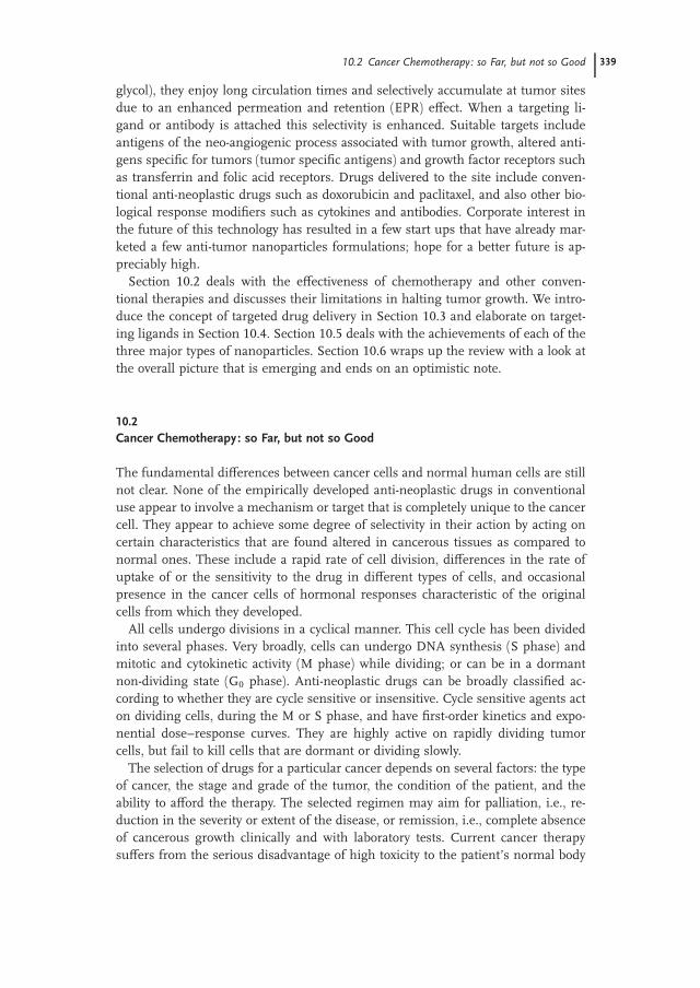
Fig. 10.1. Types of nanoparticulate drug delivery systems and
their common targeting methods. Left to right: Liposomes with
enclosed drug molecules; other polymeric nanoparticles, and
dendrimers.
342 10 Liposomes, Dendrimers and other Polymeric Nanoparticles for Targeted Delivery

10.3.2
An Overview of Targeting Methods
In this chapter we concentrate on the methods and materials by which nanopar-
ticles are targeted to neoplastic tissue. Targeting can be loosely defined in this con-
text as any means that increases the specificity of localization of nanoparticles in
tumor cell masses. Targeting does not intrinsically imply improved sensitivity but,
as we shall see, the different methods employed to increase the specificity allow
administration of higher doses of the drugs, thus also favorably increasing sensitiv-
ity. Also, as mentioned earlier, the ability of nanoparticles to cross the blood–brain
barrier and other impediments to conventional therapy increases its volume of dis-
tribution. This also results in increased sensitivity.
Targeting can be divided into two major types – passive and active. Passive tar-
geting involves modifications of nanoparticles that increase the circulation time
without addition of any component/involvement of any method that is specific to
the tumor; increased circulation time helps in accumulation of the particles in the
tumor by an EPR effect described below. Active targeting of tumors can be divided
into ‘‘physical’’ and ‘‘chemical/biological’’ targeting. Physical targeting involves di-
recting magnetic nanoparticles to tumor sites under the influence of an external
magnetic field. Chemical/biological targeting involves the modification of nanopar-
ticles’ surfaces with tumor-specific ligands.
Notably, any of these methods can used in conjunction with others. For example,
common modifications for passive targeting, such as PEGylation, are frequently
used with more ‘‘active’’ modifications like antibodies. The methods are almost in-
dependent of each other, and can be judiciously combined to increase the effective-
ness of the drugs.
This chapter does not deal extensively with the magnetic therapy of cancer and
induced hyperthermic killing of tumor cells (see Chapters 5, 8 and 9). However, in
keeping with the idea of overviewing the whole field of targeting, we have incorpo-
rated a short discussion of physical targeting by magnetically directing particles to
tumor tissues.
10.4
Means to the End: Methods for Targeting
10.4.1
Passive Targeting
Here we must make a clearer distinction between what can be described as ‘‘active’’
and ‘‘passive’’ targeting. In general terms, a targeted drug can be defined as one
modified in a fashion that allows its preferential uptake in the desired cells/tissues,
in this case cancer. Evidence has been provided that many nanoparticles, especially
long circulating ones, will show a preferential distribution to cancer sites over
healthy tissues, even without any specific targeting molecule. This was demon-
10.4 Means to the End: Methods for Targeting 343

strated for the first time my Matsumara and Maeda [10]. This is likely due to the
increased vasculature of these regions, larger fenestrations in the capillary walls for
rapid delivery of nutrients, general disordered architecture that is symbolic of the
neoplastic process, and the reduced lymphatic drainage in these regions. All these
factors lead to the enhanced permeation and retention effect (EPR) [11, 12] (Fig.
10.2). This fortuitous distribution has been taken advantage of by numerous
researchers in designing targeted drug delivery systems. As an example, in a
recent study tamoxifen was encapsulated in poly(ethylene oxide)-modified poly(e-
caprolactone) (PEO-PCL) nanoparticles and administered to a murine model of
breast cancer [13]. The poly(ethylene oxide) (PEO) coating made it a ‘‘stealth nano-
particle’’, i.e., able to avoid detection by the body’s MPS system for a considerable
time. The PEO surface modified nanoparticles showed significantly increased
levels of accumulation within tumor with time than did the native drug or surface
unmodified nanoparticles. An earlier effort by one of the authors to incorporate ta-
moxifen in nanoparticles for preferential uptake by estrogen receptor (ER) positive
breast cancer cells had been shown to be successful in vitro [14]. However, to trans-
late that success in vivo, a long-circulating nanoparticle was necessary. Another cur-
rent example is the incorporation of cisplatin in liposomal formulations [15] with
poly(ethylene glycol) (PEG) coating for gastric tumors: in preclinical and clinical
trials, this formulation has been demonstrated to have longer half-life in circula-
tion without the attendant side effects.
This targeted distribution of nanoparticles, together with the controlled release
of anticancer drugs, can bring about a type of ‘‘passive’’ but effective targeted deliv-
ery. The other type of targeting is by specific ligand–receptor interaction. Possibly,
Fig. 10.2. The enhanced permeation and retention (EPR) effect
accumulates nanoparticles to tumor sites without active
targeting.
344 10 Liposomes, Dendrimers and other Polymeric Nanoparticles for Targeted Delivery

better specificity and sensitivity can only be achieved by this more ‘‘active’’ means.
However, the introduction of a biological entity on the surface of the nanoparticle
can create several problems: the size and complexity will increase, with attendant
complexities of synthesis and characterization; the biomolecule may react adversely
with the immune system or any of the other myriad proteins in the blood and tis-
sues. This may give rise to unpredictable adverse reactions.
However, potential advantages include the ability to deliver larger amounts of
drug per target biorecognition event; increase selectivity by including more than
one type of targeting molecule, better avoidance of barriers by using different
avoidance methods and the ability to administer ‘‘localized integrated combination
therapy’’ by including multiple drugs in the same nanoparticle [1].
A method for enhancing this passive delivery to the cancer using nanoparticles
is by the use of pH- and heat-sensitive nanoparticles. The fact that the pH in the
region of cancerous growth is lower than in the rest of body, while the temperature
is raised locally because of enhanced metabolism and ongoing inflammation, has
been used to design pH-responsive nanoparticles: a lower pH causes enhanced
drug release. Wei et al. have given a recent example of this [16]. The temperature-
and pH-sensitive amphiphilic polymer poly(N-isopropylacrylamide-co-acrylic acid-
co-cholesteryl acrylate) [P(NIPAAm-co-AA-co-CHA)] was synthesized and used to
encapsulate paclitaxel in core–shell nanoparticles fabricated by a membrane dial-
ysis method. The spherical nanoparticles are below 200 nm and have a pH-
dependent lower critical solution temperature (LCST). In vitro release of paclitaxel
from the nanoparticles was responsive to external pH changes, and was faster in a
lower pH environment.
10.4.2
Magnetic Targeting of Nanoparticles
Magnetic drug targeting has been defined as ‘‘the specific delivery of chemothera-
peutic agents to their desired targets, e.g., tumors, by using magnetic nanoparticles
(ferrofluids) bound to these agents and an external magnetic field which is focused
on the tumor’’ [17]. This type of tumor targeting is aimed at concentrating the
toxic drug at the cancer, hence enhancing its efficacy and reducing systemic side
effects from high doses.
Gilchrist et al. described the first magnetic nanoparticles in 1957 [18]. These are
generally 10–200 nm in diameter, magnetic or superparamagnetic and usually
composed of iron oxide, magnetite, nickel, cobalt or neodymium; of these, magnet-
ite (Fe3O4) and maghemite (g-Fe2O3) are more biocompatible and preferred. Iron
oxide is degradable in the body and has been shown to be safe for in vivo applica-
tions [6]. However, to be effective, magnetic nanoparticles should demonstrate
high magnetization for external magnetic field control and should exceed linear
blood flow rates of 10 cm s�1 in arteries and 0.05 cm s�1 in capillaries. For this
purpose, particles composed of, for example, 20% magnetite require a field of 0.8
Tesla. Tissue depth is a limiting factor in active targeting, with deeply localized tu-
mors being hard to access.
10.4 Means to the End: Methods for Targeting 345
Lubbe et al. have utilized a complex of 4-epirubicin bound to ferrofluids in the
treatment of squamous carcinoma of the breast or head and neck [19]. They used
a field of 0.5–0.8 T at the tumor site while infusing the particles. Results showed
accumulation in tumors in 6 of 14 patients with reduced toxicity, a transient rise of
serum iron, but no demonstrable damage to kidney or hepatic function. Alexiou et
al. have used mitoxantrone complexed to 100 nm ferromagnetic particles against
VX2 squamous cell carcinoma among rabbits to demonstrate the superiority of
intra-arterial infusion to other routes of delivery, especially the intravenous route
where the particles are removed from circulation by the reticulo-endothelial system
[20]. They have also demonstrated that circulating mitoxantrone becomes concen-
trated at the tumor tissue when complexed to ferrofluids; a higher concentration of
the complexed drug (compared to the free drug) is achieved at the tumor site using
only a half to one-fifth dose [17]. The drug mainly resides in the intraluminal
space, the tumor interstitium and peritumoral area (region surrounding the tumor
a1 cm). Other researchers have investigated ferrous nanoparticles in glioblastoma
(a tumor of the brain), and demonstrated that these particles effectively cross the
blood–brain barrier [21].
The clinical applications of magnetic drug targeting have been reviewed in more
detail [22].
10.4.3
Ligands for Active Targeting
Cancer cells arise from normal cells through a complex series of genetic events.
Unlike infectious agents like bacteria, they largely share the same proteins as nor-
mal cells. However, some proteins are found in much larger numbers on cancer
cells than in normal cells [23]. These overexpressed antigens are called tumor-
associated antigens (TAA). Some proteins derived from normally silent genes or
mutated forms of normal proteins are found exclusively on cancer cells. These are
known as tumor-specific antigens (TSA). The obvious targets for targeted cancer
therapy are TSAs. However, TSAs are often difficult to characterize for a particular
tumor. When found, they are usually not extensive, i.e., they are not found in all
patients affected by the tumor, nor are they found in all the cells in a particular
tumor in the same patient. The aberrantly expressed tumor specific antigens are
produced by aberrant glycosylation in glycolipids, glycoproteins, proteoglycans,
and mucin [24]. For example, the glycosylation pattern of MUC1 mem-
brane mucin of breast cancer epithelial cells differs from normal breast epi-
thelial cells, possibly as a result of changes in expression of glycosyltransferases
[25]. TAG-72 is a mucin-like tumor-associated glycoprotein [26] that is found in
some epithelial tumors. Melanoma cells aberrantly express GM3 ganglioside on
their surface [27]. Gastrointestinal cancer cells abnormally express LeX antigens
[28].
Tumor-associated antigens are often growth factor receptors on the tumor that
are overexpressed to meet the rapidly dividing neoplastic cells’ demands. A classic
example is folic acid and congeners. These are low molecular weight pterin-based
346 10 Liposomes, Dendrimers and other Polymeric Nanoparticles for Targeted Delivery
vitamins essential for carbon metabolism and de novo nucleotide synthesis. The
presence of elevated levels of folate receptors has been demonstrated from epithe-
lial tumors of various organs such as the colon, lungs, prostate, ovaries, mammary
glands, and brain [29–40]. These present attractive targets for drug delivery. An-
other promising tumor receptor target is Her2_neu, also known as c-erbB-2. This
is a transmembrane protein with an epidermal growth factor receptor that pos-
sesses intrinsic tyrosine kinase activity [41–43]. The normal human Her-2_neu
proto-oncogene is frequently found to be overexpressed in breast and ovarian can-
cers among others. Its level may correlate with the metastatic potential of the
cancer cells [44, 45].
Another such membrane-associated tumor antigen is the transferrin receptor.
This is overexpressed in different types of cancers [46]. The levels may also corre-
late with the malignant potential of these cells [47] (Fig. 10.3).
The presence of various other tumor antigens has been demonstrated:
membrane-associated Carcinoembryonic antigen; CD10 or CALLA in leukemias,
melanomas and myelomas [48, 49]; CD20 in B cell malignancies [50]; etc. Many
others are being recognized routinely. They all represent potential goals for tar-
geted drug delivery.
10.4.3.1 Monoclonal Antibodies against Tumor-specific Antigens
Specific targeting became a possibility with the discovery of monoclonal antibodies
in 1975 [51]. As the name suggests, these are antibodies derived from a single
clone of cells. They can be mass produced in the laboratory from a single clone
and recognize only one antigen. Monoclonal antibodies are usually made by fusing
a short-lived, antibody-producing B cell to a fast-growing cell, such as a cancer cell.
The resulting hybrid cell, or hybridoma, multiplies rapidly, creating a clone that
produces large quantities of the antibody. It was hoped that monoclonal antibodies
would be able to target specific antigens on the surface of cancer cells, and initially
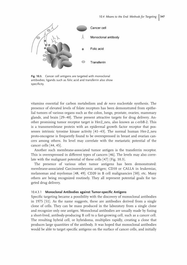
Fig. 10.3. Cancer cell antigens are targeted with monoclonal
antibodies; ligands such as folic acid and transferrin also show
specificity.
10.4 Means to the End: Methods for Targeting 347

raised high hopes for targeted cancer therapy. Animal-origin monoclonal anti-
bodies such as murine antibodies were strongly immunogenic; to avoid this, chi-
meric antibodies [52] and humanized antibodies [53, 54] were produced. More
recently, completely human monoclonal antibodies and single-chain human anti-
body fragments [55] have been introduced for immunotherapy and targeted drug
delivery.
Antigen targets for monoclonal antibody therapy (or for any other targeted anti-
cancer therapies) must have the property of specificity, i.e., present only on the
cancer cell surface and be absent from the surfaces of all normal cells; they should
be extensive, i.e., present on all the cells in a given tumor, preferably on all such
tumors in every patient; there should not be any mutation or structural variation
of the antigen; and they should be preferably involved with a critical function of
the cell, so that adaptive response of the cancer to therapy by losing the antigen
can be prevented [56]. However, naturally, such a perfect antigen is rarely, if ever,
found. Several good candidates have been discovered, and monoclonal antibodies
against these have been marketed. Examples include anti-CD20 antibody for
relapsed/refractory CD20 positive B-cell non-Hodgkin’s lymphoma and low-grade
or follicular-type lymphoma, anti-CD53 antibody for relapsed/refractory AML, and
anti-CD52 antibody for B-cell chronic lymphocytic leukemia [56, 57]. Recently,
Zhou et al. have utilized BDI-1 monoclonal antibodies to target highly toxic to blad-
der cancer cells in vitro [58]. To improve therapeutic efficacy and decrease toxicity,
we prepared arsenic trioxide-loaded aluminates immuno-nanospheres [As2O3-
(HAS-NS)-BDI-1] targeted with monoclonal antibody (McAb) BDI-1 and tested its
specific killing effect against bladder cancer cells.
Identifying Monoclonal Antibodies One strategy to identify ideal targets for mono-
clonal antibodies is to select the internalizing antibodies from phage libraries; ac-
cordingly, two antibodies to the breast cancer cell line SK-BR-3 which bind to ErbB2
have been identified [59]. Nielsen et al. have demonstrated the use of single-chain
Fv (scFv) antibodies for internalization of nanoparticles in target cells [60]. The
antibodies were recovered from a non-immune phage library and the scFv specific
for ErbB2 (F5) was re-engineered and attached to liposomes. Doxorubicin loaded in
these immunoliposomes was selectively active on ErbB2 positive breast cancer cells
and showed targeted cancer cell cytotoxicity.
Liu et al. have described a method to map the ‘‘epitope space’’ of a tumor using
monoclonal antibody libraries [61]. The expressed epitopes on the cell surface,
which constitute the ‘‘epitope space’’, is essential to targeted drug delivery. This
is ‘‘highly complex, composed of proteins, carbohydrates, and other membrane-
associated determinants including post-translational modification products, which
are difficult to probe by approaches based on gene expression’’ [61]. The authors
used monoclonal antibody libraries against prostrate cancer cells and identified
over 90 antibodies, which bind to the cancer cells selectively, with little or no bind-
ing to normal human cells. This approach does not attempt to identify the tumor-
specific antigens, but rather takes a functional approach to the problem of tumor-
target identification.
348 10 Liposomes, Dendrimers and other Polymeric Nanoparticles for Targeted Delivery

Monoclonal Antibodies and Nanoparticles Monoclonal antibodies were initially
hailed as therapy for cancers in their own right (Fig. 10.4). To introduce more
cancer cell killing ability, they can also be conjugated with anticancer drugs. This
complex can also be formulated as a prodrug, which is cleaved to release the anti-
cancer agent at the cancer site. Usually, the link between the molecules would be
peptidase cleavable, in order to separate inside the cancer cell. However, such for-
mulations are not very stable in vivo, and may undergo early cleavage. The absence
or reduction of cleavage at the site will, clearly, lower the potency of the anticancer
agent.
The greatest difficulty in using monoclonal antibodies for cancer therapy is their
rapid clearance from the bloodstream by the immune mechanisms of the body. Li
et al. have estimated that <0.01% of the administered dose of antibodies reaches
the target sites [62]. This has two serious drawbacks: firstly, monoclonal antibodies
are relatively costly, and a large dose of the therapy becomes prohibitively expen-
sive; secondly, the excess antibody can have serious toxic effects on rest of the
patient’s body. As we discussed in the introductory section on chemotherapy,
this problem, in various proportions, dogs all current therapies of cancer. Hence,
monoclonal antibodies by themselves provide no advantage over general anticancer
drugs. Attachment of the molecules to nanoparticles circumvents some of these
problems.
10.4.3.2 Targeting the Angiogenic Process
Judah Folkman discussed one of the prime candidates for targeted cancer therapy,
angiogenic factors, in 1989 [63]. Cancer cells require a significant amount of nutri-
tion to keep growing and reproducing. To obtain this they promote the ingrowth of
capillary vessels into the tumor site. This is known as angiogenesis. Tumor cells
secrete chemicals that promote angiogenesis. Since active promotion of angio-
genesis is unnecessary in most body cells, the associated antigens are of interest
Fig. 10.4. Monoclonal antibodies against cancer were
originally used either by themselves (A) or attached to drug
molecules (B). However, attachment to nanoparticles (C)
provides longer circulation times and lower toxicity.
10.4 Means to the End: Methods for Targeting 349
in targeted therapies. The anti-VEGF (vascular endothelial growth factor) drug
Avastin2 has shown moderate effectiveness in several solid tumors. This has led
to a gene-therapeutic approach to anti-angiogenesis using liposomes [64] and cat-
ionic nanoparticles [65]. In the latter, an integrin-targeting ligand was used for
specific delivery of a mutant Raf gene, and selectivity was demonstrated.
Hallahan et al. have described novel targets that depend on targeting radiation-
induced neoantigens in the cancer microvasculature [66]. They demonstrated the
presence of an RGD-containing amino acid sequence in phage-displayed peptides
obtained from irradiated tumors. This peptide binds to integrins. Immunohisto-
chemical examination confirmed the presence of the fibrinogen receptor integrin
in irradiated tumors. Targeting this receptor by fibrinogen-containing nanopar-
ticles showed enhanced anti-tumor effects.
The Intradigm Corporation (MD, USA) has utilized the RGD sequence attached
to PEG to target their siRNA-containing nanoparticles to the integrins present in
the tumor microvasculature [67]. The siRNA provide potent selective gene inhibi-
tion with high specificity. They delivered siRNA-inhibiting vascular endothelial
growth factor receptor-2 (VEGF R2) expression and, thereby, tumor angiogenesis
to a mouse tumor model using PEI-PEG nanoparticles. The siRNA was found to
be active in a highly cell-selective manner. They demonstrated that selectivity could
be incorporated in the drug itself, along with the ligand–receptor interaction.
Nucleic acid conjugates have been favored as targeting ligands for their exquisite
specificity for the targeted molecule. Farokhzad et al. have utilized nucleic acid li-
gands (aptamers) to target cells expressing prostate-specific membrane antigen, a
tumor marker for prostate cancer acinar epithelial cells [68]. They created PEG-
containing PLA nanoparticles that bind to aptamers by surface negative charge.
These aptamer conjugated nanoparticles showed 77� greater uptake by the cancer
cells than unmodified nanoparticles.
Other efforts [69] have demonstrated nanoparticle targeting to angiogenic epi-
thelium using the avb3-integrin, which is found on endothelial cells. Anti-
angiogenic effects were demonstrated in mouse models of melanoma and adeno-
carcinoma.
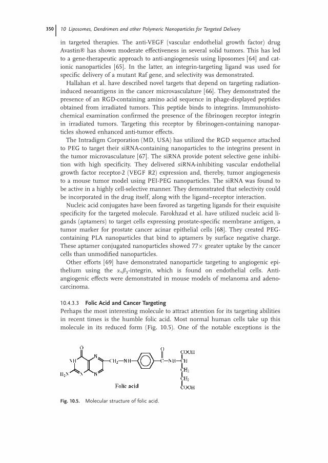
10.4.3.3 Folic Acid and Cancer Targeting
Perhaps the most interesting molecule to attract attention for its targeting abilities
in recent times is the humble folic acid. Most normal human cells take up this
molecule in its reduced form (Fig. 10.5). One of the notable exceptions is the
Fig. 10.5. Molecular structure of folic acid.
350 10 Liposomes, Dendrimers and other Polymeric Nanoparticles for Targeted Delivery
luminal sides of the gut endothelial layer, which take up the folic acid present in
food, convert it into folate, and release it into our bloodstream. Many types of
cancer cells, however, take up folic acid in its oxidized form. Since intravenously
administered nanoparticles will only come into contact with the abluminal side of
the gut epithelium, selectivity for tumor cells is very high. As mentioned above,
several endothelial tumors (e.g., derived from the ovaries, mammary glands, colon,
lungs, prostate and brain) possess elevated levels of folic acid receptors on their
surface [29, 30, 32, 33, 36–40]. Already, a large body of research has accumulated
regarding the use of folic acid receptors as cancer targets. Since folic acid, the nat-
ural choice for ligand to the folic acid receptor, can be quite easily coupled to the
nanoparticle surface, it has been used for targeting these to cancer cells.
Hattori and Maitani have reported in vitro studies of folate-linked nanoparticles
in human cancer cells [31]. They synthesized DC cholesterol–Tween 80 nanopar-
ticles with incorporated folate-PEG conjugates for steric stability and targeting.
The nanoparticles were complexed with plasmid DNA. They showed enhanced
uptake in human oral cancer cells by a folate-dependent route. Human prostate
cancer cells also showed high uptake of the nanoparticle–DNA complexes. How-
ever, based on their results, the authors suggest that the route of uptake may be
different for the two cell types.
Oyewumi and Mumper have reported the cellular uptake, distribution and tumor
retention of folate-coated and PEG-coated gadolinium nanoparticles in mice models
of human nasopharyngeal carcinomas [34]. While this was more for imaging than
therapeutic purposes, their results also indicate the efficacy of using folate as a tar-
geting agent. Baker et al. at the Center for Biologic Nanotechnology, University of
Michigan Medical School have focused on the use of dendrimers as targeted deliv-
ery vehicles (described in detail later). They used folic acid as targeting element to
deliver a triplex-forming growth-inhibitory oligonucleotide to breast, ovarian and
prostate cell lines [35]. A very recent example of folic acid receptor targeting in
squamous cell carcinoma has been demonstrated by Santra et al. [70], who em-
ployed a novel technique that uses fluorescent silica nanoparticles (FSNPs) to
detect overexpressed folate receptors.
Folic acid targeting to cancer cells has also been demonstrated in our own Cellu-
lar and Molecular Bioengineering Laboratory in the National University of Singa-
pore. Although the use of folate as a targeting agent has been extensively reported,
little has been done to continuously track the intracellular delivery of nanoparticles
grafted with folate using imaging techniques such as confocal microscope. This is
possibly due to the short lifetime of most biological fluorescent labels. The prob-
lem has been tackled using quantum dots (QDs).
QDs have several advantages over conventional fluorescent dyes and proteins
like the Green Fluorescent Protein. They exhibit a strong fluorescence emission, a
broad absorption spectra and a narrow, symmetric emission spectrum, and are
photochemically stable. The most exciting finding is that QDs also exhibit a wide
range of colors, which is exquisitely controlled by their size; a broad absorption
spectra means that a series of different-colored dots can be activated using a single
laser. These properties of QDs raise the possibility of using them to tag biomole-
10.4 Means to the End: Methods for Targeting 351
cules with an optical coding technology. This can, for example, create QD bar
codes, and the use of six colors and ten intensity levels can theoretically encode
one million biomolecules. Techniques have been developed to incorporate QDs
into polymer beads, to solve problems relating to their surface chemistry. In
general, quantum dots exhibit water insolubility, poor biocompatibility, and low
chemical stability in physiological media; encapsulation in polymer nanoparticles
can reduce these problems. In their work, Zhang et al. have incorporated lumines-
cent CdSe/ZnS QDs into polystyrene (PS) nanoparticles grafted with carboxyl
groups using an emulsion polymerization method. Nanoscale QD-incorporated
PS nanoparticles (30 nm diameter) were separated by centrifugation at high speed
in viscous solution.
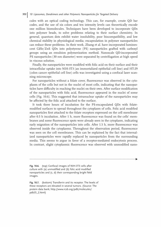
Finally, the nanoparticles were modified with folic acid on their surface and their
intracellular uptake into NIH-3T3 (an immortalized epithelial cell line) and HT-29
(colon cancer epithelial cell line) cells was investigated using a confocal laser scan-
ning microscope.
For nanoparticles without a folate cover, fluorescence was observed in the cyto-
plasm of the cells but not in the nuclei of most cells, indicating that the nanopar-
ticles have difficulty in reaching the nuclei on their own. After surface modification
of the nanoparticles with folic acid, fluorescence appeared in the nuclei of some
cells (Fig. 10.6). This suggested that intranuclear uptake of the nanoparticles may
be affected by the folic acid attached to the surface.
It took three hours of incubation for the PS-encapsulated QDs with folate-
modified surfaces to spread throughout the cytoplasm of cells. Folic acid modified
nanoparticles first attached to the folate receptors expressed on the cell membrane
after 0.5 h incubation. After 1 h, more fluorescence was found on the cells’ mem-
branes and some fluorescence spots were already seen in the cytoplasm, indicating
early migration of the nanoparticles into cells. After 1.5 h, more fluorescence was
observed inside the cytoplasms. Throughout the observation period, fluorescence
was seen on the cell membranes. This can be explained by the fact that internal-
ized nanoparticles were rapidly replaced by nanoparticles from the surrounding
media. This seems to argue in favor of a receptor-mediated endocytosis process.
In contrast, slight cytoplasmic fluorescence was observed with unmodified nano-
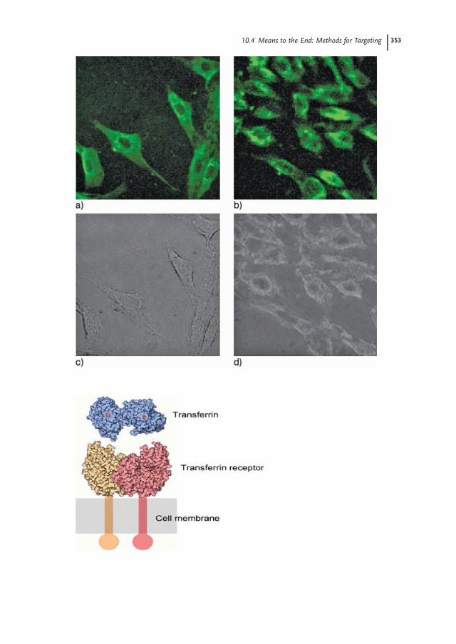
Fig. 10.6. (top) Confocal images of NIH-3T3 cells after
culture with (a) unmodified and (b) folic acid modified
nanoparticles and (c, d) their corresponding bright field
images.
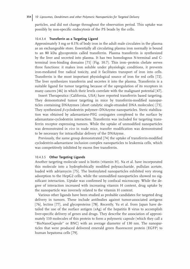
Fig. 10.7. (bottom) Transferrin and its receptor. The levels of
these receptors are elevated in several tumors. (Source: The
protein data bank; http://www.rcsb.org/pdb/molecules/
pdb35_2.html)
________________________________________________________________________________G
352 10 Liposomes, Dendrimers and other Polymeric Nanoparticles for Targeted Delivery
10.4 Means to the End: Methods for Targeting 353
particles, and did not change throughout the observation period. This uptake was
possibly by non-specific endocytosis of the PS beads by the cells.
10.4.3.4 Transferrin as a Targeting Ligand
Approximately 3 mg or 0.1% of body iron in the adult male circulates in the plasma
as an exchangeable store. Essentially all circulating plasma iron normally is bound
to an 80 kDa glycoprotein called transferrin. Plasma transferrin is synthesized
by the liver and secreted into plasma. It has two homologous N-terminal and C-
terminal iron-binding domains [71] (Fig. 10.7). This iron–protein chelate serves
three functions: it makes iron soluble under physiologic conditions, it prevents
iron-mediated free radical toxicity, and it facilitates transport of iron into cells.
Transferrin is the most important physiological source of iron for red cells [72].
The liver synthesizes transferrin and secretes it into the plasma. Transferrin is a
suitable ligand for tumor targeting because of the upregulation of its receptors in
many cancers [46] in which their levels correlate with the malignant potential [47].
Insert Therapeutics (California, USA) have reported transferrin based targeting.
They demonstrated tumor targeting in mice by transferrin-modified nanopar-
ticles containing DNAzymes (short catalytic single-stranded DNA molecules) [73].
They synthesized b-cyclodextrin polymer–DNAzyme nanoparticles. Steric stabiliza-
tion was obtained by adamantane-PEG conjugates complexed to the surface by
adamantane-cyclodextrin interaction. Transferrin was included for targeting trans-
ferrin receptor expressing tumors. While the uptake of unmodified nanoparticles
was demonstrated in vivo in nude mice, transfer modification was demonstrated
to be necessary for intracellular delivery of the DNAzyme.
Previously, the same group demonstrated [74] the uptake of transferrin-modified
cyclodextrin-adamantane inclusion complex nanoparticles to leukemia cells, which
was competitively inhibited by excess free transferrin.
10.4.3.5 Other Targeting Ligands
Another targeting molecule used is biotin (vitamin H). Na et al. have incorporated
this molecule into a hydrophobically modified polysaccharide, pullulan acetate,
loaded with adriamycin [75]. The biotinylated nanoparticles exhibited very strong
adsorption to the HepG2 cells, while the unmodified nanoparticles showed no sig-
nificant interaction. Uptake was confirmed by confocal microscopy. While the de-
gree of interaction increased with increasing vitamin H content, drug uptake by
the nanoparticle was inversely related to the vitamin H content.
Various other ligands have been studied as probable candidates for targeted drug
delivery in tumors. These include antibodies against tumor-associated antigens
[76], lectins [77], and glycoproteins [78]. Recently, Yu et al. from Japan have de-
tailed the use of the surface antigen (sAg) of the hepatitis B virus to accomplish
liver-specific delivery of genes and drugs. They describe the association of approxi-
mately 110 molecules of this protein to form a polymeric capsule (which they call a
‘‘BioNanoCapsule’’ or BNC) with an average diameter of 130 nm. The nanopar-
ticles that were produced delivered emerald green fluorescent protein (EGFP) to
human hepatoma cells [79].
354 10 Liposomes, Dendrimers and other Polymeric Nanoparticles for Targeted Delivery

10.5
Targeting with Different Types of Nanoparticles
10.5.1
Liposomes in Cancer Targeting
Currently, the most exciting event in targeted drug delivery systems is undoubtedly
the ongoing clinical trial of the world’s first tumor-targeted gene delivery vector,
Rexin-G.TM, marketed by Epeius Biotechnologies Corporation. This nanoparticle
formulation combines a proprietary vector system with a proprietary anticancer
gene controlling cell cycle. The company website claims that ‘‘when given by intra-
venous infusions, Rexin-GTM has been shown to eradicate remote metastatic can-
cers in mice and to arrest cancer growth with shrinkage and necrosis of solid tu-
mors in humans – determined by CT Scan and MRI – without eliciting systemic
side effects.’’ The first clinical trials were started in 2003 in Manila, Philippines;
and following FDA approval shortly thereafter, clinical trials were started in New
York from April, 2005 for metastatic pancreatic and colon cancer. Initial reports
are favorable and results of ongoing studies are eagerly awaited [3, 80, 81].
In this context, a few words regarding immunoliposomes are in order. In our ini-
tial classification of colloid based systems, we separated nanoparticles from lipo-
somes and block copolymer micelles. Many authors consider them part and parcel
of the same package [1, 82]. Hence, in the interest of topicality and completeness,
we consider both in some measure.
Liposomes (and, in some instances, micelles) have long been used for targeted
delivery of drugs. Of all the proposed nanoparticulate drug delivery systems,
liposome-based agents, particularly liposomal anthracyclines, have had the greatest
impact on oncology to date [82]. These lipid based formulations have several ad-
vantages as drug carriers: they are amphiphilic, hence allowing the carriage of lip-
ophilic drugs in plasma; they fuse with cell membranes and transfer their load
inside cells; and they can be embedded with targeting molecules like antibodies
on their outer surface. They have been likened to the Trojan horse in Greek my-
thology: the cancer cells fuse to the liposomal membrane and takes up the con-
tents of the liposome, which contains the anticancer formulations (the company
that is conducting the liposomal targeted-gene delivery trials has been appropri-
ately named after Epeius, the maker of the original Trojan horse).
Several problems have dogged liposomal drug delivery systems from its concep-
tion and implementation. These include a short half-life in circulation, instability
in vivo, and lack of target selectivity. Liposomes are removed rapidly from the circu-
lation by the reticulo-endothelial system (RES) [83]. Stability and circulation times
were increased, as in nanoparticles, by the introduction of a steric stabilizer on
the surface, usually PEG [84]. Like other nanoparticles, enhanced passive uptake
of liposomes has been demonstrated with longer circulation times. PEGylation
has increased therapeutic efficacy of the liposomal formulations [85, 86]. These in-
novations, affording longer and stable circulation times, have resulted in approval
of some liposomal formulations for clinical use [87]. The passive increase of lipo-
10.5 Targeting with Different Types of Nanoparticles 355

somes in cancer sites was recognized as a means of delivering toxic, poorly soluble
drugs. Liposome-encapsulated doxorubicin was approved for use in Kaposi’s sar-
coma more than a decade ago. Formulations are available now for breast and
ovarian cancers. The most recent instance deals with liposomes encapsulating cis-
platin; this has been demonstrated in preclinical and phase 1 clinical trials in hu-
mans to have almost negligible nephrotoxicity, ototoxicity and neurotoxicity, which
are the major side-effects of free cisplatin in circulation [15].
Improved target selectivity has been demonstrated using ligands and antibodies.
Active targeting of liposomes using ligands for tumor-associated receptors, and
antibodies against tumor-associated antigens, has been investigated as a means of
improving selectivity. Liposome–antibody complexes (immunoliposomes) have re-
ceived most attention [88]. The problem of rapid removal by the RES is com-
pounded by the attachment of antibodies [89, 90]. However, PEGylation has cre-
ated a generation of ‘‘stealth’’ immunoliposomes that can effectively hide from the
RES for long periods. In this context, immunoliposomes can be classified into two
groups based on the relation of the antibody to the PEG group on the liposomal
surface: antibody coupled to lipid head group 76 and antibody coupled to the distal
end of PEG [91]. The latter composition has been considered to be better for effi-
cient antibody–target coupling because of the absence of interference from PEG
chains [92]. At optimal compositions of both types of formulations, antibodies do
not interfere with PEG function and long circulation times have been demon-
strated; conversely, PEG does not interfere with antibody function, and active tar-
geting is unimpaired. Immunoliposomes complexed with PEG have been success-
fully demonstrated for in vivo targeting of brain [93] and lung [91] tumors.
Immunoliposomes with cancer targeting antibodies have been shown to be cyto-
toxic to cancer cells in vitro [94]. However, this did not translate to in vivo success.
This was because not all antibodies used to target the cancer cells result in uptake
and internalization of the liposome. The use of more specific internalizing anti-
bodies aided in uptake and cytotoxic effects in vitro. As an example, the anticancer
antibody anti-ErbB2 did not increase cytotoxicity of the liposome [95]; but when
the anti-ErbB2 Fab fragment was used, the liposome was internalized and had
high efficacy of cancer kill [96, 97]. More recently, scFV (single-chain Fv) antibodies
against ErbB2 have been utilized for cancer targeting with doxorubicin-loaded lip-
osomal formulations. The antibodies are selected from a phage display library, and
demonstrate higher internalization efficacy [98–100]. Ongoing research by Park et
al. at UCSF comprehensive cancer centre [82, 101] aims to develop immunolipo-
somes with antibodies targeted to HER 2, overexpressed in breast cancer, and
EGFR, overexpressed in several cancers such as lung, pancreas and prostate. The
targeted liposomes have been loaded with anticancer drugs like doxorubicin and
vinorelbine. The loaded immunoliposomes have shown increased effectiveness
and reduced toxicity compared with conventional chemotherapy.
The issue of internalization can be circumvented by a two-step method to release
the anticancer drugs from the liposome near the tumor. This antibody-directed en-
zyme pro-drug therapy (ADEPT) involves first administering an enzyme–antibody
conjugate that preferentially targets cancer cells, followed by administration of a
356 10 Liposomes, Dendrimers and other Polymeric Nanoparticles for Targeted Delivery

non-toxic prodrug that is activated by the bound enzyme and releases the toxic
product close to the cancer cells [102]. An improvement of the ADEPTmethod us-
ing immunoliposomes to carry the enzyme shows greater stability in the body and
has been tested pre-clinically [103].
10.5.1.1 Beyond Immunoliposomes
Similar to the development of nanoparticles, scientists have looked beyond mono-
clonal antibodies as a means of targeting liposome to cancer. This was to avoid the
intrinsic immunogenic nature of antibodies, their high cost, and the ease of pro-
curement of ligands for overexpressed growth receptors on the tumor surface. Folic
acid coupled liposomes have been developed and tested against various cancer cells
both in vitro and in vivo [85, 104, 105]. Transferrin has also been evaluated as a tar-
geting ligand for anticancer drug carrying liposomes [106].
Targeting with transferrin and folic acid has also been used to deliver anticancer
genes to tumor cells. Cationic liposome–DNA complexes (lipoplexes) that are
formed spontaneously by charge interaction exhibit the best gene-transfer effi-
ciency. However, cationic liposomes were found to be unsuitable for active
cancer cell targeting. Hence, a new anionic liposome–polyplex (anionic liposome-
entrapped polycation-condensed DNA, LPD-II) was introduced for the gene ther-
apy of cancer. Cationic lipoplexes containing liposomes and DNA were better inter-
nalized and transfected when transferrin was conjugated [107] and demonstrated
selectivity for myeloblast cells [108], adenocarcinomas and squamous cell carci-
nomas [109]. Folic acid has also been used for tumor targeting the LPD-II poly-
plexes [110]; and systemic administration of anticancer gene therapy for selective
uptake in squamous cell carcinomas of the head and neck, and breast cancer xe-
nografts have been demonstrated [109].
10.5.2
Dendrimers
Block copolymers or dendrimers were described as far back as 1985. Initial studies
elucidated the physical and chemical properties of these new materials. However,
their entry into the field of targeted cancer therapy is relatively recent.
Dendrimers are globular macromolecules that have a ‘‘starburst’’-like shape,
with multiple branches radiating from a central core. The stepwise synthesis of
dendrimers makes it possible to fine-tune its highly regular branched structure
with defined peripheral groups and adjust physical properties such as the high mo-
lecular weight and low polydispersity index. Dendrimers have several advantages
over polymeric nanoparticles derived from linear chains. Dendrimers are multiva-
lent: several different drug molecules, targeting agents and other groups can be at-
tached in a predefined fashion. Dendrimers have low polydispersity: all the synthe-
sized molecules have molecular weights within a narrow range; this makes their
behavior highly reproducible. Dendrimers have a globular rather than random
coil structure, which could lead to better biologic properties [111]. Various types of
10.5 Targeting with Different Types of Nanoparticles 357
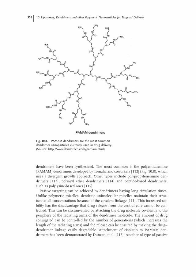
dendrimers have been synthesized. The most common is the polyamidoamine
(PAMAM) dendrimers developed by Tomalia and coworkers [112] (Fig. 10.8), which
uses a divergent growth approach. Other types include polypropyleneimine den-
drimers [113], polyaryl ether dendrimers [114] and peptide-based dendrimers,
such as polylysine-based ones [115].
Passive targeting can be achieved by dendrimers having long circulation times.
Unlike polymeric micelles, dendritic unimolecular micelles maintain their struc-
ture at all concentrations because of the covalent linkage [111]. This increased sta-
bility has the disadvantage that drug release from the central core cannot be con-
trolled. This can be circumvented by attaching the drug molecule covalently to the
periphery of the radiating arms of the dendrimer molecule. The amount of drug
conjugated can be controlled by the number of generations (which increases the
length of the radiating arms) and the release can be ensured by making the drug–
dendrimer linkage easily degradable. Attachment of cisplatin to PAMAM den-
drimers has been demonstrated by Duncan et al. [116]. Another of type of passive
Fig. 10.8. PAMAM dendrimers are the most common
dendrimer nanoparticles currently used in drug delivery.
(Source: http://www.dendritech.com/pamam.html)
358 10 Liposomes, Dendrimers and other Polymeric Nanoparticles for Targeted Delivery
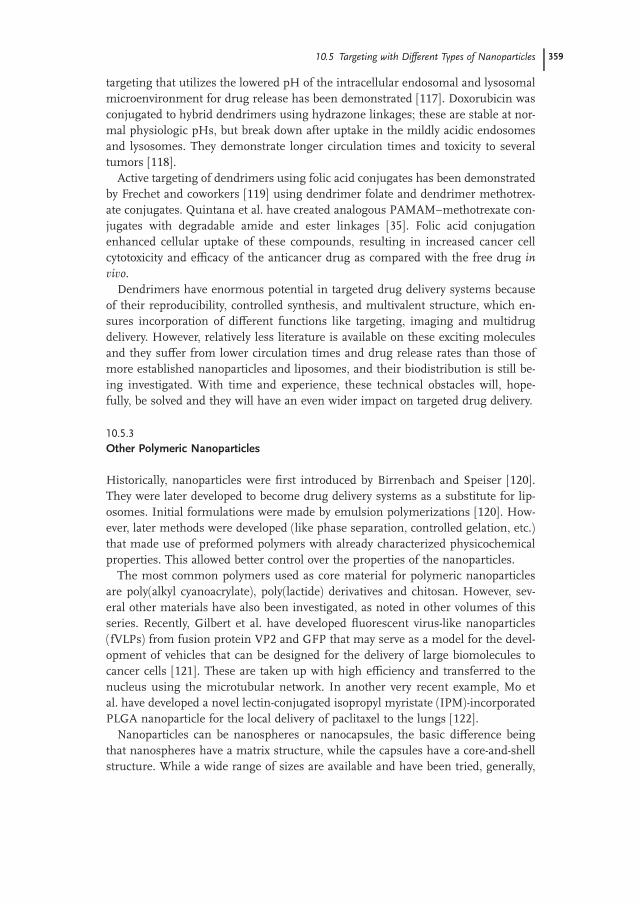
targeting that utilizes the lowered pH of the intracellular endosomal and lysosomal
microenvironment for drug release has been demonstrated [117]. Doxorubicin was
conjugated to hybrid dendrimers using hydrazone linkages; these are stable at nor-
mal physiologic pHs, but break down after uptake in the mildly acidic endosomes
and lysosomes. They demonstrate longer circulation times and toxicity to several
tumors [118].
Active targeting of dendrimers using folic acid conjugates has been demonstrated
by Frechet and coworkers [119] using dendrimer folate and dendrimer methotrex-
ate conjugates. Quintana et al. have created analogous PAMAM–methotrexate con-
jugates with degradable amide and ester linkages [35]. Folic acid conjugation
enhanced cellular uptake of these compounds, resulting in increased cancer cell
cytotoxicity and efficacy of the anticancer drug as compared with the free drug invivo.Dendrimers have enormous potential in targeted drug delivery systems because
of their reproducibility, controlled synthesis, and multivalent structure, which en-
sures incorporation of different functions like targeting, imaging and multidrug
delivery. However, relatively less literature is available on these exciting molecules
and they suffer from lower circulation times and drug release rates than those of
more established nanoparticles and liposomes, and their biodistribution is still be-
ing investigated. With time and experience, these technical obstacles will, hope-
fully, be solved and they will have an even wider impact on targeted drug delivery.
10.5.3
Other Polymeric Nanoparticles
Historically, nanoparticles were first introduced by Birrenbach and Speiser [120].
They were later developed to become drug delivery systems as a substitute for lip-
osomes. Initial formulations were made by emulsion polymerizations [120]. How-
ever, later methods were developed (like phase separation, controlled gelation, etc.)
that made use of preformed polymers with already characterized physicochemical
properties. This allowed better control over the properties of the nanoparticles.
The most common polymers used as core material for polymeric nanoparticles
are poly(alkyl cyanoacrylate), poly(lactide) derivatives and chitosan. However, sev-
eral other materials have also been investigated, as noted in other volumes of this
series. Recently, Gilbert et al. have developed fluorescent virus-like nanoparticles
(fVLPs) from fusion protein VP2 and GFP that may serve as a model for the devel-
opment of vehicles that can be designed for the delivery of large biomolecules to
cancer cells [121]. These are taken up with high efficiency and transferred to the
nucleus using the microtubular network. In another very recent example, Mo et
al. have developed a novel lectin-conjugated isopropyl myristate (IPM)-incorporated
PLGA nanoparticle for the local delivery of paclitaxel to the lungs [122].
Nanoparticles can be nanospheres or nanocapsules, the basic difference being
that nanospheres have a matrix structure, while the capsules have a core-and-shell
structure. While a wide range of sizes are available and have been tried, generally,
10.5 Targeting with Different Types of Nanoparticles 359
the particles should be less than 100 nm in diameter [123]. The greater surface
area of the nanoparticles allows attachment of targeting molecules and anticancer
agents; alternatively, the drug may form the core of the nanocapsule with a poly-
meric shell.
Nanoparticles as vehicles of targeted drug delivery enjoy several advantages. Pri-
marily, the attachment of different molecules to the same platform confers bi- and
multi-functionality. This is important because conjugating targeting molecules di-
rectly to the anticancer drug may cause decreased functionality of either or both.
Nanoparticles can be modified to incorporate not only two but more functions.
One of could report the presence of the tumor using, for example, quantum dots.
Secondly, attachment to the nanoparticle surface increases the stability of mole-
cules. This is particularly important with peptides, nucleic acids (like anti-HA-ras[124]) and anti-Ewing sarcoma [125] and small proteins (like antibodies), which
are easily removed from the circulation in the free form. It is also important for
carrying poorly soluble drugs (e.g., muramyl tripeptide cholesterol [126]) in signif-
icant quantities without the usual side effects. Controlled release can be achieved
to ensure a long-term, high level of the drug without encountering the loading
dose problem. This has been shown in the use of doxorubicin-loaded nanopar-
ticles. Doxorubicin can cause acute and chronic cardiomyopathy at high levels. By
attachment to nanoparticles, high levels of doxorubicin were achieved in the circu-
lation, with reduced cardiac levels and, consequently, less toxic effects and in-
creased effectiveness against the metastatic cancer.
Chemotherapy fails in vivo even when the cancer is shown to be highly sensitive
to the drug in vitro because of cellular ‘‘sanctuaries’’. For example, the recurrence
of acute lymphoblastic leukemia has been attributed to cell nests in the CNS. Intra-
venous drug formulations fail to cross the blood–brain barrier. Nanoparticles, how-
ever, can accomplish this feat, perhaps by trans-cellular movement after endocy-
tosis by the endothelial cells. Some examples of this include the use of polysorbate
80-coated nanospheres to deliver Kytorphin, turbocurarine NMDA antagonist and
doxorubicin to the brain. Polysorbate 80 adsorbs apolipoprotein E from the circula-
tion and becomes attached to the low-density lipoprotein receptors on the endothe-
lial surface.
The major drawback of nanoparticles is their propensity to be taken up by the
macrophages lodged in the organs of the mononuclear phagocytic system (MPS).
After intravenous administration, most of the nanoparticles accumulate in the
spleen and the liver, and to a lesser extent in the bone marrow. Steric stabilization
has been devised to avoid this. The attachment of PEG to the nanoparticle surface
allows them to ‘‘hide’’ from the MPS and stay longer in circulation, eventually find-
ing their way to the targeted regions. Poloxamine has been proposed as an alterna-
tive to PEG for producing steric stabilization.
In fact, PEG-coated poly(cyanoacrylate) (pCA) nanoparticles – made by a copoly-
mer inculcating both – have such a long circulating time that they penetrated the
brain more than any other modifications, including coating by polysorbate. This
uptake was increased in pathological situations with, presumably, higher blood–
brain barrier permeability.
360 10 Liposomes, Dendrimers and other Polymeric Nanoparticles for Targeted Delivery

Modeling of nanoparticle delivery and effects has raised the question of ‘‘diffu-
sional instability’’. Vittorio Cristini et al. have shown that the delivery of cytotoxic
agents to tumors, particularly anti-angiogenic drugs, might fractionate the lesion
into multiple satellite neoplasms [127]. This is likely due to the rearrangement of
the sources of oxygen and nutrient supply because of the anti-angiogenic therapy.
The other difficulty is the issue of control. Especially with synthetic methods de-
rived from microencapsulation techniques, there may be considerable difficulty in
encapsulating the drug and then controlling its release after encapsulation.
To summarize, a nanoparticle designed for targeted delivery of anticancer agents
will have three essential components: (a) the capsule or matrix or core that pro-
vides the platform to bind or contain; (b) the drug or anticancer agent, which binds
on the surface; and (c) the targeting molecule. This whole arrangement must be
made immune from the attacks of the immune system by steric stabilization, usu-
ally by the attachment of PEG to the surface.
Mauro Ferrari has labeled these modified nanoparticles as ‘‘nanovectors’’ [1].
The name is appropriate as an indication of their function as carriers of large ther-
apeutic payloads to target sites. He points out that while antibody conjugated thera-
pies can only deliver single molecules of drug per recognition event, nanovectors
can deposit a much larger amount. This makes nanoparticles particularly attractive
when dealing with toxic drugs.
In the described tripartite arrangement, an optimum combination of matrix,
drug and surface recognition element is required. As stated above and described
elsewhere, many matrix polymers have been tested for their drug uptake and sur-
face modification abilities (for a review see Ref. [128]). Targeting has ranged from
‘‘active’’ targeting, using antibodies, radiation-induced vascular neo-antigens, folic
acid receptors and transferrin, to passive targeting that depends on surface modifi-
cation with PEG and enhanced fenestrations of tumor vasculature to selectively de-
liver the drug.
The role of the surface-modified matrix and the targeting molecule is to effec-
tively deliver the drug to the target site. For this, they must provide stability to pep-
tides and others drugs with short half-lives in circulation, avoid uptake by the MPS
or RES, circumvent the endothelial and blood–brain barrier, be non-toxic to normal
body tissues and, finally, deliver the drug to the tumor cells, avoiding the enhanced
osmotic pressure in tumor regions [129] and ensuring uptake and action.
However, a nanoparticle may have yet further uses. As demonstrated by Baker et
al., dendrimer-based nanoparticles can be modified to not only avoid obstacles and
target tumors but also to ‘‘report’’ on the presence and extent of the tumor by the
means of an attached ‘‘reporter’’ molecule, in this case fluorescein [35]. In a similar
vein, Loo et al. have demonstrated the dual functional ability of immunotargeted
nanoshells for integrated cancer imaging and therapy [130]. They define nano-
shells as ‘‘a novel class of optically tunable nanoparticles that consist of a dielectric
core surrounded by a thin gold shell’’. The relative dimensions of the shell thick-
ness and core radius allow nanoshells to either scatter and/or absorb light over a
broad spectral range, including the near-infrared (NIR). The NIR is a wavelength
region that provides maximal penetration of light through human tissue. Thus,
10.5 Targeting with Different Types of Nanoparticles 361
with attachment of suitable antibodies, these nanoshells offer the opportunity to
design vehicles that provide, in a single nanoparticle, both diagnostic and thera-
peutic capabilities.
Targeting can be enhanced or achieved by other factors, too. One of these is the
use of drugs that act preferentially on tumor cells. While most conventional che-
motherapeutic drugs now in use have greater or lesser degrees of tumor selectivity
(usually by targeting the rapid proliferation of tumor cells), greater selectivity may
be achieved by using siRNA that are specific for tumor antigens (see below). An-
other type of targeting, demonstrated by Potineni, et al. [131], describes a method
to utilize pH differences to release drugs at tumor sites. They demonstrated the invitro release of the anticancer drug paclitaxel by biodegradable poly(ethylene oxide)-
modified poly(b-amino ester) nanoparticles. This can theoretically be reproduced at
cancer sites, which have high metabolic rates and altered pH.
10.6
Conclusion
As far back as the late 19th century, Paul Ehrlich (1854–1915) – Nobel laureate for
Physiology or Medicine in 1908 for his pioneering work in immunology and
chemotherapy and for the discovery of the first effective therapy for syphilis –
propounded and popularized the concept of a ‘‘magic bullet’’. This is a drug that
targets and destroys diseased cells selectively, to the exclusion of all others, like a
bullet that finds its mark every time. His invention, preparation 606, later called
Salvarsan, was highly effective against syphilis and harmless despite a large arsenic
content. This drug was also found to be successful in curing several other diseases.
Indeed, it seemed that a ‘‘magic bullet’’ had indeed been found [132]. We, of
course, realize that despite its effectiveness, arsenical-based therapy of syphilis
can not accurately qualify for the label of magic bullet. Indeed, for years scientists
have searched in vain for such a drug for cancer, but as our understanding of the
complexities of the cancer cell and the diverse nature of the neoplastic process
grows, more than one researcher has denounced this concept as an unachievable
objective. The search for the magic bullet for cancer has become a sort of Holy
Grail for cancer researchers worldwide – to be sought for, but not to be found.
Each new advance and discovery in this field has been accompanied by hopes that
finally the grail has been found, but in all cases these hopes have been shattered by
the realities of failure rates. On the bright side, very large strides have been made
in cancer treatment, and average life expectancy after detection has increased for
most cancers, and several treatments have chalked up impressive cure rates.
The field of nanotechnology was famously introduced by another Nobel laureate,
Richard P. Feynmann, in his lecture ‘‘There is plenty of room at the bottom’’ at the
annual meeting of the American Physical Society at the California Institute of
Technology in 1959. Many nanotechnology researchers consider this talk to be the
inspiration for their work. Feynmann, in this lecture, proposed two challenges to
future nanotechnologists, but neither, however, was related to the field of biology.
362 10 Liposomes, Dendrimers and other Polymeric Nanoparticles for Targeted Delivery

Lipid based formulations, especially liposome encapsulated doxorubicin, first
raised visions of a route to the magic bullet. However, it was still just a drug mod-
ification that allowed the transfer of toxic but effective drugs in the serum. The in-
troduction of monoclonal antibodies suddenly made active targeting an attractive
possibility. However, the high cost of these antibodies, the large amounts needed
to counter their rapid removal from circulation, and the single molecule of drug
they carried, all presented technical difficulties for their widespread use. An ant
can carry 10–20� its body weight of food; imagine how ineffective it would be if
it could carry only a mouthful at a time! Nanoparticles – introduced in the 1970s
– seemed to be the probable replacement for liposomes. However, they soon came
up against the same problem that monoclonal antibodies faced: they were rapidly
cleared from circulation by the mononuclear phagocytic system in the liver and
spleen. Scientists innovatively tried to turn this drawback into an advantage, by us-
ing nanoparticles to target the liver specifically, e.g., to transfect genes to produce
essential proteins, but in the end the goal of targeted delivery seemed to be as far
away as before.
The discovery of the steric stabilization effect of poly(ethylene glycol) (PEG)
solved that major problem. While ‘‘PEGylating’’ nanoparticles increased their cir-
culation time dramatically, it also reaped an unexpected harvest. The local environ-
ment of the tumor supports extravasation of nanoparticles through large endothe-
lial fenestrations and disordered neoplastic tissue architecture. Their retention is
ensured by a low lymphatic drainage. This enhanced permeation and retention
(EPR) effect had a dramatic influence on the distribution of long-circulating nano-
particles: they collected at tumor sites to the exclusion of other body tissues even
without any ‘‘active’’ targeting molecules. Long time of circulation and the EPR ef-
fect ensured that nanoparticles circumvented the blood–brain barrier, with impor-
tant implications for the treatment of resistant tumors with cell sanctuaries in the
brain.
Innovations continue to increase almost daily. Not satisfied with costly monoclo-
nal antibodies for tumor targeting, researchers have turned to such mundane mol-
ecules as folic acid, transferrin and biotin for enhanced selective uptake in tumors.
Folic acid in particular has a relatively long history of tumor targeting abilities, and
preferential uptake in a large number of solid tumors.
The drug carried to the tumor site is not restricted to the more traditional doxo-
rubicins and paclitaxels. There had been considerable interest in a possible gene
therapy of cancer. Indeed, at one time it had been hyped as the elusive ‘‘magic bul-
let’’. In vitro studies have conclusively demonstrated repetitively the effectiveness
of this approach against a wide variety of cancer. The obstacle had always been an
appropriate vehicle that can effectively deliver sufficient amounts of the gene to the
cancer cells. Direct injection of the naked DNA into muscle cells (the so-called
‘‘gene gun’’) showed considerable uptake and expression. But for the ultimate de-
livery vehicle, man decided to trust the vector that Nature’s evolutionary process
has skillfully crafted over millions of years for this very purpose: viruses. Initial ex-
periments with harmless adenoviridae in vitro promised success; however, clinical
trials (to supplement the mutated p53 in ovarian cancer) failed to demonstrate any
10.6 Conclusion 363
advantage over conventional therapy [133]. On September 17, 1999, eighteen-year-
old Jesse Gelsinger died during a gene therapy clinical trial for ornithine trans-
carbamylase (OTC) deficiency, a rare metabolic disorder that is marked by danger-
ous levels of ammonia in the bloodstream. He died after a vector injected into his
liver triggered an immune response that led to multiple organ failure. This inci-
dent seemed to ring the death knell to virus-delivered gene therapy.
Gene therapists turned to non-viral means of delivery – the obvious solution ap-
peared to be nanoparticles. They can be modified to carry biological macromole-
cules, are themselves harmless, and can help protect the gene from the immune
system till the cancerous tissue is reached. The complex of genetic material, espe-
cially siRNA, and targeted nanoparticle vector brought together a double selectivity
process: the targeting ligand ensures selective uptake to cancer cells, and the
siRNA ensures that even if some non-cancerous cells have taken up the particle,
only the cancer cells are affected.
The excellent results with nanoparticulate gene vector complexes brought back
the old nanoparticle workhorse, liposomes, to the forefront. Ease of DNA uptake
by the cationic liposomes, and their uptake by fusion to membranes, ensures effi-
cient transfection. With added targeting ligands, the complex promises to be the
answer to many cancer therapy problems. Another nano-sized colloidal drug deliv-
ery system made its entry into the field more recently: block copolymer micelles or
dendrimers.
With nearly all the components finally at hand, the assembly of the actively tar-
geted nanoparticle against cancer was but a matter of time. Several such complexes
have already shown promise. In a natural spin off, several start-up companies have
already begun early research into human applications. Endocyte, Inc. has taken up
folic acid as their solution, while Baker has established the Center for Biologic
Nanotechnology at the University of Michigan to espouse the cause of dendrimers.
Arguably the most exciting is the performance of Epeius Biotechnologies Corpora-
tion’s liposomal based active targeting system that delivers genetic material to treat
several cancers, including pancreatic head carcinoma, which has one of the worst
prognoses among all neoplasms.
It may be that nanoparticles fail to provide us the ‘‘magic bullet’’ against all can-
cers. However, if current interest, effort and progress in the field of nanoparticulate
targeted drug delivery systems are any indication, we may be heading towards an
era of more effective chemotherapy with less morbidity and higher cure rates.
References
1 Ferrari, M., Cancer nanotechnology:
Opportunities and challenges, Nat.Rev. Cancer, 2005, 5, 161–171.
2 Jain, K. K., Nanotechnology-based
drug delivery for cancer, Technol.Cancer Res. Treat., 2005, 4, 407–416.
3 Jain, K. K., Nanotechnology in clinical
laboratory diagnostics, Clin. Chim.Acta, 2005, 358, 37–54.
4 McNeil, S. E., Nanotechnology for the
biologist, J. Leukoc. Biol., 2005.5 Fortina, P., Kricka, L. J., Surrey, S.,
Grodzinski, P., Nanobiotechnology:
The promise and reality of new
364 10 Liposomes, Dendrimers and other Polymeric Nanoparticles for Targeted Delivery

approaches to molecular recognition,
Trends Biotechnol., 2005, 23, 168–173.6 Leuschner, C., Kumar, C. S. S. R., in
Nanofabrication Towards BiomedicalApplications, eds. Kumar C. S. S. R.,
Hormes J., Leuschner C., Wiley-
VCH, Verlag, Weinheim, 2005, pp.
289–326.
7 Courvreur, P. G. L., Lenaerts, V.,
Brasseur, F., Guiot, P., Biornacki,
A., Biodegradable PolymericNanoparticles as Drug Carrier forAntitumor Agents, ed. Guiot, P. C. P.,CRC Press, Boca Raton, 1986.
8 Florence, A. T., Hussain, N.,
Transcytosis of nanoparticle and
dendrimer delivery systems: Evolving
vistas, Adv. Drug Deliv. Rev., 2001,50(Suppl 1), S69–89.
9 Kawashima, Y., Nanoparticulate
systems for improved drug delivery,
Adv. Drug Deliv. Rev., 2001, 47, 1–2.10 Matsumura, Y., Maeda, H., A new
concept for macromolecular
therapeutics in cancer chemotherapy:
Mechanism of tumoritropic
accumulation of proteins and the
antitumor agent smancs, Cancer Res.,1986, 46, 6387–6392.
11 Sledge, G. W., Jr., Miller, K. D.,
Exploiting the hallmarks of cancer:
The future conquest of breast cancer,
Eur. J. Cancer, 2003, 39, 1668–1675.12 Teicher, B. A., Molecular targets and
cancer therapeutics: Discovery,
development and clinical validation,
Drug Resist. Updat., 2000, 3, 67–73.13 Shenoy, D. B., Amiji, M. M.,
Poly(ethylene oxide)-modified
poly(epsilon-caprolactone)
nanoparticles for targeted delivery of
tamoxifen in breast cancer, Int. J.Pharm., 2005, 293, 261–270.
14 Chawla, J. S., Amiji, M. M.,
Biodegradable poly(epsilon-
caprolactone) nanoparticles for tumor-
targeted delivery of tamoxifen, Int. J.Pharm., 2002, 249, 127–138.
15 Boulikas, T., Stathopoulos, G. P.,
Volakakis, N., Vougiouka, M.,
Systemic Lipoplatin infusion results in
preferential tumor uptake in human
studies, Anticancer Res., 2005, 25,3031–3039.
16 Wei, J. S. et al., Temperature- and
pH-sensitive core-shell nanoparticles
self-assembled from poly(n-
isopropylacrylamide-co-acrylic acid-co-
cholesteryl acrylate) for intracellular
delivery of anticancer drugs, FrontBiosci., 2005, 10, 3058–3067.
17 Alexiou, C. et al., Magnetic drug
targeting – biodistribution of the
magnetic carrier and the chemo-
therapeutic agent mitoxantrone after
locoregional cancer treatment, J. DrugTarget, 2003, 11, 139–149.
18 Gilchrist, R. K. et al., Selective
inductive heating of lymph nodes,
Ann. Surg., 1957, 146, 596–606.19 Lubbe, A. S. et al., Clinical
experiences with magnetic drug
targeting: A phase I study with 4 0-epidoxorubicin in 14 patients with
advanced solid tumors, Cancer Res.,1996, 56, 4686–4693.
20 Alexiou, C. et al., Locoregional cancer
treatment with magnetic drug target-
ing, Cancer Res., 2000, 60, 6641–6648.21 Moore, A., Marecos, E., Bogdanov,
A., Jr., Weissleder, R., Tumoral
distribution of long-circulating
dextran-coated iron oxide
nanoparticles in a rodent model,
Radiology, 2000, 214, 568–574.22 Lubbe, A. S., Alexiou, C.,
Bergemann, C., Clinical applications
of magnetic drug targeting, J. Surg.Res., 2001, 95, 200–206.
23 Browning, M., The Cancer Cell andthe Immune System, ed. Vile, R. G.,
John Wiley & Sons, New York, 1995.
24 Hakomori, S., Tumor-associatedCarbohydrate Markers: Chemical andPhysical Basis and Cell BiologicalImplications, ed. Sell, S., TheHumana Press, Totowa, NJ, 1992.
25 Taylor-Papadimitriou, J., Burchell,
J., Miles, D. W., Dalziel, M., MUC1
and cancer, Biochim. Biophys. Acta,1999, 1455, 301–313.
26 Colcher, D., Milenic, D. E.,
Ferroni, P., Roselli, M., Schlom, J.,
In vivo and in vitro clinical
applications of monoclonal antibodies
against TAG-72, Int. J. Rad. Appl.Instrum. B, 1991, 18, 395–401.
27 Hirabayashi, Y. et al., Syngeneic
References 365
monoclonal antibody against
melanoma antigen with interspecies
cross-reactivity recognizes GM3, a
prominent ganglioside of B16
melanoma, J. Biol. Chem., 1985, 260,13 328–13 333.
28 Hakomori, S., Tumor malignancy
defined by aberrant glycosylation and
sphingo(glyco)lipid metabolism,
Cancer Res., 1996, 56, 5309–5318.29 Coney, L. R. et al., Cloning of a
tumor-associated antigen: MOv18 and
MOv19 antibodies recognize a folate-
binding protein, Cancer Res., 1991, 51,6125–6132.
30 Garin-Chesa, P. et al., Trophoblast
and ovarian cancer antigen LK26.
Sensitivity and specificity in immuno-
pathology and molecular identification
as a folate-binding protein, Am. J.Pathol., 1993, 142, 557–567.
31 Hattori, Y., Maitani, Y., Enhanced
in vitro DNA transfection efficiency by
novel folate-linked nanoparticles in
human prostate cancer and oral
cancer, J. Controlled Release, 2004, 97,173–183.
32 Holm, J., Hansen, S. I., Hoier-
Madsen, M., High-affinity folate
binding in human liver membranes,
Biosci. Rep., 1991, 11, 139–145.33 Mattes, M. J. et al., Patterns of antigen
distribution in human carcinomas,
Cancer Res., 1990, 50, 880s–884s.34 Oyewumi, M. O., Yokel, R. A., Jay,
M., Coakley, T., Mumper, R. J.,
Comparison of cell uptake,
biodistribution and tumor retention of
folate-coated and PEG-coated
gadolinium nanoparticles in tumor-
bearing mice, J. Controlled Release,2004, 95, 613–626.
35 Quintana, A. et al., Design and
function of a dendrimer-based
therapeutic nanodevice targeted to
tumor cells through the folate receptor,
Pharm. Res., 2002, 19, 1310–1316.36 Ross, J. F., Chaudhuri, P. K.,
Ratnam, M., Differential regulation of
folate receptor isoforms in normal and
malignant tissues in vivo and in
established cell lines. Physiologic and
clinical implications, Cancer, 1994, 73,2432–2443.
37 Toffoli, G. et al., Overexpression of
folate binding protein in ovarian
cancers, Int. J. Cancer, 1997, 74, 193–198.
38 Weitman, S. D., Frazier, K. M.,
Kamen, B. A., The folate receptor in
central nervous system malignancies
of childhood, J. Neurooncol., 1994, 21,107–112.
39 Weitman, S. D. et al., Distribution of
the folate receptor GP38 in normal
and malignant cell lines and tissues,
Cancer Res., 1992, 52, 3396–3401.40 Weitman, S. D. et al., Cellular
localization of the folate receptor:
Potential role in drug toxicity and
folate homeostasis, Cancer Res., 1992,52, 6708–6711.
41 Coussens, L. et al., Tyrosine kinase
receptor with extensive homology to
EGF receptor shares chromosomal
location with neu oncogene, Science,1985, 230, 1132–1139.
42 Bargmann, C. I., Hung, M. C.,
Weinberg, R. A., The neu oncogene
encodes an epidermal growth factor
receptor-related protein, Nature, 1986,319, 226–230.
43 Yamamoto, T. et al., Similarity of
protein encoded by the human c-erb-
B-2 gene to epidermal growth factor
receptor, Nature, 1986, 319, 230–234.44 Slamon, D. J. et al., Human breast
cancer: Correlation of relapse and
survival with amplification of the
HER-2/neu oncogene, Science, 1987,235, 177–182.
45 Borg, A. et al., HER-2/neu
amplification predicts poor survival in
node-positive breast cancer, CancerRes., 1990, 50, 4332–4337.
46 Keer, H. N. et al., Elevated transferrin
receptor content in human prostate
cancer cell lines assessed in vitro and
in vivo, J. Urol., 1990, 143, 381–385.47 Elliott, R. L., Elliott, M. C., Wang,
F., Head, J. F., Breast carcinoma and
the role of iron metabolism. A
cytochemical, tissue culture, and
ultrastructural study, Ann. New YorkAcad. Sci., 1993, 698, 159–166.
48 LeBien, T. W., McCormack, R. T.,
The common acute lymphoblastic
leukemia antigen (CD10) –
366 10 Liposomes, Dendrimers and other Polymeric Nanoparticles for Targeted Delivery
emancipation from a functional
enigma, Blood, 1989, 73, 625–635.49 Carrel, S., Zografos, L., Schreyer,
M., Rimoldi, D., Expression of
CALLA/CD10 on human melanoma
cells, Melanoma Res., 1993, 3, 319–323.
50 Vervoordeldonk, S. F., Merle, P. A.,
van Leeuwen, E. F., von dem Borne,
A. E., Slaper-Cortenbach, I. C.,
Preclinical studies with radiolabeled
monoclonal antibodies for treatment
of patients with B-cell malignancies,
Cancer, 1994, 73, 1006–1011.51 Kohler, G., Milstein, C., Continuous
cultures of fused cells secreting
antibody of predefined specificity,
J. Immunol., 2005, 174, 2453–2455.52 Buske, C., Feuring-Buske, M.,
Unterhalt, M., Hiddemann, W.,
Monoclonal antibody therapy for B cell
non-Hodgkin’s lymphomas: Emerging
concepts of a tumour-targeted strategy,
Eur. J. Cancer, 1999, 35, 549–557.53 Jones, P. T., Dear, P. H., Foote, J.,
Neuberger, M. S., Winter, G.,
Replacing the complementarity-
determining regions in a human
antibody with those from a mouse,
Nature, 1986, 321, 522–525.54 Riechmann, L., Clark, M.,
Waldmann, H., Winter, G.,
Reshaping human antibodies for
therapy, Nature, 1988, 332, 323–327.55 Marks, J. D. et al., By-passing
immunization. Human antibodies
from V-gene libraries displayed on
phage, J. Mol. Biol., 1991, 222, 581–597.
56 Abou-Jawde, R., Choueiri, T.,
Alemany, C., Mekhail, T., An over-
view of targeted treatments in cancer,
Clin Ther., 2003, 25, 2121–2137.57 Glennie, M. J., van de Winkel, J. G.,
Renaissance of cancer therapeutic
antibodies, Drug Discov Today, 2003, 8,503–510.
58 Zhou, J. et al., Preparation of arsenic
trioxide-loaded albuminutes immuno-
nanospheres and its specific killing
effect on bladder cancer cell in vitro,
Chin. Med. J. (Engl.), 2005, 118, 50–55.59 Becerril, B., Poul, M. A., Marks,
J. D., Toward selection of internalizing
antibodies from phage libraries,
Biochem. Biophys. Res. Commun., 1999,255, 386–393.
60 Nielsen, U. B. et al., Therapeutic
efficacy of anti-ErbB2 immunolipo-
somes targeted by a phage antibody
selected for cellular endocytosis,
Biochim. Biophys. Acta, 2002, 1591,109–118.
61 Liu, B., Conrad, F., Cooperberg,
M. R., Kirpotin, D. B., Marks, J. D.,
Mapping tumor epitope space by
direct selection of single-chain Fv
antibody libraries on prostate cancer
cells, Cancer Res., 2004, 64, 704–710.62 Li, K. C., Pandit, S. D., Guccione,
S., Bednarski, M. D., Molecular
imaging applications in
nanomedicine, Biomed. Microdevices,2004, 6, 113–116.
63 Folkman, J., Successful treatment of
an angiogenic disease, N. Engl. J.Med., 1989, 320, 1211–1212.
64 Chen, Q. R., Zhang, L., Gasper, W.,
Mixson, A. J., Targeting tumor
angiogenesis with gene therapy, Mol.Genet. Metab., 2001, 74, 120–127.
65 Hood, J. D. et al., Tumor regression
by targeted gene delivery to the
neovasculature, Science, 2002, 296,2404–2407.
66 Hallahan, D. et al., Integrin-
mediated targeting of drug delivery to
irradiated tumor blood vessels, CancerCell, 2003, 3, 63–74.
67 Schiffelers, R. M. et al., Trans-
porting silence: Design of carriers for
siRNA to angiogenic endothelium,
J. Controlled Release, 2005.68 Farokhzad, O. C. et al., Nanoparticle-
aptamer bioconjugates: A new approach
for targeting prostate cancer cells,
Cancer Res., 2004, 64, 7668–7672.69 Li, L. et al., A novel antiangiogenesis
therapy using an integrin antagonist
or anti-Flk-1 antibody coated 90Y-
labeled nanoparticles, Int. J. Radiat.Oncol. Biol. Phys., 2004, 58, 1215–1227.
70 Santra, S. et al., Folate conjugated
fluorescent silica nanoparticles for
labeling neoplastic cells, J. Nanosci.Nanotechnol., 2005, 5, 899–904.
71 Huebers, H. A., Finch, C. A.,
The physiology of transferrin and
References 367
transferrin receptors, Physiol. Rev.,1987, 67, 520–582.
72 Ponka, P., Tissue-specific regulation
of iron metabolism and heme
synthesis: Distinct control
mechanisms in erythroid cells, Blood,1997, 89, 1–25.
73 Pun, S. H. et al., Targeted delivery of
RNA-cleaving DNA enzyme
(DNAzyme) to tumor tissue by
transferrin-modified, cyclodextrin-
based particles, Cancer Biol. Ther.,2004, 3, 641–650.
74 Bellocq, N. C., Pun, S. H., Jensen,
G. S., Davis, M. E., Transferrin-
containing, cyclodextrin polymer-based
particles for tumor-targeted gene
delivery, Bioconj. Chem., 2003, 14,1122–1132.
75 Na, K. et al., Self-assembled nano-
particles of hydrophobically-modified
polysaccharide bearing vitamin H as
a targeted anti-cancer drug delivery
system, Eur. J. Pharm. Sci., 2003, 18,165–173.
76 Maruyama, K. et al., Characterization
of in vivo immunoliposome targeting
to pulmonary endothelium, J. Pharm.Sci., 1990, 79, 978–984.
77 Hutchinson, F. J., Francis, S. E.,
Lyle, I. G., Jones, M. N., The
characterisation of liposomes with
covalently attached proteins, Biochim.Biophys. Acta, 1989, 978, 17–24.
78 Sarkar, D. P., Blumenthal, R., The
role of the target membrane structure
in fusion with Sendai virus, Membr.Biochem., 1987, 7, 231–247.
79 Yu, D. et al., The specific delivery of
proteins to human liver cells by
engineered bio-nanocapsules, FEBS J.,2005, 272, 3651–3660.
80 Gordon, E. M., Hall, F. L., Nano-
technology blooms, at last (Review),
Oncol. Rep., 2005, 13, 1003–1007.81 Gordon, E. M. et al., First clinical
experience using a ‘‘pathotropic’’
injectable retroviral vector (Rexin-G) as
intervention for stage IV pancreatic
cancer, Int. J. Oncol., 2004, 24, 177–185.
82 Park, J. W., Benz, C. C., Martin, F. J.,
Future directions of liposome- and
immunoliposome-based cancer
therapeutics, Semin. Oncol., 2004, 31,196–205.
83 Senior, J. H., Fate and behavior of
liposomes in vivo: A review of
controlling factors, Crit. Rev. Ther.Drug Carrier Syst., 1987, 3, 123–193.
84 Klibanov, A. L., Maruyama, K.,
Torchilin, V. P., Huang, L.,
Amphipathic polyethyleneglycols
effectively prolong the circulation time
of liposomes, FEBS Lett., 1990, 268,235–237.
85 Gabizon, A. et al., Targeting folate
receptor with folate linked to
extremities of poly(ethylene glycol)-
grafted liposomes: In vitro studies,
Bioconj. Chem., 1999, 10, 289–298.86 Storm, G., ten Kate, M. T.,
Working, P. K., Bakker-
Woudenberg, I. A., Doxorubicin
entrapped in sterically stabilized
liposomes: Effects on bacterial blood
clearance capacity of the mononuclear
phagocyte system, Clin. Cancer Res.,1998, 4, 111–115.
87 Muggia, F. M. et al., Phase II study of
liposomal doxorubicin in refractory
ovarian cancer: Antitumor activity and
toxicity modification by liposomal
encapsulation, J. Clin. Oncol., 1997,15, 987–993.
88 Lasic, D. D., Papahadjopoulos, D.,
Liposomes revisited, Science, 1995,267, 1275–1276.
89 Aragnol, D., Leserman, L. D.,
Immune clearance of liposomes
inhibited by an anti-Fc receptor
antibody in vivo, Proc. Natl. Acad. Sci.U.S.A., 1986, 83, 2699–2703.
90 Derksen, J. T., Morselt, H. W.,
Scherphof, G. L., Uptake and
processing of immunoglobulin-coated
liposomes by subpopulations of rat
liver macrophages, Biochim. Biophys.Acta, 1988, 971, 127–136.
91 Maruyama, K. et al., Targetability of
novel immunoliposomes modified
with amphipathic poly(ethylene
glycol)s conjugated at their distal
terminals to monoclonal antibodies,
Biochim. Biophys. Acta, 1995, 1234,74–80.
92 Maruyama, K., Ishida, O., Takizawa,
T., Moribe, K., Possibility of active
368 10 Liposomes, Dendrimers and other Polymeric Nanoparticles for Targeted Delivery

targeting to tumor tissues with
liposomes, Adv. Drug Deliv. Rev., 1999,40, 89–102.
93 Huwyler, J., Wu, D., Pardridge,
W. M., Brain drug delivery of small
molecules using immunoliposomes,
Proc. Natl. Acad. Sci. U.S.A., 1996, 93,14 164–14 169.
94 Berinstein, N., Matthay, K. K.,
Papahadjopoulos, D., Levy, R., Sikic,
B. I., Antibody-directed targeting of
liposomes to human cell lines: Role of
binding and internalization on growth
inhibition, Cancer Res., 1987, 47,5954–5959.
95 Goren, D. et al., Targeting of stealth
liposomes to erbB-2 (Her/2) receptor:
In vitro and in vivo studies, Br. J.Cancer, 1996, 74, 1749–1756.
96 Park, J. W. et al., Development of
anti-p185HER2 immunoliposomes for
cancer therapy, Proc. Natl. Acad. Sci.U.S.A., 1995, 92, 1327–1331.
97 Park, J. W. et al., Anti-HER2
immunoliposomes for targeted
therapy of human tumors, CancerLett., 1997, 118, 153–160.
98 Poul, M. A., Becerril, B., Nielsen,
U. B., Morisson, P., Marks, J. D.,
Selection of tumor-specific
internalizing human antibodies from
phage libraries, J. Mol. Biol., 2000,301, 1149–1161.
99 Winter, G., Griffiths, A. D.,
Hawkins, R. E., Hoogenboom, H. R.,
Making antibodies by phage display
technology, Annu. Rev. Immunol.,1994, 12, 433–455.
100 Schier, R. et al., Isolation of
picomolar affinity anti-c-erbB-2 single-
chain Fv by molecular evolution of the
complementarity determining regions
in the center of the antibody binding
site, J. Mol. Biol., 1996, 263, 551–567.101 Park, J. W., Liposome-based drug
delivery in breast cancer treatment,
Breast Cancer Res., 2002, 4, 95–99.102 Springer, C. J., Niculescu-Duvaz, I.,
Approaches to gene-directed enzyme
prodrug therapy (GDEPT), Adv. Exp.Med. Biol., 2000, 465, 403–409.
103 Vingerhoeds, M. H. et al., A new
application for liposomes in cancer
therapy. Immunoliposomes bearing
enzymes (immuno-enzymosomes) for
site-specific activation of prodrugs,
FEBS Lett., 1993, 336, 485–490.104 Lee, R. J., Low, P. S., Delivery of
liposomes into cultured KB cells via
folate receptor-mediated endocytosis,
J. Biol. Chem., 1994, 269, 3198–3204.105 Lee, R. J., Low, P. S., Folate-mediated
tumor cell targeting of liposome-
entrapped doxorubicin in vitro,
Biochim. Biophys. Acta, 1995, 1233,134–144.
106 Kirpotin, D. et al., Sterically
stabilized anti-HER2 immuno-
liposomes: Design and targeting to
human breast cancer cells in vitro,
Biochemistry, 1997, 36, 66–75.107 Morrison, S. L., Transfectomas
provide novel chimeric antibodies,
Science, 1985, 229, 1202–1207.108 Feero, W. G. et al., Selection and use
of ligands for receptor-mediated gene
delivery to myogenic cells, Gene Ther.,1997, 4, 664–674.
109 Xu, L., Pirollo, K. F., Tang, W. H.,
Rait, A., Chang, E. H., Transferrin-
liposome-mediated systemic p53 gene
therapy in combination with radiation
results in regression of human head
and neck cancer xenografts, HumGene Ther., 1999, 10, 2941–2952.
110 Lee, R. J., Huang, L., Folate-targeted,
anionic liposome-entrapped
polylysine-condensed DNA for tumor
cell-specific gene transfer, J. Biol.Chem., 1996, 271, 8481–8487.
111 Frechet, J. M. J., Tomalia, D. A.,
Dendrimers Dendritic Polym., 2001.112 Esfand, R., Tomalia, D. A.,
Poly(amidoamine) (PAMAM)
dendrimers: From biomimicry to drug
delivery and biomedical applications,
Dendrimers Dendritic Polym., 2001, 6,427–436.
113 Malik, N. et al., Dendrimers:
Relationship between structure and
biocompatibility in vitro, and
preliminary studies on the
biodistribution of 125I-labelled
polyamidoamine dendrimers in vivo,
J. Controlled Release, 2000, 65, 133–148.114 Bosman, A. W., Vestberg, R.,
Heumann, A., Frechet, J. M.,
Hawker, C. J., A modular approach
References 369
toward functionalized three-
dimensional macromolecules: From
synthetic concepts to practical
applications, J. Am. Chem. Soc., 2003,125, 715–728.
115 Dewey, R. S. et al., The synthesis of
peptides in aqueous medium. VII.
The preparation and use of 2,5-
thiazolidinediones in peptide synthesis,
J. Org. Chem., 1971, 36, 49–59.116 Malik, N., Evagorou, E. G., Duncan,
R., Dendrimer-platinate: A novel
approach to cancer chemotherapy,
Anticancer Drugs, 1999, 10, 767–776.117 Greenfield, R. S. et al., Evaluation
in vitro of adriamycin immuno-
conjugates synthesized using an acid-
sensitive hydrazone linker, Cancer Res.,1990, 50, 6600–6607.
118 Padilla De Jesus, O. L., Ihre, H. R.,
Gagne, L., Frechet, J. M., Szoka,
F. C., Jr., Polyester dendritic systems
for drug delivery applications: In vitro
and in vivo evaluation, Bioconj. Chem.,2002, 13, 453–461.
119 Kono, K., Liu, M., Frechet, J. M.,
Design of dendritic macromolecules
containing folate or methotrexate
residues, Bioconj. Chem., 1999, 10,1115–1121.
120 Birrenbach, G., Speiser, P. P.,
Polymerized micelles and their use as
adjuvants in immunology, J. Pharm.Sci., 1976, 65, 1763–1766.
121 Gilbert, L. et al., Molecular and
structural characterization of
fluorescent human parvovirus B19
virus-like particles, Biochem. Biophys.Res. Commun., 2005, 331, 527–535.
122 Mo, Y., Lim, L. Y., Preparation and in
vitro anticancer activity of wheat germ
agglutinin (WGA)-conjugated PLGA
nanoparticles loaded with paclitaxel
and isopropyl myristate, J. ControlledRelease, 2005.
123 Brigger, I., Dubernet, C.,
Couvreur, P., Nanoparticles in cancer
therapy and diagnosis, Adv. Drug Deliv.Rev., 2002, 54, 631–651.
124 Schwab, G. et al., Antisense
oligonucleotides adsorbed to
polyalkylcyanoacrylate nanoparticles
specifically inhibit mutated Ha-ras-
mediated cell proliferation and
tumorigenicity in nude mice, Proc.Natl. Acad. Sci. U.S.A., 1994, 91,10 460–10 464.
125 Lambert, G. et al., EWS fli-1 antisense
nanocapsules inhibits Ewing sarcoma-
related tumor in mice, Biochem.Biophys. Res. Commun., 2000, 279,401–406.
126 Morin, C., Barratt, G., Fessi, H.,
Devissaguet, J. P., Puisieux, F.,
Improved intracellular delivery of a
muramyl dipeptide analog by means
of nanocapsules, Int. J. Immuno-pharmacol., 1994, 16, 451–456.
127 Sinek, J., Frieboes, H., Zheng, X.,
Cristini, V., Two-dimensional
chemotherapy simulations
demonstrate fundamental transport
and tumor response limitations
involving nanoparticles, Biomed.Microdevices, 2004, 6, 297–309.
128 Duncan, R., The dawning era of
polymer therapeutics, Nat. Rev. DrugDiscov., 2003, 2, 347–360.
129 Netti, P. A., Baxter, L. T., Boucher,
Y., Skalak, R., Jain, R. K., Time-
dependent behavior of interstitial fluid
pressure in solid tumors: Implications
for drug delivery, Cancer Res., 1995,55, 5451–5458.
130 Loo, C., Lowery, A., Halas, N., West,
J., Drezek, R., Immunotargeted
nanoshells for integrated cancer
imaging and therapy, Nano Lett., 2005,5, 709–711.
131 Potineni, A., Lynn, D. M., Langer,
R., Amiji, M. M., Poly(ethylene oxide)-
modified poly(beta-amino ester)
nanoparticles as a pH-sensitive
biodegradable system for paclitaxel
delivery, J. Controlled Release, 2003, 86,223–234.
132 Ehrlich, Paul, Encyclopaedia
Britannica, 2006, Encyclopaedia
Britannica Premium Service, 31 Mar
2006, http://www.britannica.com/eb/
article-9032103.
133 Zeimet, A. G., Marth, C., Why did
p53 gene therapy fail in ovarian cancer?
Lancet Oncol., 2003, 4, 415–422.134 Zhang, Y., Huang N., Intracellular
uptake of CdSe-ZnS/polystyrene
nanobeads, J. Biomed. Mater. Res. B,Appl. Biomater., 2006, 76(1), 161–168.
370 10 Liposomes, Dendrimers and other Polymeric Nanoparticles for Targeted Delivery