nanoparticle morphology in feni–cu granular films with giant magnetoresistance
TRANSCRIPT
ARTICLE IN PRESS
0921-4526/$ - see
doi:10.1016/j.ph
�CorrespondiLiaocheng city 2
E-mail addre
Physica B 392 (2007) 72–78
www.elsevier.com/locate/physb
Nanoparticle morphology in FeNi–Cu granular films with giantmagnetoresistance
Changzheng Wanga,b,�, Xiaoguang Xiaob, Haiquan Hub, Yonghua Rongc, T.Y. Hsuc
aLaboratory of Advanced Materials, Department of Materials Science and Engineering, Tsinghua University, Beijing 100084, PR ChinabDepartment of Physics, Liaocheng University, Liaocheng city 252059, Shandong Province, PR China
cSchool of Materials Science and Engineering, Shanghai Jiaotong University, Shanghai 200030, PR China
Received 29 September 2006; received in revised form 13 October 2006; accepted 1 November 2006
Abstract
A series of (Fe50Ni50)xCu1�x granular films were prepared using the magnetron-controlled sputtering method. The nanoparticle
morphology and giant magnetoresistance (GMR) of FeNi–Cu films deposited at room temperature were investigated through the
transmission electron microscope (TEM) and the conventional four probes method under room temperature, respectively. Upon varying
the magnetic volume fraction x, the GMR of as-deposited FeNi–Cu films reached a maximum of about 1.8% at a volume fraction of
32% FeNi. In addition, TEM observation disclosed that FeNi–Cu films consist of FeNi nanoparticles dispersed in Cu matrix and form
FCC structure. For films with lower volume fraction of FeNi particles, the size distribution of FeNi particles satisfied a log-normal
function. With increasing volume fraction of FeNi particles, the size distribution of FeNi particles deviated gradually from a log-normal
function. Meanwhile, the average size of FeNi particles increased monotonically with increasing volume fraction of FeNi particles,
leading to the fact that GMR can reach a peak value at a certain middle particle size. In addition, the relationship between GMR and
nanoparticle morphology of granular films was discussed.
r 2006 Elsevier B.V. All rights reserved.
PACS: 72.15.Gd; 75.47.�m; 75.70.Cn
Keywords: Nanoparticle morphology; Giant magnetoresistance; Granular films; Size distribution
1. Introduction
Since GMR was first discovered in magnetic multilayers[1] and subsequently in magnetic granular films [2,3], it hasattracted much attention [4–6] during the last two decadesbecause of their novel transport, magnetic, optical andother physical properties and possible application asrecording heads and magnetic sensors. Especially, hetero-geneous granular film has been a hot issue [7–10] in recentlystudying GMR due to its easier preparation and complex-ity of GMR mechanism related to spintronics. It is knownthat the GMR in granular films originates from the spin-dependent scattering of conduction electrons at the inter-
front matter r 2006 Elsevier B.V. All rights reserved.
ysb.2006.11.001
ng author. Department of Physics, Liaocheng University,
52059, Shandong Province, PR China.
ss: [email protected] (C. Wang).
face between the ferromagnetic particles and nonmagneticmatrix as well as within the ferromagnetic particles, andthen has a close relationship with the local magnetizationof ferromagnetic particles [2,11]. Therefore, the particle sizeand its distribution in granular films are important toimprove GMR. According to Cullity’s classification [12],all magnetic particles in granular films can be divided intothree categories: super paramagnetic, single-domain ferro-magnetic and multi-domain ferromagnetic particles. Theycan display different magnetic properties under appliedfields, giving rise to different effects on the spin-dependentscattering related to GMR. Some experiments and theories[13–15] indicate that super paramagnetic particles play akey role in GMR, since the smaller the particle size, thelarger the ratio of surface to volume, resulting in strongerspin-dependent scattering of conduction electrons. Mean-while, the smaller the particle size, the easier the particles
ARTICLE IN PRESS
-1.5 -1.0 -0.5 0.0 0.5 1.0 1.5
-2.0
-1.8
-1.6
-1.4
-1.2
-1.0
-0.8
-0.6
-0.4
-0.2
0.0
0.2
e
d
c
b
a
GM
R (
100%
)
H (T)
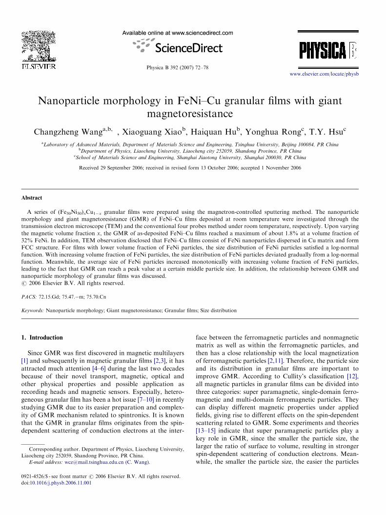
Fig. 1. Dependence of GMR on the applied field for FeNi–Cu films. (a)
15%, (b) 25%, (c) 32%, (d) 37%, (e) 42%.
C. Wang et al. / Physica B 392 (2007) 72–78 73
show super paramagnetic nature. Hence, GMR increasesmonotonically with decreasing particle size. However,Song et al. [16] found that there is no monotonicrelationship between particle size and GMR and thereexists a peak value at a certain size for s, given theannealing temperature, which is also proven theoreticallyby Gu et al. [17]. This phenomenon indicates single-domainferromagnetic particles, rather than super paramagneticparticles that predominate in GMR. In addition, Chen Xuet al. [18] confirmed that single-domain ferromagneticparticles play a key role in GMR based on effectivemedium theory. Besides, Hutten et al. [19] found that whenmagnetic particles in granular films form multi-domainstructure, their GMR will lose like bulk magnetic particles.To ascertain this discrepancy, we set FeNi–Cu granularfilms system as an example to study the relationshipbetween particle size and GMR in detail. At the same time,since the discontinuous multilayer-thin films consisted ofmagnetic phase FeNi and non-magnetic phase Ag, itdisplayed high sensitivity of GMR [20]; accordingly, it wasexpected that FeNi–Cu alloy system might be another goodcandidate.
2. Experimental procedure
In our experiments, all (Fe50Ni50)xCu1�x granular filmswere sputtered on glass and KCl substrate at roomtemperature (RT) with a spc350 multi-target magnetron-controlled sputtering system. FeNi target (the weight ratioof Fe and Ni is 1:1) and Cu target (99.99% purity) wereseparately installed on two independently controlled RFcathodes, and were alternatively employed to sputter thesefilms with 30 circles of substrate per minute. The volumefraction of FeNi in the FeNi–Cu films was controlled bychanging the sputtering power of the FeNi target and wasdetermined by means of energy dispersive spectrum,attached to a scanning electron microscope. The sputteringgas (Ar) pressure was 1.7� 10�1 Pa. The film specimens forTEM observation were obtained by putting a sputteredsubstrate into de-ionized water with a little acetone, thenby using a copper grid to hold the film floating on the waterafter the dissolution of the KCl substrate. TEM observa-tions were carried out on JEM-100CX. The GMR weremeasured with the conventional four probes method underroom temperature and the applied fields were swept fromabout �1.25 to 1.25 T.
3. Results and discussion
Fig. 1 shows the dependence of GMR on the appliedfield in FeNi–Cu films with various volume fractions ofFeNi particles. The GMR ratio is defined asDr=r ¼ ½rðHÞ � rð0Þ�=rð0Þ, where r(0) and r(H) are theresitivities at zero field and field H, respectively. As can beseen from Fig. 1, GMR first increases and then decreaseswith increasing volume fraction of FeNi particles, reachinga maximum of 1.8% at about 32 vol% FeNi. On the other
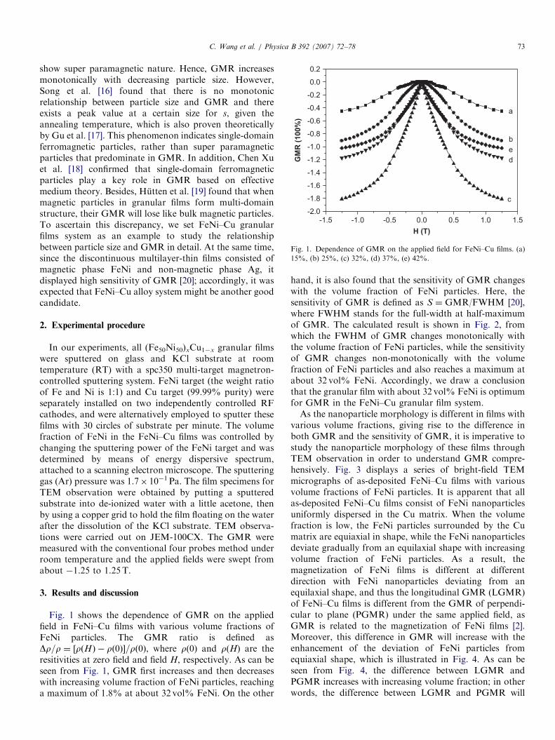
hand, it is also found that the sensitivity of GMR changeswith the volume fraction of FeNi particles. Here, thesensitivity of GMR is defined as S ¼ GMR/FWHM [20],where FWHM stands for the full-width at half-maximumof GMR. The calculated result is shown in Fig. 2, fromwhich the FWHM of GMR changes monotonically withthe volume fraction of FeNi particles, while the sensitivityof GMR changes non-monotonically with the volumefraction of FeNi particles and also reaches a maximum atabout 32 vol% FeNi. Accordingly, we draw a conclusionthat the granular film with about 32 vol% FeNi is optimumfor GMR in the FeNi–Cu granular film system.As the nanoparticle morphology is different in films with
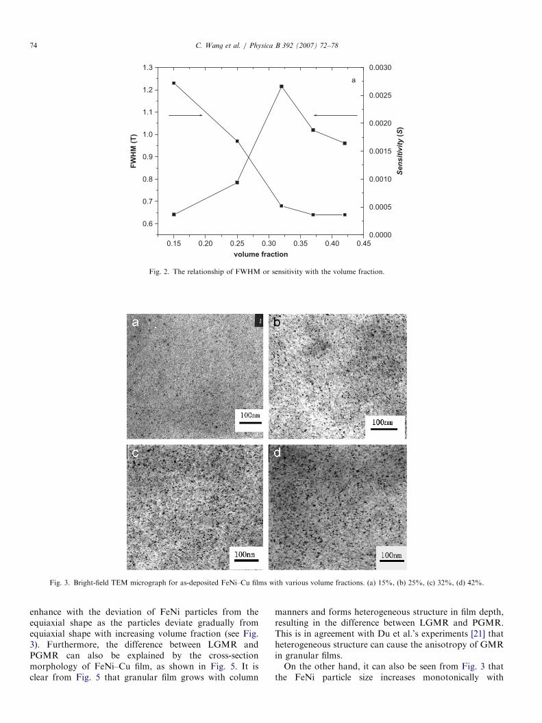
various volume fractions, giving rise to the difference inboth GMR and the sensitivity of GMR, it is imperative tostudy the nanoparticle morphology of these films throughTEM observation in order to understand GMR compre-hensively. Fig. 3 displays a series of bright-field TEMmicrographs of as-deposited FeNi–Cu films with variousvolume fractions of FeNi particles. It is apparent that allas-deposited FeNi–Cu films consist of FeNi nanoparticlesuniformly dispersed in the Cu matrix. When the volumefraction is low, the FeNi particles surrounded by the Cumatrix are equiaxial in shape, while the FeNi nanoparticlesdeviate gradually from an equilaxial shape with increasingvolume fraction of FeNi particles. As a result, themagnetization of FeNi films is different at differentdirection with FeNi nanoparticles deviating from anequilaxial shape, and thus the longitudinal GMR (LGMR)of FeNi–Cu films is different from the GMR of perpendi-cular to plane (PGMR) under the same applied field, asGMR is related to the magnetization of FeNi films [2].Moreover, this difference in GMR will increase with theenhancement of the deviation of FeNi particles fromequiaxial shape, which is illustrated in Fig. 4. As can beseen from Fig. 4, the difference between LGMR andPGMR increases with increasing volume fraction; in otherwords, the difference between LGMR and PGMR will
ARTICLE IN PRESS
0.15 0.20 0.25 0.30 0.35 0.40 0.45
0.6
0.7
0.8
0.9
1.0
1.1
1.2
1.3
0.0000
0.0005
0.0010
0.0015
0.0020
0.0025
0.0030
Sen
sit
ivit
y (
S)
FW
HM
(T
)
volume fraction
a
Fig. 2. The relationship of FWHM or sensitivity with the volume fraction.
Fig. 3. Bright-field TEM micrograph for as-deposited FeNi–Cu films with various volume fractions. (a) 15%, (b) 25%, (c) 32%, (d) 42%.
C. Wang et al. / Physica B 392 (2007) 72–7874
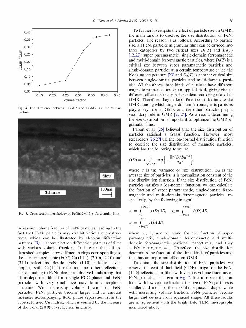
enhance with the deviation of FeNi particles from theequiaxial shape as the particles deviate gradually fromequiaxial shape with increasing volume fraction (see Fig.3). Furthermore, the difference between LGMR andPGMR can also be explained by the cross-sectionmorphology of FeNi–Cu film, as shown in Fig. 5. It isclear from Fig. 5 that granular film grows with column
manners and forms heterogeneous structure in film depth,resulting in the difference between LGMR and PGMR.This is in agreement with Du et al.’s experiments [21] thatheterogeneous structure can cause the anisotropy of GMRin granular films.On the other hand, it can also be seen from Fig. 3 that
the FeNi particle size increases monotonically with
ARTICLE IN PRESS
0.15 0.20 0.25 0.30 0.35 0.40 0.45
0.00
0.05
0.10
0.15
0.20
0.25
0.30
0.35
0.40
LG
MR
-PG
MR
volume fraction
Fig. 4. The difference between LGMR and PGMR vs. the volume
fraction.
Fig. 5. Cross-section morphology of FeNi(32 vol%)–Cu granular films.
C. Wang et al. / Physica B 392 (2007) 72–78 75
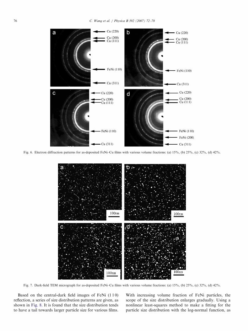
increasing volume fraction of FeNi particles, leading to thefact that FeNi particles may exhibit various microstruc-tures, which can be illustrated by electron diffractionpatterns. Fig. 6 shows electron diffraction patterns of filmswith various volume fractions. It is clear that all as-deposited samples show diffraction rings corresponding tothe face-centered cubic (FCC) Cu (1 1 1), (2 0 0), (2 2 0) and(3 1 1) reflections. Besides FeNi (1 1 0) reflection over-lapping with Cu(1 1 1) reflection, no other reflectionscorresponding to FeNi phase are observed, indicating thatall as-deposited films form single FCC phase and FeNiparticles with very small size may form amorphousstructure. With increasing volume fraction of FeNiparticles, FeNi particles become larger and its amountincreases accompanying BCC phase separation from thesupersaturated Cu matrix, which is verified by the increaseof the FeNi (2 0 0)BCC reflection intensity.
To further investigate the effect of particle size on GMR,the main task is to disclose the size distribution of FeNiparticles. The reason is as follows. According to particlesize, all FeNi particles in granular films can be divided intothree categories by two critical sizes D1(T) and D2(T)[12,22]: super paramagnetic, single-domain ferromagneticand multi-domain ferromagnetic particles, where D1(T) is acritical size between super paramagnetic particles andsingle-domain particles at a certain temperature called theblocking temperature [23] and D2(T) is another critical sizebetween single-domain particles and multi-domain parti-cles. All the above three kinds of particles have differentmagnetic properties under an applied field, giving rise todifferent effects on the spin-dependent scattering related toGMR. Therefore, they make different contributions to theGMR, among which single-domain ferromagnetic particlesplay a key role in GMR and the other particles play asecondary role in GMR [22,24]. As a result, determiningthe size distribution is important to optimize the GMR ofgranular films.Parent et al. [25] believed that the size distribution of
particles satisfied s Guass function. However, mostresearchers [26,27] use the log-normal distribution functionto describe the size distribution of magnetic particles,which has the following formula:
f ðDÞ ¼ A1ffiffiffiffiffiffi2pp
sexp �
½lnðD=D0Þ�2
2s2
� �,
where s is the variance of size distribution, D0 is theaverage size of particles, A is normalization constant of thesize distribution function. If the size distribution of FeNiparticles satisfies a log-normal function, we can calculatethe fraction of super paramagnetic, single-domain ferro-magnetic and multi-domain ferromagnetic particles, re-spectively, by the following integral:
x1 ¼
Z D1ðTÞ
0
f ðDÞdD; x2 ¼
Z D2ðTÞ
DðTÞ
f ðDÞdD,
x3 ¼
Z 1D2ðTÞ
f ðDÞdD,
where x1, x2 and x3 stand for the fraction of superparamagnetic, single-domain ferromagnetic and multi-domain ferromagnetic particles, respectively, and theysatisfy x1+x2+x3 ¼ 1. Therefore, the size distributiondetermines the fraction of the three kinds of particles andthus has an important effect on GMR.To obtain the size distribution of FeNi particles, we
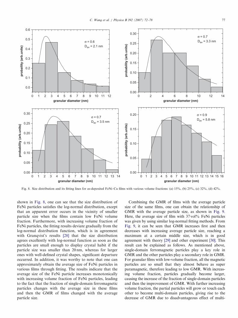
observe the central dark field (CDF) images of the FeNi(1 1 0) reflection for films with various volume fractions ofFeNi particles, as shown in Fig. 7. It can be seen that forfilms with low volume fraction, the size of FeNi particles issmaller and most of them exhibit equiaxial shape, whilewith increasing volume fraction, FeNi particles becomelarger and deviate from equiaxial shape. All these resultsare in agreement with the bright-field TEM micrographsmentioned above.
ARTICLE IN PRESS
Fig. 6. Electron diffraction patterns for as-deposited FeNi–Cu films with various volume fractions: (a) 15%, (b) 25%, (c) 32%, (d) 42%.
Fig. 7. Dark-field TEM micrograph for as-deposited FeNi–Cu films with various volume fractions: (a) 15%, (b) 25%, (c) 32%, (d) 42%.
C. Wang et al. / Physica B 392 (2007) 72–7876
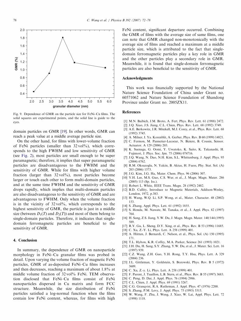
Based on the central-dark field images of FeNi (1 1 0)reflection, a series of size distribution patterns are given, asshown in Fig. 8. It is found that the size distribution tendsto have a tail towards larger particle size for various films.
With increasing volume fraction of FeNi particles, thescope of the size distribution enlarges gradually. Using anonlinear least-squares method to make a fitting for theparticle size distribution with the log-normal function, as
ARTICLE IN PRESS
0 2 4 6 9 10 11 12
0.0
0.1
0.2
0.3
0.4
0.5
0.6
σ = 0.6
Dav = 2.1 nm
pro
bailit
y (
arb
.un
its)
granular diameter (nm)
0 2 4 6 8 10 12 14
0.00
0.05
0.10
0.15
0.20
0.25
0.30
σ = 0.7
Dav = 3.3 nm
pro
bab
ilit
y (
arb
.un
its)
granular diameter (nm)
0 6 9 10 11 12 13 14
0.00
0.05
0.10
0.15
0.20
0.25
0.30
σ = 0.7
Dav = 3.5 nm
pro
bab
ilit
y (
arb
.un
its)
granular diameter (nm)
0 2 5 8 10 11 12 13 14 15 16
0.00
0.05
0.10
0.15
0.20 σ = 0.9
Dav = 5.8 nm
pro
bab
ilit
y (
arb
.un
its)
granular diameter (nm)
1 3 5 7 8
1 3 4 6 7 91 2 3 4 5 7 8
Fig. 8. Size distribution and its fitting lines for as-deposited FeNi–Cu films with various volume fractions: (a) 15%, (b) 25%, (c) 32%, (d) 42%.
C. Wang et al. / Physica B 392 (2007) 72–78 77
shown in Fig. 8, one can see that the size distribution ofFeNi particles satisfies the log-normal distribution, exceptthat an apparent error occurs in the vicinity of smallerparticle size when the films contain low FeNi volumefraction. Furthermore, with increasing volume fraction ofFeNi particles, the fitting results deviate gradually from thelog-normal distribution function, which is in agreementwith Granqvist’s results [28] that the size distributionagrees excellently with log-normal function as soon as theparticles are small enough to display crystal habit if theparticle size was smaller than 20 nm, whereas for largerones with well-defined crystal shapes, significant departureoccurred. In addition, it was worthy to note that one canapproximately obtain the average size of FeNi particles invarious films through fitting. The results indicate that theaverage size of the FeNi particle increases monotonicallywith increasing volume fraction of FeNi particles, leadingto the fact that the fraction of single-domain ferromagneticparticles changes with the average size in these filmsand then the GMR of films changed with the averageparticle size.
Combining the GMR of films with the average particlesize of the same films, one can obtain the relationship ofGMR with the average particle size, as shown in Fig. 9.Here, the average size of film with 37vol% FeNi particleswas given by using similar log-normal fitting methods. FromFig. 9, it can be seen that GMR increases first and thendecreases with increasing average particle size, reaching amaximum at a certain middle size, which is in goodagreement with theory [29] and other experiment [30]. Thisresult can be explained as follows. As mentioned above,single-domain ferromagnetic particles play a key role inGMR and the other particles play a secondary role in GMR.For granular films with low-volume fraction, all the magneticparticles are so small that they almost behave as superparamagnetic, therefore leading to low GMR. With increas-ing volume fraction, particles gradually become larger,causing the increase of the fraction of single-domain particlesand then the improvement of GMR. With further increasingvolume fraction, the partial particles will grow or touch eachother to become multi-domain particles, giving rise to thedecrease of GMR due to disadvantageous effect of multi-
ARTICLE IN PRESS
2.0 2.5 3.0 3.5 4.0 4.5 5.0 5.5 6.0
0.4
0.6
0.8
1.0
1.2
1.4
1.6
1.8
2.0
GM
R (
%)
granular diameter (nm)
Fig. 9. Dependence of GMR on the particle size for FeNi–Cu films. The
solid squares are experimental points, and the solid line is guide to the
eyes.
C. Wang et al. / Physica B 392 (2007) 72–7878
domain particles on GMR [19]. In other words, GMR canreach a peak value at a middle average particle size.
On the other hand, for films with lower-volume fractionof FeNi particles (smaller than 32 vol%), which corre-sponds to the high FWHM and low sensitivity of GMR(see Fig. 2), most particles are small enough to be superparamagnetic; therefore, it implies that super paramagneticparticles are disadvantageous to the FWHM and thesensitivity of GMR. While for films with higher volumefraction (larger than 32 vol%), most particles becomelarger or touch each other to form multi-domain particles,and at the same time FWHM and the sensitivity of GMRdrops rapidly, which implies that multi-domain particlesare also disadvantageous to the sensitivity of GMR and areadvantageous to FWHM. Only when the volume fractionis in the vicinity of 32 vol%, which corresponds to thehighest sensitivity of GMR, the particle is just in a middlesize (between D1(T) and D2(T)) and most of them belong tosingle-domain particles. Therefore, it indicates that single-domain ferromagnetic particles are beneficial to thesensitivity of GMR.
4. Conclusion
In summary, the dependence of GMR on nanoparticlemorphology in FeNi–Cu granular films was probed indetail. Upon varying the volume fraction of magnetic FeNiparticles, GMR of as-deposited FeNi–Cu films increasesand then decreases, reaching a maximum of about 1.8% atmiddle volume fraction of 32 vol% FeNi. TEM observa-tion disclosed that FeNi–Cu films consist of FeNinanoparticles dispersed in Cu matrix and form FCCstructure. Meanwhile, the size distribution of FeNiparticles satisfied a log-normal function when the filmscontain low FeNi content, whereas, for films with high
FeNi content, significant departure occurred. Combiningthe GMR of films with the average size of same films, onecan note that GMR changed non-monotonically with theaverage size of films and reached a maximum at a middleparticle size, which is attributed to the fact that single-domain ferromagnetic particles play a key role in GMRand the other particles play a secondary role in GMR.Meanwhile, it is found that single-domain ferromagneticparticles are also beneficial to the sensitivity of GMR.
Acknowledgments
This work was financially supported by the NationalNature Science Foundation of China under Grant no.60571062 and Nature Science Foundation of ShandongProvince under Grant no. 2005ZX11.
References
[1] M.N. Baibich, J.M. Broto, A. Fert, Phys. Rev. Lett. 61 (1988) 2472.
[2] J.Q. Xiao, J.S. Jiang, C.L. Chien, Phys. Rev. Lett. 68 (1992) 3749.
[3] A.E. Berkowitz, J.R. Mitekell, M.J. Corey, et al., Phys. Rev. Lett. 68
(1992) 3745.
[4] A. Milner, I. Ya. Korenblit, A. Gerber, Phys. Rev. B 60 (1999) 14821.
[5] C. Fermon, M. Pannetier-Lecoeur, N. Biziere, B. Cousin, Sensor.
Actuator. A 129 (2006) 203.
[6] K. Suenaga, G. Oomi, Y. Uwatoko, K. Saito, K. Takanashi, H.
Fujimori, J. Phys. Soc. Jpn. 75 (2006) 074716.
[7] J.Q. Wang, N. Dao, N.H. Kim, S.L. Whittenburg, J. Appl. Phys. 95
(2004) 6762.
[8] R.M. Oksuzoglu, N. Yalcin, B. Aktas, H. Fuess, Phy. Stat. Sol. (A)
203 (2006) 1573.
[9] J.G. Kim, J.G. Ha, Mater. Chem. Phys. 96 (2006) 307.
[10] Y.H. Lee, M.S. Guo, C.S. Wur, et al., J. Magn. Magn. Mater. 286
(2005) 113 (Sp. Iss.).
[11] Robert L. White, IEEE Trans. Magn. 28 (1992) 2482.
[12] B.D. Cullity, Introduce to Magnetic Materials, Addison-Wesley,
London, 1972, p.383.
[13] H. Wang, W.Q. Li, S.P. Wong, et al., Mater. Character. 48 (2002)
153.
[14] S. Zhang, Appl. Phys. Lett. 61 (1992) 1855.
[15] S. Honda, M. Nawate, M. Tanaka, et al., J. Appl. Phys. 82 (1997)
764.
[16] H. Sang, Z.S. Jiang, Y.W. Du, J. Magn. Magn. Mater. 140/144 (1995)
589.
[17] R.Y. Gu, L. Sheng, D.Y. Xing, et al., Phys. Rev. B 53 (1996) 11685.
[18] C. Xu, Z.-Y. Li, Phys. Lett. A 258 (1999) 401.
[19] A. Hutten, J. Bernardi, C. Nelson, et al., Phys. Sol. (A) 150 (1995)
171.
[20] T.L. Hylton, K.R. Coffey, M.A. Parker, Science 261 (1993) 1021.
[21] J.H. Du, H. Sang, S.Y. Zhang, Y.W. Du, et al., J. Mater. Sci. Lett. 16
(1997) 939.
[22] C.Z. Wang, Z.H. Guo, Y.H. Rong, T.Y. Hsu, Phys. Lett. A 329
(2004) 236.
[23] J.L. Gittleman, Y. Goldstein, S. Bozowski, Phys. Rev. B 5 (1972)
3609.
[24] C. Xu, Z.-y. Li, Phys. Lett. A 258 (1999) 401.
[25] F. Parent, J. Tuaillon, L.B. Stern, et al., Phys. Rev. B 55 (1997) 3683.
[26] C. Peng, D. Dai, J. Appl. Phys. 76 (1994) 2986.
[27] C.L. Chien, J. Appl. Phys. 69 (1991) 5267.
[28] C.G. Granqvist, R.A. Ruthrman, J. Appl. Phys. 47 (1976) 2200.
[29] S. Zhang, P.M. Levy, J. Appl. Phys. 75 (1993) 5315.
[30] W. Wang, F. Zhu, J. Weng, J. Xiao, W. Lai, Appl. Phys. Lett. 72
(1998) 1118.