nanoparticle-based sandwich electrochemical immunoassay for carbohydrate antigen 125 with signal...
TRANSCRIPT

Nanoparticle-Based Sandwich ElectrochemicalImmunoassay for Carbohydrate Antigen 125 withSignal Enhancement Using Enzyme-CoatedNanometer-Sized Enzyme-Doped Silica Beads
Dianping Tang,* Biling Su, Juan Tang, Jingjing Ren, and Guonan Chen*
Key Laboratory of Analysis and Detection for Food Safety (Ministry of Education), and Fujian Provincial KeyLaboratory of Analysis and Detection for Food Safety, Department of Chemistry,Fuzhou University, Fuzhou 350108, China
A novel nanoparticle-based electrochemical immunoassayof carbohydrate antigen 125 (CA125) as a model wasdesigned to couple with a microfluidic strategy using anti-CA125-functionalized magnetic beads as immunosensingprobes.Toconstructtheimmunoassay,thionine-horseradishperoxidase conjugation (TH-HRP) was initially doped intonanosilica particles using the reverse micelle method, andthen HRP-labeled anti-CA125 antibodies (HRP-anti-CA125) were bound onto the surface of the synthesizednanoparticles, which were used as recognition elements.Different from conventional nanoparticle-based electro-chemical immunoassays, the recognition elements of theimmunoassay simultaneously contained electron mediatorand enzyme labels and simplified the electrochemicalmeasurement process. The sandwich-type immunoassayformat was used for the online formation of the immuno-complex in an incubation cell and captured in the detec-tion cell with an external magnet. The electrochemicalsignals derived from the carried HRP toward the reduc-tion of H2O2 using the doped thionine as electron media-tor. Under optimal conditions, the electrochemical im-munoassay exhibited a wide working range from 0.1 to450 U/mL with a detection limit of 0.1 U/mL CA125.The precision, reproducibility, and stability of the immu-noassay were acceptable. The assay was evaluated forclinical serum samples, receiving in excellent accordancewith results obtained from the standard enzyme-linkedimmunosorbent assay (ELISA) method. Concluding, thenanoparticle-based assay format provides a promisingapproach in clinical application and thus represents aversatile detection method.
Immunoassay, as a promising approach for selective andsensitive analysis, has recently gained increasing attention.1-4
Various immunoassays have been developed in different fieldsincluding environmental monitoring, food safety, and clinicaldiagnosis.5-11 Nanoparticle-based assays hold great promise inrealizing highly sensitive and selective detection at attomolar
protein concentrations without requiring complex amplificationmethods including polymerase chain reaction (PCR).12-15 Mirkinand his colleagues reported a nanoparticle-based approach for thedetection of free prostate-specific antigen (PSA) at low attomolarconcentration by adding an antibody-labeled magnetic micropar-ticle, DNA barcodes, and conjugating a second antibody to theDNA-conjugated gold nanoparticle.16 Liu’s group reported ananoparticle-based assay for the highly sensitive PSA detectionat concentrations as low as 500 attomolar on a single disposablechip using a light scattering method.12 The nanoparticle-basedassay allows for the detection of low concentration levels withsignal amplification and reduces the sample pretreatment require-ment owing to the presence of magnetic particles.17-19
Usually, the nanoparticle-based assay consisted of two kindsof particles: (i) magnetic microparticles coated with monoclonalantibodies as immunosensing probe and (ii) multifunctionalnanoparticles (including nanogold particles) decorated with poly-clonal antibodies or unique “barcode” DNA sequences as recogni-tion elements. Iron oxide (Fe3O4) nanoparticles, as a well-known
* To whom correspondence should be addressed. E-mail: [email protected] or [email protected] (D.T.); [email protected] (G.C.).Phone/Fax: +86 591 2286 6135.
(1) Thangawng, A.; Howell, P., Jr.; Richards, J.; Erickson, J.; Ligler, F. LabChip 2009, 9, 3126.
(2) Golden, J.; Kim, J.; Erickson, J.; Hilliard, L.; Howell, P.; Anderson, G.; Nasir,M.; Ligler, F. Lab Chip 2009, 9, 1942.
(3) Rieger, M.; Cervino, C.; Sauceda, J.; Niessner, R.; Knopp, D. Anal. Chem.2009, 81, 2373.
(4) Wolter, A.; Niessner, R.; Seidel, M. Anal. Chem. 2008, 80, 5854.(5) Lai, G.; Yan, F.; Ju, H. Anal. Chem. 2009, 81, 9730.(6) Liu, H.; Fu, Z.; Yang, Z.; Yan, F.; Ju, H. Anal. Chem. 2008, 80, 5654.(7) Kim, J.; Anderson, G.; Erickson, J.; Golden, J.; Nasir, M.; Ligler, F. Anal.
Chem. 2009, 81, 5426.(8) Wojciechowski, J.; Shriver-Lake, L.; Yamaguchi, M.; Fureder, E.; Pieler,
R.; Schamesberger, M.; Winder, C.; Ligler, F. Anal. Chem. 2009, 81, 519.(9) Ligler, F. S. Anal. Chem. 2009, 81, 519.
(10) Erickson, J.; Ligler, F. Nature 2008, 456, 178.(11) Johnson, B.; Delehanty, J.; Lin, B.; Ligler, F. Anal. Chem. 2008, 80, 2113.(12) Goluch, E.; Nam, J.; Georganopoulou, D.; Chiesl, T.; Shaikh, K.; Ryu, K.;
Barron, A.; Mirkin, C.; Liu, C. Lab Chip 2006, 6, 1293.(13) Kim, D.; Daniel, W.; Mirkin, C. Anal. Chem. 2009, 81, 9183.(14) Dhar, S.; Daniel, W.; Giljohann, D.; Mirkin, C.; Lippard, S. J. Am. Chem.
Soc. 2009, 131, 14652.(15) Hurst, S.; Hill, H.; Macfarlane, R.; Wu, J.; Dravid, V.; Mirkin, C. Small 2009,
5, 2156.(16) Nam, J.; Thaxton, C.; Mirkin, C. Science 2003, 301, 1884.(17) Banholzer, M.; Qin, L.; Millstone, J.; Osberg, K.; Mirkin, C. Nat. Protoc.
2009, 4, 838.(18) Daniel, W.; Han, M.; Lee, J.; Mirkin, C. J. Am. Chem. Soc. 2009, 131, 6362.(19) Millstone, J.; Stoeva, S.; Lee, J.; Shaikh, K.; Mirkin, C.; Liu, C. Biosens.
Bioelectron. 2009, 24, 2397.
Anal. Chem. 2010, 82, 1527–1534
10.1021/ac902768f 2010 American Chemical Society 1527Analytical Chemistry, Vol. 82, No. 4, February 15, 2010Published on Web 01/22/2010

hard magnetic material, were extensively applied in the nanopar-ticle-based assays since they have good biocompatibility and canbe separated very readily from reaction mixtures with the help ofan external magnetic field.20-23 The functionalized probes couldpull antibodies bound to magnetic nanoparticles from one laminarflow path to another by applying a local magnetic field gradientand selectively removing them from flowing biological fluidswithout any washing step. Moreover, it has been successfullyapplied as matrixes for the electrochemical detection of biomar-kers in our past works.24-27 So, Fe3O4 nanoparticles were usedfor the fabrication of the nanoparticle-based immunosensingprobes in this study.
For the successful development of nanoparticle-based electro-chemical immunoassays, there are two basal issues to realize theapplication of electrochemical technique.28 First, signaling ampli-fication and noise reduction are very crucial for obtaining lowdetection limits. In general, they are achieved by using an indicatorsystem that results in the amplification of the measured productincluding enzyme label and nanolabels.29-32 Nanogold particleslabeled with HRP-bound antibodies are commonly exploited assignal transduction tools in the electrochemical immunoassays.33,34
The traditional method often exhibited low signaling; however,there is because of sterical reasons usually a ratio of 1:1 forenzyme and detection antibody used. To improve the labeledmethod, we utilize HRP-doped silica nanoparticles for the labelof HRP-bound antibodies. With the use of this method, there aremany HRP molecules inside and outside of the synthesizedbionanolabels. The carried HRP molecules will be entered andparticipated in the catalytic reaction, suspecting, when oneantibody among them reacts with the corresponding antigen. Thishigh efficiency makes them especially suitable for ultrasensitivebioanalysis with signal amplification.
Another key factor is to provide a good pathway of electrontransfer between the redox center of enzymes and the electrodesurface.35-38 Recently, we developed a reusable electrochemicalimmunoassay for aflatoxin B1 in food using a multifunctionalmagnetic bead as an immunosensing probe with a magneticCoFe2O4 nanoparticle as core, Prussian blue nanoparticle-doped
silica as shell, and gold nanoparticles labeled with HRP-boundanti-AFB1 antibodies as recognition elements.27 Although thedoped Prussian blue could enhance the electron communicationbetween the immobilized HRP and the base electrode, the reactivetime of the redox was relatively long, and the sensitivity was notso high (i.e., nanoamps of current response). The reason mightbe the fact that the electron mediator (Prussian blue) and bioactiveHRP enzyme were immobilized on two sets of nanoparticles,respectively, which lengthened the pathway of electron transferand lessened the redox of the HRP-H2O2 system. To solve thisproblem, we prepared a novel multifunctional silica nanoparticle,which simultaneously contained bioactive HRP enzyme andelectroactive thionine mediator, and the synthesized nanosilicaparticles were labeled onto HRP-bound antibodies as recognitionelements in this paper.
The aim of this work is to exploit an advanced nanoparticle-based electrochemical immunoassay for the detection of carbo-hydrate antigen 125 (CA125) as a model analyte in clinicaldiagnosis. The magnetic immunosensing probe not only favorsthe rapid separation and purification of sandwich-type bionano-composites but also facilitates the fabrication of the microfluidicimmunosensing interface via using an external magnet. Theelectrochemical signaling of the captured multifunctional silicananoparticles could be rapidly and adequately achieved becauseof the catalytic reaction of the carried HRP relative to theH2O2-thionine system. The performance and factors influencingthe nanoparticle-based assay are investigated and discussed inthe following sections.
EXPERIMENTAL SECTIONMaterials. Mouse monoclonal anti-CA125 (primary antibod-
ies), HRP-labeled anti-CA125 (HRP-anti-CA125, secondary anti-bodies), CA125, and HRP (EC 1.11.1.7, RZ > 3.0, A > 250 U/mg)were purchased from Sigma (St. Louis, MO). Thionine (TH),gultaraldehyde, bovine serum albumin (BSA, 96%-99%), (3-glycidyloxypropyl) trimethoxysilane (C9H20O5Si, GOPS), tet-ramethoxysilane (TEOS), and bis-(2-ethylhexyl) sodium sulfos-uccinate (AOT) were obtained from Sigma-Aldrich. Cyclohexane,n-hexanol, and ammonium hydroxide (25 wt %) were purchasedfrom Merck (Darmstadt, Germany). Magnetic Fe3O4 nanoparticles(particle size: 100 nm) in an aqueous suspension with a concentra-tion of 25 mg/mL were obtained from Chemicell GmbH (Berlin,Germany). All other reagents were of analytical grade and wereused without further purification. Deionized and distilled waterwas used throughout the study. The 0.1 M phosphate-bufferedsaline (PBS, pH 7.4) was prepared by adding 12.2 g of K2HPO4,1.36 g of KH2PO4, and 8.5 g of NaCl in 1000 mL of deionized water.
Fabrication of Immunosensing Probes. Prior to the experi-ment, Fe3O4 nanoparticles were separated using an externalmagnet, and dried in the vacuum at 80 °C for 1 h. Following that,250 mg of Fe3O4 nanoparticles was added into 5.0 mL of 5% GOPS(v/v) in dry toluene and stirred with 500 rpm for 12 h at roomtemperature (RT).39 With the aid of an external magnet, the GOPS-functionalized Fe3O4 nanoparticles were separated and purified.Afterward, the synthesized nanoparticles were washed three timeswith toluene and ethanol solution, respectively. The purifiednanoparticles were dried and activated in an oven at 80 °C for 1 h
(20) Yang, Z.; Liu, H.; Zong, C.; Yan, F.; Ju, H. Anal. Chem. 2009, 81, 5484.(21) Corchero, J.; Villaverde, A. Trends Biotechnol. 2009, 27, 468.(22) Schubayev, V.; Pisanic, T., II; Jin, S. Adv. Drug Delivery Rev. 2009, 61,
467.(23) Xie, J.; Huang, J.; Li, X.; Sun, X.; Chen, X. Curr. Med. Chem. 2009, 16,
1278.(24) Tang, D.; Yuan, R.; Chai, Y. Clin. Chem. 2007, 53, 1323.(25) Tang, D.; Yuan, R.; Chai, Y.; An, H. Adv. Funct. Mater. 2007, 17, 976.(26) Tang, D.; Yuan, R.; Chai, Y. J. Phys. Chem. 2006, 110, 11640.(27) Tang, D.; Zhong, Z.; Niessner, R.; Knopp, D. Analyst 2009, 134, 1554.(28) Tushar, R.; Amit, A.; Shuming, N. Anal. Chem. 2006, 78, 5627.(29) Yu, X.; Munge, B.; Patel, V.; Jensen, G.; Bhirde, A.; Gong, J.; Kim, S.;
Gillespie, J.; Gutkind, J.; Papadimitrakopoulos, F.; Rusling, J. J. Am. Chem.Soc. 2006, 128, 11199.
(30) Sun, N.; McMullan, M.; Papakonstantinou, P.; Gao, H.; Zhang, X.; Mihailovic,D.; Li, M. Anal. Chem. 2008, 80, 3593.
(31) Das, J.; Aziz, M.; Yang, H. J. Am. Chem. Soc. 2006, 128, 16022.(32) Wang, J.; Liu, G.; Engelhard, M.; Lin, Y. Anal. Chem. 2006, 78, 6974.(33) Shim, S.; Lim, D.; Nam, J. Nanomedicine 2008, 3, 215.(34) Kim, E.; Stanton, J.; Korber, B.; Krebs, K.; Bogdan, D.; Kunstman, K.; Wu,
S.; Wolinsky, S. Nanomedicine 2008, 3, 293.(35) Lanci, M.; Remy, M.; Kaminsky, W.; Mayer, J.; Sanford, M. J. Am. Chem.
Soc. 2009, 131, 15618.(36) Palacin, T.; Le Khanh, H.; Jousselme, B.; Jeqou, P.; Filoramo, A.; Ehli, C.;
Guldi, D.; Campidelli, S. J. Am. Chem. Soc. 2009, 131, 15394.(37) Dai, Z.; Yan, F.; Yu, H.; Hu, X.; Ju, H. J. Immunol. Methods 2004, 287, 13.(38) Wu, J.; Yan, F.; Tang, J.; Zhai, C.; Ju, H. Clin. Chem. 2007, 53, 1495. (39) Wu, Y.; Chen, C.; Liu, S. Anal. Chem. 2009, 81, 1600.
1528 Analytical Chemistry, Vol. 82, No. 4, February 15, 2010

under a nitrogen atmosphere. Afterward, 100 µL of anti-CA125(500 ng/mL) was injected into the functionalized Fe3O4 aqueoussolution with the concentration of 25 mg/mL. The mixture wasslightly stirred for 12 h at 4 °C to make the -NH2 groups of anti-CA125 antibodies conjugate to epoxy groups. The obtained anti-CA125-Fe3O4 bionanoparticles (i.e., immunosensing probes,Scheme 1) were incubated with 10 mg/mL BSA-PBS at 37 °Cfor 1 h to block the unreacted and nonspecific sites. Finally, theas-prepared bionanoparticles were stored in pH 7.4 PBS with afinal concentration of 25 mg/mL at 4 °C when not in use.
Preparation of Thionine-HRP Conjugation (TH-HRP).Amounts of 500 µL of HRP (1.5 × 10⁻³ mol/L) and 500 µL ofthionine (3.0 × 10⁻⁵ mol/L) were initially dissolved into 1 mL of0.01 M PBS, and the pH was adjusted to 9.5 by using 10 wt %K2CO3, and then 100 µL of gultaraldehyde solution was addedinto the mixture. After being stirred for 2 h, the mixture wasadjusted to pH 7.0 by using 1.0 M NaH2PO4. The unbound thioninewas removed using ultrafiltration for 12-15 times until the peakcorresponding to thionine in the elution disappeared. Finally, theobtained TH-HRP was prepared into 2 mL of aqueous solution.
Synthesis of Recognition Elements. Scheme 1 displays theconstruction of the nanosilica-based recognition elements (RE).The TH-HRP-doped silica nanoparticles were prepared at RTusing a reverse micelle method according to these literatures withsome modification,40,41 which contained the following three steps:(i) 5.3 g of Triton X-100, 5.0 mL of n-hexanol, and 1.0 mL ofTH-HRP were added into 20 mL of cyclohexane solution in turnand continuously stirred for 20 min, (ii) 2.5 mL of TEOS and 1.8mL of NH3 ·H2O (25 wt %) were slowly dropped into the stirringmixture and vigorously stirred for 24 h, and (iii) 5.0 mL of acetonewas added, and the mixture was centrifuged and washed for fivetimes with ethanol and water. The obtained TH-HRP-doped silicananoparticles (designed as SiO2(TH-HRP)) were used for thefabrication of recognition elements.
Similar with the preparation process of the immunosensingprobes, the functionalization of the SiO2(TH-HRP) was carriedout using the amine-epoxy interaction. To maintain the bioactivityof the doped HRP, the drying and activation of the SiO2(TH-HRP)
were performed at 40 °C. The process included the synthesis ofepoxy-functionalized SiO2(TH-HRP) and the modification ofHRP-anti-CA125 on the epoxy-functionalized SiO2(TH-HRP).Finally, the synthesized HRP-anti-CA125-SiO2(TH-HRP) bio-nanoparticles were fixed with a concentration of 25 mg/mL andstill stored at 4 °C when not in use.
Electrochemical Measurement. Electrochemical measure-ments were carried out with a microAutoLab Type III system (EcoChemie, The Netherlands) in combination with a flow-throughdetection cell. The detection system comprised an indium-tinoxide (ITO, 5 wt % In2O3 + SnO2) working electrode, a platinumwire as auxiliary electrode, and a Ag/AgCl reference electrode(the schematic illustration is shown in our previous report27). Theflow injection system consists of a six-way connected valveequipped with a 1 mL syringe pump and connected through aTeflon tubing to the flow cell. The analytical flow stream enteredfrom the other side into the center of the flow cell at 0.5 mL/min. The ITO electrode was installed at the bottom of the cell,and the bar electromagnet was set under the ITO electrode. Allthe experiments were carried out in pH 7.4 PBS containing 1.0mM H2O2 with a potential sweep rate of 50 mV/s at RT. With thesandwich-type immunoassay format, the carried HRP on therecognition elements exhibited a stable current at +120 mV (vsAg/AgCl). The changes in cathodic currents were collected andrecorded versus the CA125 concentrations.
Scheme 1 represents the sandwich-type immunoassay protocoland measurement method. During the process, the collection,incubation, and detection of the nanoparticle-based immunoassaywere performed in the flow cell. The measurement processconsisted of five steps: (i) 50 µL of immunosensing probes (25mg/mL) was injected into the cell and collected on the ITOsurface with an external magnet; (ii) 50 µL of CA125 standards/specimens with various concentrations was added to the cell andincubated for 18 min without the magnetic field to form theimmunocomplexes on the Fe3O4 surface; (iii) the immunocom-plexes were accumulated and washed with the external magneticfield; (iv) 100 µL of recognition elements (25 mg/mL) was flowedinto the cell and incubated for another 18 min without themagnetic field to construct a sandwich-type immunocomplex; (v)the H2O2-PBS (pH 7.4) was flowed through the cell with the
(40) Kumar, R.; Maitra, A.; Patanjali, P.; Sharma, P. Biomaterials 2005, 26, 6743.(41) Zhong, Z.; Li, M.; Xiang, D.; Dai, N.; Qing, Y.; Wang, D.; Tang, D. Biosens.
Bioelectron. 2009, 24, 2246.
Scheme 1. Construction of the Immunosensing Probe and Recognition Element (RE) and Measurement Protocol ofthe Nanoparticle-Based Electrochemical Immunoassay with a Sandwich-Type Format
1529Analytical Chemistry, Vol. 82, No. 4, February 15, 2010

magnetic field, and cyclic voltammograms were registered at theflow stop. After each step, the cell should be washed with pH 7.4PBS.
RESULTS AND DISCUSSIONCharacteristics of the Nanoparticle-Based Immunoassay.
In this study, the nanoparticle-based immunoassay contains twokinds of nanoparticles including anti-CA125-Fe3O4 and HRP-anti-CA125-SiO2(TH-HRP). In the presence of the analyte (CA125antigen), these two sets of nanoparticles form a sandwich-typeimmunocomplex, and the immunocomplex increases with theincrement of the CA125 concentration in the sample, thusfabricating a three-dimensional network with the interleaving ofFe3O4 and SiO2 nanoparticles. With an external magnet, theimmunocomplex is captured on the surface of the ITO electrode.The electrochemical detection is based on the immobilized HRPon the silica nanoparticles toward the reduction of H2O2 with thehelp of the doped (thionine) electron mediator. The more theamount of CA125 antigens in the sample was, the more the amountof the cross-linked HRP-anti-CA125-SiO2(TH-HRP) in theimmunocomplex was; thus, the catalytic current was increased.
To successfully carry out the nanoparticle-based immunoassay,the preparation, modification, and functionalization of the nano-particles were very crucial. In this paper, anti-CA125 and HRP-anti-CA125 were immobilized on the surface of nanoparticles throughthe epoxy-amino reaction.42,43 Figure 1 shows the transmission
electron microscopy (TEM, H600, Hitachi Instrument, Japan)images of Fe3O4 and SiO2 nanoparticles before and after modifiedwith the biomolecules, respectively. Seen from Figure 1, the meansizes of the formed bionanoparticles were 100 and 60 nm for Fe3O4
and SiO2(TH-HRP), respectively. Moreover, the surface of thesenanoparticles became rougher after modification, indicating anti-CA125 and HRP-anti-CA125 could be linked to the nanoparticles(inset of Figure 1).
To further verify the formation of the functional nanoparticles,we used UV-vis absorption spectrometry (Hitachi Instrument,Japan) to investigate the fabricated process of the bionanoparticles(Figure 2). Figure 2a-c shows the absorption spectra of anti-CA125 antibodies, pure HRP, and thionine, respectively. Aftermagnetic Fe3O4 nanoparticles were modified with anti-CA125antibodies, a 278 nm absorption peak was observed in comparisonwith that of pure Fe3O4 nanoparticles (Figure 2, curves d and e),suggesting antibodies could be covalently bound to Fe3O4 nano-particles. Seen from Figure 2, curves f and g, the absorption peaksof HRP and thionine could be simultaneously appeared at theSiO2(TH-HRP) nanoparticles. This result indicated that theHRP-thionine conjugation could be doped into the silica particlesusing the reverse micelle method. Furthermore, the 268 nmabsorption peak was achieved when HRP-anti-CA125 biomol-ecules were conjugated to the SiO2(TH-HRP) nanoparticles. Theabsorption wavelength was obviously less than that of pure anti-CA125 (Figure 2a). The reason might be attributed to theinteroverlapping of 278 nm (for HRP) and 250 nm (for thionine).These results suggested that anti-CA125-Fe3O4 and HRP-anti-
(42) Hu, P.; Huang, C.; Li, Y.; Ling, J.; Liu, Y.; Fei, Y.; Xie, J. Anal. Chem. 2008,80, 1819.
(43) Wu, Y.; Chen, C.; Liu, S. Anal. Chem. 2009, 81, 1600.
Figure 1. TEM images of (a) Fe3O4 and (b) HRP-TH-doped SiO2 nanoparticles. Insets: TEM images of anti-CA125-Fe3O4 and HRP-anti-CA125-SiO2(TH-HRP), respectively.
Figure 2. UV-vis absorption spectra of (a) anti-CA125, (b) HRP, (c) thionine, (d) nano-Fe3O4 colloids, (e) anti-CA125-Fe3O4 colloids, (f)nano-SiO2 colloids, and (g) HRP-TH-doped nano-SiO2 colloids.
1530 Analytical Chemistry, Vol. 82, No. 4, February 15, 2010

CA125-SiO2(TH-HRP) could be constructed via using thereverse micelle method and the epoxy-amino reaction.
Electrochemical Characteristics of Nanoparticle-BasedImmunoassay. Figure 3A displays the cyclic voltammograms ofthe nanoparticle-based immunoassay for the various steps at 50mV/s in pH 7.4 PBS. The formed immunocomplex was capturedon the surface of the ITO electrode with an external magnet afterbeing reacted with 100 U/mL CA125. No peak was observed forthe anti-CA125-Fe3O4 and CA125-anti-CA125-Fe3O4 (Figure 3,curves a and b). The high background current after theantigen-antibody interaction was mainly attributed to the for-mation of the immunocomplex on the nanoparticle surface,which hindered the electron transfer. After HRP-anti-CA125-SiO2(TH-HRP) bionanoparticles were reacted with theCA125-anti-CA125-Fe3O4 bionanoparticles, however, the formedimmunocomplex exhibited a couple of redox peak at +120 and220 mV (Figure 3c). The result indicated that the doped thionine,as a good electron mediator, still remained their redox properties,which could provide a fast pathway of electron transfer for thereduction of H2O2. Seen from Figure 3d, an obvious redox reactionwas appeared with a distinct increase of the reduction currentand a decrease of the oxidation current upon the addition of H2O2
in pH 7.4 PBS. Moreover, the reduction current (ipc) increasedwith the increment of CA125 concentration in the sample. Thecatalytic current mainly derived from the immobilized HRP towardthe reduction of H2O2 with the aid of the doped thionine asmediator.44,45 Thus, we might quantitatively evaluate the concen-tration of CA125 according to the reduction current.
To clarify the advantage of the developed immunoassay usingmultifunctional silica nanoparticles in place of the conventionalnanogold particles, we fabricated two kinds of recognition ele-ments including HRP-anti-CA125-SiO2(TH-HRP) and HRP-anti-CA125-nanogold, which were used for the detection of CA125by using the sandwich-type immunoassay and the same anti-CA125-Fe3O4 probes. The preparation and labeling of nanogoldparticles were according to our previous report46 and measuredin pH 7.4 thionine-PBS. As shown in Figure 3B, HRP-anti-CA125-SiO2(TH-HRP) exhibited higher response and sensitivity
(i.e., slope) than that of using HRP-anti-CA125-nanogold asrecognition elements. The reason might be attributed to theproximity of the TH and the fact that the HRP molecules wereimmobilized inside and outside of silica nanoparticles, whereasthey were only on the surface of gold nanoparticles. We mightspeculate that the immobilized amount of the HRP molecules forHRP-anti-CA125-SiO2(TH-HRP)wasmorethanthatofHRP-anti-CA125-nanogold and thus exhibited high catalytic efficiencytoward the reduction of H2O2. In addition, the synthesizedHRP-anti-CA125-SiO2(TH-HRP) could avoid the addition ofelectron mediator.
Optimization of Experimental Conditions. To achieve anoptimal electrochemical signaling, the ratio of HRP and thioninedoped into silica nanoparticles was very crucial owing to the almostsame amount of the HRP-anti-CA125 on the nanosilica surface.Various molar ratios of HRP and thionine including 4:1, 3:1, 2:1,1:1, 1:2, 1:3, and 1:4 (w/w) were used for the preparation ofrecognition elements, which was applied for the detection of 100U/mL CA125. Seen from Figure 4a, the optimal current responsewas obtained at the molar ratio of 2:1. Higher or lower concentra-tion of thionine as an electron mediator would affect the catalyticperformance of the immobilized HRP toward the reduction ofH2O2. Thus, 2:1 molar ratio of HRP and thionine was selected forthe preparation of recognition elements.
In the sandwich-type immunoassays, temperature and time forthe antigen-antibody interaction greatly influenced the sensitivityof the developed immunoassay. Considering the practical applica-tion of the proposed system in clinical immunoassays, all experi-ments were carried out at RT (25 ± 1.0 °C). Following that, weinvestigated the effect of various incubation times (from 5 to 30min) on the current of the immunoassay toward 100 U/mL CA125.As shown in Figure 4b, the reduction current increased with theincreasing incubation time and trended to level off after 18 min,indicating the optimal formation of the sandwich-type immuno-complexes. Longer incubation time could not improve the re-sponse. So, 18 min of incubation time was used for the detectionof CA125 in this study.
Calibration Curve of the Nanoparticle-Based Immunoas-say. Under the optimal conditions, a sandwich-type immunoassayformat was employed for the detection of CA125 standards usingthe biomolecules-functionalized silica nanoparticles (HRP-anti-
(44) Tang, D.; Yuan, R.; Chai, Y. Anal. Chem. 2008, 80, 1582.(45) Tang, D.; Ren, J. Anal. Chem. 2008, 80, 8064.(46) Tang, D.; Sauceda, J.; Lin, Z.; Ott, S.; Basova, E.; Goryacheva, I.; Biselli, S.;
Niessner, R.; Knopp, D. Biosens. Bioelectron. 2009, 25, 514.
Figure 3. (A) Cyclic voltammograms of (a) anti-CA125-Fe3O4, (b) CA125-anti-CA125-Fe3O4, (c) CA125-anti-CA125-Fe3O4 after beingreacted with HRP-anti-CA125-SiO2(TH-HRP) in pH 7.4 PBS, and (d) complex c in pH 7.4 PBS containing 1.0 mM H2O2. (B) Comparison ofcurrent responses of the developed immunoassay using various detection antibodies: (a) HRP-anti-CA125-SiO2(TH-HRP) and (b) HRP-anti-CA125.
1531Analytical Chemistry, Vol. 82, No. 4, February 15, 2010

CA125-SiO2(TH-HRP)) as tracer and H2O2 as enzyme substrate.The catalytic current increased with the increment of CA125concentration in the sample (Figure 5a), which exhibits a widelinear dynamic range of 0.1-450 U/mL and a detection limit(LOD) of 0.1 U/mL CA125. The linear regression equation is ipc
(µA) ) 12.3 + 7.7 × Ln C[CA125] (U/mL, R2 ) 0.943). When theCA125 concentration was higher than 300 U/mL, an appropriatedilution of sample was necessary in the preincubation step. Sincethe cutoff value of CA125 in diagnostic is 35 U/mL, the sensitivityof the developed immunoassay was enough to practical application.
For comparison, we prepared another two sets of recognitionelements, including HRP-anti-CA125-labled thionine-doped silicananoparticles (HRP-anti-CA125-SiO2(TH)) and anti-CA125-labledthionine and HRP-doped silica nanoparticles (anti-CA125-SiO2(TH-HRP)), which were used for the determinationof CA125 standards using the same assay protocol. The linearranges and LODs toward CA125 were 2.5-230 U/mL with an LODof 2.5 U/mL and 4.5-200 U/mL with an LOD of 4.5 U/mL forHRP-anti-CA125-SiO2(TH) and anti-CA125-SiO2(TH-HRP),respectively (Figure 5, curves b and c). These results adequatelysuggested that the synthesized multifunctional silica nanoparticlescould greatly improve the sensitivity and working range of the
electrochemical immunoassay. In addition, we also compared theproperties of the immunoassay with other CA125 immunoassaysreported previously including standard enzyme-linked immun-osorbent assay (ELISA). Seen from Table 1, the immunoassayexhibits wide linear range and low detection limit. Significantly,the developed method was capable of continuously carrying outall steps in less than 40 min for one sample including 36 min ofincubation, separation, and detection, which is shorter than thatof the commercial ELISA.
Precision, Reproducibility, Selectivity, and Stability. Theprecision and reproducibility of the immunoassay were evaluatedby using the variation coefficient (CV) of the intra- and interassays.Three CA125 standards (including 1.0, 150, and 350 U/mL CA125)were used as examples, and each CV represents the average valueof three assays. Analyzed from experimental results, the CVs ofthe intra-assay were 6.9%, 5.7%, and 6.2% at 1.0, 150, and 350 U/mLCA125, respectively, whereas the CVs of the interassay usingdifferent nanoparticles with various batches were 7.2%, 5.8%, and6.5% at 1.0, 150, and 350 U/mL CA125, respectively. Thus, theprecision and reproducibility of the immunoassay were acceptable.
To investigate the differences in response of the immunoassayto interference degree or crossing recognition level, carcinoem-bryonic antigen (CEA), R-fetoprotein (AFP), human chorionicgonadotropin (HCG), and prostate-specific antigen (PSA) withvarious concentrations were injected into the detection system,respectively. The current responses to each type of antigen wererecorded, and the results are described in Table 2. Seen fromTable 2, the interference degree of variability between lineage-different tumor markers is acceptable. When normal (negative)serum samples were analyzed using the developed immunoassayas the control tests, the current shift before and after the
(47) http://www.biocompare.com/ProductDetails/1044399/Human-CA-125-EIA-Kit.html.
(48) Sok, D.; Clarizia, L.; Farris, L.; McDonald, M. Anal. Bioanal. Chem. 2009,393, 1521.
(49) Yang, Z.; Xie, Z.; Liu, H.; Yan, F.; Ju, H. Adv. Funct. Mater. 2008, 18, 3991.(50) Suwansa-ard, S.; Kanatharana, P.; Asawatreratanakui, P.; Wongkittisuksa,
B.; Limsakul, C.; Thavarungkul, P. Biosens. Bioelectron. 2009, 24, 3436.(51) Bangar, M.; Shirale, D.; Chen, W.; Myung, N.; Mulchandani, A. Anal. Chem.
2009, 81, 2168.(52) Tang, D.; Yuan, R.; Chai, Y. Analyst 2008, 133, 933.(53) Wu, J.; Yan, Y.; Yan, F.; Ju, H. Anal. Chem. 2008, 80, 6072.(54) Fu, Z.; Yan, F.; Liu, H.; Lin, J.; Ju, H. Biosens. Bioelectron. 2008, 23, 1422.(55) Fu, Z.; Yang, Z.; Tang, J.; Liu, H.; Yan, F.; Ju, H. Anal. Chem. 2007, 79,
7376.(56) Fu, Z.; Liu, H.; Ju, H. Anal. Chem. 2006, 78, 6999.
Figure 4. Effects of (a) molar ratios of HRP and thionine for the preparation of HRP-TH-doped silica nanoparticles and (b) incubation time onthe current responses in the presence of 100 U/mL CA125.
Figure 5. Calibration curves of the electrochemical immunoassaysusing various detection antibodies: (a) HRP-anti-CA125-SiO2(TH-HRP), (b) HRP-anti-CA125-SiO2(TH), and (c)anti-CA125-SiO2(TH-HRP) in pH 7.4 PBS containing 1.0 mM H2O2
after incubation with CA125 standards.
1532 Analytical Chemistry, Vol. 82, No. 4, February 15, 2010
incubation was very low (∆i < 4.0 µA) in contrast to the resultsobtained when positive serum samples were assayed. Theseresults revealed the significant response difference between thelineage-specific recognition and the nonspecific adsorption.
When the immunosensing probes and recognition elementswere not in use, they could be stored in pH 7.4 PBS containing0.1% NaN3 at 4 °C for at least 1 month without obvious signalchange. We speculate that the long-term stability mainly attributedto the following two issues: (i) anti-CA125 and HRP-anti-CA125molecules were covalently immobilized on the surface of thenanoparticles, and (ii) the synthesized method could efficientlyprevent the leakage of the doped HRP and thionine fromrecognition elements.
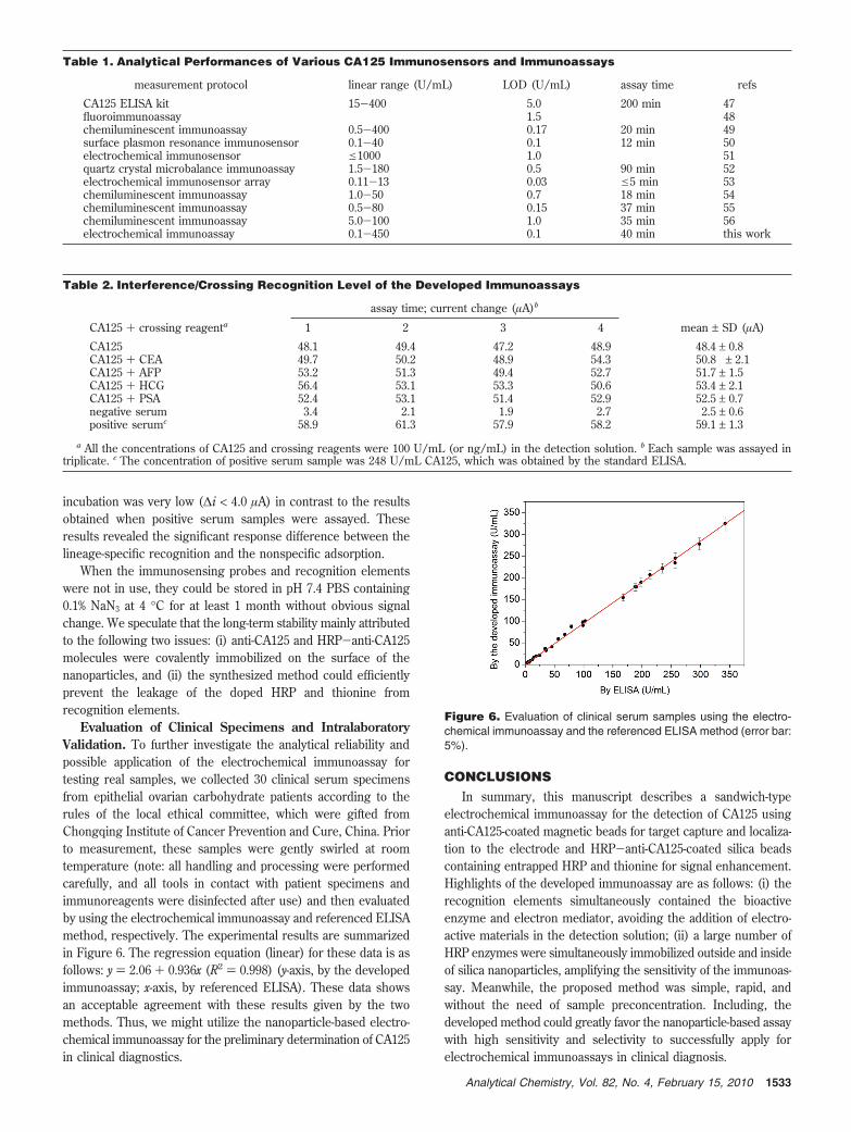
Evaluation of Clinical Specimens and IntralaboratoryValidation. To further investigate the analytical reliability andpossible application of the electrochemical immunoassay fortesting real samples, we collected 30 clinical serum specimensfrom epithelial ovarian carbohydrate patients according to therules of the local ethical committee, which were gifted fromChongqing Institute of Cancer Prevention and Cure, China. Priorto measurement, these samples were gently swirled at roomtemperature (note: all handling and processing were performedcarefully, and all tools in contact with patient specimens andimmunoreagents were disinfected after use) and then evaluatedby using the electrochemical immunoassay and referenced ELISAmethod, respectively. The experimental results are summarizedin Figure 6. The regression equation (linear) for these data is asfollows: y ) 2.06 + 0.936x (R2 ) 0.998) (y-axis, by the developedimmunoassay; x-axis, by referenced ELISA). These data showsan acceptable agreement with these results given by the twomethods. Thus, we might utilize the nanoparticle-based electro-chemical immunoassay for the preliminary determination of CA125in clinical diagnostics.
CONCLUSIONSIn summary, this manuscript describes a sandwich-type
electrochemical immunoassay for the detection of CA125 usinganti-CA125-coated magnetic beads for target capture and localiza-tion to the electrode and HRP-anti-CA125-coated silica beadscontaining entrapped HRP and thionine for signal enhancement.Highlights of the developed immunoassay are as follows: (i) therecognition elements simultaneously contained the bioactiveenzyme and electron mediator, avoiding the addition of electro-active materials in the detection solution; (ii) a large number ofHRP enzymes were simultaneously immobilized outside and insideof silica nanoparticles, amplifying the sensitivity of the immunoas-say. Meanwhile, the proposed method was simple, rapid, andwithout the need of sample preconcentration. Including, thedeveloped method could greatly favor the nanoparticle-based assaywith high sensitivity and selectivity to successfully apply forelectrochemical immunoassays in clinical diagnosis.
Table 1. Analytical Performances of Various CA125 Immunosensors and Immunoassays
measurement protocol linear range (U/mL) LOD (U/mL) assay time refs
CA125 ELISA kit 15-400 5.0 200 min 47fluoroimmunoassay 1.5 48chemiluminescent immunoassay 0.5-400 0.17 20 min 49surface plasmon resonance immunosensor 0.1-40 0.1 12 min 50electrochemical immunosensor e1000 1.0 51quartz crystal microbalance immunoassay 1.5-180 0.5 90 min 52electrochemical immunosensor array 0.11-13 0.03 e5 min 53chemiluminescent immunoassay 1.0-50 0.7 18 min 54chemiluminescent immunoassay 0.5-80 0.15 37 min 55chemiluminescent immunoassay 5.0-100 1.0 35 min 56electrochemical immunoassay 0.1-450 0.1 40 min this work
Table 2. Interference/Crossing Recognition Level of the Developed Immunoassays
assay time; current change (µA)b
CA125 + crossing reagenta 1 2 3 4 mean ± SD (µA)
CA125 48.1 49.4 47.2 48.9 48.4 ± 0.8CA125 + CEA 49.7 50.2 48.9 54.3 50.8 ± 2.1CA125 + AFP 53.2 51.3 49.4 52.7 51.7 ± 1.5CA125 + HCG 56.4 53.1 53.3 50.6 53.4 ± 2.1CA125 + PSA 52.4 53.1 51.4 52.9 52.5 ± 0.7negative serum 3.4 2.1 1.9 2.7 2.5 ± 0.6positive serumc 58.9 61.3 57.9 58.2 59.1 ± 1.3
a All the concentrations of CA125 and crossing reagents were 100 U/mL (or ng/mL) in the detection solution. b Each sample was assayed intriplicate. c The concentration of positive serum sample was 248 U/mL CA125, which was obtained by the standard ELISA.
Figure 6. Evaluation of clinical serum samples using the electro-chemical immunoassay and the referenced ELISA method (error bar:5%).
1533Analytical Chemistry, Vol. 82, No. 4, February 15, 2010

ACKNOWLEDGMENTThis work was jointly supported by the National Basic Research
Program of China (2010CB732403), the National Natural ScienceFoundation of China (20877019 and 20735002), the Key NaturalSciences Foundation of Fujian Province, China (D0520001), theKey Program of Science and Technology Department of FujianProvince, China (2007Y0026), NTU-MOE Academic Research
Funds (RG65/08), and the High-Qualified Talent Funding ofFuzhou University (0460-022275).
Received for review December 5, 2009. Accepted January12, 2010.
AC902768F
1534 Analytical Chemistry, Vol. 82, No. 4, February 15, 2010