mycobacteria. causative agents of tuberculosis. pathogenesis, laboratory diagnostics, prophylaxis...
TRANSCRIPT

Mycobacteria. Causative agents of tuberculosis.
Pathogenesis, laboratory diagnostics, prophylaxis and
therapy of diseases caused by mycobacteria.
Vinnitsa National Pirogov Memorial Medical University / Department of microbiology

Taxonomy and classification of mycobacteria
Family Mycobacteriacea
Genus Mycobacterium includes about 40 species
Pathogenic mycobacteria:
Species causing tuberculosis:M.tuberculosis, M.bovis, M.africanum, M.microtii
Species causing leprosy is M.leprae

Morphology of tubercle bacilli

Cultivation of M.tuberculosis
It is obligate aerobe with the slow growth in culture
Optimum temperature is 370C, growth range is between 250C and 400C
Optimum pH is 6.4-7.0 Culture media for tubercle bacilli
Lowenstein-Jensen potato-glycerol broth citrate blood

Colonies of M.tuberculosis on the LJ nutrient medium

Virulent factors of tubercle bacilli
Cord –factor Fatty acids, lipids tuberculoprotein

Epidemiology of tuberculosis
The source of tubercle bacilli may be: Humans Animals
Tuberculosis is air-borne infection.
The mode of transmission is by direct inhalation
Infection with M.bovis arises by ingestion of contaminated non-pasteurized milk and dairy products

Main clinical signs of tuberculosis
A person with active TB will have the following A person with active TB will have the following symptoms that get more severe over timesymptoms that get more severe over time
a bad cough that is worse in the morning chest pain greenish or bloody sputum weakness or fatigue weight loss night sweats chills fever
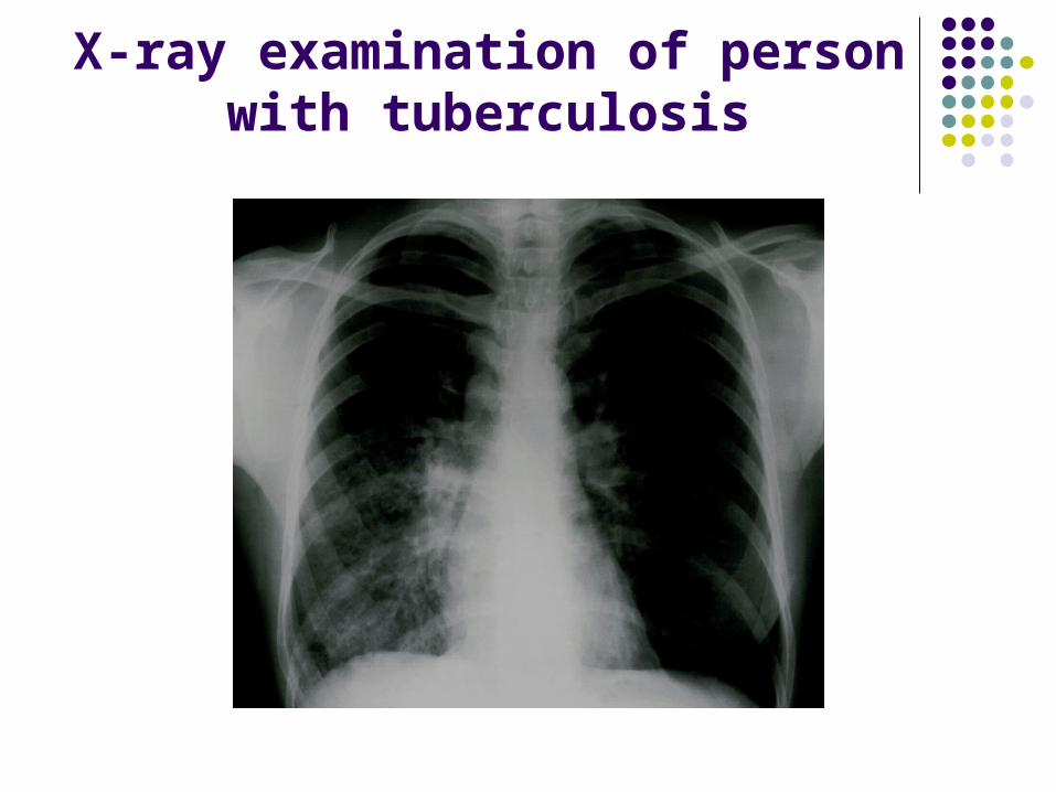
X-ray examination of person with tuberculosis

Laboratory diagnostics (microscopy)
1. Microscopy : Direct microscopy Fluorescent microscopy
2. Culture method3. Biological or experimental infection4. Allergic test (Mantoux test)5. Serology

Immunofluorescence test Direct microscopy after Zheel-Neelsen staining

Mantoux test

Prevention and treatment of tuberculosis in persons with
positive test
Immunoprophylaxis is with alive attenuated BCG vaccine
Persons with negative Mantoux test must be vaccinated with BCG vaccine

Therapy Specific antituberculous chemotherapeutic
drugs are used: Isoniasid Ethambutol Pyrazinamide Rifampicin Streptomycin

M.leprae

Cultural properties It is not cultivated onto nutrient media (!) Experimental infection may be caused
- by injection into foot of mouse – local change
- by infection of armadillo (tatou)– generalized infection
Virulent factors : microcapsule, fatty acids, cord-factor, allergins (lepromin)

EpidemiologySource of infection– a person with
lepromatous formRoute of transmission - By inhalation of air droplets- By direct contact
Leprosy is low contagious endemic disease (!)

Pathogenesis
Due to immunological reactiveness of host there are two types of leprosy:
- ТТ – tuberculoid type
- LT– lepromatous type
- intermediate type

Tyberculoid type of leprosy

Lepromatous type

Laboratory diagnosticsMicroscopy Serology Allergic skin test with lepromin

Mycobacteria