myasthenia gravis (mg) - terumo bct overview myasthenia gravis (mg) is a rare autoimmune disease...
TRANSCRIPT

MYASTHENIA GRAVIS (MG)CLINICAL SUMMARY SHEET

2
OVERVIEW
Myasthenia gravis (MG) is a rare autoimmune disease caused by antibodies directed against proteins in the postsynaptic membrane of the neuromuscular junction (NMJ: Figure 1).1 These pathogenic antibodies result in a classic pathology,2 translating into reduced, fatigable3,4 muscle function.5 A particular challenge with MG is the management of myasthenic crisis, which leads to acute respiratory failure that requires ventilatory support.6-8 Many stressors can precipitate a myasthenic crisis,9 and approximately 15 to 20 percent of patients with MG experience at least one episode of myasthenic crisis.7
Figure 1: NMJ showing the major components implicated in MG10
AChR, acetylcholine receptor; ColQ, collagen-tailed subunit of acetylcholinesterase; Kv1.4, voltage-gated potassium channel 1.4; LRP4, low-density lipoprotein receptor-related protein 4; MG, myasthenia gravis; MuSK, muscle-specific kinase; NMJ, neuromuscular junction; RyR, ryanodine receptor.
MG comprises several different conditions that are classified according to antibodies, age of onset, and accompanying thymic involvement (Table 1).10-13

3
Table 1: Characteristics of MG subgroups10-13
EPIDEMIOLOGY
Worldwide prevalence of MG is 7.8 cases per 100,000 people.14 In the United States, the incidence of MG is 1 per 100,000,4 and the prevalence is estimated to be 20 per 100,000.15 The disease is most common in females younger than 40 years and males older than 50 years.1,16,17 Juvenile MG is rare.18
ASSOCIATIONS
Other autoimmune diseases are commonly associated with MG (Table 2), and the most common comorbidity is autoimmune thyroid disease (10%).19
Table 2: MG subgroups and associated autoimmune disorders13
LRP4, low-density lipoprotein receptor-related protein 4; MG, myasthenia gravis; MuSK, muscle-specific kinase; POEMS, polyneuropathy, organomegaly, endocrinopathy, monoclonal gammopathy, and skin changes.
Prompt treatment for comorbidities is important for quality of life, performance of daily functions, short- and long-term prognosis, and survival.11 Muscle weakness associated with MG may increase the risk of respiratory infections, osteoporosis, and weight gain. General, widespread myopathy may develop in patients with MG.20
AChR, acetylcholine receptor; LRP4, low-density lipoprotein receptor-related protein 4; MG, myasthenia gravis; MuSK, muscle-specific kinase.
SUBGROUP ANTIBODY ADDITIONAL ANTIBODIES AGE AT ONSET PATIENTS (%) AFFECTED
MUSCLES THYMUS
Early-onset Anti-AChR Rare < 50 years 15–25 Generalized Hyperplasia common
Late-onset Anti-AChR Common > 50 years 35–45 Generalized Atrophy common
Thymoma Anti-AChR Very common Any 10Ocular,
bulbar, neck, generalized
Lymphoepithelioma
MuSK Anti-MuSK Rare Any 1–10Ocular,
bulbar, neck, generalized
Normal
LRP4 Anti-LRP4 Rare Any 1–5 Bulbar Normal
SeronegativeNone of the above
detectedVariable Any 10–15
Ocular, generalized
Variable
Ocular Variable Rare Any 15 Ocular Variable
MG SUBGROUP ASSOCIATED AUTOIMMUNE DISORDERS
Early-onset
Autoimmune thyroid disease, systemic lupus erythematosus, type 1 diabetes mellitus, alopecia areata totalis, giant cell myocarditis, neuromyelitis optica, myositis, pure red cell aplasia, autoimmune hepatitis, Sjögren’s syndrome, Addison’s disease, dermatomyositis/polymyositis, Guillain-Barré syndrome
Late-onset Hashimoto’s disease, systemic lupus erythematosus, multiple sclerosis
ThymomaSystemic lupus erythematosus, neuromyotonia, Sjögren’s syndrome, autoimmune hemolytic anemia, POEMS syndrome
MuSK Anti-MuSK
LRP4 Lambert-Eaton myasthenic syndrome, neuromyelitis optica
Seronegative –
Ocular Autoimmune thyroid disease

4
PATHOGENESIS
Eighty-five percent of patients with generalized MG, as well as 50 percent with ocular MG, have pathogenic immunoglobulin G1 (IgG1) and IgG33 antibodies to acetylcholine receptors (AChRs; Figure 2).21,22 Classical complement activation forms membrane attack complexes (MACs) on the muscle membrane and results in a characteristic NMJ rearrangement with internalization and degradation of surface AChRs.10 These pathogenic processes reduce ACh function in the NMJ.10
Figure 2: Major pathogenic mechanisms of the AChR antibodies in MG10
1) Complement activation at NMJ; 2) Antigenic modulation; 3) Binding of AChR antibodies; 4) Normal mechanisms for maintenance of the organization of the NMJ (see Figure 1). Antibodies with known pathogenic involvement in MG are shown in red. AChR, acetylcholine receptor; MAC, membrane attack complex; MG, myasthenia gravis; NMJ, neuromuscular junction.
Other MG patients have autoantibodies that are reactive to muscle-specific kinase (MuSK), a transmembrane tyrosine receptor kinase that is required for the development and maintenance of AChR clusters at the NMJ.12,22,23 The post-synaptic structure is disrupted when pathogenic IgG4 MuSK antibodies bind to MuSK, preventing low-density lipoprotein receptor-related protein 4 (LRP4) from binding MuSK and thereby inhibiting agrin-stimulated MuSK phosphorylation.24 Other patients can have antibodies against LRP4 with similar deleterious effects.12,25 Some patients with MG do not have detectable antibodies against AChR, MuSK, or LRP4 and are classified as having the seronegative form of disease.10
SYMPTOMS AND SIGNS
The hallmark characteristic of patients with MG is fatigable weakness,26 often including ptosis and diplopia (Table 3).4
Table 3: Clinical features of MG15
CATEGORY SIGNS AND SYMPTOMS
Ocular1. Ptosis—asymmetric, fatigue with upgaze2. Diplopia—the most commonly involved extraocular muscle is the medial rectus
Bulbar
1. Dysarthria—lingual, buccal, palatal (nasal speech)2. Dysphagia—excessive clearing of the throat, recurrent pneumonias (subtle signs)3. Dysphonia—hoarseness4. Masticatory weakness—jaw fatigue, jaw closure more affected than jaw opening
Facial1. Eyelid closure—inability to bury eyelashes with forced eye closure2. Lower face—poor cheek puff, drooling
Limb muscles1. Commonly proximal, symmetric2. Arms more affected than legs3. Rarely focal
Axial muscles1. Neck flexion2. Neck extension (head drop)
Respiratory muscles1. Exertional dyspnea—weak sniff and cough2. Orthopnea, tachypnea3. Respiratory failure

5
Certain common prescription medications are associated with worsening MG symptoms:1
DIAGNOSTIC CRITERIA AND TESTS
A number of tests are available to confirm a diagnosis of MG (Table 4).
Table 4: Diagnostic tests for MG15
AChR, acetylcholine receptor; CT, computed tomography; MG, myasthenia gravis; MRI, magnetic resonance imaging; MuSK, muscle-specific kinase.
Certain diseases may closely mimic MG and should be considered in the differential diagnosis (Table 5).
Table 5: Differential diagnosis of MG1
MG, myasthenia gravis.
TEST DETAILS
Bedside Edrophonium test Ice pack test
Reliable in patients with ptosis/extraocular weaknessUsed only when assessing improvement in ptosis
Electrophysiological Repetitive nerve stimulation Single-fiber electromyography
Sensitivity is 75% in generalized MG and < 50% in ocular MG patientsHighly sensitive (95%–99%), but not specific
Immunological (autoantibodies) Anti-AChR (binding) Anti-MuSK Low-affinity anti-AChR Anti-titin Anti-ryanodine receptor
Sensitivity is 85% in generalized MG and 50% in ocular MGDetected in 40% of AChR-negative generalized MGDetected in 66% of AChR and MuSK-negative generalized MGDetected in 95% thymomatous MG and 50% late-onset non-thymomatous MGDetected in 70% of thymomatous MG (more severe disease)
Other CT scan or MRI of chest Thyroid function testing
Obtain in all patients after diagnostic confirmation of MG–
DISEASE THAT MAY CLINICALLY MIMIC MG DIFFERENTIATING FEATURES
Lambert-Eaton myasthenic syndrome
Proximal extremity weaknessAreflexiaDry mouthVoltage-gated calcium channel antibodies
Congenital myasthenic syndromes Usually starts in infancy/childhood
Miller-Fisher syndromeAreflexiaSensory ataxiaGQ1b antibodies
Brainstem strokesNeighborhood signs (facial numbness, Horner’s syndrome)Acute onset
Mitochondrial myopathiesProgressive external ophthalmoplegia without diplopiaVery gradual onset
1. Telithromycin
2. Azithromycin
3. Ciprofloxacin/levofloxacin
4. Gentamicin/neomycin
5. Botulinum toxin
6. Corticosteroids
7. Quinine
8. Procainamide
9. Beta-adrenergic receptor blockers
10. Magnesium
11. D-penicillamine
12. Live, attenuated vaccines (shingles, nasal influenza); should be avoided in patients receiving immunosuppressants

6
CURRENT MANAGEMENT
Symptomatic pharmacologic treatment, immunomodulatory treatment, plasma exchange, and thymectomy are the main therapeutic options for MG12,27 and individualized for each MG subgroup (Figure 3).10
Figure 3: Restoring the function of the NMJ10
AChR, acetylcholine receptor; MG, myasthenia gravis; NMJ, neuromuscular junction.
In 2016, the Myasthenia Gravis Foundation published an international formal consensus-based guidance for the management of MG.28 The guidance for various treatment options for MG is summarized below, along with additional supporting data.
Acetylcholinesterase (AChE) Inhibitors
First-line therapy for nearly all patients with MG should be the reversible cholinesterase inhibitor pyridostigmine.12,28,29
Pyridostigmine27 is most effective in patients with generalized and ocular MG1 and less effective in those with MuSK-positive MG.30,31 In patients who do not meet treatment goals by following a regimen of pyridostigmine, corticosteroids or immunosuppressive therapy should be considered.28
Common adverse events associated with AChE inhibitors include nausea; vomiting; diarrhea; abdominal cramps; increased peristalsis, salivation, and bronchial secretions; miosis; diaphoresis; muscle cramps; fasciculation; and weakness.29
Key Study Covering AChE Inhibitors
Table 6 summarizes data from a key study covering AChE inhibitors that was included in a systematic review of the literature through July 2014.32
Table 6: Key study covering AChe inhibitors32
AChe, acetylcholinesterase; MG, myasthenia gravis.
TRIAL DETAILS ACHE INHIBITOR OUTCOME
Badrising 199633: Randomized controlled trial.
n = 10
4.5 mg intranasal neostigmine or placebo three times daily for 2 consecutive weeks preceded by a baseline observation week.
1. Improvement in generalized MG occurred in two of five participants with ocular symptoms, four of four with bulbar symptoms, and four of seven participants with impaired muscle power.
2. Dyspnea improved in two participants with this symptom.3. One participant experienced no effect.4. One of three participants with ocular MG had less ptosis with neostigmine.5. None of the participants receiving a placebo showed improvement.

7
IMMUNOSUPPRESSIVE AGENTS
Generally, corticosteroids (prednisone and prednisolone) improve muscle strength in all MG subtypes,27 and corticosteroid administration in patients with ocular MG may prevent progression to generalized MG.34,35 If treatment goals are reached, the dose of corticosteroid should be tapered, and a low-dose corticosteroid given long-term is often necessary.28 When corticosteroids are contraindicated, not tolerated, ineffective, or refused, an immunosuppressant agent should be considered.28
Azathioprine, cyclosporine, methotrexate, mycophenolate mofetil, and tacrolimus are used to treat MG.28 Azathioprine is the preferred first-line non-steroid immunosuppressive agent in MG and is often co-administered with corticosteroids.12,28 Cyclosporine is effective;28 however, it is not typically used because of safety risks and drug-drug interactions.28 Mycophenolate mofetil and tacrolimus are widely used to treat MG and are recommended by some national treatment guidelines.28,36,37 Recent results of a 12-month randomized, double-blind, placebo-controlled trial of methotrexate versus placebo in patients with MG showed that methotrexate had no steroid-sparing effect.38
Once patients have met and maintained treatment goals for 6 months to 2 years, the non-steroidal immunosuppressive agent should be tapered to the lowest effective dose.28 It is common for patients to require immunosuppressive drugs for many years, and sometimes for the rest of their lives.28 Patients with MuSK-MG often have a favorable response to immunosuppressive therapy.39
Key Studies Covering Immunosuppressive Agents
Table 7 summarizes key studies covering immunosuppressive agents that were included in a systematic review of the literature through July 2007.40
Table 7: Key studies covering immunosuppressive agents40
TRIAL DETAILS AGENT OUTCOME
De Feo 200241: Randomized, double-blind, controlled trial of cyclophosphamide plus prednisolone versus prednisolone plus placebo.
n = 23; 12 in the cyclophosphamide (CP) plus corticosteroids group, 11 in the corticosteroids plus placebo group.
Intravenous cyclophosphamide (CP) 500 mg/m2 of body surface and titrated according to response or side effects versus placebo.
1. Reductions of methylprednisone doses were noted in both groups but were more pronounced in patients receiving CP than placebo at 6 months P < 0.05) and at 12 months (P < 0.03).
2. At 12 months, 5 patients on CP had tapered off their steroids; no patient on placebo achieved further reductions (P < 0.03).
3. CP improved muscle strength at 3 and 6 months, and this improvement reached statistical significance compared with placebo at 12 months, mainly in the bulbar and masticatory (P < 0.009) and extraocular muscles (P < 0.03).
Gajdos 199342: Randomized, unblinded, controlled trial of azathioprine plus initial prednisolone versus prednisolone.
n = 41; 21 were randomized to receive prednisolone for the first month plus azathioprine, and 20 received prednisolone.
Prednisolone (1 mg/kg daily for 1 month, subsequently reduced to 0.5 mg/kg daily by 5 months
and to 0.25 mg/kg daily by 10 months) versus azathioprine (3 mg/kg daily for 1 year and then 2 mg/kg daily). In the latter group, participants were also given prednisolone 1 mg/kg daily for 1 month, which was then gradually tapered and discontinued.
1. 21 patients showed deterioration: 12 in the prednisone group and 9 in the azathioprine group (P = 0.40) during the 30-month follow-up.
2. No differences were noted between groups in muscular score and functional grade or in tolerance.
3. Treatment failure occurred in 12 patients in the prednisone group and in 5 patients in the azathioprine group (P = 0.02).

8
TRIAL DETAILS AGENT OUTCOME
Meriggioli 200343: Randomized, double-blind, controlled pilot study. One group received mycophenolate mofetil (MMF) plus either cyclosporine or prednisolone or no immunosuppressants; another group received placebo plus either cyclosporine or prednisolone or no immunosuppressants.
n = 14; 7 in the MMF plus either cyclosporine or prednisolone or no immunosuppressants group, and 7 in the placebo plus either cyclosporine or prednisolone or no immunosuppressants group.
MMF (1 g twice daily) in one group versus placebo in the other group. Five participants in each group were also on prednisolone. One participant in each group was also on cyclosporine.
Two participants in each group were on MMF monotherapy.
1. Patients who received MMF improved by 2.86 QMG points versus 0.29 points on average for placebo (P = 0.30).
2. Median change of antibody titer was 1.1 nmol/L in the MMF group and was 0.1 nmol/L in the placebo group (P = 0.52).
Nagane 200544: Randomized, unblinded, non-placebo-controlled pilot trial of tacrolimus plus corticosteroids with/without plasma exchange versus no tacrolimus plus corticosteroids with/without plasma exchange.
n = 34; 18 in the tacrolimus plus corticosteroids with/without plasma exchange group, 16 in the no tacrolimus plus corticosteroids with/without plasma exchange group.
Tacrolimus (FK506) was administered orally at a dose of 3 mg/day. For early-phase therapy, participants received tacrolimus or prednisolone or both. The prednisolone dose was tapered and adjusted to the minimal dose needed to maintain minimal manifestations.
For the follow-up phase, participants were treated to maintain minimal manifestations for one year; plasma exchange plus high-dose intravenous methylprednisolone, high-dose intravenous methylprednisolone
alone, or pyridostigmine was administered as needed to maintain minimal manifestations..
1. Low-dose FK506 reduced the duration of early-phase therapy in hospital (P < 0.05) and the need for combined therapy with plasma exchange and high-dose intravenous methylprednisolone or high-dose intravenous methylprednisolone alone (P < 0.05).
2. Low-dose FK506 reduced the daily dose of prednisolone (P < 0.05) required to maintain minimal manifestations of Myasthenia Gravis Foundation of America post-intervention status.
Palace 199845: Randomized, double-blind, controlled trial of azathioprine plus prednisolone versus prednisolone plus placebo.
n = 34; 15 in the azathioprine plus prednisolone group and 19 in the prednisolone plus placebo group
All participants received initial high-dose prednisolone (1.5 mg/kg or 100 mg on alternate days). Participants were also randomized to receive either azathioprine (2.5 mg/kg daily) or the equivalent dose of a matching placebo. The initial prednisolone dose was maintained until remission and then tapered to the minimum required to maintain remission.
1. No significant differences were noted between the two treatment groups in objective or subjective measurement scores at 12 months.
2. There was no significant difference between the two treatment groups in terms of number of participants experiencing treatment failure.
3. Duration of remission was significantly longer in the azathioprine plus prednisolone group compared with the prednisolone plus placebo group, giving a relative risk of 0.28 (95%, CI 0.08 to 0.94).
CI, confidence interval; QMG, quantitative myasthenia gravis score.
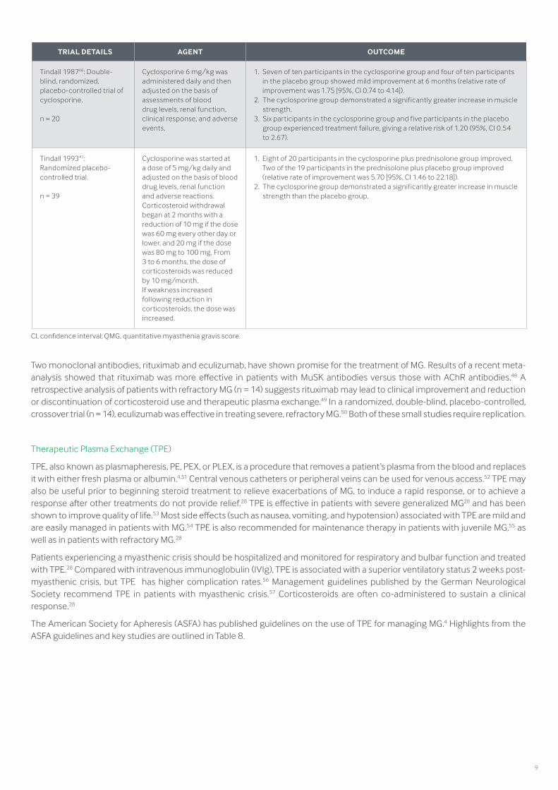
9
CI, confidence interval; QMG, quantitative myasthenia gravis score.
Two monoclonal antibodies, rituximab and eculizumab, have shown promise for the treatment of MG. Results of a recent meta-analysis showed that rituximab was more effective in patients with MuSK antibodies versus those with AChR antibodies.48 A retrospective analysis of patients with refractory MG (n = 14) suggests rituximab may lead to clinical improvement and reduction or discontinuation of corticosteroid use and therapeutic plasma exchange.49 In a randomized, double-blind, placebo-controlled, crossover trial (n = 14), eculizumab was effective in treating severe, refractory MG.50 Both of these small studies require replication.
Therapeutic Plasma Exchange (TPE)
TPE, also known as plasmapheresis, PE, PEX, or PLEX, is a procedure that removes a patient’s plasma from the blood and replaces it with either fresh plasma or albumin.4,51 Central venous catheters or peripheral veins can be used for venous access.52 TPE may also be useful prior to beginning steroid treatment to relieve exacerbations of MG, to induce a rapid response, or to achieve a response after other treatments do not provide relief.28 TPE is effective in patients with severe generalized MG28 and has been shown to improve quality of life.53 Most side effects (such as nausea, vomiting, and hypotension) associated with TPE are mild and are easily managed in patients with MG.54 TPE is also recommended for maintenance therapy in patients with juvenile MG,55 as well as in patients with refractory MG.28
Patients experiencing a myasthenic crisis should be hospitalized and monitored for respiratory and bulbar function and treated with TPE.28 Compared with intravenous immunoglobulin (IVIg), TPE is associated with a superior ventilatory status 2 weeks post-myasthenic crisis, but TPE has higher complication rates.56 Management guidelines published by the German Neurological Society recommend TPE in patients with myasthenic crisis.57 Corticosteroids are often co-administered to sustain a clinical response.28
The American Society for Apheresis (ASFA) has published guidelines on the use of TPE for managing MG.4 Highlights from the ASFA guidelines and key studies are outlined in Table 8.
TRIAL DETAILS AGENT OUTCOME
Tindall 198746: Double-blind, randomized, placebo-controlled trial of cyclosporine.
n = 20
Cyclosporine 6 mg/kg was administered daily and then adjusted on the basis of assessments of blooddrug levels, renal function, clinical response, and adverse events.
1. Seven of ten participants in the cyclosporine group and four of ten participants in the placebo group showed mild improvement at 6 months (relative rate of improvement was 1.75 [95%, CI 0.74 to 4.14]).
2. The cyclosporine group demonstrated a significantly greater increase in muscle strength.
3. Six participants in the cyclosporine group and five participants in the placebo group experienced treatment failure, giving a relative risk of 1.20 (95%, CI 0.54 to 2.67).
Tindall 199347: Randomized placebo-controlled trial.
n = 39
Cyclosporine was started at a dose of 5 mg/kg daily and adjusted on the basis of blood drug levels, renal function and adverse reactions. Corticosteroid withdrawal began at 2 months with a reduction of 10 mg if the dose was 60 mg every other day or lower, and 20 mg if the dose was 80 mg to 100 mg. From 3 to 6 months, the dose of corticosteroids was reduced by 10 mg/month.If weakness increased following reduction in corticosteroids, the dose was increased.
1. Eight of 20 participants in the cyclosporine plus prednisolone group improved. Two of the 19 participants in the prednisolone plus placebo group improved (relative rate of improvement was 5.70 [95%, CI 1.46 to 22.18]).
2. The cyclosporine group demonstrated a significantly greater increase in muscle strength than the placebo group.

10
Table 8: Category and grade recommendation for TPE for the management of MG according to ASFA4
*Number of trials/case series (total number of enrolled participants/patients). †6 (405) case series contained both groups of patients; case series added anti-MuSK 110, with rippling muscle disease 2 (10).4 Category I: Disorders for which apheresis is accepted as first-line therapy, either as a primary standalone treatment or in conjunction with other modes of treatment. Grade 1B: Strong recommendation with moderate-quality evidence; can apply to most patients in most circumstances without reservation. Grade 1C: Strong recommendation with low-quality or very low-quality evidence; recommendation may change when higher-quality evidence becomes available.
ASFA, American Society for Apheresis; MG, myasthenia gravis; TPE, therapeutic plasma exchange.
Similarly, the European Federation of Neurological Societies recommends TPE for the short-term management of MG, especially in patients with severe disease or those undergoing surgery.27 The Association of British Neurologists recommends TPE in patients with specific risk factors for IVIg.37 The International consensus guidance for management of MG28 suggests that TPE may be more effective than IVIg in the treatment of MuSK-MG. TPE and IVIg are used as short-term treatments for impending and manifest myasthenic crisis, as well as in patients with significant respiratory dysfunction. Although clinical trials suggest that TPE and IVIg are equally effective, this expert consensus suggests that TPE is more effective and works more quickly.
Key Studies Covering TPE
Table 9 summarizes the key studies covering TPE that were included in a systematic review of the literature through June 2002.58
Table 9: Key studies covering TPE58
CI, confidence interval; ICU, intensive care unit; IVIg, intravenous immunoglobulin; MMS, myasthenic muscle score; NS, not significant; QMG, quantitative myasthenia gravis score; SD, standard deviation; TPE, therapeutic plasma exchange.
INDICATION CATEGORY RECOMMENDATIONRANDOMIZED CONTROLLED
TRIALS
CONTROLLED TRIALS CASE SERIES
Moderate-severe I Grade 1B 8 (279)* 8 (2,837) 30 (556)†
Pre-thymectomy I Grade 1C 0 5 (342) 2 (51)†
TRIAL DETAILS INTERVENTION OUTCOME
Gajdos 198359: Randomized controlled trial.
n = 14
Group 1 received prednisone.
Group 2 received prednisone and TPE.
1. Improvement in mean (SD) quantitative muscle score was not significantly greater in patients treated with TPE plus prednisone than in patients treated with prednisone alone 1 month after initiating treatment (79 [22] versus 62 [22]).
2. Mean (SD) muscle score values after 1 year were 82 (19) in the prednisone and plasma exchange group and 91 (6) in the prednisone-alone group.
3. More relapses (5) were observed in the TPE plus prednisone group in the first year compared with the prednisone-alone group (1).
Gajdos 199760: Randomized controlled trial, two parallel groups.
n = 87
Group 1 received 3 TPE treatments.
The mean (SD) change in the MMS score at day 15 was 16.6 (16) in the TPE group and 15.6 (15.9) in the IVIg group.
Kamel 200961: Randomized controlled trial, two parallel groups.
n = 35
Group 1 received 3 TPE treatments before thymectomy.
Group 2 received no TPE treatments before thymectomy.
1. Duration of postoperative mechanical ventilation was 1.8 (1.3) hours in group 1 versus 2.9 (1.7) hours in group 2, with a mean difference of 1.10 hours (95% CI -2.12 to -0.08, P = 0.03).
2. Duration of ICU and hospital stay was lower in group 1 than in group 2.
Ronager 200162: Randomized controlled crossover trial.
n = 12
Group 1 received IVIg 0.4 g/kg for 5 days and then, 16 weeks later, received 5 TPE treatments.
Group 2 received the same treatments on the opposite schedule.
1. Mean decrease in QMG from baseline to 1 week after TPE was 0.23 (P < 0.05) and after IVIg was 0.10 (NS).
2. From baseline to 4 weeks, the mean decrease in QMG after TPE was still significant and after IVIg was 0.23 (P < 0.05); change at 8 or 16 weeks was not significant in either group.

11
Intravenous Immunoglobulin (IVIg)
In a similar way to TPE, IVIg is reserved for specific situations in which acute treatment is necessary.1,28 IVIg is effective in patients with severe generalized MG, but it may not be as efficacious in patients with milder MG or ocular MG.28 IVIg may also not be as effective in patients with MuSK-MG.28 IVIg may be used as maintenance therapy in patients with refractory MG or in cases in which immunosuppressive agents are contraindicated.28 Like TPE, IVIg has been shown to improve quality of life in patients with severe MG and worsening symptoms.53 IVIg is recommended for maintenance therapy in patients with juvenile MG, although it may not result in responses that are as consistent as those seen with TPE.55
Patients experiencing myasthenic crisis should be hospitalized and monitored for respiratory and bulbar function and may be treated with IVIg.28 Corticosteroids are often also co-administered to these patients to sustain a clinical response.28
The AAN cautiously recommends IVIg for the treatment of MG, noting that IVIg is probably effective.63 There is insufficient evidence to compare the efficacy of IVIg and TPE.63
Key Studies Covering IVIg
Table 10 contains key studies covering IVIg that were included in a systematic review of the literature through September 2011.64
Table 10: Key studies covering IVIg64
*Unpublished data.
AChR, acetylcholine receptor; CI, confidence interval; IVIg, intravenous immunoglobulin; PE, plasma exchange; QMG score, quantitative myasthenia gravis score; SD, standard deviation; TPE, therapeutic plasma exchange.
TRIAL DETAILS INTERVENTION OUTCOME
Barth 201165: Randomized controlled trial, two parallel groups.
n = 84
Group 1 received IVIg 2g/kg.
Group 2 received five TPE treatments.
1. IVIg and TPE reduced the QMG, and IVIg was comparable to TPE in efficacy.
2. The presence of AChR antibodies and greater baseline disease severity predicted a better response to therapy.
3. Percentage of patients improved with treatment: 69% on IVIg and 65% on TPE.
Gajdos 199760: Randomized controlled trial, two parallel groups.
n = 87
Group 1 received three TPE treatments.
Group 2a received IVIg 2 g/kg.
Group 2b received IVIg 1.2 g/kg.
Myasthenic muscular score variation was similar in both groups (median value +18 in the TPE group and +15.5 in the IVIg group; (P = 0.65).
Gajdos 200566: Randomized controlled trial, two parallel groups.
n = 173
Group 1 received IVIg 1g/kg.
Group 2 received IVIg 2 g/kg.
Mean improvements in the myasthenic muscular scores after 2 weeks were 15.49 points (95% CI, 12.09 to 18.90 points) in group 1 and 19.33 points (95% CI, 15.82 to 22.85 points) in group 2 (P = 0.12).
Ronager 200162: Randomized controlled crossover trial.
n = 12
Group 2b received IVIg 1.2 g/kg. 1. Mean decrease in QMG from baseline to 1 week after TPE was 0.23 (P < 0.05) and after IVIg was 0.10 (NS).
2. From baseline to 4 weeks, the mean decrease in QMG after TPE was still significant and after IVIg was 0.23 (P < 0.05); change at 8 or 16 weeks was not significant in either group.
Schuchardt 200264: Randomized controlled trial, two parallel groups.
n = 33*
Group 1 received methylprednisolone 1 mg/kg to 1.5 mg/kg for 14 days.
Group 2 received IVIg 30 g/day for 5 days.
The mean (SD) sum of the two most pathological items of the QMG at day 0 was 3.9 (1.1) for the IVIg group and 4.2 (0.7) for the methylprednisolone group.
Wolfe 200267: Randomized controlled trial, two parallel groups.
n = 15
Group 1 received IVIg 1 g/kg for 2 days followed by IVIg 1 g/kg on day 22.
Group 2 received 5% albumin for 2 days followed by 5% albumin on day 22.
1. At day 42, there was no significant difference between groups in terms of change in QMG.
2. The study was terminated early because of insufficient IVIg inventory.
Zinman 200768: Randomized controlled trial, two parallel groups.
n = 51
Group1 received IVIg 1 g/kg for 2 days.
Group 2 received 5% dextrose for 2 days.
1. Clinically meaningful improvement in QMG score for disease severity was observed at day 14 and persisted at day 28 in patients who received IVIg.
2. The greatest improvement occurred in patients with more severe disease as defined by a QMG score for disease severity > 10.5.

12
Thymectomy
In patients with thymoma, thymectomy should be performed to remove the tumor.28 Removal of the tumor may not immediately alleviate symptoms of MG,28 and clinical improvement may take years to achieve.4
Results of a recent multicenter, randomized trial indicate that thymectomy improves clinical outcomes in patients with non-thymomatous MG.69 Patients (n = 126) were randomized to a thymectomy-plus-prednisone group and a prednisone-only group,69 and quantitative myasthenia gravis score (QMG; scale 0 to 39, with 39 indicating more severe disease) was assessed.69 Over a 3-year period, patients randomized to thymectomy plus prednisone had a lower-weighted mean QMG than patients who received prednisone alone (6.15 versus 8.99, P < 0.001).69 The prednisone dose for patients in the thymectomy group was lower than that in the prednisone-only group (44 mg versus 60 mg, P < 0.001).69 Fewer exacerbations were reported in the thymectomy group, with 9 percent of patients requiring hospitalization for exacerbations versus 37 percent in the prednisone group (P < 0.001).69
A proposed treatment algorithm for patients with MG is shown in Figure 4.
Figure 4: Treatment flowchart15
AChR, acetylcholine receptor; IVIg, intravenous immunoglobulin; MG, myasthenia gravis; MuSK, muscle-specific kinase; PE, plasma exchange.
PROGNOSIS
Available treatment options have decreased mortality due to MG from 30 percent to less than 5 percent.4,12 Patients with mild to moderate symptoms often make a full recovery or at least substantial improvement.12 However, patients with refractory MG should be referred to a physician or a medical center with expertise in the management of MG.28 Similarly, the mortality rate due to myasthenic crisis is less than 5 percent.70
The MG composite scale is a validated and reliable outcome measure for MG (Table 11).71 This scale, which can be used to assess the clinical status of patients with MG, is composed of physician- and patient-reported test items.71

13
Table 11: The MG composite scale71
*Moderate weakness for neck and limb items should be construed as weakness that equals roughly 50% ± 15% of expected normal strength. Any weakness milder than that would be classified as mild, and any weakness more severe than that would be classified as severe.
MG, myasthenia gravis.
SUMMARY
1. MG is an autoimmune disease of the NMJ with pathogenic antibodies targeting proteins in the postsynaptic membrane.
2. MG is characterized by fatigable weakness. There are seven main MG subgroups, which are characterized according to antibody involvement, age at onset, affected muscles, and thymus involvement.
3. Diagnostic tests include clinical, electrophysiological, and immunological tests, as well as other tests, such as CT scan or MRI and thyroid function tests.
4. Acetylcholinesterase inhibitors, immunosuppressive agents, TPE, and thymectomy are the accepted treatments for MG.
5. Prognosis is good for most patients, with mortality rates less than 5 percent.
6. TPE may be more effective than IVIg in the treatment of MuSK-MG.
7. Although clinical trials suggest that IVIg and TPE are equally effective in the treatment of impending or manifest myasthenic crisis, expert consensus suggests that TPE is more effective and works more quickly than IVIg.28
Ptosis, upward gaze (physician examination)
> 45 seconds = 0 11–45 seconds = 1 1–10 seconds = 2 Immediate = 3
Double vision on lateral gaze, left or right (physician examination)
> 45 seconds = 0 11–45 seconds = 1 1–10 seconds = 3 Immediate = 4
Eye closure (physician examination)
Normal = 0Mild weakness (can be forced open with effort) = 0
Moderate weakness (can be forced open easily) = 1
Severe weakness (unable to keep eyes closed) = 2
Talking (patient history) Normal = 0Intermittent slurring or nasal speech = 2
Constant slurring or nasal but can be understood = 4
Difficult to understand speech = 6
Chewing (patient history) Normal = 0 Fatigue with solid food = 2 Fatigue with soft food = 4 Gastric tube = 6
Swallowing (patient history)
Normal = 0Rare episode of choking or trouble swallowing = 2
Frequent trouble swallowing, e.g., necessitating changes in diet = 5
Gastric tube = 6
Breathing (thought to be caused by MG)
Normal = 0Shortness of breath with exertion = 2
Shortness of breath at rest = 4
Ventilator dependence = 9
Neck flexion or extension (weakest) (physician examination)
Normal = 0 Mild weakness = 1Moderate weakness (i.e.,
~50% weak, ± 15%) = 3*Severe weakness = 4
Shoulder abduction (physician examination)
Normal = 0 Mild weakness = 2Moderate weakness (i.e.,
~50% weak, ± 15%) = 4*Severe weakness = 5
Hip flexion (physician examination)
Normal = 0 Mild weakness = 2Moderate weakness (i.e.,
~50% weak, ± 15%) = 4*Severe weakness = 5

14
REFERENCES
1. Gwathmey KG, Burns TM. Myasthenia gravis. Semin Neurol. 2015;35(4):327-339.
2. Drachman DB. Myasthenia gravis. N Engl J Med. 1994;330(25):1797-1810.
3. Kernich CA. Patient and family fact sheet. Myasthenia gravis: maximizing function. Neurologist. 2008;14(1):75-76.
4. Schwartz J, Padmanabhan A, Aqui N, et al. Guidelines on the use of therapeutic apheresis in clinical practice-Evidence-based approach from the Writing Committee of the American Society for Apheresis: The Seventh Special Issue. J Clin Apher. 2016;31(3):149-338.
5. Oger J, Frykman H. An update on laboratory diagnosis in myasthenia gravis. Clin Chim Acta. 2015;444:126-131.
6. O’Riordan JI, Miller DH, Mottershead JP, Hirsch NP, Howard RS. The management and outcome of patients with myasthenia gravis treated acutely in a neurological intensive care unit. Eur J Neurol. 1998;5(2):137-142.
7. Ropper AH GD, Diringer MN, Green DM, Mayer SA, Bleck TP. Treatment of the Critically Ill Patient With Myasthenia Gravis. Neurological and Neurosurgical Intensive Care. Philadelphia, PA: Lipincott Williams & Wilkins; 2004.
8. Thomas CE, Mayer SA, Gungor Y, et al. Myasthenic crisis: clinical features, mortality, complications, and risk factors for prolonged intubation. Neurology. 1997;48(5):1253-1260.
9. Wendell LC, Levine JM. Myasthenic crisis. Neurohospitalist. 2011;1(1):16-22.
10. Gilhus NE, Skeie GO, Romi F, Lazaridis K, Zisimopoulou P, Tzartos S. Myasthenia gravis–autoantibody characteristics and their implications for therapy. Nat Rev Neurol. 2016;12(5):259-268.
11. Gilhus NE, Nacu A, Andersen JB, Owe JF. Myasthenia gravis and risks for comorbidity. Eur J Neurol. 2015;22(1):17-23.
12. Gilhus NE, Verschuuren JJ. Myasthenia gravis: subgroup classification and therapeutic strategies. Lancet Neurol. 2015;14(10):1023-1036.
13. Nacu A, Andersen JB, Lisnic V, Owe JF, Gilhus NE. Complicating autoimmune diseases in myasthenia gravis: a review. Autoimmunity. 2015;48(6):362-368.
14. Carr AS, Cardwell CR, McCarron PO, McConville J. A systematic review of population based epidemiological studies in myasthenia gravis. BMC Neurol. 2010;10:46.
15. Meriggioli MN, Sanders DB. Autoimmune myasthenia gravis: emerging clinical and biological heterogeneity. Lancet Neurol. 2009;8(5):475-490.
16. Grob D, Brunner N, Namba T, Pagala M. Lifetime course of myasthenia gravis. Muscle Nerve. 2008;37(2):141-149.
17. Mantegazza R, Baggi F, Antozzi C, et al. Myasthenia gravis (MG): epidemiological data and prognostic factors. Ann N Y Acad Sci. 2003;998:413-423.
18. Liew WK, Kang PB. Update on juvenile myasthenia gravis. Curr Opin Pediatr. 2013;25(6):694-700.
19. Mao ZF, Yang LX, Mo XA, et al. Frequency of autoimmune diseases in myasthenia gravis: a systematic review. Int J Neurosci. 2011;121(3):121-129.
20. Liewluck T. Immune-mediated rippling muscle disease: another inflammatory myopathy in myasthenia gravis. Arch Neurol. 2010;67(7):896-897; author reply 897.
21. Berrih-Aknin S, Frenkian-Cuvelier M, Eymard B. Diagnostic and clinical classification of autoimmune myasthenia gravis. J Autoimmun. 2014;48-49:143-148.
22. Phillips WD, Vincent A. Pathogenesis of myasthenia gravis: update on disease types, models, and mechanisms. F1000Res. 2016;5.
23. Otsuka K, Ito M, Ohkawara B, et al. Collagen Q and anti-MuSK autoantibody competitively suppress agrin/LRP4/MuSK signaling. Sci Rep. 2015;5:13928.
24. Huijbers MG, Zhang W, Klooster R, et al. MuSK IgG4 autoantibodies cause myasthenia gravis by inhibiting binding between MuSK and Lrp4. Proc Natl Acad Sci USA. 2013;110(51):20783-20788.
25. Messeant J, Dobbertin A, Girard E, et al. MuSK frizzled-like domain is critical for mammalian neuromuscular junction formation and maintenance. J Neurosci. 2015;35(12):4926-4941.
26. Benatar M. Pearls: myasthenia. Semin Neurol. 2010;30(1):35-37.
27. Skeie GO, Apostolski S, Evoli A, et al. Guidelines for treatment of autoimmune neuromuscular transmission disorders. Eur J Neurol. 2010;17(7):893-902.
28. Sanders DB, Wolfe GI, Benatar M, et al. International consensus guidance for management of myasthenia gravis: Executive summary. Neurology. 2016;87(4):419-425.
29. MESTINON (pyridostigmine bromide syrup) Prescribing Information. 2013.
30. Gilhus NE, Owe JF, Hoff JM, Romi F, Skeie GO, Aarli JA. Myasthenia gravis: a review of available treatment approaches. Autoimmune Dis. 2011;2011:847393.
31. Hatanaka Y, Hemmi S, Morgan MB, et al. Nonresponsiveness to anticholinesterase agents in patients with MuSK-antibody-positive MG. Neurology. 2005;65(9):1508-1509.
32. Mehndiratta MM, Pandey S, Kuntzer T. Acetylcholinesterase inhibitor treatment for myasthenia gravis. Cochrane Database Syst Rev. 2014(10):CD006986.
33. Badrising U BH, van Hilten J, Brietl P, Wintzen A. Intranasal neostigmine as add-on therapy in myasthenia gravis. Journal of Neurology 1996;243(Suppl):59.
34. Benatar M, Kaminski H. Medical and surgical treatment for ocular myasthenia. Cochrane Database Syst Rev. 2012;12:CD005081.
35. Kerty E, Elsais A, Argov Z, Evoli A, Gilhus NE. EFNS/ENS Guidelines for the treatment of ocular myasthenia. Eur J Neurol. 2014;21(5):687-693.
36. Murai H. Japanese clinical guidelines for myasthenia gravis: putting into practice. Clin Exp Neuroimmunol. 2015;6:21-31.
37. Sussman J, Farrugia ME, Maddison P, Hill M, Leite MI, Hilton-Jones D. Myasthenia gravis: Association of British Neurologists’ management guidelines. Pract Neurol. 2015;15(3):199-206.
38. Pasnoor M, He J, Herbelin L, et al. A randomized controlled trial of methotrexate for patients with generalized myasthenia gravis. Neurology. 2016;87(1):57-64.
15
39. Pasnoor M, Wolfe GI, Nations S, et al. Clinical findings in MuSK-antibody positive myasthenia gravis: a U.S. experience. Muscle Nerve. 2010;41(3):370-374.
40. Hart IK, Sathasivam S, Sharshar T. Immunosuppressive agents for myasthenia gravis. Cochrane Database Syst Rev. 2007(4):CD005224.
41. De Feo LG, Schottlender J, Martelli NA, Molfino NA. Use of intravenous pulsed cyclophosphamide in severe, generalized myasthenia gravis. Muscle Nerve. 2002;26(1):31-36.
42. Myasthenia Gravis Clinical Study Group. A randomised clinical trial comparing prednisone and azathioprine in myasthenia gravis. Results of the second interim analysis. J Neurol Neurosurg Psychiatry. 1993;56(11):1157-1163.
43. Meriggioli MN, Rowin J, Richman JG, Leurgans S. Mycophenolate mofetil for myasthenia gravis: a double-blind, placebo-controlled pilot study. Ann N Y Acad Sci. 2003;998:494-499.
44. Nagane Y, Utsugisawa K, Obara D, Kondoh R, Terayama Y. Efficacy of low-dose FK506 in the treatment of Myasthenia gravis–a randomized pilot study. Eur Neurol. 2005;53(3):146-150.
45. Palace J, Newsom-Davis J, Lecky B. A randomized double-blind trial of prednisolone alone or with azathioprine in myasthenia gravis. Myasthenia Gravis Study Group. Neurology. 1998;50(6):1778-1783.
46. Tindall RS, Rollins JA, Phillips JT, Greenlee RG, Wells L, Belendiuk G. Preliminary results of a double-blind, randomized, placebo-controlled trial of cyclosporine in myasthenia gravis. N Engl J Med. 1987;316(12):719-724.
47. Tindall RS, Phillips JT, Rollins JA, Wells L, Hall K. A clinical therapeutic trial of cyclosporine in myasthenia gravis. Ann N Y Acad Sci. 1993;681:539-551.
48. Iorio R, Damato V, Alboini PE, Evoli A. Efficacy and safety of rituximab for myasthenia gravis: a systematic review and meta-analysis. J Neurol. 2015;262(5):1115-1119.
49. Nowak RJ, Dicapua DB, Zebardast N, Goldstein JM. Response of patients with refractory myasthenia gravis to rituximab: a retrospective study. Ther Adv Neurol Disord. 2011;4(5):259-266.
50. Howard JF, Jr., Barohn RJ, Cutter GR, et al. A randomized, double-blind, placebo-controlled phase II study of eculizumab in patients with refractory generalized myasthenia gravis. Muscle Nerve. 2013;48(1):76-84.
51. Cortese I, Chaudhry V, So YT, Cantor F, Cornblath DR, Rae-Grant A. Evidence-based guideline update: Plasmapheresis in neurologic disorders: report of the Therapeutics and Technology Assessment Subcommittee of the American Academy of Neurology. Neurology. 2011;76(3):294-300.
52. Guptill JT, Oakley D, Kuchibhatla M, et al. A retrospective study of complications of therapeutic plasma exchange in myasthenia. Muscle Nerve. 2013;47(2):170-176.
53. Barnett C, Wilson G, Barth D, Katzberg HD, Bril V. Changes in quality of life scores with intravenous immunoglobulin or plasmapheresis in patients with myasthenia gravis. J Neurol Neurosurg Psychiatry. 2013;84(1):94-97.
54. Ebadi H, Barth D, Bril V. Safety of plasma exchange therapy in patients with myasthenia gravis. Muscle Nerve. 2013;47(4):510-514.
55. Liew WK, Powell CA, Sloan SR, et al. Comparison of plasmapheresis and intravenous immunoglobulin as maintenance therapies for juvenile myasthenia gravis. JAMA Neurol. 2014;71(5):575-580.
56. Qureshi AI, Choudhry MA, Akbar MS, et al. Plasma exchange versus intravenous immunoglobulin treatment in myasthenic crisis. Neurology. 1999;52(3):629-632.
57. Melzer N, Ruck T, Fuhr P, et al. Clinical features, pathogenesis, and treatment of myasthenia gravis: a supplement to the Guidelines of the German Neurological Society. J Neurol. 2016;263(8):1473-1494.
58. Gajdos P, Chevret S, Toyka K. Plasma exchange for myasthenia gravis. Cochrane Database Syst Rev. 2002(4):CD002275.
59. Gajdos P, Simon N, de Rohan-Chabot P, Raphael JC, Goulon M. Long-term effects of plasma exchange in myasthenia. Results of a randomized study. Presse Med. 1983;12(15):939-942.
60. Gajdos P, Chevret S, Clair B, Tranchant C, Chastang C. Clinical trial of plasma exchange and high-dose intravenous immunoglobulin in myasthenia gravis. Myasthenia Gravis Clinical Study Group. Ann Neurol. 1997;41(6):789-796.
61. Kamel A EM. Effectiveness of prethymecthomy plasmapheresis on short-term outcome of non-thymomatous generalized myasthenia gravis. Egyptian Journal Neurology, Psychiatry and Neurosurgery. 2009;46(1):161-168.
62. Ronager J, Ravnborg M, Hermansen I, Vorstrup S. Immunoglobulin treatment versus plasma exchange in patients with chronic moderate to severe myasthenia gravis. Artif Organs. 2001;25(12):967-973.
63. Patwa HS, Chaudhry V, Katzberg H, Rae-Grant AD, So YT. Evidence-based guideline: intravenous immunoglobulin in the treatment of neuromuscular disorders: report of the Therapeutics and Technology Assessment Subcommittee of the American Academy of Neurology. Neurology. 2012;78(13):1009-1015.
64. Gajdos P, Chevret S, Toyka KV. Intravenous immunoglobulin for myasthenia gravis. Cochrane Database Syst Rev. 2012;12:CD002277.
65. Barth D, Nabavi Nouri M, Ng E, Nwe P, Bril V. Comparison of IVIg and PLEX in patients with myasthenia gravis. Neurology. 2011;76(23):2017-2023.
66. Gajdos P, Tranchant C, Clair B, et al. Treatment of myasthenia gravis exacerbation with intravenous immunoglobulin: a randomized double-blind clinical trial. Arch Neurol. 2005;62(11):1689-1693.
67. Wolfe GI, Barohn RJ, Foster BM, et al. Randomized, controlled trial of intravenous immunoglobulin in myasthenia gravis. Muscle Nerve. 2002;26(4):549-552.
68. Zinman L, Ng E, Bril V. IV immunoglobulin in patients with myasthenia gravis: a randomized controlled trial. Neurology. 2007;68(11):837-841.
69. Wolfe GI, Kaminski HJ, Aban IB, et al. Randomized Trial of Thymectomy in Myasthenia Gravis. N Engl J Med. 2016;375(6):511-522.
70. Godoy DA, Mello LJ, Masotti L, Di Napoli M. The myasthenic patient in crisis: an update of the management in Neurointensive Care Unit. Arq Neuropsiquiatr. 2013;71(9A):627-639.
71. Burns TM, Conaway M, Sanders DB. The MG Composite: A valid and reliable outcome measure for myasthenia gravis. Neurology. 2010;74(18):1434-1440.

©2017 Terumo BCT, Inc. / PN 305200043C
Terumo BCT (Asia Pacific) Ltd.
89 Science Park Drive#04-25 (Lobby B)The RutherfordSingapore 18261
Phone: +65.6715.3778Fax: +65.6774.1419
Terumo BCT Europe N.V.
Europe, Middle East and AfricaIkaroslaan 411930 ZaventemBelgium
Phone: +32.2.715.05.90 Fax: +32.2.721.07.70
Terumo BCT, Inc.
10811 West Collins Ave.Lakewood, Colorado 80215-4440USA
USA Phone: 1. 877. 339.4228Phone: +1. 303 . 231.4357Fax: +1. 303 . 542 .5215
Terumo BCT Latin America S.A.
La Pampa 1517–12th FloorC1428DZEBuenos Aires Argentina
Phone: +54.11.5530.5200 Fax: +54.11.5530.5201
Terumo BCT Japan, Inc.
Takanawa Park Tower 13F20-14, 3-chome,Higashi Gotanda, Shinagawa-ku,Tokyo, 141-0022 Japan
Phone: +81.3.6743.7890Fax: +81.3.6743.9800
As a global leader in blood component, therapeutic apheresis and cellular technologies, we believe in the potential of blood to do even more for patients than it does today. This belief inspires our innovation and strengthens our collaboration with customers.
UNLOCKING THE POTENTIAL OF BLOOD | TERUMOBCT.COM