mvz. edgar abundis-santamaria. edited by roberto a ... · mvz. edgar abundis-santamaria. edited by...
TRANSCRIPT
Edgar Abundis-Santamaría
1
Aspergillosis in birds of prey
MVZ. Edgar Abundis-Santamaria.
Edited by Roberto A. Cervantes-Olivares Ph.D.
Aspergillosis in birds of prey
2
ABSTRACT
Aspergillosis is a common disease in raptors held in captivity, and Aspergillus fumigatus
is frequently found in affected birds. This disease is divided in acute and chronic. The
acute form occurs when birds are exposed to an overwhelming dose of spores. The
chronic form affects birds under conditions of immunosuppression. Some species are
more susceptible to the disease such as gyrfalcons (Falco rusticolus), goshawks
(Accipiter gentilis) and red-tailed hawks (Buteo jamaicensis). The clinical signs are very
non-specific and a variety of diagnostic procedures can help, including: hematology,
endoscopy, radiology, cytology and serology. Indirect ELISA is a very useful tool for the
diagnosis or monitorig the progress or response to treatment. The prognosis for infected
birds is poor, especially if treatment is not started early. Prevention is the best way for
the control of the disease.
Edgar Abundis-Santamaría
3
INTRODUCTION
Aspergillosis is an infectious, non-contagious disease, caused by fungus of the genus
Aspergillus that affect humans, mammals and mainly wild or domestic birds.1-5 This
genus was described for the first time in a lesion of a bird in 1842 by Rayer and
Montagne in the air sacs of a bullfinch, but molds likely belonging to the genus
Aspergillus, were described in wild birds in the early 1800s. 3,4,6
The fungus is a ubiquitous, soil saprophyte and grows in organic matter at temperatures
up to 25°C.1,2,3,7,8,9 The species frecuently isolated are Aspergillus fumigatus, A. flavus,
A. nidulans, A. niger and A. terreus, 10-18 but A. fumigatus is isolated from 95% of
raptors with aspergillosis. 8,19 This species was first found in the lungs of a bustard (Otis
tarda) in 1863 by Fresenius, he also applied the term aspergillosis to this respiratory
disease. 6
Aspergillosis is an opportunistic infection, causing the disease under
immunocompromised situations of the host or when the bird is exposed to an
overwhelming number of spores.1,5,18,20,21,22 Stress seems to be the major predisposing
factor in the development of the disease and can be caused by shipping, heat, recently
capture, or changes in management. Aspergillosis also occurs associated with
prolonged disease or when immunosuppresive doses of corticosteroids are
used.1,2,4,5,9,10,11,14,15,16,21,23,24,25
Malnutrition, vitamin deficiencies (especially vitamin A), long term antibiotic use, age
(young or old) also contributes to the presentation of the disease, as well as trauma,
dusty environments, lead poisoning and irritants of the airways like smoke of cigarrette
or ammonia. 1,2,3,4,11,19,21,23
Environmental factors play an important role in the development of the disease,
increasing the number of spores to which the birds are exposed. Poor sanitation in nests
as well as food or fecal accumulation promote fungal growth. Poor ventilation, in
conjunction with these factors, increase the possibility that airborne spores may invade
and infect a bird's respiratory system. Wood shavings used as bedding material have
been frequently implicated in outbreaks of the disease. 1,4,10,19,21
Aspergillosis in birds of prey
4
Some species of the genus Aspergillus produce mycotoxins, like aflatoxins, produced by
A. flavus and A. parasiticus and ochratoxins produced by A. ochraceus, that affect
poultry when they consume contaminated food. Other important aspects of the genus
Aspergillus is that they may cause the brooder pneumonia, originated by hatcher or
brooder contamination with spores of this fungus. These spores get into the egg shell,
porducing embryo and neonatal death. 3,5,7,26
Multiple clinical signs may be seen on presentation. This makes aspergillosis one of the
most difficult diseases to treat,21,25,28 and a good program of preventive medicine is
needed, not just to avoid aspergillosis but any other disease that can affect wildlife. Also,
it is important to make a quick diagnosis to have a good prognosis, because the
treatment is ineffective in advanced cases.
Aspergillosis occurs frequently in birds of prey held in captivity, 3,5,13,20,28 i.e. in zoos (for
conservancy, research or education), falconry and those that are mantained in
rehabilitation centers. This disease is responsible for 15 to 30% of deaths in birds of prey
exhibited in zoos, but rarely occurs in free-living birds. 19
Birds of prey include diurnal birds, mainly from the order falconiforms and also nocturnal
birds of the order strigiforms. Falconiforms are divided in families that include vultures,
condors, eagles, hawks and falcons. Strigiforms are all kind of owls. 5,19
The species more susceptible to this disease are: Goshawk (Accipiter gentilis),
Gyrfalcon (Falco rusticolus), Snowy owl (Nyctea scandiaca), Rough-legged hawk (Buteo
lagopus), immature Red-tailed hawk (Buteo jamaicensis), Golden eagle (Aquila
chrysaetos). While the resistant species are: Prairie falcon (Falco mexicanus) and
Harris hawk (Parabuteo unicinctus). 2,5,9,10,11,19,20,21,22,23,25,28 This disease is one of the
principal causes of death in captive penguins. 3,4,10,11,13,16,19,21,25,26,29,30. The Bald eagle
(Haliaeetus leucocephalus) is more susceptible in cases of lead poisoning. 21,25
Edgar Abundis-Santamaría
5
ETHIOLOGY Aspergillus is classified as: division: Eumycota, subdivision: Deuteromicotina (fungi
imperfecti), class: Hyphomycetes, order: Moniliales, family: Moniliaceae. 6,26,31 Most of
the Aspergillus are classified as fungi imperfecti (asexual reproduction), but the perfect
state has been found in A. nidulans that can produced ascospores.8,26,32 Genus
Aspergillus was classified in 1729 by Micheli and many species have been described. 32
Species of the genus Aspergillus are molds formed by septate hyphae that are tubular
branched structures from 2 to 10 µ. when the growth starts, hyphae get together forming
a mycelia. Vegetative mycelia consists of superficial hyphae and hyphae that are on the
surface are the aerial mycelia. These structures produce conidiophores.7,29
Conidiophores are formed in foot cells and end in a vesicle. In this structure grow one
line of phialides or one line of metulae and over these structures one line of phialides
that produce chains of conidia or spores. Conidia are uni- or multi-nucleated, but always
one-celled. The shape can be globose, elliptical or oval, and the surface can be smooth,
finely rough or echuinulated. The color of the conidia determines the color of the conidial
head, which also usually determines the color of the colony. The function of the conidia
is air dissemination of the fungus. 26,29,32,33
Vesicle and chains of conidia are called conidial head and the shape can be columnar or
spherical. If phialides are produced just in the vesicle surface, the shape of the head is
columnar as in A. fumigatus and A. terreus, if phialides are produced over the whole
surface, conidia chains are radiated and the shape of the head is spherical as in A. Niger
and A. flavus. 26,32,33
This fungus has a worldwide distribution; air and soil of almost any part of the world
contain conidia of different species.15,26 Genus Aspergillus is usable as a food in many
different substances because of the number of enzymes that it can produce. The only
two main requirements of substrates for the fungal growth are organic matter and
moisture. If both factors are present, the fungus can grow in almost any substance. 26
Aspergillosis in birds of prey
6
The species that can cause infections in birds are: A. fumigatus, A. flavus, A. niger,
A. nidulans and A. terreus, but the most important species is A. fumigatus because is
isolated in 95% of affected birds. 3,10,11,12,13,14,15,19
A. fumigatus produce haemolysins, proteolytic enzymes and other toxic factors but their
role in the pathogenesis is not know. 8 However, it is known that an antibiotic produced
by this species named fumagilin, may have poor antibacterial activity but is a potent
amoebicide, specially against Entamoeba histolytica. 8,26 A. flavus produce aflatoxins that
will be described later.
Edgar Abundis-Santamaría
7
PATHOGENESIS Infection generally occurs when the bird inhales airborne spores. The organism may
then penetrate respiratory tissues, reproducing by simple division of tubular hyphae to
form mycelia. Tissue invasion incites an inflammatory response, with heterophils,
lymphocytes, monocytes and some giant cells infiltrating the lesion. 1,2,6,11,34
Severity of lesions depend on chronicity of infection, organs affected and the number of
spores inhalated. This disease is divided in acute and chronic. 1,3,4,21,35
ACUTE FORM
Seen most often in wild birds or psittacines under poor sanitary or ventilation conditions.
It occurs following the inhalation of an overwhelming number of spores. 2,10,21,23,35 In this
form of the disease, massive, rapid colonization of the lungs occurs, and the lungs
become diffusely infiltrated with miliary granulomas. Severe dyspnea is often seen, with
rapid progression to death, because treatment is usually ineffective. The course of the
acute form is usually less than a week, but anecdotal reports indicate that apparently
healthy raptors exposed to moldy hay have died within 48 hours. 2,10,15,21,23,25 Diagnosis is
made at necropsy, where these pulmonic nodules are visible grossly.1,3,19,35
Histologically, multiple foci containing fungal hyphae and rimmed by hemorrhage and
heterophilic, mononuclear and multinuclear cell infiltrates are seen. The fungus can be
isolated from many other tissues like liver, spleen or blood. 1,10,19
CHRONIC FORM
This is the most commonly observed form of the disease and follows a stressful event or
immunosuppression. Under these circumstances, the bird is unable to effectively
eliminate or contain even small numbers of Aspergillus organisms.1,2,15,21,35 Chronic
aspergillosis is divided into focal and generalized. It is possible to find both forms present
in one patient. Focal aspergillosis has better response to treatment, but in generalized
form, the treatment is prolonged and generally ineffective and the prognosis is
poor.2,4,6,11,12,21,25
Aspergillosis in birds of prey
8
o Focal
NASAL: is presented as a solid mycotic plug or aspergilloma localized in nares or
choana. The organism invades the sinus, and nasal bones. It may present as a unilateral
lesion. Combined infections of gram-negative bacteria and Aspergillus are frequent and
the fungus can not be detected if the diagnosis is made based solely on bacterial
culture. Histopathological examination of granulomas generally shows a necrotic foci
surrounded by macrophages, heterophils and giant cells, sometimes within a connective
tissue capsule. 10,11,21
TRACHEAL: is caracterized by the presence of mycotic colonies above the syrinx or at
the bifurcation due to air turbulence which deposits spores in this section of the air ways,
in addition, the narrowing in the respiratory tract at this point may predispose it to
blockage with necrotic debris and caseous exudate. The lesions reduce air movement
through the lower airway, so inspiratory dyspnea exists. Mycelia penetrate walls and in
combination with inflammatory cells and connective tissue, granulomatous nodules are
formed. Treatment can be successful if dyspnea is corrected and treatment is started
immediately. 1,4,5,10,11,21,23,25
CUTANEOUS: Necrotic granulomatous dermatitis has been described and A. fumigatus
was isolated from infected tissue. Skin lesions have been encountered under moisture-
retaining bandages. Cutaneous lesions and their manifestations are rare in birds.
Atkinson (1998) reports one case in a wing of a Great horn owl (Bubo virginianus) and
Abrams (2000) on the head of an hybrid peregrine-gyrfalcon (Falco peregrinus-Falco
rusticolus). 2,3,6,9,23
OPHTHALMITIS: exist 2 presentations: one superficial and the affected tissues are
conjunctiva and external surfaces of the eye with the development of a cheesy exudate
or plaque forming beneath the nictitating membrane. 3,6,9,17,36 The second presentation is
deeper and occurs rarely and probably as a result of hematogenous dissemination of the
organism from a primary respiratory infection, reaching the posterior eye. Pathological
changes can occur in the vitrous humor and extend into adjacent tissue. Pecten edema
can be present with heterophil infiltration and mononuclear cells. Also it is possible to
observe fungal hyphae, heterophils, macrophages and cell debris in the retina. 6,23
Edgar Abundis-Santamaría
9
In rare cases, well-encapsulated Aspergillus granulomas can be found in the trachea,
esophagus or under the skin. These probably represent an effective walling-off of early
fungal infection on the part of the bird.19
o Generalized
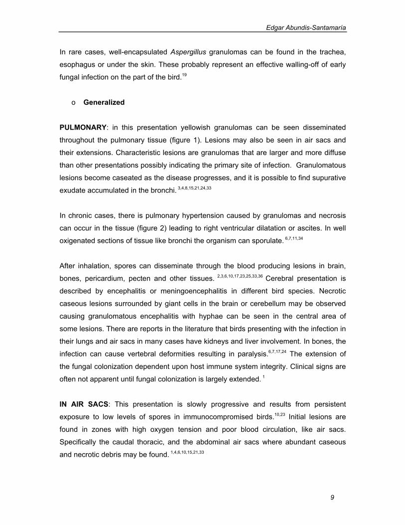
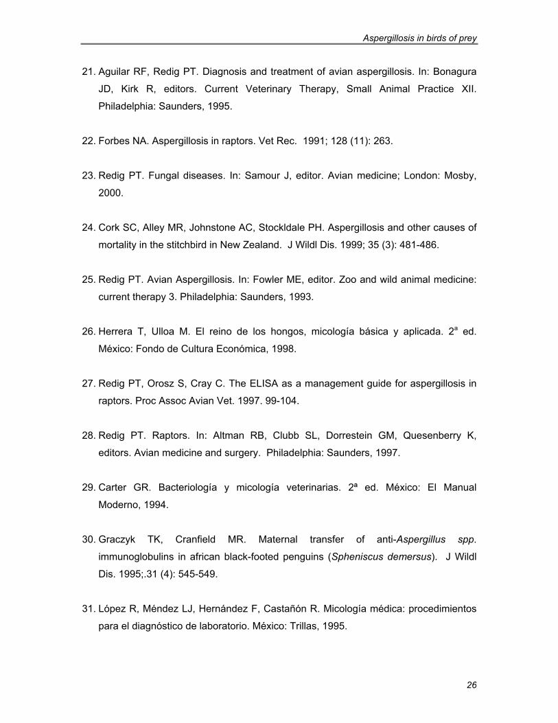
PULMONARY: in this presentation yellowish granulomas can be seen disseminated
throughout the pulmonary tissue (figure 1). Lesions may also be seen in air sacs and
their extensions. Characteristic lesions are granulomas that are larger and more diffuse
than other presentations possibly indicating the primary site of infection. Granulomatous
lesions become caseated as the disease progresses, and it is possible to find supurative
exudate accumulated in the bronchi. 3,4,8,15,21,24,33
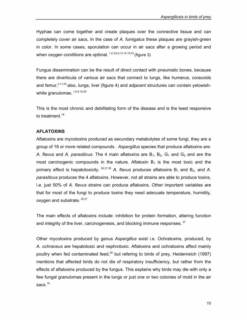
In chronic cases, there is pulmonary hypertension caused by granulomas and necrosis
can occur in the tissue (figure 2) leading to right ventricular dilatation or ascites. In well
oxigenated sections of tissue like bronchi the organism can sporulate. 6,7,11,34
After inhalation, spores can disseminate through the blood producing lesions in brain,
bones, pericardium, pecten and other tissues. 2,3,6,10,17,23,25,33,36 Cerebral presentation is
described by encephalitis or meningoencephalitis in different bird species. Necrotic
caseous lesions surrounded by giant cells in the brain or cerebellum may be observed
causing granulomatous encephalitis with hyphae can be seen in the central area of
some lesions. There are reports in the literature that birds presenting with the infection in
their lungs and air sacs in many cases have kidneys and liver involvement. In bones, the
infection can cause vertebral deformities resulting in paralysis.6,7,17,24 The extension of
the fungal colonization dependent upon host immune system integrity. Clinical signs are
often not apparent until fungal colonization is largely extended. 1
IN AIR SACS: This presentation is slowly progressive and results from persistent
exposure to low levels of spores in immunocompromised birds.10,23 Initial lesions are
found in zones with high oxygen tension and poor blood circulation, like air sacs.
Specifically the caudal thoracic, and the abdominal air sacs where abundant caseous
and necrotic debris may be found. 1,4,6,10,15,21,33
Aspergillosis in birds of prey
10
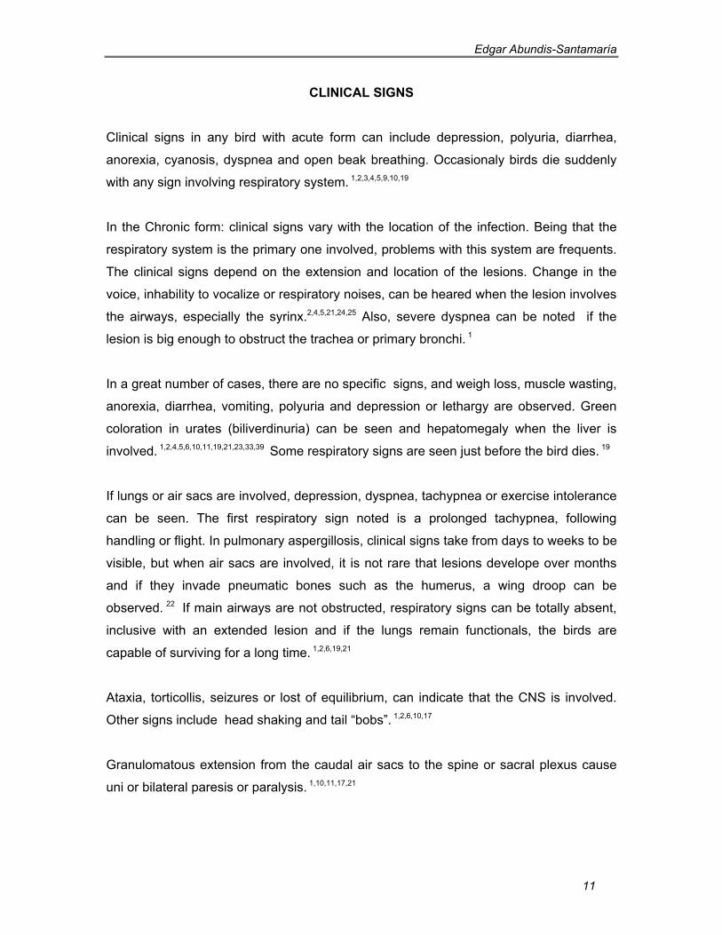
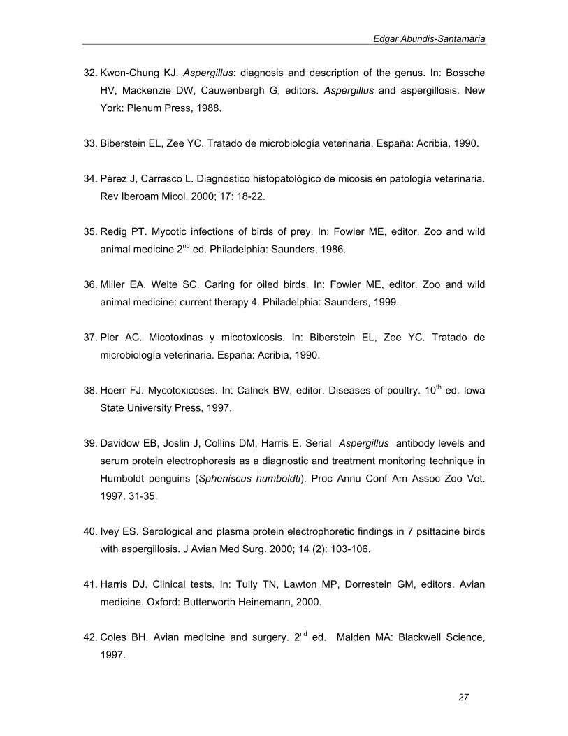
Hyphae can come together and create plaques over the connective tissue and can
completely cover air sacs, In the case of A. fumigatus these plaques are grayish-green
in color. In some cases, sporulation can occur in air sacs after a growing period and
when oxygen conditions are optimal. 1,2,3,6,8,14,15,19,33 (figure 3)
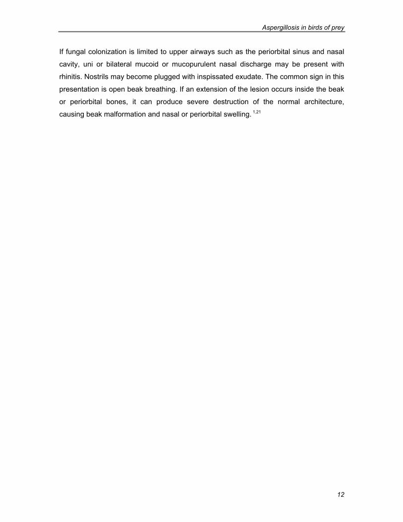
Fungus dissemination can be the result of direct contact with pneumatic bones, because
there are diverticula of various air sacs that connect to lungs, like humerus, coracoids
and femur,2,11,23 also, lungs, liver (figure 4) and adjacent structures can contain yelowish-
white granulomas. 1,6,8,19,29
This is the most chronic and debilitating form of the disease and is the least responsive
to treatment.19
AFLATOXINS
Aflatoxins are mycotoxins produced as secundary metabolytes of some fungi, they are a
group of 18 or more related compounds . Aspergillus species that produce aflatoxins are:
A. flavus and A. parasiticus. The 4 main aflatoxins are B1, B2, G1 and G2 and are the
most carcinogenic compounds in the nature. Aflatoxin B1 is the most toxic and the
primary effect is hepatotoxicity. 26,37,38 A. flavus produces aflatoxins B1 and B2, and A.
parasiticus produces the 4 aflatoxins. However, not all strains are able to produce toxins,
i.e. just 50% of A. flavus strains can produce aflatoxins. Other important variables are
that for most of the fungi to produce toxins they need adecuate temperature, humidity,
oxygen and substrate. 26,37
The main effects of aflatoxins include: inhibition for protein formation, altering function
and integrity of the liver, carcinogenesis, and blocking immune responses. 37
Other mycotoxins produced by genus Aspergillus exist i.e. Ochratoxins, produced, by
A. ochraceus are hepatotoxic and nephrotoxic. Aflatoxins and ochratoxins affect mainly
poultry when fed contaminated feed,26 but refering to birds of prey, Heidenreich (1997)
mentions that affected birds do not die of respiratory insufficiency, but rather from the
effects of aflatoxins produced by the fungus. This explains why birds may die with only a
few fungal granulomas present in the lungs or just one or two colonies of mold in the air
sacs.19
Edgar Abundis-Santamaría
11
CLINICAL SIGNS Clinical signs in any bird with acute form can include depression, polyuria, diarrhea,
anorexia, cyanosis, dyspnea and open beak breathing. Occasionaly birds die suddenly
with any sign involving respiratory system. 1,2,3,4,5,9,10,19
In the Chronic form: clinical signs vary with the location of the infection. Being that the
respiratory system is the primary one involved, problems with this system are frequents.
The clinical signs depend on the extension and location of the lesions. Change in the
voice, inhability to vocalize or respiratory noises, can be heared when the lesion involves
the airways, especially the syrinx.2,4,5,21,24,25 Also, severe dyspnea can be noted if the
lesion is big enough to obstruct the trachea or primary bronchi. 1
In a great number of cases, there are no specific signs, and weigh loss, muscle wasting,
anorexia, diarrhea, vomiting, polyuria and depression or lethargy are observed. Green
coloration in urates (biliverdinuria) can be seen and hepatomegaly when the liver is
involved. 1,2,4,5,6,10,11,19,21,23,33,39 Some respiratory signs are seen just before the bird dies. 19
If lungs or air sacs are involved, depression, dyspnea, tachypnea or exercise intolerance
can be seen. The first respiratory sign noted is a prolonged tachypnea, following
handling or flight. In pulmonary aspergillosis, clinical signs take from days to weeks to be
visible, but when air sacs are involved, it is not rare that lesions develope over months
and if they invade pneumatic bones such as the humerus, a wing droop can be
observed. 22 If main airways are not obstructed, respiratory signs can be totally absent,
inclusive with an extended lesion and if the lungs remain functionals, the birds are
capable of surviving for a long time. 1,2,6,19,21
Ataxia, torticollis, seizures or lost of equilibrium, can indicate that the CNS is involved.
Other signs include head shaking and tail “bobs”. 1,2,6,10,17
Granulomatous extension from the caudal air sacs to the spine or sacral plexus cause
uni or bilateral paresis or paralysis. 1,10,11,17,21
Aspergillosis in birds of prey
12
If fungal colonization is limited to upper airways such as the periorbital sinus and nasal
cavity, uni or bilateral mucoid or mucopurulent nasal discharge may be present with
rhinitis. Nostrils may become plugged with inspissated exudate. The common sign in this
presentation is open beak breathing. If an extension of the lesion occurs inside the beak
or periorbital bones, it can produce severe destruction of the normal architecture,
causing beak malformation and nasal or periorbital swelling. 1,21
Edgar Abundis-Santamaría
13
DIAGNOSIS Ante-mortem aspergillosis diagnosis can be difficult, principally in chronic cases. The
clinical signs are nonspecific and fungus mycelia are generally intimately associated with
the tissues, making them rarely visible in exudates or body fluids. A careful clinical
history can reveal the presence of poor sanitation in the environment. An
immunosuppressing factor, chronic weakness history, weight loss, change in voice or
exercise intolerance, also species susceptibility to this disease must be considered.
Aspergillosis must be suspected in cases of weak animals that do not response to or get
worst with antibiotic treatment. 1,2,7,11,12,19,21,25
It is important to mention that to establish a definitive diagnosis, many tests should be
made, like hematology, serology, cytology, radiographs and endoscopy, because this
fungus is ubiquitous and it is easy to contaminate the sample when the cultures are
made which may affect the results. 1,2,40
• Hemogram Severe leucocytosis of 20, 000 or more than 100, 000 cells/µl is common. The
differential count usually reveals heterophilia with a left shift, monocytosis and
lymphopenia.1,2,4,19,28,35 However, gyrfalcons (Falco rusticolus) have poor responses in
white cells production, compared with other birds of prey and total white cells count may
range as low as12, 000 to 15, 000 cells/µl. 1,12,19,21,25
In chronic infection non-regenerative anemia can be present, an increase in total serum
proteins and in the globulin portion, and an increase in AST and bilie acids when the
liver is involved are noted. 1,19,21,25
• Serology Indirect ELISA is a useful tool for the diagnosis and for monitoring the progress and the
response to the treatment of aspergillosis in birds of prey. A specific conjugated anti-
falcon exists, because falconiforms don't have a good response to anti-turkey
conjugated that is useful for other species. This test can detect antibodies against
A. fumigatus. 10,19,21,23,25,27
Aspergillosis in birds of prey
14
Experimentally, antibodies can be detected one week after infection, which is much
earlier than the clinical signs of the disease are present and the treatment can be started
having better results. 19,25
The available conjugate for falconiforms do not recognize immunoglobulins from owls
and there are any anti-owl conjugated available.2,23,27
Other tests that have been used but with limited diagnostic value are: immunodiffusion in
agar gel, indirect hemaglutination, serum electrophoresis and indirect
immunofluorescence. 5,6,8,12,33,41
• Endoscopy
Endoscopy is an invaluable tool for the diagnosis of aspergillosis. In-patients with severe
dyspnea, tracheoscopy can reveal a lesion obstructing the trachea or syrinx. In the
lumen, plaques or white discharges can be noted, samples for cytology and culture
should be taken. Tracheoscopy requires injectable or inhalant anesthesia.1,5,25,40
Air sac endoscopy can reveal diffuse opacity or the presence of white or yellow fungal
plaques, these plaques can be covered with a grayish mold. Samples are taken directly
or with air sacs washes for cytology and culture.1 To perform the air sac endoscopy, the
bird is put under anesthesia. After surgical preparation of the selected site (The
incision is done behind the last rib, just posterior to the dorsal part of the last rib and
above the level of the junction of the vertebral and sternal parts of the rib cage and in
birds above 1kg between the last two ribs) the skin is picked up with a rat-toothed
forceps to form a “tent”. A nick is made with scissors and their points used to
spread and change the hole to reveal the underlying muscle and ribs. The point
of a closed pair of a straight mosquito forceps or round pointed scissors is quickly
thrust through the muscle and into the abdominal cavity The points of the
instrument used are then opened sufficiently to enable insertion of the
endoscope. 42
Edgar Abundis-Santamaría
15
• Radiographs
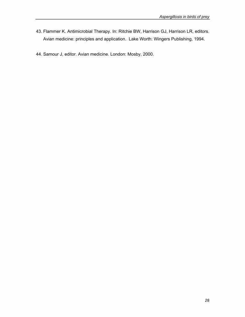
Radiographic changes may not be visible in early cases. However, in advanced cases
radiographic abnormalities include a prominent parabronchial pattern, (figure 5) lost of
definition and asymmetry in air sacs by consolidation, enlargement of air sacs and/or
focal densities in lungs or air sacs. Many of the lesions in air sacs are found in the
cranial part of the abdominal air sac, next to the lungs. 2,21,25 Nephromegaly or
hepatomegaly are seen when these organs are involved. When radiographic changes
are visible prognosis is poor because of the disease is in an advanced stage. 1,2,4,9,10,11,19,25
• Organism identification
The organism identification is made by histopathological or cytological study of the
lesions and by organism culture from the site of infection, but it must be considered that
isolation of Aspergillus species by culture is not a definitive diagnosis, because the
organism is in the environment and contamination is common. 1,8,9,10,11
• Culture
To do the culture from the air sacs the procedure is the same as in the endoscopy to
access to this structure. 3 to 5 ml/kg of saline are used and the air sac is irrigated using
a sirynge and a urinary catheter, then the liquid is recovered and cultured.23 This
procedure should be done in as sterile a fashion as possible.
Trachea culture is done using a sterile nasopharyngeal swab, introducing it deep into the
trachea during the inspiratory process, when glottis is open and trying to not make
contact with oral cavity to avoid sample contamination.19,25,28 This procedure can be done
with or without anesthesia. The recovered material in the swab is immediately transfered
to an appropriate culture media and incubated at 37°C. Also, a tracheal wash can be
performed, using a syringe, a feline urinary catheter and 3ml/kg of saline. The catheter is
introduced into the trachea, the liquid is injected and recovered immediately for culture
and cytological study. 19,23

Aspergillosis in birds of prey
16
Aspergillus species grow at temperatures from 25 to 37°C, some species can grow at
45°C in Sabouraud dextrose agar or blood agar and antibiotics like cloramphenicol must
be used to avoid bacterial contamination, however, Aspergillus species are sensitive to
cycloheximides.1,6,8,17,24,26,29,33 Following up colony characteristics of some species are
described.
Aspergillus fumigatus colonies have a diameter of approximately 3 to 4cm in 7 days. The
flat colonies are white at first, then bluish green as conidia begin to mature, especially
near to the center of the colony. As the colony matures, conidial masses become gray-
green, while the colony edge remains white. A distinctive feature of A. fumigatus is the
development of columnar masses of chains of conidia arising from the vesicle. These
conidial chains can reach a length of up to 400µ. This organism is resistant to high
temperatures and can grow well at 45°C. These are the most typical characteristics, but
variation occur in colony color, both surface and reverse, and colony morphology. 1,6,8,31
A. flavus grows very rapidly, obtaining a colony diameter of 6-7cm in 10 days at 25°C.
The colony begins with a white color, turning yellowish to yellow-green with a white edge
as conidia develop. Mature colonies may become somewhat olive-green. 6,8,31
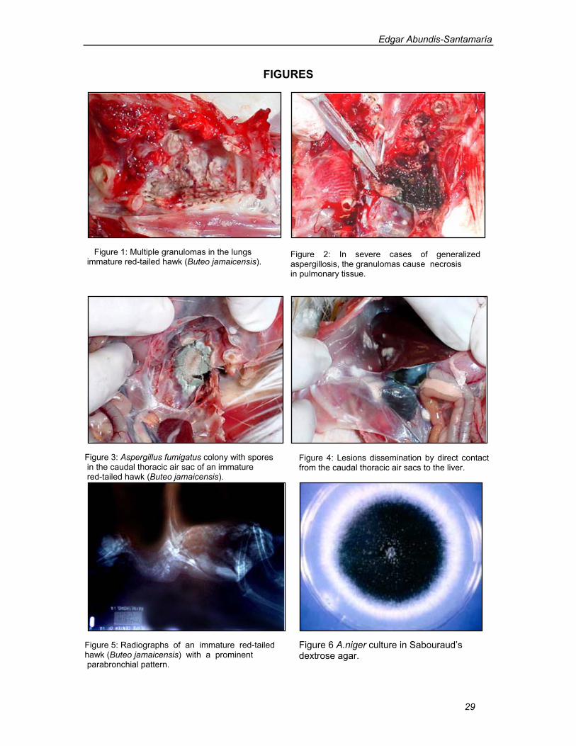
A. niger begins with a white color, but rapidly develops a black color as the conidia
mature.( figure 6) The colony reverse is yellowish. 8,31
• Microscopic exam
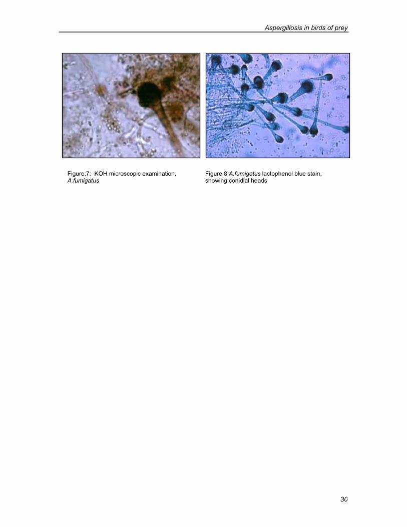
Samples can be seen microscopically, using wet preparations with 20% KOH, (figure
7),lactophenol blue, calcofluor white or new methylene blue. First put some drops of the
stain on the slide, and then add the sample and covering with the coverslip. When KOH
is used, the slide may be heated over a flame. 6,7,8,11,29,33
Organisms can be seen with slides stained with Hematoxilin-Eosin, PAS or
Grocott’s.1,7,8,11,24,29,34 Microscopic identification also, can be made using the culture
colonies, using lactophenol blue stain (figure 8) or new methylene blue. 1,11,14
Edgar Abundis-Santamaría
17
Microscopically, Aspergillus mycelia are composed by tubular septate hyphae.
Occasionally, fruiting bodies are seen when samples come from lungs or air sacs, which
are places where the organism can mature. 1,11,14
Aspergillus fumigatus presents smooth conidiophores with a length up to 300µ and a
diameter from 5 to 8µ . Vesicles are from 20 to 30µ in diameter, with a single series of
phialides. Phialides are from 6 to 8µ in length and are arranged upward paralleling the
axis of the conidiophore. Conidia are echinulated, spherical to semispherical and from 2
to 3µ in diameter. 6,8,29,31
A. flavus contains conidiophores of up to 100µ in length and 10 to 65µ in diameter.
Vesicles are spherical, from 30 to 45µ in diameter and phialides in 2 series over the
vesicle surface, but can be in one series. Conidia are elliptical or spherical, echinulated
and from 3 to 6µ in diameter. 6,8,29,31
A. niger is observed with big fruiting bodies. Vesicle is spherical from 50 to 75µ. Conidia
are black, rounded and smooth with phialides in one or two series. 8,31
DIFFERENTIAL DIAGNOSIS Differential diagnosis in a mature bird showing weight loss, and severe heterophilia can
include chlamydiophylosis and tuberculosis.4,10,11 Neoplasias can some times cause
weight loss and heterophilia. Differential for dyspnea include increase in abdominal
pressure from a mass, ascites, hepatomegaly, pneumonia and inhalation of foreign
bodies.11 Vitamin A deficiency can cause ocular lesions. Some granulomas can be
produced by bacteria. Other mycotic infections must be suspected like candidiasis. 2,10,19
Aspergillosis in birds of prey
18
TREATMENT
Treatment for aspergillosis is complicated, because the drugs used do not reach the
fungus that is walled-off by the bird’s inflammatory response and therefore isolated of the
bloodstream. This disease has a poor prognosis when the infection in the tissues is
extensive and when only systemic drugs are used. The best treatment results if the
granulomatous lesions are debrided and a topic treatment, in conjunction with a systemic
therapy is given.1,9,12,19,25 When the patient shows cachexia, dyspnea and vomiting is
beyond of the point of treatment.19,25
Options for the treatment of aspergillosis are limited, The drugs used include:
itraconazole, flucytosine, fluconazole, clotrimazole, miconazole, ketoconazole and
amphotericin B.1,2,4,5,6,9,25,28,29
ITRACONAZOLE
Itraconazole has a high specificity against Aspergillus,43 is given orally, using a dose of
5mg/kg twice a day, or 10mg/kg once a day, 15mg/kg twice a day can be given, this
depends of the reaction of the bird to the drug, because if it is given in high doses it can
cause depression and anorexia and if these signs continue, the use must be suspended.
These adverse effects have been related to hepatic toxicity and can vary between
different bird species. The absorption of this drug is increased with fat
consumption.1,12,23,43 Itraconazole is the treatment of choice for this disease. 9
Jones (2000) describes itraconazole pharmacokinetics in red-tailed hawks (Buteo
jamaicensis) and he observed that plasma disposition after daily doses of 5 and
10mg/kg for 14 days is minor to the granivorous birds, however, disposition in tissues
like liver, lungs, kidneys, small intestine and air sacs is good, except cerebrum who had
a low disposition. Also demonstrated was a better concentration when 10mg/kg were
used.20
In severe aspergillosis cases, treatment can be combined with amphotericin B IV. Also is
used in nebulizations with clotrimazole 1% in 2 or 3ml saline 1.5 hours/day for 4 to 6
Edgar Abundis-Santamaría
19
weeks or with amphotericin B besides intratracheally if bronchial and tracheal lesions are
detected. Long treatment is recommended, in many cases for some months.1,11,12,20,21,42
Abrams (2001) explains the treatment in a hybrid of peregrine falcon - gyrfalcon (Falcon
peregrinus x Falco rusticolus) who presented with blepharitis and dermatitis caused by
Aspergillus sp. In the beginning the diagnosis of a fungus was obscured because of the
more common presentation of trauma and secondary bacterial infection in birds of prey.
After antibacterial treatment, condition progressed to severe blepharitis and dermatitis
that involved upper and lower eyelids of both eyes and the head. Diagnosis consisted of
histopathological exam of the lesions where septated hyphae were seen, and sample
culture, recognizing fungus of the genus Aspergillus. Treatment started with
itraconazole, dose of 15mg/kg once a day, 2 weeks later, head lesions were debrided,
the dose was increased to 15mg/kg twice a day and a topic ointment was applied twice a
day. A month and a half after the use of the itraconazole, lesions decreased and
itraconazole and clotrimazole were still used at the same dose and frequency, at 2
months the skin was normal on the top of the head and around the eyes. Use of
itraconazole continued for one more month, and the total time of treatment was 3
months. 9
AMPHOTERICIN B
Amphotericin B can be used for initial treatment of severe infections. It is fungicidal and
rarely resistance occurs for Aspergillus species. This drug is not well absorbed when is
given orally and causes irritation with intramuscular or subcutaneous injections. In
severe cases of aspergillosis affecting respiratory system, amphotericin B, IV can be
given simultaneously, intratechal and in nebulizations, followed by itraconazole or
flucytosine. Treatment is started with IV applications at a dose of 1.5mg/kg TID for 3 to 5
days.1,23,24,25,42 It is worth mentioning that this drug is potentially nephrotoxic in birds, but
the prolonged use in birds of prey has not been associated with nephrotoxicity. 11,43
When nebulization is used, the drug needs to reach the internal surface of the air sacs,
to achieve this, the drop size must be under 5µ in diameter, otherwise, drops do not
remain suspended enough in the air stream to reach the intended area. Vaporization of
drugs does not work due to the fact that drops are too big and get condensed in the
Aspergillosis in birds of prey
20
upper airways. The drug needs to be given from a nebulizator to a chamber big enough
to keep the bird comfortable in order to eliminate material loss from dead space effects.
When amphotericin B is used for this treatment, 100 mg of the drug in 15 ml of saline, or
it is even better if can be used in a vehicle like tyloxapol, because this substance helps
the drug to have a better dispersion. Nebulizations are given for 20 minutes, 3 to 4 times
a day. Nebulization is useful for the drug to reach the air sacs without systemic
absorption.19,23,42
Amphotericin B can be used directly in the air sacs or coelomic cavity using a dose of
0.5mg/ml (50mg/L using saline) when lesions have been identified through endoscopy,
and removal of mycotic plaques or granulomas is needed. 1,19,35,44
In nasal aspergillosis, treatment depends on successful plug removal or dissolution by
nasal, choanal or sinus washes, and systemic fungistatic treatment should be initiated
immediately and continued until total remission. Vigorous flushing with saline solution
under isoflurane anesthesia is sufficient to initiate breakdown of the plug. Surgical
excision by trephination should considered if mechanical breakdown fails. Once partial
patency to the choana is established, a combined fungicidal/proteolytic solution can be
used to flush the nares. The solution is made by combining 0.2 to 0.4ml of a commercial
neomycin-chymotrypsin-trypsin-hydrocortisone ointment with 1.0mg/kg amphotericin B
and then diluting the mix in 20ml of saline solution. The resulting combination has
proven to be effective in dissolving and dislodging the caseous plug; 10 ml should be
flushed vigorously but in small amounts through each naris. Patency through the upper
respiratory tract should be verified by observation of drainage through the choana.
Anesthesia and tracheal intubation are seldom necessary once patency to the oral cavity
is established. After the third of fourth sinus flush, the plug ussualy becomes dislodged,
and is seen to appear to the choana. Flushes should be continued using only physical
restraint. Mantaining the unsedated bird in a vertical position is critical to preventing
aspiration. If the solution enters the lower airway, it can cause severe tissue damage.
Daily topical treatment of the nares and sinuses should continue until the lesion and
signs disappear. Treatment by sinus lavage rarely exceeds 7 days. 21
The treatment for the tracheal or syrinx form of Aspergillosis may be successful if the
lesion can be removed surgically through an incision in the lower portion of the trachea
Edgar Abundis-Santamaría
21
or removing it endoscopically,23,35 or if the growth is controlled using antifungal drugs.
When the bird presents with open beak breathing, noticeable neck extension,
discomfort, or acute respiratory crisis, air sac cannulation is indicated.21 This procedure
can be made using a cut-off endotracheal tube as a cannula, the bird is put under
anesthesia and the cannula is inserted into the left side of the bird’s abdomen, just
posterior to the dorsal part of the last rib and above the level of the junction of the
vertebral and sternal parts of the rib cage. The purpose is to insert the cannula into the
caudal thoracic air sac and not into the abdominal air sac which is deeper in the
abdomen. In larger birds (above 1 kg) it may be possible to insert the cannula between
the last two ribs. In cases of severe airway obstruction, this procedure is made just with
physical restraint of the bird and if done quickly, seems to cause a little discomfort in the
bird. After surgical preparation of the selected site the skin is picked up with a rat-toothed
forceps to form a “tent”. A nick is made with scissors and their points used to spread the
hole to reveal the underlying muscle and ribs. The point of a closed pair of a straight
mosquito forceps or round pointed scissors is quickly thrust through the muscle and into
the abdominal cavity. The point of the instrument used are then opened sufficiently to
enable insertion of the plastic cannula which can be sutured in place. The surgeon can
check if air is moving freely through the tube by holding a wisp of cotton wool or fine
suture material near to the opening and watching for fluctuation during each
respiration.23,42 This procedure can also be performed by securing a large-bore (18
gauge or larger) flexible Teflon intravenous catheter in one of the abdominal air sacs to
relieve any dyspnea by extratracheal airflow. Another air sac that can be used is the
clavicular.5 Intratracheal injection of amphotericin B (1mg/kg diluted in 2ml/kg saline)
twice a day using a feline urinary catheter,1,23 may eliminate the granuloma because
amphotericin B is a fungicide and direct contact with the lesion can be an effective
method of treatment. The granuloma may become larger until it bursts at the center,
giving it the appereance of a rosette. This lesion will then rapidly decrease in size and
become dislodged. The debris fall as a plug into the syrinx or one of the bronchi. Total
obstruction of the lower airway may initiate an acute respiratory crisis. Extratracheal
respiration through an air sac cannula prevents asphyxia. Complete blockage, if it
occurs, is fatal. Once endoscopy reveals no visible lesion, the air sac cannula can be
removed. As part of treatment itraconazole can be used orally for 90 to120 days, but
treatment must be suspended if the bird presents with anorexia or depression. Positive
results have also been noted using flucytosine. 21
Aspergillosis in birds of prey
22
FLUCYTOSINE Flucytosine can be given orally using doses of 20 to 60mg/kg twice a day in conjunction
with amphotericin B. 1,19,25,43 This drug is fungistatic and must be given for a long period,
in many cases for 6 or more months. Flucytosine is widely distributed to tissues that are
difficult to penetrate such as, the CSF, eye and joints. Resistance develops quickly when
the drug is used alone and should be used in combination with itraconazole or
amphotericin B. Hemogram monitoring is recommended because this drug can cause
bone marrow toxicity. 1,43
FLUCONAZOLE Fluconazole, in contrast to ketoconazole and itraconazole is highly water soluble and is
readily absorbed from GI tract regardless of acidity or food intake. It penetrates the CSF,
brain tissue, ocular fluids and sputum and is the drug of choice in situations where
penetration into the CFS is desirable. It can be used in combination with itraconazole or
ketoconazole. The dose used is 15mg/kg twice a day, orally. 21,42,43
KETOCONAZOLE Ketoconazole is used orally at doses of 20 to 30mg/kg twice a day for 2 to 6 weeks and
can be used in conjunction with other antifungal drugs. It is widely distributed to tissues
but is highly protein-bound and does not significantly penetrate into the cerebrospinal or
ocular fluids. Ketoconazole is considered less active and potentially more toxic than
fluconazole and itraconazole. 1,42,43
There are different antifungal drugs in ointments for the topical treatment of Aspergillosis
like amphotericin B, miconazole and enilconazole. 9,23,43
Birds with chronic Aspergillosis show a severe immunosuppression and support therapy
is needed, including fluids, force feeding and a warm environment, also it is important to
decrease any factor that can cause stress in the birds, in gyrfalcons (Falco rusticolus)
psychological well-being seems to play an important role. 19
Many times Aspergillosis treatment must be continued for some months and to monitor
the treatment response, white blood cells count and indirect ELISA are useful. 23,43
Edgar Abundis-Santamaría
23
PREVENTION
Aspergillus genus is an opportunistic pathogen, therefore every attempt should be made
to reduce predisposing immunosuppresive factors such as stress and malnutrition, good
managment of the birds is essential.1,7,10,11,19,21,24,25 To avoid inhalation of a large number
of spores, birds should be housed in a well-ventilated area, with a bedding changes
daily. When treating other illnesses, the benefits of long term or repeated antibiotic
usage, or the use of immunosuppresive doses of corticosteroids, must be weighed
against the possibility of opportunistic deep mycotic infections.1
Flucytosine at dose of 50 to 60mg/kg twice a day for 10 days or itraconazole, at dose of
10mg/kg once a day for 10 days (both orally) have been used prophylactically in birds
deemed to be at high risk for the development of aspergillosis. This type of prophylaxis is
of value for any endangered raptor species during stressful periods such as transport,
after injuries and during medical treatment.6,12,19,28
In recent years some breeders have tried to counteract the susceptibility of gyrfalcon to
aspergillosis by crossing them with the relatively resistant saker falcon. Indeed,
gyrfalcon - saker falcon hybrid (Falco rusticolus x Falco cherrug) appear to be less
vulnerable to fungal infection. As these hybrids are back-crossed to gyrfalcons, their
susceptibility seems to rise proportionally to the degree of gyrfalcon genes. 19
IMMUNIZATION
Some autogenous vaccines have been applied and seem to be effective in decreasing
cases of aspergillosis, but there is not much information about this vaccine. This
measure seems to be the most important for prevention to this disease, but more studies
are needed. 6,10,11,19,25,28
Aspergillosis in birds of prey
24
REFERENCES
1. Oglesbee BL. Mycotic Diseases. In: Altman RB, Clubb SL, Dorrestein GM,
Quesenberry K, editors. Avian medicine and surgery. Philadelphia: Saunders, 1997.
2. Atkinson R, Brojer C. Unusual presentations of aspergillosis in wild birds. Proc Assoc
Avian Vet. 1998; 177-181.
3. Friend M, Franson J, editors. Field manual of wildlife diseases: general field
procedures and diseases of birds. US Geological Survey, 1999.
4. Ramírez LJ, Chávez SL, Velasco G. Reporte de un caso de aspergilosis en un ave
búho cornudo (Pseudocops clamator) en el zoológico Miguel Alvarez del Toro,
Tuxtla Gutiérrez, Chiapas. Memorias del XIX Simposio Sobre Fauna Silvestre, Gral.
MV Manuel Cabrera Valtierra; 2002 noviembre 27-29; México: 2002.
5. Forbes NA. Rapaces. In: Beynon PH, Cooper JE, editors. Manual de animales
exóticos. España: Harcourt Brace, 1999.
6. Richard JL. Aspergillosis. In: Calnek BW, editor. Diseases of poultry. 10th ed. Iowa
State University Press, 1997.
7. Jordan FT, Pattison M. Poultry diseases. 4th ed. London: Saunders, 1997.
8. Quinn PJ, Carter ME, Markey BK, Carter GR. Clinical veterinary microbiology.
London: Wolfe, 1994.
9. Abrams GA, Paul-Murphy J, Ramer JC, Murphy CJ. Aspergillus blepharitis and
dermatitis in a peregrine falcon-gyrfalcon hybrid (Falco peregrinus x Falco
rusticolus), J Avian Med Surg. 2001; 15 (2): 114-120.
10. Rosskopf W, Woerpel R. Diseases of cage and aviary birds. 3rd ed. Baltimore:
Williams and Wilkins, 1997.
Edgar Abundis-Santamaría
25
11. Bauck L. Mycoses. In: Ritchie BW, Harrison GJ, Harrison LR, editors. Avian
medicine: principles and application. Lake Worth: Wingers Publishing, 1994.
12. Redig PT, Ackermann J. Raptors. In: Tully TN, Lawton MP, Dorrestein GM, editors.
Avian medicine. Oxford: Butterworth Heinemann, 2000.
13. Faucette TG, Loomis M, Reininger K, Zombeck D, Stout H, Porter C, Dykstra MJ. A
Three-year study of viable airborne fungi in the North Carolina Zoological Park R.J.R.
Nabisco Rocky Coast Alcid Exhibit. J Zoo Wildl Med. 1999; 30 (1): 44-53.
14. Dykstra MJ, Loomis M, Reininger K, Zombeck D, Faucette T. A comparison of
sampling methods for airborne fungal spores during an outbreak of aspergillosis in
the forest aviary of the North Carolina Zoological Park. J Zoo Wildl Med. 1997; 28
(4): 454-463.
15. Young EA, Cornish TE, Little SE. Concomitant mycotic and verminous pneumonia in
a blue jay from Georgia, J Wildl Dis. 1998; 34 (3): 625-628.
16. German AC, Shankland GS, Edwards J, Flach EJ. Development of an Indirect
ELISA for the detection of serum antibodies to Aspergillus fumigatus in captive
penguins. Vet Rec. 2002; 150 (16): 513-518.
17. Akan M, Haziroğlu R, Ilhan Z, Sareyyüpoğlu B, Tunca R. A case of aspergillosis in a
broiler breeder flock, Avian Dis. 2002; 46 (2): 497-501.
18. Stone WB, Okoniewski JC. Necropsy findings and environmental contaminants in
common loons from New York. J Wildl Dis. 2001; 37 (1): 178-184.
19. Heidenreich M. Birds of prey: medicine and management. Malden MA: Blackwell
Science, 1997.
20. Jones MP, Orosz SE, Cox SK, Frazier DL. Pharmacokinetic disposition of
itraconazole in red-tailed hawks (Buteo jamaicensis). J Avian Med Surg. 2000; 14
(1): 15-22.
Aspergillosis in birds of prey
26
21. Aguilar RF, Redig PT. Diagnosis and treatment of avian aspergillosis. In: Bonagura
JD, Kirk R, editors. Current Veterinary Therapy, Small Animal Practice XII.
Philadelphia: Saunders, 1995.
22. Forbes NA. Aspergillosis in raptors. Vet Rec. 1991; 128 (11): 263.
23. Redig PT. Fungal diseases. In: Samour J, editor. Avian medicine; London: Mosby,
2000.
24. Cork SC, Alley MR, Johnstone AC, Stockldale PH. Aspergillosis and other causes of
mortality in the stitchbird in New Zealand. J Wildl Dis. 1999; 35 (3): 481-486.
25. Redig PT. Avian Aspergillosis. In: Fowler ME, editor. Zoo and wild animal medicine:
current therapy 3. Philadelphia: Saunders, 1993.
26. Herrera T, Ulloa M. El reino de los hongos, micología básica y aplicada. 2a ed.
México: Fondo de Cultura Económica, 1998.
27. Redig PT, Orosz S, Cray C. The ELISA as a management guide for aspergillosis in
raptors. Proc Assoc Avian Vet. 1997. 99-104.
28. Redig PT. Raptors. In: Altman RB, Clubb SL, Dorrestein GM, Quesenberry K,
editors. Avian medicine and surgery. Philadelphia: Saunders, 1997.
29. Carter GR. Bacteriología y micología veterinarias. 2ª ed. México: El Manual
Moderno, 1994.
30. Graczyk TK, Cranfield MR. Maternal transfer of anti-Aspergillus spp.
immunoglobulins in african black-footed penguins (Spheniscus demersus). J Wildl
Dis. 1995;.31 (4): 545-549.
31. López R, Méndez LJ, Hernández F, Castañón R. Micología médica: procedimientos
para el diagnóstico de laboratorio. México: Trillas, 1995.
Edgar Abundis-Santamaría
27
32. Kwon-Chung KJ. Aspergillus: diagnosis and description of the genus. In: Bossche
HV, Mackenzie DW, Cauwenbergh G, editors. Aspergillus and aspergillosis. New
York: Plenum Press, 1988.
33. Biberstein EL, Zee YC. Tratado de microbiología veterinaria. España: Acribia, 1990.
34. Pérez J, Carrasco L. Diagnóstico histopatológico de micosis en patología veterinaria.
Rev Iberoam Micol. 2000; 17: 18-22.
35. Redig PT. Mycotic infections of birds of prey. In: Fowler ME, editor. Zoo and wild
animal medicine 2nd ed. Philadelphia: Saunders, 1986.
36. Miller EA, Welte SC. Caring for oiled birds. In: Fowler ME, editor. Zoo and wild
animal medicine: current therapy 4. Philadelphia: Saunders, 1999.
37. Pier AC. Micotoxinas y micotoxicosis. In: Biberstein EL, Zee YC. Tratado de
microbiología veterinaria. España: Acribia, 1990.
38. Hoerr FJ. Mycotoxicoses. In: Calnek BW, editor. Diseases of poultry. 10th ed. Iowa
State University Press, 1997.
39. Davidow EB, Joslin J, Collins DM, Harris E. Serial Aspergillus antibody levels and
serum protein electrophoresis as a diagnostic and treatment monitoring technique in
Humboldt penguins (Spheniscus humboldti). Proc Annu Conf Am Assoc Zoo Vet.
1997. 31-35.
40. Ivey ES. Serological and plasma protein electrophoretic findings in 7 psittacine birds
with aspergillosis. J Avian Med Surg. 2000; 14 (2): 103-106.
41. Harris DJ. Clinical tests. In: Tully TN, Lawton MP, Dorrestein GM, editors. Avian
medicine. Oxford: Butterworth Heinemann, 2000.
42. Coles BH. Avian medicine and surgery. 2nd ed. Malden MA: Blackwell Science,
1997.
Aspergillosis in birds of prey
28
43. Flammer K. Antimicrobial Therapy. In: Ritchie BW, Harrison GJ, Harrison LR, editors.
Avian medicine: principles and application. Lake Worth: Wingers Publishing, 1994.
44. Samour J, editor. Avian medicine. London: Mosby, 2000.
Edgar Abundis-Santamaría
29
FIGURES
Figure 1: Multiple granulomas in the lungs immature red-tailed hawk (Buteo jamaicensis).
Figure 3: Aspergillus fumigatus colony with spores in the caudal thoracic air sac of an immature red-tailed hawk (Buteo jamaicensis).
Figure 5: Radiographs of an immature red-tailed hawk (Buteo jamaicensis) with a prominent parabronchial pattern.
Figure 2: In severe cases of generalized aspergillosis, the granulomas cause necrosis in pulmonary tissue.
Figure 4: Lesions dissemination by direct contact from the caudal thoracic air sacs to the liver.
Figure 6 A.niger culture in Sabouraud’s dextrose agar.
Aspergillosis in birds of prey
30
Figure:7: KOH microscopic examination, A.fumigatus
Figure 8 A.fumigatus lactophenol blue stain, showing conidial heads
Edgar Abundis-Santamaría
31
Appendix I Indirect ELISA in falconiforms.23
A positive result indicates either active infection, long term exposition or a high level of
antibodies against Aspergillus fumigatus, resulting from a previous infection. A negative
result indicates no antibodies, either as a result of lack of disease or inability to produce
them. This test was developed at the Raptor Center (1929 Fitch Avenue, St Paul,
Minnesota, 55108, USA). You can still obtain results from them.
In the test three categories of response are used:
1. Below cut-off (optical density below to 0.12), which implies no detectable
antibodies, and is a category in which false negatives have been encountered only in
circumstances where the patient, in addition to having aspergillosis, had another
debilitating condition such as tuberculosis or lead poisoning.
2. Mid-range (optical density between 0.13 and 0.30), which implies exposure and low
level antibody production either due to low level disease development or poor
immune response to severe diseases.
3. High-range (optical density between 0.31 up to slightly over 1.0) which is associated
with vigorous immune response and may bode well for recovery. An affected bird
often yields a mid-range response early in the disease that increases into the high
range during the second to fourth week of treatment. Failure to show increasing
optical density readings during treatment implies lack of antibody response and may
be indicative of a guarded prognosis.