muscle injuries and repair: the role of prostaglandins … regeneration, ... muscle injuries and...
TRANSCRIPT

Summary. Skeletal muscle injuries are a commonproblem in trauma and orthopaedic surgery. Muscleinjuries undergo the healing phases of degeneration,inflammation, regeneration, and fibrosis. Current andexperimental therapies to improve muscle regenerationand limit muscle fibrosis include conservative andsurgical principles with the adjuvant use of non-steroidalanti-inflammatory drugs (NSAIDs) and growth factormanipulation. NSAIDs appear to have a paradoxicaleffect on the healing of muscle injuries with early signsof improvement and subsequent late impairment infunctional capacity and histology. In vitro and in vivostudies have explored the role of the cyclooxygenasesand prostaglandins in the biological processes of healingmuscle, including precursor cell activation, myoblastproliferation, myoblast fusion, and muscle proteinsynthesis. Through use of more specific cyclooxygenaseinhibitors, we may be able to better understand the roleof inflammation in muscle healing.
Key words: Muscle injury, Prostaglandins, NSAIDs,Cyclooxygenase
Introduction
Skeletal muscle injuries constitute the majority ofsports-related injuries in many epidemiological studies(Garrett, 1996; Croisier et al., 2002). Moderate to severemuscle injuries may result in the inability to train orcompete for several weeks and have a high tendency torecur (Verrall et al., 2001; Orchard and Best, 2002).Muscle injuries result from a variety of mechanisms,including direct and indirect causes. Direct causesinclude traumatic processes such as lacerations,contusions, and strains (Hughes et al., 1995;
Kasemkijwattana et al., 1998, 2000; Fukushima et al.,2001). Indirect causes are those that result from anothermedical condition such as ischemia or neurologicdysfunction (Day et al., 2002; Paoni et al., 2002).
Additionally, repeated eccentric muscle contractionscan result in delayed-onset muscle soreness (DOMS)with symptoms similar to muscle injuries, includingdecreased function, stiffness, and pain (Warren et al.,2002). DOMS is attributable to a distinct process inmuscle that includes an inflammatory response andchanges in the structural integrity of muscle resulting inthe loss of functional capacity (Barash et al., 2002;Lieber et al., 2002). Moreover, mechanical damage andleukocyte infiltration after intense eccentric exercise areknown to coincide with torque reductions (MacIntyre etal., 1996).
Years of research have clarified the time-dependentand interrelated processes that occur after skeletalmuscle is injured (Hurme et al., 1991; Kaariainen et al.,2000; Huard et al., 2002). Muscle injuries undergo adistinct set of healing phases, including degeneration,inflammation, regeneration, and fibrosis. In this paper,we describe these biological events, which occur duringthe first few weeks following muscle injury. We thenoutline the latest discoveries made with regard toimproving the regenerative process and reducing fibrosisformation both experimentally and therapeutically toimprove muscle functional recovery. Finally, we focuson the role of the inflammatory phase of muscle healing,giving the most attention to the cyclooxygenases, theprostaglandins, and their inhibitors.
The phases of muscle healing after injury
Injured skeletal muscle undergoes the healing phasesof degeneration, inflammation, regeneration, andfibrosis. Although the biological processes of thesephases have a great deal of overlap, the different phasesof muscle healing, as depicted by histological analysis,can be very distinct at sequential time points (Fig. 1).Below, we discuss these phases in more detail.
Review
Muscle injuries and repair: The role of prostaglandins and inflammationV. Prisk1 and J. Huard1,2
1Growth and Development Laboratory, Department of Orthopaedic Surgery Children�s Hospital of Pittsburgh and University of
Pittsburgh, Pittsburgh, USA and 2Department of Molecular Genetics and Biochemistry, University of Pittsburgh, Pittsburgh, PA, USA
Histol Histopathol (2003) 18: 1243-1256
Offprint requests to: Johnny Huard, Ph.D., Director, Growth andDevelopment Laboratory, 4151 Rangos Research Center, Children�sHospital of Pittsburgh, 3705 Fifth Avenue Pittsburgh, PA 15213-2583.Fax: 412-692-7095. e-mail: [email protected]
http://www.hh.um.es
Histology andHistopathology
Cellular and Molecular Biology

Degeneration
Trauma to muscle tissue disrupts the integrity of thesarcomere, sarcolemma, and basal lamina, leading to theingress of extracellular calcium as well as the activationof the complement cascade (Carpenter and Karpati,1989; Orimo et al., 1991). Intrinsic proteases autodigestdisrupted and subsequently necrotic myofibers (Ebisui etal., 1995; Mbebi et al., 1999). The tendon-myofiber-tendon units are disrupted and the ruptured myofibersretract forming a gap (Kaariainen et al., 2000). Skeletalmuscle is richly vascularized, and capillary injury resultsin a hematoma that fills this gap. The upregulation ofadhesion molecules and cytokines influences localvascular permeability and blood flow, thus acceleratingthe ensuing inflammatory response and resultant edema.Toxic free radical species develop and can impairexcitation-contraction coupling, induction of proteolysis,and subsequent necrosis of myofibers both in thetraumatized tissue and in healthy tissue located nearby(Clanton et al., 1999). In addition to mechanical damage,the myofibers become denervated through destruction of
intramuscular nerve branches and separation of the fibersegment containing the neuromuscular junction from theremainder of the myofiber (Rantanen et al., 1995b).
Little attention has been paid to finding ways to limitthe degeneration that occurs with muscle injury. In orderto affect this phase, one must employ preventativemedicine techniques. Some have suggested the use ofantioxidant substances prior to sporting activities.However, studies on antioxidant or free radicalinactivating substances like vitamin E, vitamin C, and N-acetyl cysteine have failed to show promising results(Childs et al., 2001; Beaton et al., 2002).
Inflammation
Inflammation is an early response to muscle tissueinjury and involves coordination between the immunesystem and the injured tissue. This phase is, perhaps, theleast distinct phase as its processes overlap with all ofthe other phases of muscle injury and repair (Fig. 1). It isknown that within the first day after muscle injury, smallblood vessels, neutrophils, activated macrophages, and
1244
Prostaglandins and muscle repair
Fig. 1. Injured muscle undergoes the healing phases of degeneration, inflammation, regeneration, and fibrosis. Muscle inflammation after injury resultsin the release of growth factors, cytokines, free radicals, and other mediators, which set up a microenvironment that overlaps with all of the healingphases. Muscle degeneration involves marked myofiber necrosis and early inflammatory cell infiltration. Muscle regeneration is most clearly defined bythe presence of centronucleated myofibers. Muscle fibrosis results from collagen deposition between regenerated myofibers.

T-lymphocytes infiltrate the hematoma between theruptured myofibers (Fielding et al., 1993; Tidball, 1995;Frenette et al., 2000; MacIntyre et al., 2000). Theinvading neutrophils are followed by differentpopulations of macrophages (Lapointe et al., 2002).Some macrophages are believed to be involved mainlyin phagocytosis and removal of cellular debris, thoughthey also may participate with neutrophils in evokingnonspecific tissue damage due to the spillage ofdegradative enzymes (Lapointe et al., 2002). Likeneutrophils, these activated macrophages release pro-inflammatory cytokines, prostanoids, collagenases, andmany potentially cytotoxic compounds such asperoxynitrite, which can lead to further muscledegeneration and excitation-contraction uncouplingoutside of the zone of injury (Beckman and Koppenol,1996; Witko-Sarsat et al., 2000). Other populations ofmacrophages do not phagocytose degenerating skeletalmuscle fibers; rather, they produce early cytokine andgrowth factor signals during the regenerative process(McLennan, 1993; St Pierre and Tidball, 1994).
In addition to the inflammatory cell response, themyogenic cells are potentially capable of secretinggrowth factors, cytokines, and prostanoids, which affectthe regenerative microenvironment and contribute to thesymptomatology of muscle injuries. Various growthfactors released during the inflammatory phase havewell described roles in both the muscle regenerationphase and the formation of muscle fibrosis after severeinjury (Huard et al., 2002; Li and Huard, 2002). Alteringthe inflammatory process may have both beneficial anddetrimental effects. Limiting inflammation maytheoretically reduce excessive muscle degeneration andsignals for scar formation, but reducing the availabilityof growth factors, cytokines, and prostaglandins mayinhibit strong signals that promote the regenerativeprocess as well. This paradox is explored in more detaillater in this review.
Regeneration
Whereas active muscle degeneration andinflammation are occurring at the injury site in the firstfew days, muscle regeneration begins to take precedenceduring the first week post-injury (Kaariainen et al., 2000;Huard et al., 2002). Satellite cells are activated as earlyas 24 hours post-injury, but the beginning of theregenerative phase is marked by the subsequentmyoblast proliferation, which culminates in myoblastdifferentiation and fusion into multinucleated myofibersand eventually mature myofibers (Rantanen et al.,1995a; Zammit and Beauchamp, 2001; Huard et al.,2002). In fact, some researchers theorize that thedisruption of the sarcolemma and basal lamina aftermuscle injury releases and activates previously quiescentsatellite cells and muscle stem cells residing betweenthese structures (Hurme and Kalimo, 1992; Bischoff,1994; Qu-Petersen et al., 2002). Growth factors releasedat the injury site, including Insulin-like Growth Factor-1
(IGF-1), basic Fibroblast Growth Factor (bFGF),Epidermal Growth Factor (EGF), Hepatocyte GrowthFactor (HGF), and Transforming Growth Factor Beta-1(TGF-ß1), have been shown to influence theproliferation and differentiation of myoblasts and musclestem cells in vitro (Sheehan and Allen, 1999; Deasy etal., 2002; Huard et al., 2002). In vivo studies of musclelaceration, contusion, and strain have shown that IGF-1,bFGF, and, to a lesser extent, Nerve Growth Factor(NGF) injected at early time points post-injury (2, 5, and7 days) are all capable of enhancing muscle regeneration(Kasemkijwattna et al., 1998, 2000; Menetrey et al.,2000). Of particular interest, IGF-1 plays an influentialrole in the muscle regenerative process by stimulatingmyoblast proliferation, differentiation, and, eventually,myofiber protein synthesis and hypertrophy (Engert etal., 1996; Damon et al., 1998). As the local environmentduring muscle regeneration overlaps with theinflammatory process, prostaglandins released in theinjured muscle may also contribute or be essential to theaction of growth factors in myofiber regeneration. Wediscuss the role of inflammatory mediators in satellitecell activation, growth factor regulation, and myoblastdifferentiation in further detail below.
Fibrosis
The growth factors released from residentmacrophages, myogenic precursor cells, and othercellular mediators of the injury process do not always actto augment the regenerative process. In fact, EGF,myostatin, and TGF-ß1 have been shown to inhibitskeletal muscle regenerative processes both in vitro andin vivo (Doumit et al., 1993; Mendler et al., 2000;Yamanouchi et al., 2000; Fukushima et al., 2001; Tayloret al., 2001; Rios et al., 2002; Langley et al., 2002; Liand Huard, 2002). It has been documented that thehematoma in the necrotic muscle gap begins to bereplaced by a connective tissue scar made of type III andthen type I collagen starting as early as the third daypost-injury (Kaariainen et al., 2000). This fibrotic tissueprovides early support for ruptured myofibers, but as itbecomes increasingly dense over the course of seven tofourteen days post-injury, it restricts the regenerativegrowth of myofibers (Kaariainen et al., 2000; Li andHuard, 2002). The dense fibrotic tissue that developsafter severe muscle injuries not only prevents themyofiber stumps from rejoining but also may preventnew axons from reaching muscle fibers to createneuromuscular junctions (Kaariainen et al., 2000). Thus,those fibers may undergo atrophy following denervation.
TGF-ß1 has been implicated in the pathogenesis offibrosis in many tissues, including those of the lung,kidney, central nervous system, heart, and liver (Czaja etal., 1989; Khalil et al., 1993; Logan et al., 1994; Lijnenet al., 2000; Ina et al., 2002; Venkatesan et al., 2002).Similarly, TGF-ß1 has been found to be associated withthe muscle necrosis and fibrosis that occur in Duchennemuscular dystrophy and dermatomyositis as documented
1245
Prostaglandins and muscle repair
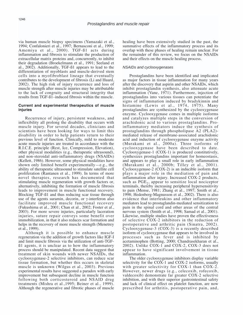
via human muscle biopsy specimens (Yamazaki et al.,1994; Confalonieri et al., 1997; Bernasconi et al., 1999;Amemiya et al., 2000). TGF-ß1 acts duringinflammation and fibrosis to stimulate the production ofextracellular matrix proteins and, concurrently, to inhibittheir degradation (Broekelmann et al., 1991; Seeland etal., 2002). Additionally, TGF-ß1 appears to lead to thedifferentiation of myoblasts and muscle-derived stemcells into a myofibroblast lineage that eventuallycontributes to the development of fibrosis (Li and Huard,2002). The high risk of injury recurrence and loss ofmuscle strength after muscle injuries may be attributableto the lack of congruity and structural integrity thatresults from TGF-ß1–induced fibrosis within the muscle.
Current and experimental therapeutics of muscleinjuries
Recurrence of injury, persistent weakness, andinflexibility all prolong the disability that occurs withmuscle injury. For several decades, clinicians andscientists have been looking for ways to limit thisdisability in order to help patients return to theirprevious level of function. Clinically, mild to moderateacute muscle injuries are treated in accordance with theR.I.C.E. principle (Rest, Ice, Compression, Elevation),other physical modalities (e.g., therapeutic ultrasound),and non-steroidal anti-inflammatory drugs (NSAIDs)(Kellett, 1986). However, some physical modalities haveshown only limited benefit experimentally—e.g., theability of therapeutic ultrasound to stimulate satellite cellproliferation (Rantanen et al., 1999). In terms of morenovel therapies, research has documented thatstimulating muscle regeneration with growth factors or,alternatively, inhibiting the formation of muscle fibrosisleads to improvement in muscle functional recovery.Blocking TGF-ß1 and thus reducing scar tissue throughuse of the agents suramin, decorin, or γ-interferon alsofacilitate improved muscle functional recovery(Fukushima et al., 2001; Chan et al., 2002; Foster et al.,2003). For more severe injuries, particularly lacerationinjuries, suture repair conveys some benefit overimmobilization, in that it also reduces scar formation andhelps in the recovery of more muscle strength (Menetreyet al., 1999).
Although it is possible to enhance muscleregeneration via the addition of growth factors like IGF-Iand limit muscle fibrosis via the utilization of anti-TGF-ß1 agents, it is unclear as to how the inflammatoryprocess should be manipulated. Recent data suggest thattreatment of skin wounds with newer NSAIDs, thecyclooxygenase-2 selective inhibitors, can reduce scartissue formation, but whether this occurs in skeletalmuscle is unknown (Wilgus et al., 2003). Previousexperimental results have suggested a paradox with earlyimprovement but subsequent decline in muscle functionfollowing both corticosteroid and NSAID drugtreatments (Mishra et al.,1995; Beiner et al., 1999).Although the regenerative and fibrotic phases of muscle
healing have been extensively studied in the past, thesummative effects of the inflammatory process and itsoverlap with these phases of healing remain unclear. Forthe remainder of this review, we focus on the NSAIDsand their effects on the muscle healing process.
NSAIDs and cyclooxygenases
Prostaglandins have been identified and implicatedas major factors in tissue inflammation for many yearsafter the discovery that aspirin and other NSAIDs, whichinhibit prostaglandin synthesis, also attenuate acuteinflammation (Vane, 1971). Furthermore, injection ofprostaglandins into various tissues can potentiate thesigns of inflammation induced by bradykinnin andhistamine (Lewis et al., 1974, 1975). Manyprostaglandins are synthesized by the cyclooxygenaseenzyme. Cyclooxygenase comes in multiple isoformsand catalyzes multiple steps in the conversion ofarachidonic acid to various prostaglandins. Pro-inflammatory mediators induce the synthesis ofprostaglandins through phospholipase A2 (PLA2)-mediated release of membrane-associated arachidonicacid and induction of cyclooxygenase enzyme activity(Murakami et al., 2000a). Three isoforms ofcyclooxygenase have been described to date.Cyclooxygenase-1 (COX-1) is produced constitutively,synthesizes prostaglandins important for homeostasis,and appears to play a small role in early inflammation(Murakami et al., 2000b; Tilley et al., 2001).Cyclooxygenase-2 (COX-2) is an inducible isoform thatplays a major role in the mediation of pain andinflammation after injury. Increased COX-2 products,such as PGE2, appear to sensitize local nociceptorterminals, thereby increasing peripheral hypersensitivityto pain (Mense, 1981; Zhang et al., 1997; Smith et al.,1998; Hedenberg-Magnusson et al., 2002). There is alsoevidence that interleukins and other inflammatorymediators lead to prostaglandin-mediated sensitization topain in the spinal cord and other areas of the centralnervous system (Smith et al., 1998; Samad et al., 2001).Likewise, multiple studies have proven the effectivenessof selective COX-2 inhibitors in the reduction ofpostoperative and arthritic pain (Sinatra, 2002).Cyclooxygenase-3 (COX-3) is a recently describedisoform of cyclooxygenase that appears to be involved inprocesses such as fever and is inhibited byacetaminophen (Botting, 2000; Chandrasekharan et al.,2002). Unlike COX-1 and COX-2, COX-3 does notappear to have significant involvement in tissueinflammation.
The older cyclooxygenase inhibitors display variableselectivity for the COX-1 and COX-2 isoforms, usuallywith greater selectivity for COX-1 than COX-2.However, newer drugs (e.g., celecoxib, rofecoxib,valdecoxib) demonstrate far greater COX-2 selectiveinhibition, and with their superior gastrointestinal safetyand lack of clinical effect on platelet function, are nowprescribed for arthritis, postoperative pain, and,
1246
Prostaglandins and muscle repair

occasionally, acute soft tissue injury. These agents areclearly capable of reducing the pain associated withvarious insults, but it remains unclear whether they havebeneficial or detrimental effects on the complex processof muscle repair and recovery of strength. Althoughmany studies have looked at the role of non-selectiveNSAIDs in muscle healing, there is a paucity ofinformation regarding the effects of the COX-2 selectiveinhibitors in this process.
NSAIDs and muscle functional recovery
In addition to the R.I.C.E. principle, clinicians oftenrecommend that NSAIDs be taken in the first few daysafter injury to limit inflammation and pain. However,some studies indicate early functional and histologicalimprovement with the use of NSAIDs but concomitantloss of functional capacity at later time points (Fig. 2). Inanimal models and humans, NSAIDs are capable ofdecreasing prostaglandin concentrations, limiting edema,and delaying the inflammatory process in traumatized
skeletal muscle (Almekinders, 1999; Trappe et al.,2001). Salminen and Kihlstrom (1987) noted thatNSAIDs provide a cytoprotective effect on exercisedmuscle with a reduction in myofibrillar inflammatorycells and myonecrosis at 48 hours post-exercise injury inmice. However, Almekinders and Gilbert (1986)reported a delay in muscle regeneration and nosignificant effect on tensile strength recovery (force torupture) in a rat tibialis anterior strain injury whentreated with the NSAID piroxicam.
In a later study, Obremsky et al. (1994) investigatedcontractile strength recovery and tensile strength, andconducted histological analysis of strain-injured musclein rabbits. In their study, contractile forces at day 1 post-injury showed approximately 20% improvement withpiroxicam treatment as compared to untreated controls.No further significant improvements were observed at 2,4, or 7 days follow-up. As with Almekinders and Gilbert(1986), no change in tensile strength was noted at anytime point. The rabbit model utilized by Obremsky et al.also demonstrated similar histologic responses to
1247
Prostaglandins and muscle repair
Fig. 2. The NSAIDs have been shown to improve early functional capacity of injured muscle but lead to deficits in late functional capacity. NSAIDs mayimprove early outcome by limiting the destructive effects of inflammation, while NSAID inhibition of regenerative signals from the inflammatory processmay result in late functional deficits.
piroxicam treatment, including the inhibition ofinflammatory cell infiltration, myonecrosis, and collagendeposition but limited myofiber regeneration. Thus,though the NSAID piroxicam improved contractileforces and limited inflammation very early, it isuncertain as to whether the associated inhibition ofmyofiber regeneration could lead to deficits in functionalcapacity at later time points post-injury.
To investigate this matter further, Mishra et al.(1995) studied the effects of the NSAID flurbiprofen oneccentric contraction–induced muscle injury in theextensor digitorum longus of the rabbit up to 28 dayspost-injury. They found that flurbiprofen conveyed ashort-term protective effect in the first week with morecomplete recovery of muscle strength. Yet, again, theirresults also suggested a delay in the regenerative processbetween 3 and 7 days. Even though functional recoverywas improved at early time points upon flurbiprofentreatment, at 28 days the researchers recorded anunexplained reduction in torque production by thetreated muscles. The reason for this occurrence isunknown as no follow-up studies were performed at time
points between 7 and 28 days. When NSAID therapies have been examined in
human muscle injuries, more conflicting results havebeen observed. Reynolds et al. (1995) studied the effectsof the NSAIDs meclofenamate and diclofenac in adouble-blind, placebo-controlled study of healing afteracute hamstring muscle strains. Unexpectedly, thegroups with severe muscle strains experiencedsignificantly more persistent pain when treated with theNSAIDs as compared to the control group. Similarly,Bourgeois et al. (1999) found no significant difference inperceived, post-exercise muscle soreness upon treatmentwith the NSAID naproxen, though significantimprovement occurred at 48 hours, including the returnof voluntary knee extension torque to baseline.Conversely, a more recent study by Sayers et al. (2001)showed that ketoprofen treatment 36 hours after intenseeccentric muscle contraction exercise reduced sorenessand improved maximal isometric force production.NSAIDs are typically given to patients after muscleinjury to limit pain. Whether or not this practice resultsin a premature return to activity and subsequent early re-
1248
Prostaglandins and muscle repair
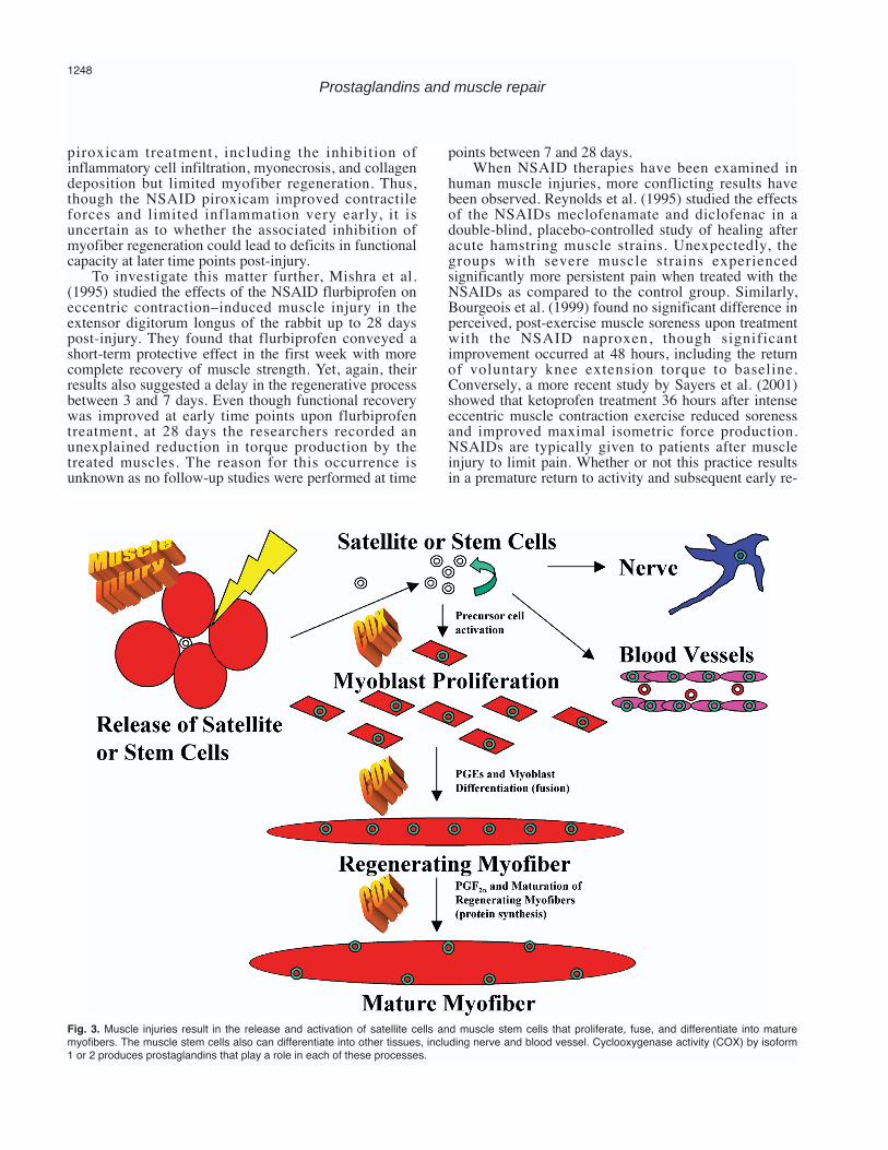
Fig. 3. Muscle injuries result in the release and activation of satellite cells and muscle stem cells that proliferate, fuse, and differentiate into maturemyofibers. The muscle stem cells also can differentiate into other tissues, including nerve and blood vessel. Cyclooxygenase activity (COX) by isoform1 or 2 produces prostaglandins that play a role in each of these processes.

injury of the previously traumatized muscle is unclear. Making comparisons amongst the many studies
using NSAIDs to treat muscle injuries is complex due toa great deal of variability in the experimental methods.Inconsistencies among the mentioned studies areconfounded by the use of different NSAIDs withdifferent specificities for various cyclooxygenaseisoforms, timing of outcome measurements, dosage andadministration routes of drugs, magnitude or type ofdamaging force (i.e., laceration, contusion, strain),and/or muscle type injured. Intervention with NSAIDSduring the early inflammatory process may significantlyaffect the long-term outcomes of skeletal muscleinjuries, and thus necessitates further investigation tounderstand the effects of inactivating thecyclooxygenase enzymes during skeletal muscle repair.
Prostaglandins and muscle repair biology
In the remainder of this paper, we focus on thebiological processes in which prostaglandins are knownto play a role during muscle repair. The prostaglandins,which are largely produced during the inflammatoryphase after muscle injury, also have a role in theregenerative and fibrosis phases of muscle repair.Mechanistically, the regenerative phase of musclehealing can be broken down into the components ofprecursor cell activation, proliferation, fusion, anddifferentiation into mature myofibers (Fig. 3). Throughfurther examination of past research it may be possibleto formulate an understanding of the paradoxical earlyfunctional improvement and later functional impairmentoccasionally seen with NSAID therapies.
Prostaglandins in muscle inflammation
Skeletal muscle can produce PGE1, PGE2, PGF2α,and other prostaglandins both in situ and in vitro (Youngand Sparks, 1979; Berlin et al., 1979; Rodemann andGoldberg, 1982; Palmer et al., 1983; Wennmalm andFitzgerald, 1988; McLennan, 1991). In particular, PGE2concentrations have been shown to be increased ininjured or painful muscle (McArdle et al., 1994;Hedenberg-Magnusson et al., 2002). Additionally, bothdystrophin-deficient mdx mouse muscle and muscle inpatients with Duchenne muscular dystrophy release anincreased amount of PGE2 in response to contractileactivity (Jackson et al., 1991; McArdle et al., 1991,1992). McArdle et al. (1994) further noted thatregenerating myofibers display enhanced phospholipaseactivity and release more PGE2.
PGE2 appears to have multiple functions in themuscle inflammatory process, including chemotaxis ofinflammatory cells, stimulation of pro-inflammatorycytokines, induction of nitric oxide synthase, andvasodilatation with increased vascular permeability(Murata et al., 1997; Lapointe et al., 2002). Manyinflammatory mediators (IL1ß, TNFα, and others) arecapable of enhancing PGE2 production by a variety of
cell types (Jourdan et al., 1999; Walch and Morris, 2002;Bradbury et al., 2002). Further enhancement of theinflammatory phase by the presence of PGE2 may leadto excess degradation of myofibers within the injuredarea. The added inflammation, pain, disruption ofexcitation-contraction coupling, and calciumhomeostasis attributable to PGE2 likely reduces earlyrecovery of muscle functional capacity in the injuredathlete. Thus, inhibition of this process through the useof NSAIDs could explain the early functionalimprovement that may occur with these drugs.
Prostaglandins in precursor cell activation and myoblastproliferation
Prostaglandins released during the inflammatoryphase contribute toward the microenvironment thatoverlaps with precursor cell activation and proliferationduring the regenerative phase of muscle healing. Acomplex interaction appears to exist between growthfactors and prostaglandins in control of cell proliferation.Past experiments commonly employed a peptide growthfactor with a prostaglandin, like PGF2α, to achieve thepassage of a larger number of cells through the cell cycle(de Asua et al., 1977; O’Farrell et al., 1979). Theconnection between PGF2α and cell proliferation wasdescribed many years ago but is still not completelyunderstood (Taylor and Polgar, 1977). Although it isclear that prostaglandins have a function in cellularproliferation in vitro, it is unclear what roleprostaglandins play in muscle precursor cell activationand/or subsequent myoblast proliferation anddifferentiation after injury in situ.
Immediately after muscle injury, satellite cells andstem cells within the basal lamina of myofibers arereleased and activated from the quiescent state to theactivated state to divide and eventually participate in theregeneration of myofibers (Rantanen et al., 1995a;Huard et al., 2002). Mitogenic stimulation of multiplequiescent cell types by the addition of serum to mediumtypically results in increased expression of COX-2 invitro (O’Banion et al., 1992; Pilbeam et al., 1993). Thisfinding correlates with the complex role of COX-2 in thecell cycle dynamics of neoplasia, and one couldpostulate that COX-2 plays a related role in initiatingproliferation of satellite cells or muscle stem cells (Rossiet al., 1989; Steiner et al., 1995; Cao and Prescott, 2002).Moreover, Steiner et al. (1995) noted that the rise inCOX-2 protein levels after changing from serumdepletion to serum stimulation of myoblasts is onlytransient, suggesting that COX-2 expression might onlybe of significance in stimulating re-entry of satellite cellsinto the cell cycle rather than being essential forcontinued exponential proliferation of those cells.Studies in human fibroblasts have shown that COX-2induction by IL1-b is more pronounced in quiescent cellswithin the G0 phase than in cells in cycle (Gilroy et al.,2001). Theoretically, NSAID inhibition of COX-2 maylead to a decreased number of active muscle stem cells,
1249
Prostaglandins and muscle repair

satellite cells, or myofibroblasts contributing tomyogenic, vascular, neurologic, or fibrotic tissuesynthesis. How this might contribute to early versus laterecovery of muscle functional capacity is too complex todecipher at this time.
Prostaglandins in myoblast fusion and differentiation
Cyclooxygenase products not only affect cellproliferation and growth, but also display complexmechanisms in muscle differentiation and maturation.After muscle injury, activated, previously quiescentsatellite cells proliferate and fuse to form multinucleatedmyofibers that later differentiate into mature myofibers(Huard et al., 2002). Early work by Zalin (1977)revealed that NSAIDs prevent cell fusion in culturedchick myoblasts. Zalin (1987) later suggested that achange from PGF2α production to E series prostaglandinproduction contributed to the transition from theproliferating state to the differentiated state. Overall, thecontrol of myoblast fusion involves a complexinteraction between ion channels, prostaglandins, andacetylcholine receptors (Entwistle et al., 1988a,b).Entwistle et al. (1988a) suggested that fusion iscontrolled by depolarization initiated through activationof the acetylcholine receptor or prostaglandin actions onchloride channels.
Hausman et al. have published numerous studies onthe role of prostaglandins in early cell surface eventsleading to the fusion of myoblasts (Hausman andVelleman, 1981; Hausman et al., 1986, 1990; Hausmanand Berggrun, 1987; Santini et al., 1987, 1988; Elgendyand Hausman, 1990). These studies have led to theconclusion that a G-protein–mediated event results fromthe binding of prostaglandins to their receptors andsubsequent membrane organization events, which allowfor cell-cell adhesion and fusion into myotubes (Santiniet al., 1987, 1988; Hausman et al., 1990; Elgendy andHausman, 1990). Myoblasts grown in the NSAIDindomethacin fail to differentiate into myotubes, and thisblock of cell-cell adhesion can be reversed via additionof exogenous prostaglandin to the cell culture medium(Santini et al., 1988). Furthermore, administration ofindomethacin or aspirin to chick embryos has beenfound to decrease the number of myonuclei incorporatedinto the embryo muscles (McLennan, 1987b). Overall,the prostaglandins appear to be important mediators ofmyoblast fusion and formation of multinucleatedmyofibers.
Schutzle et al. (1984) have suggested that E seriesprostaglandins may contribute to muscle differentiationby stimulating the production of muscle-specificproteins. They observed that indomethacin treatmentreduced creatine kinase accumulation in cultured chickmyotubes and that this outcome could be reversed byadding PGE1 and PGE2 to the medium (Schutzle et al.,1984). Similarly, chick embryos treated with aspirin orindomethacin develop disrupted myofibrils and lackcreatine kinase, resulting in a muscular dystrophy-like
myopathy (McLennan, 1985, 1987a). McLennan (1987a)also observed the loss of both thick and thin filamentsfrom the myofibrillar apparatus with abnormal contactsbetween the developing myofibers. The simultaneousadministration of PGE1 reversed the effects of theNSAIDs on creatine kinase levels and muscle structure(Schutzle et al., 1984; McLennan, 1985, 1987a). Eventhough prostaglandins may be essential for embryonicmyogenesis, their importance to adult muscle celldifferentiation from myoblast to myofiber after injury isunclear. For instance, Thorsson et al. (1998) used a ratgastrocnemius contusion injury model to demonstratethat intramuscular injection of the NSAID naproxen didnot appear to affect the formation of regeneratingmyofibers from satellite cells. In looking at the NSAIDparadox of early versus late effects on the recovery offunctional capacity in injured muscle, it is conceivablethat inhibition or delay of myoblast differentiation due toinhibition of prostaglandin synthesis could lead todemonstrated losses at later time points.
Prostaglandins and muscle protein synthesis
PGF2α and PGE2 appear to perform conflictingactions when it comes to protein turnover rates inskeletal muscle. Early on it was discovered that PGE2could lead to net protein degradation, possibly viaactivation of the lysosomal apparatus, and that PGF2αplayed a role in stimulating muscle protein synthesis andgrowth, especially under insulin stimulation (Palmer etal., 1989; Thompson et al., 1993; Hussey and Tisdale,2000). Additionally, prostaglandins appear to act asmessengers in the regulation of tension-induced muscleprotein turnover rates (Smith et al., 1983; Palmer et al.,1983; Vandenburgh et al., 1990, 1995; Trappe et al.,2002). McMillan et al. (1987) showed that a stimulus formuscle hypertrophy increased muscle protein synthesisand that the NSAID fenbufen inhibited this increase.Furthermore, Trappe et al. (2002) reported that over-the-counter dosing of both ibuprofen and acetaminophencould suppress the normal rise in protein synthesis thatoccurs after eccentric contraction exercise in humans.Their follow-up studies revealed that PGF2α levels wereelevated significantly in human vastus lateralis biopsiesfollowing a high intensity eccentric contraction exerciseprotocol and that these levels could be reduced by bothibuprofen and acetaminophen (Trappe et al., 2001).
Vandenburgh et al. (1993) revealed that more than90% of the prostaglandin production during musclestretch arises from the myofibers rather than fromfibroblasts. Vandenburgh et al. (1995) furtherdemonstrated that cyclooxygenase activity and PGF2αproduction are elevated greatly within 24 hours bymechanical stretch of mature avian myoblast cultures.Moreover, the stretched skeletal myofibers displayedhigher COX-2 expression as compared to nearlyundetectable constitutive COX-1 expression. Althoughconflicting studies exist, the aforementioned datasuggest that the inducible COX-2 enzyme and its
1250
Prostaglandins and muscle repair

products play an important role in muscle proteinsynthesis and that inhibition of this enzyme may lead toa reduction in the accumulation of contractile orbioenergetic proteins required for later recovery of fullfunctional capacity (Turinsky and Loegering, 1985;McKinley and Turinsky, 1986; Barnett and Ellis, 1987;McElligott et al., 1988).
Prostaglandins and muscle fibrosis
Work in our laboratory has demonstrated that TGF-ß1 is a significant contributor to the formation of scartissue in injured muscle (Li and Huard, 2002). Therelationship between inflammation and scar tissueformation has been extensively studied in models ofpulmonary fibrosis and wound healing. However, therole of inflammatory prostaglandins in the formation ofmuscle fibrosis after injury is undefined. By looking atother tissue types, however, it is possible to theorize howprostaglandins and myofibroblasts might interact aftermuscle injury.
Multiple studies have demonstrated a lack ofinflammation in early trimester fetal wound healing thatcorrelates with scarless repair (Liechty et al., 2000a,b).Likewise, several studies have shown that the addition ofpro-inflammatory cytokines, PGE2, and TGF-ß1transformed the process of scarless wound healing intoone of fibrotic scar formation (Haynes et al., 1994;Lanning et al., 2000). As previously mentioned, Wilguset al. (2003) demonstrated that the COX-2 specificinhibitor celecoxib reduced scar tissue formation in skinwounds with a concomitant reduction in inflammatorycell infiltration and TGF-ß1 production. However, itremains to be determined whether inhibition of COX-2and inflammation limits the formation of scar tissue afterinjury in muscle as it appears to do in skin wounds.
As previously mentioned in regards to satellite cells,COX-2 appears to play a role in mitogen-stimulatedfibroblast cell proliferation (Kujubu et al., 1991;Scheuren et al., 2002; Frungieri et al., 2002). The COX-2 product, PGE2, is the most prevalent prostaglandinproduced by fibroblasts and is a potent regulator ofTGF-ß1–stimulated fibroblast proliferation and collagensynthesis (Saltzman et al., 1982; Goldstein and Polgar,1982; Elias, 1988; McAnulty et al., 1997). AlthoughPGE2 has been shown to increase fibroblast proliferationand collagen production, higher concentrations of PGE2play a negative feedback role on stimulators of collagensynthesis in fibroblasts (Goldstein and Polgar, 1982;Elias, 1988; McAnulty et al., 1997). In fact, TGF-ß1upregulates COX-2 expression and increases PGE2production in fibroblasts (Diaz et al., 1989;Keerthisingam et al., 2001). Lower concentrations ofPGE2 appear to contribute to fibroblast proliferation andcollagen production, while higher concentrations inhibitthis process. Because regenerating skeletal muscleproduces high levels of PGE2, it is difficult to determinewhether the fibrotic phase of muscle repair is augmentedor inhibited by COX-2 specific inhibition. All things
considered, the role of cyclooxygenases andprostaglandins in TGF-ß1 negative feedback, combinedwith their roles in fibroblast proliferation andextracellular matrix synthesis, makes it difficult topredict the effect of NSAIDs on muscle fibrosis.
Conclusion
In 1971, Vane proposed that aspirin-like drugsinhibit cyclooxygenase and thus block the formation ofthe prostaglandins, essential mediators of inflammation(Vane, 1971). These aspirin-like drugs, now known asNSAIDs, since have been shown to perform actionsindependent of their inhibition of cyclooxygenase(Tegeder et al., 2001). The role of NSAIDs in theinhibition of NF-κB and many other transcription factorsstrengthens the argument that there is much more behindthe anti-inflammatory action of NSAIDs than simplychanges in prostaglandin synthesis (Tegeder et al.,2001). Some NSAIDs act directly on the cell membraneto alter fluidity (Abramson and Weissmann, 1989). Thus,NSAIDs may independently influence immune cellproliferation, differentiation, lysosomal enzyme release,and chemotaxis without affecting cyclooxygenase. Intrying to understand the role of prostaglandins in musclecell biology, it is best to take into consideration whichNSAIDs are being used to manipulate the syntheticpathways. Most of the studies mentioned in this reviewused non-selective COX inhibitors that limit thesynthesis of COX-1 and COX-2 products. We areunaware of any studies examining post-traumatic musclerepair with new, more selective COX-2 inhibitors.
There are a few aspects of this review that should behighlighted. First, the possibility that COX-2 may drivethe quiescent cell into an activated state is of greatinterest. Our laboratory has characterized a population ofmuscle-derived stem cells that display an improvedtransplantation capacity (relative to satellite cells)partially attributable to their high self-renewal abilityand multipotent behavior (Qu-Petersen et al., 2002). Animproved understanding of the physiologic processesdriving the proliferation and differentiation of these cellsduring the regeneration of musculoskeletal tissuesshould prove quite valuable to the basic and clinicalsciences. Second, it appears as though COX-2 and itsproducts are important mediators of cellular responses togrowth factors. Thus, the prostaglandins producedduring the inflammatory phase after muscle injury mayplay an integral role in the subsequent regenerative andfibrotic phases of muscle healing. Research has clearlyshown that NSAID inhibition of prostaglandinproduction after muscle injury results in changes in theregenerative process and long-term deficits in musclefunctional capacity. The exact mechanism by which thisoccurs remains unknown, but disruption of growthfactor–driven processes is a likely culprit. Third, itappears as though COX-2 and the prostaglandins havean important function with regards to the formation offibrosis within diseased tissues. Whether or not they play
1251
Prostaglandins and muscle repair
a similar role in injured skeletal muscle is unclear. Recent studies have already shown that the acute
administration of COX-2 inhibitors can limit the healingof fractured bone (Simon et al., 2002). Is it possible thatthe same will occur with muscle healing? Will the COX-2 inhibitors limit muscle fibrosis after injury as theyappear to do in skin wound healing? By using specificCOX-1 or COX-2 inhibitors, as well as COX-1 and -2knockout mice, we are currently attempting to clarifysome of the mechanisms behind the paradox of earlyfunctional improvement and late functional impairmentobserved with use of NSAIDs in muscle injuries andrepair.
Acknowledgements. The authors wish to thank Ryan Sauder for hiseditorial assistance with the manuscript.
References
Abramson S.B. and Weissmann G. (1989). The mechanisms of action ofnonsteroidal antiinflammatory drugs. Arthritis Rheum. 32, 1-9.
Almekinders L.C. (1999). Anti-inflammatory treatment of muscularinjuries in sport. An update of recent studies. Sports Med. 28, 383-388.
Almekinders L.C. and Gilbert J.A. (1986). Healing of experimentalmuscle strains and the effects of nonsteroidal antiinflammatorymedication. Am. J. Sports Med. 14, 303-308.
Amemiya K., Semino-Mora C., Granger R.P. and Dalakas M.C. (2000).Downregulation of TGF-beta1 mRNA and protein in the muscles ofpatients with inflammatory myopathies after treatment with high-dose intravenous immunoglobulin. Clin. Immunol. 94, 99-104.
Barash I.A., Peters D., Friden J., Lutz G.J. and Lieber R.L. (2002).Desmin cytoskeletal modifications after a bout of eccentric exercisein the rat. Am. J. Physiol. Regul. Integr. Comp. Physiol. 283, R958-R963.
Barnett J.G. and Ellis S. (1987). Prostaglandin E2 and the regulation ofprotein degradation in skeletal muscle. Muscle Nerve 10, 556-559.
Beaton L.J., Allan D.A., Tarnopolsky M.A., Tiidus P.M. and Phillips S.M.(2002). Contraction-induced muscle damage is unaffected byvitamin E supplementation. Med. Sci. Sports Exerc. 34, 798-805.
Beckman J.S. and Koppenol W.H. (1996). Nitric oxide, superoxide, andperoxynitrite: the good, the bad, and ugly. Am. J. Physiol. 271,C1424-C1437.
Beiner J.M., Jokl P., Cholewicki J. and Panjabi M.M. (1999). The effectof anabolic steroids and corticosteroids on healing of musclecontusion injury. Am. J. Sports Med. 27, 2-9.
Berlin T., Cronestrand R., Nowak J., Sonnenfeld T. and Wennmalm A.(1979). Conversion of arachidonic acid to prostaglandins inhomogenates of human skeletal muscle and kidney. Acta Physiol.Scand. 106, 441-445.
Bernasconi P., Di Blasi C., Mora M., Morandi L., Galbiati S., ConfalonieriP., Cornelio F. and Mantegazza R. (1999). Transforming growthfactor-beta1 and fibrosis in congenital muscular dystrophies.Neuromuscul. Disord. 9, 28-33.
Bischoff R. (1994). The satellite cell and muscle regeneration. In:Myology. Basic and Clinical. 2nd ed. Engel A.G. and Franzini-Armstrong C. (eds). McGraw-Hill. New York. pp 97-118.
Botting R.M. (2000). Mechanism of action of acetaminophen: is there a
cyclooxygenase 3? Clin. Infect. Dis. 31, S202-S210. Bourgeois J., MacDougall D., MacDonald J. and Tarnopolsky M. (1999).
Naproxen does not alter indices of muscle damage in resistance-exercise trained men. Med. Sci. Sports Exerc. 31, 4-9.
Bradbury D.A., Newton R., Zhu Y.M., Stocks J., Corbett L., HollandE.D., Pang L.H. and Knox A.J. (2002). Effect of bradykinin, TGF-beta1, IL-1beta, and hypoxia on COX-2 expression in pulmonaryartery smooth muscle cells. Am. J. Physiol. Lung Cell. Mol. Physiol.283, L717-L725.
Broekelmann T.J., Limper A.H., Colby T.V. and McDonald J.A.(1991).Transforming growth factor beta 1 is present at sites of extracellularmatrix gene expression in human pulmonary fibrosis. Proc. Natl.Acad. Sci. USA 88, 6642-6646.
Cao Y. and Prescott S.M. (2002). Many actions of cyclooxygenase-2 incellular dynamics and in cancer. J. Cell. Physiol. 190, 279-286.
Carpenter S. and Karpati G. (1989). Segmental necrosis and itsdemarcation in experimental micropuncture injury of skeletal musclefibers. J. Neuropathol. Exp. Neurol. 48, 154-170.
Chan Y., Li Y., Horaguchi T. and Foster W. (2002) The antifibroticeffects of suramin in injured skeletal muscle [abstr]. 48th AnnualMeeting of the Orthopaedic Research Society. Dallas, TX.
Chandrasekharan N.V., Dai H., Roos K.L., Evanson N.K., Tomsik J.,Elton T.S. and Simmons D.L. (2002). COX-3, a cyclooxygenase-1variant inhibited by acetaminophen and other analgesic/antipyreticdrugs: cloning, structure, and expression. Proc. Natl. Acad. Sci. USA99, 13926-13931.
Childs A., Jacobs C., Kaminski T., Halliwell B. and Leeuwenburgh C.(2001). Supplementation with vitamin C and N-acetyl-cysteineincreases oxidative stress in humans after an acute muscle injuryinduced by eccentric exercise. Free Radic. Biol. Med. 31, 745-753.
Clanton T.L., Zuo L. and Klawitter P. (1999). Oxidants and skeletalmuscle function: physiologic and pathophysiologic implications.Proc. Soc. Exp. Biol. Med. 222, 253-262.
Confalonieri P., Bernasconi P., Cornelio F. and Mantegazza R. (1997).Transforming growth factor-beta 1 in polymyosit is anddermatomyositis correlates with fibrosis but not with mononuclearcell infiltrate. J. Neuropathol. Exp. Neurol. 56, 479-484.
Croisier J.L., Forthomme B., Namurois M.H., Vanderthommen M. andCrielaard J.M. (2002). Hamstring muscle strain recurrence andstrength performance disorders. Am. J. Sports Med. 30, 199-203.
Czaja M.J., Weiner F.R., Flanders K.C., Giambrone M.A., Wind R.,Biempica L. and Zern M.A. (1989). In vitro and in vivo association oftransforming growth factor-beta 1 with hepatic fibrosis. J. Cell Biol.108, 2477-2482.
Damon S.E., Haugk K.L., Birnbaum R.S. and Quinn L.S. (1998).Retrovirally mediated over-expression of insulin-like growth factorbinding protein 4: evidence that insulin-like growth factor is requiredfor skeletal muscle differentiation. J. Cell. Physiol. 175, 109-120
Day C.S., Buranapanitkit B., Riano F.A., Tomaino M.M., Somogyi G.,Sotereanos D.G., Kuroda R. and Huard J. (2002). Insulin growthfactor-1 decreases muscle atrophy following denervation.Microsurgery 22, 144-151.
de Asua L.J., O'Farrell M.K., Clingan D. and Rudland P.S. (1977).Temporal sequence of hormonal interactions during theprereplicative phase of quiescent cultured 3T3 fibroblasts. Proc.Natl. Acad. Sci. USA 74, 3845-3849.
Deasy B.M., Qu-Peterson Z., Greenberger J.S. and Huard J. (2002).Mechanisms of muscle stem cell expansion with cytokines. StemCells 20, 50-60.
1252
Prostaglandins and muscle repair

Diaz A., Varga J. and Jimenez S.A. (1989). Transforming growth factor-beta stimulation of lung fibroblast prostaglandin E2 production. J.Biol. Chem. 264, 11554-11557.
Doumit M.E., Cook D.R. and Merkel R.A. (1993). Fibroblast growthfactor, epidermal growth factor, insulin-like growth factors, andplatelet-derived growth factor-BB stimulate proliferation of clonallyderived porcine myogenic satellite cells. J. Cell. Physiol. 157, 326-332.
Ebisui C., Tsujinaka T., Morimoto T., Kan K., Iijima S., Yano M.,Kominami E., Tanaka K. and Monden M. (1995). Interleukin-6induces proteolysis by activating intracellular proteases (cathepsinsB and L, proteasome) in C2C12 myotubes. Clin. Sci. (Lond). 89,431-439.
Elgendy H. and Hausman R.E. (1990). Prostaglandin-dependentphosphatidylinositol signaling during embryonic chick myogenesis.Cell Differ. Dev. 32, 109-115.
Elias J.A. (1988). Tumor necrosis factor interacts with interleukin-1 andinterferons to inhibit f ibroblast proliferation via f ibroblastprostaglandin-dependent and -independent mechanisms. Am. Rev.Respir. Dis. 138, 652-658.
Engert J.C., Berglund E.B. and Rosenthal N. (1996). Proliferationprecedes differentiation in IGF-1 stimulated myogenesis. J. Cell Biol.135, 431-440.
Entwistle A., Zalin R.J., Bevan S. and Warner A.E. (1988a). The controlof chick myoblast fusion by ion channels operated by prostaglandinsand acetylcholine. J. Cell Biol. 106, 1693-1702.
Entwistle A., Zalin R.J., Warner A.E. and Bevan S. (1988b). A role foracetylcholine receptors in the fusion of chick myoblasts. J. Cell Biol.106, 1703-1712.
Fielding R.A., Manfredi T.J., Ding W., Fiatarone M.A., Evans W.J. andCannon J.G. (1993). Acute phase response in exercise. III.Neutrophil and IL-1 beta accumulation in skeletal muscle. Am. J.Physiol. Regul. Integr. Comp. Physiol. 265, R166-R172.
Foster W., Li Y., Usas A., Somogyi G. and Huard J. (2003). Gammainterferon as an antifibrosis agent in skeletal muscle. J. Orthop. Res.(In press).
Frenette J., Cai B. and Tidball J. (2000). Complement activationpromotes muscle inflammation during modified muscle use. Am. J.Pathol. 156, 2103-2110.
Frungieri M.B., Weidinger S., Meineke V., Kohn F.M. and Mayerhofer A.(2002). Proliferative action of mast-cell tryptase is mediated byPAR2, COX2, prostaglandins, and PPARgamma: possible relevanceto human fibrotic disorders. Proc. Natl. Acad. Sci. USA 99, 15072-15077.
Fukushima K., Badlani N., Usas A., Riano F., Fu F. and Huard J. (2001).The use of an antifibrosis agent to improve muscle recovery afterlaceration. Am. J. Sports Med. 29, 394-402.
Garrett W.E. (1996). Muscle strain injuries. Am. J. Sports Med. 24, S2-S8.
Gilroy D.W., Saunders M.A. and Wu K.K. (2001). COX-2 expression andcell cycle progression in human fibroblasts. Am. J. Physiol. CellPhysiol. 281, C188-C194.
Goldstein R.H. and Polgar P. (1982). The effect and interaction ofbradykinin and prostaglandins on protein and collagen production bylung fibroblasts. J. Biol. Chem. 257, 8630-8633.
Hausman R.E. and Berggrun D.A. (1987). Prostaglandin binding doesnot require direct cell-cell contact during chick myogenesis in vitro.Exp. Cell Res. 168, 457-462.
Hausman R.E., Dobi E.T., Woodford E.J., Petrides S., Ernst M. and
Nichols E.B. (1986). Prostaglandin binding activity and myoblastfusion in aggregates of avian myoblasts. Dev. Biol. 113, 40-48.
Hausman R.E., Elgendy H. and Craft F. (1990). Requirement for Gprotein activity at a specif ic t ime during embryonic chickmyogenesis. Cell Differ. Dev. 29, 13-20.
Hausman R.E. and Velleman S.G. (1981). Prostaglandin E1 receptorson chick embryo myoblasts. Biochem. Biophys. Res. Commun. 103,213-218.
Haynes J.H., Johnson D.E., Mast B.A., Diegelmann R.F., Salzberg D.A.,Cohen I.K. and Krummel T.M. (1994). Platelet-derived growth factorinduces fetal wound fibrosis. J. Pediatr. Surg. 29, 1405-1408.
Hedenberg-Magnusson B., Ernberg M., Alstergren P. and Kopp S.(2002). Effect on prostaglandin E2 and leukotriene B4 levels by localadministration of glucocorticoid in human masseter muscle myalgia.Acta Odontol. Scand. 60, 29-36.
Huard J., Li Y. and Fu F.H. (2002). Muscle injuries and repair: currenttrends in research. J. Bone Joint Surg. Am. 84-A, 822-832.
Hughes C. 4th, Hasselman C.T., Best T.M., Martinez S. and GarrettW.E. (1995). Incomplete, intrasubstance strain injuries of the rectusfemoris muscle. Am. J. Sports Med. 23, 500-506.
Hurme T., Kalimo H., Lehto M. and Jarvinen M. (1991). Healing ofskeletal muscle injury: an ultrastructural and immunohistochemicalstudy. Med. Sci. Sports Exerc. 23, 801-810.
Hurme T. and Kalimo H. (1992). Activation of myogenic precursor cellsafter muscle injury. Med. Sci. Sports Exerc. 24, 197-205.
Hussey H.J. and Tisdale M.J. (2000). Effect of the specif iccyclooxygenase-2 inhibitor meloxicam on tumour growth andcachexia in a murine model. Int. J. Cancer 87, 95-100.
Ina K., Kitamura H., Tatsukawa S., Takayama T., Fujikura Y. andShimada T. (2002). Transformation of interstitial fibroblasts andtubulointerstitial fibrosis in diabetic nephropathy. Med. ElectronMicrosc. 35, 87-95.
Jackson M.J., Brooke M.H., Kaiser K. and Edwards R.H. (1991).Creatine kinase and prostaglandin E2 release from isolatedDuchenne muscle. Neurology 41, 101-104.
Jourdan K.B., Evans T.W., Goldstraw P. and Mitchell J.A.(1999).Isoprostanes and PGE2 production in human isolated pulmonaryartery smooth muscle cells: concomitant and differential release.FASEB J. 13, 1025-1030.
Kaariainen M., Jarvinen T., Jarvinen M., Rantanen J. and Kalimo H.(2000). Relation between myofibers and connective tissue duringmuscle injury repair. Scand. J. Med. Sci. Sports 10, 332-337.
Kasemkijwattna C., Menetrey J., Bosch P., Moreland M.S., Fu F.,Buranapanitkit B., Watkins S.C. and Huard J. (2000). The use ofgrowth factors to improve muscle healing after strain injury. Clin.Orthop. 370, 272-285.
Kasemkijwattana C., Menetrey J., Somogyi G., Moreland M.S., Fu F.,Buranapanitkit B., Watkins S.C. and Huard J. (1998). Developmentof approaches to improve the healing following muscle contusion.Cell Transplant. 7, 585-598.
Keerthingam C.B., Jenkins R.G. Harrison N.K., Hernandez-RodriguezN.A., Booth H., Laurent G.J., Hart S.L., Foster M.L. and McAnultyR.J. (2001). Cyclooxygenase-2 deficiency results in a loss of theanti-prloiferative response to transforming growth factor-beta inhuman fibrotic lung fibroblasts and promotes bleomycin-inducedpulmonary fibrosis in mice. Am. J. Pathol. 158, 1411-1422.
Kellett J. (1986). Acute soft tissue injuries�a review of the literature.Med. Sci. Sports Exerc. 18, 489-500.
Khalil N., Whitman C., Zuo L., Danielpour D. and Greenberg A. (1993).
1253
Prostaglandins and muscle repair

Regulation of alveolar macrophage transforming growth factor-betasecretion by corticosteroids in bleomycin-induced pulmonaryinflammation in the rat. J. Clin. Invest. 92, 1812-1818.
Kujubu D.A., Fletcher B.S., Varnum B.C., Lim R.W. and HerschmanH.R. (1991). TIS10, a phorbol ester tumor promoter-inducible mRNAfrom Swiss 3T3 cells, encodes a novel prostaglandinsynthase/cyclooxygenase homologue. J. Biol. Chem. 266, 12866-12872.
Langley B., Thomas M., Bishop A., Sharma M., Gilmour S. andKambadur R. (2002). Myostatin inhibits myoblast differentiation bydown regulating MyoD expression. J. Biol. Chem. 277, 49831-49840.
Lanning D.A., Diegelmann R.F., Yager D.R., Wallace M.L., Bagwell C.E.and Haynes J.H. (2000). Myofibroblast induction with transforminggrowth factor-beta1 and -beta3 in cutaneous fetal excisionalwounds. J. Pediatr. Surg. 35, 183-187.
Lapointe B.M., Frenette J. and Cote C.H. (2002). Lengtheningcontraction-induced inflammation is linked to secondary damage butdevoid of neutrophil invasion. J. Appl. Physiol. 92, 1995-2004.
Lewis A.J., Nelson D.J. and Sugrue M.F. (1974). Proceedings:Potentiation by prostaglandin E1 and arachidonic acid of oedema inthe rat paw induced by various phlogogenic agents. Br. J.Pharmacol. 50, 468P-469P.
Lewis A.J., Nelson D.J. and Sugrue M.F.(1975). On the ability ofprostaglandin E1, and arachidonic acid to modulate experimentallyinduced oedema in the rat paw. Br. J. Pharmacol. 55, 51-56.
Li Y. and Huard J. (2002). Differentiation of muscle-derived cells intomyofibroblasts in injured skeletal muscle. Am. J. Pathol. 161, 895-907.
Lieber R.L., Shah S. and Friden J. (2002). Cytoskeletal disruption aftereccentric contraction-induced muscle injury. Clin. Orthop. 403, S90-S99.
Liechty K.W., Adzick N.S. and Crombleholme T.M. (2000a). Diminishedinterleukin 6 (IL-6) production during scarless human fetal woundrepair. Cytokine 12, 671-676.
Liechty K.W., Kim H.B., Adzick N.S. and Crombleholme T.M. (2000b).Fetal wound repair results in scar formation in interleukin-10-deficient mice in a syngeneic murine model of scarless fetal woundrepair. J. Pediatr. Surg. 35, 866-872.
Lijnen P.J., Petrov V.V. and Fagard R.H. (2000). Induction of cardiacfibrosis by transforming growth factor-beta(1). Mol. Genet. Metab.71, 418-435.
Logan A., Berry M., Gonzalez A.M., Frautschy S.A., Sporn M.B. andBaird A. (1994). Effects of transforming growth factor beta 1 on scarproduction in the injured central nervous system of the rat. Eur. J.Neurosci. 6, 355-363.
MacIntyre D.L., Reid W.D., Lyster D.M. and McKenzie D.C. (2000).Different effects of strenuous eccentric exercise on the accumulationof neutrophils in muscle in women and men. Eur. J. Appl. Physiol.81, 47-53.
MacIntyre D.L., Reid W.D., Lyster D.M., Szasz I.J. and McKenzie D.C.(1996). Presence of WBC, decreased strength, and delayedsoreness in muscle after eccentric exercise. J. Appl. Physiol. 80,1006-1013.
Mbebi C., Hantai D., Jandrot-Perrus M., Doyennette M.A. and Verdiere-Sahuque M. (1999). Protease nexin I expression is up-regulated inhuman skeletal muscle by injury-related factors. J. Cell. Physiol.179, 305-314.
McAnulty R.J., Hernandez-Rodriguez N.A., Mutsaers S.E., Coker R.K.
and Laurent G.J. (1997). Indomethacin suppresses the anti-proliferative effects of transforming growth factor-beta isoforms onfibroblast cell cultures. Biochem. J. 321, 639-643.
McArdle A., Edwards R.H. and Jackson M.J. (1991). Effects ofcontractile activity on muscle damage in the dystrophin-deficientmdx mouse. Clin. Sci. (Lond). 80, 367-371.
McArdle A., Edwards R.H. and Jackson M.J. (1992). Accumulation ofcalcium by normal and dystrophin-deficient mouse muscle duringcontractile activity in vitro. Clin. Sci. (Lond). 82, 455-459.
McArdle A., Edwards R.H. and Jackson MJ. (1994). Release of creatinekinase and prostaglandin E2 from regenerating skeletal musclefibers. J. Appl. Physiol. 76, 1274-1278.
McElligott M.A., Chaung L.Y., Baracos V. and Gulve E.A. (1988).Prostaglandin production in myotube cultures. Influence on proteinturnover. Biochem. J. 253, 745-749.
McKinley C.J. and Turinsky J. (1986). Prostaglandin E2 and muscleproteolysis: effect of burn injury and cycloheximide. Am. J. Physiol.250, R207-R210.
McLennan I.S. (1985). Inhibition of prostaglandin synthesis produces amuscular dystrophy-like myopathy. Exp. Neurol. 89, 616-621.
McLennan I.S. (1987a). Characterization of a prostaglandin dysfunctionmyopathy. Muscle Nerve 10, 801-809.
McLennan I.S. (1987b). Hormonal regulation of myoblast proliferationand myotube production in vivo: influence of prostaglandins. J. Exp.Zool. 241, 237-245.
McLennan I.S. (1991). E and F alpha series prostaglandins indeveloping muscles. Prostaglandins Leukot. Essent. Fatty Acids 43,77-82.
McLennan I.S. (1993). Resident macrophages (ED2- and ED3-positive)do not phagocytose degenerating rat skeletal muscle fibres. CellTissue Res. 272, 193-196.
McMillan D.N., Reeds P.J., Lobley G.E. and Palmer R.M. (1987).Changes in protein turnover in hypertrophying plantaris muscles ofrats: effect of fenbufen�an inhibitor of prostaglandin synthesis.Prostaglandins 34, 841-852.
Mendler L., Zador E., Ver Heyen M., Dux L. and Wuytack F. (2000).Myostatin levels in regenerating rat muscles and in myogenic cellcultures. J. Muscle Res. Cell Motil. 21, 551-563.
Menetrey J., Kasemkijwattana C., Day C.S., Bosch P., Vogt M., Fu F.H.,Moreland M.S. and Huard J. (2000). Growth factors improve musclehealing in vivo. J. Bone Joint Surg. Br. 82, 131-137.
Menetrey J., Kasemkijwattana C., Fu F.H., Moreland M.S. and Huard J.(1999). Suturing versus immobilization of a muscle laceration. Amorphological and functional study in a mouse model. Am. J. SportsMed. 27, 222-229.
Mense S. (1981). Sensitization of group IV muscle receptors tobradykinin by 5-hydroxytryptamine and prostaglandin E2. Brain Res.225, 95-105.
Mishra D.K., Friden J., Schmitz M.C. and Lieber R.L. (1995). Anti-inflammatory medication after muscle injury. A treatment resulting inshort-term improvement but subsequent loss of muscle function. J.Bone Joint Surg. Am. 77, 1510-1519.
Murakami M., Nakatani Y., Kuwata H. and Kudo I. (2000a). Cellularcomponents that functionally interact with signaling phospholipaseA(2)s. Biochim. Biophys. Acta 1488, 159-166.
Murakami M., Naraba H., Tanioka T., Semmyo N., Nakatani Y., KojimaF., Ikeda T., Fueki M., Ueno A., Oh S. and Kudo I. (2000b).Regulation of prostaglandin E2 biosynthesis by induciblemembrane-associated prostaglandin E2 synthase that acts in
1254
Prostaglandins and muscle repair

concert with cyclooxygenase-2. J. Biol. Chem. 275, 32783-32792.Murata T., Ushikubi F., Matsuoka T., Hirata M., Yamasaki A., Sugimoto
Y., Ichikawa A., Aze Y., Tanaka T., Yoshida N., Ueno A., Oh-ishi S.and Narumiya S. (1997). Altered pain perception and inflammatoryresponse in mice lacking prostacyclin receptor. Nature. 388, 678-682.
O'Banion M.K., Winn V.D. and Young D.A. (1992). cDNA cloning andfunctional activity of a glucocorticoid-regulated inflammatorycyclooxygenase. Proc. Natl. Acad. Sci. USA 89, 4888-4892.
Obremsky W.T., Seaber A.V., Ribbeck B.M. and Garrett W.E. Jr. (1994).Biomechanical and histologic assessment of a controlled musclestrain injury treated with piroxicam. Am. J. Sports Med. 22, 558-561.
O'Farrell M.K., Clingan D., Rudland P.S. and Jimenez de Asua L.(1979). Stimulation of the initiation of DNA synthesis and celldivision in several cultured mouse cell types. Effect of growth-promoting hormones and nutrients. Exp. Cell. Res. 118, 311-321.
Orchard J. and Best T. (2002). The management of muscle straininjuries: an early return versus the risk of recurrence. Clin. J. SportMed. 12, 3-5.
Orimo S., Hiyamuta E., Arahata K. and Sugita H. (1991). Analysis ofinflammatory cells and complement C3 in bupivacaine-inducedmyonecrosis. Muscle Nerve. 14, 515-520.
Palmer R.M., Campbell G.P., Whitelaw P.F., Brown D.S., Bain P.A. andHesketh J.E. (1989). The cyclo-oxygenase inhibitors indomethacinand ibuprofen inhibit the insulin-induced stimulation of ribosomalRNA synthesis in L6 myoblasts. Biochem. J. 264, 101-106.
Palmer R.M., Reeds P.J., Atkinson T. and Smith R.H. (1983). Theinfluence of changes in tension on protein synthesis andprostaglandin release in isolated rabbit muscles. Biochem. J. 214,1011-1014.
Paoni N.F., Peale F., Wang F., Errett-Baroncini C., Steinmetz H., ToyK., Bai W., Williams M., Bunting S., Gerritsen M.E. and Powell-Braxton L. (2002). Time course of skeletal muscle repair and geneexpression following acute hind limb ischemia in the mouse. Physiol.Genomics. 11, 263-272.
Pilbeam C.C., Kawaguchi H., Hakeda Y., Voznesensky O., Alander C.B.and Raisz L.G. (1993). Differential regulation of inducible andconstitutive prostaglandin endoperoxide synthase in osteoblasticMC3T3-E1 cells. J. Biol. Chem. 268, 25643-25649.
Qu-Petersen Z., Deasy B., Jankowski R., Ikezawa M., Cummins J.,Pruchnic R., Mytinger J., Cao B., Gates C., Wernig A. and Huard J.(2002). Identification of a novel population of muscle stem cells inmice: potential for muscle regeneration. J. Cell. Biol. 157, 851-864.
Rantanen J., Ranne J., Hurme T. and Kalimo H. (1995a). Satellite cellproliferation and expression of myogenin and desmin inregenerating skeletal muscle: evidence for two different populationsof satellite cells. Lab. Invest. 72, 341-347.
Rantanen J., Ranne J., Hurme T. and Kalimo H. (1995b). Denervatedsegments of injured skeletal muscle fibers are reinnervated by newlyformed neuromuscular junctions. J. Neuropathol. Exp. Neurol. 54,188-194.
Rantanen J., Thorsson O., Wollmer P., Hurme T. and Kalimo H. (1999).Effects of therapeutic ultrasound on the regeneration of skeletalmyofibers after experimental muscle injury. Am. J. Sports Med. 27,54-59.
Reynolds J.F., Noakes T.D., Schwellnus M.P., Windt A. and BowerbankP. (1995). Non-steroidal anti-inflammatory drugs fail to enhancehealing of acute hamstring injuries treated with physiotherapy. S.Afr. Med. J. 85, 517-522.
Rios R., Carneiro I., Arce V.M. and Devesa J. (2002). Myostatin is aninhibitor of myogenic differentiation. Am. J. Physiol. Cell Physiol.282, C993-C999.
Rodemann H.P. and Goldberg A.L. (1982). Arachidonic acid,prostaglandin E2 and F2 alpha influence rates of protein turnover inskeletal and cardiac muscle. J. Biol. Chem. 257, 1632-1638.
Rossi M.J., Clark M.A. and Steiner S.M. (1989). Possible role ofprostaglandins in the regulation of mouse myoblasts. J. Cell.Physiol. 141, 142-147.
Salminen A. and Kihlstrom M. (1987). Protective effect of indomethacinagainst exercise-induced injuries in mouse skeletal muscle fibers.Int. J. Sports Med. 8, 46-49.
Saltzman L.E., Moss J., Berg R.A., Hom B. and Crystal R.G. (1982).Modulation of collagen production by fibroblasts. Effects of chronicexposure to agonists that increase intracellular cyclic AMP.Biochem. J. 204, 25-30.
Samad T.A., Moore K.A., Sapirstein A., Billet S., Allchorne A., Poole S.,Bonventre J.V. and Woolf C.J. (2001). Interleukin-1beta-mediatedinduction of Cox-2 in the CNS contributes to inflammatory painhypersensitivity. Nature. 410, 471-475.
Santini M.T., Indovina P.L. and Hausman R.E. (1987). Changes inmyoblast membrane order during differentiation as measured byEPR. Biochim. Biophys. Acta 896, 19-25.
Santini M.T., Indovina P.L. and Hausman R.E. (1988). Prostaglandindependence of membrane order changes during myogenesis invitro. Biochim. Biophys. Acta 938, 489-492.
Sayers S.P., Knight C.A., Clarkson P.M., Van Wegen E.H. and KamenG. (2001). Effect of ketoprofen on muscle function and sEMGactivity after eccentric exercise. Med. Sci. Sports Exerc. 33, 702-710.
Scheuren N., Jacobs M., Ertl G. and Schorb W. (2002).Cyclooxygenase-2 in myocardium stimulation by angiotensin-II incultured cardiac fibroblasts and role at acute myocardial infarction.J. Mol. Cell. Cardiol. 34, 29-37.
Schutzle U.B., Wakelam M.J. and Pette D. (1984). Prostaglandins andcyclic AMP stimulate creatine kinase synthesis but not fusion incultured embryonic chick muscle cells. Biochim. Biophys. Acta. 805,204-210.
Seeland U., Haeuseler C., Hinrichs R., Rosenkranz S., Pfitzner T.,Scharffetter-Kochanek K. and Bohm M. (2002). Myocardial fibrosisin transforming growth factor-beta(1) (TGF-beta(1)) transgenic miceis associated with inhibition of interstitial collagenase. Eur. J. Clin.Invest. 32, 295-303.
Sheehan S.M. and Allen R.E. (1999). Skeletal muscle satellite cellproliferation in response to members of the fibroblast growth factorfamily and hepatocyte growth factor. J. Cell. Physiol. 181, 499-506.
Simon A.M., Manigrasso M.B. and O'Connor J.P. (2002). Cyclo-oxygenase 2 function is essential for bone fracture healing. J. BoneMiner. Res. 17, 963-976.
Sinatra R. (2002). Role of COX-2 inhibitors in the evolution of acute painmanagement. J. Pain Symptom Manage. 24, S18-S27.
Smith C.J., Zhang Y., Koboldt C.M., Muhammad J., Zweifel B.S.,Shaffer A., Talley J.J., Masferrer J.L., Seibert K. and Isakson P.C.(1998). Pharmacological analysis of cyclooxygenase-1 ininflammation. Proc. Natl. Acad. Sci. USA. 95, 13313-13318.
Smith R.H., Palmer R.M. and Reeds PJ. (1983). Protein synthesis inisolated rabbit forelimb muscles. The possible role of metabolites ofarachidonic acid in the response to intermittent stretching. Biochem.J. 214, 153-161.
1255
Prostaglandins and muscle repair
St Pierre B.A. and Tidball J.G. (1994). Differential response ofmacrophage subpopulations to soleus muscle reloading after rathindlimb suspension. J. Appl. Physiol. 77, 290-297.
Steiner S.M., Hu Y. and Steiner MR. (1995). Regulation of prostaglandinH synthase 1 and 2 in MyoD transfected cells. Exp. Cell. Res. 218,389-393.
Taylor L. and Polgar P. (1977). Self regulation of growth by humandiploid fibroblasts via prostaglandin production. FEBS Lett. 79, 69-72.
Taylor W.E., Bhasin S., Artaza J., Byhower F., Azam M., Willard D.H. Jr,Kull F.C. Jr and Gonzalez-Cadavid N. (2001). Myostatin inhibits cellproliferation and protein synthesis in C2C12 muscle cells. Am. J.Physiol. Endocrinol. Metab. 280, E221-E228.
Tegeder I., Pfeilschifter J. and Geisslinger G. (2001). Cyclooxygenase-independent actions of cyclooxygenase inhibitors. FASEB J. 15,2057-2072.
Thompson M.G., Acamovic F., Mackie S.C., Morrison K.S. and PalmerR.M. (1993). Arachidonate activation of protein kinase C may beinvolved in the stimulation of protein synthesis by insulin in L6myoblasts. Biosci. Rep. 13, 359-366.
Thorsson O., Rantanen J., Hurme T. and Kalimo H. (1998). Effects ofnonsteroidal anti inflammatory medication on satell i te cellproliferation during muscle regeneration. Am. J. Sports Med. 26,172-176.
Tidball J.G. (1995). Inflammatory cell response to acute muscle injury.Med. Sci. Sports Exerc. 27, 1022-1032.
Tilley S.L., Coffman T.M. and Koller B.H. (2001). Mixed messages:modulation of inflammation and immune responses byprostaglandins and thromboxanes. J. Clin. Invest. 108, 15-23.
Trappe T.A., Fluckey J.D., White F., Lambert C.P. and Evans W.J.(2001). Skeletal muscle PGF(2)(alpha) and PGE(2) in response toeccentric resistance exercise: influence of ibuprofen acetaminophen.J. Clin. Endocrinol. Metab. 86, 5067-5070.
Trappe T.A., White F., Lambert C.P., Cesar D., Hellerstein M. andEvans W.J. (2002). Effect of ibuprofen and acetaminophen onpostexercise muscle protein synthesis. Am. J. Physiol. Endocrinol.Metab. 282, E551-E556.
Turinsky J. and Loegering D.J. (1985). Prostaglandin E2 and muscleprotein turnover in Pseudomonas aeruginosa sepsis. Biochim.Biophys. Acta. 840, 137-140.
Vandenburgh H.H., Hatfaludy S., Sohar I. and Shansky J. (1990).Stretch-induced prostaglandins and protein turnover in culturedskeletal muscle. Am. J. Physiol. 259, C232-C240.
Vandenburgh H.H., Shansky J., Karlisch P. and Solerssi R.L. (1993).Mechanical stimulation of skeletal muscle generates lipid-relatedsecond messengers by phospholipase activation. J. Cell. Physiol.155, 63-71.
Vandenburgh H.H., Shansky J., Solerssi R. and Chromiak J. (1995).Mechanical stimulation of skeletal muscle increases prostaglandin
F2 alpha production, cyclooxygenase activity, and cell growth by apertussis toxin sensitive mechanism. J. Cell. Physiol. 163, 285-294.
Vane J.R. (1971). Inhibition of prostaglandin synthesis as a mechanismof action for aspirin-like drugs. Nat. New Biol. 231, 232-235.
Venkatesan N., Roughley P.J. and Ludwig M.S. (2002). Proteoglycanexpression in bleomycin lung fibroblasts: role of transforming growthfactor-beta(1) and interferon-gamma. Am. J. Physiol. Lung Cell. Mol.Physiol. 283, L806-L814.
Verrall G.M., Slavotinek J.P., Barnes P.G., Fon G.T. and Spriggins A.J.(2001). Clinical risk factors for hamstring muscle strain injury: aprospective study with correlation of injury by magnetic resonanceimaging. Br. J. Sports Med. 35, 435-439; discussion 440.
Walch L. and Morris P.L. (2002). Cyclooxygenase 2 pathway mediatesIL-1beta regulation of IL-1alpha, -1beta, and IL-6 mRNA levels inLeydig cell progenitors. Endocrinology 143, 3276-3283.
Warren G.L., Ingalls C.P., Lowe D.A. and Armstrong R.B. (2002). Whatmechanisms contribute to the strength loss that occurs during and inthe recovery from skeletal muscle injury? J. Orthop. Sports Phys.Ther. 32, 58-64.
Wennmalm A. and Fitzgerald G.A. (1988). Excretion of prostacyclin andthromboxane A2 metabolites during leg exercise in humans. Am. J.Physiol. 255, H15-H18.
Wilgus T.A., Vodovotz Y., Vittadini E., Clubbs E.A. and Oberyszyn T.M.(2003). Reduction of scar formation in full-thickness wounds withtopical celecoxib treatment. Wound Repair Regen. 11, 25-34.
Witko-Sarsat V., Rieu P., Descamps-Latscha B., Lesavre P. andHalbwachs-Mecarelli L. (2000). Neutrophils: molecules, functionsand pathophysiological aspects. Lab. Invest. 80, 617-653.
Yamanouchi K., Soeta C., Naito K. and Tojo H. (2000). Expression ofmyostatin gene in regenerating skeletal muscle of the rat and itslocalization. Biochem. Biophys. Res. Commun. 270, 510-516.
Yamazaki M., Minota S., Sakurai H., Miyazono K., Yamada A.,Kanazawa I. and Kawai M. (1994). Expression of transforminggrowth factor-beta 1 and its relation to endomysial fibrosis inprogressive muscular dystrophy. Am. J. Pathol. 144, 221-226.
Young E.W. and Sparks H.V. (1979). Prostaglandin E release from dogskeletal muscle during restricted flow exercise. Am. J. Physiol. 236,H596-H599.
Zalin R.J. (1977). Prostaglandins and myoblast fusion. Dev. Biol. 59,241-248.
Zammit P. and Beauchamp J. (2001). The skeletal muscle satellite cell:stem cell or son of stem cell? Differentiation 68, 193-204.
Zhang Y., Shaffer A., Portanova J., Seibert K. and Isakson P.C. (1997).Inhibition of cyclooxygenase-2 rapidly reverses inflammatoryhyperalgesia and prostaglandin E2 production. J. Pharmacol. Exp.Ther. 283, 1069-1075.
Accepted May 7, 2003
1256
Prostaglandins and muscle repair