multiple candidate effectors from the oomycete pathogen ... · multiple candidate effectors from...
TRANSCRIPT
Multiple Candidate Effectors from the OomycetePathogen Hyaloperonospora arabidopsidis SuppressHost Plant ImmunityGeorgina Fabro1¤, Jens Steinbrenner2, Mary Coates2, Naveed Ishaque1, Laura Baxter2,3, David J.
Studholme1,4, Evelyn Korner1,5, Rebecca L. Allen2, Sophie J. M. Piquerez1, Alejandra Rougon-Cardoso1,6,
David Greenshields1,7, Rita Lei1, Jorge L. Badel1, Marie-Cecile Caillaud1, Kee-Hoon Sohn1, Guido Van den
Ackerveken8, Jane E. Parker9, Jim Beynon2, Jonathan D. G. Jones1*
1 The Sainsbury Laboratory, John Innes Centre, Norwich, United Kingdom, 2 School of Life Sciences, Warwick University, Wellesbourne, United Kingdom, 3 Warwick
Systems Biology, Warwick University, Coventry, United Kingdom, 4 Biosciences, College of Life and Environmental Sciences, University of Exeter, Exeter, United Kingdom,
5 John Innes Centre, Norwich, United Kingdom, 6 Laboratorio Nacional de Genomica para la Biodiversidad, CINVESTAV Irapuato, Mexico, 7 National Research Council
Canada, Plant Biotechnology Institute, Saskatoon, Canada, 8 Plant-Microbe interactions, Department of Biology, Utrecht University, Utrecht, and Center for Biosystems
Genomics, Wageningen, The Netherlands, 9 Department of Plant-Microbe Interactions, Max-Planck Institute for Plant Breeding Research, Cologne, Germany
Abstract
Oomycete pathogens cause diverse plant diseases. To successfully colonize their hosts, they deliver a suite of effectorproteins that can attenuate plant defenses. In the oomycete downy mildews, effectors carry a signal peptide and an RxLRmotif. Hyaloperonospora arabidopsidis (Hpa) causes downy mildew on the model plant Arabidopsis thaliana (Arabidopsis).We investigated if candidate effectors predicted in the genome sequence of Hpa isolate Emoy2 (HaRxLs) were able tomanipulate host defenses in different Arabidopsis accessions. We developed a rapid and sensitive screening method totest HaRxLs by delivering them via the bacterial type-three secretion system (TTSS) of Pseudomonas syringae pv tomatoDC3000-LUX (Pst-LUX) and assessing changes in Pst-LUX growth in planta on 12 Arabidopsis accessions. The majority(,70%) of the 64 candidates tested positively contributed to Pst-LUX growth on more than one accession indicating thatHpa virulence likely involves multiple effectors with weak accession-specific effects. Further screening with a Pst mutant(DCEL) showed that HaRxLs that allow enhanced Pst-LUX growth usually suppress callose deposition, a hallmark ofpathogen-associated molecular pattern (PAMP)-triggered immunity (PTI). We found that HaRxLs are rarely strongavirulence determinants. Although some decreased Pst-LUX growth in particular accessions, none activated macroscopiccell death. Fewer HaRxLs conferred enhanced Pst growth on turnip, a non-host for Hpa, while several reduced it,consistent with the idea that turnip’s non-host resistance against Hpa could involve a combination of recognized HaRxLsand ineffective HaRxLs. We verified our results by constitutively expressing in Arabidopsis a sub-set of HaRxLs. Severaltransgenic lines showed increased susceptibility to Hpa and attenuation of Arabidopsis PTI responses, confirming theHaRxLs’ role in Hpa virulence. This study shows TTSS screening system provides a useful tool to test whether candidateeffectors from eukaryotic pathogens can suppress/trigger plant defense mechanisms and to rank their effectiveness priorto subsequent mechanistic investigation.
Citation: Fabro G, Steinbrenner J, Coates M, Ishaque N, Baxter L, et al. (2011) Multiple Candidate Effectors from the Oomycete Pathogen Hyaloperonosporaarabidopsidis Suppress Host Plant Immunity. PLoS Pathog 7(11): e1002348. doi:10.1371/journal.ppat.1002348
Editor: Frederick M. Ausubel, Massachusetts General Hospital, Harvard Medical School, United States of America
Received February 17, 2011; Accepted September 17, 2011; Published November 3, 2011
Copyright: � 2011 Fabro et al. This is an open-access article distributed under the terms of the Creative Commons Attribution License, which permitsunrestricted use, distribution, and reproduction in any medium, provided the original author and source are credited.
Funding: This work was supported by the following grants: The ERA-PG Effectoromics project funded by the British Biotechnological and Biological SciencesResearch Council (BBSRC), the German Research Foundation (Deutsche Forschungsgemeinshaft, JEP/DFG) and the Germany-Netherlands Genomics Initiative/Netherlands Organization for Scientific Research (NGI/NOW) to GF, JS, MC, RLA, JEP, GV, JB and JDGJ. The HFSP grant RGP0057/20067-C to DG, JLB and JDGJ; TheGatsby Foundation GAT2545 to SJMP, AR, RL, DJS, EK and GF. The BBSRC grants BB/F0161901, BB/E024882/1 to NI and JDGJ; The BBSRC CASE studentship T12144to NI and a Marie Curie early stage training program fellowship (019727) to SJMP. The EMBO ALTF 614–2009 and Marie Curie FP7-PEOPLE-2009-IEF funded MCC.The BBSRC grants BB/E024815/1, BB/G015066/1, BB/F005806/1 supported JB. The funders had no role in experimental design, data collection and analysis,decision to publish or preparation of the manuscript.
Competing Interests: The authors have declared that no competing interests exist.
* E-mail: [email protected]
¤ Current address: CIQUIBIC-CONICET, Departamento de Quımica Biologica, Facultad de Ciencias Quımicas, Universidad Nacional de Cordoba, Cordoba, Argentina
Introduction
Plants face constant attacks by a wide array of microorganisms
including bacteria, fungi and oomycetes. Obligate biotrophic
pathogens are particularly interesting because they can effectively
evade or suppress host recognition, thus thwarting host defenses
and enabling pathogen growth and reproduction [1].
In natural environments, plant disease is rare because plants
activate a multilayered defense to most potential pathogens [2].
Relatively conserved molecules, called pathogen (or microbe)-
associated molecular patterns (PAMPs), are recognized by the
plants via pattern recognition receptor proteins (PRRs) [3,4]. This
interaction results in pattern-triggered immunity (PTI). Successful
pathogens target effector proteins to the host cell cytoplasm to
PLoS Pathogens | www.plospathogens.org 1 November 2011 | Volume 7 | Issue 11 | e1002348
suppress PTI [5]. To counteract this, plants have evolved a second
line of defense comprising resistance (R) proteins that recognize
particular effectors either directly or through their activities on
plant targets. This recognition leads to effector-triggered immunity
(ETI) [2,5].
It has been proposed that the ‘‘effector repertoire’’ of a given
pathogen specifies its ability to infect a given host genotype [6,7,8].
Recent publications report many effector candidates predicted in
the genomes of filamentous obligate biotrophs [9,10,11]. Com-
parison of effector sets of phylogenetically related species of
obligate biotrophs that grow on different hosts reveals little
overlap, suggesting host species-specific adaptation [10]. However,
there are few studies about the functionality of obligate biotroph
effectors on their hosts [12].
The downy mildew Hyaloperonospora arabidopsidis (Hpa) is an
obligate biotroph that can only grow on Arabidopsis thaliana [13].
The Hpa-Arabidopsis pathosystem has been used to study host/
parasite co-evolution and allowed the identification of cognate
avirulent (AVRs) and resistance (R) proteins involved in specific
Arabidopsis/Hpa interactions [14,15]. The sequencing of the Hpa
isolate Emoy2 genome revealed its potential to encode at least 134
candidate effectors (HaRxLs) [9]. We report here assessments of
the contribution of many of these HaRxLs to Arabidopsis immunity
suppression.
Filamentous pathogens likely secrete their effectors from
intercellular hyphae or haustoria [16]. Several studies have
defined apoplastic and cytoplasmic effectors, based on their target
sites in the host [17,18,19]. Cytoplasmic effector proteins have
been inferred from either their localization inside the host cell (e.g.
Uromyces fabae RTP1 protein) [20] or their recognition by host
cytoplasmic R proteins; examples include Melampsora lini AVRs
(AvrL567, AvrM, AvrP123, AvrP4), Leptosphaeria maculans
(AvrLm1) and Blumeria graminis f.sp. hordei (AVRa10, AVRk1)
[21,22,23]. In oomycetes, the cloning of four AVR genes, Avr1b-1
(Phytophthora sojae), Avr3a (Phytophthora infestans, P.i.), ATR1 and
ATR13 (Hpa) ([24,25,26,27]) revealed a common N-terminal
organization with signal peptides, enabling secretion from the
pathogen, followed by a region that includes the amino acid motifs
RxLR (for arginine (Arg), any amino acid, leucine (Leu), Arg) and
EER (for glutamine (Glu, Glu, Arg) [28]. Functional analysis of
Avr3a demonstrated that it accumulates in and is secreted from
P.i. haustoria before its translocation into the host cell and its
RxLR and EER motifs are required for delivery [29]. Avr1b
requires its RxLR and EER motifs for uptake independently of the
presence of the pathogen [30]. Binding of the RxLR EER and
RxLR-like motifs of several fungal and oomycete proteins to
phosphatidyl-inositol 3-phosphate (PI-3-P) has been proposed to
mediate their entry into host cells [31]. In summary, the oomycete
and fungal RxLR-like motifs, and the recently described
LXLFLAK motif in Crinkler proteins [32] are conserved
sequences involved in effector translocation into the host
[33,34]. For Hpa, no apoplastic effectors have been reported and
the few effector candidates of Hpa that have LXLFLAK motifs,
also carry overlapping RxLR motifs. For that reason we focused
our ‘‘effectoromics’’ studies on predicted HaRxL-type effector
candidates.
Unlike Phytophthora spp., Hpa is not transformable [35,36].
Previous reports indicate that the bacterial type-three secretion
system (TTSS) can be used to study how non-bacterial effectors
can manipulate host cell functions [37,38]. The phytopathogenic
bacterium Pseudomonas syringae possesses a TTSS that translocates
effectors to the host cell cytoplasm [39] via signals located on
their N-termini [40]. P. syringae pv tomato DC3000 (Pst DC3000)
grows on multiple Arabidopsis accessions [41]. Its growth in planta
increases in PTI-compromised mutants like fls2, cerk1, sdf2, and
crt3 [42,43,44,45], and decreases due to ETI when it delivers
bacterial AVRs in plants carrying the cognate R proteins
[46,47,48,49]. The Hpa effectors ATR1 and ATR13 can be
delivered from P. syringae using fusions to the N-terminus of the
bacterial effectors AvrRps4 and AvrRpm1 [37,50]. This tech-
nique has enabled the study of Hpa cytoplasmic effectors by
monitoring growth in planta of P. syringae delivering different
alleles of ATR1 and ATR13 into Arabidopsis accessions that carry
(or not) the cognate R proteins RPP1 and RPP13. Although
enhanced pathogen growth due to interference with host defence
can be detected, it is likely that effectors whose prime role is to
promote the elaboration of haustoria would be missed in this kind
of assay.
By genomic and expression analysis of the Hpa isolate Emoy2
we defined 140 HaRxLs that carry a signal peptide and RxLR
motif, and ranked them taking into account allelic diversity and
expression level. Our aim was to survey a broad set of candidate
HaRxLs to investigate if they might play a role in suppressing
PTI and/or ETI. For this purpose the Effector Detector Vector
(EDV) system [37], with a luciferase-expressing Pst DC3000
strain (Pst-LUX), was used for an initial assessment of whether 64
of these HaRxLs could enhance Pst-LUX growth on at least some
Arabidopsis accessions. The majority of HaRxLs were found to
increase host susceptibility on multiple accessions revealing a
correlation with increased callose suppression. Interestingly,
many HaRxLs were not effective on all accessions, implying that
host effector targets might evolve to be refractory to effector
action. However, although a few HaRxLs reduced bacterial
growth on certain accessions, avirulence was rare. Selected
HaRxLs were studied in more detail in transgenic plants,
confirming their disease-promoting activities. On turnip, a non-
host plant for Hpa, fewer HaRxLs enhanced Pst-LUX growth,
and more reduced it, providing interesting clues into mechanisms
that underpin non-host resistance. In addition to providing novel
insights into how parasites impose host susceptibility, these data
reveal several high priority HaRxLs for future mechanistic
investigations.
Author Summary
Hyaloperonospora arabidopsidis (Hpa) is an obligatebiotroph whose population coevolves with its host,Arabidopsis thaliana. The Hpa isolate Emoy2 genome hasbeen sequenced, allowing the discovery of dozens ofsecreted candidate effectors. We set out to assignfunctions to these candidate effectors, investigating ifthey suppress host defenses. We analyzed a sub-set of Hpacandidate effectors (HaRxLs) that carry the RxLR motif,using a bacterial system for in planta delivery. To oursurprise, we found that most of the HaRxLs enhancedplant susceptibility on at least some accessions, while fewdecreased it. These phenotypes were mostly confirmed onArabidopsis transgenic lines stably expressing HaRxLs thatbecame more susceptible to compatible Hpa isolates.Furthermore, effectors that conferred enhanced virulencegenerally suppressed callose deposition, a hallmark ofplant defense. This indicates that the ‘‘effectorome’’ of Hpacomprises multiple distinct effectors that can attenuateArabidopsis immunity. We found that many HaRxLs did notconfer enhanced virulence on all host accessions, and alsothat only ,50% of the effectors that conferred enhancedPst growth on Arabidopsis, were able to do so on turnip, anon-host for Hpa. Our data reveal interesting HaRxLs fordetailed mechanistic investigation in future experiments.
Testing the Arabidopsis Downy Mildew Effectorome
PLoS Pathogens | www.plospathogens.org 2 November 2011 | Volume 7 | Issue 11 | e1002348
Results
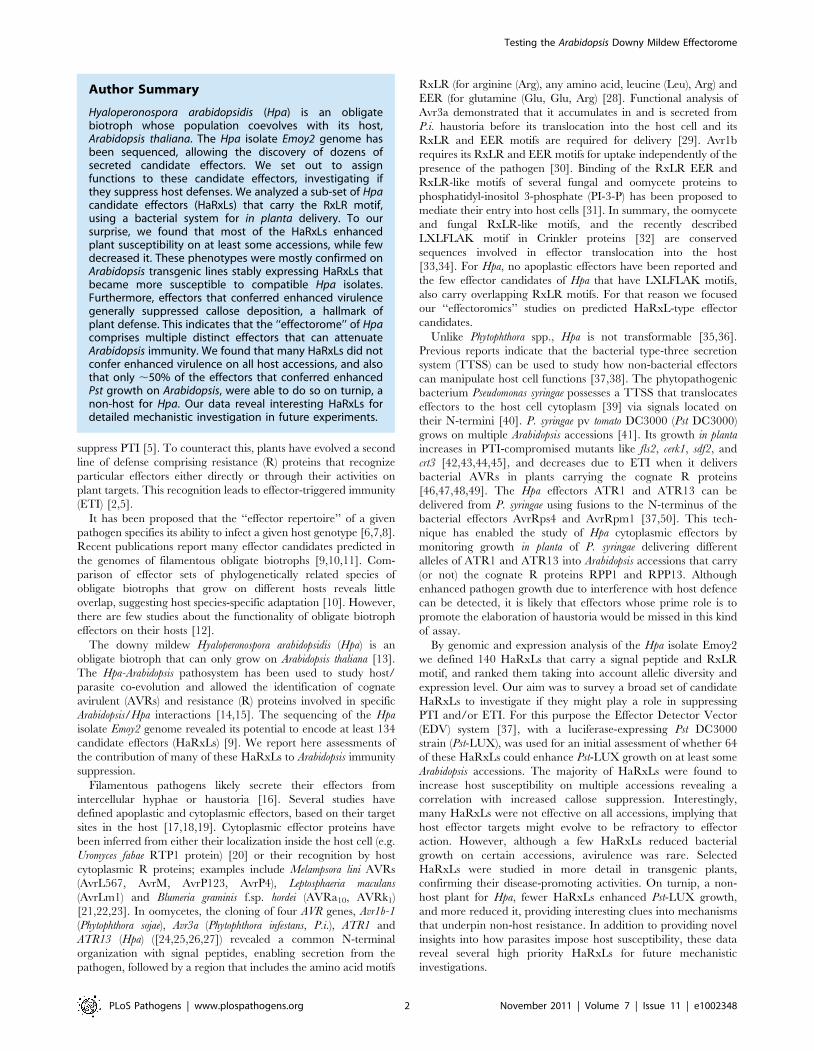
Identification of HaRxLs in the Hpa Emoy2 genomeTo establish an inventory of the RxLR effector secretome of
Hpa, we scanned the draft genome of Emoy2 (http://vmd.vbi.vt.
edu) for all possible open reading frames (ORFs) encoding putative
proteins longer than 100 amino acids. We then searched these
sequences for the presence of signal peptides and from those we
extracted gene models carrying RxLR-like motifs (RxLR/Q; RxL)
with the sequence and positional constrains defined in Figure 1
(see Materials and Methods). Different sets of HaRxLs were
identified depending on the version of the genome used (versions
3.0, 6.0 and 8.3.2). We merged different lists to define a set of 191
HaRxL genes that included the known effector genes ATR1 [26]
and ATR13 [27]. Most of the encoded proteins were smaller than
300 amino acids. Approximately 37% had an acidic motif (EE/
EER) after the RxLR and ,6% had a predicted nuclear
localization signal (data not shown). This collection included
HaRxLs identified by others using similar search algorithms
[9,28].
We next tested which HaRxLs were expressed during the
oomycete life cycle and whether these were correctly predicted. A
set of Sanger ESTs from germinating Hpa spores ([9] and two
different cDNA libraries from infected Arabidopsis plants at 3 and 7
days post inoculation (dpi) were used (see Materials and Methods).
We verified expression of 140 of the 191 predicted HaRxLs.
Ninety of them were expressed from the asexual spore stage,
perhaps ensuring their early availability upon initiation of
infection, and remained expressed in planta until 7 dpi. The
remaining 51 HaRxLs were expressed either at 3 dpi, 7 dpi or
both. Data in column M of Table S1 illustrate the expression
pattern of the sub-sets of effectors tested in this work. Of those for
which we could confirm expression, the majority (90%) of the
HaRxLs was correctly predicted and none had introns (data not
shown). Highly expressed and accurately predicted HaRxLs were
prioritized for cloning.
We then looked for evidence of polymorphism in effector
candidates between seven Hpa isolates (Cala2, Emco5, Emoy2,
Hind2, Maks9, Noco2, Waco9). Single Nucleotide Polymorphisms
(SNPs) were detected either on PCR products (Baxter et al.,
unpublished data) or partial assemblies of Illumina short reads (N.
Ishaque, unpublished data). Our results indicated that 12% of the
HaRxLs were not polymorphic, 56% had between 1 and 10 SNPs,
and 31% showed more than 10 and up to 38 SNPs. We classified
them as not polymorphic (0 SNPs), low ($1 SNPs #5), medium
($6 SNPS #15) and high polymorphic (.16 SNPs) candidates
(Column L, Table S1). For some HaRxLs it was difficult to
distinguish heterozygosity from paralogous family members. In
consequence, the real level of polymorphism might be underes-
timated. Recognized Hpa effectors like ATR1 and ATR13 show
high levels of polymorphism [26,27] while we hypothesize that
non-recognized virulent effectors, adapted to interact with a
specific host target, might have low sequence variability. Hence,
candidates belonging to all four above described categories were
used in this study.
Construction and verification of an HaRxL library forfunctional screening using the EDV system
The Effector Detector Vector (EDV) delivers individual effector
candidates to host plant cells using the TTSS of Pseudomonas syringae
[37]. Seventy-four HaRxLs were cloned into pENTR/pEDV
vectors (pEDV-HaRxLs) (Table S1). We obtained 71 fusion
proteins (AvrRPS4N1–136–HA tag-HaRxL). Two candidate effec-
tors could not be cloned correctly in pEDV, and another one was
truncated and further used as a negative control (NC2, Table S1).
Correct in-frame constructs were introduced by conjugation into
Pst DC3000 and derivative strains, particularly one expressing the
luciferase (luxCDABE) operon of Photorhabdus luminescens (Pst-LUX)
[41] (see Materials and Methods for full details). No differences in
bacterial growth (either in liquid or solid media) were observed in
Pst-LUX clones carrying any of the 71 AvrRPS4N1–136–HA tag-
HaRxL fusion proteins regarding the growth of Pst-LUX
harbouring AvrRPS4N1–136–HA tag-GFP/AvrRPS4AAAA (data
not shown).
We performed in vitro secretion assays to check that the 71 fusion
proteins obtained were made in bacteria and secreted to the
medium in TTSS-inducing conditions. Secreted protein could be
detected as illustrated in Figure S1A. Proteins of the expected size
were produced by Pseudomonas for 64 of the pEDV5/6-HaRxLs
cloned. No proteins, or protein bands of incorrect size, were
observed for the remaining 7 HaRxLs, which were not used in
further assays (Table S1, column H). Thus, our library comprised
64 Emoy2 pEDV-HaRxLs.
Figure 1. Bioinformatic pipeline used for the identification ofHyaloperonospora arabidopsidis (Hpa) HaRxLs. (*) The genomebrowser is maintained at the Sainsbury Laboratory (gbrowse2.tsl.ac.uk/cgi-bin/gb2/gbrowse/hpa_emoy2_publication).doi:10.1371/journal.ppat.1002348.g001
Testing the Arabidopsis Downy Mildew Effectorome
PLoS Pathogens | www.plospathogens.org 3 November 2011 | Volume 7 | Issue 11 | e1002348

In Pst DC3000, the TTSS, encoded by the hrp-hrc (hypersensitive
response [HR] and pathogenicity-hr conserved) gene cluster, is
required for elicitation of HR in non-host plants like tobacco and
for full pathogenicity in host plants like tomato [51,52]. To verify
that the HaRxLs proteins did not alter Pseudomonas growth in planta
by blocking the TTSS, we performed HR cell death tests in
tobacco (Nicotiana tabacum cv. Petit Havana) and disease assays in
tomato (Solanum lycopersicum cv. Moneymaker). Of the 64 pEDV-
HaRxLs delivered by Pst-LUX in tobacco, only 1 attenuated HR
in tobacco while four reduced disease symptoms in tomato. No
candidate impaired both activities or completely abolished HR or
disease (Table S2 columns C, D and representative examples in
Figure S1 B, C). We infer from these results that none of the
pEDV-HaRxLs constructs blocked Pst-LUX TTSS translocation
of effectors.
The Pst-LUX strain was designed for screening multiple
Arabidopsis mutants/accessions that vary in resistance to
PstDC3000 [41]. To evaluate the sensitivity of this system, we
carefully validated the correlation between the level of bacterial
bioluminescence and bacterial growth in planta using ATR1 and
ATR13 (Figure S2). ATR1Emoy2/Cala2 and ATR13Emoy2/Emco5
conferred enhanced growth to Pst-LUX in the susceptible
genotype Col-0, as did ATR13Emoy2 on Nd-0 plants. This
phenotype could be detected as an increase in bioluminescence
that correlated with higher numbers of bacteria (colony forming
units (cfu)/cm2) (Figure S2). We were also able to detect decreased
growth conferred to Pst-LUX by ATR13Emco5 or ATR1Emoy2 in
Nd-0 plants (Figure S2).
Several HaRxLs delivered via Pst-LUX increase bacterialgrowth on multiple Arabidopsis accessions
Using spray inoculation, a protocol was developed to assay
bacterial growth in a sub-set of the 96 accessions described by
Nordborg et al., [53], selected to maximize variability (Bay-0, Br-
0, Col-0, Ksk-1, Ler-0, Nd-0, Oy-0, Shakdara, Ts-1, Tsu-0, Wei-0,
Ws-0) (Figure 2). Plants were spray-inoculated with Pst-LUX
carrying EDV constructs that delivered either a control protein or
an HaRxL via TTSS. At 3 dpi, the bioluminescence (photons/
fresh weight) emitted by the bacteria was quantified as a measure
of bacterial growth (Figure 2, see details in Materials and
Methods). The growth of Pst-LUX in planta carrying each HaRxL
was compared to a control (see below) and expressed as a ratio.
This assay allowed us to establish whether a given HaRxL was
able to enhance or decrease Pst-LUX growth, manifested as
quantitative differences in bioluminescence, on multiple host
accessions in parallel.
Sixty-four pEDV-HaRxLs and 3 control proteins (EDV5:HA-
AvrRPS41–136, EDV6:HA-YFP, EDV6:HA-AvrRps4-AAAA) were
delivered via Pst-LUX to 12 different Arabidopsis accessions. At
three days post spray-inoculation, the photons/second/g fresh
weight (CPS/Fw) were scored for five plants of each accession and
averages, standard deviations and errors calculated. The ratio of
increase or decrease in the CPS/Fw emitted by a Pst-LUX strain
delivering a given pEDV-HaRxL, versus control (in the corre-
sponding EDV5 or EDV6 backbone) was determined, as well as its
statistical significance (one tailed T-test, unequal variances
assumed) (Figure 2, Table S2). Experiments were repeated at
least three times. Given the variability between experiments, the
final outcome of each pEDV-HaRxL effect per accession was
assessed across experiments and categorized according to the
following criteria: i) a reproducible ratio higher or lower than one,
showing the same trend on at least two experiments with a
minimum statistical significance of p,0.05 on each of them, was
considered as either ‘‘Enhanced’’ or ‘‘Decreased’’ growth and
labeled with (+) or (2) respectively; ii) a non-reproducible ratio
showing opposite statistically significant trends or the same trend
but not statistically significant was considered as ‘‘No Change’’
and scored as ( = ) (see Table S2, columns R, S, T). A graphical
synopsis of the screening outcome per effector across the 12 host
accessions is presented in Figure 3, with the most effective pEDV-
HaRxL (HaRxL62) at the top, conferring enhanced Pst growth on
all 12 accessions.
To distinguish the effector-driven Enhanced/Decreased Pst-LUX
growth patterns from the random phenotypes that can be obtained
by delivering any given protein into the plant via the EDV system,
we included four internal controls (negative controls, NCs). These
constructs were truncated versions of HaRxLs (NC2 and NC3),
non-secreted proteins with an RxLR-like motif (NC1) or a small
bacterial protein with similarity to a xylosidase (NC4, genebank:
AP12030.1). NC1 is part of a larger Hpa ORF encoding a putative
transposase. NC2 is an early C-terminal truncated version of
HaRxL143 (before the RxLR motif), while NC3 is a frame-shift
version of HaRxL77 (Table S1). Functional ATR13Emoy2 and
ATR13Emco5 alleles were also included. The pattern shown by these
internal controls allowed us to establish threshold levels to assess
whether a given HaRxL had a credible effect on Pst-LUX growth
(Figure 3, Table S2). NC3 and NC4 did not impact Pst-LUX
bioluminescence. We attributed the residual effect of NC1 and NC2
on Pst-LUX growth to the random variability of the system (Figure 3)
and therefore we set the thresholds as follows: for an effector to be
considered as an ‘‘enhancer’’ of Pst-LUX growth it had to show an
increased significant change in bioluminescence in four or more
accessions. Conversely, as the control ATR13Emco5 was recognized
in only 1 accession out of the 12 tested, any effector that decreased
the growth of Pst-LUX on one or more accessions was classified as
capable of being recognized (Figure 3).
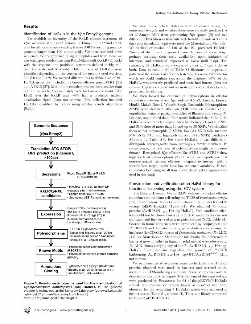
Our results indicate that 43 pEDV-HaRxLs enhanced Pst-
LUX growth in planta, while 28 decreased Pst-LUX activity
(Figure 3). The majority (72%) of the pEDV-HaRxLs that
increased growth in $4 accessions did not decrease it in any
accession (Green/black bars, Figure 3). This suggests most
pEDV-HaRxLs can suppress plant defenses and avoid recogni-
tion by the host, although their effectiveness varies between
accessions. We found only one pEDV-HaRxL capable of
enhancing Pst-LUX growth in all accessions tested (HaRxL62);
we infer that its plant target(s) might have little natural variation
between accessions and that no R gene(s) recognize it in these
accessions. In addition, pEDV-HaRxL9, 17, 21, 45b, 53, 73 and
HaRxLL464 were able to increase Pst-LUX growth in 8 or more
accessions and had no negative effect on the remaining ones. The
pEDV-HaRxLs that decreased Pst-LUX growth did so mainly in
#3 accessions (68%). This pattern of accession-specific Pst-LUX
growth reduction was observed for both alleles of ATR13, and
also for pEDV-HaRxL4, 44, 45, 57, 106, 108, HaRxLL445, 483
and 495. Only nine pEDV-HaRxLs conferred decreased
bacterial growth in .3 and ,8 accessions. Given the lack of
accession-specificity of their phenotype we speculate these
HaRxLs might affect the virulence activity of Pst effectors
(Magenta areas, Figure 3).
Verification via growth curves of HaRxL-induced changesin Pst-LUX growth highlights a subset of effective HaRxLs
We extended the concordance analysis developed with
ATR1Emoy2/Cala2 and ATR13Emoy2/Emco5 (Figure S2) to 13 other
pEDV-HaRXLs delivered from Pst-LUX by conducting growth
curve assays using seven Arabidopsis accessions (Table S3). For this
test we selected some HaRXLs representative of the different
patterns we observed in Figure 3. Briefly, we tested HaRxL62
Testing the Arabidopsis Downy Mildew Effectorome
PLoS Pathogens | www.plospathogens.org 4 November 2011 | Volume 7 | Issue 11 | e1002348

because it enhanced Pst-LUX luminescence in all accessions;
HaRxL14, HaRxL21, HaRxLL60, HaRxLL464, and HaRxLL492
because they enhanced Pst-LUX luminescence in $6, but did not
decreased it in any accession. HaRxL44, 45, 57 and 106 also
increased bacterial luminescence in $6 accessions but decreased it
in 1–3 accessions. HaRxL70 was selected among the group of ‘‘non-
effective’’ effectors, and HaRxL79 because it reduced Pst-LUX
bioluminescence in .3 accessions.
For 32 of 35 combinations (pEDV-HaRXL6accession) we
confirmed the correlation between enhanced bioluminescence and
increased bacterial growth. These data verified that Pst-LUX
bioluminescence reveals the effect of HaRXLs on Pst-LUX
growth. We also observed that some HaRXLs have a substantial
positive effect on bacterial growth on multiple accessions, and can
increase Pst-LUX growth ,10-fold (Table S3 and Figure S3). In
particular, we confirmed that HaRxL62 and HaRxL14 render
multiple host accessions more susceptible to bacterial infection
(Figure S3). Accession-specific effects were verified for
HaRxLL464 and HaRxL21 while putative recognition events,
leading to a decrease in bacterial growth, were verified for
HaRxL44 in Ler-0, HaRxL57 in Ksk-1 (Figure S3, Table S3), and
HaRxL106 in Col-0 (Table S3). No effect was observed for
HaRxL70 in Col-0 while the decrease in bacterial growth caused
by HaRxL79 was only observed when plants were spray
inoculated (Table S3). These data reinforced the usefulness of
the EDV Pst-LUX assay for selecting candidates for further work,
Figure 2. Functional screening method. Hpa effector candidates (HaRxLs) were delivered on 12 Arabidopsis accessions through the bacterialTTSS of the Pst-LUX strain. Levels of bacterial growth were measured quantifying bioluminescence (photon counts) emitted by the bacteria presenton whole plants. The ratio of the average photon counts per second (CPS) per gram of fresh weight (FW) emitted by the bacteria delivering a givenHaRxL versus the bacteria delivering control proteins was determined per accession. Experiments were repeated at least three times and statisticaltests applied. Results and conclusions are shown in Table S2 and Figure 3.doi:10.1371/journal.ppat.1002348.g002
Testing the Arabidopsis Downy Mildew Effectorome
PLoS Pathogens | www.plospathogens.org 5 November 2011 | Volume 7 | Issue 11 | e1002348
and confirmed several candidates as a high priority for further
investigation.
Host genotypes and levels of HaRxL polymorphism arenot correlated with effector-induced changes in Pst-LUXgrowth
To evaluate if host genotypes influenced the pattern of
Arabidopsis responsiveness to the set of HaRxLs tested, the
spectrum of effective HaRxLs per accession was analyzed. We
found that an average of 42% of the pEDV-HaRxLs enhanced
Pst-LUX growth on any given accession, while only ,11%
reduced Pst-LUX growth. Many combinations (46%) did not
cause any change in Pst-LUX growth (Figure S4). Enhancement or
decrease of susceptibility was not restricted to a particular set of
accessions, and did not correlate with those accessions showing
resistance or susceptibility to the infection by the Hpa isolate
Emoy2 (Figure S4). The only deviations from this pattern were
Nd-0, in which most of the pEDV-HaRXLs (73%) increased Pst
growth and only ATR13Emco5 was able to decrease it, and Br-0 in
which fewer pEDV-HaRXLs in total were effective (31%
compared to the average of 42% for all other accessions) (Figure
S4). These results are consistent with the idea that some effector
targets are widely conserved while others vary between accessions.
The level of polymorphism of HaRxLs did not correlate with
the capacity to enhance Pst-LUX growth. Among the 64
candidates tested, 11 were highly polymorphic, 21 had a medium
level and 32 showed low polymorphism. HaRxLs categorized in
these three groups showed ability to increase bacterial lumines-
cence in an average of 662.54, 663.15 or 562.66 accessions,
respectively. For example, HaRxLL464 and HaRxL57 showing
low or no polymorphism, and the highly polymorphic HaRxL106
and HaRxL21 were all capable of increasing Pst-LUX growth in 8
or more host accessions.
HaRxLs did not trigger hypersensitive recognition in anyArabidopsis accession after EDV delivery
Isolate Emoy2 is recognized by certain Arabidopsis accessions,
indicating effector recognition by R protein(s). In order to identify
avirulent HaRxLs in the library, we analysed in detail Pst-LUX
growth assays in each of the 12 Arabidopsis accessions (Figure 3,
Figure S4). Possible recognition of pEDV-HaRxL strains in our
assays was indicated by the decrease in Pst-LUX growth, usually in
an accession-specific manner (Figure 3, Table S2). Potentially
novel ATR proteins may have been detected in interactions with
accessions Col-0, Ler-0, Br-0 and Ksk-1 (Figure S4).
ETI is strongly correlated with HR-like cell death [54,55]
although HR is not always required for resistance [49,56]. We
tested possible recognitions using a weakly virulent Pst DC3000
DCEL (Pst-DCEL) strain and a modified P fluorescens carrying a
functional TTSS (Pf0-1) [57] to deliver potentially recognized
HaRxLs to the corresponding ‘‘resistant’’ accessions. We per-
formed localized leaf infiltrations using high doses of bacteria and
looked for macroscopic (leaf collapse) and microscopic (dead cells
Figure 3. Hpa HaRxLs can promote or decrease Pst-LUX growthin different Arabidopsis accessions. The graph illustrates theoutcome of the interaction between 12 Arabidopsis accessions (X axis)and Pst-LUX clones delivering 64 different Hpa effector candidates(HaRxLs, Y axis). Bars indicate the number of host accessions where thedelivery of a given Hpa RxLR-like candidate effector by Pst-LUX
conferred either enhanced (green bars), decreased (magenta bars) orno change (black bars) in bacterial growth, measured as biolumines-cence, compared to the controls. The arrow indicates the threshold setup to consider that a given HaRxL truly enhances Pst-LUX biolumines-cence. The asterisks indicate HaRxLs that suppress callose deposition inCol-0 when delivered via Pst-DCEL. High suppression levels are markedwith (+). For details see Table S2, columns R,S and T. NC 1,2,3,4: negativeinternal controls.doi:10.1371/journal.ppat.1002348.g003
Testing the Arabidopsis Downy Mildew Effectorome
PLoS Pathogens | www.plospathogens.org 6 November 2011 | Volume 7 | Issue 11 | e1002348

stained with trypan blue) indicators of HR-like cell death 24 h post
infiltration.
Surprisingly, no HaRxLs besides the positive control
(ATR13Emco5 in Nd-0) provoked clear signs of macroscopic HR.
We then stained infiltrated leaves with trypan blue and examined
for microscopic lesions. All micro-HR lesions were much smaller
and weaker than those triggered by bacterial effectors like
AvrRpm1 or AvrRpt2 (data not shown) or by the Hpa effector
ATR13Emco5 in Nd-0 (Table S4). In 78 pEDV-HaRXL/accession
combinations, we saw micro-HR-like cell death in only 7
interactions, comprising just 4 candidate effectors (HaRxL4, 18,
70 and 80; in bold in Table S4). Similar results were obtained with
both Pf0-1 and Pst-DCEL strains, except for HaRxL106 where no
HR was detected in Col-0 and Ksk-1 when delivered through Pf0-
1 (Table S4). Nevertheless, the decrease in bacterial growth
observed for Pst-LUX delivering each of these candidate effectors
in the corresponding accessions was confirmed by reduction in
disease symptoms and bacterial growth using Pst-DCEL (data not
shown).
None of the mild recognition patterns matched with profiles
expected for ATR4, ATR5 and the putative ATR(s) recognized in
Ksk-1 and Br-0 (Figure S4). Interestingly, two HaRxLs (HaRxL18
and 70) were weakly recognized in Bay-0, an accession susceptible
to isolate Emoy2. These results suggest that the decreases in Pst
growth we see in some HaRxL/accession combinations are not
due to strong R/AVR interactions. Also, weak recognition of some
HaRxLs might not result in HR [49,56].
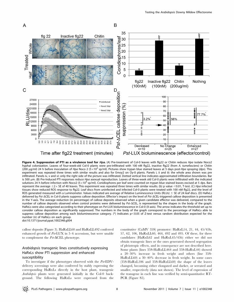
Importance of PTI suppression for Hpa infectionMany HaRxLs delivered in planta by Pst-LUX confer increased
growth of an already virulent pathogen. Enhanced susceptibility to
adapted pathogens is often a result of PTI suppression [42,43,44,
45]. PTI- responses, like callose deposition, likely limit the growth
of Hpa during infection [58,59,60]. Also, ATR13Emoy2/Emco5 can
complement HopM1-mediated suppression of callose deposition
when delivered by Pst-DCEL [37]. Therefore, we investigated if
PTI affects Hpa growth, and whether Hpa is able to actively
suppress PTI. As no known PAMP has been identified for Hpa, we
tested whether Hpa infection alters responses to known PAMPs.
To test if PTI can attenuate Hpa growth during a compatible
interaction, we pre-treated young Col-0 plants with flg22
(100 nM), an inactive flg22 (from Agrobacterium tumefaciens), or
chitin (200 mg/ml) 24 h before the plants were sprayed with
spores of Hpa isolate Noco2. Reduced hyphal growth was
observed in the areas where the PAMPs were applied, as assessed
by trypan blue staining of the pathogen in the leaves (Figure 4 A).
We also noticed a decrease in the rate of Hpa sporulation (Figure 4
B). These phenotypes were not observed with inactive flg22
(Figure 4 A, B), or when the treatment was applied on plants
mutant for these PAMP receptors (fls2-1 and cerk1-1, data not
shown). These data suggest that the phenomenon is specific for
PTI. We also noticed that the ‘‘protection’’ that flg22 and chitin
conferred to the plants was higher near the infiltrated site, was
dose-dependent, and diminished with the time of pre-infiltration
relative to Hpa infection (24 hs.48 hs.72 hs) (Figure 4 A lower
panel and data not shown), consistent with the transient nature of
the local PTI response [61]. We did not observe extensive local
micro-HR in flg22-treated leaves [62]. The ‘‘local’’ effect of flg22
and chitin in restricting Hpa hyphal growth (Figure 4 A) might
indicate that either we did not induce systemic acquired
resistance (SAR) [63], or we applied Hpa before SAR was
established.
One of the earliest PTI responses is the generation of reactive
oxygen species (ROS burst) [64]. To determine if Hpa can suppress
ROS, we measured flg22-induced ROS in infected leaf tissues. We
observed a highly reproducible reduction by ,50% in ROS
accumulation induced by flg22 if leaves were pre-infected with Hpa
isolate Noco2 (Figure 4C). Oy-0 plants infected with Emoy2
showed the same pattern (data not shown). Thus, Hpa infection
can dampen PTI responses.
Most HaRxLs that increase bacterial growth in Col-0 cansuppress Pst-DCEL-induced callose deposition
PTI results in callose deposition in the cell wall [65] and
microbial effectors that impair PTI also suppress callose deposition
[66,67,68,69]. Pst-DCEL is unable to suppress callose deposition
due to lack of HopM1 and AvrE [70]. We introduced pEDV-
HaRxL constructs into Pst-DCEL and evaluated if they could
restore callose suppression when infiltrated in Col-0.
Sixty-two pEDV-HaRxLs and 2 control proteins were delivered
through this system. Due to variability between leaf reactions in
the same plant and among experiments, we established a threshold
to define significant reductions in callose deposition (see Materials
and Methods). Taking into account the maximum levels of
random callose suppression observed after delivery of the negative
controls (NC1 and NC2), we set up the threshold to 40% of callose
suppression because negative controls could reduce up to 29% or
34% of the callose dots found when Pst-DCEL delivered the
additional controls (EDV5:HA-AvrRPS41–136, EDV6:HA-YFP or
EDV6:HA-AvrRps4AAAA). Using this stringent criterion we found
that 35 HaRxLs were able to suppress callose deposition by more
than 40%, while 27 HaRxLs did not. Those effectors comple-
menting the phenotype of Pst-DCEL are indicated with asterisks (*)
in Figure 3.
We noticed that most of the HaRxLs able to complement Pst-
DCEL were also able to enhance Pst-LUX growth in four or more
host accessions (Figure 3). To establish the degree of correlation
between both phenotypes, the extent of callose suppression was
compared with the changes in Pst-LUX growth produced by each
effector in the accession Col-0 (Figure 4 D). For this, we classified
the effector’s conferred phenotype in the host as follows: enhanced
susceptibility = 27 HaRxLs (right side of the graph), decreased
susceptibilty = 10 HaRxLs (left side of the graph), no change = 25
HaRxLs (axes intersection). When we plotted the percentage of
callose suppression of each of the effectors in these groups, we
found 77.7% of HaRxLs candidate effectors with a positive effect
on Pst-LUX growth in Col-0 were able to suppress callose
deposition, while only 3.2% of those decreasing Pst-LUX growth
could reduce callose levels (Figure 4 D). These percentages deviate
significantly from those expected for a random distribution (Z-test
p,0.05). The HaRxLs located at the top right side of Figure 4 D
were those that strongly suppressed callose deposition, and are
indicated with plus (+) signs in Figure 3. The HaRxLs with no
effect on Pst-LUX growth showed no clear trend in ability to
suppress callose deposition.
We conclude that most HaRxLs that enhance Pst growth also
suppress PTI, increasing host-susceptibility. Based on the data
presented in Figure 3, Tables S2 and S3, Figure S3 and Figure 4D,
we prioritized HaRxL14, 21, 44, 45/45b, 57, 62, 106,
HaRxLL60, 464, 492 and 495 for further detailed studies.
HaRxL14, 21, 44, 57 and 62 were chosen because they increased
Pst-LUX growth in more than 6 accessions and strongly
suppressed callose deposition in Col-0 (.60%) (Figure 3 and top
right side on Figure 4D). HaRxLL464 and HaRxL45/45b
strongly enhanced Pst-LUX growth in 9 or more accessions, but
showed a mild reduction in callose deposits (Figure 3 and Table
S2). HaRxL106 conferred enhanced susceptibility to Pst-LUX in
several ecotypes, except Col-0, where it nevertheless reduced
Testing the Arabidopsis Downy Mildew Effectorome
PLoS Pathogens | www.plospathogens.org 7 November 2011 | Volume 7 | Issue 11 | e1002348
callose deposits (Figure 3). HaRxLL60 and HaRxLL492 conferred
enhanced growth of Pst-LUX in 5–6 accessions, but were unable
to complement the Pst-DCEL phenotype.
Arabidopsis transgenic lines constitutively expressingHaRxLs show PTI suppression and enhancedsusceptibility
To investigate if the phenotypes observed with the Pst-EDV-
delivery screenings were also conferred by stably expressing the
corresponding HaRxLs directly in the host plant, transgenic
Arabidopsis plants were generated initially in the Col-0 back-
ground. The following HaRxLs were expressed from the
constitutive (CaMV 35S) promoter: HaRxL14, 21, 44, 45/45b,
57, 62, 106, HaRxLL60, 464, 492 and 495. Of these, for three
candidates (HaRxL62 and HaRxL45/45b) either we did not
obtain transgenic lines or the ones generated showed segregation
of pleiotropic effects, and in consequence are not described here.
Some plants (lines 35S-HaRxLL464 and 35S-HaRxL44) showed
a 20–30% increase in fresh weight and others (line 35S-
HaRxLL60) a 30–40% decrease in fresh weight. In some cases
(35S-HaRxL106 and 35S-HaRxLL60) the shape of the leaves
changed, becoming either elongated and darker, or serrated and
smaller, respectively (data not shown). The level of expression of
the transgene in each line was verified by semi-quantitative RT-
PCR (Figure S5).
Figure 4. Suppression of PTI as a virulence tool for Hpa. (A) Pre-treatment of Col-0 leaves with flg22 or Chitin reduces Hpa isolate Noco2hyphal colonization. Leaves of four-week-old Col-0 plants were pre-infiltrated with 100 nM flg22, inactive flg22 (from A. tumefasciens) or Chitin(200 mg/ml) 24 h before inoculation of Hpa Noco 2 (56104 sp/ml). Pictures show trypan blue stained leaves at 5 days post-Hpa spraying (dps). Thisexperiment was repeated three times with similar results and also for Emoy2 on Oy-0 plants. Panels i, ii and iii: the whole area shown was preinfiltrated. Panels iv, v and vi: only the right side of the picture was infiltrated. Dotted vertical line indicates approximated infiltration boundaries. Baris 500 mm. (B) Pre-Induced PTI responses reduce Hpa asexual reproduction. Leaves of three-week old Col-0 plants were infiltrated with the indicatedsolutions 24 h before infection with Noco2 (56104 sp/ml). Conidiophores per leaf were counted on trypan blue stained leaves excised at 5 dps. Barsrepresent the average 626SE of 40 leaves. This experiment was repeated three times with similar results. (b) p value ,0.01, T-test. (C) Hpa infectedtissues show reduced ROS response to flg22. Leaf discs from uninfected and infected Col-0 plants were treated with 100 nM flg22, and the level ofROS generated measured with a Luminometer. Values indicated are average of Relative Luminescence Units (RLUs) 6 SE of 24 leaf discs. (D) HaRxLsdelivered by Pst-DCEL in Col-0 plants suppress callose deposition. Effector’s impact on the level of Pst-DCEL-triggered callose deposition is presentedin the Y-axis. The average reduction (in percentage) of callose deposits observed when a given candidate effector was delivered, compared to thenumber of callose deposits observed when control proteins were delivered by Pst-DCEL, is represented by the shapes in the body of the graph.HaRxLs were also categorized according to their phenotype on Pst-LUX bioluminescence in Col-0 (X-axis). The arrow indicates the threshold set up toconsider callose deposition as significantly suppressed. The numbers in the body of the graph correspond to the percentage of HaRxLs able tosuppress callose deposition among each bioluminescence category. (*) Indicates p,0.05 of Z-test versus random distribution expected for thenumber (n) of HaRxLs on each group.doi:10.1371/journal.ppat.1002348.g004
Testing the Arabidopsis Downy Mildew Effectorome
PLoS Pathogens | www.plospathogens.org 8 November 2011 | Volume 7 | Issue 11 | e1002348

Using three independent lines for the remaining nine candidate
effectors, that did not showed perturbed growth, we assessed
whether in planta over-expression altered pathogen development,
PTI or ETI. We characterized the responses of these transgenic
lines to both bacterial (Pst-LUX, Pst DavrPto/DavrPtoB) and
oomycete pathogens (Hpa isolates Noco2 and Emoy2) (Figure 5
and Figure S6). Transgenic lines expressing different HaRXLs
showed increased susceptibility to Pst-LUX when spray-inoculated
(8 lines, Figure S6), or to Pst DavrPto/DavrPtoB when infiltrated (7
lines, Figure 5 A). Also, seven lines showed enhanced susceptibility
to Hpa isolate Noco2 (Figure 5 B). These phenotypes were
observed in at least two out of the three transgenic lines recovered
for each effector.
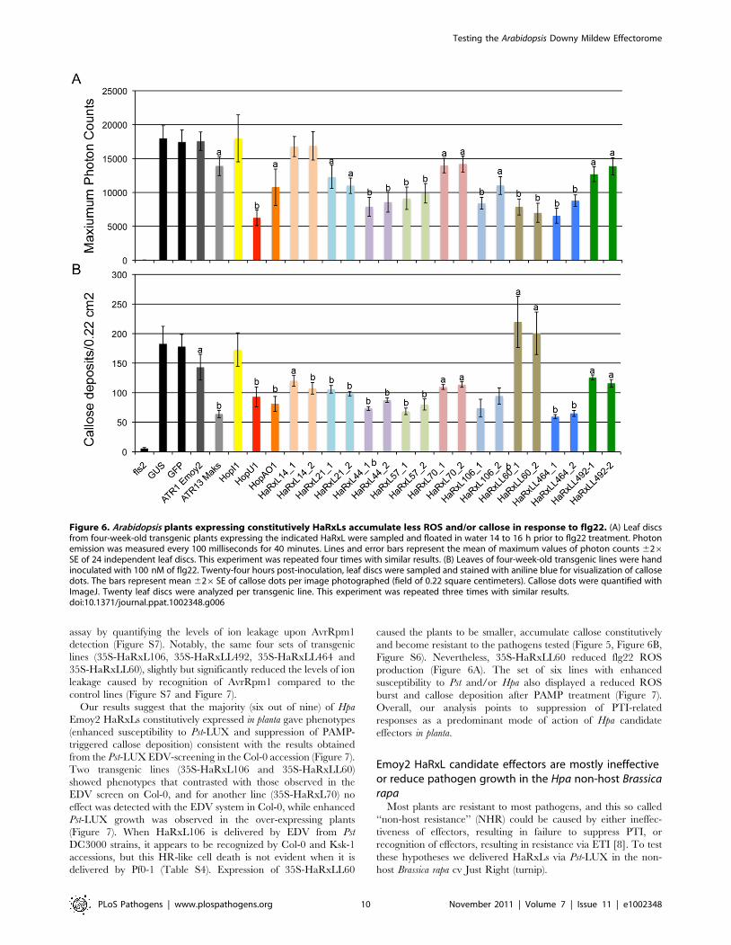
We investigated if the transgenic lines were compromised in
ROS burst and callose deposition in response to flg22 (Figure 6).
Eight 35S-HaRxLs were able to reduce flg22-triggered ROS
accumulation by 22 to 65% compared to controls (Figure 6 A).
Also, callose deposition was diminished by an average of 40%
compared to controls (Figure 6 B). The ROS and callose
suppression in transgenic lines expressing HaRxL 14, 21, 44, 57,
106, HaRXLL 464 was comparable to that observed in plants that
express the bacterial effectors HopU1 and HopAO1 (Figure 6 A,
B). In summary, six different Hpa HaRxLs, when stably-expressed
in planta, displayed a positive correlation between increased
susceptibility to Pst and/or Hpa and reduced levels of ROS and
callose deposition elicited by flg22 (Figure 7).
To establish if any of the nine HaRxLs could also compromise
ETI, we tested transgenic lines for altered resistance to Hpa Emoy2
which is recognized in Col-0 [71]. Two-week-old seedlings were
sprayed with Emoy2 conidiospores and trypan blue-stained at
5 dpi. While some restricted hyphal growth was detected, we did
not observe asexual or sexual reproduction -in true leaves- in any
line (summarized in Figure 7 and data not shown). We then
studied the ETI response to AvrRpm1 from P. syringae pv.
maculicola [72]. AvrRpm1 was delivered via Pf0-1 in leaves of 4-
week-old plants and a macroscopic HR recorded. The onset of
HR was delayed but not completely suppressed in four different
lines (data not shown). We therefore performed a more sensitive
Figure 5. Arabidopsis Col-0 plants expressing constitutively HaRxLs support enhanced growth of P. syringae DavrPto/DavrPtoB andHpa isolate Noco2. (A) Four leaves of three five-week-old plants of two independent transgenic lines per HaRxL were infiltrated with Pst-DavrPto/DavrPtoB at OD600 = 0.0005. Bacterial growth was determined at 3 dpi by traditional growth curve assays. Bacterial populations immediately afterinoculation (3 h; 0 dpi) were averaged among plants and are represented by the solid black horizontal line, with 26SE represented by the dashedhorizontal lines. (a) T-test p value,0.05, (b) T-test p value,0.01.This experiment was repeated two times with similar results. (B) Two-week-oldseedlings were spray inoculated with a suspension of 16104 conidiospores per ml of Hpa isolate Noco2. At 6 dps, whole seedlings were cut andstained with Trypan blue. The number of conidiophores per leaf was counted in 4 leaves per seedling. Ten seedlings were analyzed per transgenicline per HaRxL. The horizontal black and dashed lines represent the average 626SE of the number of conidiophores per leaf found in the hyper-susceptible mutant Col-0 eds1-2. (a) T-test p value,0.01, (b) T-test p value,0.05. This experiment was repeated three times with similar results.doi:10.1371/journal.ppat.1002348.g005
Testing the Arabidopsis Downy Mildew Effectorome
PLoS Pathogens | www.plospathogens.org 9 November 2011 | Volume 7 | Issue 11 | e1002348
assay by quantifying the levels of ion leakage upon AvrRpm1
detection (Figure S7). Notably, the same four sets of transgenic
lines (35S-HaRxL106, 35S-HaRxLL492, 35S-HaRxLL464 and
35S-HaRxLL60), slightly but significantly reduced the levels of ion
leakage caused by recognition of AvrRpm1 compared to the
control lines (Figure S7 and Figure 7).
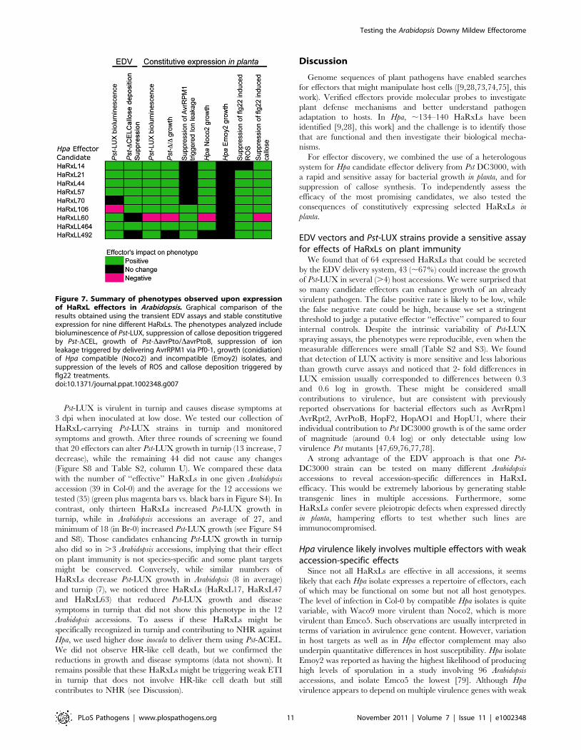
Our results suggest that the majority (six out of nine) of Hpa
Emoy2 HaRxLs constitutively expressed in planta gave phenotypes
(enhanced susceptibility to Pst-LUX and suppression of PAMP-
triggered callose deposition) consistent with the results obtained
from the Pst-LUX EDV-screening in the Col-0 accession (Figure 7).
Two transgenic lines (35S-HaRxL106 and 35S-HaRxLL60)
showed phenotypes that contrasted with those observed in the
EDV screen on Col-0, and for another line (35S-HaRxL70) no
effect was detected with the EDV system in Col-0, while enhanced
Pst-LUX growth was observed in the over-expressing plants
(Figure 7). When HaRxL106 is delivered by EDV from Pst
DC3000 strains, it appears to be recognized by Col-0 and Ksk-1
accessions, but this HR-like cell death is not evident when it is
delivered by Pf0-1 (Table S4). Expression of 35S-HaRxLL60
caused the plants to be smaller, accumulate callose constitutively
and become resistant to the pathogens tested (Figure 5, Figure 6B,
Figure S6). Nevertheless, 35S-HaRxLL60 reduced flg22 ROS
production (Figure 6A). The set of six lines with enhanced
susceptibility to Pst and/or Hpa also displayed a reduced ROS
burst and callose deposition after PAMP treatment (Figure 7).
Overall, our analysis points to suppression of PTI-related
responses as a predominant mode of action of Hpa candidate
effectors in planta.
Emoy2 HaRxL candidate effectors are mostly ineffectiveor reduce pathogen growth in the Hpa non-host Brassicarapa
Most plants are resistant to most pathogens, and this so called
‘‘non-host resistance’’ (NHR) could be caused by either ineffec-
tiveness of effectors, resulting in failure to suppress PTI, or
recognition of effectors, resulting in resistance via ETI [8]. To test
these hypotheses we delivered HaRxLs via Pst-LUX in the non-
host Brassica rapa cv Just Right (turnip).
Figure 6. Arabidopsis plants expressing constitutively HaRxLs accumulate less ROS and/or callose in response to flg22. (A) Leaf discsfrom four-week-old transgenic plants expressing the indicated HaRxL were sampled and floated in water 14 to 16 h prior to flg22 treatment. Photonemission was measured every 100 milliseconds for 40 minutes. Lines and error bars represent the mean of maximum values of photon counts 626SE of 24 independent leaf discs. This experiment was repeated four times with similar results. (B) Leaves of four-week-old transgenic lines were handinoculated with 100 nM of flg22. Twenty-four hours post-inoculation, leaf discs were sampled and stained with aniline blue for visualization of callosedots. The bars represent mean 626SE of callose dots per image photographed (field of 0.22 square centimeters). Callose dots were quantified withImageJ. Twenty leaf discs were analyzed per transgenic line. This experiment was repeated three times with similar results.doi:10.1371/journal.ppat.1002348.g006
Testing the Arabidopsis Downy Mildew Effectorome
PLoS Pathogens | www.plospathogens.org 10 November 2011 | Volume 7 | Issue 11 | e1002348
Pst-LUX is virulent in turnip and causes disease symptoms at
3 dpi when inoculated at low dose. We tested our collection of
HaRxL-carrying Pst-LUX strains in turnip and monitored
symptoms and growth. After three rounds of screening we found
that 20 effectors can alter Pst-LUX growth in turnip (13 increase, 7
decrease), while the remaining 44 did not cause any changes
(Figure S8 and Table S2, column U). We compared these data
with the number of ‘‘effective’’ HaRxLs in one given Arabidopsis
accession (39 in Col-0) and the average for the 12 accessions we
tested (35) (green plus magenta bars vs. black bars in Figure S4). In
contrast, only thirteen HaRxLs increased Pst-LUX growth in
turnip, while in Arabidopsis accessions an average of 27, and
minimum of 18 (in Br-0) increased Pst-LUX growth (see Figure S4
and S8). Those candidates enhancing Pst-LUX growth in turnip
also did so in .3 Arabidopsis accessions, implying that their effect
on plant immunity is not species-specific and some plant targets
might be conserved. Conversely, while similar numbers of
HaRxLs decrease Pst-LUX growth in Arabidopsis (8 in average)
and turnip (7), we noticed three HaRxLs (HaRxL17, HaRxL47
and HaRxL63) that reduced Pst-LUX growth and disease
symptoms in turnip that did not show this phenotype in the 12
Arabidopsis accessions. To assess if these HaRxLs might be
specifically recognized in turnip and contributing to NHR against
Hpa, we used higher dose inocula to deliver them using Pst-DCEL.
We did not observe HR-like cell death, but we confirmed the
reductions in growth and disease symptoms (data not shown). It
remains possible that these HaRxLs might be triggering weak ETI
in turnip that does not involve HR-like cell death but still
contributes to NHR (see Discussion).
Discussion
Genome sequences of plant pathogens have enabled searches
for effectors that might manipulate host cells ([9,28,73,74,75], this
work). Verified effectors provide molecular probes to investigate
plant defense mechanisms and better understand pathogen
adaptation to hosts. In Hpa, ,134–140 HaRxLs have been
identified [9,28], this work] and the challenge is to identify those
that are functional and then investigate their biological mecha-
nisms.
For effector discovery, we combined the use of a heterologous
system for Hpa candidate effector delivery from Pst DC3000, with
a rapid and sensitive assay for bacterial growth in planta, and for
suppression of callose synthesis. To independently assess the
efficacy of the most promising candidates, we also tested the
consequences of constitutively expressing selected HaRxLs in
planta.
EDV vectors and Pst-LUX strains provide a sensitive assayfor effects of HaRxLs on plant immunity
We found that of 64 expressed HaRxLs that could be secreted
by the EDV delivery system, 43 (,67%) could increase the growth
of Pst-LUX in several (.4) host accessions. We were surprised that
so many candidate effectors can enhance growth of an already
virulent pathogen. The false positive rate is likely to be low, while
the false negative rate could be high, because we set a stringent
threshold to judge a putative effector ‘‘effective’’ compared to four
internal controls. Despite the intrinsic variability of Pst-LUX
spraying assays, the phenotypes were reproducible, even when the
measurable differences were small (Table S2 and S3). We found
that detection of LUX activity is more sensitive and less laborious
than growth curve assays and noticed that 2- fold differences in
LUX emission usually corresponded to differences between 0.3
and 0.6 log in growth. These might be considered small
contributions to virulence, but are consistent with previously
reported observations for bacterial effectors such as AvrRpm1
AvrRpt2, AvrPtoB, HopF2, HopAO1 and HopU1, where their
individual contribution to Pst DC3000 growth is of the same order
of magnitude (around 0.4 log) or only detectable using low
virulence Pst mutants [47,69,76,77,78].
A strong advantage of the EDV approach is that one Pst-
DC3000 strain can be tested on many different Arabidopsis
accessions to reveal accession-specific differences in HaRxL
efficacy. This would be extremely laborious by generating stable
transgenic lines in multiple accessions. Furthermore, some
HaRxLs confer severe pleiotropic defects when expressed directly
in planta, hampering efforts to test whether such lines are
immunocompromised.
Hpa virulence likely involves multiple effectors with weakaccession-specific effects
Since not all HaRxLs are effective in all accessions, it seems
likely that each Hpa isolate expresses a repertoire of effectors, each
of which may be functional on some but not all host genotypes.
The level of infection in Col-0 by compatible Hpa isolates is quite
variable, with Waco9 more virulent than Noco2, which is more
virulent than Emco5. Such observations are usually interpreted in
terms of variation in avirulence gene content. However, variation
in host targets as well as in Hpa effector complement may also
underpin quantitative differences in host susceptibility. Hpa isolate
Emoy2 was reported as having the highest likelihood of producing
high levels of sporulation in a study involving 96 Arabidopsis
accessions, and isolate Emco5 the lowest [79]. Although Hpa
virulence appears to depend on multiple virulence genes with weak
Figure 7. Summary of phenotypes observed upon expressionof HaRxL effectors in Arabidopsis. Graphical comparison of theresults obtained using the transient EDV assays and stable constitutiveexpression for nine different HaRxLs. The phenotypes analyzed includebioluminescence of Pst-LUX, suppression of callose deposition triggeredby Pst-DCEL, growth of Pst-DavrPto/DavrPtoB, suppression of ionleakage triggered by delivering AvrRPM1 via Pf0-1, growth (conidiation)of Hpa compatible (Noco2) and incompatible (Emoy2) isolates, andsuppression of the levels of ROS and callose deposition triggered byflg22 treatments.doi:10.1371/journal.ppat.1002348.g007
Testing the Arabidopsis Downy Mildew Effectorome
PLoS Pathogens | www.plospathogens.org 11 November 2011 | Volume 7 | Issue 11 | e1002348

effects, rather than a few genes with strong effects, some effectors,
such as HaRxL62, 14, 44, 57 and 106, are particularly effective
and will repay detailed mechanistic investigation in the future.
HaRxLs that enhance Pst growth usually suppress callosedeposition
Significantly, we found that most of the HaRxLs (77%) that
increase Pst-LUX growth in Col-0 were also able to suppress Pst-
DCEL-induced callose deposition. Conversely, those HaRxLs
reducing Pst-LUX growth in Col-0 were generally unable to
suppress callose deposition. Callose deposition is a late PTI
response (though also associated with ETI). We speculate that
HaRxLs may enhance Pst-LUX growth via additional PTI
suppression, either alone or in conjunction with Pst effectors.
The increased susceptibility to Pst-LUX observed using the EDV
system was usually consistent with phenotypes of plant lines that
constitutively express the corresponding HaRxLs (Figure 7).
Moreover, seven of the transgenic lines also showed increased
susceptibility to Hpa isolate Noco2. We infer that enhanced
susceptibility results from suppression of host mechanisms that are
active against diverse pathogens. The fact that they further elevate
virulence conditioned by Pst-DC3000 effectors may reflect
HaRxLs interference with targets that are not identical to those
of Pst-DC3000, resulting in an additive effect.
Infection with Hpa, and many Hpa effectors, suppress PTIAssays using flg22-induced ROS or callose deposition on stable
transgenic lines indicate that the main target of HaRxLs is PTI. All
of the transgenic lines tested showed either reduced levels of flg22-
induced ROS or callose deposition or both. The PAMP
complement in Hpa is unknown, as are their receptors and
downstream signal transduction pathways in Arabidopsis. Several
molecules have been reported as oomycete PAMPs [17,80] but their
existence in Hpa is not known [9,81]. We show that pre-elicitation of
PTI by bacterial and fungal PAMPs impairs Hpa growth and
reproduction, indicating that to infect, Hpa must counteract these
host responses. Moreover, in host tissues where high numbers of
haustoria are established, PTI responses are attenuated.
PTI involves multiple processes that can be attenuated by
diverse pathogen effectors [76,77,78,82]. Our data support the
idea that the function of the majority of the effector proteins is to
inhibit plant immunity [4,83,84]. For Pst, 13 out of 28 active
effectors (12 belonging to Pst-DC3000) have been reported to
suppress PTI [84,85,86]. Thus, ,50% of this bacterial pathogen’s
effector repertoire targets PTI in one host. Importantly, this
hemibiotroph can infect Solanaceae as well as Brassicaceae, so
more effectors might emerge as PTI suppressors when other host
species are studied. It has also been observed that 91% of Pst-
DC3000 effectors, when delivered at high titers from Pf0-1, are
able to suppress the HR induced by the bacterial effector HopA1
(from P. syringae syringae) in tobacco [38]. Since tobacco is a non-
host for Pst-DC3000, this study again points to a high functional
redundancy between effectors in suppressing HR. In oomycetes,
experimental characterization of several RxLR effector genes
suggests that many function to suppress host defenses [37,50,86].
Also, 3 out of 32 P. infestans RXLR candidate effectors were able to
suppress PAMP- triggered programmed cell death (PCD) in N.
benthamiana, while another 13 induced either non-specific or R-
mediated PCD [87].
HaRxLs are rarely avirulence determinantsWe also identified HaRxLs that reduced Pst-LUX growth in the
interaction with Arabidopsis accessions, and investigated whether
they are new avirulence determinants (ATRs). Surprisingly, we
observed that strong incompatibility caused by HaRxLs is rare.
None was able to trigger macroscopic ETI when delivered in planta
at high titer, as ATR13Emco5 did in Nd-0. Instead, several were
identified that can reduce Pst-LUX growth in specific Arabidopsis
accessions and four triggered micro HR-like lesions when
delivered via Pst-DCEL. Since we tested only 64 HaRxLs from
just one isolate, on only 12 host accessions, our survey was not
exhaustive, and the anticipated ATR4 might not have been in our
repertoire. Alternatively, the EDV assay may not be sensitive
enough to detect new Hpa ATRs because these ATR-RPP
recognitions are weaker than those already described with this
system (ATR13-RPP13 or ATR1-RPP1). Conceivably, some
ATRs might not carry an RxLR motif and therefore were not
identified as candidate effectors in our bioinformatic analysis, as
with the recently cloned ATR5 [88]. It also remains possible that
either the sub-cellular localization or post-translational modifica-
tions of the EDV-delivered HaRxLs are not similar enough to
their native form to be able to elicit ETI, although this has not
been the case for ATR1 and ATR13 alleles [37,50].
Non-host resistance could involve a combination ofrecognized HaRxLs and ineffective HaRxLs
We also tested if the recognition or non-functionality of HaRxLs
could be involved in non-host resistance to Hpa in Brassica rapa
(turnip). We found that HaRxLs were ‘‘less effective’’ in turnip, but
those HaRxLs that enhanced Pst-LUX growth in B. rapa also did
so in Arabidopsis, suggesting conservation in their targets. Notably,
three HaRxLs conferred reduced Pst-LUX and Pst-DCEL growth
in B. rapa, but did not reduce growth in any Arabidopsis accession.
Therefore, the inability of Hpa to grow in turnip might result not
only from reduced ‘‘effectiveness’’ of the effector complement, but
also from recognition in the ‘‘non-host’’ of a subset of effectors that
are not recognized by most Arabidopsis accessions.
Concluding observationsAs with any screening protocol, this heterologous system has
some limitations. For example, HaRxLs that require extensive
post-translational modifications will not be correctly produced by a
prokaryotic system. Also, the co-delivery of an HaRxLs with c.a 30
effectors from Pst DC3000 might alter the outcome of the assay if
positive or negative interactions exist between them. This might
explain some of the discrepancies we observed between results
obtained with the EDV system and those generated by expressing
the candidate effectors directly in the plant. A further potential
limitation of the EDV system is that effectors required to elaborate
a haustorium inside the host cell might not be revealed as
promoting Pst growth by the assays we developed. Despite such
limitations, most of the phenotypes observed with the EDV system
were confirmed in the transgenic lines.
In conclusion, the EDV-based system has enabled the
systematic analysis of the biological relevance of effector candidate
proteins. The Pst-LUX and Pst-DCEL screens allowed the
generation of a ‘‘ranking of effectors’’ that permitted the selection
of highly interesting candidates as targets for subsequent
mechanistic studies. Further detailed investigations of Hpa effectors
will help reveal how Hpa alters host cellular processes to promote
its growth and reproduction.
Materials and Methods
Bioinformatic identification of HaRxLsDifferent versions of the genome of Hpa isolate Emoy2 (http://
vmd.vbi.vt.edu; v3.0, v6.0 and v8.3.2) were translated in all 6
Testing the Arabidopsis Downy Mildew Effectorome
PLoS Pathogens | www.plospathogens.org 12 November 2011 | Volume 7 | Issue 11 | e1002348

reading frames. ORFs from ATG to Stop codon were identified
using FgenesH (www.softberry.com) and GETORF (http://
emboss.sourceforge.net). Only sequences that encoding $100
aminoacids were considered. Secreted proteins were identified
using SignalP v3.0 (score cutt-off .0.9), TargetP and PSort (www.
psort.org) [89]. Proteins were considered as secreted if two out of
three programs called the Signal Peptide as significant. HaRxLs
were selected as fulfilling the following criteria: i) Signal Peptide
(SP) length ,30 amino acids, ii) RxLR-like motif (RxLR/Q, RxL)
between 4 and 60 amino acids from the SP cleavage site, iii)
predicted protein had .40 amino acids after the RxLR like motif.
Redundancy in the ORF dataset was corrected using BlastP.
Sequences with 100% identity and E,1025 were clustered and
simplified to the one with highest SP score. A sub-set of RxLR-EE
proteins was identified carrying an acidic motif (EE, EER/G/D)
[28] between 4 and 30 aminoacids from the RxLR like motif. The
expression of HaRxLs was verified using the following resources: i)
ESTs generated by Sanger sequencing and 454 sequencing from
cDNA extracted from Emoy2 conidiospores [6], ii) Illumina
sequence tags (SAGE using 39 tags from 7 dpi infected tissue), iii)
Illumina normalized/concatamerized cDNA (from 3 and 7 dpi
infected tissue) (Ishaque et al., unpublished) and iv) RT-PCR with
primers designed at 100 bp flanking the ORF sequence (Baxter
et al., unpublished). The presence of the predicted and alternative
ATGs and Stops Codons, as well as introns, was verified.
Nucleotidic sequence polymorphisms on the HaRxLs accross
seven Hpa isolates was assessed using either PCR products or in
silico assemblies of Illumina short reads (Ishaque et al., unpub-
lished). The HaRxLs were roughly classified as: No polymorphic
(0 SNPs), Low ($1 SNPs #5), Medium ($6 SNPS #15) and High
Polymorphic (.16 SNPs). To complete the characterization of the
Hpa Emoy2 HaRxLs set, its sub-cellular localization (PSORTII)
and presence of known conserved protein domains using Coil,
Gene3D, HMMPfam, HMMSmart, HMMTigr, PFAM and
Prosite was recorded. These data are available in Table S1 for
the subset of HaRxLs cloned in this work.
Cloning of HaRxLsSelected HaRxLs were amplified from genomic DNA extracted
from conidiospores of isolate Emoy2 using proofreading polymer-
ase (Accuprime Pfx, Invitrogen) and standard PCR conditions. To
generate the HaRxLs collection, the primers were designed to
amplify from the signal peptide cleavage site or the RxLR
(inclusive) until after the stop codon (39 untranslated region, UTR).
For cloning in pEDV3 or pEDV5 [37], the primers were designed
to have SalI/ClaI and BamHI/BglII restriction sites at the 59 and
39 ends respectively. For cloning in pEDV6, a Gateway destination
version of pEDV3, the sequence CACC was added at the 59 of the
Forward primer. PCR products were gel purified (Qiagen) and
ligated (EDV3/5) or recombined in pENTRY-SD-D-TOPO/
pDONR221 following the manufacturer’s instructions and
electroporated in Escherichia coli DH5/. Gentamycin (EDV3/5)
or kanamycin (pENTR/pDONR) resistant colonies were selected
on plates and colony PCR performed with M13F and M13R
primers. Colonies carrying the right size insert were selected for
plasmid purification and sequencing. For EDV6, the correct
inserts on pENTRY/pDONR vectors were recombined using
Gateway LR clonase or LR clonase II enzyme mix (Invitrogen).
The in frame fusion of vector-HA tag-HaRxL sequences were
confirmed by sequencing with M13F and M13R primers. Plasmids
were mobilized from E.coli DH5/ to wild-type or mutant Pst
strains by standard triparental matings using E. coli HB101
(pRK2013) as a helper strain. Bacterial growth in vitro was
controlled at 12, 24, 32 and 44 hs post inoculation of 10 ml Kings
B media with a dilution corresponding to 0.00001 OD of an
overnight culture of each of the Pst-LUX clones harbouring a
different HaRxL or control proteins (GFP, AvrRPS4AAAA). Three
colonies per clone were assayed in different experiments. Growth
was measured assessing turbidity at OD600 (for liquid cultures) or
counting colonies of plated dilutions (in solid media). No
significant differences in growth kinetics were observed for the
Pst-LUX carrying HaRxLs regarding the clones carrying control
proteins or the empty vector pEDV5.
Bacterial strainsBacterial strains used in this study include E. coli DH5/,
Pseudomonas syringae pv tomato DC3000 carrying the luxCDABE
operon from Photorhabdus luminescens (Pst-LUX) [41], Pseudomonas
syringae pv tomato DC3000 mutant DCEL [90], Pseudomonas syringae
pv tomato DC3000 double mutant DavrPto/DavrPtoB [91],
Pseudomonas fluorescens Pf0-1 carrying a functional TTSS [57] and
Agrobacterium tumefasciens GV3101 (pMP90 RK). E. coli, and
Agrobacterium were grown in low salt Luria-Bertani broth at 37uC(E. coli) or 28uC (Agro) using either liquid media or petri dishes.
Pseudomonas strains were grown in either LB or King’s B medium at
28uC in liquid media or petri dishes. Antibiotics concentrations
(mg/ml) were as follows: Rifampicin 100, Kanamycin 50,
Gentamycin 25, Spectinomycin 50, Chloramphenicol 50, Tetra-
cycline 10, Carbenicillin 50.
Plant materials and growthArabidopsis accessions used in this study were obtained from
NASC. The fls2-1 mutant was obtained from Cyril Zipfel and the
cerk1-1 mutant was a kind gift of JP Rathjen. Transgenic lines
constitutively expressing HopU1 and HopAO1 were kindly
provided by Jim Alfano. Turnip seeds (Brassica rapa cv Just Right)
were purchased from Gurney’s seeds (http://gurneys.com).
Tobacco (Nicotiana tabacum cv petit Havana) and tomato (Solanum
lycopersicum cv Moneymaker) seeds were obtained from John Innes
Horticultural services. Arabidopsis plants were grown in Scotts and
Levington F1 modular compost in controlled environment rooms
under short day cycles (10 h/14 h day/night and 150–200 mE/
m2s) at 22uC and 60% relative humidity and slightly watered every
day from below. Tobacco, tomato and turnip plants were grown
under similar conditions as Arabidopsis for 5 weeks post-germina-
tion. Plants expressing constitutively HaRxLs were generated by
recombining the corresponding ORFs cloned in pDONR221 in
the Gateway destination binary vector pB2GW7 [92] under the
control of the CaMV 35S promoter. Constructs were transferred
to A. tumefaciens strain GV3101 (pMP90 RK) [93] and transformed
into Arabidopsis accession Col-0 by the floral dipping method.
Primary transformants (T1) were selected on soil containing
BASTA (Bayer CropScience, Wolfenbuttel, Germany) and self-
pollinated. The progeny of the T2 generation was observed and
3:1 (BASTA-resistant/BASTA-susceptible) segregating lines were
taken further. Homozygous lines were selected by examining the
BASTA resistance of T3 seedlings. Three independent transgenic
lines per HaRxL (T4s) were analysed and for simplicity we present
results for two.
Pathogen growth and inoculationsPrimary streaks of Pst-LUX complemented with the controls or
HaRxLs were made from isolated colonies onto selective King’s B
plates and grown overnight at room temperature. Selected
individual colonies were then spread with a sterile loop in solid
KB plates and incubated overnight at room temperature to
produce even bacterial lawns. Cells were scraped from plates with
a sterile loop and suspended in 50 to 100 ml of 10 mM MgCl2 to a
Testing the Arabidopsis Downy Mildew Effectorome
PLoS Pathogens | www.plospathogens.org 13 November 2011 | Volume 7 | Issue 11 | e1002348

final OD600 of 1. Dilution series were made from these suspensions
to: spray (OD600: 0.2) or infiltrate (OD600 = 0.001) Arabidopsis
plants, or to infiltrate tobacco (OD600 = 0.01), tomato
(OD600 = 0.001) or turnip (OD600 = 0.001) plants. For tobacco
and turnip, leaf panels of the third- to fifth-oldest leaves of were
infiltrated by pricking the leaves with a dissecting needle and
infiltrating with a blunt syringe. pEDV-HaRxLs were compared
with controls on the same leaf. For tomato, leaflets of the third and
fourth most recently expanded leaves were used. Concentrations
of other bacterial pathogens used in this work are stated on the
corresponding figure legends or other sections of M and M. Hpa
isolates Emoy2 and Noco2 were maintained in compatible host
accessions and inoculated onto 2-week-old plants at 1 or 56104
conidiospores/ml. After infection, plants were covered with a
transparent lid to maintain high humidity (90–100%) conditions in
a growth chamber at 16uC for 7 days in short day (10 h/14 h day/
night) cycle. To increase the ratio pathogen/host biomass for gene
expression analysis, plants were sprayed with the conidiospores
solution (or water as control) were kept uncovered in low humidity
(60%).
EDV-Pst LUX screening methodArabidopsis plants of all 12 different accessions were grown in
arrays of five individual cells and shuffled randomly before
inoculation. Five four-week-old plants of each accession were
sprayed with Pst-LUX bacterial suspensions (26108 cfu/ml,
0.03% (v/v) Silwet L-77) carrying pEDVs encoding for control
or HaRxL proteins. Spraying was done using an airbursh system
attached to a compressor (GS, model AS18). About 3 mls of
bacterial suspension was used per five plants at a pressure of 10–12
psi. Sprayed plants were kept under a transparent lid to keep high
humidity conditions. At three days post-spraying, sets of whole five
plants were placed in an ultra low light CCD camera (Photek,
www.photek.com). Photons emitted per second were scored per
plant and referred to the whole plant fresh weight to account for
the foliar area. The average level of Photon Counts per Second
per gram of Fresh Weight was obtained for each Pst-LUX EDV-
delivered protein and the ratio versus the control was scored on
each accession (Figure 2). To verify the correspondence between
increase on photon emission and bacterial population growth, leaf
discs were sampled from the five plants sprayed to generate at least
6 technical replicates. The leaves were surface sterilized (30 s in
70% ethanol, then 30 s sterile distilled water). Traditional growth
curve assays were performed as described [94].
Gene expression analysisTotal RNA was extracted from three-week-old HaRxL
overexpressing lines (T4 generation) using the RNeasy Plant Mini
Kit (Qiagen, Hilden, Germany). 1 mg DNAse-treated total RNA
was reverse transcribed using the QuantiTect Reverse Transcrip-
tion Kit (Qiagen). For semi-quantitative RT-PCR 1 ml of cDNA
was used per reaction and amplified with an initial denaturation
step at 95uC for 3 minutes followed by 23 cycles with the following
conditions: 45 s at 94uC, 30 s at 55uC and 30 s at 72uC. In a last
cycle a final elongation step at 72uC for 5 min was added. PCR
products were separated on 1.5% TAE agarose gels. To control
the equal amount of cDNA in every reaction the Actin2 gene
(At3g18780) was used. The specificity of the primers for
amplification of the HaRxLs transcripts was tested on pDONR221
clones harboring the corresponding HaRxLs. In case of control
RT-PCRs on Arabidopsis Col-0 wild type plants cDNA, no
unspecific amplification using the HaRxLs’ primers was observed.
The sequences of primers used in this study are available on
request.
Measurement of Reactive Oxygen Species (ROS)generation
ROS released by the leaf tissue in response to flg22 was
measured using a chemiluminescent assay [95]. Briefly, leaf discs
(0.38 cm2) from Col-0, fls2-1 mutant, transgenic lines expressing a
given 35S-HaRxL lines, and control lines were sampled. At least
24 leaf discs from four five-week-old plants per accession/line were
sampled and floated for 14–16 h in sterile distilled water in a 96
flat-bootom white multiwell plate (Greiner Bio-One) kept in the
dark. ROS production in response to flg22 was measured by
replacing the water by 100 ul of a working solution of Luminol
(34 mg/ml final concentration, Sigma), Horseradish Peroxidase
(20 mg/ml final concentration, Sigma) and flg22 (100 nM final
concentration, Peptron, South Korea). The plate was immediately
introduced in a Luminometer (Varioskan, Thermo Scientific) and
photon counts were recorded every 40 seconds for at least
40 minutes.
Callose and Trypan Blue Stainings and microscopicanalysis
Leaves of five-week-old plants were hand-infiltrated at the
bottom of the leaf area with 56107 cfu/ml (OD600 = 0.05) Pst-
DCEL suspensions. Bacteria complemented with control or
HaRxL proteins were infiltrated in different plants of the same
set. Twenty leaf samples were taken 12 to 14 h after inoculation at
the top of the infiltrated area to avoid visualization of mechanical
damage induced callose. Leaf discs were cleared 2 times (1 h) in
96% ethanol and then re-hydrated in ethanol/water series (70%,
50%, 30%) per 30 min each. Leaf disc were floated in water 1 h
and then transferred to a solution of 0.01% (w/v) Aniline Blue
(Gurr, BHD, England) in K2HPO4 Buffer (150 mM pH 9.5) for
1 h. The samples were then transferred to Glycerol 60% (v/v) and
mounted for observation with a Leica DM R fluorescence
microscope using UV light and I3 filter (A4-UV). Pictures were
taken with a Leica DFC 300 FX Digital Camera. Images were
analyzed and callose dots quantified using Image J software and an
in house written Macro. The same imaging system was used to
visualize Hpa infection structures after staining with lactophenol
trypan blue. The number of conidiophores per leaf was scored by
manually scanning the abaxial and adaxial surfaces of each leaf on
at least four true leaves of five-ten plants per accession/transgenic
line analyzed.
Ion leakage measurementsFive-week-old plants were infiltrated with 16108 cfu/ml
(OD600 = 0.1) P. fluorescens Pf0-1 carrying pVSP61-AvrRPM1. To
be able to detect HR symptoms the bacteria had to be grown in
plates. Leaf discs were collected right after infiltration using a cork
borer number 4. Twenty-four leaf discs were collected per
transgenic line and shacked in falcon tubes with 45 ml distilled
water for 1 h at room temperature. Four leaf discs were
transferred multi-well plates with 2 ml distilled water. The level
of ion leakage caused by AvrRPM1 recognition by RPM1 in the
Col-0 background was detected as an increment in conductivity.
This was measured with a Conductivity meter (Horiba Twin B-
173) every 60 min over 14 h on at least 6 technical replicates per
transgenic line.
Secretion assay and protein analysisTo verify the accumulation proteins corresponding to the
AvrRPS4-effector fusions in Pseudomonas and its secretion by the
TTSS, the strains complemented with Hpa HaRxLs were grown
overnight at 28uC in 25 ml liquid LB media to a final
Testing the Arabidopsis Downy Mildew Effectorome
PLoS Pathogens | www.plospathogens.org 14 November 2011 | Volume 7 | Issue 11 | e1002348

OD600 = 0.3, centrifuged, washed twice with 10 mM MgCl2 and
re-suspended in 20 ml minimal media (hrp-inducing minimal
medium, MM) [50 mM potassium phosphate buffer, 7.6 mM
(NH4) 2SO4, 1.7 mM MgCl2, 1.7 mM NaCl, pH 6.0, with
20 mM glucose added]. The cultures were incubated at 22uC at
200 rpm on a rotator shaker to a final OD600 of 0.5. The pellet
and supernatant fractions were separated by centrifugation at
5,2006 g for 15 min at 4uC. The pellets were re-suspended in
300 ul of bacterial protein extraction buffer [100 mM NaCl,
25 mM Tris-HCl pH 8, 10% glycerol, 10 mM DTT and 16protease inhibitors cocktail (Complete EDTA-free tablets,
Roche)], sonicated 3 times (10 s), and centrifuged at
12.000 rpm for 5 minutes. Then 100 ml of supernatant were
taken and 25 ml of SDS loading buffer were added. Samples were
boiled for 5 minutes at 96uC before loading. The supernatant of
the 20 ml MM culture was filtered through a 0.2 mm pore filter
and concentrated using centricon YM-10 columns (Amicon
Bioseparation) by sequential centrifugations of 30 min at
5000 g at 4uC. The process was repeated several times. Proteins
in the final 2 ml of concentrated supernatant were precipitated
using Strataclean beads (Stratagene). Five to ten ml of beads were
used per ml of supernatant; incubated 10 min at 4uC inverting
gently, centrifuged at 2000 g for 2 min and re-suspended in 25 ul
Laemmli buffer containing 0.1 M NaOH. Samples were boiled
for 5 minutes at 96uC and centrifuged at 12.000 rpm for
2 minutes before loading. The pellet fraction (15 ml) and the
culture fluid fraction (25 ml) were analyzed by SDS–PAGE,
electro blotted onto PVDF membrane (Bio-Rad), and probed
with horseradish peroxidase- conjugated high affinity anti–HA
antibody (Roche) and re-probed with anti-NPTII antibody
(Upstate). Bands were visualized using PICO kit (Thermo
Scientific) and imaged with Kodak scientific imaging film.
Supporting Information
Figure S1 Verification of the functionality of the TTSSof Pst-LUX clones expressing HaRxLs. (A) Immunoblot
showing the accumulation of AvrRPS4 1–136-HA-HaRxL fusions
on the bacterial pellet (left panel) and its secretion into TTSS-
inducing minimal media (right panel, supernatant). Approximat-
ed molecular weights in KDa are shown on the left of each panel.
(B) Four-week old tobacco (N. tabacum cv. petit havana) leaves were
infiltrated with Pst-LUX clones carrying either control proteins
(YFP, AvrRPS4-AAAA), or HaRxLs at OD600 = 0.01. Pst-hrcC
and Pst-hrcC-LUX strains were included as positive controls for
TTSS impairment. Symptoms of HR cell death were screened at
2,3 and 4 dpi. Picture was taken at 2 dpi. (C) Five-week old
tomato (Solanum lycopersicum cv. moneymaker) leaflets syringe
infiltrated with OD600 = 0.001 of different Pst-LUX clones
expressing HaRxLs or controls (YFP). Leaflets were detached
and imaged at 3 dpi with a Photek camera to detect
bioluminescence.
(TIF)
Figure S2 Correlation between Pst-LUX biolumines-cence and its growth in planta. (A), (C) Five-week-old plants
of the indicated Arabidopsis accessions were spray-inoculated at
OD600 = 0.2 with Pst-LUX delivering the indicated Hpa effector or
control proteins. At 3 dpi, five whole plants per treatment were
imaged using a Photek camera to record photons counts per
second. Bars illustrate the average photon counts per gram of plant
fresh 6 SD. (a) p value of T-test assuming unequal variances
,0.05, (b) p,0.01. (B), (D) Twenty-four leaf discs obtained from
the above mentioned plants were excised and used to determine
the number of bacteria per leaf area, showed in Log10 scale. Bars
indicate the average 6 SD of six technical replicates. One-way
ANOVA test was applied with (a) p,0.05, (b) p,0.01.
(TIF)
Figure S3 Behavior of Pst-LUX delivering HaRxLsassessed via growth curves. Histograms illustrate the changes
in growth levels (measured as colony forming units –CFU-) of Pst-
LUX strains delivering the indicated HaRxLs, compared to
control strains, on different Arabidopsis accessions.
(TIF)
Figure S4 Pattern of HaRxLs induced changes in Pst-LUX virulence plotted per Arabidopsis accession. Bars
indicate the number of HaRxLs that enhanced (green), decreased
(red) or did not changed (black) the growth of Pst-LUX on each
Arabidopsis accession tested. The outcome of the interaction of the
Hpa isolate Emoy2 with each accession is indicated as Susceptible
(S) or Resistant (R). Known and predicted ATR/RPP interactions
are described. (?) indicate putative/unknown ATR/RPP genes.
(TIF)
Figure S5 Semi-quantitative RT-PCR applied to RNAextracted from the stable transgenic lines generated andtested in this paper. Expression levels of each HaRxL were
tested in two independent homozygous transgenic lines (1,2). For
comparison, the expression of the Arabidopsis housekeeping gene
actin (Act2) is shown.
(TIF)
Figure S6 Arabidopsis Col-0 plants expressing consti-tutively HaRxLs support enhanced growth of Pst-LUX.Five four-week-old plants of two independent transgenic lines
expressing the corresponding Hpa candidate effector and one line
per control protein, were sprayed at OD600 = 0.2 with Pst-LUX.
At 3 dpi, photon counts per plant were measured, as well as plant’s
fresh weight. Bars represent means of 5 replicates 626Standard
Errors (SE). (a) p value of T-test assuming unequal variances
,0.05; (b) p,0.01. This experiment was repeated three times with
similar results.
(TIF)
Figure S7 Constitutive expression of four differentHaRxLs caused a mild reduction on the levels of ionleakage triggered by AvrRPM1 recognition. Five-week-old
plants were hand infiltrated with Pf0-1 delivering P. maculicola
AvrRPM1 at OD600 = 0.1. Twenty-four leaf discs were sampled
from four infiltrated transgenic plants per line per HaRxL in each
of five different experiments. At least six technical replicates were
done per line. Conductivity was measured in the water were discs
were floating, as an indication of ion leakage into the media.
Measurements were taken every hour until the peak of ion leakage
was detected in the wild type and control lines (around 14 hours
post-infiltration). Bars correspond to the average 626 SE of the
maximum level of ion leakage observed for each line expressed as
a percentage of the value displayed by Col-0 wild type (100%).
Values take into account averaged results of 5 different
experiments. (a) p value,0.05 of two tailed Z-test.
(TIF)
Figure S8 Fewer HaRxLs can alter the growth of Pst-LUX in the Hpa-non-host Brassica rapa compared toArabidopsis. Columns show the distribution of the number of
candidate effectors that enhance or decrease Pst-LUX virulence in
the non-host B. rapa compared to one accession (Col-0) and the
average results of the screening in the set of twelve accessions of
the host Arabidopsis.
(TIF)
Testing the Arabidopsis Downy Mildew Effectorome
PLoS Pathogens | www.plospathogens.org 15 November 2011 | Volume 7 | Issue 11 | e1002348
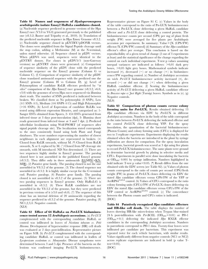
Table S1 Names and sequences of Hyaloperonosporaarabidopsidis isolate Emoy2 HaRxLs candidates cloned.(a) Nucleotide sequence predicted in genome versions of the Hpa
Emoy2 race (V3.0 to V6.0) generated previously to the published
one (v8.3.2; Baxter and Tripathy et al., 2010). (b) Translation of
the predicted nucleotide sequence in Hpa Emoy Genome v8.3.2.
(c) Nucleotidic sequence cloned in pENTRY/pDONR vectors.
The clones were amplified from the Signal Peptide cleavage until
the stop codon, adding a Methionine (M) at the N-terminal,
unless stated otherwise. (d) Sequence cloned in pEDV vectors.
When pEDV6 was used, sequences were identical to the
pENTRY donors. For clones in pEDV3/5 (non-Gateway
versions) no pENTRY clones were generated. (e) Comparison
of sequence similarity of the pEDV clone with the predicted
nucleotide sequence on the Emoy2 genome (Column G vs
Column C). (f) Comparison of sequence similarity of the pEDV
clone translated aminoacid sequence with the predicted one the
Emoy2 genome (Column H vs Column D). (g) Level of
Polymorphism of candidate RxLR effectors predicted by ‘‘in
silico’’ comparison of the Hpa Emoy2 race genome (v8.3.2, v6.0,
v3.0) with the genomes of seven Hpa races sequenced via illumina
short reads. The number of SNPs predicted is indicated between
brakets. We classified them as No polymorphic (0 SNPs), Low
($1 SNPs #5), Medium ($6 SNPS #15) and High Polymorphic
(.16 SNPs). (h) Level of Expression of candidate RxLRs was
tested using different expression libraries, and categorized as: 1-
Sanger ESTs obtained from Spores, 2- 454 ESTs generated from
infected tissue at 3 days post-inoculation (dpi), 3- Illumina short
reads generated from infected tissue at 3 and 7 dpi. (i) Predicted
subcellular localization using Wolf-PSORT (of sequences after
signal peptide cleavage site). The localization stated corresponds
to the ones consistently found using both Plant and Fungi
databases. The score numbers representing the number of closest
neighbours in each alignment are represented in brakets. (+)
Cloned from RxLR onwards, without M. (*) Cloned from RxLR
onwards, X or L replaced by M. (u) Cloned from SP cleavage site
onwards, with M introduced. ND Not determined. (1) There are
two sequences highly similar in Emoy2 genome. The paralog
cloned here is not assembled in the published Emoy2 genome
(v8.3.2). They differ only in three aminoacids (EGMIW6KN-
MIR). (2) Putative gene family. The paralog cloned is not the one
assembled in V8.3.2 of the genome. (3) The cloned sequence not
assembled in v8.3.2. It is highly similar except for the C-terminal
end. Putative paralogs. (4) Putative gene family. The paralog
cloned is not assembled in v8.3.2 of the genome. (5) There are
two paralog sequences in Emoy2 genome. Only HaRxL45 is
assembled in v8.3.2. (6) These RxLR candidates are not
assembled in the V8.3.2 of the genome, but they were predicted
in previous versions of it (v3.0 and v6.0). (7) The sequence of this
clone has an internal deletion of 20 aminoacids regarding the
sequence predicted in v8.3.2 of the genome. Putative paralog. (8)
NC1,2,3,4: Negative controls.
(XLS)
Table S2 Effect of 64 HaRxLs on Pst-LUX biolumines-cence tested across 12 Arabidopsis accessions. (a) Pst-LUX
complemented with the corresponding candidate HaRxL or
control was infiltrated in leaves of Nicotiana tabacum cv. Petit
Havana. Development of hypersensitive response (HR) cell death
was evaluated at 2 days post-infiltration. Representative picture
on Figure S1B. (b) Pst-LUX complemented with the correspond-
ing candidate HaRxL or control was infiltrated in leaflets of
Lycopersicum esculentum cv. Moneymaker. Disease sympthoms were
determined between 3 and 5 dpi. Presence of the bacteria on the
lessions was confirmed imaging Pst-LUX bioluminescence.
Representative picture on Figure S1 C. (c) Values in the body
of the table correspond to the ratio of Pst-LUX bioluminescence
between a Pst-LUX clone delivering a given HaRxL candidate
effector and a Pst-LUX clone delivering a control protein. The
bioluminescence counts per second (CPS) per mg of plant fresh
weight (FW) were averaged for five plants per Arabidopsis
accession per experiment. In summary: Value = [X (CPS/FW)
effector/X (CPS/FW) control] (d) Summary of the Hpa candidate
effector’s effect per ecotype. This conclusion is based on the
reproducibility of a given trend of change (2 out of 3 experiments
at least) and the statistical significance of the change regarding the
control on each individual experiment. T-test p values assuming
unequal variances are indicated as follows: .0,01 dark grey
boxes, .0,05 light grey boxes. Mathematical symbols indicate
increased (+), decreased (2) or no change ( = ) in biolumines-
cence/FW regarding control. (e) Number of Arabidopsis accessions
on wich Pst-LUX bioluminescence activity increased (+), de-
creased (2) or did not change ( = ) when delivering a given
HaRxL candidate effector. (f) Scoring the bioluminescence
activity of Pst-LUX delivering a given HaRxL candidate effector
in Brassica rapa cv Just Right (Turnip) leaves. Symbols as in (e). (g)
Negative controls.
(XLS)
Table S3 Comparison of photon counts versus colonyforming units for Pst-LUX. Results obtained delivering 13
Hpa candidate effectors via EDV by Pst-LUX in different
Arabidopsis accessions. Numbers in the body of the table correspond
to the ratio between Pst-LUX delivering the indicated effector and
the control Pst-LUX clone delivering YFP. For the spray
inoculation, the quantization of the bacterial bioluminescence
(Photon Counts) and colony forming units (CFUs) is displayed for
two to 3 replicate experiments. Experiments displaying the results
obtained when the bacteria are introduced in the plant by syringe
infiltration are shown for comparison. For the spray inoculation
experiments, bacterial growth was scored at 3 dpi using five plants
to record Pst-LUX bioluminescence. The same plants were ground
to determine bacterial growth by plating in selective media and
CFUs. Experiments in parallel were done inoculating the bacteria
at OD600: 0.001 by syringe infiltration. Numbers highlighted in
bold indicate T-test p value,0.05. (*) Result differs from the one
obtained with the EDV screen (see Table S2). (a) Values of photon
counts correspond to the ratio of counts per second (CPS)/fresh
weight (FW) in grams of Pst-LUX clones delivering via EDV the
stated Hpa candidate effectors versus CPS/FW of the YFP or
AvrRPS4AAAA control. (b) Values of CFUs correspond to the ratio
colony forming units (CFUs)/FW of Pst-LUX clones delivering via
EDV the stated Hpa candidate effectors versus CFUs/FW of the
YFP control or AvrRPS4AAAA control. (c) The concordance
between the Photon counts and CFUs ratios is indicated.
(DOC)
Table S4 Putatively recognized Hpa candidate effectorsand HR-like cell death. The table displays the number of
leaves showing HR-like micro-lesions when Trypan blue-stained
24 h post-infiltration with Pst-DCEL (OD600 = 0.01) or Pf0-1
(OD600 = 0.1) delivering the indicated Hpa RXLR effector
candidates in the corresponding Arabidopsis accessions. Numbers
in parenthesis correspond to Pf0-1 data. Twenty-four leaves were
infiltrated per candidate per bacterium. This experiment was
repeated twice for each vehicle bacterium, with similar results.
Values significantly different from negative controls (NC1 or NC2)
across replicate experiments are indicated in bold (p value T-
test,0.05).
(DOC)
Testing the Arabidopsis Downy Mildew Effectorome
PLoS Pathogens | www.plospathogens.org 16 November 2011 | Volume 7 | Issue 11 | e1002348

Acknowledgments
We thank the JIC horticulturist team, Matthew Smoker, Avril Soards, Tina
Payne and Jaqueline Bautor for technical assistance. We thank Craig
Brooks-Rooney, Nicolas Battich, Belen Tejada-Romero, Sharon Hall,
Peter Bittner-Eddy and Astrild Vaudaine for contributing with the cloning
of HaRxLs. We thank Cyril Zipfel, Sophien Kamoun, Ruslan Yatusevik
and Adriana Cruz-Cabral for helpful comments on the manuscript. This
paper is dedicated to Tiziano and Francesco Lunello-Fabro and to
Federico Lunello.
Author Contributions
Conceived and designed the experiments: JDGJ JB JEP GvA GF.
Performed the experiments: GF JS MC EK RLA SJMP ARC DG RL
JLB MCC. Analyzed the data: GF LB NI DJS JB JDGJ. Contributed
reagents/materials/analysis tools: GF JS MC NI EK RLA SJMP ARC DG
RL JLB MCC KHS LB NI DJS. Wrote the paper: GF JDGJ. Edit the
paper: JB JS JEP GvA MCC GF JDGJ.
References
1. Oliver R, Ipcho S (2004) Arabidopsis pathology breathes new life into the
necrotrophs-vs.-biotrophs classification of fungal pathogens. Mol Plant Pathol 5:
347–352.
2. Jones JD, Dangl JL (2006) The plant immune system. Nature 444: 323–329.
3. Zipfel C (2009) Early molecular events in PAMP-triggered immunity. Curr Opin
Plant Biol 12: 414–420.
4. Boller T, He SY (2009) Innate immunity in plants: an arms race between pattern
recognition receptors in plants and effectors in microbial pathogens. Science
324: 742–744.
5. van der Hoorn RA, Kamoun S (2008) From Guard to Decoy: a new model for
perception of plant pathogen effectors. Plant Cell 20: 2009–2017.
6. Raffaele S, Farrer RA, Cano LM, Studholme DJ, MacLean D, et al. (2010)
Genome evolution following host jumps in the Irish potato famine pathogen
lineage. Science 330: 1540–1543.
7. Hajri A, Brin C, Hunault G, Lardeux F, Lemaire C, et al. (2009) A ‘‘repertoire
for repertoire’’ hypothesis: repertoires of type three effectors are candidate
determinants of host specificity in Xanthomonas. PLoS One 4: e6632.
8. Lipka U, Fuchs R, Lipka V (2008) Arabidopsis non-host resistance to powdery
mildews. Curr Opin Plant Biol 11: 404–411.
9. Baxter L, Tripathy S, Ishaque N, Boot N, Cabral A, et al. (2010) Signatures of
adaptation to obligate biotrophy in the Hyaloperonospora arabidopsidis
genome. Science 330: 1549–1551.
10. Spanu PD, Abbott JC, Amselem J, Burgis TA, Soanes DM, et al. (2010) Genome
expansion and gene loss in powdery mildew fungi reveal tradeoffs in extreme
parasitism. Science 330: 1543–1546.
11. Schirawski J, Mannhaupt G, Munch K, Brefort T, Schipper K, et al. (2010)
Pathogenicity determinants in smut fungi revealed by genome comparison.
Science 330: 1546–1548.
12. Oliva R, Win J, Raffaele S, Boutemy L, Bozkurt TO, et al. (2010) Recent
developments in effector biology of filamentous plant pathogens. Cell Microbiol
12: 1015.
13. Slusarenko AJ, Schlaich NL (2003) Downy mildew of Arabidopsis thaliana
caused by Hyaloperonospora parasitica (formerly Peronospora parasitica). Mol
Plant Pathol 4: 159–170.
14. Holub EB (2008) Natural history of Arabidopsis thaliana and oomycete
symbioses. Eur J Plant Pathol 122: 91–109.
15. Coates ME, Beynon JL (2010) Hyaloperonospora arabidopsidis as a pathogen
model. Annu Rev Phytopathol 48: 329–345.
16. Torto TA, Li SS, Styer A, Huitema E, Testa A, et al. (2003) EST Mining and
Functional Expression Assays Identify Extracellular Effector Proteins From the
Plant Pathogen Phytophthora. Genome Res 13: 1675–1685.
17. Kamoun S (2006) A catalogue of the effector secretome of plant pathogenic
oomycetes. Annu Rev of Phytopathol 44: 41–60.
18. Birch PRJ, Rehmany AP, Pritchard L, Kamoun S, Beynon JL (2006)
Trafficking arms: oomycete effectors enter host plant cells. Trends Microbiol
14: 8–11.
19. Qutob D, Kemmerling B, Brunner F, Kufner I, Engelhardt S, et al. (2006)
Phytotoxicity and Innate Immune Responses Induced by Nep1-Like Proteins.
Plant Cell 12: 3721–3744.
20. Kemen E, Kemen AC, Rafiqi M, Hempel U, Mendgen K, et al. (2005)
Identification of a protein from rust fungi transferred from haustoria into
infected plant cells. Mol Plant Microbe In 18: 1130–1139.
21. Catanzariti AM, Dodds PN, Lawrence GJ, Ayliffe MA, Ellis JG (2006)
Haustorially expressed secreted proteins from flax rust are highly enriched for
avirulence elicitors. Plant Cell 18: 243–256.
22. Gout L, Fudal I, Kuhn ML, Blaise F, Eckert M, et al. (2006) Lost in the middle
of nowhere: the AvrLm1 avirulence gene of the Dothideomycete Leptosphaeria
maculans. Mol Microbiol 60: 67–80.
23. Ridout CJ, Skamnioti P, Porritt O, Sacristan S, Jones JD, et al. (2006) Multiple
avirulence paralogues in cereal powdery mildew fungi may contribute to parasite
fitness and defeat of plant resistance. Plant Cell 18: 2402–2414.
24. Shan W, Cao M, Leung D, Tyler BM (2004) The Avr1b locus of Phytophthora
sojae encodes an elicitor and a regulator required for avirulence on soybean
plants carrying resistance gene Rps1b. Mol Plant Microbe In 17: 394–403.
25. Armstrong MR, Whisson SC, Pritchard L, Bos JI, Venter E, et al. (2005) An
ancestral oomycete locus contains late blight avirulence gene Avr3a, encoding a
protein that is recognized in the host cytoplasm. Proc Natl Acad Sci U S A 102:
7766–7771.
26. Allen RL, Bittner-Eddy PD, Grenvitte-Briggs LJ, Meitz JC, Rehmany AP, et al.
(2004) Host-parasite coevolutionary conflict between Arabidopsis and downy
mildew. Science 306: 1957–1960.
27. Rehmany AP, Gordon A, Rose LE, Allen RL, Armstrong MR, et al. (2005)
Differential recognition of highly divergent downy mildew avirulence gene alleles
by RPP1 resistance genes from two Arabidopsis lines. Plant Cell 17: 1839–1850.
28. Win J, Morgan W, Bos J, Krasileva KV, Cano LM, et al. (2007) Adaptive
evolution has targeted the C-terminal domain of the RXLR effectors of plant
pathogenic oomycetes. Plant Cell 19: 2349–2369.
29. Whisson SC, Boevink PC, Moleleki L, Avrova AO, Morales JG, et al. (2007) A
translocation signal for delivery of oomycete effector proteins into host plant
cells. Nature 450: 115.
30. Dou D, Kale SD, Wang X, Jiang RH, Bruce NA, et al. (2008) RXLR-mediated
entry of Phytophthora sojae effector Avr1b into soybean cells does not require
pathogen-encoded machinery. Plant Cell 20: 1930–1947.
31. Kale SD, Gu B, Capelluto DG, Dou D, Feldman E, et al. (2010) External lipid
PI3P mediates entry of eukaryotic pathogen effectors into plant and animal host
cells. Cell 142: 284–295.
32. Schornack S, van Damme M, Bozkurt TO, Cano LM, Smoker M, et al. (2010)
Ancient class of translocated oomycete effectors targets the host nucleus. Proc
Natl Acad Sci U S A 107: 17421–17426.
33. Birch PR, Boevink PC, Gilroy EM, Hein I, Pritchard L, et al. (2008) Oomycete
RXLR effectors: delivery, functional redundancy and durable disease resistance.
Curr Opin Plant Biol 11: 373–379.
34. Panstruga R, Dodds PN (2009) Terrific protein traffic: the mystery of effector
protein delivery by filamentous plant pathogens. Science 324: 748–750.
35. Judelson HS, Tyler BM, Michelmore RW (1991) Transformation of the
oomycete pathogen, Phytophthora infestans. Mol Plant Microbe In 4: 602–607.
36. Huitema E, Smoker M, Kamoun S (2010) A straightforward protocol for electro-
transformation of Phytophthora capsici zoospores. Methods Mol Biol 712: 129–135.
37. Sohn KH, Lei R, Nemri A, Jones JDG (2007) The downy mildew effector
proteins ATR1 and ATR13 promote disease susceptibility in Arabidopsis
thaliana. Plant Cell 19: 4077–4090.
38. Guo M, Tian F, Wamboldt Y, Alfano JR (2009) The majority of the type III
effector inventory of Pseudomonas syringae pv. tomato DC3000 can suppress
plant immunity. Mol Plant Microbe In 22: 1069–1080.
39. Cornelis GR (2010) The type III secretion injectisome, a complex nanomachine
for intracellular toxin delivery. Biol Chem 391: 745–751.
40. Mudgett MB, Chesnokova O, Dahlbeck D, Clark ET, Rossier O, et al. (2000)
Molecular signals required for type III secretion and translocation of the
Xanthomonas campestris AvrBs2 protein to pepper plants. Proc Natl Acad Sci
USA 97: 13324–13329.
41. Fan J, Crooks C, Lamb C (2008) High-throughput quantitative luminescence
assay of the growth in planta of Pseudomonas syringae chromosomally tagged
with Photorhabdus luminescens luxCDABE. Plant J 53: 393–399.
42. Zipfel C, Robatzek S, Navarro L, Oakeley EJ, Jones JD, et al. (2004) Bacterial
disease resistance in Arabidopsis through flagellin perception. Nature 428:
764–767.
43. Gimenez-Ibanez S, Ntoukakis V, Rathjen JP (2009) The LysM receptor kinase
CERK1 mediates bacterial perception in Arabidopsis. Plant Signal Behav 4:
539–541.
44. Nekrasov V, Li J, Batoux M, Roux M, Chu ZH, et al. (2009) Control of the
pattern-recognition receptor EFR by an ER protein complex in plant immunity.
Embo J 28: 3428–3438.
45. Li J, Zhao-Hui C, Batoux M, Nekrasov V, Roux M, et al. (2009) Specific ER
quality control components required for biogenesis of the plant innate immune
receptor EFR. Proc Natl Acad Sci U S A 106: 15973–15978.
46. Salmeron JM, Staskawicz BJ (1993) Molecular characterization and h r p
dependence of the avirulence gene avrPro from Pseudomonas syringae pv. tomato.
Mol Gen Genet 239: 6–16.
47. Belkhadir Y, Nimchuk Z, Hubert DA, Mackey D, Dangl JL (2004) Arabidopsis
RIN4 negatively regulates disease resistance mediated by RPS2 and RPM1
downstream or independent of the NDR1 signal modulator and is not required
for the virulence functions of bacterial type III effectors AvrRpt2 or AvrRpm1.
Plant Cell 16: 2822–2835.
48. Axtell MJ, Staskawicz BJ (2003) Initiation of RPS2-specified disease resistance in
Arabidopsis is coupled to the AvrRpt2-directed elimination of RIN4. Cell 112:
369–377.
Testing the Arabidopsis Downy Mildew Effectorome
PLoS Pathogens | www.plospathogens.org 17 November 2011 | Volume 7 | Issue 11 | e1002348
49. Gassmann W, Hinsch ME, Staskawicz BJ (1999) The Arabidopsis RPS4
bacterial-resistance gene is a member of the TIR-NBS-LRR family of disease-resistance genes. Plant J 20: 265–277.
50. Rentel MC, Leonelli L, Dahlbeck D, Zhao B, Staskawicz BJ (2008) Recognition
of the Hyaloperonospora parasitica effector ATR13 triggers resistance againstoomycete, bacterial, and viral pathogens. Proc Natl Acad Sci USA 105:
1091–1096.51. Lindeberg M, Stavrinides J, Chang JH, Alfano JR, Collmer A, et al. (2005)
Proposed guidelines for a unified nomenclature and phylogenetic analysis of type
III Hop effector proteins in the plant pathogen Pseudomonas syringae. Mol PlantMicrobe In18: 275–282.
52. Lin NC, Martin GB (2005) An avrPto/avrPtoB mutant of Pseudomonas syringaepv. tomato DC3000 does not elicit Pto-mediated resistance and is less virulent on
tomato. Mol Plant Microbe In 18: 43–51.53. Nordborg M, Hu TT, Ishino Y, Jhaveri J, Toomajian C, et al. (2005) The
Pattern of Polymorphism in Arabidopsis thaliana. PLoS Biology 3: 1289–1299.
54. Goodman RN, Novacky AJ (1994) The Hypersensitive Response in Plants toPathogens: A Resistance Phenomenon. St. Paul: APS Press.
55. Greenberg JT, Yao N (2004) The role and regulation of programmed cell deathin plant-pathogen interactions. Cell Microbiol 6: 201–211.
56. Yu IC, Parker J, Bent AF (1998) Gene-for-gene disease resistance without the
hypersensitive response in Arabidopsis dnd1 mutant. Proc Natl Acad Sci USA95: 7819–7824.
57. Thomas WJ, Thireault CA, Kimbrel JA, Chang JH (2009) Recombineering andstable integration of the Pseudomonas syringae pv. syringae 61 hrp/hrc cluster
into the genome of the soil bacterium Pseudomonas fluorescens Pf0-1. Plant J 60:919–928.
58. Donofrio NM, Delaney TP (2001) Abnormal callose response phenotype and
hypersusceptibility to Peronospoara parasitica in defence-compromised arabi-dopsis nim1-1 and salicylate hydroxylase-expressing plants. Mol Plant Microbe
In 14: 439–450.59. Dong X, Hong Z, Chatterjee J, Kim S, Verma DP (2008) Expression of callose
synthase genes and its connection with Npr1 signaling pathway during pathogen
infection. Planta 229: 87–98.60. Meyer D, Pajonk S, Micali C, O’Connell R, Schulze-Lefert P (2009)
Extracellular transport and integration of plant secretory proteins intopathogen-induced cell wall compartments. Plant J 57: 986–999.
61. Robatzek S, Chinchilla D, Boller T (2006) Ligand-induced endocytosis of thepattern recognition receptor FLS2 in Arabidopsis. Genes Dev 20: 537–542.
62. Naito K, Taguchi F, Suzuki T, Inagaki Y, Toyoda K, et al. (2008) Amino Acid
sequence of bacterial microbe-associated molecular pattern flg22 is required forvirulence. Mol Plant Microbe In 9: 1165–1174.
63. Mishina T, Zeier J (2007) Pathogen-associated molecular pattern recognitionrather than development of tissue necrosis contributes to bacterial induction of
systemic aquired resistance in Arabidopsis. Plant J 50: 500–513.
64. Gomez-Gomez L, Boller T (2002) Flagellin perception: a paradigm for innateimmunity. Trends Plant Sci 7: 251–256.
65. Gomez-Gomez L, Felix G, Boller T (1999) A single locus determines sensitivityto bacterial flagellin in Arabidopsis thaliana. Plant J 18: 277–284.
66. Hauck P, Thilmony R, He SY (2003) A Pseudomonas syringae type III effectorsuppresses cell wall-based extracellular defense in susceptible Arabidopsis plants.
Proc Natl Acad Sci U S A 100: 8577–8582.
67. Nomura K, Debroy S, Lee YH, Pumplin N, Jones J, et al. (2006) A bacterialvirulence protein suppresses host innate immunity to cause plant disease. Science
313: 220–223.68. Zhang J, Shao F, Li Y, Cui H, Chen L, et al. (2007) A Pseudomonas syringae
Effector Inactivates MAPKs to Suppress PAMP-Induced Immunity in Plants.
Cell Host Microbe 1: 175–185.69. Underwood W, Zhang S, He SY (2007) The Pseudomonas syringae type III
effector tyrosine phosphatase HopAO1 suppresses innate immunity inArabidopsis thaliana. Plant J 52: 658–672.
70. DebRoy S, Thilmony R, Kwack YB, Nomura K, He SY (2004) A family of
conserved bacterial effectors inhibits salicylic acid-mediated basal immunity andpromotes disease necrosis in plants. Proc Natl Acad Sci U S A 101: 9927–9932.
71. Tor M, Holub EB, Brose E, Musker R, Gunn N, et al. (1994) Map positions ofthree loci in Arabidopsis thaliana associated with isolate-specific recognition of
Peronospora parasitica. (downy mildew). Mol Plant Microbe In 7: 214–222.72. Rohmer L (2003) Nucleothide sequence, functional characterization and
evolution of pFKN, a virulence plasmid in Pseudomonas syringae pathovar
maculicola. Mol Microbiol 47: 1545–1562.
73. Tyler BM, Tripathy S, Zhang X, Dehal P, Jiang RH, et al. (2006) Phytophthora
genome sequences uncover evolutionary origins and mechanisms of pathogen-
esis. Science 313: 1261–1266.
74. Jiang RH, Tripathy S, Govers F, Tyler BM (2008) RXLR effector reservoir in
two Phytophthora species is dominated by a single rapidly evolving superfamily
with more than 700 members. Proc Natl Acad Sci U S A 105: 4874–4879.
75. Haas BJ, Kamoun S, Zody MC, Jiang RHY, Handsaker RE, et al. (2009)
Genome sequence and analysis of the Irish potato famine pathogen
Phytophthora infestans. Nature 461: 393–398.
76. Fu ZQ, Guo M, Jeong BR, Tian F, Elthon TE, et al. (2007) A type III effector
ADP-ribosylates RNA-binding proteins and quells plant immunity. Nature 447:
284–281.
77. de Torres M, Mansfield JW, Grabov N, Brown IR, Ammouneh H, et al. (2006)
Pseudomonas syringae effector AvrPtoB suppresses basal defence in Arabidopsis.
Plant J 47: 368–382.
78. Wilton M, Subramaniam R, Elmore J, Felsensteiner C, Coaker G, et al. (2010)
The type III effector HopF2(Pto) targets Arabidopsis RIN4 protein to promote
Pseudomonas syringae virulence. Proc Natl Acad Sci USA 107: 2349–2354.
79. Nemri A, Atwell S, Tarone AM, Huang YS, Zhao K, et al. (2010) Genome-wide
survey of Arabidopsis natural variation in downy mildew resistance using
combined association and linkage mapping. Proc Natl Acad Sci USA 107:10302–10307.
80. Gaulin E, Drame N, Lafitte C, Torto-Alalibo T, Martinez Y, et al. (2006)
Cellulose Binding Domains of a Phytophthora Cell Wall Protein Are NovelPathogen-Associated Molecular Patterns. Plant Cell 18: 1766–1777.
81. Tor M (2008) Pattern-recognition receptors [PRRs] in plants. Jo Leukocyte Biol84: A1–A2.
82. Kvitko BH, Park DH, Velasquez AC, Wei CF, Russell AB, et al. (2009)
Deletions in the Repertoire of Pseudomonas syringae pv. tomato DC3000 TypeIII Secretion Effector Genes Reveal Functional Overlap among Effectors. PLoS
Pathog 5: e1000388. doi:10.1371/journal.ppat.1000388.
83. Block A, Li GY, Fu ZQ, Alfano JR (2008) Phytopathogen type III effectorweaponry and their plant targets. Curr Op Plant Biol 11: 396–403.
84. Cunnac S, Lindeberg M, Collmer A (2009) Pseudomonas syringae type III
secretion system effectors: repertoires in search of functions. Curr Op Microbiol12: 53–60.
85. Hann DR, Rathjen JP (2010) The long and winding road: virulence effector
proteins of plant pathogenic bacteria. Cell Mol Life Sci 67: 3425–3434.
86. Dou D, Kale SD, Wang X, Chen Y, Wang Q, et al. (2008) Conserved C-terminal motifs required for avirulence and suppression of cell death by
Phytophthora sojae effector Avr1b. Plant Cell 20: 1118–1133.
87. Oh SK, Young C, Lee M, Oliva R, Bozkurt TO, et al. (2009) In PlantaExpression Screens of Phytophthora infestans RXLR Effectors Reveal Diverse
Phenotypes, Including Activation of the Solanum bulbocastanum DiseaseResistance Protein Rpi-blb2. Plant Cell 21: 2928–2947.
88. Bailey K, Cevik V, Holton N, Byrne-Richardson J, Sohn K, et al. (2011)
Molecular Cloning of ATR5Emoy2 from Hyaloperonospora arabidopsidis, anavirulence determinant that triggers RPP5-mediated defense in Arabidopsis.
Mol Plant Microbe In 24: 827–838.
89. Emanuelsson O, Brunak S, von Heijne G, Nielsen H (2007) Locating proteins inthe cell using TargetP, SignalP and related tools. Nat Protoc 2: 953–971.
90. Badel JL, Nomura K, Bandyopadhyay S, Shimizu R, Collmer A, et al. (2003)
Pseudomonas syringae pv. tomato DC3000 HopPtoM (CEL ORF3) is importantfor lesion formation but not growth in tomato and is secreted and translocated
by the Hrp type III secretion system in a chaperone-dependent manner. Mol
Microbiol 49: 1239–1251.
91. Lin NC, Martin GB (2005) An avrPto/avrPtoB mutant of Pseudomonas syringae
pv. tomato DC3000 does not elicit Pto-mediated resistance and is less virulent on
tomato. Mol Plant Microbe In 18: 43–51.
92. Karimi M, Inze D, Depicker A (2002) GATEWAY((TM)) vectors for
Agrobacterium-mediated plant transformation. Trends Plant Sci 7: 193–195.
93. Koncz C, Schell J (1986) The promoter of the TL -DNA gene 5 controls thetissue-specific expression of chimaeric genes carried by a novel type of
Agrobacterium binary vector. Mol Gen Genet 204: 383–396.
94. Katagiri F, Thilmony R, He SY (2002) The Arabidopsis Thaliana-PseudomonasSyringae Interaction. Arabidopsis Book 1: 1–35.
95. Keppler LD, Baker CJ, Atkinson MM (1989) Active oxygen production during a
bacteria-induced hypersensitive reaction in tobacco suspension cells. Phytopa-thology 79: 974–978.
Testing the Arabidopsis Downy Mildew Effectorome
PLoS Pathogens | www.plospathogens.org 18 November 2011 | Volume 7 | Issue 11 | e1002348