moyamoya disease - neurosurgery education & training ... filemoyamoya disease moderators: dr a....
TRANSCRIPT
Moyamoya disease
Moderators: Dr A. SuriModerators: Dr A. Suri
Dr D. Gupta
Presented by: Dr Amitabha Das
Introduction
• A chronic occlusive cerebro-vascular disease affecting arteries around the ‘circle of Willis’ & formation of extensive collaterals at the base of the brainof the brain
• Presents with ischemic and hemorrhagic symptoms
• Characteristic angiographic finding

History
• First described in Japan- Takeuchi & Shimizu
(1957)
• Spontaneous occlusion of the ‘ circle of • Spontaneous occlusion of the ‘ circle of
Willis’- Kudo (1968)
• Moyamoya means ‘puff of smoke’
• Coined by Suzuki and Takaku in 1969

Epidemiology
• Highest incidence in Japan (0.35/ lakh)
• Incidence in Western countries- 1/10th of Japan
• F:M= 2:1• F:M= 2:1
• Bimodal age distribution: larger peak in 1st
decade & smaller peak around 30-49years
• 10-15% have familial form

Etiology
• Multifactorial: genetic predisposition and
environmental stimuli
• Genetic loci: chromosome 3, 6, 8 & 17

Associated conditions
• Immunologic: Grave’s disease/ thyrotoxicosis
• Infections: Leptospirosis and tuberculosis
• Hematologic disorders: Aplastic anemia,
Fanconi anemia, sickle cell anemia, and
lupus anticoagulant
Associated conditions 2
• Congenital syndromes: Apert syndrome, Down syndrome, Marfan syndrome, tuberous sclerosis, Turner syndrome, NF-1& Hirschsprung disease Hirschsprung disease
• Vascular diseases: Atherosclerosis, coarctation of aorta, fibromuscular dysplasia & hypertension
Associated conditions 3
• Others: Head injury, Head neck irradiation for optic glioma, pituitary tumor, craniopharyngioma.
• These are not causative, but warrant consideration during treatment.
Patho-physiology 1
• Smooth muscle hyperplasia of vessel wall &
luminal thrombosis
�Fibro cellular thickening of intima, �Fibro cellular thickening of intima,
�Attenuation of media
�Disruption of internal elastic lamina
• No evidence of inflammation or arteriosclerosis
Patho-physiology 2
• Site: supra-clinoidal ICA, ACA & MCA
• Rare involvement of PCA & BA
• Extra-cranial involvement: STA
• Role of pleuripotent peptides, enzymes &
receptors: primary or secondary

Clinical features
Symptoms
Ischemic Hemorrhagic
Pediatric population
• Ischemic symptoms: 70-80% cases
• Stroke or TIA: 6% of childhood strokes
• Occurs in watershed areas
• Precipitating factors:
� Hyperventilation
� Dehydration
Pediatric population 2
• Features:
�Hemi paresis
�Speech disturbance
Cognitive impairment�Cognitive impairment
�Seizure
�Subtle deficits: developmental delay, syncope, personality changes, visual disturbance
Pediatric population 3
• Hemorrhage: IVH, intraparenchymal or
subarachnoidsubarachnoid
• Headache
• Choreiform movements

Adult population
• Hemorrhage: 66% cases
• Intra or periventricular bleeding
• Annual rebleeding rate 7%
• High morbidity & mortality• High morbidity & mortality
• Sources:
�Fragile collateral vessels
�Micro aneurysms in the circle of Willis
�Periventricular pseudo aneurysms
�Saccular aneurysms in vertebro-basilar system
Adult population
• Ischemic symptoms predominate in Western
world
• Low morbidity and mortality• Low morbidity and mortality
• Pregnancy and delivery increase the risk
Imaging
• Angiography: Gold standard
• MRI & MRA: steno-occlusive carotid lesion
and basal Moyamoyaand basal Moyamoya
• Plain CT: helps in acute stage
• Cerebral blood flow studies: xenon enhanced
CT, PET, SPECT
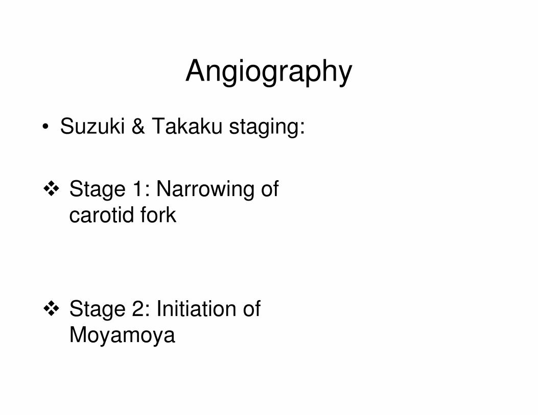
Angiography
• Suzuki & Takaku staging:
� Stage 1: Narrowing of carotid forkcarotid fork
� Stage 2: Initiation of Moyamoya
Angiography 2
� Stage 3: Intensification of Moyamoya
� Stage 4: Minimization of Moyamoya
Angiography 3
� Stage 5: Reduction of Moyamoya
� Stage 6: Disappearance of Moyamoya

Angiography 2
• Types:
�Basal Moyamoya
�Ethmoidal Moyamoya
�Vault Moyamoya
Management
• No definite treatment available
• Medical treatment: not effective
• Aspirin
• Anticoagulants
• Calcium channel blockers
• Steroids
Surgical management
• Aim:
�Augment cerebral blood flow
�Improve cerebral hemodynamics
• Methods:
�Direct revascularization
�Indirect revascularization
�Combined
Surgical management 2
• Criteria for revascularization:
1. Symptomatic patients with good neurological status
2. Infarction <2cm on CT & all previous 2. Infarction <2cm on CT & all previous hemorrhages resolved completely
3. Angiographic stage II to IV
4. Timing: > 2 months after the most recent attack

Direct revascularization
• Indicated when donor & recipient vessel
diameter >1mm
• Immediate selective perfusion of ischemic • Immediate selective perfusion of ischemic
area
• Chance of hyper perfusion syndrome
• Usually done in adults
Direct revascularization 2
• STA-MCA bypass- Donaghy & Yasargil
(1967)(1967)
• STA-ACA bypass
• STA- PCA bypass

Indirect revascularization
• Aimed at stimulating neovascularization
• Extent of revascularization unpredictable• Extent of revascularization unpredictable
• Useful in pediatric population
Indirect revascularization 2
• Encephalomyosynangiosis (EMS): implantation of temporalis muscle on lateral brain surface and secured to dura
• Encephaloduroarteriosynangiosis (EDAS): dissected STA is laid onto the cortical surface
Indirect revascularization 3
• Ribbon EDAS: pedicle of galea inserted
into interhemispheric fissureinto interhemispheric fissure
• Autogenic omentum transplantation as
free graft
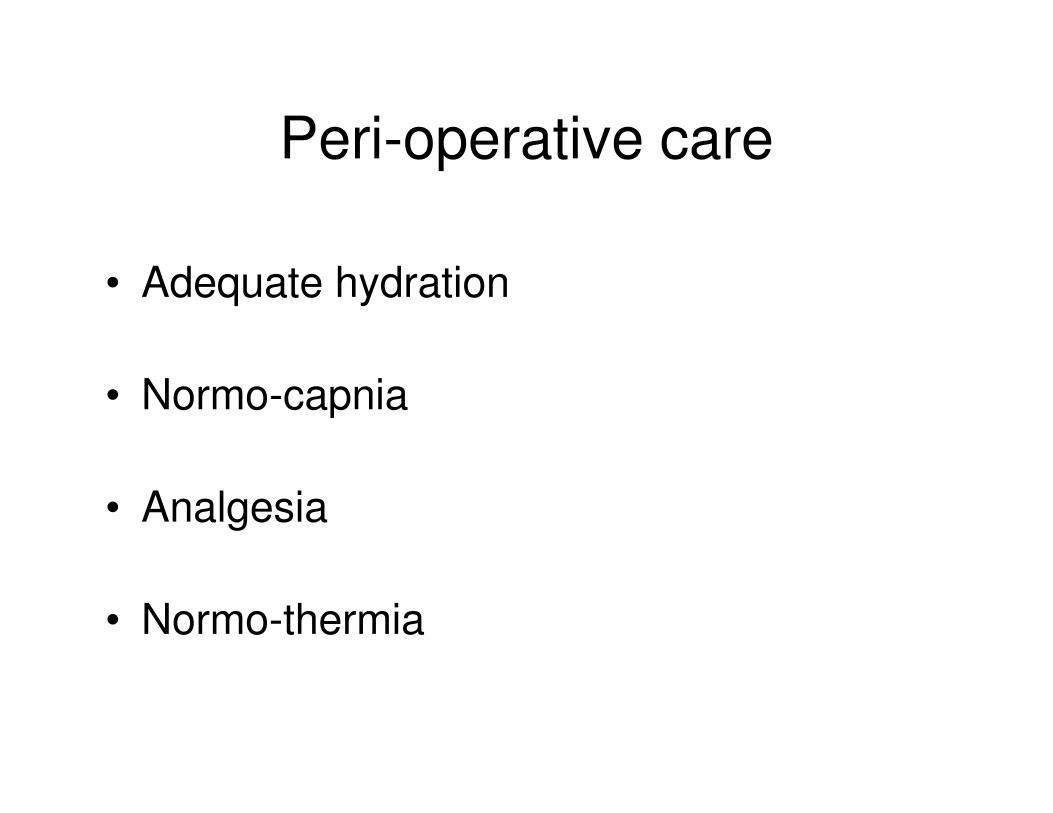
Peri-operative care
• Adequate hydration
• Normo-capnia• Normo-capnia
• Analgesia
• Normo-thermia
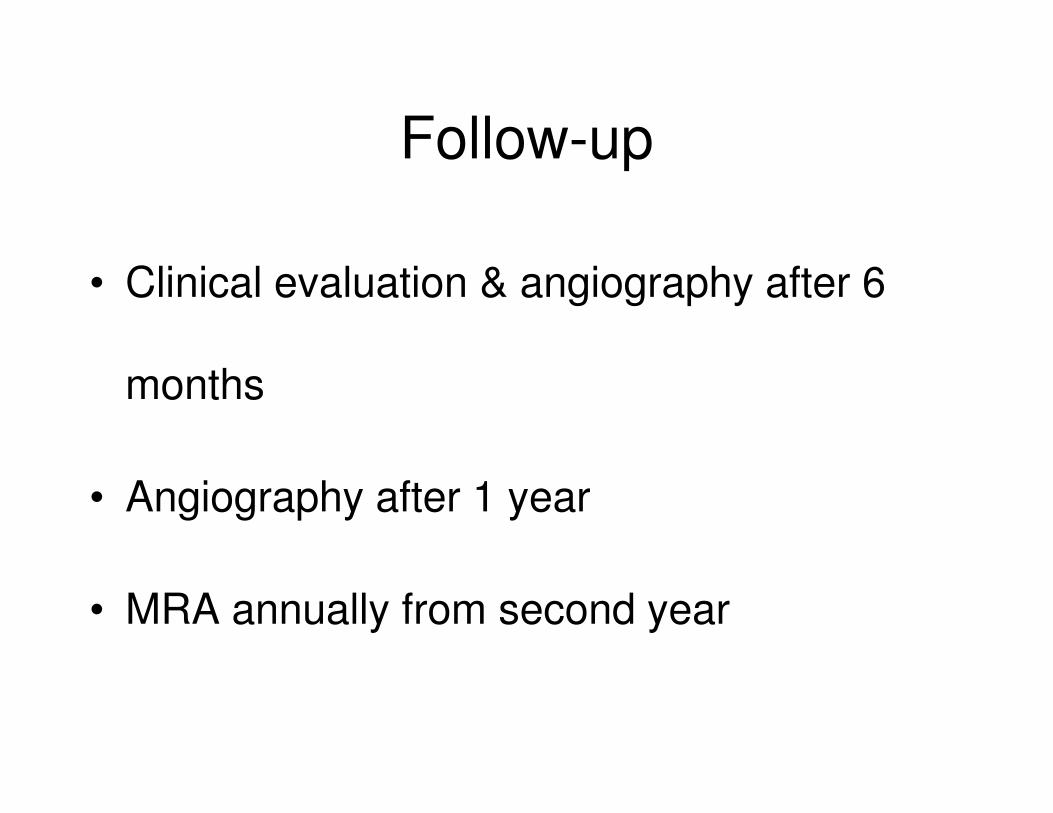
Follow-up
• Clinical evaluation & angiography after 6
months months
• Angiography after 1 year
• MRA annually from second year
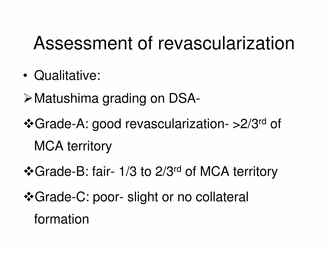
Assessment of revascularization
• Qualitative:
�Matushima grading on DSA-
�Grade-A: good revascularization- >2/3rd of �Grade-A: good revascularization- >2/3 of
MCA territory
�Grade-B: fair- 1/3 to 2/3rd of MCA territory
�Grade-C: poor- slight or no collateral
formation
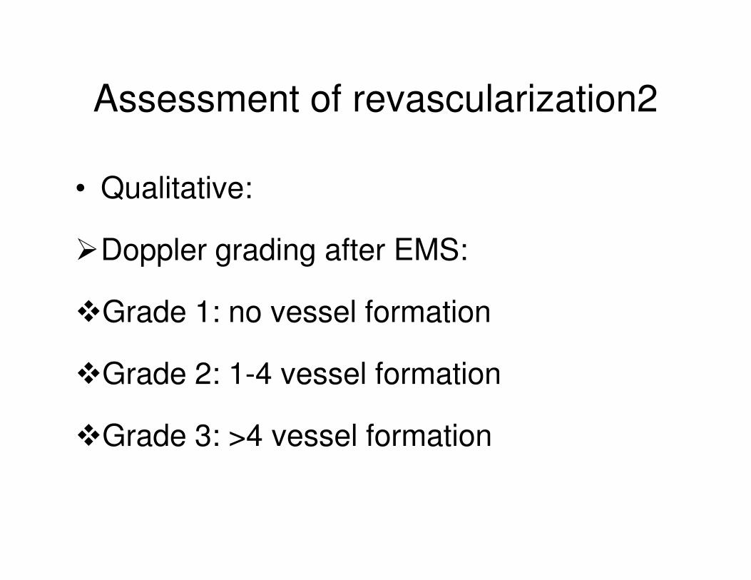
Assessment of revascularization2
• Qualitative:
�Doppler grading after EMS:
�Grade 1: no vessel formation
�Grade 2: 1-4 vessel formation
�Grade 3: >4 vessel formation
Assessment of revascularization3
• Quantitative:
• Study published in Neurosurgery in March 2012
• Quantitative assessment of RV on DSA• Quantitative assessment of RV on DSA
• Revascularization of MCA territory against supratentorial area of the ipsilateral hemisphere
• Best result following combined procedure
Prognosis
• Benign course in 75-80%
• Rebleeding occurs in 30-65%
• Revascularization reduces rebleeding & TIAs
• Unilateral disease progresses to bilateral
involvement in 7-27%
Future prospects
• Role of endothelial progenitor cells
• Role of cytokines and growth factors• Role of cytokines and growth factors
• Quantitative assessment of RV
AIIMS data
• Ten-year experience of 44 patients with Moyamoya disease from a single institution
• Published in Journal of Clinical Neurosciences in April 2010Neurosciences in April 2010
• Adult population predominates: 59% vs 41%
• Hemorrhagic symptoms more common: 68% vs 32%
AIIMS data 2
• Revascularization done in 11 patients: 9 indirect & 2 combined
• No new episode in revascularized patients
• In conservatively managed 19 patients 7 • In conservatively managed 19 patients 7 developed new episodes
• In hospital mortality: 3 patients with hemorrhagic symptoms died
Conclusion
• The unpredictable and relentless course of
the MMD, coupled with irreversible nature of
deficits once present dictates a need for deficits once present dictates a need for
early diagnosis, prompt treatment and
regular follow-up