moodie, 1928
DESCRIPTION
By RoY L. MOODIE Ossified tendons, under the term "tendon bones," have been known to paleontologists for a long time, and their occurrence in the dino- saurs, especially, has been a matter of common knowledge. It would be of no especial value for me to review the literature relating to the occur- rence of these objects among fossil reptiles, so I shall merely cite two Number 3-11 THE AmEniCAN .%ewm JrNATURAL HISTORY April 25, 1928 Published by 56,81,9:14.74 Paper. 2TRANSCRIPT

AMERICAN MUSEUM NOVITATESPublished by
Number 3-11 THE AmEniCAN .%ewm JrNATURAL HISTORY April 25, 1928
56,81,9:14.74
THE HISTOLOGICAL NATURE OF OSSIFIED TENDONSFOUND IN DINOSAURS
By RoY L. MOODIE
Ossified tendons, under the term "tendon bones," have been knownto paleontologists for a long time, and their occurrence in the dino-saurs, especially, has been a matter of common knowledge. It would beof no especial value for me to review the literature relating to the occur-rence of these objects among fossil reptiles, so I shall merely cite twopapers by Barnum Brown", 2 who has written the most recent and mostcomplete accounts of these structures.
I was attracted by the possibility of explaining the nature of someof the vertebral lesions in Smilodon3, on the basii of the coniparativehistology of the structures involved. If those curious lesions in thesabre-tooth are really myositis, then there should be some evidence of amodification in the histology of the lesions toward a ligamentous ortendinous structure. Such proved not to be the case. and I now believethose vertebral lesions of Smilodon to be parosteal, due to injury of thevertebrae.
Mr. Brown sent me specimens of ossified tendons from two genera ofdinosaurs, Trachodon and Ankylosaurus. He also secured funds to payfor making the photomicrographs, shown on the accompanying figures.The tendons sent for study were of the size of lead-pencils, and whereweathered showed the osseous appearance so well known to experiencedfossil-hunters. Their fibers run longitudinally without interruption.
Although there have been a number of studies on the histologicalnature of fossil bone, no attempt has been made previously to describethe histology of petrified tendons, so the results given herewith are all
'Brown, Barnum. 1916. Corythosaurus casuarius. Skeleton, Musculature and Epidermis. SecondPaper.
Bull. Amer; Mus. Nat. Hist., XXXV, Art. XXXVIII, pp. 709-716, Pls. xIII-xxII. On page 711,under the caption "Tendons and musculature," is given the pertinent discussion.
2 -- 1917. A complete Skeleton of the Horned Dinosaur Monoclonius, and Descriptionof a second Skeleton showing Skin Impressions.
Ibid, XXXVII, Art. X, pp. 281-306, Pls. xI-xix. "Ossified tendons," page 290. Plates xii andxvi show the arrangement of the tendons especially well.
3Moodie, Roy L. 1927. Studies in Paleopathology, XIX. Vertebral Lesions in the Sabre-tooth,Pleistocene of California, resembling the so-called Myositis ossificans progressiva, compared with certainrOssifications in the Dinosaurs. Annals of Medical History. IX, No. 1, pp. 91-102, Figs. 1-11.

AMERICAN MUSEUM NOVITATES [No. 311
new. I have reviewed elsewhere' the literature of fossil histology, andfurther reference to that subject need not be made here.
Cross and longitudinal sections of the ossified tendons of Trachodonand Ankylosaurus reveal the histological characters. One who is accus-tomed to examining the microscopical structures of ancient fossil bone isat once impressed with the absence of vascular spaces, which are ex-tremely abundant in skeletal bone. The arrangement of Haversiansystems is strikingly regular, and there is no evidence of stellate cells, orbundles of tissue, so characteristic of recent tendons. Osteoid tissue, soabundant in the skeletal parts of fossil reptiles, is strikingly absent fromthe fossil tendons. There is an interesting similarity in the histology ofthe ossified tendons and pathological fossil bone in the tendency in bothto the production of fairly perfect Haversian systems. The systems seenin the tendons are quite perfectly formed and include all the principalelements, i. e., lacunae, lamellaX, canals, fibrillar ground substance, andabsorption areas. I cannot be sure of an intercommunication between thecanaliculi of adjoining lacunae. The lacunae, in fact, on closer examina-tion (Fig. 2, B), are not definitely arranged in concentric layers, betweenthe quite apparent lamellae. The lacunae show a number of definitecanaliculi, springing both from the two poles and from the sides of thespindles. However, they agree in general with the structure, form, andsize of other dinosaurian lacunae.
Bacterial invasion, through vascular channels, of the lacunae andcanaliculi is known to have taken place with similar results from theDevonian ostracoderms down to the Pleistocene mammals and tothis common occurrence the ossified tendons of the two Cretaceousdinosaurs offer no exception. Fig. 6, A, shows several lacunae invadedand enlarged by the bacteria of decay, whose activities were stopped andtheir effects preserved by fossilization.
SUMMARY
The histology of the ossified tendons of two genera of dinosaurs isstructurally unlike that of the skeletal bone of fossil reptiles, in thepresence of fairly well-developed Haversian systems. In this respect thetendons approach the structure of mammalian long bones. There is anabsence of osteoid tissue in the tendons. The concentric lamellae are few,rarely more than six. The lacunae are shaped like those seen in skeletalbones of dinosaurs. and the canaliculi are few, short, and unbranched.
'Moodie, Roy L. 1926. Studies in Paleopathology, XIII. The Elements of the Haversian Systemin normal and Pathological Structures among fossil Vertebrates. Biologia Generalis, II, pp. 63-95,12 Plates, 10 figs.
2

Figures 1 to 6
3
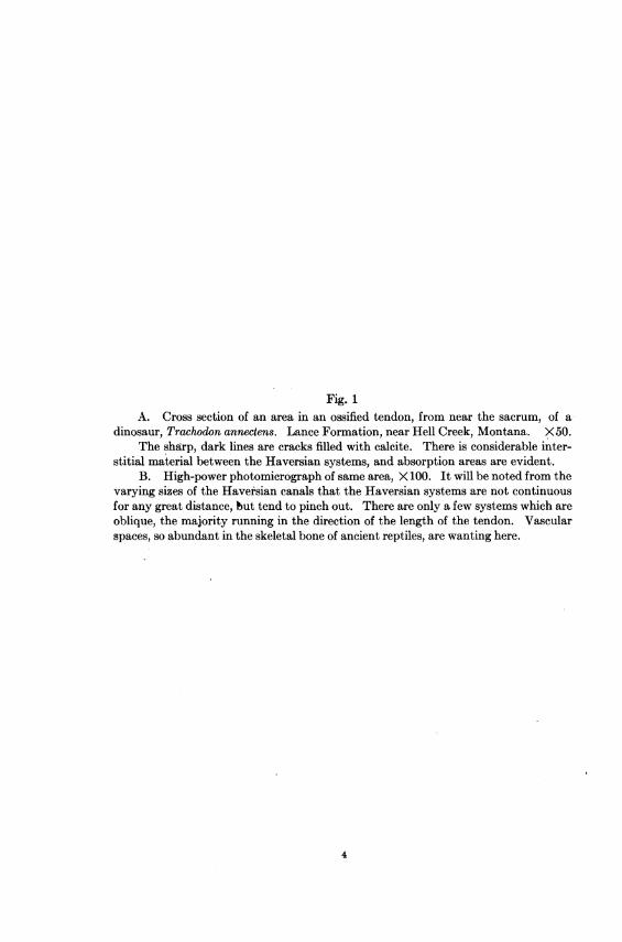
Fig. 1A. Cross section of an area in an ossified tendon, from near the sacrum, of a
dinosaur, Trachodon annectens. Lance Formation, near Hell Creek, Montana. X50.The sharp, dark lines are cracks filled with calcite. There is considerable inter-
stitial material between the Haversian systems, and absorption areas are evident.B. High-power photomicrograph of same area, X 100. It will be noted from the
varying sizes of the Haversian canals that the Haversian systems are not continuousfor any great distance, but tend to pinch out. There are only a few systems which areoblique, the majority running in the direction of the length of the tendon. Vascularspaces, so abundant in the skeletal bone of ancient reptiles, are wanting here.
4
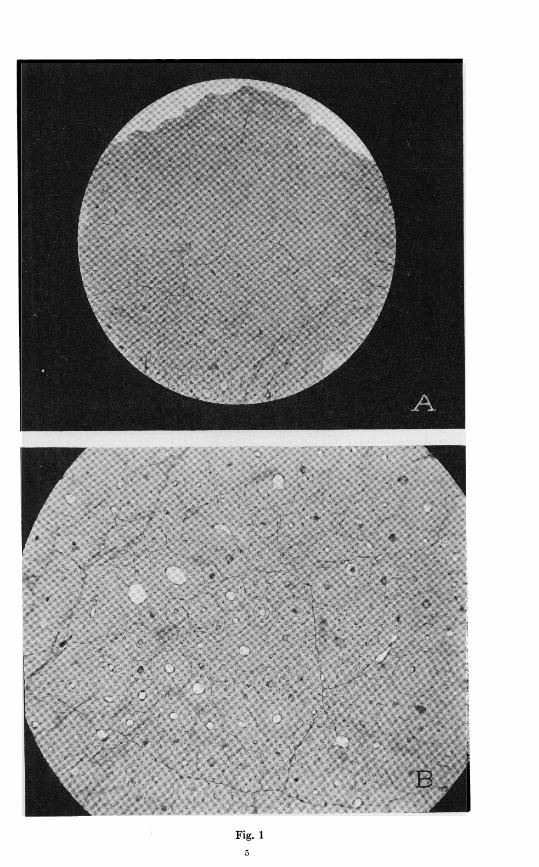
Fig. 1
5

Fig. 2A. Another area of the same slide, X 100, showing less distinct boundaries to
the small Haversian systems.B. Same section, X300. Enlarged view of some of the Haversian systems,
showing varying sizes. The concentric lamelloe are evident in the upper right-handcorner of the figure, and adjoining this system is an absorption area which does not differessentially from an area in modern human bone. The interstitial ground substancehas laculna of unmodified type. The dark line running obliquely across the bottom ofthe figure is a post-fossilization crack.
Fig. 2
7
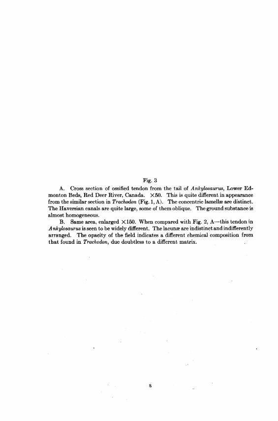
Fig. 3A. Cross section of ossified tendon from the tail of Ankylosaurus, Lower Ed-
monton Beds, Red Deer River, Canada. X)50. This is quite different in appearancefrom the similar section in Trachodon (Fig. 1, A). The concentric lamellae are distinct.The Haversian canals are quite large, some of them oblique. The ground substance isalmost homogeneous.
B. Same area, enlarged X 150. When compared with Fig. 2, A-this tendon inAnkylosaurus is seen to be widely different. The lacunw are indistinct and indifferentlyarranged. The opacity of the field indicates a different chemical composition fromthat found in Trachodon, due doubtless to a different matrix.
$

;G-W+'>''z''5' 6f ; '> '
g< . . , f . : *: : «x- ~~~
S .4 Li. SVXi*S}FiE {rB.<
Fig 39

Fig. 4A. Longitudinal section of an ossified dinosaur tendon, from near the sacrum
of Trachodon annectens, from the Lance Formation; near Hell Creek, Montana. X70.Two Haversian canals are shown, one extending across the field. Others are
cut obliquely. The orderly arrangement of the lacunme is attractive. The lacunaare small and numerous.
B. Same section, enlarged X150. Upper part of field has numerous cracks.Some of the lacunme in the lower part of the field are quite large, as if invaded bybacteria.
10

Fig. 4
11

Fig. 5A. Longitudinal section of an ossified tendon from near the sacrum of Trachodon
annectens. Lance Formation, near Hell Creek, Montana. X300.This shows the early effect of the invasion of lacunae by bacteria of decay, before
fossilization. The canaliculi are hypertrophied. Normal lacunae in dinosaur bonepossess only brief canaliculi. The three lacune shown in the middle above are figuredenlarged to 600 diameters in Fig. 6, A.
B. Longitudinal section of an ossified tendon from the tail of Ankylosauruts.Lower Edmonton Beds, Red Deer River, Canada. X50.
The Haversian canals have the appearance of vascular spaces. The lamelleand lacu'ne are indistinct. The entire area presents a dull opacity wanting in thesections of Trachodon.
12

K-~~~~~~~~~~~~~~~~-~ ~ ~ ~ ~~~~~I
4w
4~~~~~~~~~~~~~~~~~~~~~~~~~~~~~~,tea~~~~~~~~~~~~~~~~~~~~~~~~~~~~~~~~~~~~~~~~~~~~~~,
tAt~~~~~~~~~~~~~t
*4~~~~~~~~~~~~~~~~~~~~~~~~4
-C.--p~~~~~~~~~~~~~~~~~..~~~~~~~~~~~~~~~W
Fig. 513
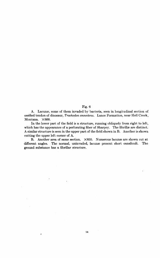
Fig. 6A. Lacunae, some of them invaded by bacteria, seen in longitudinal section of
ossified tendon of dinosaur, Trachodon annectens. Lance Formation, near Hell Creek,Montana. X600.
In the lower part of the field is a structure, running obliquely from right to left,which has the appearance of a perforating fiber of Sharpey. The fibrillee are distinct.A similar structure is seen in the upper part of the field shown in B. Another is showncutting the upper left corner of A.
B. Another area of same section. X600. Numerous lacune are shown cut atdifferent angles. The normal, uninvaded, lacunoe present short canaliculi. Theground substance has a fibrillar structure.
14

Fig 615
