monoamine transporters in female human...
TRANSCRIPT

LUND UNIVERSITY
PO Box 117221 00 Lund+46 46-222 00 00
Monoamine transporters in female human reproduction.
Bottalico, Barbara
Published: 2007-01-01
Link to publication
Citation for published version (APA):Bottalico, B. (2007). Monoamine transporters in female human reproduction. Department of Obstetrics andGynecology, Lund University
General rightsCopyright and moral rights for the publications made accessible in the public portal are retained by the authorsand/or other copyright owners and it is a condition of accessing publications that users recognise and abide by thelegal requirements associated with these rights.
• Users may download and print one copy of any publication from the public portal for the purpose of privatestudy or research. • You may not further distribute the material or use it for any profit-making activity or commercial gain • You may freely distribute the URL identifying the publication in the public portalTake down policyIf you believe that this document breaches copyright please contact us providing details, and we will removeaccess to the work immediately and investigate your claim.

Download date: 06. Jun. 2018
ABSTRACTThe present study explored the gene and protein expression of the monoamine transporters in human endometrium throughout the menstrual cycle, in early decidua and in placentas from normal as well as preeclamptic pregnancies using in-situ hybridization, real time-PCR, immunohistochemistry and primary tissue cultures.
Four distinguishable patterns were observed in the endometrium over the menstrual cycle: (1) epithelial expres-sion of norepinephrine transporter (NET) mRNA, (2) Stromal co- expression of vesicular monoamine transporter 2 (VMAT2) and plasma membrane monoamine transporter (PMAT) mRNAs with maximal intensity in the proli-ferative phase; (3) increasing epithelial expression of VMAT2 mRNA with a maximum in the late secretory phase; (4) stromal expression of extra-neuronal monoamine transporter (EMT) mRNA with a peak in the early secretory phase. The presence of functional EMT and VMAT2 transporter proteins throughout the menstrual cycle was shown by uptake of radiolabelled histamine.
A similar expression pattern of monoamine transporters was seen in normal and preeclamptic placentas. In particular, NET mRNA was detected in the chorionic and anchoring villi while EMT mRNA was expressed in scattered cells in placental vessels as well as in intralobular septa cells. Serotonin transporter (SERT) mRNA was mainly detected in the chorionic villi. VMAT2 mRNA was detected in the deeper layers of the placenta bed biopsies in trophoblast cells. A small number of cells in the intima layer of some placental vessels showed mRNA expression of the organic cation transporters 1 and 2 (OCT1 and OCT2). Although the expression pattern was si-
versus normal pregnancies.
maintaining adequate levels of extra cellular monoamines. Their presence and dynamic expression suggests an important role during the menstrual cycle and pregnancy. Moreover a defective gene expression or function of the monoamine transporters might be determinant in the onset of preeclampsia and its alteration in the vascular bed. Knowledge of the regulation of monoamine metabolism in the endometrium, decidua and placenta will increase the understanding of infertility problems and may offer new pharmacological approaches to optimise assisted reproduction and treatment of preeclampsia.
Key words: Monoamine transporters, histamine, PMAT, VMAT2, OCT, NET, in-situ hybridization histo-chemistry, decidualization, human endometrium, placenta, preeclampsia
109

Cover by Nobert Robles Back picture: Nobert Robles artistic interpretation of the authors’ scientific work
Printed by Media-Tryck, Lund University, Sweden Barbara Bottalico
ISSN 1652-8220
ISBN 978-91-85897-13-1
Lund University, Faculty of Medicine Doctoral Dissertation Series 2007:135

Ad Alessandro con amore


“REGARDLESS OF WHETHER I BELIEVE OR NOT, WHETHER I AM A CHRISTIAN OR NOT, I WOULD PLAY MY PART IN THE COLLECTIVE BUILDING OF THE CATHEDRAL.” FOUR SCREENPLAYS OF INGMAR BERGMAN (1918-2007)

TABLE OF CONTENTS
LIST OF PAPERS
ABBREVIATIONS
INTRODUCTION 15Monoamines 15Monoamines in human reproduction 16Monoamine transporters 17Monoamine transporters, SSRI and drug addiction 21Normal human endometrium 22The implantation window and placentation 23Preeclampsia 24
AIMS OF THE STUDY 26
MATERIALS AND METHODS 27
RESULTS AND COMMENTS 31Monoamine transporters in normal endometrium and early pregnancy deciduas 31Monoamine transporters in normal and preeclamptic placentas 32Functional study: Histamine uptake 33Comments 33
DISCUSSION 35Monoamine transporters in cyclic endometrium 35Monoamine transporters in normal and preeclamptic placentas 36Endometrial cell cultures and specific histamine uptake 38
SUMMARY 41
CONCLUSIONS 43
FUTURE DIRECTIONS 45
POPULÄRVETENSKAPLIG SAMMANFATTNING PÅ SVENSKA 46
RIASSUNTO DIVULGATIVO IN ITALIANO 48
ACKNOWLEDGEMENTS 50
REFERENCES 52
APPENDIX I 69
APPENDIX II 77
APPENDIX III 91
APPENDIX IV 101

LIST OF PAPERS
I Bottalico, B., Pilka, R., Larsson, I., Casslen, B., Marsal, K. and Hansson, S.R. Plasma membrane and vesicular monoamine transporters in normal endometrium and early pregnancy decidua. Molecular Human Reproduction, 2003, 9, 389-394
II Bottalico, B., Larsson, I., Brodszki, J., Hernandez-Andrade, E., Casslen, B., Marsal, K. and Hansson, S.R. Norepinephrine transporter (NET), serotonin transporter (SERT), vesicular monoamine transporter (VMAT2) and organic cation transporters (OCT1,2 and EMT) in human placenta from preeclamptic and normotensive pregnancies. Placenta, 2004, 25, 518-529
III Noskova, V., Bottalico, B., Olsson, H., Ehinger, A., Pilka, R., Casslen, B. and Hansson, S. R. Histamine uptake by human endometrial cells expressing the organic cation transporter EMT and the vesicular monoamine transporter-2. Molecular Human Reproduction, 2006, 12, 483-489
IV Bottalico, B., Noskova, V., Pilka, R., Larsson, I., Domanski, H., Casslen, B. and Hansson, S.R. The organic cation transporters (OCT1, OCT2, EMT) and the plasma membrane monoamine transporter (PMAT) show differential distribution and cyclic expression pattern in human endometrium and early pregnancy decidua. Molecular Reproductive Development, 2007, 74 (10), 1303-1311
Published papers are reproduced with permission from Oxford University Press (I, III), Elsevier (II), and Wiley-Liss, Inc. a subsidiary of John Wiley & Sons, Inc (IV).

ABBREVIATIONS
35S-UTP 35Sulfur labeled uridine triphosphate 5HT serotonin or 5-hydroxytryptamine 5HTT serotonin transporter ABC avidin biotin complex BSA bovine serum albumin BSS Hank’s balanced salt solution cATP cyclic adenosine triphosphate cDNA complementary deoxyribonucleic acid CNS central nervous system COMT catechol-O-methyltransferase cRNA complementary deoxyribonucleic acid C-terminal carboxy terminal (3’-terminal) DA dopamine DAB diaminobenzidine DAT dopamine transporter DNase deoxyribonuclease dNTP deoxynucleosidetriphosphate E epinephrine EDTA ethylenediamine tetra acetic acid EMT extra-neuronal monoamine transporter ENT equilibrative nucleoside transporter GABA gamma amino butyric acid HDC histamine decarboxylase HI histamine HRP horseradish peroxidase Ig G immunoglobulin G IUGR intrauterine growth restriction ISHH In situ hybridization histochemistry IVF in vitro fertilization MAO monoamine oxidase MMP+ 1-methyl-4-phenylpyridinium mRNA messenger ribonucleic acid NE norepinephrine NET norepinephrine transporter NT nucleotides NTB nuclear track emulsion N-terminal amino terminal (5’-terminal) OCT organic cation transporters PBS phosphate-buffered saline PCR polymerase chain reaction PMAT plasma membrane monoamine transporter PNMT phenyletanolamine-N-methyltransferase RNase ribonuclease RNA ribonucleic acid RT room temperature RT-PCR reversed transcriptase polymerase chain reaction

SDS sodium dodecyl sulfate SERT serotonin transporter SSC standard saline citrate buffer SSRI serotonin specific re-uptake inhibition TMDs transmembrane domains TRIS tris(hydroxymethyl) amino methane tRNA transfer ribonucleic acid VMAT vesicular monoamine transporter

INTRODUCTION
Monoamines
Monoamines are neurotransmitters and neuro-modulators that derive from aromatic amino acids. They contain one amino group connected to an aromatic ring by means of a two-carbon chain (-CH2-CH2-). Monamines include:
- catecholamines such as epinephrine (E), norepinephrine (NE), dopamine (DA) - serotonin or 5-hydroxytryptamine (5-HT), an indolamin - histamine (HI).
Norepinephrine is the principal neurotransmitter in the sympathetic nervous system and together with epinephrine is a potent stress hormone released into the blood by the adrenal medulla (Kandel and Schwartz 1985). Histamine also has a dual role, being a neurotransmitter in the central nervous system (Steinbusch and Mulder 1984) and a potent peripheral vasoregulator, e.g. when released by mast cells during allergic reactions (White 1990). Dopamine is another neurohormone released by the hypothalamus. As a hormone, its main function is to inhibit the release of prolactin from the anterior lobe of the pituitary gland.
As neurotransmitters, monoamines take part in a wide variety of processes such as modulation of motor functions, arousal, attention, mood, and anxiety. Neuroendocrine cells in the gastrointestinal and respiratory tracts also contain monoamines, which play important paracrine roles in these organs (Axelrod 1971).
Monoamines are among the earliest neurotransmitters to be detected during human fetal brain development (Olson et al. 1973). Since monoamines and their synthesizing enzymes are present before the nerve terminal areas are developed, their early role differs from their role in the synapsis. Serotonin is the first monoamine to appear and has been detected at embryonic day 12 (E12) in the fetal rat brain, followed by DA (E13) and NE (E14) (Olson et al. 1973; Olson and Seiger 1972). During development, monoamines regulate morphogenesis through cell differentiation and migration; some have also been shown to release trophic factors and induce trophic effects. In particular, 5-HT plays a role in early gastrulation, induction of neurogenesis and neural differentiation, formation of the neural tube and migration of cranial neural crest cells. At a later stage, it inhibits synaptogenesis and causes release of neuronal growth factors (Buznikov 1984; Fox 1995; Marshak 1990; Moiseiwitsch and Lauder 1995; Whitaker-Azmitia et al. 1996; Whitaker-Azmitia et al. 1990). Synthesis of catecholamines is mediated by two enzymes: tyrosine hydroxylase, which is a rate limiting enzyme and converts tyrosine to dopamine; and dopamine beta hydroxylase that converts dopamine to norepinephrine. NE is then converted in epinephrine by phenyletanolamine-N-methyltransferase (PNMT). Serotonin is synthesized from tryptophan by tryptophan hydroxylase. Histidine decarboxylase (HDC) is the rate limiting enzyme for histamine biosynthesis (Kandel and Schwartz 1985).
15

Neuronal monoamine degradation is mediated by MAO (monoamine oxidase), while the extraneuronal catabolism of 5-HT, NE and DA is mediated by MAO-A, and of NE and E by catechol-O-methyltransfrase (COMT) (Eisenhofer 2001). Histamine is broken down by histamine-N-methyltransferase and diamine oxidase. Unlike most transmitter substances, monoamines are recycled rather than enzymatically inactivated, and over 70% of recaptured catecholamines are sequestered into storage vesicles rather than being deaminated (Eisenhofer 2001). Their intracellular fate is influenced by their lower affinity for inactivating enzymes than for vesicular transporters located in intracellular vesicles (see monoamine transporters) (Erickson and Eiden 1993; Peter et al. 1995b). Thus, cells are capable not only of storing and degrading monoamines, but also of recycling and releasing them in a controlled fashion.
Monoamines in human reproduction
The uterus has an extensive innervation of sympathetic neurons and the adrenergic nerve fibres have been shown to play a role in uterine contractility. In the central nervous system, monoamines together with sex hormones, regulate sexual behaviour and neuro-endocrine responses (Fabre-Nys 1998).
Enzymes involved in monoamine-synthesis have been demonstrated in normal endometrium as well as in early pregnancy decidua, with the exception of histidine decarboxylase. Local synthesis of the monoamines plays an important physiological paracrine role (Manyonda et al. 1998). An abnormal synthesis and concentration of monoamines in the early stages of pregnancy can lead to miscarriage. Mifepristone (RU486) and DL111-IT (a non-hormonal contra gestational agent) cause early pregnancy arrest, reducing monoamine synthesis in the uterus with subsequent decidual cell injury that leads to miscarriage (Shentu et al. 2001). Adequate monoamine concentrations are essential for a proper implantation and physiological evolution of pregnancy. Many factors are involved in this regulation, for example degradation of catecholamines is increased by progesterone that stimulates COMT and MAO activities in endometrium and decidua (Hobel et al. 1981).
Serotonin and histamine play a role in decidualization, implantation and, in the case of histamine, immune-modulation (Cocchiara et al. 1986; Dey 1981; Hatanaka et al. 1982; Maekawa and Yamanouchi 1996 1223; Mitchell et al. 1983). There is a great deal of evidence in this respect. Barash et al. reported that local injury to the endometrium, caused by taking a biopsy, increased the incidence of implantation in IVF patients (Barash et al. 2003). Thus, it is likely that inflammatory mediators, including histamine, which are normally released during tissue repair and remodelling, are important mediators of decidualization and implantation. Implantation in rats was also induced by histamine when combined with suboptimal doses of estrogen (Johnson and Dey 1980), while intrauterine application of inhibitors or antagonists to histamine receptors inhibits decidua formation (Hatanaka et al. 1982; Shelesnyak 1952; Shelesnyak 1957).
In mice, the rate-limiting enzyme in histamine synthesis, histidine decarboxylase (HDC), has been observed in uterine epithelial cells with peak expression at the time of implantation (Paria et al. 1998). The blastocyst expresses a histamine type 2 receptor, (H2), which is the target for histamine activation (Zhao et al. 2000). After implantation, the
16
placenta is developed by the invasion of trophoblasts into the endometrium and maternal vasculature. Histamine enhances the invasion by activating the histamine type 1 (H1) receptor on the cytotrophoblasts (Liu et al. 2004). Monoamines are potent vasoactive mediators that regulate blood flow and, in the case of histamine, capillary permeability (Barkai and Kraicer 1996). Regulation of the uterine blood flow is important both during menstruation and pregnancy. Prostaglandins are of crucial importance for implantation and survival of the blastocyst. Their synthesis can also be modulated by NE, DA and HI (Schrey et al. 1995a; Skarzynski et al. 1999; Yanagawa et al. 1997).
Sexual response Research has shown that histamine is released from mast cells in the genitals as part of the human orgasm, and this release has been connected to the sex flush among women. If this response is lacking, it may indicate histapenia. Conversely, men with high histamine levels may suffer from premature ejaculations (Kirmaz et al. 2005; Meston and Frohlich 2000).
Monoamine transporters
Since monoamines are potent mediators of physiological and pathophysiological events throughout the body, their extracellular concentrations are tightly regulated. Unlike other transmitter substances, monoamines are recycled rather than enzymatically inactivated. Specific membrane bond transporter proteins mediate re-uptake of monoamines from the synaptic cleft and extra cellular fluids and accumulate them intracellularly (Amara and Kuhar 1993; Brownstein and Hoffman 1994). Monoamine transporters can be grouped in three main families:
- the neuronal monoamine transporters - the vesicular monoamine transporters - the non neuronal monoamine transporters
Neuronal monoamine transporters:
Monoaminergic signaling is terminated by reuptake of the transmitter substance by specific cell membrane transporter proteins, which were first described in neuronal cells and are therefore referred to as neuronal transporters (Amara and Kuhar 1993; Brownstein and Hoffman 1994).
Neuronal monoamine transporters are membrane proteins, containing 12 putative trans-membrane domains (TMDs). The mechanism by which the transporter proteins mediate monoamine uptake involves sequential binding and co-transport of Na+ and Cl- ions. DAT transports two Na+ ions and one Cl- ion with its substrate, whereas NET and SERT co-transport their substrates with one Na+ and one Cl- ion (Torres et al. 2003a).
The presence of an active transport system for NE uptake at sympathetic nerve endings was first suggested in early 1960 (Axelrod 1971; Whitby et al. 1961). Chemical signaling by neural cells is terminated by the reuptake of the monoamine from the synaptic cleft by the presynaptic neuron and at extra junctional sites (Figure 1).
17
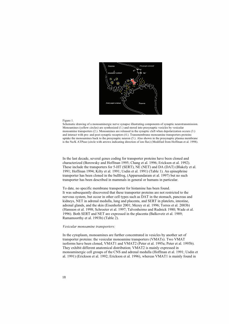
Figure 1. Schematic drawing of a monoaminergic nerve synapse illustrating components of synaptic neurotransmission. Monoamines (yellow circles) are synthesized (1.) and stored into presynaptic vesicles by vesicular monoamine transporters (2.). Monoamines are released in the synaptic cleft when depolarization occurs (3.) and interact with pre- and post-synaptic receptors (4.). Transmembrane monoamine transporters proteins uptake the monoamines back to the presynaptic neuron (5.). Also shown in the presynaptic plasma membrane is the Na/K ATPase (circle with arrows indicating direction of ion flux) (Modified from Hoffman et al. 1998).
In the last decade, several genes coding for transporter proteins have been cloned and characterized (Borowsky and Hoffman 1995; Chang et al. 1996; Erickson et al. 1992). These include the transporters for 5-HT (SERT), NE (NET) and DA (DAT) (Blakely et al. 1991; Hoffman 1994; Kilty et al. 1991; Usdin et al. 1991) (Table 1). An epinephrine transporter has been cloned in the bullfrog, (Apparsundaram et al. 1997) but no such transporter has been described in mammals in general or humans in particular.
To date, no specific membrane transporter for histamine has been found.It was subsequently discovered that these transporter proteins are not restricted to the nervous system, but occur in other cell types such as DAT in the stomach, pancreas and kidneys, NET in adrenal medulla, lung and placenta, and SERT in platelets, intestine, adrenal glands, and the skin (Eisenhofer 2001; Mezey et al. 1996; Torres et al. 2003b) (Hansson et al. 1998; Schroeter et al. 1997; Talvenheimo and Rudnick 1980; Wade et al. 1996). Both SERT and NET are expressed in the placenta (Balkovetz et al. 1989; Ramamoorthy et al. 1993b) (Table 2).
Vesicular monoamine transporters:
In the cytoplasm, monoamines are further concentrated in vesicles by another set of transporter proteins: the vesicular monoamine transporters (VMATs). Two VMAT isoforms have been cloned, VMAT1 and VMAT2 (Peter et al. 1995a; Peter et al. 1995b). They exhibit different anatomical distribution; VMAT2 is mainly expressed in monoaminergic cell groups of the CNS and adrenal medulla (Hoffman et al. 1991; Usdin et al. 1991) (Erickson et al. 1992; Erickson et al. 1996), whereas VMAT1 is mainly found in
18

the adrenal medulla and neuroendocrine cells of the gastrointestinal tract (Weihe et al. 1994; Weihe et al. 1996). Thus, monoamines are preferentially transferred from the cytoplasm to storage vesicles, from which they can be subsequently released on demand. Cell membrane bound monoamine transporters therefore function not only as part of a metabolizing system, but also as part of a recycling system operating together with the VMATs to replenish the transmitter stores.
Non neuronal monoamine transporters:
Organic cation transporters: It has been known for many years that another family of plasma membrane transporters, the organic cation transporters (OCT), is responsible for the so-called type 2 uptake of monoamines (Iversen 1965). OCTs, also known as non-neuronal membrane transporters, were only recently cloned (Gorboulev et al. 1997; Grundemann et al. 1998; Koehler et al. 1997; Zhang et al. 1997). Unlike neuronal monoamine transporters, which mainly regulate synaptic levels of monoamines, OCTs play a key role in the clearance of monoamines from the blood stream. In contrast to the neuronal transporters, OCTs have a broad affinity for all biogenic amines as well as exogenous drugs, xenobiotics and organic anions and cations (Eisenhofer 2001; Grundemann et al. 1999; Hayer-Zillgen et al. 2002; Koepsell 2004; Wieland et al. 2000). Compared to neuronal monoamine transporters, OCTs have 12 putative TMDs, a lower affinity for catecholamines (i.e., higher Km), favour EPI over NE, and exhibit a higher maximum rate of catecholamine uptake (i.e., higher Vmax). In addition, uptake2 is not an Na+- and Cl- -dependent process (Eisenhofer, 2001 #1213) (Table 1). OCTs include OCT1, OCT 2 and OCT3 or the “extra-neuronal monoamine transporter” (EMT) since their functional properties match those of the corticosterone-sensitive catecholamine transport system (Eisenhofer 2001). 1-Methyl-4-phenylpyridinium (MPP+) is an excellent substrate for all transporters. The transport efficiency of OCT1 and OCT2 for dopamine, noradrenaline, adrenaline and 5-HT in general are rather low, 5%-15% of the range of MPP+. This suggests that OCT1 and OCT2 are not primarily dedicated to transport these monoamine transmitters, however EMT plays a significant role in catecholamine inactivation (Amphoux et al. 2006b; Lazar et al. 2007; Schomig et al. 2006). Their pharmacological properties have recently been analysed in stably transfected cell lines (Hayer-Zillgen et al. 2002). OCTs 1 and 2 as well as EMT are greatly inhibited by corticosterone, progesterone and 17 -estradiol (Hayer-Zillgen et al. 2002)(Table 1). In humans, non-neuronal monoamine transporters are mainly expressed by the liver (OCT-1, EMT), intestine (OCT-1), kidney and brain (OCT-2, EMT). EMT has a broad tissue distribution and is also found in the heart, blood vessels, placenta and retina (Eisenhofer 2001)(Table 2).
Plasma membrane monamine transporters -PMAT: Another non-neuronal monoamine transporter, the plasma membrane monoamine transporter (PMAT), which has functional and structural similarities to the OCT transporters, has recently been cloned and characterized (Engel and Wang 2005; Engel et al. 2004). PMAT is a protein characterized by 11 TMDs and in common with OCTs its uptake is Na+ independent (Engel et al. 2004). A peculiarity of this transporter is its ability to function bidirectionally (Engel et al. 2004).
19
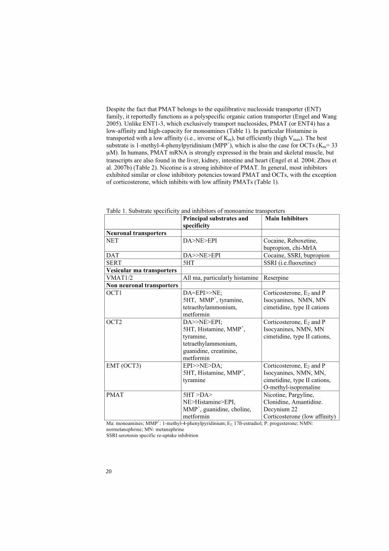
Despite the fact that PMAT belongs to the equilibrative nucleoside transporter (ENT) family, it reportedly functions as a polyspecific organic cation transporter (Engel and Wang 2005). Unlike ENT1-3, which exclusively transport nucleosides, PMAT (or ENT4) has a low-affinity and high-capacity for monoamines (Table 1). In particular Histamine is transported with a low affinity (i.e., inverse of Km), but efficiently (high Vmax). The best substrate is 1-methyl-4-phenylpyridinium (MPP+), which is also the case for OCTs (Km= 33
M). In humans, PMAT mRNA is strongly expressed in the brain and skeletal muscle, but transcripts are also found in the liver, kidney, intestine and heart (Engel et al. 2004; Zhou et al. 2007b) (Table 2). Nicotine is a strong inhibitor of PMAT. In general, most inhibitors exhibited similar or close inhibitory potencies toward PMAT and OCTs, with the exception of corticosterone, which inhibits with low affinity PMATs (Table 1).
Table 1. Substrate specificity and inhibitors of monoamine transporters Principal substrates and
specificity Main Inhibitors
Neuronal transporters NET DA>NE>EPI Cocaine, Reboxetine,
bupropion, chi-MrIA DAT DA>>NE>EPI Cocaine, SSRI, bupropion SERT 5HT SSRI (i.e.fluoxetine) Vesicular ma transporters VMAT1/2 All ma, particularly histamine Reserpine Non neuronal transporters OCT1 DA=EPI>>NE;
5HT, MMP+, tyramine, tetraethylammonium, metformin
Corticosterone, E2 and P Isocyanines, NMN, MN cimetidine, type II cations
OCT2 DA>>NE>EPI; 5HT, Histamine, MMP+,tyramine, tetraethylammonium, guanidine, creatinine, metformin
Corticosterone, E2 and P Isocyanines, NMN, MN cimetidine, type II cations,
EMT (OCT3) EPI>>NE>DA; 5HT, Histamine, MMP+,tyramine
Corticosterone, E2 and P Isocyanines, NMN, MN, cimetidine, type II cations, O-methyl-isoprenaline
PMAT 5HT >DA> NE>Histamine>EPI, MMP+, guanidine, choline, metformin
Nicotine, Pargyline, Clonidine, Amantidine. Decynium 22 Corticosterone (low affinity)
Ma: monoamines; MMP+: 1-methyl-4-phenylpyridinium; E2: 17ß-estradiol; P: progesterone; NMN: normetanephrine; MN: metanephrine SSRI serotonin specific re-uptake inhibition
20
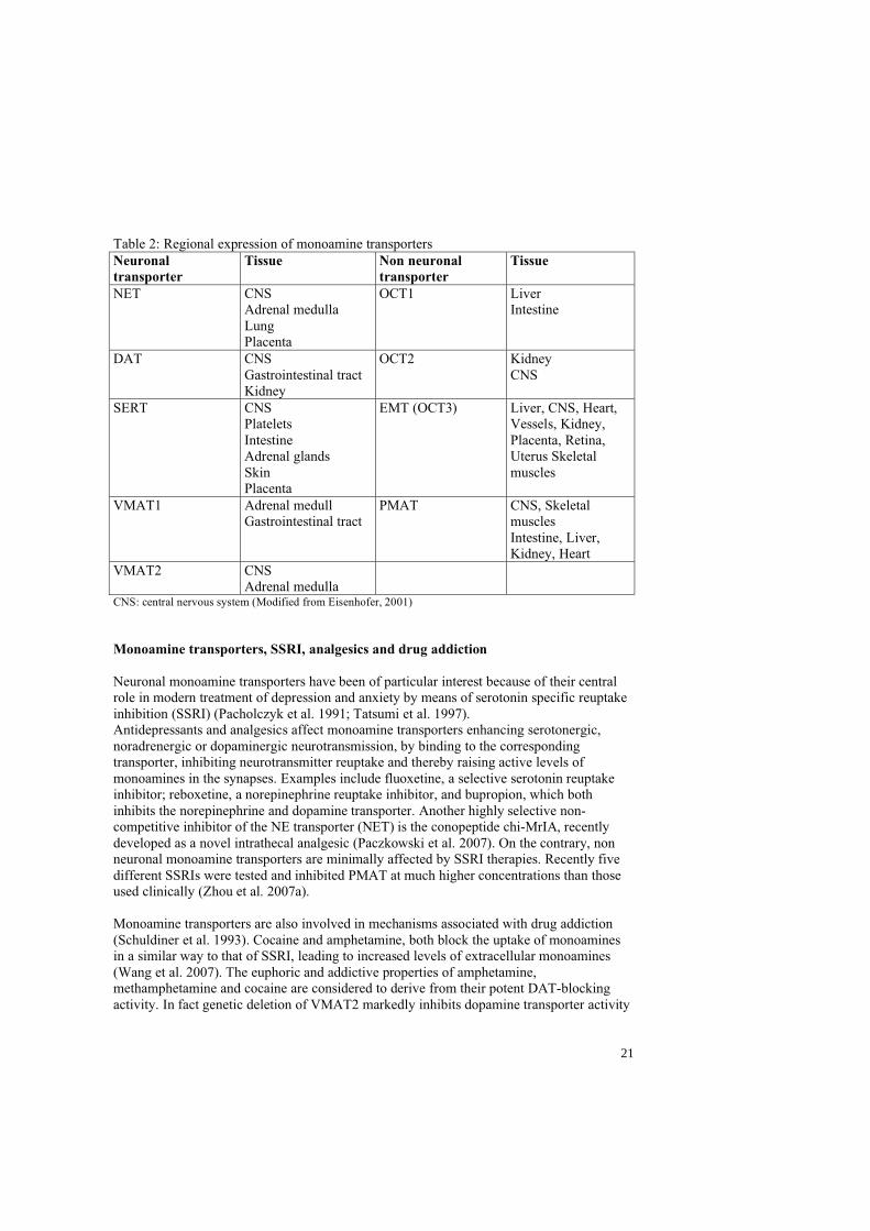
Table 2: Regional expression of monoamine transporters Neuronaltransporter
Tissue Non neuronal transporter
Tissue
NET CNS Adrenal medulla Lung Placenta
OCT1 Liver Intestine
DAT CNS Gastrointestinal tract Kidney
OCT2 Kidney CNS
SERT CNS Platelets Intestine Adrenal glands SkinPlacenta
EMT (OCT3) Liver, CNS, Heart, Vessels, Kidney, Placenta, Retina, Uterus Skeletal muscles
VMAT1 Adrenal medull Gastrointestinal tract
PMAT CNS, Skeletal muscles Intestine, Liver, Kidney, Heart
VMAT2 CNS Adrenal medulla
CNS: central nervous system (Modified from Eisenhofer, 2001)
Monoamine transporters, SSRI, analgesics and drug addiction
Neuronal monoamine transporters have been of particular interest because of their central role in modern treatment of depression and anxiety by means of serotonin specific reuptake inhibition (SSRI) (Pacholczyk et al. 1991; Tatsumi et al. 1997). Antidepressants and analgesics affect monoamine transporters enhancing serotonergic, noradrenergic or dopaminergic neurotransmission, by binding to the corresponding transporter, inhibiting neurotransmitter reuptake and thereby raising active levels of monoamines in the synapses. Examples include fluoxetine, a selective serotonin reuptake inhibitor; reboxetine, a norepinephrine reuptake inhibitor, and bupropion, which both inhibits the norepinephrine and dopamine transporter. Another highly selective non-competitive inhibitor of the NE transporter (NET) is the conopeptide chi-MrIA, recently developed as a novel intrathecal analgesic (Paczkowski et al. 2007). On the contrary, non neuronal monoamine transporters are minimally affected by SSRI therapies. Recently five different SSRIs were tested and inhibited PMAT at much higher concentrations than those used clinically (Zhou et al. 2007a).
Monoamine transporters are also involved in mechanisms associated with drug addiction (Schuldiner et al. 1993). Cocaine and amphetamine, both block the uptake of monoamines in a similar way to that of SSRI, leading to increased levels of extracellular monoamines (Wang et al. 2007). The euphoric and addictive properties of amphetamine, methamphetamine and cocaine are considered to derive from their potent DAT-blocking activity. In fact genetic deletion of VMAT2 markedly inhibits dopamine transporter activity
21

(Yamamoto et al. 2007).Cocaine users and sufferers of cocaine-induced mood disorders, display a marked reduction in VMAT2 immunoreactivity, which may reflect damage to striatal dopaminergic fibers (Little et al. 2003). It has been recently shown, that in monoaminergic neurons, high doses as well as therapeutic doses of amphetamine and methylphenidate selectively redistribute (“trafficking”) the vesicular monoamine transporter-2 (Riddle et al. 2007). These neuronal changes could play a role in causing mood disorder and the motivational processes seen in severely addicted users.
Monoamine transporters; neurological and psychiatric diseases:
Monoamine transporters have also been associated to neurological and psychiatric diseases. VMAT1 gene has been associated with schizophrenia (Richards et al. 2006) and the norepinephrine transporter (NET) has been associated with the attention deficit hyperactivity disorder and neurodegenerative disorders such as Alzheimer's disease and Parkinson's disease (Ding et al. 2006).
It has become evident that genetic variants of monoamine transporters may also contribute to individual differences such as behavior and neuropsychiatric disorders. Genetic studies of the dopamine transporter (hDAT; SLC6A3), the serotonin transporter (hSERT; SLC6A4), and the norepinephrine transporter (hNET; SLC6A2) have shown that genomic variations can influence pharmacology and brain physiology by various mechanisms, including substrate affinities, transport velocity, transporter expression levels (density), extra-cellular membrane expression, trafficking and turnover, as well as neurotransmitter release. (Lin and Madras 2006).
Normal human endometrium
Hitchmann and Adler were the first to describe cyclic changes of the endometrium in 1908 (Hitchmann and Adler 1908). The endometrial lining of the uterine cavity is composed of a functional layer (upper two thirds of the endometrium) and a basal layer. The functional layer is composed of epithelial cells, which form the luminal surface of the glands and the supporting stroma. The endocrine influence of gonadal steroids initiates a cascade of paracrine interactions between these cell types (Arnold et al. 2001). During the menstrual cycle in particular, the functional layer undergoes structural and functional changes in response to estrogen and progesterone. Estrogens initiate endometrial proliferation during the follicular phase of the ovarian cycle, and progesterone results in endometrial differentiation during the luteal phase. Three phases can be distinguished in the cycling endometrium. Growth of all compartments from the basal layer occurs under the influence of estrogen in the proliferative phase; tissue differentiation with secretory activity in the glands in response to progesterone in the secretory phase (Cooke et al. 1998; Kurita et al. 2000) and the menstrual phase with discharge of the functional layer started by progesterone withdrawal secondary to corpus luteum regression, if no pregnancy has been established. The proliferative and secretory phases can be sub-divided into early, mid and late.
Estrogen and progesterone interact with the endometrial cells by means of specific nuclear receptors for estrogens (ER and ER ) and for progesterone (PR-A and PR-B) (Conneely and Lydon 2000; Enmark et al. 1997). The expression of endometrial sex steroid receptors
22
varies temporally and spatially across the menstrual cycle. Both ER( and PR are up-regulated during the proliferative phase by estradiol and down regulated in the secretory phase by progesterone acting at both the transcriptional and the post-transcriptional level (Lecce et al. 2001; Lessey et al. 1988; Mertens et al. 2001).PR is an estrogen-responsive gene, and its presence is considered evidence of a functional ER mediated pathway.
The stroma is characterized by a dynamic leukocyte population. Endometrial leukocytes include T and B cells, mast cells, macrophages, neutrophils, and the uterine natural killer (uNK) cells. These cells vary across the menstrual cycle. The uNK cells are mainly present in the late secretory phase and early pregnancy; macrophages are always present but increase in number in the mid-late secretory phase and in decidua; mast cell distribution does not vary during the cycle, although the cells are activated in the late secretory phase, prior to menstruation (Poropatich et al. 1987). Mast cells are a well known source of histamine and could contribute to the histamine pool present in the endometrium.
The implantation window and placentation
The implantation window is defined as the limited period during which the endometrium is receptive to implantation of the blastocyst (Bourgain and Devroey 2003). During this period the functional endometrial layer undergoes structural and functional changes in response to estrogen, progesterone and blastocyst-derived factors, which transform the endometrium into decidua (Cooke et al. 1998; Kurita et al. 2000).
Monoamines in general and histamine in particular have been shown to play a role in the process of decidualization of endometrial tissue in the secretory phase (Barkai and Kraicer 1996; Dey 1981; Dey et al. 1979a; Dey et al. 1979b).
Implantation is a complex process that involves intimate "cross-talk" between the embryo and uterus. Successful implantation also involves participation of locally derived growth factors, cytokines, prostaglandins, transcription factors and lipid mediators (Paria et al. 2001).
Implantation of the human blastocyst may be divided into three phases: (1) the free blastocyst phase, in which the embryo has not interacted with the endometrium; (2) the adhesion and intrusion phase, which includes attachment of the blastocyst and penetration of the surface epithelium (this has never been observed in humans); (3) the placentation phase, during which the placenta is formed (Enders 2000; Engel and Wang 2005; Lopata et al. 2002).
The human trophoblast is highly invasive in vivo and can penetrate the thickness of the endometrium, reaching as far as the basal third of the endometrium (Enders 1997). The microenvironment of the uterus is decisive for the proper invasion of the trophoblast and progression of pregnancy. An inadequate toxin free milieu in the uterine cavity may lead to inadequate implantation and early miscarriage. Non neuronal monoamine transmitters have broad affinity for many substrates such as exogenous drugs, xenobiotics and toxins in general (Eisenhofer 2001), therefore they can maintain a proper setting for implantation.
23
Normal embryonic development is dependent upon a sufficient oxygen, nutrient and waste exchange through the placenta. In primates including humans, this exchange is attained by successful hemochorial placentation which requires the transformation of maternal intramyometrial spiral arterioles by trophoblast invasion in order to achieve uteroplacental circulation as well as the establishment and maintenance of a competent fetoplacental vasculature. Trophoblast and endothelial cell differentiation, proliferation and invasion occur during placentation (Wulff et al. 2003). Trophoblast cells can be divided into chorionic, villous, and extravillous trophoblasts. There are two types of extravillous trophoblasts: endovascular trophoblast that forms the definitive placenta by occlusion of the spiral arterioles at the implantation site; and interstitial extravillous trophoblast that is responsible for the anatomical erosion of the distal spiral arterioles and the secretion of angiogenic and vasodilator signals to improve uterine blood flow (Chaddha et al. 2004) The importance of tight regulation of angiogenic factors and their endogenous antagonists for normal development of the placenta is demonstrated by failure of this system, resulting in abnormal placenta vascularization and trophoblast invasion associated with intrauterine growth retardation or preeclampsia (Wulff et al. 2003).
Preeclampsia
Preeclampsia is one of the most common causes of perinatal and maternal morbidity and mortality. The overall incidence is 3-7% of pregnancies, and mainly primigravidae are affected. The clinical manifestations of preeclampsia appear after 20 weeks of gestation and are characterized by low circulating blood volume, hemo-concentration, high blood pressure and proteinuria. Furthermore, severe cases of preeclampsia involve pathologic activation of the coagulation and fibrinolytic systems. Without intervention the condition may develop into severe epileptic seizures – eclampsia. Only symptomatic treatment of the condition is available and termination of the pregnancy remains the only curative intervention (Schroeder 2002). Removal of the placenta is believed to be the underlying mechanism for resolution of the symptoms (Roberts et al. 1989).
The spiral arteries are modified during early gestation. Trophoblast cells invade the muscular layer of these vessels in order to make them distended, thin-walled and less responsive to vaso-active substances, thus ensuring proper blood flow (Robertson et al. 1986). Modification of the spiral arteries has been shown to be inadequate in preeclampsia and seems to be a background key factor for its aetiology and early onset IUGR (de Groot and Taylor 1993; Khong et al. 1986; Pijnenborg et al. 1991). Defective endovascular erosion could in fact render the basal plate inadequate for meeting the demands of fetal growth (Chaddha et al. 2004). A generalized inflammatory process in vascular endothelial cells is a hallmark of preeclampsia (de Groot and Taylor 1993; Granger et al. 2001; Roberts and Cooper 2001; Roberts et al. 1989). Factors associated with the placenta have been suggested, but so far no specific culprit has been found. Inadequate regulation of monoamine levels during implantation and placental formation may influence trophoblast invasion. Histamine plays a central role in this modification (Dey 1981; Hatanaka et al. 1982) as well as in local blood flow regulation (Barkai and Kraicer 1996).
24

The hypertension is due to peripheral vasoconstriction and elevated resistance. Increased activity in the sympathetic nervous system and elevated concentrations of circulating vaso-active substances are both important factors in the pathophysiology of hypertension. In fact, sympathetic over-activity is a significant finding in preeclampsia (Schobel et al. 1996). NE is the principal neurotransmitter in the sympathetic nervous system. Increased blood levels of monoamines such as norepinephrine and serotonin have been shown in preeclampsia as well as in IUGR (Kaaja et al. 1999; Manyonda et al. 1998; Middelkoop et al. 1993). Serotonin has been shown to affect the migration of neuronal crest cells during early development (Moiseiwitsch and Lauder 1995), and may also regulate cell migration in the placenta. During early gestation in rats, SERT has been demonstrated in migrating cells of the ectoplacental cone (Hansson et al. 1998; Yavarone et al. 1993), suggesting a need for regulated uptake during placenta formation.
After re-uptake from the extra cellular space, the enzyme MAO degrades NE. Several studies have shown that the expression and activity of MAO-A is decreased in the placenta from pre-eclamptic pregnancies (Carrasco et al. 2000a; Sivasubramaniam et al. 2002). This enzyme also metabolizes 5-HT, another monoamine known to be elevated in preeclampsia (Middelkoop et al. 1993). The high 5-HT plasma levels are mainly due to reduced enzymatic degradation.
Cocaine abuse reportedly gives rise to a preeclampsia-like condition in pregnant women (Towers et al. 1993). Cocaine is a known inhibitor of monoamine transporters.
25
AIMS OF THE STUDY
The general objective of the present study was to investigate the gene expression of the monoamine transporters in female human reproduction. Since the transporter proteins critically regulate extra-cellular monoamine concentrations, knowledge of their distribution and cyclic variation is of great importance for a deeper understanding of the contribution of monoaminergic mechanisms to the reproductive process.
The specific aims were:
-To study the gene expression of the monoamine transporters in normal endometrium, early pregnancy decidua, and placentas.
-To investigate whether the gene expression of monoamine transporters showed differences in placentas of normal pregnancies vs. placentas of pregnancies complicated by preeclampsia, in order to enhance the understanding of monoaminergic mechanisms in the pathogenesis of preeclampsia.
-To demonstrate specific in vitro histamine uptake in human endometrial cells by EMT and VMAT2, and to better comprehend the storage and release mechanisms of histamine and its important role in the initial stages of pregnancy and early fetal development.
26
MATERIALS AND METHODS
Patients and tissue sampling
Tissue was collected at the Department of Obstetrics and Gynaecology, Lund University Hospital, Lund. Sampling after informed consent was approved by the Research Ethics Committee Review Board for studies in human subjects.
Papers I, III and IV Endometrial tissue was collected from healthy women (n=39) under 45 years of age, who were undergoing hysterectomy or diagnostic curettage for benign reasons unrelated to endometrial dysfunction e.g., cervical dysplasia, myoma or uterine prolaps, etc. Each sample was evaluated by a histopathologist in order to exclude endometrial pathology and identify the cyclic phase (Hendrickson and Kempson 1980; Noyes et al. 1950). Decidual tissue was collected from first trimester elective abortions (n=5).
Paper II Placental tissue was collected from preeclamptic (n=10), and normal pregnancies (n=7). The controls were matched for maternal age and parity. Patients with essential hypertension and renal or other systemic diseases were excluded. Tissue samples were taken from the placenta immediately after delivery and, in the case of caesarean section (29% in the control and 50% in the preeclamptic group), from the uterine wall. Two specific areas within the placenta were sampled: (1) a section, 10x10x4 mm, from the maternal surface, at the entrance of the spiral arteries and (2) a cube 10x10x10 mm from the central part of the placenta consisting of villi. Placenta bed biopsies were collected from the uterine wall during caesarean section in order to study gene expression in the modified spiral arteries.
In-situ hybridization histochemistry (Papers I-IV) RNA Probes A probe of approximately 400-560 nucleotids was used for the human 5HTT, NET, DAT, VMAT1, VMAT2, OCT1, OCT2, EMT, PMAT mRNAs (Engel et al. 2004; Erickson et al. 1992; Gorboulev et al. 1997; Grundemann et al. 1998; Koehler et al. 1997; Pacholczyk et al. 1991; Ramamoorthy et al. 1993b; Uhl and Kitayama 1993; Zhang et al. 1997)
PCR
DNA templates were generated by a polymerase chain reaction (PCR) from various cDNAs using bipartite primers consisting of either a T7 RNA promoter and a downstream gene-specific sequence (anti-sense) or a T3 RNA promoter and an upstream gene-specific primer (sense). DNA templates were purified from agarose gels and thereafter sequenced using a cycle sequencing reaction kit. Complementary RNA (cRNA) probes were transcribed from a gel-purified DNA template using 35S-UTP and either T3 or T7 RNA polymerase to generate sense and antisense probes respectively.
Tissue sections
Tissue sections were cut on a cryostat (12 m), thaw mounted onto silanized slides, and stored at -80°C prior to hybridization. Fresh frozen tissue as opposed to fixative-treated
27
tissue, was used in order to maximize mRNA detection. In order to ensure the best possible tissue integrity, the tissue was not thawed prior to sectioning.
RNA Hybridization
Prior to hybridization, tissue sections were fixed, dehydrated and delipidated as previously described (Bradley et al. 1992). Sections were hybridised overnight with a 35S-radiolabeled probe. After washing to remove excess probe, slides were apposed to film and coated with film emulsion. Following four weeks of exposure, the slides were developed, fixed and counterstained. All slides in papers I and IV were evaluated by two independent investigators (S.H and B.B.) who were blinded for the experimental conditions.
Real-time PCR
RNA extractions Total RNA was extracted from frozen tissue. Proteoglycan and polysaccharide contamination was removed. Only samples with visible 18S/28S bands were included for further analysis.
cDNA synthesis
RNA was reversed transcribed. The samples were stored at -20°C until further use.
Real-time PCR amplification
Primers and probes were designed using Assays on-Design/Demand™ (Applied Biosystems). In order to rule out the presence of hybridization artefacts to a specific part of the gene, the real time probes were designed as one overlapping with the in-situ probe and one non overlapping; both probes revealed identical patterns throughout the experiments. Each primer pair was located on different exons of the investigated gene in order to avoid genomic DNA contamination. Oligonucleotide probes were labelled with fluorogenic dye, 6 carboxyfluorescein (Fam) and quenched with 6 carboxy-tetramethylrhodamine (Tamra). Two negative controls, without a template, were included in each amplification. RNA samples were tested for genomic DNA contamination prior to further investigation. Duplicate or triplicate assays were made for each reaction. Transcript of ß-actin, as a housekeeping gene, was quantified as endogenous reference RNA in order to normalize each sample. Quantification was achieved by means of a calibration curve obtained from 10-fold serial dilutions of the DNA template. Results are expressed as relative values.
Immuno-histochemistry
Paper II Frozen sections were fixed with paraformaldehyde and then blocked in bovine serum albumin (BSA). The presence of cytokeratin, an epithelial cell marker, was detected using a mouse monoclonal anti-human cytokeratin (Dako). Sections were incubated overnight. Endogenous peroxidase activity was blocked. Biotin-SP-conjugated anti mouse IgG was used as a secondary antibody. The primary and secondary antiserums were diluted. Slides were incubated in streptavidin-HRP. Immune complexes were visualized in diamonbenzidine (DAB). Sections were air-dried and coverslipped.
28
Paper IV A polyclonal goat anti human EMT and a corresponding blocking peptide were used in order to detect EMT protein in fresh frozen tissue sections. A polyclonal rabbit anti goat IgG antibody was used as a secondary antibody and a Rabbit System-HRP as the detection system. Sections were fixed with formalin. Endogenous peroxidase activity was blocked by 1% H2O2 in PBS. Sections were incubated overnight with the primary antibody. They were subsequently incubated with the rabbit anti goat IgG antibody and EnVision Rabbit System-HRP. The immune complexes were visualized with diaminobenzidine (DAB). Sections were counter-stained with hematoxylin, dehydrated and coverslipped.
Tissue culture
Endometrial tissue was obtained under sterile conditions, subsequently cut and incubated in dissociation solution with collagenase and DNAse. Gentle pipetting up and down assisted dissociation. Cells were fractionated over sterile sieves. A nylon sieve was used to remove undigested pieces of tissue, and another sieve retained the glands, while stromal cells were collected in the flow-through. The glands were back-washed from the sieve, and both cell types were collected by centrifugation, and plated. Stromal cells were grown in culture flasks with a medium. The cells were transferred to 12-well plates and subcultured before 3H-labelled histamine uptake was assayed. Glands and fragments of glands were plated in 12-well tissue culture plates. Overnight incubation allowed glands to attach to the plastic, preserving cell polarized morphology and hence the functional integrity of the glands.
Cellular uptake of 3H-labelled histamine
Uptake of 3H-histamine was assayed by incubating subconfluent cultures of endometrial stromal cells or attached glands for 15 min at 37°C with 3H-histamine 0.1 mol/L in uptake buffer. Subsequently, the cultures were washed and lysed, and the cell lysate was assayed for radioactivity in a liquid scintillator. To determine background nonspecific uptake/binding of 3H-histamine, identical experiments were performed in the presence of 100-fold unlabelled histamine. We estimated cellular binding as compared with active uptake by incubating the cultures with 3H-labelled histamine 0.1 mol/L at 0°C. Non-specific binding/uptake was measured for each experiment and subtracted from the total counts to yield the specific binding/uptake. Stromal cell cultures from two different patients were used for these experiments, and each treatment was performed in quadruplicate.
Uptake was also measured after pretreatment with either an inhibitor of the organic cation transporter EMT, i.e. corticosterone, or an inhibitor of VMAT2 i.e. reserpine. The cytosols were assayed for protein content using bovine serum albumin as a standard. Finally, the results were expressed as uptake of 3H-histamine/mg cellular protein. Each inhibitor concentration was assayed in quadruplicate for each patient.
Statistics
Results are presented as box plots, with the exception of paper IV, in which they are presented as scatter plots. The Mann-Whitney U test was used to evaluate the significant differences between groups. In paper III all data are presented as mean and SEM, since data distribution within each group appeared normal. Differences between groups were evaluated using the Wilcoxon signed rank test or Mann-Whitney U-test. Concentration-dependent reduction of uptake by
29

inhibitors was evaluated by means of the chi-square test for trend (Altman 1991). All tests were two sided and p<0.05 was considered statistically significant.
30

RESULTS AND COMMENTS
The various in-situ hybridization histochemistry probes used in the study were hybridized on control tissue in order to verify their specificity. None of the sense probes showed specific hybridization. Hybridization was repeated on at least two occasions with consistently reproducible results.
Monoamine transporters in normal endometrium and early pregnancy decidua (papers I,
III and IV)
In situ hybridization revealed endometrial expression of monoamine transporter genes. Four distinguishable patterns were observed over the menstrual cycle: (1) epithelial expression of NET mRNA; (2) Stromal coexpression of VMAT2 and PMAT mRNAs with maximal intensity in the late proliferative phase; (3) increasing epithelial expression of VMAT2 mRNA with a maximum in the late secretory phase; (4) stromal expression of EMT mRNA with a peak in the early secretory phase (Figure 2).
Figure 2. Brightfield images of normal endometrial tissue showing four distinguishable patterns observed over the menstrual cycle. 1. Norepinephrine transporter (NET) mRNA in glandular epithelial cells (arrow) in the late proliferative phase 2. PMAT mRNA in stromal cells in the late proliferative phase 3. VMAT2 mRNA in epithelial cells of the late secretory phase. Some cells of the glandular epithelium (arrows) were distinguishable by an even stronger signal, as shown in the magnified area 4. EMT mRNA in stromal cells of the secretory phase. G: glandular epithelial cells; S: stroma Scale bars 1,3=100 m; 2,4=50 m
31
Neuronal and vesicular monoamine transporters
NET mRNA was found with ISHH consistently in the late proliferative phase in glandular epithelial cells. In contrast, real time PCR revealed low expression levels of NET in all phases, and a trend toward lower expression in the secretory than in the proliferative phase was observed, but the difference was not significant. VMAT2 mRNA exhibited a dynamic pattern over the menstrual cycle. Stromal expression increased in the proliferative phase with a peak in the late part of the phase followed by a decrease in the secretory phase. In addition, a strong epithelial expression increased in the early secretory phase and reached a maximum in the late secretory phase. Real time PCR quantification confirmed the higher expression of VMAT2 mRNA in the secretory compared to the proliferative phase, although levels peaked in the mid rather than late secretory phase.
VMAT1 mRNA was not detected in any phase of the menstrual cycle. SERT mRNA was found occasionally in stromal cells of two secretory phase endometria, and sporadically in early decidua epithelial cells. DAT mRNA was found in one case of early pregnancy decidua.
Non neuronal monoamine transporters
In the stroma, PMAT mRNA showed a similar pattern to that of VMAT2 mRNA. It was not detected in the glandular cells. Real time PCR confirmed this pattern, as well as a significant difference between the proliferative and secretory phases. EMT mRNA was also restricted to the stroma. Intensity of the expression increased during the proliferative phase, reached a peak in the early secretory phase, and then decreased gradually during the remainder of the secretory phase, a pattern confirmed by real time PCR results. The levels expressed in decidual tissue were comparable to those in the early proliferative phase. Immunohistochemistry showed stromal distribution of EMT protein that matched the distribution of EMT mRNA. It also showed a weak immunohistochemical staining in the epithelial cells.
ISHH did not reveal OCT1 mRNA in any phase of the menstrual cycle. Real-time PCR showed a very weak expression of OCT1 mRNA, which did not vary over the menstrual cycle. OCT2 mRNA was detected in a few scattered cells in the stroma with no differences between the phases. Real-time PCR did not detect a significant amount of OCT2 mRNA in any sample, presumably due to the very low number of positive cells. None of the neuronal and non neuronal monoamine transporters were detected in the menstrual phase or decidua, except for DAT (see above).
Monoamine transporters in normal and pre-eclamptic placentas (paper II)
In situ hybridization showed a similar expression pattern of monoamine transporters in normal and preeclamptic placentas. In particular, NET mRNA was seen in syncytiotrophoblast cells lining chorionic villi with a low intensity expression, and in trophoblast cells of the anchoring villi with a stronger intensity expression. Positive cells were located at the maternal surface surrounding the spiral arteries and were cytokeratin positive at immuno histochemical staining. ISHH performed on the placenta bed biopsies showed NET mRNA expression in the superficial layers. EMT mRNA was expressed in scattered adventitia cells in placental
32
vessels as well as in intralobular septa cells. A real time PCR assay of tissue extracts demonstrated a significantly lower expression of NET mRNA and EMT mRNA in the central placenta from preeclamptic compared to normal pregnancies. SERT mRNA was mainly detected in trophoblast cells of chorionic villi and real time PCR showed no significant difference between groups. VMAT2 mRNA was detected by ISHH in the deeper layers of the placenta bed biopsies in trophoblast cells (cytokeratin positive cells); real time PCR detected low expression levels in the central placenta, with no significant difference between preeclamptic and normal placentas. A small number of cells in the intima layer of some placental vessels hybridized with OCT1 and OCT2 mRNA probes. Real time PCR confirmed low expression levels, without a significant difference, but with a trend toward lower levels in the preeclamptic group.
Functional study: Histamine uptake (paper III)
In the preliminary experiments (see Materials and Methods) it was found that the mean specific uptake of histamine was 44%, specific binding 12%, non-specific uptake 13% and non-specific binding 31%. Specific uptake in the control groups of stromal cell cultures did not differ in the proliferative or secretory phase.
Stromal cell cultures pre-treated with corticosterone, a highly efficient inhibitor of EMT-mediated uptake (Hayer-Zillgen et al.), showed a significant inhibition of 3H-histamine specific uptake in both the proliferative and the secretory phase at a concentration of 10 mol/l. However, higher concentrations of corticosterone resulted in a further decrease in histamine uptake only in proliferative phase cultures.
Pre-treatment of stromal cell cultures with reserpine, a selective inhibitor of VMAT2, at a concentration of 10 mol/l inhibited histamine uptake in the proliferative phase but not in secretory phase cultures. Higher concentrations of reserpine resulted in a dose-dependent reduction of uptake in proliferative as well as secretory phase cultures.
No specific histamine uptake was detected in the gland cultures, either in the proliferative or secretory phase. When the same cultures were incubated for a longer period, three hours instead of fifteen minutes, in order to allow diffusion of 3H-histamine into the cells, glands obtained during the secretory phase accumulated significantly more 3H-histamine over time than those from the proliferative phase.
Pre-treatment of gland cultures with 10 mol/l of reserpine for three hours resulted in a reduction of accumulated 3H-histamine in glands from the secretory but not from the proliferative phase.
Comments
The peak expression of NET detected by means of ISHH in the late proliferative phase was not confirmed by quantitative PCR. Instead, real time PCR showed low expression levels throughout the cycle which, however, tended to be higher in the proliferative phase. Hybridization artefacts were considered unlikely, since both real time probes that overlapped with the in-situ probe and one that did not, showed identical patterns. Both
33

methods can detect low levels of RNA in tissue but only ISSH allows identification of low intensity mRNA expression in single cells. A possible risk when analysing low expression transcripts, such as NET, from heterogeneous tissue, is that cyclic variations can be masked as the tissue composition changes. Endometrial morphology changes significantly from the proliferative to the secretory phase when the glands increase in size and the stroma becomes denser. Such changes may help to explain discrepancies between the results obtained with ISHH and with real time PCR, respectively.
34

DISCUSSION
Monoamine transporters in cyclic endometrium
Our results show that two families of monoamine transporters, neuronal and extraneuronal monoamine transporters, are present in human endometrium (I, IV). Their different expression patterns and signal intensity imply that they have biological roles in the cyclic endometrium and reproductive process.
The pattern of NET expression suggests a role associated with the uptake of NE, mainly in the proliferative phase (I). In-vitro uptake of NE in the human endometrium and myometrium has previously been shown (Pedroza-Garcia et al. 1975). NET uptake is cocaine-sensitive and enhanced by estradiol, as previous animal experiments have shown glandular epithelial cell uptake of NE with these characteristics (Alm et al. 1975; Declercq de Perez Bedes and Garcia Bienere 1975; Kennedy and Lande de la 1986). DA cannot be ruled out as a potential transmitter in human endometrium in spite of the fact that DAT mRNA was not detected, especially as NET has a higher affinity for DA than NE (Eisenhofer 2001; Pacholczyk et al. 1991).
PMAT and VMAT2 mRNAs are expressed in stromal cells with peak expression in the late proliferative phase (I, IV). PMAT has been shown to function bidirectionally (Engel and Wang), releasing monoamines in the form of regulated efflux. Our results suggest uptake, storage capacity and a potentially regulated release of monoamines by stromal cells in the proliferative phase. VMAT2 mRNA was also strongly expressed in the epithelial cells during the secretory phase (I). A slow increase of VMAT2 mRNA throughout the secretory phase suggests a gradual accumulation of monoamines in the epithelial cells.
Monoamines have been shown to play a role in the process of decidualization of endometrial tissue in the secretory phase. The mechanisms are, however not known, although histamine has been extensively studied in this respect (Barkai and Kraicer 1996; Dey 1981; Dey et al. 1979a; Dey et al. 1979b). VMAT2 transports all monoamines and thus has the highest affinity for histamine (Erickson et al. 1992).
Monoamines in general are potent vasoactive agents. Regulation of uterine blood flow is important in the cycling endometrium and during menstruation, but most of all during pregnancy when blood flow increases markedly. By inhibiting NE uptake, changes in the contractile response of the porcine uterine artery have been demonstrated in early pregnancy (Laporte and DeRoth 1997). NET, therefore, may have a role as a protective mechanism, preventing sympathetic hyperactivity from reducing uterine blood flow. During non-fertile cycles, the secretory endometrium eventually disintegrates and menstruation is initiated. Monoamines, accumulated into vesicles by VMAT2 during the secretory phase, could be released en mass and play a role in vasoconstriction of the uterine spiral arteries, thereby diminishing excessive bleeding.
Among non-neuronal transporters, EMT and PMAT mRNAs are expressed in the endometrium, predominantly in the stroma. PMAT mRNA exhibits peak expression in the proliferative phase and EMT in the secretory phase. OCTs and PMAT are sensitive to 17 -
35

estradiol and progesterone (Amphoux et al. 2006a). The cyclic variations of these hormones may modulate EMT and PMAT uptake differently in the proliferative and secretory phases.
Both EMT and PMAT contribute strongly to the inactivation of catecholamines present in extracellular fluids (Eisenhofer 2001; Engel and Wang 2005; Engel et al. 2004). OCTs and PMAT mediate cellular uptake of a wide variety of substances, in particular neuro-toxins. They have a broad tissue distribution, especially in toxin eliminating organs such as liver, kidney and placenta (Engel et al. 2004; Hayer-Zillgen et al. 2002). Impaired detoxification of xenobiotics has been associated with endometriosis and malignant transformation (Baxter et al. 2001). Therefore, EMT and PMAT may have a protective function in the endometrium of the non-pregnant uterus. The uterine environment is also of crucial importance for optimising proper implantation and survival of the blastocyst. Environmental toxins have been shown to affect both the pre-implantation embryo and the embryonic growth (Godfrey 2002). Therefore EMT and PMAT may reflect a safety mechanism to ensure an optimal toxin free milieu in the uterine cavity and may be a pre-requisite for normal programming of the conceptus.
Nicotine is a strong inhibitor of PMAT, a fact that could explain the higher rate of miscarriages in smokers compared to non smokers (Shiverick and Salafia 1999). High levels of stress hormones, such as corticosterone, also have a negative impact on reproduction. Since corticosterone is a selective high affinity inhibitor of EMT (Hayer-Zillgen et al. 2002) and a low affinity inhibitor of PMAT (Engel and Wang 2005), our results suggest a possible cellular mechanism explaining impaired implantation during stress.
Monoamine transporters in normal and preeclamptic placentas (II)
It has been known for some years that SERT, NET and the OCTs are expressed in the placenta (Balkovetz et al. 1989; Grundemann et al. 1998; Hansson et al. 1998; Hayer-Zillgen et al. 2002; Prasad et al. 1996; Ramamoorthy et al. 1993b; Yavarone et al. 1993). Since monoamine transporters regulate extracellular monoamine concentrations, they could play a role in maintaining homeostasis in the amniotic fluid and fetal circulation (Ganapathy and Leibach 1995; Ganapathy et al. 1993; Prasad et al. 1994). The feto-placental circulation is crucial for fetal development. Monoamine transporters may represent a protection against fluctuating levels of vaso-active substances by uptake from both the maternal and fetal circulation, thereby controlling vasoconstriction in the placental vascular bed.
Based on the anatomical distribution in the placenta, our results suggest that the transporters may form three lines of defense against monoamines in blood involving spiral arteries and myofibroblasts. The first line of defense may consist of trophoblast cells lining the spiral arteries closest to the myometrium expressing both NET and VMAT2 mRNA, indicating that these cells are capable of uptake and regulated release (Figure 3). By controlling local monoamine levels, a regional blood flow control may be achieved and adequate perfusion maintained. It should be noted that unperfused areas of placenta have been identified by means of ultrasound in normal pregnancies without apparent vascular pathology in the placenta (Gudmundsson et al. 2000).
36
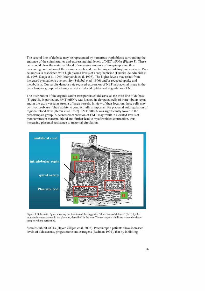
The second line of defense may be represented by numerous trophoblasts surrounding the entrance of the spiral arteries and expressing high levels of NET mRNA (Figure 3). These cells could clear the maternal blood of excessive amounts of norepinephrine, thus preventing contraction of the uterine vessels and maintaining circulatory homeostasis. Pre-eclampsia is associated with high plasma levels of norepinephrine (Ferreira-de-Almeida et al. 1998; Kaaja et al. 1999; Manyonda et al. 1998). The higher levels may result from increased sympathetic overactivity (Schobel et al. 1996) and/or reduced uptake and metabolism. Our results demonstrate reduced expression of NET in placental tissue in the preeclampsia group, which may reflect a reduced uptake and degradation of NE.
The distribution of the organic cation transporters could serve as the third line of defense (Figure 3). In particular, EMT mRNA was located in elongated cells of intra lobular septa and in the extra vascular stroma of large vessels. In view of their location, these cells may be myofibroblasts. Their ability to contract villi is important for placental autoregulation of regional blood flow (Demir et al. 1997). EMT mRNA was significantly lower in the preeclampsia group. A decreased expression of EMT may result in elevated levels of monoamines in maternal blood and further lead to myofibroblast contraction, thus increasing placental resistance to maternal circulation.
Figure 3: Schematic figure showing the location of the suggested “three lines of defence” (I-III) by the monoamine transporters in the placenta, described in the text. The rectangulars indicate where the tissue samples where performed.
Steroids inhibit OCTs (Hayer-Zillgen et al. 2002). Preeclamptic patients show increased levels of aldosterone, progesterone and estrogens (Redman 1991), that by inhibiting
37
monoamine transporters in the placenta, could contribute to elevated monoamine plasma levels.
In addition to the differential gene expression of the transporters, the function of the transporter protein must be considered. If the significant decrease in the expression of NET and EMT mRNAs in pre-eclamptic placentae reflect a decreased transporter function, the “three lines of defense” hypothesized above could help to explain some of the altered hemodynamics that characterize preeclampsia.
Endometrial cell cultures and specific histamine uptake
Primary cell cultures of endometrial cells show specific uptake of histamine, indicating the presence of functional transporter proteins. A common denominator for EMT and VMAT2 is that both have high affinity for histamine (Erickson et al. 1992; Grundemann et al. 1999; Merickel and Edwards 1995 627). Since histamine uptake is inhibited by corticosterone and reserpine, the presence of functional proteins for EMT and VMAT2 is indicated.
Basal uptake of radiolabeled histamine did not differ between the proliferative and secretory phase in stromal cells. In spite of the fact that EMT mRNA is mainly expressed in the secretory phase, PMAT mRNA was present in the proliferative phase. Despite low affinity for histamine, PMAT transports histamine at a high rate (Engel and Wang 2005). In addition, the expression of VMAT2 and the weak expression of EMT and OCT2 genes may add to the uptake of histamine in the proliferative phase. The combined effects of these transporter mechanisms provide an explanation for the lack of difference in basal uptake between the cyclic phases.
Corticosterone inibited uptake in both the proliferative and the secretory phase endometrium. The full inhibitory effect was seen at a higher concentration of corticosterone in the proliferative phase. The possible contribution of OCT2 and PMAT to uptake in the proliferative phase may explain the difference, since their sensitivity to corticosterone is less compared to that of EMT (Grundemann et al. 1998; Hayer-Zillgen et al. 2002). Reserpine showed an inhibitory effect both in the proliferative and in the secretory phase. Despite our ISHH results, this suggests that VMAT2 protein, synthesized in the proliferative phase, persists into the secretory phase, or that a yet unidentified transporter protein with similar sensitivity to reserpine is expressed in this phase. Reserpine inhibition reached a plateau at increasing inhibitor concentrations in the proliferative phase, but not in the secretory phase, when VMAT2 mRNA was not detected in stroma cells. There is a similarity between the inhibition patterns of EMT and VMAT2, i.e. inhibition reaches a plateau in the cyclic phase where the transporter mRNA is strongly expressed and the transporter protein is likely to be abundant. Differences between the cyclic phases may suggest that higher concentration of the inhibitor is required at a lower number of transporter molecules, or that there may be a variation in the sensitivity of transporter proteins to inhibitors between the cyclic phases. In accordance with our ISHH results, the uptake data on human endometrial epithelial cells suggest that there is no plasma membrane transporter with affinity for histamine. NET has a higher affinity for DA than NE, but nothing has been reported on its ability to transport
38
histamine. We found that radiolabeled histamine was not available to VMAT2 during short-term incubations. Prolongation of incubation allowed 3H-histamine to accumulate in the cells by diffusion with higher concentrations in the secretory phase. A possible explanation could be that, in the epithelial cells of the secretory phase, the presence of VMAT2 allows accumulation in vesicles, creating a diffusion gradient and thus a new diffusion of histamine from extracellular fluid into the cytoplasma. Under these conditions, reserpine only inhibited the accumulation of histamine in the secretory phase, suggesting the presence of functional VMAT2 protein.
The cellular origin of endometrial histamine is still controversial. Expression of a rate-limiting enzyme in histamine synthesis, histidine decarboxylase (HDC), has not been reported in human endometrium. Synthesis by migratory mast cells is a well-known source of histamine, and these are known to be present in human endometrium and uterine fluid (Casslen et al. 1982). Uptake of histamine from the environment by EMT, PMAT and VMAT2 offers an alternative source of endometrial histamine. Even very low concentrations of histamine in blood and extra-cellular fluids may serve as substrate for the high affinity transporters in the stationary endometrial cells.
Histamine reportedly plays a role in decidualization, implantation, placenta formation, immunemodulation and blood flow regulation (Barkai and Kraicer 1996; Cocchiara et al. 1986; Dey 1981; Hatanaka et al. 1982). In particular, decidualization is initiated by the presence of the conceptus after appropriate priming with estradiol and progesterone and involves edema and hyperemia, two classical effects of histamine (Hoffman et al. 1990; Paria et al. 2000; Rockwell et al. 2002). Johnson & Dey showed that estrogen induced implantation was inhibited in mice treated with dexamethasone (Dey 1981), possibly through inhibition of organic cation transporters and their histamine uptake.
In mice HDC has been demonstrated in uterine epithelial cells with peak expression at the time of implantation (Paria et al. 1998). The blastocyst expresses histamine type 2 receptor (H2), which is the target for histamine activation (Zhao et al. 2000). After implantation, the placenta is developed by invasion of trophoblasts into the endometrium and maternal vasculature. Histamine enhances the invasion by the activation of the histamine type 1 (H1) receptor on the cytotrophoblasts (Liu et al. 2004).
Modification of the spiral arteries has been shown to be inadequate in preeclampsia (de Groot and Taylor 1993; Khong et al. 1986; Pijnenborg et al. 1991). Inadequate regulation of monoamine levels, and in particular of histamine, during implantation and placental formation may influence the trophoblast invasion into the spiral arteries. Histamine is also a potent vaso-active agent, that modulates local blood flow and capillary permeability (Barkai and Kraicer 1996). Regulation of the uterine blood flow is important during both menstruation and pregnancy, when the blood flow increases dramatically to meet the demands of the fetus.
Interestingly, none of the examined plasma membrane transporters showed an overlapping expression pattern with the vesicular transporter in the secretory phase. If histamine is accumulated in stromal cell vesicles during the proliferative phase, it may be released to the extracellular fluid and subsequently enter epithelial cells in the secretory phase for storage in vesicles. A hypothesis could be that histamine is taken up in epithelial vesicles by
39

VMAT2 and may then be released in response to embryonic histamine releasing factor (HFR) to stimulate decidua formation and support implantation.
40
SUMMARY
Monoamines are potent mediators of physiological and pathophysiological events throughout the body, including the reproductive system. Specific membrane bond transporter proteins tightly regulate their extracellular concentrations. The monoamine transporters have been extensively studied, mainly in the CNS. The aim of this thesis was to study monoamine transporters and their involvement in human reproduction.
Neuronal and non-neuronal monoamine transporters are expressed in human endometrium and placenta. This suggests involvement in the reproductive process by regulating reuptake and release of monoamines.
Monoamine transporters are expressed in endometrial tissue in a cycle-dependent way (I - IV). Among neuronal monoamine transporters, NET mRNA was mainly expressed in the proliferative phase in glandular epithelial cells. VMAT2 mRNA showed a dynamic pattern over the menstrual cycle: stromal expression mainly in the proliferative phase, and strong epithelial expression in the secretory phase. Non-neuronal monoamine transporters were restricted to the stroma. PMAT mRNA was mainly expressed in the proliferative phase, while EMT mRNA expression increased during the proliferative phase, reached a peak in the early secretory phase, and then decreased gradually. Monoamine transporter uptake may be regulated differently in the proliferative and secretory phases by the cyclic variations in estradiol and progesterone that may have a protective function in the non-pregnant uterus as well as during pregnancy by preventing sympathetic hyperactivity and by regulating the uterine blood flow. Ensuring an optimal toxin free milieu in the uterine cavity could also be of crucial importance for the proper implantation and survival of the blastocyst.
Based on the anatomical distribution in the placenta, our results suggest that the transporters may form three lines of defense against monoamines in blood involving spiral arteries and myofibroblasts (Figure 3). A first line of defense may consist of trophoblast cells lining the spiral arteries closest to the myometrium expressing both NET and VMAT2 mRNA. These cells, capable of uptake and regulated release of monoamines, could be important for controlling regional blood flow and maintaining adequate perfusion. A second line of defense might be represented by numerous trophoblasts surrounding the entrance of the spiral arteries and expressing high levels of NET mRNA. These cells could clear the maternal blood of excessive amounts of norepinephrine, thus preventing contraction of the uterine vessels and maintaining circulatory homeostasis. A third line of defense might be the organic cation transporters, in particular myofibroblasts expressing EMT mRNA, that, by contraction of villi, could be important for placental autoregulation of local blood flow. In situ hybridization showed a similar pattern of monoamine transporter expression in normal and preeclamptic placentas. Real time PCR demonstrated a significantly lower expression of NET mRNA and EMT mRNA in placentas from preeclamptic compared to normal pregnancies. If the significant decrease in the expression of NET and EMT mRNAs in preeclamptic placentas reflects a lower transporter protein function, the “three lines of defense”
41
hypothesis might help to explain some of the altered hemodynamics that characterizes preeclampsia.
Demonstration of monoamine transporter mRNA does not necessarily prove the presence of functional transporter protein activities. Future studies applying other methods should address this question. Primary cell cultures of endometrial cells showed specific uptake of histamine, indicating the presence of functional transporter proteins (III). The fact that corticosterone and reserpine inhibited histamine uptake suggests the presence of EMT and VMAT2 functional proteins.
Basal uptake of radiolabeled histamine in stromal cells remained the same during the endometrial cycle. Corticosterone and reserpine inibited uptake in both the proliferative and the secretory phase. There was a similarity between the inhibition patterns for EMT and VMAT2: inhibition reached a plateau in the cyclic phase where the mRNA transporter was strongly expressed and transporter protein was likely to be abundant. In accordance with our ISHH results, the uptake data on human endometrial epithelial cells suggested that there is no plasma membrane transporter with affinity for histamine. We found that radiolabeled histamine was not available to VMAT during short-term incubation. Prolongation of incubation allowed 3H-histamine to accumulate in the cells by diffusion with higher concentrations in the secretory phase. Under these conditions, reserpine only inhibited the accumulation of histamine in the secretory phase, suggesting the presence of functional VMAT2 protein in epithelial cells.
Uptake of histamine from the environment by EMT, PMAT and VMAT2 could be a source of endometrial histamine, since the rate-limiting enzyme in histamine synthesis, HDC, has not been found in human endometrium. Histamine plays a role in decidualization, implantation, placenta formation, immunomodulation and blood flow regulation. None of the examined plasma membrane transporters showed an overlapping expression pattern with the vesicular transporter in the secretory phase. We hypothesized that histamine, accumulated in stromal cell vesicles during the proliferative phase, may be released to the extra-cellular fluid, then enter epithelial cells in the secretory phase for storage in vesicles, to be released in response to embryonic HFR (histamine releasing factor) stimulating decidua formation and support implantation. Another hypothesis is that the VMAT 2 protein is relocated from the stromal cells to the epithelial cells, in what has been recently described in monoaminergic neurons as “trafficking” (Riddle et al. 2007).
42
CONCLUSIONS
- Norepinephrine transporter (NET) mRNA expression was detected in the proliferative phase in glandular epithelial cells of the endometrium
- Vesicular monoamine transporter 2 (VMAT2) mRNA was expressed in the endometrial stroma during the menstrual cycle, mainly in the late proliferative phase
- VMAT2 m RNA showed a strong epithelial expression in the secretory phase
- Plasma membrane monoamine transporter (PMAT) mRNA expression was strong in the stroma during the proliferative phase
- Extra-neuronal monoamine transporter (EMT) mRNA was mainly observed in the stroma during the secretory phase
- No neuronal or non-neuronal monoamine transporters were detected in the endometrium during the menstrual phases or the decidua
- NET mRNA was found in syncytiotrophoblast cells lining chorionic villi with a low intensity expression and in trophoblast cells of the anchoring villi with a stronger intensity expression
- EMT mRNA was expressed in scattered adventitia cells in placental vessels as well as in intralobular septa cells
- In situ hybridization histochemistry showed a similar monoamine transporter expression pattern in normal and preeclamptic placentas
- Real time PCR assay of tissue extracts demonstrated a significantly lower expression of NET mRNA and EMT mRNA in the centre of placentas from preeclamptic as compared to normal pregnancies
- Specific uptake of stromal cell cultures did not differ between the proliferative and the secretory phase of the endometrium
- Stromal cell cultures pre-treated with corticosterone showed a significant inhibition of 3H-histamine specific uptake in both the proliferative and the secretory phase
- Reserpine pre-treatment of stromal cell cultures inhibited histamine uptake in the proliferative but not in the secretory phase
- No specific histamine uptake was detected in the gland cultures, either in the proliferative or in the secretory phase culture
- Prolongation of incubation in glandular cell cultures allowed diffusion of 3H-histamine into the cells. The accumulation of histamine was significantly higher in the secretory phase
43

- Reserpine pre-treatment of gland cultures after prolongation of incubation resulted in a reduction of accumulated 3H-histamine in secretory but not proliferative phase glands
Our results suggest that monoamine transporters may have specific functions in female human reproduction by maintaining adequate levels of extracellular monoamines. Their presence and dynamic expression suggest an important role during the menstrual cycle and pregnancy. Moreover a defective gene expression or function of the monoamine transporters might be determinant in the onset of preeclampsia and its alteration in the vascular bed. Knowledge of the regulation of monoamine metabolism in the endometrium, decidua and placenta will increase the understanding of infertility problems and may offer new pharmacological approaches to optimise assisted reproduction and treatment of preeclampsia.
44
FUTURE DIRECTIONS
The presence of transporters mRNAs doesn’t necessarily reflect the presence of functional proteins. We have shown functional EMT and VMAT2 transporter proteins throughout the menstrual cycle, further functional studies on the other monoamine transporters should be performed.
The effects of antidepressants and drug addiction on monoamine transporters during pregnancy could also be an interesting topic to explore.
Our data should also stimulate further studies on the regulation of monoamines, in particular of histamine, in the endometrium and deciduas, in fertile as well as infertile women in order to increase the understanding of infertility problems and offer new pharmacological approaches to optimize assisted reproduction.
Most studies correlating monoamine transporters with diseases, such as preeclampsia in this study, have been performed in vitro. The results have never been verified via in vivo
methods because imaging of monoamine transporters in living systems has been hampered due to the lack of suitable radioligands. Once new ligands will be available PET studies on the endometrium would be of great interest.
45
POPULÄRVETENSKAPLIG SAMMANFATTNING PÅ SVENSKA (Popularized summary in Swedish)
Monoaminer
Adrenalin, noradrenalin, dopamine, serotonin och histamin är samtliga monoaminer som fungerar som signalsubstanser och hormoner. Monoaminer är viktiga i många fysiologiska processer och obalans i deras nivåer ger ett brett spektrum av störningar. De klassiska är: serotoninbrist som ger depression, ångest och sömnstörningar, dopaminbrist ger motoriska störningar som ses vid Parkinsons sjukdom. Histamin frisätts i stora mängder vid allergiska reaktioner vilket leder till blodtrycksfall och en förträngning av luftvägarna. Vidare har serotonin och histamin viktiga roller i samband med att fostret fäster till livmoderslemhinnan, och dels vid reglering av blodgenomströmning till moderkakan.
Monoamintransportörer
Eftersom monoaminer är potenta signalsubstanser som medierar flera fysiologiska funktioner, är det viktigt att deras nivåer regleras. Denna koncentrationsreglering sker I huvudsak av monoamintransportörerna. Dessa är lokaliserade på cellers ytmembran och utgör där en por som selektivt kan suga upp (dvs transportera in i cellen) och inaktivera signalsubstanserna ifrån miljön utanför cellen. Detta innebär att kommunikationen mellan cellerna upphör. Inne i cellen lagras signalsubstansen i små blåsor med hjälp av ytterliggare en typ av transportörer och kan sedan återanvändas vid förnyad signalering.
Man har hittills identifierat en specifik transportör för varje monoamin utom histamin. De grupperas i huvudsakligen två familjer: -de neuronala, som främst uttrycks i hjärnan och är substansspecifika - de icke neuronala, som återfinns i andra organ såsom njurar och lever där de, förutom att transportera monoaminer, även kan inaktivera vissa miljögifter.
Ytterliggare en familj av transportörer finns, vesikeltransportörerna. Strukturellt skiljer sig inte dessa från de andra, men pumpmekanismen drivs av olika saltjoner. Vesikeltransportörerna pumpar alla monoaminerna men effektivast histamin.
Farmakologisk blockering av monoamintransportörerna har revolutionerat behandlingen av depressioner. Moderna antidepressiva preparat (ex Fontex®) blockerar specifikt serotoninpumpen och höjer därmed de låga nivåerna av serotonin som är typiska vid depression och ångest.
Målsättning
Målet med avhandlingen var; -att studera distribution och funktion av monoamintransportörerna i livmoderslemhinnan under den normala menstruationscykeln samt i tidig graviditet. -att studera distrubution och funktion av monoamintransportörerna i moderkakan vid havandeskapsförgiftning (preeklampsi) samt vid normal graviditet.
Resultat
Fyra olika expressionsmönster av de olika monoamintransportörerna kunde urskiljas i menstrationscykelns två huvudfaser, proliferations- respektive sekretionsfasen (arbete I, III). Vidare visades, i celler isolerade ifrån livmoderslemhinnan, att histamin aktivt kan tas
46

upp med hjälp av monoamintransportörer lokaliserade i cellernas ytmembran samt att histamin kan lagras inuti cellernas små blåsor för att därifrån åter aktivt frisättas (arbete IV). I moderkakan beskrivs en lägre nivå, av framförallt noradrenalintransportören, vid havandeskapsförgiftning (arbete II).
Slutsatser
Våra resultat påvisar en tidigare icke känd distribution av monoamintransportörer i livmoderslemhinnan. Fynden talar för att monoaminer har en viktig roll i såväl den normala menstruationscykeln som under graviditeten. Monoamintransportörerna dynamiska expression indikerar att de spelar en viktig roll vid finreglering av de fysiologiska nivåerna av monoaminer generellt och mer specifikt histamin. Denna nya kunskap kan få betydelse för utvecklande av framtida behandlingsstrategier vid infertilitet samt vid havandeskapsförgiftning.
47

RIASSUNTO DIVULGATIVO IN ITALIANO (Popularized summary in Italian)
Le monoamine
L’adrenalina, la noreadrenalina, la dopamina, la serotonina e l’istamina sono monoamine, e sono tutte dei neurotrasmettitori che permettono alle cellule di scambiarsi dei segnali. Esse sono importanti in molti processi come l’attenzione, la modulazione delle funzioni motorie, il risveglio, l’umore, l’ansia e il comportamento sessuale. Inoltre la serotonina e l’istamina sono determinanti nella gravidanza per la formazione della placenta e l’impianto dell’embrione. In generale le monoamine sono anche importanti nel regolare il flusso di sangue nei vasi.
I trasportatori delle monoamine
Visto che le monoamine sono dei mediatori di eventi fisiologici e patologici molto importanti, la loro concentrazione extracellulare, e quindi la loro azione, sono sotto stretto controllo. I trasportatori delle monoamine sono delle proteine che si trovano nelle membrane cellulari e svolgono la funzione di regolare l’azione delle monoamine. Essi assomigliano a dei canali che permettono il passaggio delle monoamine, all’interno delle cellule, nel citoplasma. Qui le monoamine, anziche´ essere distrutte, vengono in gran parte reciclate per essere riutilizzate. Si conoscono due grandi famiglie di trasportatori delle monoamine di membrana; essi sono: - I trasportatori neuronali, studiati da piu´ tempo soprattutto a livello del sistema nervoso centrale - I trasportatori non neuronali, scoperti piu´ di recente. Questi ultimi non sono specifici per le monoamine ma trasportano all’interno della cellula molte atre sostanze. Esistono inoltre i trasportatori delle monoamine vescicolari che si trovano nel citoplasma e permettono di conservare le monoamine in piccole vescicole nel citoplasma, come riserva.
I trasportatori delle monoamine sono molto interessanti anche in considerazione della loro interazione con farmaci per la depressione, le droghe, gli analgesici, e alcune malattie neurologiche e psichiatriche.
In questo studio i trasportatori delle monoamine sono stati studiati in relazione ad un loro possible coinvolgimento nella riproduzione umana.
Sono stati eseguiti degli esperimenti in laboratorio su tessuti prelevati in sala operatoria. Per esempio sono state utilizzate sonde fluorescenti, quindi visibili al microscopio, specifiche per i singoli transportatori, che a contatto con i tessuti, ed in presenza dei trasportatori, rimangono adese. Sono state inoltre utilizzate altre metodiche di laboratorio che permettono di dare informazioni quantitative (PCR), o funzionali.
Innanzitutto è stato possible osservare la presenza dei trasportatori delle monoamine in determinati tessuti.
48
In particolare sono stati identificati e localizzati nella placenta e per la prima volta nell’endometrio, il tessuto che riveste la cavità uterina. Si è visto che nell’endometrio la loro presenza subisce delle variazioni in funzione delle fasi del ciclo mestruale. Sono state inoltre messi a confronto tessuti di placente ottenuti da gravidanze normali e da gravidanze complicate da preeclampsia, una patologia molto frequente in garividanza di cui si conosce poco. Questa malattia consiste principalmente in un innalzamento della pressione arteriosa, nella perdita di proteine dalle urine e riduzione del flusso di sangue. Essa può degenerare in crisi epilettiche ed ha una prognosi grave sia per la madre che per il feto. Si è dimostrato che esiste una differenza in termini quantitativi della presenza dei trasportatori delle monoamine nei due gruppi. In particolare nelle gravidanze complicate dalla preeclampsia la presenza di tali trasportatori è inferiore.
Questo studio apre nuovi orizzonti sulla funzione dei trasportatori delle monoamine nella riproduzione umana e permette un nuovo approccio nel tentativo di identificare le cause dell’insorgenza della preeclampsia.
49
AKNOWLEDGEMENTS
1996 was my first time in Sweden and my first approach to research as a young medical student, since then many things have happened in my life and at work. I wish to thank all the people I met during this period that made me love Sweden and my job. I would particularly like to mention the following:
Stefan Hansson, my supervisor and head tutor, for guiding me in the fascinating world of monoamine transporters, but most of all for his support, knowledge, energy, irony, friendship and incredible laughs. Thank you “CAPO” for believing in me!
Professor Karel Marsal, for welcoming and giving me the chance to take part to the life of the Department of Obstetrics and Gynecology as a student and later for introducing me to BMC and research.
Professor Luigi Marzetti for giving me the opportunity to come to Sweden and believing in this experience and in my research.
Isis Amer Wåhlin, the reason for me to become a gynecologist, for her support, strenght, smiles and for being there even in the toughest moments.
Håkan Rydström for his knowledge and patience during our neverending statistics discussions that I always will miss.
David Ley for very interesting discussions, questions and very precious help.
Göran Lingman for his kindness and support.
Bertil Casslen for his collaboration, nice discussions, and interesting hints.
Barbro Gustavsson and Irene Larsson for their patience in teaching me how to survive in a lab and how to perform ISHH with a joyful touch.
Thanks to all at the Department of Obstetrics and Gynecology at the University Hospital of Lund for kindness and interesting discussions. Thank you Maria and Lefteria for your generous help. Thank you Ingrid Cordesius for teaching me how natural and marvellous a delivery can/should be!
Thanks to all my colleagues at BMC, in particular to Katrien, Vera, Emir and Andreas with our nice coffee breaks and chats.
A special thank and lots of gratitude to my “Swedish family”: the Wåhlins, for a cosy house, hot meals, warmth, chats and interesting discussions on research and life. Thank you lilla Julia for teaching me the few sentences in Swedish I remember!
50
I would also like to thank my dear friends from Swemsic-IFMSA: Lars, Karin, Johan, Kevin, Kathy, Maria, Hans and Attila that with their friendship represented one of the reason for which I loved Sweden at first.
Grazie Renato, Inge, Norbert, Eva, Christina, and Gunilla for warm friendship, relaxing evenings, good food, beautiful art, and more to come.
Eva, Annamaria, and Katrin I will always miss our dinners with Italian/Swedish lessons and can’t wait to materialize our summer together with your beautiful partners and sons!
If travelling and working abroad is an happy event is also thanks to the people who stay home, love and wait for you: la Tata, Tita, Pia, Luisa, Barbara, Fausta, Marcia, Carrie, Amy and friends and colleagues who make it possible, Elena, Laura, Chiara, Azzurra, Assunta, Barbara.
A special and energetic thank to my very good friends M&M, just for being there.
Thank you Franca and Giulio for teaching me the joy of a good meal together and for baby sitting!
Above all these I wish to thank my parents, Nello e Grazia, and sister Sonia for teaching me to love life and for supporting and enjoying all my travels.
Alessandro, Giulia and Andrea for reminding me how beautiful life really is.
51
REFERENCES
Alm P, Falck B, Owman C, Sjöberg NO, Thorbert G. 1975. Reduced level of uterine norepinephrine transmitter during hCG-induced pseudopregnancy in the rabbit. endocrin 96(3):819-822.
Altman DG. 1991. Practical statistics for medical research. London: Chapman & Hall.
Amara SG, Kuhar MJ. 1993. Neurotransmitter transporters: recent progress. AnnuRevNeurosci 16:73-93.
Amphoux A, Vialou V, Drescher E, Bruss M, La Cour CM, Rochat C, Millan MJ, Giros B, Bonisch H, Gautron S. 2006a. Differential pharmacological in vitro properties of organic cation transporters and regional distribution in rat brain. Neuropharmacology.
Amphoux A, Vialou V, Drescher E, Bruss M, Mannoury La Cour C, Rochat C, Millan MJ, Giros B, Bonisch H, Gautron S. 2006b. Differential pharmacological in vitro properties of organic cation transporters and regional distribution in rat brain. Neuropharmacology 50(8):941-952.
Apparsundaram S, Moore KR, Malone MD, Hartzell CH, Blakely RD. 1997. Molecular Cloning and Characterization of an L-Epinephrine Transporter from Sympathetic Ganglia of the Bullfrog, Rana catesbiana. JNeurosci 17(8):2691-2702.
Arnold JT, Kaufman DG, Seppala M, Lessey BA. 2001. Endometrial stromal cells regulate epithelial cell growth in vitro: a new co-culture model. 16(5):836-845.
Axelrod J. 1971. Brain monoamines. Biosynthesis and fate. NeurosciResProgramBull 9:188-196.
Balkovetz DF, Tiruppathi C, Leibach FH, Mahesh VB, Ganapathy V. 1989. Evidence for an imipramine-sensitive serotonin transporter in human placental brush-border membranes. J BiolChem 264:2195-2198.
Barash A, Dekel N, Fieldust S, Segal I, Schechtman E, Granot I. 2003. Local injury to the endometrium doubles the incidence of successful pregnancies in patients undergoing in vitro fertilization. Fertil Steril 79(6):1317-1322.
Barkai U, Kraicer PF. 1996. Intrauterine signaling and embryonic implantation. biological signals 5:111-121.
Baxter SW, Thomas EJ, Campbell IG. 2001. GSTM1 null polymorphism and susceptibility to endometriosis and ovarian cancer. Carcinogenesis 22(1):63-65.
Beer DJ, Matloff SM, Rocklin RE. 1984. The influence of histamine on immune and inflammatory responses. advances in Immunology 35:209-268.
52
Bel'skaia GD. 1990. The role of biogenic amines in the mechanism of uterine hemorrhage in uterine myoma. Akush Ginekol (Mosk) 2:17-21.
Blakely RD, Berson HE, Fremeau RT, Jr., Caron MG, Peek MM, Prince HK, Bradley CC. 1991. Cloning and expression of a functional serotonin transporter from rat brain. Nature 354:66-70.
Borowsky B, Hoffman BJ. 1995. Neurotransmitter transporters: molecular biology, function, and regulation. IntRevNeurobiology 38:139-199.
Bottalico B, Larsson I, Casslen B, Broszki J, Hernandez-Andrade E, Marsal K, Hansson RS. 2004. Norepinephrine (NET), Serotonin (SERT), vesicular monoamine (VMAT2) and organic cation transporters (OCT1,2 and EMT) in human placenta from preeclamptic and normotensive pregnancies". Placenta 25(6):518-529.
Bottalico B, Noskova V, Pilka R, Larsson I, Domanski H, Casslen B, Hansson SR. 2007. Organic cation transporter (OCT 1, OCT 2 and EMT) and the plasma membrane monoamine transporter (PMAT) show differential distribution and cyclic expression in human endometrium and early pregnancy decidua. Molecular Reproduction and Development 74 (10):1303-1311.
Bottalico B, Pilka R, Larsson I, Casslen B, Marsal K, Hansson S. 2003. Plasma membrane and vesicular monoamine transporters in normal endometrium and early pregnancy decidua. Mol Hum Reprod 9(7):389-394.
Bourgain C, Devroey P. 2003. The endometrium in stimulated cycles for IVF. Human Reproduction Update 9(6):515-522.
Bradley DJ, Towle HC, Young WS, III. 1992. Spatial and Temporal Expression of - and -Thyroid Hormone Receptor mRNAs, including the 2-Subtype, in the developing
Mammalian Nervous System. JNeurosci 12(6):2288-2302.
Brownstein MJ, Hoffman BJ. 1994. Neurotransmitter transporters. RecentProgHormRes 49:27-42.
Budde IK, Aalberse RC. 2003. Histamine-releasing factors, a heterogeneous group of different activities. Clin Exp Allergy 33(9):1175-1182.
Buznikov GA. 1984. The action of neurotransmitters and related substances on early embryogenesis. PharmacolTher 25:23-59.
Buznikov GA, Shmukler YB, Lauder JM. 1996. From oocyte to neuron: do neurotransmitters function in the same way throughout development? Cell MolNeurobiol 16:537-559.
Carrasco G, Cruz MA, Dominguez A, Gallardo V, Miguel P, Gonzalez C. 2000a. The expression and activity of monoamine oxidase A, but not of the serotonin
53

transporter, is decreased in human placenta from pre-eclamptic pregnancies. Life Sciences 67:2961-2969.
Carrasco G, Cruz MA, Dominguez A, Gallardo V, Miguel P, Gonzalez C. 2000b. The expression and activity of monoamine oxidase A, but not of the serotonin transporter, is decreased in human placenta from pre-eclamptic pregnancies. Life Sciences 67(24):2961-2969.
Casslen B, Kobayashi TK, Stormby N. 1982. Cyclic variation of the cellular components in human uterine fluid. J Reprod Fertil 66(1):213-218.
Casslen B, Nordengren J, Gustavsson B, Nilbert M, Lund LR. 1995. Progesterone stimulates degradation of urokinase plasminogen activator (u-PA) in endometrial stromal cells by increasing its inhibitor and surface expression of the u-PA receptor. Journal of Clinical Endocrinology and Metabolism 80(9):2776-2784.
Chaddha V, Viero S, Huppertz B, Kingdom J. 2004. Developmental biology of the placenta and the origins of placental insufficiency. Semin Fetal Neonatal Med 9(5):357-369.
Chang AS, Chang SM, Starnes DM, Schroeter S, Bauman AL, Blakely RD. 1996. Cloning and expression of the mouse serotonin transporter. Brain ResMolBrain Res 43:185-192.
Cocchiara R, Di Trapani G, Azzolina A, Albeggiani G, Geraci D. 1986. Early embryonic histamine-releasing factor: a new model for human implantation. human reproduction 1(7):445-447.
Conneely OM, Lydon JP. 2000. Progesterone receptors in reproduction: functional impact of the A and B isoforms. Steroids 65(10-11):571-577.
Cooke PS, Buchanan DL, Lubahn DB, Cunha GR. 1998. Mechanism of estrogen action: Lessons from the estrogen receptor-alfa knockout mouse. Biol Reprod 59:470-475.
de Groot CJM, Taylor RN. 1993. New Insights into the etiology of pre-eclampsia. Annals of Medicine 25:243-249.
Declercq de Perez Bedes G, Garcia Bienere W. 1975. Retention of noradrenaline by the rat endometrial epithelium. Action of estradiol. Ann Endocrinol (Paris) 36(4):185-187.
Demir R, Kosanke G, Kohnen G, Kertschanska S, Kaufmann P. 1997. Classification of Human Placental Stem Villi:
Review of Structural and Functional Aspects. MICROSCOPY RESEARCH AND TECHNIQUE 38(29-41).
Dey SK. 1981. Role of histamine in implantation:inhibition of histidine decarboxylase induced delayed implantation in the rabbit. biology of reproduction 24:867-869.
54
Dey SK, Johnson DC. 1980. Histamine formation by mouse preimplantation embryos. J Reprod Fert 60:457-460.
Dey SK, Johnson DC, Santos JG. 1979a. Is histamine production by the blastocyst required for implantation in the rabbit? Biol Reprod 21(5):1169-1173.
Dey SK, Villanueva C, Abdou NI. 1979b. Histamine receptors on rabbit blastocyst and endometrial cell membranes. Nature 278(12):648-649.
Ding YS, Lin KS, Logan J. 2006. PET imaging of norepinephrine transporters. Curr Pharm Des 12(30):3831-3845.
Eisenhofer G. 2001. The role of neuronal and extraneuronal plasma membrane transporters in the activation of peripheral catecholamines. Pharmacology and therapeutics 91:35-62.
Enders AC. 1997. Implantation, Embryology. San Diego: Academic Press.
Enders AC. 2000. Trophoblast-uterine interactions in the first days of implantation: models for the study of implantation events in the human. Semin Reprod Med 18(3):255-263.
Engel K, Wang J. 2005. Interaction of organic cations with a newly identified plasma membrane monoamine transporter.
Engel K, Wang J. Mol Pharmacol 68(5):1397-1407.
Engel K, Zhou M, Wang J. 2004. Identification and characterization of a novel monoamine transporter in the human brain. J Biol Chem 279(48):50042-50049.
Enmark E, Pelto-Huikko M, Grandien K, Lagercrantz S, Lagercrantz J, Fried G, Nordenskjold M, Gustafsson JA. 1997. Human estrogen receptor beta-gene structure, chromosomal localization, and expression pattern. J Clin Endocrinol Metab 82(12):4258-4265.
Erickson JD, Eiden LE. 1993. Functional identification and molecular cloning of a human brain vesicle monoamine transporter. JNeurochem 61:2314-2317.
Erickson JD, Eiden LE, Hoffman BJ. 1992. Expression cloning of a reserpine-sensitive vesicular monoamine transporter. ProcNatlAcadSciUSA 89:10993-10997.
Erickson JD, Schafer MK, Bonner TI, Eiden LE, Weihe E. 1996. Distinct pharmacological properties and distribution in neurons and endocrine cells of two isoforms of the human vesicular monoamine transporter. ProcNatlAcadSciUSA 93:5166-5171.
Fabre-Nys C. 1998. Steroid control of monoamines in relation to sexual behaviour. Reviews of Reproduction 3:31-41.
55
Ferreira-de-Almeida JA, Amenta F, Cardoso F, Polonia JJ. 1998. Association of circulating endothelin and noradrenaline with increased calcium-channel binding sites in the placental bed in pre-eclampsia. British Journal of Obstetrics and Gynecology 105(Oct):1104-1112.
Fong SK, Liu CQ, Chan HC. 1998. Cellular mechanisms of adrenaline-stimulated anion secretion by the mouse endometrial epithelium. Biol Reprod 59(6):1342-1348.
Fox K. 1995. The critical period for long-term potentiation in primary sensory cortex. Neuron 15:485-488.
Ganapathy V, Leibach FH. 1995. Placental biogenic amines and their transporters. In: Ramasastry BV, editor. Placental Toxicology. Boca Raton. FL.: CRC. pp 161-174.
Ganapathy V, Ramamoorthy S, Leibach FH. 1993. Transport and metabolism of monoamines in the human placenta. Trophoblast Res 7:35-51.
Garris DR, Dar MS. 1985. Decidua-associated changes in guinea pig uterine blood flow and volume:relation to uterine norepinephrine concentrations. Anat Rec 211(4):410-413.
Genbacev O, McMaster MT, Zdravkovic T, Fisher SJ. 2003. Disruption of oxygen-regulated responses underlies pathological changes in the placentas of women who smoke or who are passively exposed to smoke during pregnancy. Reprod Toxicol 17(5):509-518.
Godfrey KM. 2002. The role of the placenta in fetal programming-a review. Placenta 23(Suppl A):S20-27.
Gorboulev V, Ulzheimer JC, Akhoundova A, Ulzheimer-Teuber I, Karbach U, Quester S, Baumann C, Lang F, Busch AE, Koepsell H. 1997. Cloning and characterization of two human polyspecific organic cation transporters. DNA Cell Biol 16(7):871-881.
Granger JP, Alexander BT, Bennett WA, Khalil RA. 2001. Pathophysiology of pregnancy-induced hypertension. Am J Hypertens 14:178S-185S.
Grundemann D, Liebich G, Kiefer N, Koste S, Schomig E. 1999. Selective substrates for non-neuronal monoamine transporters. Molecular Pharmacology 56(1):1-10.
Grundemann D, Schechinger B, Rappold GA, Schomig E. 1998. Molecular identification of the corticosterone-sensitive extraneuronal catecholamine transporter. Nat Neurosci 1(5):349-351.
Gudmundsson S, Marsal J, Dubiel P, Maly P, Marsal K, Laurini R. 2000. Power doppler, x-ray contrast angiography and morphological imaging of the human placenta. Prenat Neonat Med 5:27-34.
56
Hansson SR, Mezey E, Hoffman BJ. 1998. Serotonin Transporter mRNA Expression in Neuronal Crest-derived Structures and Sensory Pathways of the Developing Rat Embryo. Neuroscience 89(1):243-265.
Hart PH. 2001. Regulation of the inflammatory response in asthma by mast cell products. Immunol Cell Biol 79(2):149-153.
Hatanaka K, Kitamura Y, Maeyama K, Watanabe T, Matsumoto K. 1982. Deciduoma formation in uterus of genetically mast cell-deficient W/W mice. biology of reproduction 27:25-28.
Hayer-Zillgen M, Bruss M, Bonisch H. 2002. Expression and pharmacological profile of the human organic cation transporters hOCT1, hOCT2 and hOCT3. Br J Pharmacol 136(6):829-836.
Hendrickson MR, Kempson RL. 1980. Surgical pathology of the uterine corpus. Philadelphia: Saunders.
Hitchmann, Adler. 1908. Acta Obstetrica et Gynaecologica Japonica 17(9):957-964.
Hobel CJ, Parvez H, Parvez S, Lirette M, Papiernik E. 1981. Enzymes for epinephrine synthesis and metabolism in the myometrium, endometrium, red blood cells, and plasma of pregnant human subjects. american journal of obstetrics and gynecology 141(8):1009-1018.
Hoffman BJ. 1994. Expression cloning of a serotonin transporter: a new way to study antidepressant drugs. Pharmacopsychiatry 27:16-22.
Hoffman BJ, Hansson SR, Mezey E, Palkovits M. 1998. Distribution of Monoamine Neurotransmitter Transporters in the Rat Brain. Frontiers in Neuroendocrinology.
Hoffman BJ, Mezey E, Brownstein MJ. 1991. Cloning of a serotonin transporter affected by antidepressants. Science 254:579-580.
Hoffman LH, Winfrey VP, Hoos PC. 1990. Sites of endometrial vascular leakage during implantation in the rabbit. Anat Rec 227(1):47-61.
Iversen LL. 1965. The uptake of catecholamine at high perfusion concentrations in the rat isolated heart: a novel catecholamine uptake process. BritJPharmacol 25:18-33.
Johnson DC, Dey SK. 1980. Role of histamine in implantation: dexamethasone inhibits estradiol-induced implantation in the rat. Biol Reprod 22(5):1136-1141.
Kaaja RJ, Moore PM, Yandle TG, Ylikorkala O, Frampton CM, Nicholls GM. 1999. Blood pressure and vasoactive hormones in mild preeclampsia and normal pregnancy. Hypertension in Pregnancy 18(2):173-187.
57

Kandel ER, Schwartz JH. 1985. Principles of Neural Science. Kandel ER, Schwartz JH, editors. New York: Elsevier.
Kekuda R, Prasad PD, Wu X, Wang H, Fei Y-J, Leibach FH, Ganapathy V. 1998. Cloning and Functional Characterization of a Potential-sensitive,
Polyspecific Organic Cation Transporter (OCT3) Most Abundantly
Expressed in Placenta. Journal of Biological Chemistry 273( 26):15971–15979.
Kennedy JA, Lande de la IS. 1986. Effect of progesterone on the metabolism of noradrenaline in rabbit uterine endometrium and myometrium. Naunyn Schmiedebergs Arch Pharmacol 333(4):368-376.
Khong TY, De Wolf F, Robertson WB, Brosens I. 1986. Inadequate maternal vascular response to placentation in pregnancies complicated by pre-eclampsia and small-for-gestational age infants. British Journal of Obstetrics and Gynecology 93(10):1049-1059.
Kilty JE, Lorang D, Amara SG. 1991. Cloning and expression of a cocaine-sensitive rat dopamine transporter. Science 254:578-579.
Kirmaz C, Aydemir O, Bayrak P, Yuksel H, Ozenturk O, Degirmenci S. 2005. Sexual dysfunction in patients with allergic rhinoconjunctivitis. Ann Allergy Asthma Immunol 95(6):525-529.
Koehler MR, Wissinger B, Gorboulev V, Koepsell H, Schmid M. 1997. The two human organic cation transporter genes SLC22A1 and SLC22A2 are located on chromosome 6q26. Cytogenet Cell Genet 79(3-4):198-200.
Koepsell H. 2004. Polyspecific organic cation transporters: their functions and interactions with drugs. Trends Pharmacol Sci 25(7):375-381.
Kurita T, Lee K, Cooke PS, Taylor JA, Lubahn DB, Cunha GR. 2000. Paracrine regulation of epithelial progesterone receptor by estradiol in the mouse female reproductive tract. Biol Reprod 62:821-830.
Laporte R, DeRoth L. 1997. Modulation of the effects of norepinephrine uptake inhibitors on the norepinephrine-induced contractile response of the porcine uterine artery during early pregnancy. Can J Vet Res 61(3):214-220.
Lazar A, Walitza S, Jetter A, Gerlach M, Warnke A, Herpertz-Dahlmann B, Grundemann D, Grimberg G, Schulz E, Remschmidt H, Wewetzer C, Schomig E. 2007. Novel mutations of the extraneuronal monoamine transporter gene in children and adolescents with obsessive-compulsive disorder. Int J Neuropsychopharmacol:1-14.
58
Lecce G, Meduri G, Ancelin M, Bergeron C, Perrot-Applanat M. 2001. Presence of estrogen receptor beta in the human endometrium through the cycle: expression in glandular, stromal, and vascular cells. J Clin Endocrinol Metab 86(3):1379-1386.
Lessey BA, Killam AP, Metzger DA, Haney AF, Greene GL, McCarty KS, Jr. 1988. Immunohistochemical analysis of human uterine estrogen and progesterone receptors throughout the menstrual cycle. J Clin Endocrinol Metab 67(2):334-340.
Lin Z, Madras BK. 2006. Human genetics and pharmacology of neurotransmitter transporters. Handb Exp Pharmacol(175):327-371.
Lingman G, Marsal K. 1986. Fetal central blood circulation in the third trimester of normal pregnancy. Early Hum Dev 13:137-150.
Little KY, Krolewski DM, Zhang L, Cassin BJ. 2003. Loss of striatal vesicular monoamine transporter protein (VMAT2) in human cocaine users. Am J Psychiatry 160(1):47-55.
Liu Z, Kilburn BA, Leach RE, Romero R, Paria BC, Armant DR. 2004. Histamine enhances cytotrophoblast invasion by inducing intracellular calcium transients through the histamine type-1 receptor. Mol Reprod Dev 68(3):345-353.
Lopata A, Bentin-Ley U, Enders A. 2002. "Pinopodes" and implantation. Rev Endocr Metab Disord 3(2):77-86.
Maekawa F, Yamanouchi K. 1996. Effect of deprivation of serotonin by p-chlorophenylalanine on induction and maintenance of pseudopregnancy in female rats. Brain Res Bull 39(5):317-321.
Manyonda IT, Fenske C, Hole D, Choy MY, Wilson C. 1998. A role for noradrenaline in pre-eclampsia: towards a unifying hypothesis for the pathophysiology. British Journal of Obstetrics and Gynaecology 105:641-648.
Marshak DR. 1990. S100 beta as a neurotrophic factor. ProgBrain Res 86:169-181.
Merickel A, Edwards RH. 1995. Transport of histamine by vesicular monoamine transporter-2. Neuropharmacology 34:1543-1547.
Mertens HJ, Heineman MJ, Theunissen PH, de Jong FH, Evers JL. 2001. Androgen, estrogen and progesterone receptor expression in the human uterus during the menstrual cycle. Eur J Obstet Gynecol Reprod Biol 98(1):58-65.
Meston CM, Frohlich PF. 2000. The neurobiology of sexual function. Arch Gen Psychiatry 57(11):1012-1030.
59

Mezey E, Eisenhofer G, Harta G, Hansson S, Gould L, Hunyady B, Hoffman BJ. 1996. A novel nonneuronal catecholaminergic system: exocrine pancreas synthesizes and releases dopamine. ProcNatlAcadSciUSA 93:10377-10382.
Middelkoop CM, Dekker GA, Kraayenbrink AA, Popp-Snijders C. 1993. Platelet-poor plasma serotonin in normal and preeclamptic pregnancy. Clin Chem 39(8):1675-1678.
Mitchell JA, Hammer RE, Goldman H. 1983. Serotonin-induced disruption of implantation in the rat: II. Suppression of decidualization. Biol Reprod 29(1):151-156.
Moiseiwitsch JR, Lauder JM. 1995. Serotonin regulates mouse cranial neural crest migration. ProcNatlAcadSciUSA 92:7182-7186.
Noskova V, Bottalico B, Orbona I, Pilka R, Larsson I, Casslen B, Marsal K, Hansson SR. 2006. Histamine uptake by organic cation transporter (EMT) and vesicular monoamine transporter (VMAT2) in cultured human endometrial stromal and epithelial cells. Mol Human Reproduction 12(8):483-489.
Noyes RW, Hertig AT, Rock J. 1950. Dating the endometrial biopsy. Fertility and Sterility 1:3-25.
Ogasawara M, Yamauchi K, Satoh Y, Yamaji R, Inui K, Jonker JW, Schinkel AH, Maeyama K. 2006. Recent advances in molecular pharmacology of the histamine systems: organic cation transporters as a histamine transporter and histamine metabolism. J Pharmacol Sci 101(1):24-30.
Oikawa K, Kosugi Y, Ohbayashi T, Kameta A, Isaka K, Takayama M, Kuroda M, Mukai K. 2003. Increased expression of IgE-dependent histamine-releasing factor in endometriotic implants. J Pathol 199(3):318-323.
Olson L, Boreus LO, Seiger A. 1973. Histochemical demonstration and mapping of 5-hydroxytryptamine- and catecholamine-containing neuron systems in the human fetal brain. ZAnatEntwicklungsgesch 139:259-282.
Olson L, Seiger A. 1972. Early prenatal ontogeny of central monoamine neurons in the rat: fluorescence histochemical observations. ZAnatEntwicklungsgesch 137:301-316.
Pacholczyk T, Blakely RD, Amara SG. 1991. Expression cloning of a cocaine- and antidepressant-sensitive human noradrenaline transporter. Nature 350:350-354.
Paczkowski FA, Sharpe IA, Dutertre S, Lewis RJ. 2007. chi-Conotoxin and tricyclic antidepressant interactions at the norepinephrine transporter define a new transporter model. J Biol Chem 282(24):17837-17844.
60
Paria BC, Das N, Das SK, Zhao X, Dileepan KN, Dey SK. 1998. Histidine decarboxylase gene in the mouse uterus is regulated by progesterone and correlates with uterine differentiation for blastocyst implantation. endocrin 139(9):3958-3966.
Paria BC, Lim H, Das SK, Reese J, Dey SK. 2000. Molecular signaling in uterine receptivity for implantation. Semin Cell Dev Biol 11(2):67-76.
Paria BC, Song H, Dey SK. 2001. Implantation: molecular basis of embryo-uterine dialogue. Int J Dev Biol 45(3):597-605.
Pedroza-Garcia E, Garcia Bienere W, Inglesi J, Szerezewski A, Rosner JM. 1975. Interactions in the uptake between catecholamines and estradiol in the human uterus. american journal of obstetrics and gynecology 122(1):96-100.
Peter D, Jimenez J, Liu Y, Kim J, Edwards RH. 1994. The chromaffin granule and synaptic vesicle amine transporters differ in substrate recognition and sensitivity to inhibitors. JBiolChem 269:7231-7237.
Peter D, Liu Y, Brecha N, Edwards RH. 1995a. The transport of neurotransmitters into synaptic vesicles. ProgBrain Res 105:273-281.
Peter D, Liu Y, Sternini C, de Giorgio R, Brecha N, Edwards RH. 1995b. Differential expression of two vesicular monoamine transporters. JNeurosci 15:6179-6188.
Pijnenborg R, Anthony J, Davey DA, Rees A, Tiltman A, Vercruysse L, van Assche A. 1991. Placental bed spiral arteries in the hypertensive disorders of pregnancy. British Journal of Obstetrics and Gynecology 98(7):648-655.
Pilka R, Domanski H, Hansson SR, Eriksson P, Casslén B. 2004. Endometrial TIMP-4 mRNA is high at midcycle and in hyperplasia, but down-regulated in malignant tumours. Coordinated expression with MMP-26. Mol Hum Reprod 10(9):s.641-650.
Pilka R, Whatling C, Eriksson P, Domanski H, Hansson SR, Casslen B. 2003. Epithelial expression of matrix metallo-proteinase-26 (MMP-26) is elevated at midcycle in the human endometrium. Mol Hum Reprod 9(5):271-277.
Poropatich C, Rojas M, Silverberg SG. 1987. Polymorphonuclear leukocytes in the endometrium during the normal menstrual cycle. Int J Gynecol Pathol 6(3):230-234.
Prasad PD, Hoffmans BJ, Moe AJ, Smith CH, Leibach FH, Ganapathy V. 1996. Functional expression of the plasma membrane serotonin transporter but not the vesicular monoamine transporter in human placental trophoblasts and choriocarcinoma cells. Placenta 17:201-207.
Prasad PD, Leibach FH, Mahesh VB, Ganapathy V. 1994. Human placenta as a target organ for cocaine action: interaction of cocaine with the placental serotonin transporter. Placenta 15:267-278.
61

Ramamoorthy S, Bauman AL, Moore KR, Han H, Yang-Feng T, Chang AS, Ganapathy V, Blakely RD. 1993a. Antidepressant- and cocaine-sensitive human serotonin transporter: molecular cloning, expression, and chromosomal localization. Proc Natl Acad Sci U S A 90(6):2542-2546.
Ramamoorthy S, Prasad PD, Kulanthaivel P, Leibach FH, Blakely RD, Ganapathy V. 1993b. Expression of a cocaine-sensitive norepinephrine transporter in the human placental syncytiotrophoblast. Biochemistry 32:1346-1353.
Redman CW. 1991. Immunology of preeclampsia. Semin Perinatol 15(3):257-262.
Richards M, Iijima Y, Kondo H, Shizuno T, Hori H, Arima K, Saitoh O, Kunugi H. 2006. Association study of the vesicular monoamine transporter 1 (VMAT1) gene with schizophrenia in a Japanese population. Behav Brain Funct 2:39.
Riddle EL, Hanson GR, Fleckenstein AE. 2007. Therapeutic doses of amphetamine and methylphenidate selectively redistribute the vesicular monoamine transporter-2. Eur J Pharmacol 571(1):25-28.
Roberts JM, Cooper DW. 2001. Pathogenesis and genetics of pre-eclampsia. Lancet 357(9249):53-56.
Roberts JM, Taylor RN, Musci TJ, Rodgers GM, Hubel CA, McLaughlin MK. 1989. Preeclampsia: An endothelial cell disorder. Am J Obstet Gynecol 161(5):1200-1204.
Robertson WB, Khong TY, Brosens I, De Wolf F, Sheppard BL, Bonnar J. 1986. The placental bed biopsy: review from three European centers. american journal of obstetrics and gynecology 155(2):401-412.
Rockwell LC, Pillai S, Olson CE, Koos RD. 2002. Inhibition of vascular endothelial growth factor/vascular permeability factor action blocks estrogen-induced uterine edema and implantation in rodents. Biol Reprod 67(6):1804-1810.
Salamonsen LA, Jeziorska M, Newlands GF, Dey SK, Woolley DE. 1996. Evidence against a significant role for mast cells in blastocyst implantation in the rat and mouse. Reprod Fertil Dev 8(8):1157-1164.
Schobel HP, Fischer T, Heuszer K, Geiger H, Schmieder RE. 1996. Preeclampsia-a state of sympathetic overactivity. N Engl J Med 335(20):1480-1485.
Schomig E, Lazar A, Grundemann D. 2006. Extraneuronal monoamine transporter and organic cation transporters 1 and 2: a review of transport efficiency. Handb Exp Pharmacol(175):151-180.
Schrey MP, Hare AL, Ilson SL, Walters MP. 1995a. Decidual histamine release and amplification of prostaglandin F2 alpha production by histamine in interleukin-1
62
beta-primed decidual cells: potential interactive role for inflammatory mediators in uterine function at term. J Clin Endocrinol Metab 80(2):648-653.
Schrey MP, Hare AL, Ilson SL, Walters MP. 1995b. Decidual histamine release and amplification of prostaglandin F2 alpha production by histamine in interleukin-1 beta-primed decidual cells: potential interactive role for inflammatory mediators in uterine function at term. J Clin Endocrinol Metab 80(2):648-653.
Schroeder BM. 2002. ACOG practice bulletin on diagnosing and managing preeclampsia and eclampsia. Am Fam Physician 66(2):330-331.
Schroeter S, Levey AI, Blakely RD. 1997. Polarized expression of the antidepressant-sensitive serotonin transporter in epinephrine-synthesizing chromaffin cells of the rat adrenal gland. MolCell Neurosci 9:170-184.
Schuldiner S, Steiner-Mordoch S, Yelin R, Wall SC, Rudnick G. 1993. Amphetamine derivatives interact with both plasma membrane and secretory vesicle biogenic amine transporters. MolPharmacol 44:1227-1231.
Senat MV, Minoui S, Multon O, Fernandez H, Frydman R, Ville Y. 1998. Effect of dexamethasone and betamethasone on fetal heart rate variability in preterm labour: a randomised study. British Journal of Obstetrics and Gynecology 105(7):749-755.
Shelesnyak MC. 1952. Inhibition of decidual cell formation in the pseudopregnant rat by histamine antagonists. Am J Physiol 170(3):522-527.
Shelesnyak MC. 1957. Some experimental studies on the mechanism of ova-implantation in the rat. Recent Prog Horm Res 13:269-322.
Shentu J, Zhou H, He Q, Wang L, Fang R. 2001. Effects of DL111-IT or combined with RU486 on uterine polyamines biosynthesis in rats during early gestation. Contraception 63(5):283-287.
Shiverick KT, Salafia C. 1999. Cigarette smoking and pregnancy I: ovarian, uterine and placental effects. Placenta 20(4):265-272.
Sivasubramaniam SD, Finch CC, Billett MA, Baker PN, Billett EE. 2002. Monoamine oxidase expression and activity in human placentae from pre-eclamptic and normotensive pregnancies. Placenta 23:163-171.
Skarzynski DJ, Uenoyama Y, Kotwica J, Okuda K. 1999. Noradrenaline stimulates the production of prostaglandin f2alpha in cultured bovine endometrial cells. Biol Reprod 60(2):277-282.
Steinbusch HWM, Mulder AH. 1984. Immunohistochemical localization of histamine in neurons and mast cells in the rat brain. In: Bjorklund A, Hokfelt T, Kuhar MJ,
63

editors. Handbook of Chemical Anatomy, Vol 3: Classical Transmitters and Transmitter Receptors in the CNS, Part 11. London: Elsevier Science. pp 126-140.
Talvenheimo J, Rudnick G. 1980. Solubilization of the platelet plasma membrane serotonin transporter in an active form. J Biol Chem 255(18):8606-8611.
Tatsumi M, Groshan K, Blakely RD, Richelson E. 1997. Pharmacological profile of antidepressants and related compounds at human monoamine transporters. Eur J Pharmacol 340:249-258.
Torres GE, Gainetdinov RR, Caron MG. 2003a. Plasma membrane monoamine transporters: structure, regulation and function. Nat Rev Neurosci 4(1):13-25.
Torres GE, Gainetdinov RR, Caron MG. 2003b. Plasma membrane monoamine transporters: structure, regulation and function. Nat Rev Neurosci 4(1):13-25.
Towers CV, Pircon RA, Nageotte MP, Porto M, Garite TJ. 1993. Cocaine intoxication presenting as preeclampsia and eclampsia. Obstet Gynecol 81(4):545-547.
Uhl GR, Kitayama S. 1993. A cloned dopamine transporter. Potential insights into Parkinson's disease pathogenesis. AdvNeurol 60:321-324.
Usdin TB, Mezey E, Chen C, Brownstein MJ, Hoffman BJ. 1991. Cloning of the cocaine-sensitive bovine dopamine transporter. ProcNatlAcadSciUSA 88:11168-11171.
Verhaagh S, Barlow PD, Zwart R. 2001. The extraneuronal monoamine transporter Slc22a3/Orct3 co-localizes with the Maoa metabolizing enzyme in mouse placenta. Mechanisms of Development 100:127-130.
Verhaagh S, Schweifer N, Barlow PD, Zwart R. 1999. Cloning of the Mouse and Human Solute Carrier 22a3 (Slc22a3/SLC22A3) Identifies a Conserved Cluster of Three Organic Cation Transporters on Mouse Chromosome 17 and Human 6q26–q27. Genomics 55:209-218.
Wade PR, Chen J, Jaffe B, Kassem IS, Blakely RD, Gershon MD. 1996. Localization and function of a 5-HT transporter in crypt epithelia of the gastrointestinal tract. J Neurosci 16:2352-2364.
Wallace EM, Baker LS. 1999. Effect of antenatal betamethasone administration on placental vascular resistance. Lancet 353.
Wang Z, Ordway GA, Woolverton W. 2007. Effects of cocaine on monoamine uptake as measured ex vivo. Neurosci Lett 413(3):191-195.
Weihe E, Schafer MK, Erickson JD, Eiden LE. 1994. Localization of vesicular monoamine transporter isoforms (VMAT1 and VMAT2) to endocrine cells and neurons in rat. JMolNeurosci 5:149-164.
64

Weihe E, Tao-Cheng JH, Schafer MK, Erickson JD, Eiden LE. 1996. Visualization of the vesicular acetylcholine transporter in cholinergic nerve terminals and its targeting to a specific population of small synaptic vesicles. ProcNatlAcadSciUSA 93:3547-3552.
Whitaker-Azmitia PM, Druse M, Walker P, Lauder JM. 1996. Serotonin as a developmental signal. BehavBrain Res 73:19-29.
Whitaker-Azmitia PM, Shemer AV, Caruso J, Molino L, Azmitia EC. 1990. Role of high affinity serotonin receptors in neuronal growth. AnnNYAcadSci 600:315-330.
Whitby LG, Axelrod J, Weil-Malherbe H. 1961. The fate of H3-Norepinephrine in animals. J Pharmacol ExpTher 132:193-201.
White MV. 1990. The role of histamine in allergic diseases. J Allergy Clin Immunol 86(4 Pt 2):599-605.
Wieland A, Hayer-Zillgen M, Bonisch H, Bruss M. 2000. Analysis of the gene structure of the human (SLC22A3) and murine (Slc22a3) extraneuronal monoamine transporter. J Neural Transm 107(10):1149-1157.
Wulff C, Weigand M, Kreienberg R, Fraser HM. 2003. Angiogenesis during primate placentation in health and disease. Reproduction 126(5):569-577.
Yamamoto H, Kamegaya E, Hagino Y, Imai K, Fujikawa A, Tamura K, Enokiya T, Yamamoto T, Takeshima T, Koga H, Uhl GR, Ikeda K, Sora I. 2007. Genetic deletion of vesicular monoamine transporter-2 (VMAT2) reduces dopamine transporter activity in mesencephalic neurons in primary culture. Neurochem Int 51(2-4):237-244.
Yanagawa T, Kishimoto Y, Tada K, Arai F, Kondo Y, Kudo T. 1997. Presence of dopamine DA-1 receptors in human decidua. Placenta 18(2-3):169-172.
Yavarone MS, Shuey DL, Sadler TW, Lauder JM. 1993. Serotonin uptake in the ectoplacental cone and placenta of the mouse. Placenta 14:149-161.
Zhang L, Dresser MJ, Gray AT, Yost SC, Terashita S, Giacomini KM. 1997. Cloning and functional expression of a human liver organic cation transporter. Molecular Pharmacology 51(6):913-921.
Zhao X, Ma W, Das SK, Dey SK, Paria BC. 2000. Blastocyst H(2) receptor is the target for uterine histamine in implantation in the mouse. Development 127(12):2643-2651.
Zhou M, Engel K, Wang J. 2007a. Evidence for significant contribution of a newly identified monoamine transporter (PMAT) to serotonin uptake in the human brain. Biochem Pharmacol 73(1):147-154.
65

Zhou M, Xia L, Wang J. 2007b. Metformin Transport by a Newly Cloned Proton-stimulated Organic Cation Transporter (PMAT) Expressed in Human Intestine. Drug Metab Dispos.
66