molecular medicine ireland guidelines for standardized ... · guidelines molecular medicine ireland...
TRANSCRIPT
GUIDELINES
Molecular Medicine Ireland Guidelinesfor Standardized Biobanking
Jan S. Guerin,1 David W. Murray,2 Mary M. McGrath,2 Martin A. Yuille,3
Joseph M. McPartlin,4 and Peter P. Doran2
Background to Molecular Medicine Ireland
Molecular Medicine Ireland (MMI) is a researchpartnership in molecular medicine formed by the
National University of Ireland Galway, the Royal College ofSurgeons in Ireland, University College Cork, UniversityCollege Dublin, and Trinity College Dublin and their asso-ciated academic hospitals to build a sustainable nationalsystem to coordinate, support, and promote translationaland clinical research. Molecular Medicine Ireland was for-mally established as a not-for-profit company in April 2008,through funding from the Irish Government’s Higher Edu-cation Authority’s (HEA) Programme for Research in ThirdLevel Institutions (PRTLI) Cycle 4.
Introduction
Progress in efforts to develop novel treatments for com-plex disease is driven by the availability of large sets of well-documented, up-to-date, epidemiological, clinical, biologicaland molecular information and corresponding material fromlarge numbers of patients and healthy persons. Biobanks ofhuman biological material are recognized increasingly asmajor assets in disease research. As medical research movesfrom the study of simple monogenetic disorders to the in-vestigation of complex diseases, the availability of biologicalmaterials will determine our ability to dissect out the inter-play of genetic and other factors that contribute to complexpolygenic diseases.1 In order to investigate the complex andoften subtle effects of genes on health and disease, there is aneed to study information and samples from a large cohortof people.2–5 Typically, researchers will study patient groupswith the same disease or trait of interest. The data generatedare then compared with samples from a group of ‘‘normal/healthy’’ people. By comparing information from each ofthese groups, we can discover more about the genes in-volved in disease.
The availability of large, well phenotyped, standardizedbiobanks is an increasingly important driver of translationaland clinical research programs as it:
� Facilitates cutting edge disease research.� Creates the critical infrastructure, to partner with ge-
netics and biotechnology industries.� Provides valuable datasets for additional health re-
search.� Assists investigators in leveraging additional research
funding.� Avoids duplication of resources and effort.� Provides significant opportunities for international col-
laboration to enhance research program outputs
Molecular Medicine Ireland (MMI) has recognized the im-portance of a strategic approach to standardized biobankingas a key pillar for clinical and translational research in Ireland.To this end MMI, in association with Queen’s UniversityBelfast and University of Ulster put together the design phaseof GeneLibrary Ireland, an all-island control bioresource,to be developed jointly in two jurisdictions—Ireland andNorthern Ireland. GeneLibrary Ireland, as proposed would bean all-island reference library of biological specimens fromhealthy volunteers with linked health and lifestyle informa-tion to provide a shared control group for a wide range ofstudies by health and biomedical scientists.6 A number ofEuropean countries have already established well-organizedpopulation-based, disease-oriented and/or case-control bio-resources. These biobanks, include HUNT in Norway,7 theUK Biobank,8 the Estonian Population Biobank,9 IcelandicdeCode biobank,10 and Generation Scotland.11 The UK DNABanking Network undertakes ISO 9001–2000 management onbehalf of over 30 large disease-specific collections.12 There are123 large population-based cohorts described in the catalogueof the global biobanking project, the P3G Consortium.13 Ty-pically these biobanks contain data on health, environmentalrisk factors, nutrition, demographic, socioeconomic, and life-style variables pertaining to corresponding biospecimens fromhealthy and disease populations. While these biocollectionshave tremendous value at a national level, the exchange ofdata and material across national boundaries has proveddifficult due to variations in legislation and ethical issues. Thishas contributed, in Europe for example, to the fragmentation
1Molecular Medicine Ireland, Dublin, Ireland.2University College Dublin, Clinical Research Centre, Dublin, Ireland.3UK DNA Banking Network, Centre for Integrated Genomic Medical Research, The University of Manchester, UK.4Trinity Biobank, Trinity College Dublin, Ireland.
BIOPRESERVATION & BIOBANKINGVolume 8, Number 1, 2010ª Mary Ann Liebert, Inc.DOI: 10.1089/bio.2010.8101
3
of its biobanking activities.14 There is no doubt that thesecurrently established biobanks and biomolecular resources area unique European strength, but the ethical and legislativebarriers to amalgamating these national bioresources, which isrequired for statistically powered studies, has been a signifi-cant limiting factor.
If Europe is to realize the full research potential fromhuman biobanks, there is a need for convergence, coordi-nation and harmonization of the biobanking and biomolec-ular resources infrastructure across Europe. In an effort toaddress this challenge, the European Commission, throughFramework Program 7 (FP7), has funded the preparatoryphase of the Biobanking and Biomolecular Resources Infra-structure (BBMRI) project. BBMRI is being funded to estab-lish a pan-European biobanking network through thecoordination and harmonization of the biomolecular re-source infrastructure, including population-based cohorts,disease-based cohorts, twin registries and clinical case/control studies. Networking and harmonization of biobank-ing across Europe will increase the success of coordinated,large-scale biomarker discovery and validation; facilitate theidentification of susceptibility genes and their associationwith environment and lifestyle factors; elucidate etiologicalpathways for multifactorial diseases and facilitate discoveryof new drugs and therapies.15,16
A major challenge in the convergence of biobanks estab-lished in different countries and institutions and by differentgroups is to promote the harmonization of collection andprocessing methodologies. This harmonization is critical ifsamples from different collections are to be analyzed and thedata generated compared. Once biobanks in different juris-dictions are harmonized, they will be able to address thequestion of which procedures require standardization. In thispublication, a set of integrated procedures for sample col-lection and processing to address this critical issue is pro-posed.
In order to harness the immense potential and power ofwell-annotated biological materials, a strong focus is neededon the harmonization and quality of biological samplesacross biobanks internationally. A number of internationalorganizations including ISBER, WHO-IARC and OECD havedeveloped guidelines based on best practices that addresskey elements for the establishment, management, organizationand governance of bio-repositories which have significantlyadvanced biobanking.17–21 The adoption of standardizedmanagement procedures will ensure the consistency, integ-rity and quality of biological materials across individualbiobanks so that the molecular changes identified in subse-quent adequately powered research studies reflect biologyand not process or sample variability particularly as in-creasingly sensitive diagnostic tests and experimental plat-forms become available. This technological progress meansthere is an increasing need for careful preparation andmaintenance of biological specimens. As a first step towardsthe development of detailed interoperable procedures toensure harmonization of biological materials across juris-dictions, MMI has developed guidelines for the collection,
processing and storage of biological specimens that stemfrom the design phase of GeneLibrary Ireland. The use ofstandardized protocols for sample collection, processing, andstorage will help to provide the proper safeguards and as-surances required for sample quality, consistency, and in-tegrity among biocollections at different sites. Thisharmonization will facilitate the global movement of bio-logical materials across research sites and the aggregation ofsamples for research studies. This will expedite high-qualityresearch and reduce its costs.
These guidelines do not specifically address the ethicaland legal issues that are fundamental to the collection andstorage of biological materials from human subjects for re-search purposes and their international exchange across ju-risdictions with differing legislative frameworks. Theseissues have been addressed in detail in international bestpractice guidelines, in the report of the design phase ofGeneLibrary Ireland, and are the focus of BBMRI’s Ethical,Legal and Societal Issues Working Group.6,18–22 Theseguidelines provide general principles for obtaining informedconsent for use of human biological samples for researchpurposes that is at the cornerstone of research involvinghuman subjects. All studies involving human biologicalmaterials require approval from an ethics committee. Whilethese guidelines do not address the process required forethical and regulatory approval, it is necessary that appro-priate ethics and regulatory approvals be in place, based onnational regulations prior to initiation of any study involvinghuman biological materials.
The MMI Guidelines have been drafted with reference tointernational best practice guidelines in biobanking, in-cluding validation studies conducted by the UK Biobankand reflect the collective experience of MMI partnersengaged in biobanking.17–21,23,24 The adoption of theseguidelines by the biobanking community will serve as animportant first step towards a more structured frameworkto standardize the collection, processing and storage of bi-ological specimens. Adoption will ensure the consistency ofhigh-quality samples harmonized across sites, creating oneof the pillars of BBMRI’s vision of a pan-European networkof biobanks, which in turn will allow free movement ofknowledge and biological materials between scientistsacross borders.
These guidelines are intended to be evolutionary in natureand will be reviewed in a timely fashion and revised in lightof experience gained and developments in best practice forbiobanking.
Scope
The scope of these guidelines is to provide guidance tostandardize procedures for the collection, processing, andstorage of biological materials (including blood, urine, DNA,RNA, cells, and tissue) that can be used across differentclinical and research centers.
These guidelines are divided into three parts pre-clinical,clinical, and laboratory standard operating procedures.
4 GUERIN ET AL.
1.1 Principles of Informed Consent
1. Obtaining informed consent from research participants for all research and clinical studies will be conducted in com-pliance with Good Clinical Practice, the Declaration of Helsinki, and relevant Irish and European legislation.
2. Suitably qualified research personnel should obtain informed consent from research participants.3. The research participant must be deemed competent to give true written informed consent prior to initial involvement in
any research or clinical study conducted. A provision for a parent/legal guardian to provide consent may be necessarydependant on the participant’s age or in the case of an incapacitated participant and in accordance with the relevantlegislation.
4. The research participant will not be coerced in any way by research personnel to participate in a research project or clinicaltrial and it will be made clear that he/she may decline to take part in and have the right to withdraw at any time from theresearch project or clinical trial. It will also be made clear that the research participant will have the right to withdraw theirconsent to the use of his/her samples and data at any time. The research participant will be advised that it is not possible towithdraw data that has already been accrued and analyzed on him/her but that no new data will be generated and thathis/her remaining samples will be destroyed provided that samples and data have not been annonymized.
5. If the research participant is a hospital inpatient or outpatient, it will be stressed that if he/she declines to participate in orwithdraw from a research project or clinical trial, it will not affect any future treatment or care that he/she receives.
6. Research personnel will allow the research participant sufficient time to reflect on the implications of participating in theresearch project or clinical trial.
7. Research personnel will allow potential research participants to give informed consent by ensuring that they understandthe following:
� the research has been approved by a recognized research ethics committee� the purpose of the research� the practicalities and procedures involved in participating in the study� the benefits and risks of participation and the alternative therapies� their role if they agree to participate in the research� that their participation is voluntary� have the right to withdraw from the study at any time, without giving any reason and without compromising their
future treatment� the informed consent form� the patient information leaflet� the amount and nature of tissues, organs, or body fluids which will be taken� that permission for access to their medical records may be required� how data about them will be stored and published� how information will be provided to them throughout the study� how clinically relevant results will be fed back to them� how individual research results will be not be communicated to participant but rather as cumulative group research
findings� that their samples and data will be protected by a unique study identification code and information stored in a
password protected database� that records identifying the research participant will be kept confidential� the insurance indemnity arrangements for the conduct of the research is in place where appropriate� that their data will be stored in accordance with the Data Protection legislation� who will have access to their samples and data and how access will be managed� whether there is any potential for commercialization from this research� the sponsor and funding body of the research� contact details, should they have further questions or want to withdraw
PART I: PRE-CLINICAL STANDARD OPERATING PROCEDURES
1.1 Principles of Informed Consent. . . . . . . . . . . . . . . . . . . . . . . . . . . . . . . . . . . . 51.2 Guiding Principles for Quality Assurance in Biological Repositories . . . . . . . . 6SOP 1.3 Assessment of the Research Participant . . . . . . . . . . . . . . . . . . . . . . . . . . . . . 8SOP 1.4 Safety Guidelines . . . . . . . . . . . . . . . . . . . . . . . . . . . . . . . . . . . . . . . . . . . . . . 8SOP 1.4.1 Safety Guidelines for Blood Collection . . . . . . . . . . . . . . . . . . . . . . . . . . . . . . 8SOP 1.4.2 Safety Guidelines for Urine and Feces Collection . . . . . . . . . . . . . . . . . . . . . . 9SOP 1.4.3 Safety Guidelines for Handling of Biological Materials . . . . . . . . . . . . . . . . . 10SOP 1.4.4 Safety for Disposal of Sharps . . . . . . . . . . . . . . . . . . . . . . . . . . . . . . . . . . . . 11SOP 1.4.5 Safety Guidelines for Handling Chemical Hazards . . . . . . . . . . . . . . . . . . . . 12SOP 1.4.6 Safety Guidelines for Handling Dry Ice . . . . . . . . . . . . . . . . . . . . . . . . . . . . 12SOP 1.4.7 Safety Guidelines for Handling Liquid Nitrogen . . . . . . . . . . . . . . . . . . . . . . 13SOP 1.5 Specimen Identification and Labeling . . . . . . . . . . . . . . . . . . . . . . . . . . . . . . 14
MMI GUIDELINES FOR STANDARDIZED BIOBANKING 5
8. The scientific potential of samples and data can often only be fully realized if their use is not confined to individ-ual research projects specifiable in advance. Consideration should be given to the use of the model of broad consentthat is used in a number of international biobanks where research participants will be asked to give generalized consentto the use of their samples and data for the purposes of medical, including genetic research and including future,unspecified use. This model of consent recognizes the significant pace at which research develops and avoids researchparticipants having to be re-contacted for each research study involving their biological material. These research studieswill have ethical oversight. The use of a standardized consent form across difference sites would be of considerablevalue.
9. A patient information sheet about the research or clinical study will be given to the research participant to keep. Researchpersonnel will ensure that the research participant has the opportunity and appropriate time to read and consider thepatient information leaflet.
10. The research participant will be given every opportunity to ask questions prior to providing written informed consent.11. Research personnel will ensure that all fields on the research or clinical study informed consent form have been com-
pleted. All signatures will be accompanied by the printed name of the signatory and the date will also be recorded.12. The appropriate responsible person, for example the principal investigator will review and sign all informed consent
forms at his or her convenience.13. A copy of the signed informed consent form will be made available for the research participant to take away with him/
her or will be forwarded to the research participant when available.14. The name and signature of the person obtaining informed consent will also be recorded in the participants file along with
a copy of the signed informed consent form.
1.2 Guiding Principles for Quality Assurance in Biological Repositories
A biological repository should be established, governed and managed in accordance with the highest scientific, ethical andlegal standards to protect participant’s privacy and confidentiality. The recommended principles and best practices arecovered in detail in a number of International Best Practice Guidelines, including the OECD, ISBER, NCI and IARC.17–21 Thissection provides an overview of guiding principles for quality assurance taken from these International Best PracticeGuidelines for consideration by those managing biological repositories.
1. Quality Management System: International Best Practice recommends biorepositories to establish, document, and im-plement a quality management system and commit to a quality assurance program. A quality improvement systemshould be in place to continuously improve the efficiency and effectiveness of the quality management system. Theappointment of a Quality Manager is recommended to develop, implement, and maintain the quality managementsystem. However if it is not possible to have a formal quality management system with dedicated staff, a process shouldbe in place to document and review procedures and assess the quality and efficiency of the operation of the biorepositoryto ensure that all biospecimens are handled uniformly.
2. Standard Operating Procedures (SOPs): A biorepository should develop and implement standardized operatingprocedures to provide written detail for all processes relating to sample handling to ensure uniformity, quality,and reproducibility. SOPs should be reviewed accordingly to defined timelines to ensure that the procedure iscurrent. A system should be in place to update, approve, and adopt SOPs and to ensure that the current version isin use.
3. Infrastructure, Storage Facilities and Environment: The ISBER International Best Practice Guidelines provides significantdetail in relation to the appropriate facilities and the storage equipment and environment for the establishment andoperation of a biorepository. Policies, procedures and schedules should be developed for equipment inspection, main-tenance, repair and calibration according to the manufacturers’ instructions for use. A system should also be in place torecord daily operations and incidents either using logbooks or an electronic system.
4. Tracking Informed Consent: A system should be in place to link the informed consent to the biospecimen to ensure thatits future use is consistent with the original consent. In addition a mechanism should be in place to facilitate theidentification and destruction of all unused biospecimen when consent is withdrawn.
5. Choice of Method: Consideration should be given to the analytes under investigation and downstream analysis whenchoosing the collection, processing, and storage methods. It may also be important to consider which anticoagulant and/or stabilizing agents are acceptable given the particular analyte under investigation. The methods chosen should aim topreserve the greatest number of analytes.
6. Recording of Methods: The SOPs used should be recorded in the study specific documentation or the data managementsystem and any deviations or nonconformances that occur should be recorded.
7. Recording of Data: The data to be recorded for each specimen type and study protocol should be identified and theprocedure for recording this data defined in the study specific documentation or data management system.
8. Pilot Study: Biorepositories should consider conducting a small-scale pilot study/proof of performance study whenimplementing new protocols, validating new processing equipment or laboratory procedures for sample collection,processing, and storage.
9. Personnel Training: The competency of all staff to perform tasks according to SOPs should be verified on a regular basis(e.g., annually) and in accordance with local policy and procedures. Personnel should also be trained in the possible
6 GUERIN ET AL.
biohazards for working with potentially pathological specimens. A system should be in place to document trainingrecords for all staff.
10. Recording of Time: The time of sample collection, processing, and storage should be recorded for all specimens in a clearlegible format in the study specific documentation or data management system in accordance with local policy andprocedures. If tissue samples are to be collected, the time of ischemia should also be recorded. This will allow confir-mation that the sample(s) has been collected, processed, and stored within the timelines defined in the SOP or alter-natively to record the variation in time outside that stipulated in the SOP as it may affect the results in downstreamanalysis.
11. Identification of Biospecimens: Each biospecimen container should have a unique identifier and/or combination ofidentifiers affixed that can withstand the storage conditions and facilitate efficient retrieval. These should be documentedin the inventory tracking system.
12. Data Management System: The development of a centralized, well-planned integrated, secure, interoperable and com-pliant information management platform is integral to the efficient operation of a biorepository. The information man-agement system should support all aspects of the bioresource including required levels of security; participant enrollmentand informed consent; collection and storage of phenotypic data; biospecimen collection, processing, storing, trackingand dissemination; quality control and quality assurance; documentation management; reporting and return of data,including ‘‘omics’’ data.
13. Biospecimen Tracking: A system either paper or electronic, such as a Laboratory Information Management System shouldbe in place to facilitate the tracking of biospecimens from collection to processing, storage, retrieval and shipment from abiorepository.
14. Biospecimen Storage: SOPs should be applied consistently to ensure biospecimens are stored uniformly. Appropriatesample size aliquots should be used to avoid thawing and refreezing of biospecimens. The use of inventory trackingmethods will minimize the disruption of the stabilized storage environment during sample retrieval. Care should also betaken to use storage containers and labels that have been validated to withstand the required storage temperature andduration.
15. Storage System Monitoring: Automated security systems are available that continually monitor storage equipment andproduce an alarm in the event of freezer failure. An alarm response procedure should be in place and tested on a regularbasis. Detailed debriefing should be held after any incident to identify possible preventive actions and to improveemergency responses. A back-up system should be in place such as an alternative power supply that is automaticallyactivated when necessary.
16. Shipping Conditions: Packing and shipping of biospecimens should conform to all governing regulations. TheISBER Best Practice Guidelines and WHO Guidance on regulations for the Transport of Infectious Substances2009–2010 contain detailed guidelines with regard to shipment of biospecimens including, regulation of sampletemperature during shipping, verification of packaging, material transfer agreements, documentation required,shipment log, labeling and regulatory requirements. The International Air Transport Association regulations2006 should be consulted for guidelines with regard to classification and shipment of samples by air internation-ally.20,25,26 Personnel should be trained appropriately in handling specimen shipment and training should bedocumented.
17. Quality Control: Points for critical checks in the process should be identified and the quality control measure to beperformed should be defined in a SOP.
18. Quality Checks: International Best Practice recommends that a representative sample of a biorepository be checked annuallyincluding assessment of specimen quality, electronic/paper records and storage location. This check should include:
� Physical verification of the specimen location and of the durability of the storage container� Verification of annotation of specimens and data records� Formalized quality control check should be developed to verify sample quality, biological activity and integrity, for example
extraction and analysis of DNA, RNA and other biomolecules should be conducted. The Canadian Tumour RepositoryNetwork has developed standardized procedures to evaluate the quality of nucleic acids such as DNA and RNA.27
� Confirmation of use of SOPs for sample collection, processing, and storage processes and verification that they havebeen adhered to.
� Quality control results should be recorded, feedback obtained and inputted into the continuous improvement processand made available for examination upon request for audit.
19. Access to Samples and Return of Data: A biorepository should have clearly defined policies and procedures withregard to access to biospecimens and return of data which is in keeping with its core objectives and overall ethicsapproval.
20. Certification of Biorepositories: ISO9001:2000 Requirements of Quality Management Systems is the recognized internationalquality standard that biorepositories are working to implement.
MMI GUIDELINES FOR STANDARDIZED BIOBANKING 7
SOP 1.3 Assessment of the Research Participant
SOP Number: 1.3Version Number 1.0
PurposeThis SOP describes the procedure for assessment of the research participant.
ResponsibilityIt is the responsibility of the research personnel carrying out this procedure to ensure that all steps are completed bothcompetently and safely.
Procedure
1. The research personnel will greet the research participant in the waiting area, identify themselves, and then escort theresearch participant to the interview room.
2. The research participant will be correctly identified by their name and date of birth.3. Assessment of research participants will be conducted as per the relevant study protocol.4. The research personnel will explain the assessment procedure to the research participant.5. Current information with regard to the research participant’s medical history and medications will be documented at
appropriate visits in accordance with the study specific protocol.6. The research personnel will decide following assessment, the research participant’s suitability for enrolment in a particular
research or clinical study.7. The research personnel will then discuss the informed consent process with research participants (see section 1.1 Guiding
Principles of Informed Consent)
SOP 1.4 Safety Guidelines
SOP 1.4.1 Safety Guidelines for Blood Collection
SOP Number: 1.4.1Version Number 1.0
PurposeThis SOP describes the procedure for safe blood collection.
Change History
SOP Number Effective Date Significant Change Previous SOP No.
Effective DateVersion Number
Name Title Date
Author
Authorizer
Effective DateVersion Number
Name Title Date
Author
Authorizer
8 GUERIN ET AL.
ResponsibilityIt is the responsibility of the research personnel carrying out this procedure to ensure that all steps are completed bothcompetently and safely.
Procedure
1. Research personnel will have completed the appropriate training and be deemed competent in the procedure of veni-puncture prior to any blood collection in accordance with local policy and procedures.
2. Research personnel will have been deemed competent to respond in the event of fainting or any other adverse eventduring or after the blood collection procedure.
3. Research personnel will greet the research participant, identify themselves, and then explain the blood collection pro-cedure to the research participant.
4. The research participant will be approached in a friendly calm manner and their cooperation will be gained prior to bloodcollection.
5. The research participant will be correctly identified prior to blood collection by asking them to give their name and dateof birth.
6. The research participant will be positioned safely and comfortably in the chair/couch provided for venipuncture, en-suring that the protective arm is in the correct position to support the research participant in the event of fainting or anyother adverse event.
7. The research participant’s mouth will be free from food or gum prior to venipuncture.8. All sample containers and equipment needed to competently and efficiently carry out the venipuncture will be assembled
prior to the procedure.9. Research personnel will wear gloves at all times during venipuncture.
10. Research personnel will use appropriate barrier protection, such as gloves, gowns, masks, and protective eyewear toprevent exposure to skin and mucus membranes when working with known infectious research participants.
11. Research personnel will ensure that needles should never be broken, bent or recapped.12. Research personnel will take care to prevent needle stick injuries when using and disposing of needles. Local policy and
procedures should be followed in the event of a needle stick injury.13. Blood collection tubes will not be labelled in advance of venipuncture.14. A unique study identification number and/or bar-coded label will be applied to all blood samples immediately after
venipuncture. The time of collection will also be recorded in the study specific documentation or data managementsystem.
15. A puncture-resistant incineration container will be placed as close to the use-area as practical.16. Using gloves, blood spillages will be covered with Milton or ‘‘Presept’’ granules, mopped up with paper towels and
discarded into puncture-resistant incineration containers.
SOP 1.4.2 Safety Guidelines for Urine and Feces Collection
SOP Number: 1.4.2Version Number 1.0
PurposeThis SOP describes the procedure for safe collection of urine and feces.
Change History
SOP Number Effective Date Significant Change Previous SOP No.
Effective DateVersion Number
Name Title Date
Author
Authorizer
MMI GUIDELINES FOR STANDARDIZED BIOBANKING 9
ResponsibilityIt is the responsibility of the research personnel carrying out this procedure to ensure that all steps are completed bothcompetently and safely.
Procedure
1. Research personnel will have been deemed competent in the procedure of urine and feces collection.2. Research personnel will wear gloves at all times during specimen collection.3. Research personnel will use appropriate barrier protection, such as gloves, gowns, masks, and protective eyewear to
prevent exposure to skin and mucus membranes when working with known infectious research participants.4. Specimen containers will not be labelled in advance of urine/feces collection.5. A unique study identification number and/or bar-coded label will be applied to all specimens immediately after collection.
The time of collection will also be recorded in the study specific documentation or data management system.6. Research personnel will ensure that the lids of the specimen containers are securely replaced so that leakage does not occur
during transport.7. Using gloves, all spillages will be covered with Milton or ‘‘Presept’’ granules, cleaned up with paper towels and discarded
into puncture-resistant incineration containers.
SOP 1.4.3 Safety Guidelines for Handling of Biological Materials
SOP Number: 1.4.3Version Number 1.0
Purpose
This SOP describes the procedure for safe handling of biological materials.
Responsibility
It is the responsibility of all research personnel carrying out this procedure to ensure that all steps are completed bothcompetently and safely.
Procedure
1. Research personnel will be aware of the correct procedures for the handling of biological materials.2. All research personnel will have received the appropriate immunizations as per local policy and procedures prior to
working with potentially infectious materials. Regular controls of immunization status will be performed as per localpolicy and procedures.
3. All biological materials collected will be treated as being potentially infectious for blood-borne diseases regardless of theirknown infectious status.
4. Universal precautions will be applied to all blood, body fluids, and tissue specimen collections, regardless of theirinfectious status even when they do not contain visible blood.
5. Research personnel will use appropriate barrier protection, such as gloves, gowns, masks, and protective eyewear toprevent exposure to skin and mucus membranes when working with biological materials.
Change History
SOP Number Effective Date Significant Change Previous SOP No.
Effective DateVersion Number
Name Title Date
Author
Authorizer
10 GUERIN ET AL.
6. Gloves will be changed after the handling of each biological material or when contaminated and will be disposed ofcorrectly in the appropriate waste disposal bins provided.
7. Hands will be washed immediately after removing gloves, using a hand-washing technique defined by local standardizedprocedures.
8. Specimens of biological materials (e.g., blood) will be placed in a secure secondary container to prevent breakage andleakage during transport and transported in accordance with the relevant regulations.25,26
SOP 1.4.4 Safety for Disposal of Sharps
SOP Number: 1.4.4Version Number 1.0
Purpose
This SOP describes the procedure for safe disposal of used sharps.
Responsibility
It is the responsibility of all research personnel carrying out this procedure to ensure that all steps are completed bothcompetently and safely.
Procedure
1. Research personnel will be responsible for the safety and disposal of used sharps.2. Sharps will be placed in the sharps container as soon as possible after use and will not be left lying around.3. Sharps containers will be placed as near as possible to the site of use.4. Sharps containers will not be overfilled and will be securely closed. Used and sealed sharps containers will be stored in a
location which will prevent risk of injury to staff, research participants and other personnel, while awaiting collection byappropriate personnel.
5. Research personnel will never discard needles or other sharps, including plastic pipette tips into polythene bags.
Change History
SOP Number Effective Date Significant Change Previous SOP No.
Effective DateVersion Number
Name Title Date
Author
Authorizer
Change History
SOP Number Effective Date Significant Change Previous SOP No.
MMI GUIDELINES FOR STANDARDIZED BIOBANKING 11
SOP 1.4.5 Safety Guidelines for Handling Chemical Hazards
SOP Number: 1.4.5Version Number 1.0
PurposeThis SOP describes the procedure for safe handling of chemical hazards.
ResponsibilityIt is the responsibility of the research laboratory personnel carrying out this procedure to ensure that all steps are completedboth competently and safely.
Procedure
1. All research laboratory personnel will receive the appropriate training and education to develop and implement workpractices to minimize personal and co-worker exposure to the chemicals in the laboratory. Based on the realization that allchemicals inherently present hazards in certain conditions, exposure to all chemicals shall be minimized.
2. Research laboratory personnel will be required to read and understand the Material Safety Data Sheet (MSDS) and SOP forthe chemical and/or process that they are working with. This will allow research laboratory personnel to be familiar with thesymptoms of exposure for the chemicals with which they work and the precautions necessary to prevent exposure.
3. General precautions to be followed for the handling and use of all chemicals are as follows:
� Skin contact with all chemicals shall be avoided.� Research personnel will wash all areas of exposed skin prior to leaving the laboratory.� Mouth suction for pipetting or starting a siphon is prohibited.� Eating, drinking, smoking, gum chewing or application of cosmetics in areas where laboratory chemicals are present
shall not be permitted. Hands shall be thoroughly washed prior to performing these activities.� Storage, handling and consumption of food or beverages shall not occur in chemical storage areas, laboratories or
refrigerators, nor shall any glassware or utensils used for laboratory operations be used in the handling of food orbeverages.
� Any chemical mixture shall be assumed to be as toxic as its most toxic component.� Substances of unknown toxicity shall be assumed to be toxic.
SOP 1.4.6 Safety Guidelines for Handling Dry Ice
SOP Number: 1.4.6Version Number 1.0
Effective DateVersion Number
Name Title Date
Author
Authorizer
Change History
SOP Number Effective Date Significant Change Previous SOP No.
Effective DateVersion Number
Name Title Date
Author
Authorizer
12 GUERIN ET AL.
PurposeThis SOP describes the procedure for safe handling of dry ice.
ResponsibilityIt is the responsibility of all research personnel carrying out this procedure to ensure that all steps are completed bothcompetently and safely.
Procedure
1. Avoid contact with skin and eyes when handling dry ice as it can cause severe burning and frostbite within seconds.NEVER handle dry ice with bare hands. Dry ice should not be put in the mouth or ingested.
2. When handling dry ice wear insulated gloves, safety glasses or goggles, long sleeves, long pants and shoes. Tongs may beused to handle large blocks of dry ice as required.
3. Never store dry ice in glass or other sealed/air-tight containers as it would be liable to cause an explosion. Do not store dryice in a confined space, only store in a well ventilated area. Do not store dry ice in a freezer/fridge.
4. Do not use dry ice in a confined area. Do not place on a tilted surface or on laminated counter tops as it may destroybonding agents. Only work with dry ice on a solid wooden board.
5. Do not dump dry ice; allow it to sublime in a well-ventilated area where there is no opportunity for the gas to build up. Donot dispose of dry ice in sewers, sinks, or toilets—the extreme cold may damage pipes.
6. If a dry ice spillage occurs isolate the area. Put on personal protective equipment, i.e. gloves, safety goggles and longsleeves. Using a dust pan and brush, carefully collect the spilt dry ice and dispose of in the usual disposal area.
7. Carbon dioxide monitors that alarm when levels are too high should be used in areas where dry ice will be stored or used.
SOP 1.4.7 Safety Guidelines for Handling Liquid Nitrogen
SOP Number: 1.4.7Version Number 1.0
Purpose
This SOP describes the procedure for safe handling of liquid nitrogen.
Responsibility
It is the responsibility of all research personnel carrying out this procedure to ensure that all steps are completed bothcompetently and safely.
Procedure
1. Liquid nitrogen is very cold (�1968C) and can quickly freeze the skin. Only persons trained in the safe handling should beallowed use liquid nitrogen.
2. Users must first read the relevant Material Safety Data Sheet for liquid nitrogen.3. Personal protective equipment must be worn, including protective gloves specifically designed for cryogenic handling, a
closed lab coat, a face shield, and shoes when working with liquid nitrogen.4. Pour liquid nitrogen slowly and carefully to minimize splashing and rapid cooling of the receiving container. Never
overfill containers.
Effective DateVersion Number
Name Title Date
Author
Authorizer
Change History
SOP Number Effective Date Significant Change Previous SOP No.
MMI GUIDELINES FOR STANDARDIZED BIOBANKING 13
5. Use tongs when placing in or removing items from liquid nitrogen.6. Use dip sticks to check liquid depth in dewars. Do not use fingers.7. The area for use should be well ventilated.8. Oxygen sensors and alarms should be put in place to detect a drop in ambient oxygen if a spill occurs.
SOP 1.5 Specimen Identification and Labeling
SOP Number: 1.5Version Number 1.0
PurposeThis SOP describes the procedure for specimen identification and labeling.
ResponsibilityIt is the responsibility of the research personnel carrying out this procedure to ensure that all steps are completed bothcompetently and safely.
Procedure
1. Specimen containers will not be labelled in advance of specimen collection.2. The appropriate allocated unique study identification number and/or bar-coded label will be applied to the specimen
container immediately following collection from the research participant thereby ensuring correct labelling.3. Each specimen will be labelled with labels that have been previously tested and proven to survive potential storage
conditions, for example �808C and liquid nitrogen and to the conditions to which the vial will be exposed in downstreamprocesses (e.g., heat blocks). The labels should also be tested to withstand exposure to common chemicals used in thelaboratory.
4. Where barcode labels are not in use, research personnel will ensure that the research unique study identification number iswritten legibly in permanent marker on the specimen container immediately following collection.
5. Research personnel will ensure that the correct unique study identification number and/or bar code label appropriate tothe research study and written informed consent form is applied to the specimen.
6. Research personnel will record the time of collection in the study-specific documentation or data management system.7. Research personnel will ensure that the lid of the specimen container is securely replaced to avoid potential leakage. The
label should be tested to ensure that exposure of material will not render it illegible.8. Where research personnel are in doubt as to the identity of a particular specimen it will be destroyed appropriately
according to local policy and procedures.
Change History
SOP Number Effective Date Significant Change Previous SOP No.
Effective DateVersion Number
Name Title Date
Author
Authorizer
Change History
SOP Number Effective Date Significant Change Previous SOP No.
14 GUERIN ET AL.
2.1 Overview Table for Sample Collection Based on AnalyteSee page 16 for the Overview Table
2.2 General Principles for Blood Collection
1. Follow the standard procedures outlined in Part I: Pre-Clinical SOPs, in particular the SOPs for safety, handling ofbiological materials, disposal of sharps and sample identification and labeling (SOPs 1.4.1–1.4.7).
2. Transfer of specimens collected using a syringe and needle to a blood collection tube is not recommended as thisadditional manipulation of sharps such as hollow bore needles increases the potential for needle-stick injury.
3. Transfer of specimens from a syringe to an evacuated tube using a non-sharps device should be performed with cautionfor the reasons outlined below:
� Depressing the syringe plunger during transfer can create a positive pressure, forcefully displacing the stopper andsample, causing splatter and potential blood exposure.
PART II: CLINICAL STANDARD OPERATING PROCEDURES
2.1 Overview Table for Sample Collection Based on Analyte . . . . . . . . . . . . . . 152.2 General Principles for Blood Collection . . . . . . . . . . . . . . . . . . . . . . . . . . . . 152.3 Equipment and Reagents for Blood Collection and Immediate Processing . . 172.4 Recommended Order for Blood Draw. . . . . . . . . . . . . . . . . . . . . . . . . . . . . 17SOP 2.5 Blood Collection - Venipuncture . . . . . . . . . . . . . . . . . . . . . . . . . . . . . . . . . 17SOP 2.5.1 Blood Collection for Plasma . . . . . . . . . . . . . . . . . . . . . . . . . . . . . . . . . . . . 18SOP 2.5.2 Blood Collection for Serum . . . . . . . . . . . . . . . . . . . . . . . . . . . . . . . . . . . . . 20SOP 2.5.3 Blood Collection for DNA Extraction . . . . . . . . . . . . . . . . . . . . . . . . . . . . . 21SOP 2.5.4 Blood Collection for RNA Isolation . . . . . . . . . . . . . . . . . . . . . . . . . . . . . . . 22SOP 2.5.5 Blood Collection for Proteomics Using Plasma . . . . . . . . . . . . . . . . . . . . . . 23SOP 2.5.6 Blood Collection for Proteomics Using Serum . . . . . . . . . . . . . . . . . . . . . . . 24SOP 2.5.7 Blood Collection for Biochemistry . . . . . . . . . . . . . . . . . . . . . . . . . . . . . . . . 26SOP 2.5.8 Blood Collection for Hematology . . . . . . . . . . . . . . . . . . . . . . . . . . . . . . . . 27SOP 2.5.9 Blood Collection for Metabolomics Using Serum. . . . . . . . . . . . . . . . . . . . . 27SOP 2.5.10 Blood Collection for Isolation of Peripheral Blood Mononuclear Cells . . . . . 28SOP 2.6 Saliva Collection . . . . . . . . . . . . . . . . . . . . . . . . . . . . . . . . . . . . . . . . . . . . . 29SOP 2.7 Urine Collection . . . . . . . . . . . . . . . . . . . . . . . . . . . . . . . . . . . . . . . . . . . . . 30SOP 2.7.1 Mid-Stream Urine Specimen . . . . . . . . . . . . . . . . . . . . . . . . . . . . . . . . . . . . 30SOP 2.7.2 24-Hour Urine Collection . . . . . . . . . . . . . . . . . . . . . . . . . . . . . . . . . . . . . . 31SOP 2.7.3 Catheter Specimen of Urine . . . . . . . . . . . . . . . . . . . . . . . . . . . . . . . . . . . . 32SOP 2.8 Feces Collection . . . . . . . . . . . . . . . . . . . . . . . . . . . . . . . . . . . . . . . . . . . . . 33SOP 2.9 Buccal Collection . . . . . . . . . . . . . . . . . . . . . . . . . . . . . . . . . . . . . . . . . . . . 342.10 Guiding Principles for Tissue Collection, Processing and Storage . . . . . . . . 35
MMI GUIDELINES FOR STANDARDIZED BIOBANKING 15
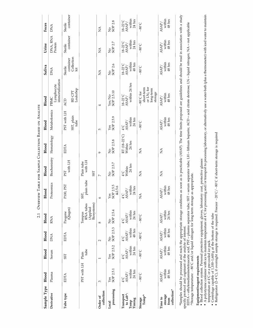
2.1
Ov
er
vie
wT
ab
le
fo
rS
am
pl
eC
ol
le
ct
io
nB
ase
do
nA
na
ly
te
Sa
mp
leT
yp
eB
loo
dB
loo
dB
loo
dB
loo
dB
loo
dB
loo
dB
loo
dB
loo
dB
loo
dS
ali
va
Uri
ne
Fe
ces
De
riv
ati
ve
Pla
sma
Ser
um
DN
AR
NA
Pro
teo
mic
sB
ioch
emis
try
Hem
ato
log
yM
etab
olo
mic
sP
BM
CL
ym
ph
ocy
teim
mo
rtal
izat
ion
DN
AD
NA
,R
NA
Pro
tein
DN
A
Tu
be
typ
eE
DT
AS
ST
ED
TA
Pax
gen
e(Q
iag
en)
P10
0,P
ST
PS
T wit
hL
HE
DT
AP
ST
wit
hL
H
SS
T,
pla
intu
be
AC
D BD
CP
TL
euk
oS
ep
Ste
rile
con
tain
erC
oll
ecti
on
kit
Ste
rile
con
tain
erS
teri
leco
nta
iner
PS
Tw
ith
LH
Pla
in tub
eT
emp
us
RN
Atu
bes
(Ap
pli
edB
iosy
stem
s)
SS
T,
pla
intu
be
Pla
intu
be
wit
hL
H
SS
T
Ord
er
of
coll
ect
ion
31
28
67
95
4N
AN
AN
A
Lo
cal
pro
cess
ing
Yes
SO
P2.
5.1
Yes
SO
P2.
5.2
No
SO
P2.
5.3
No
SO
P2.
5.4
Yes
SO
P2.
5.5
&2.
5.6
No
SO
P2.
5.7
No
SO
P2.
5.8
Yes
SO
P2.
5.9
Yes
/N
oS
OP
2.5.
10N
oS
OP
2.6
No
SO
P2.
7N
oS
OP
2.8
Tra
nsp
ort
toL
ab
*:T
em
pT
imin
g
48C
AS
AP
/w
ith
in24
hrs
48C
AS
AP
/w
ith
in24
hrs
48C
AS
AP
/w
ith
in48
hrs
48C
AS
AP
/w
ith
in24
hrs
48C
AS
AP
/w
ith
in24
hrs
48C
Wit
hin
24h
rs
RT
(18–
228C
)W
ith
in24
hrs
48C
AS
AP
/w
ith
in24
hrs
18–2
28C
AS
AP
/w
ith
in24
hrs
18–2
28C
AS
AP
/w
ith
in48
hrs
18–2
28C
AS
AP
/w
ith
in24
hrs
18–2
28C
AS
AP
/w
ith
in24
hrs
Sto
rag
eT
em
p^
�808C
�808C
�808C
�808C
NA
NA
NA
�80
8C�
808C
for
sho
rt-t
erm
or
LN
2fo
rlo
ng
-ter
mst
ora
ge
�808C
�808C
�808C
Tim
eto
sto
rag
efr
om
coll
ect
ion
*
AS
AP
/w
ith
in48
hrs
AS
AP
/w
ith
in48
hrs
AS
AP
/w
ith
in48
hrs
AS
AP
/w
ith
in24
hrs
AS
AP
/w
ith
in48
hrs
NA
NA
AS
AP
/w
ith
in48
hrs
AS
AP
/w
ith
in48
hrs
AS
AP
/w
ith
in48
hrs
AS
AP
/w
ith
in48
hrs
AS
AP
/w
ith
in48
hrs
*Sam
ple
ssh
ou
ldb
ep
roce
ssed
and
reac
hth
eap
pro
pri
ate
sto
rag
eco
nd
itio
ns
asso
on
asis
pra
ctic
able
(AS
AP
).T
he
tim
eli
mit
sp
rop
ose
dar
eg
uid
elin
esan
dsh
ou
ldb
ere
adin
asso
ciat
ion
wit
ha
stu
dy
spec
ific
pro
toco
lan
dco
gn
isan
to
fth
ean
aly
teo
fin
tere
st.
ED
TA¼
eth
yle
ned
iam
inet
etra
acet
icac
id;
PS
T¼
pla
sma
sep
arat
or
tub
e;S
ST¼
seru
mse
par
ato
rtu
be;
LH¼
lith
ium
hep
arin
;A
CD¼
acid
citr
ate
dex
tro
se;
LN¼
liq
uid
nit
rog
en;
NA¼
no
tap
pli
cab
le^
Sto
rag
ete
mp
erat
ure
:�
808C
and
/o
rli
qu
idn
itro
gen
for
lon
g-t
erm
sto
rag
eas
app
rop
riat
e.
Eq
uip
me
nt/
rea
gen
tre
qu
irem
en
ts�
Blo
od
coll
ecti
on
syst
em.
Per
son
alp
rote
ctiv
eeq
uip
men
t;g
lov
es,
lab
ora
tory
coat
,p
rote
ctiv
eg
lass
es�
Ap
oly
sty
ren
eco
nta
iner
wit
hic
eto
mai
nta
inte
mp
erat
ure
at48
Cfo
rp
roce
ssin
gan
d/
or
tran
spo
rtto
pro
cess
ing
lab
ora
tory
,or
alte
rnat
ivel
yu
sea
wat
er-b
ath
(plu
sa
ther
mo
met
er)
wit
hic
edw
ater
tom
ain
tain
tem
per
atu
reat
48C
or
ap
re-c
oo
led
ice
pac
k.
�C
entr
ifu
ge
cap
able
of
1,10
0–1,
600
gat
the
bo
tto
mo
fth
etu
be
�R
efri
ger
ato
r(2
–48C
),if
ov
ern
igh
tsa
mp
lest
ora
ge
isre
qu
ired
.F
reez
er�
208C
/�
808C
ifsh
ort
-ter
mst
ora
ge
isre
qu
ired
� Using a syringe for blood transfer may also cause overfilling or underfilling of tubes, resulting in an incorrect blood-to-additive ratio and potentially incorrect analytical results.
� Evacuated tubes are designed to draw the volume indicated. Filling is complete when vacuum no longer continues todraw, though some tubes may partially fill due to plunger resistance when filled from a syringe. The laboratory shouldbe consulted regarding the use of these samples.
4. If blood is collected through an intravenous (I.V.) line, ensure that the line has been cleared of I.V. solution beforebeginning to fill the blood collection tubes. This is critical to avoid erroneous laboratory/analytical results from I.V. fluidcontamination.
5. Overfilling or underfilling of tubes will result in an incorrect blood-to-additive ratio and may lead to incorrect laboratory/analytical results or poor product performance.
6. During collection it is important to avoid possible backflow from blood collection tubes that contain chemical additiveswhich may result in the possibility of an adverse patient reaction.
2.3 Equipment and Reagents for Blood Collection and Immediate Processing
1. General blood taking equipment, correct evacuated tubes dependent on sample type, appropriate gauge needle, bloodcollection set, tourniquet, alcohol wipes, cotton wool, adhesive bandage, and sharps disposal system will be required priorto blood collection.
2. Personal protective equipment including, gloves, eye protection glasses, and laboratory coat will be worn as necessary forprotection from exposure to blood borne pathogens.
3. Centrifuge capable of generating a G force of 1,100–3,000 g at the bottom of the tube. Counter balanced test tubes filled withwater/saline for use to balance blood collection tubes during centrifugation. Disposable transfer plastic/Pasteur pipettes.
4. A refrigerator (48C) and/or freezer (�208C/�808C) as necessary dependent on immediate processing requirements andwhether overnight and/or short-term storage of samples is required. It may also be important to have access to dry icesupplies as appropriate dependent on sample type and transport requirements.
5. A polystyrene container with ice to maintain temperature at 48C for immediate processing and/or transport to theprocessing laboratory, or alternatively use a water-bath (plus a thermometer) with iced water to bring the temperature to48C or a pre-conditioned gel pack at 48C.
SOP 2.4 Recommended Order for Blood Draw
The recommended order for blood draw based on blood collection tube type is outlined as follows:
1. Blood collection tubes for sterile samples2. Blood collection tubes without additives3. Blood collection tubes for coagulation studies (e.g. with citrate additive)4. Blood collection tubes with other lyophilised additives (vacutainers, heparin, EDTA, plasma, BD P100, or serum separator tubes)5. Blood collection tubes with other liquid additives (e.g. PAXgene� and BD vacutainer CPT)
SOP 2.5 Blood Collection—Venipuncture
SOP Number: 2.5Version Number 1.0
Effective DateVersion Number
Name Title Date
Author
Authorizer
Change History
SOP Number Effective Date Significant Change Previous SOP No.
MMI GUIDELINES FOR STANDARDIZED BIOBANKING 17
PurposeThis SOP describes the procedure for blood collection (venipuncture) from research participants.
ResponsibilityIt is the responsibility of the research personnel carrying out this procedure to ensure that all steps are completed bothcompetently and safely.
Procedure
1. The research participant’s arm will be hyperextended and positioned comfortably on the armrest of the venipuncturechair/couch as appropriate.
2. The tourniquet will be applied 3–4 inches above the selected puncture site and will not be left in position for longer thantwo minutes.
3. The research participant will be asked to make a fist without pumping the hand.4. A vacuum collection system (for example Monovette) will be used for venipuncture where possible. Syringes and needles
will be used in place of the vacuum collection system in special circumstances.5. The puncture site will be cleansed using a Sterets pre-injection swab in a circular motion from the center to the
periphery.6. The cleansed site will be allowed to air dry prior to venipuncture.7. The research participant’s vein will be anchored and the needle will then be inserted through the skin, bevel edge
uppermost, into the lumen of the vein.8. The tourniquet will be released when the last collection tube to be drawn is filling.9. Tubes containing anticoagulants must be properly mixed immediately after each is drawn by inverting the tube. See
manufacturer instructions for number of inversions required.10. Clean dry gauze or cotton wool will be placed on the venipuncture site and the needle will be removed in a swift
backward motion using a needle protector.11. The research personnel will press down on the gauze/cotton wool once the needle has been drawn out of the vein
applying adequate pressure to avoid formation of a haematoma.12. The research participants arm will not be placed in a bent position at any time following venipuncture.13. The research participants arm will be inspected to ensure bleeding has stopped and a band-aid strip will be applied.14. The research personnel will ensure that the research participant has not experienced any adverse events from the
venipuncture and will then assist them from the chair.15. All contaminated materials/supplies will be disposed of in the designated containers.16. All blood collection tubes will be labelled immediately following collection with the appropriate research study labels, for
example a unique study identification number and/or bar code label. The time of collection will also be recorded in thestudy specific documentation and/or data management system.
17. The research personnel will arrange for the blood specimens to be transported to the research laboratory as applicable.
Safety precaution: During collection it is important to avoid possible backflow from blood collection tubes that containchemical additives, for example BD Vacutainer CPT which may result in the possibility of an adverse patient reaction. Thefollowing precautions should be observed:
� Use a blood collection set with a safety lock for example; a BD Vacutainer� Safety-Lok� Blood Collection Set.� Place arm in a downward position� Hold tube with stopper upper-most� Release tourniquet as soon as blood starts to flow into the tube� Ensure that tube additives do not touch the stopper or the end of the needle during venipuncture
Change History
SOP Number Effective Date Significant Change Previous SOP No.
18 GUERIN ET AL.
SOP 2.5.1 Blood Collection for Plasma
SOP Number: 2.5.1Version Number 1.0
Purpose
This SOP describes the procedure for blood collection for plasma.
Responsibility
It is the responsibility of the research personnel carrying out this procedure to ensure that all steps are completed bothcompetently and safely.
Equipment/reagent requirements
� Blood collection system� Personal protective equipment; gloves, laboratory coat, protective glasses� Blood collection tube: EDTA, plasma separator tube (PST) with lithium heparin� A polystyrene container with ice to maintain temperature at 48C for processing and/or transport to processing labo-
ratory, or alternatively use a water-bath (plus a thermometer) with iced water to maintain the temperature at 48C or apre-conditioned gel pack at 48C
� Centrifuge capable of generating 1,100–1,300 g at the bottom of the tube� Refrigerator (2–48C) if overnight sample storage is required� Freezer �208C/�808C if short-term storage is required
Procedure
1. Draw blood directly into the evacuated tube. Filling the blood collection tube to the black mark on the tube label indicatesthat the correct amount of blood has been drawn. Underfilling or overfilling of the tube may affect laboratory results dueto the incorrect blood/additive ratio.
2. The blood collection tube is appropriately labeled either with a unique study identification number and/or a bar code labelgenerated electronically.
3. Record the time that the sample was taken in the study specific documentation or data management system.4. Invert the tube 8–10 times immediately after collection. This helps to prevent the formation of fibrin which may affect
subsequent analysis.5. Maintain tubes at 48C at all times following collection and during processing. Centrifuge tubes within 2 hours of collection
to separate plasma from cells. Place the blood collection tubes in a centrifuge and spin at 1,300 g for 10 minutes at 48C.Record the time processing was initiated in the study specific documentation or data management system.
6. Avoid mixing/agitation of PST tubes between centrifugation and separation or transport to the laboratory as this may leadto mixing and/or re-suspension of cells and platelets that were previously on or near the gel surface.
7. Using a plastic Pasteur/transfer pipette collect plasma, being sure to stay above the gel/cell layer so that no cells or portions ofthe gel are collected. Distribute the plasma (clear liquid) among cryostorage tube(s) maintained at 48C which have beenlabeled as per point 2 above. Record the volume in each tube in the study specific documentation or data management system.
8. Transfer tubes to a �808C freezer for storage. If there is not a �808C freezer on site store at �208C. If neither is availabletransport to the processing laboratory at 48C in a polystyrene container on ice. The specimen should reach the �808C freezerwithin 48 hours of collection. Record the time of storage in the study specific documentation or data management system.Note: As a general rule samples should be processed and reach the appropriate storage conditions as soon as ispracticable. The maximum time limits proposed are guidelines and should be read in association with a study specificprotocol.
Change History
SOP Number Effective Date Significant Change Previous SOP No.
Effective DateVersion Number
Name Title Date
Author
Authorizer
MMI GUIDELINES FOR STANDARDIZED BIOBANKING 19
SOP 2.5.2 Blood Collection for Serum
SOP Number: 2.5.2Version Number 1.0
PurposeThis SOP describes the procedure for blood collection for serum.
ResponsibilityIt is the responsibility of the research personnel carrying out this procedure to ensure that all steps are completed bothcompetently and safely.
Equipment/reagent requirements
� Blood collection system� Personal protective equipment; gloves, laboratory coat, protective glasses� Blood collection tube: serum separator tube (SST) or plain tube� A polystyrene container with ice to maintain temperature at 48C for processing and/or transport to processing labo-
ratory, or alternatively use a water-bath (plus a thermometer) with iced water to maintain the temperature at 48C or apre-conditioned gel pack at 48C
� Centrifuge capable of generating 1,100–1,600 g at the bottom of the tube� Refrigerator (2–48C) if overnight sample storage is required� Freezer �208C/�808C if short-term storage is required
Procedure
1. Draw blood directly into the evacuated tube. Filling up the blood collection tube to the black mark on the tube labelindicates that the correct amount of blood has been drawn.
2. The blood collection tube is appropriately labeled either with a unique study identification number and/or a barcode labelgenerated electronically.
3. Note the time that the sample was taken in the study specific documentation or data management system.4. Allow the blood to clot for 15 to 30 minutes at room temperature (RT) (18–228C). The time for clotting is dependent on tube
type so refer to the manufacturer’s instructions for use for recommended time for specific tube types.5. Place tubes in the centrifuge and spin at 1,600 g at RT (18–228C) for 10 minutes. This speed, time and temperature will
minimize platelet contamination of the specimen which may affect sample analysis. Record the time processing wasinitiated in the study specific documentation or data management system.
6. Using a plastic transfer/Pasteur pipette collect the serum being careful not to disrupt the clot or to collect any of the gel.Transfer the serum (straw coloured liquid) into 0.5 mL cryostorage tubes maintained at 48C which have been labeled as perpoint 2 above.
7. Transfer tubes to a�808C freezer for storage. If there is not a�808C freezer on site store at�208C. If neither is available transporttubes to the processing laboratory at 48C in a polystyrene container on ice. The specimen should reach the �808C freezer within48 hours of collection. Record the time of storage in the study specific documentation or data management system.Note: As a general rule samples should be processed and reach the appropriate storage conditions as soon as is practicable.The maximum time limits proposed are guidelines and should be read in association with a study specific protocol.
Effective DateVersion Number
Name Title Date
Author
Authorizer
Change History
SOP Number Effective Date Significant Change Previous SOP No.
20 GUERIN ET AL.
SOP 2.5.3 Blood Collection for DNA Extraction
SOP Number: 2.5.3Version Number 1.0
PurposeThis SOP describes the procedure for blood collection for extraction of DNA.
ResponsibilityIt is the responsibility of the research personnel carrying out this procedure to ensure that all steps are completed bothcompetently and safely.
Equipment/reagent requirements
� Blood collection system� Personal protective equipment; gloves, laboratory coat, protective glasses� Blood collection tube: EDTA.� Note: Lithium heparin is not recommended for blood collection for DNA extraction as the heparin co-purifies with the
DNA and can interfere with enzymatic reactions.� A polystyrene container with ice to maintain temperature at 48C for processing and/or transport to processing labo-
ratory, or alternatively use a water-bath with iced water to maintain the temperature at 48C (plus a thermometer) or apre-conditioned gel pack at 48C
� Refrigerator (2–48C) if overnight sample storage is required� Freezer �208C/�808C if short-term storage is required
Procedure
1. Draw blood directly into the evacuated tube. Filling up the blood collection tube to the black mark on the tube labelindicates that the correct amount of blood has been drawn. Underfilling or overfilling of the tube can affect results due tothe incorrect blood/additive ratio.
2. The blood collection tube is appropriately labeled either with a unique study identification number and/or a bar code labelgenerated electronically.
3. Invert the tube 8–10 times to avoid the formation of microclots.4. Record the time that the sample was taken in the study specific documentation or data management system.5. Maintain the tubes at 48C in a refrigerator/polystyrene container with ice. Transport tubes to the processing laboratory as
soon as is practicable or within a maximum of 48 hours for immediate processing of DNA or for direct storage at �808C.Tubes should be transported at 48C in a polystyrene container on ice. Record date and time of processing of DNA and thedata/time that DNA is frozen in the study specific documentation or data management system.
6. If a sample for DNA is frozen locally at �208C then the sample should be transported frozen, using dry ice to theprocessing laboratory. Vacutainers should be tested to ensure that they can withstand storage temperature and re-thaw. Ifa sample is thawed DNAse enzymes break down the DNA rapidly.Note: As a general rule samples should be processed and reach the appropriate storage conditions as soon as is practicable.The maximum time limits proposed are guidelines and should be read in association with a study specific protocol.
Effective DateVersion Number
Name Title Date
Author
Authorizer
Change History
SOP Number Effective Date Significant Change Previous SOP No.
MMI GUIDELINES FOR STANDARDIZED BIOBANKING 21
SOP 2.5.4 Blood Collection for RNA isolation
SOP Number: 2.5.4Version Number 1.0
PurposeThis SOP describes the procedure for blood collection for extraction of RNA.
ResponsibilityIt is the responsibility of the research personnel carrying out this procedure to ensure that all steps are completed bothcompetently and safely.
Equipment/reagent requirements
� Blood collection system� Personal protective equipment; gloves, laboratory coat, protective glasses� Blood collection tube: ACD tube, Tempus� Blood RNA Tubes (Applied Biosystems) or PaxGene (Qiagen)� A polystyrene container with ice to maintain temperature at 48C for processing and/or transport to processing labo-
ratory, or alternatively use a water-bath (plus a thermometer) with iced water to maintain the temperature at 48C or apre-conditioned gel pack at 48C
� Refrigerator (2–48C) if overnight sample storage is required� Freezer �208C/�808C if short-term storage is required� Vortex for sample mixing
Procedure
Using ACD tubes
1. Draw blood directly into the evacuated tube. Filling the tube to the black mark on the tube label indicates that the correctamount of blood has been drawn. Underfilling or overfilling of the tube can affect laboratory results.
2. The blood collection tube is labeled appropriately either with a unique identification study number and/or a bar code labelgenerated electronically.
3. Record the time that the sample was taken in the study specific documentation or data management system as available.4. Maintain tubes at RT (18–228C) and transport to the processing laboratory within 24 hours at RT (18–228C) for processing.5. If immediate transfer is not possible, samples can be maintained at RT (18–228C) and transferred to the processing
laboratory for RNA isolation as soon as is practicable or within a maximum of 24 hours. Record the time of processing inthe study specific documentation or data management system.Note: As a general rule samples should be processed and reach the appropriate storage conditions as soon as is practicable.The maximum time limits proposed are guidelines and should be read in association with a study specific protocol.
Using Tempus Blood RNA Tubes
1. Draw blood directly into the evacuated Tempus Blood RNA Tube. Filling the blood collection tube to the black mark onthe tube label indicates that the correct amount of blood has been drawn. Underfilling or overfilling of the tube can affectlaboratory results due to the incorrect blood/additive ratio.
2. Immediately after the Tempus tube is filled, stabilize the blood by shaking the tube vigorously or vortexing the contentsfor 10 seconds to ensure that the stabilizing reagent makes uniform contact with the sample.IMPORTANT: Failure to mix the stabilizing reagent with the blood leads to inadequate stabilization of the gene ex-pression profile and the formation of microclots that can potentially clog the purification filter.
3. The Tempus Blood RNA tube is appropriately labeled either with a unique study identification number and/or a bar codelabel generated electronically.
4. Record the time that the sample was taken in the study specific documentation or data management system.5. Maintain the tubes at 48C using a refrigerator/polystyrene container with ice. Transport tubes to the processing laboratory
as soon as is practicable or within a maximum of 24 hours for immediate processing of RNA or for direct storage at �808C.Tubes should be transported at 48C in a polystyrene container on ice. Record the time of processing in the study specificdocumentation or data management system.
Effective DateVersion Number
Name Title Date
Author
Authorizer
22 GUERIN ET AL.
Note: As a general rule samples should be processed and reach the appropriate storage conditions as soon as is practicable.The maximum time limits proposed are guidelines and should be read in association with a study specific protocol.
Using Paxgene tubes
1. Draw blood directly into the evacuated Paxgene tube. Filling the blood collection tube to the black mark on the tube labelindicates that the correct amount of blood has been drawn. Underfilling or overfilling of the tube can affect laboratoryresults due to the incorrect blood/additive ratio.
2. The tube is gently inverted 8–10 times.IMPORTANT: It is critical to RNA quality and yield that tubes are thoroughly mixed by inversion at the time of collection,that a full tube of blood be taken and that nothing is placed over the black fill mark on the manufacturer’s label of the tube.
3. The Paxgene tube is appropriately labeled either with a unique study identification number and/or a bar code labelgenerated electronically.
4. Record the time that the sample was taken in the study specific documentation or data management system.5. Maintain the tubes at 48C in a refrigerator/polystyrene container with ice. Transport tubes to the processing laboratory as
soon as is practicable or within a maximum of 24 hours for immediate processing of RNA or for direct storage at �808C.Tubes should be transported at 48C in a polystyrene container on ice. Record the time of processing in the study specificdocumentation or data management system.Note: As a general rule samples should be processed and reach the appropriate storage conditions as soon as is practicable.The maximum time limits proposed are guidelines and should be read in association with a study specific protocol.
SOP 2.5.5 Blood Collection for Proteomics using Plasma
SOP Number: 2.5.5Version Number 1.0
PurposeThis SOP describes the procedure for blood collection for plasma isolation for proteomic studies.
ResponsibilityIt is the responsibility of the research personnel carrying out this procedure to ensure that all steps are completed bothcompetently and safely.
Equipment/reagent requirements
� Blood collection system� Personal protective equipment; gloves, laboratory coat, protective glasses� Blood collection tube: plasma separator tube, BD P100 proteomics tube� A polystyrene container with ice to maintain temperature at 48C for processing and/or transport to processing labo-
ratory, or alternatively use a water-bath (plus a thermometer) with iced water to maintain the temperature at 48C or apre-conditioned gel pack at 48C
� Refrigerator (2–48C) if overnight sample storage is required� Freezer �208C/�808C if short-term storage is required
Change History
SOP Number Effective Date Significant Change Previous SOP No.
Effective DateVersion Number
Name Title Date
Author
Authorizer
MMI GUIDELINES FOR STANDARDIZED BIOBANKING 23
� Centrifuge capable of generating a G force of 1,100–1,300 g at the bottom of the tube
SAFETY PRECAUTION: The BD P100 evacuated blood collection tube contains chemical additives. It is important to avoidpossible backflow from the tube, which may lead to the possibility of adverse patient reaction. To guard against backflowobserve the following precautions:
� Use a blood collection set with a safety lock, for example a BD Vacutainer� Safety- Lok� Blood Collection Set.� Place the arm in a downward position.� Hold tube with stopper upper-most.� Release tourniquet as soon as blood starts to flow into the tube.� Ensure that tube additives do not touch the stopper or the end of the needle during venipuncture.
Procedure
1. Draw blood directly into the evacuated tube. Filling the blood collection tube to the black mark on the tube label indicatesthat the correct amount of blood has been drawn. Underfilling or overfilling of the tube may affect laboratory results.
2. Invert the tube 8–10 times to avoid the formation of microclots.3. The blood collection tube is labeled appropriately either with a unique study identification number and/or a bar code label
generated electronically4. Record the time that the sample was taken in the study specific documentation or data management system.5. Centrifuge tubes within 2 hours of collection to separate plasma from cells. Maintain tubes at 48C during processing. Place
the blood collection tubes in a centrifuge and spin at 1,300 g for 10 minutes at 48C. Record the time processing was initiatedin the study specific documentation or data management system.
6. Using a plastic Pasteur/transfer pipette collect plasma being sure not to get too close to the cell layer or gel. Distributethe plasma (clear liquid) among 0.5 mL cryostorage tube(s) maintained at 48C which have been labeled as per point 3above.
7. Transfer tubes to a �808C freezer for storage. If there is not a �808C freezer on site store at �208C. If neither is availabletransport to the processing laboratory at 48C in a polystyrene container on ice. The specimen should reach the �808Cfreezer as soon as is practicable or within a maximum of 48 hours following collection. Record the time of storage in thestudy specific documentation or data management system.Note: As a general rule samples should be processed and reach the appropriate storage conditions as soon as is practicable.The maximum time limits proposed are guidelines and should be read in association with a study specific protocol.
SOP 2.5.6 Blood Collection for Proteomics Using Serum
SOP Number: 2.5.6Version Number 1.0
PurposeThis SOP describes the procedure for blood collection for serum isolation for proteomic studies.
Change History
SOP Number Effective Date Significant Change Previous SOP No.
Effective DateVersion Number
Name Title Date
Author
Authorizer
24 GUERIN ET AL.
ResponsibilityIt is the responsibility of the research personnel carrying out this procedure to ensure that all steps are completed bothcompetently and safely.
Equipment/reagent requirements
� Blood collection system� Personal protective equipment; gloves, laboratory coat, protective glasses� Blood collection tube: SST or plain tube� Plain plastic 15 mL conical centrifuge tube� Protease Inhibitor Cocktail (for example P3840 Sigma Aldrich)� A polystyrene container with ice to maintain temperature at 48C for immediate processing and/or transport to pro-
cessing laboratory, or alternatively use a water-bath with iced water to maintain the temperature at 48C (plus a ther-mometer) or a pre-conditioned gel pack at 48C
� Refrigerator (2–48C) if overnight sample storage is required� Freezer �208C/�808C if short-term storage is required� Centrifuge capable of generating a G force of 1,100–1,600 g at the bottom of the tube
Procedure
1. Draw blood directly into the evacuated tube. Filling the tube to the black mark on the tube label indicates that the correctamount of blood has been drawn.
2. The blood collection tube is labeled appropriately either with a unique study identification number and/or a bar code labelgenerated electronically.
3. Record the time that the sample was taken in the study specific documentation or data management system.4. Allow the blood to clot for 15 to 30 minutes on ice (48C). Note the clotting time in the study specific documentation or data
management system.5. Centrifuge tubes within 2 hours of collection to separate serum from cells. Maintain tubes at 48C during processing. Place
the blood collection tubes in a centrifuge and spin at 1,600 g for 10 minutes at 48C. Record the time processing was initiatedin the study specific documentation or data management system.
6. Using a plastic transfer/Pasteur pipette collect serum being sure not to disrupt the clot or to collect any of the gel. Transferthe serum (straw colored liquid) to a plain plastic 15 mL conical centrifuge tube.
7. Add 100� protease inhibitor solution, for example 10 ul of protease inhibitor solution to 1 mL of serum and mix byinversion at least 8–10 times.
8. Using a plastic transfer/Pasteur pipette transfer serum into each 0.5 mL cryostorage tube maintained at 48C which havebeen labeled as per point 2 above.
9. Transfer tubes to a �808C freezer for storage. If there is not a �808C freezer on site store at �208C. If neither is availabletransport to the processing laboratory at 48C in a polystyrene container on ice. The specimen should reach the �808Cfreezer as soon as is practicable or within 48 hours following collection. Record the time of storage in the study specificdocumentation or data management system.Note: As a general rule samples should be processed and reach the appropriate storage conditions as soon as ispracticable. The maximum time limits proposed are guidelines and should be read in association with a study specificprotocol.
Change History
SOP Number Effective Date Significant Change Previous SOP No.
MMI GUIDELINES FOR STANDARDIZED BIOBANKING 25
SOP 2.5.7 Blood Collection for Biochemistry
SOP Number: 2.5.7Version Number 1.0
PurposeThis SOP describes the procedure for blood collection for biochemical analysis.
ResponsibilityIt is the responsibility of the research personnel carrying out this procedure to ensure that all steps are completed bothcompetently and safely.
Equipment/reagent requirements
� Blood collection system� Personal protective equipment; gloves, laboratory coat, protective glasses� Blood collection tube: PST with lithium heparin or a plain tube with lithium heparin. SST or plain tube.� A polystyrene container with ice to maintain temperature at 48C for processing and/or transport to processing labo-
ratory, or alternatively use a water-bath with iced water to maintain the temperature at 48C (plus a thermometer) or apre-conditioned gel pack at 48C
� Refrigerator (2–48C), if overnight sample storage is required� Centrifuge capable of generating a G force of 1,100–1,300 g at the bottom of the tube
Procedure
1. Draw blood directly into the evacuated tube. Filling the tube to the black mark on the tube label indicates that the correctamount of blood has been drawn. Underfilling or overfilling of the tube can affect laboratory results due to the incorrectblood/additive ratio.
2. Invert the tube 8–10 times immediately after collection, this helps to prevent the formation of fibrin which may affect thelaboratory result.
3. The blood collection tube is labeled appropriately with a unique study identification number generated and/or a bar codelabel generated electronically.
4. Record the time that the sample was taken in the study specific documentation or data management system.5. Transport directly (within 4 hours) to the Biochemistry laboratory for processing. If transport to the Biochemistry labo-
ratory within 4 hours is not possible separate the plasma from the blood samples by following the procedure outlined frompoints 6–8 below.
6. Centrifuge tubes within 2 hours of collection to separate plasma from cells. Maintain tubes at 48C during processing. Placethe blood collection tubes in a centrifuge and spin at 1,300 g for 10 minutes at 48C. Record the time processing was initiatedin the study specific documentation or data management system.
7. Using a plastic Pasteur/transfer pipette collect plasma, being sure not to disrupt the cell layer or gel. Transfer the plasma(clear liquid) to a 0.5 mL cryostorage tube maintained at 48C which have been labeled as per point 3 above.
8. Maintain the plasma sample at 48C and transfer to the biochemistry laboratory within 24 hours for processing.Note: Immediate separation of plasma/serum from cells provides optimal analyte stability at RT. Storage of un-centri-fuged specimens after 24 hours has resulted in clinically significant changes in measured analytes. When prolongedcontact of plasma/serum with cells is unavoidable use of serum is recommended because of the higher instability ofplasma analytes.28
Effective DateVersion Number
Name Title Date
Author
Authorizer
Change History
SOP Number Effective Date Significant Change Previous SOP No.
26 GUERIN ET AL.
SOP 2.5.8 Blood Collection for Hematology
SOP Number: 2.5.8Version Number 1.0
PurposeThis SOP describes the procedure for blood collection for hematological analysis.
ResponsibilityIt is the responsibility of the research personnel carrying out this procedure to ensure that all steps are completed bothcompetently and safely.
Equipment/reagent requirements
� Blood collection system� Personal protective equipment; gloves, laboratory coat, protective glasses� Blood collection tube: EDTA
Procedure
1. Draw blood directly into the evacuated EDTA tube. Filling up the tube to the black mark on the tube label indicates thatthe correct amount of blood has been drawn. Underfilling or overfilling of the tube can affect laboratory results due to theincorrect blood/additive ratio.
2. The blood collection tube is labeled appropriately with a unique study identification number generated and/or a bar codelabel generated electronically.
3. Invert the tube 8–10 times to avoid the formation of microclots.4. Record the time that the sample was taken in the study specific documentation or data management system.5. Transport to the Hematology laboratory within 4 hours at RT (18–228C) for processing.
SOP 2.5.9 Blood Collection for Metabolomics Using Serum
SOP Number: 2.5.9Version Number 1.0
Effective DateVersion Number
Name Title Date
Author
Authorizer
Effective DateVersion Number
Name Title Date
Author
Authorizer
Change History
SOP Number Effective Date Significant Change Previous SOP No.
MMI GUIDELINES FOR STANDARDIZED BIOBANKING 27
PurposeThis SOP describes the procedure for blood collection for serum isolation for metabolomic studies.
ResponsibilityIt is the responsibility of the research personnel carrying out this procedure to ensure that all steps are completed bothcompetently and safely.
Equipment/reagent requirements
� Blood collection system� Personal protective equipment; gloves, laboratory coat, protective glasses� Blood collection tube: SST or plain tube� A polystyrene container with ice to maintain temperature at 48C for processing and/or transport to processing labo-
ratory, or alternatively use a water-bath (plus a thermometer) with iced water to maintain the temperature at 48C or apre-conditioned gel pack at 48C
� Refrigerator (2–48C) if overnight sample storage is required� Centrifuge capable of generating a G force of 1,100–1,600 g at the bottom of the tube
Procedure
1. Draw blood directly into the evacuated tube. Filling the tube to the black mark on the tube label indicates that the correctamount of blood has been drawn.
2. The blood collection tube is labeled appropriately either with a unique study identification number and/or a bar code labelgenerated electronically.
3. Record the time that the sample was taken in the study specific documentation or data management system.4. Allow the blood to clot for 15–30 minutes at RT (18–228C). Record clotting time in the study specific documentation or data
management system.5. Centrifuge tubes within 2 hours of collection to separate serum from cells. Maintain tubes at 48C during processing.
Place the blood collection tubes in a centrifuge and spin at 1,600 g for 10 mins at 48C. Record the time processinginitiated.
6. Using a plastic transfer/Pasteur pipette collect serum being sure not to disrupt the clot or gel. Transfer the serum into0.5 mL cryostorage tube(s) maintained at 48C which have been labeled as per point 2 above.
7. Transfer tubes to a �808C freezer for storage. If there is not a �808C freezer on site transport to the processing laboratory at48C in a polystyrene container on ice. The specimen should reach the �808C as soon as is practicable or within 48 hours ofcollection. Record the time of storage in the study specific documentation or data management system.Note: As a general rule samples should be processed and reach the appropriate storage conditions as soon as is practicable.The maximum time limits proposed are guidelines and should be read in association with a study specific protocol.
SOP 2.5.10 Blood Collection for Isolation of Peripheral Blood Mononuclear Cells
SOP Number: 2.5.10Version Number 1.0
Change History
SOP Number Effective Date Significant Change Previous SOP No.
Effective DateVersion Number
Name Title Date
Author
Authorizer
28 GUERIN ET AL.

PurposeThis SOP describes the procedure for blood collection for isolation of peripheral blood mononuclear cells.
ResponsibilityIt is the responsibility of the research personnel carrying out this procedure to ensure that all steps are completed bothcompetently and safely.
Equipment/reagent requirements
� Blood collection system� Personal protective equipment; gloves, laboratory coat, protective glasses� Blood collection tube: Acid Citrate Dextrose (ACD) tubes or BD CPT tube� If using a BD CPT tube a centrifuge capable of generating a G force of 1,100– 1,300 g at the bottom of the tube is required.
ProcedureUsing ACD tubes
1. Draw blood directly into the evacuated tube. Filling the tube to the black mark on the tube label indicates that the correctamount of blood has been drawn. Underfilling or overfilling of the tube can affect laboratory results.
2. The blood collection tube is labeled appropriately either with a unique identification study number and/or a bar code labelgenerated electronically.
3. Record the time that the sample was taken in the study specific documentation or data management system.4. Maintain tubes at RT (18–228C) and transport to the processing laboratory within 24 hours at RT (18–228C) for pro-
cessing.Note: As a general rule samples should be processed and reach the appropriate storage conditions as soon as is practicable.The maximum time limits proposed are guidelines and should be read in association with a study specific protocol.
Note: For BD CPT follow the manufacturer’s instructions outlined in the information for use.
SOP 2.6 Saliva Collection
SOP Number: 2.6Version Number 1.0
Purpose
This SOP describes the procedure for saliva collection from research participants.
Responsibility
It is the responsibility of the research personnel carrying out this procedure to ensure that all steps are completed bothcompetently and safely.
Effective DateVersion Number
Name Title Date
Author
Authorizer
Change History
SOP Number Effective Date Significant Change Previous SOP No.
MMI GUIDELINES FOR STANDARDIZED BIOBANKING 29
Equipment
� Personal protective equipment: gloves (optional)� Specimen container or saliva collection kit (for example Oragene DNA Emergo Europe)� Sterile container with warm water
Procedure
1. Research personnel will greet the research participant, identify themselves, and then explain the saliva collection proce-dure to the research participant.
2. The research participant will be approached in a friendly calm manner and their co-operation will be gained prior to salivacollection.
3. The research participant will be correctly identified prior to saliva collection, by asking them to give their name and date ofbirth.
4. All sample containers and equipment needed to competently and efficiently carry out the saliva collection will be as-sembled prior to the procedure. For specific saliva collection kits follow the manufacturer’s instruction for sample col-lection as outlined in the information for use, for example Oragene DNA saliva collection kit (Emergo Europe).
5. The research participant will rinse mouth out with warm water. Avoid use of mouthwashes or fluoride rinses. In addition,the research participant should not drink, smoke or chew gum for 30 minutes before giving a saliva sample.
6. The research participant will be asked to fill the specimen container to a certain marked level with saliva.7. The inside of the specimen container will not be touched and the outer surfaces of the specimen container will not be
contaminated with saliva.8. A unique study identification number or bar code label will be applied to the sample immediately after saliva collection.
Record the time of saliva collection in the study specific documentation or data management system.9. Transport to the processing laboratory as soon as is practicable or within a maximum of 48 hours for immediate processing
of DNA or for direct storage at �808C. Saliva collection tubes should be transported at 48C in a polystyrene container onice. Alternatively when using commercial collection kits such as Oragene DNA the sample may be stored at RT for up to 18months in the cell lysis buffer in the container prior to DNA processing. The saliva collection kit should be transported tothe processing laboratory at RT. Follow the manufacturer’s instructions for use in the information for use/package insertin all cases. Record date/time processing was initiated and date/time of storage in the study specific documentation ordata management system.Note: As a general rule samples should be processed and reach the appropriate storage conditions as soon as is practicable.The maximum time limits proposed are guidelines and should be read in association with a study specific protocol.
SOP 2.7 Urine Collection
SOP 2.7.1 Mid-Stream Urine Specimen
SOP Number: 2.7.1Version Number 1.0
PurposeThis SOP describes the procedure for mid-stream urine collection from research participants.
Change History
SOP Number Effective Date Significant Change Previous SOP No.
Effective DateVersion Number
Name Title Date
Author
Authorizer
30 GUERIN ET AL.
Responsibility
It is the responsibility of the research personnel carrying out this procedure to ensure that all steps are completed bothcompetently and safely.
Equipment
� Personal protective equipment: gloves� Sterile urine container
Procedure
1. Research personnel will greet the research participant, identify themselves, and then explain the urine collection procedureto the research participant.
2. The research participant will be approached in a friendly calm manner and their co-operation will be gained prior to urinecollection.
3. The research participant will be correctly identified prior to urine collection, by asking them to give their name and date ofbirth.
4. All sample containers and equipment needed to competently and efficiently carry out the urine collection will be as-sembled prior to the procedure.
5. The research participant will be asked to provide a urine sample by instructing them to discard the first part of the voidedurine and without disrupting the flow to collect approximately 10 mls of urine in the sterile container.
6. A unique study identification number and/or bar code label will be applied to the sample immediately after urinecollection. Record the time of urine collection in the study-specific documentation or data management system.
7. Transport tubes to the processing laboratory as soon as is practicable or within a maximum of 24 hours for RNA isolationor 48 hours for DNA and/or protein extraction or for direct storage at �808C. Tubes should be transported at 48C in apolystyrene container on ice. Record the time processing was initiated and time of storage in the study specific docu-mentation or data management system.Note: As a general rule samples should be processed and reach the appropriate storage conditions as soon as ispracticable. The maximum time limits proposed are guidelines and should be read in association with a study specificprotocol.
SOP 2.7.2 24-Hour Urine Collection
SOP Number: 2.7.2Version Number 1.0
PurposeThis SOP describes the procedure for 24-hour urine collection from research participants.
ResponsibilityIt is the responsibility of the research personnel carrying out this procedure to ensure that all steps are completed bothcompetently and safely.
Change History
SOP Number Effective Date Significant Change Previous SOP No.
Effective DateVersion Number
Name Title Date
Author
Authorizer
MMI GUIDELINES FOR STANDARDIZED BIOBANKING 31
Equipment
� Personal protective equipment: gloves� 24-hour sterile urine collection container
Procedure
1. Research personnel will greet the research participant, identify themselves, and then explain the urine collection procedureto the research participant.
2. The research participant will be approached in a friendly calm manner and their co-operation will be gained prior to urinecollection.
3. The research participant will be correctly identified prior to urine collection, by asking them to give their name and date ofbirth.
4. All sample containers and equipment needed to competently and efficiently carry out the urine collection will be as-sembled prior to the procedure.
5. The research participant will be asked to empty his/her bladder and discard the first morning urine void.6. The 24-hour urine collection will commence with the empty bladder and the research participant will note the time of
emptying.7. All urine over the next 24 hours including the last specimen at the same time the next day will be saved and collected in the
container provided.8. Research personnel will ensure that an accurate 24-hour urine collection has been made by the research participant prior to
labeling and transport of the specimen to the processing laboratory.9. Transport tubes to the processing laboratory as soon as is practicable or within a maximum of 24 hours for RNA isolation
and 48 hours for DNA and/or protein extraction or for direct storage at �808C. Tubes should be transported at 48C in apolystyrene container on ice. Record the time processing was initiated and time of storage in the study specific docu-mentation or data management system.Note: As a general rule samples should be processed and reach the appropriate storage conditions as soon as is practicable.The maximum time limits proposed are guidelines and should be read in association with a study specific protocol.
SOP 2.7.3 Catheter Specimen of Urine
SOP Number: 2.7.3Version Number 1.0
Purpose
This SOP describes the procedure for collection of a catheter specimen of urine from research participants.
Responsibility
It is the responsibility of the research personnel carrying out this procedure to ensure that all steps are completed bothcompetently and safely.
Change History
SOP Number Effective Date Significant Change Previous SOP No.
Effective DateVersion Number
Name Title Date
Author
Authorizer
32 GUERIN ET AL.
Equipment
� Personal protective equipment: gloves� Sterile urine container
Procedure
1. Research personnel will greet the research participant, identify themselves, and then explain the urine collection procedureto the research participant.
2. The research participant will be approached in a friendly calm manner and their co-operation will be gained prior to urinecollection.
3. The research participant will be correctly identified prior to urine collection, by asking them to give their name and date ofbirth.
4. All sample containers and equipment needed to competently and efficiently carry out the urine collection will be as-sembled prior to the procedure.
5. If the catheter specimen of urine is to be collected from an indwelling catheter the specimen will be obtained asepticallyfrom a sample port in the catheter tubing. The sample will not be obtained from the collection bag.
6. If the catheter specimen of urine is to be collected from a transient catheter the specimen will be obtained via a catheterpassed aseptically into the bladder.
7. A unique study identification number or bar code label will be applied to the sample immediately after urine collection.8. Transport tubes to the processing laboratory as soon as is practicable or within a maximum of 24 hours for RNA isolation
and 48 hours for DNA and/or protein extraction or for direct storage at �808C. Tubes should be transported at 48C in apolystyrene container on ice. Record the time processing was initiated and the time of storage in the study specificdocumentation or data management system.Note: As a general rule samples should be processed and reach the appropriate storage conditions as soon as ispracticable. The maximum time limits proposed are guidelines and should be read in association with a study specificprotocol.
SOP 2.8 Feces Collection
SOP Number: 2.8Version Number 1.0
PurposeThis SOP describes the procedure for feces collection from research participants.
ResponsibilityIt is the responsibility of the research personnel carrying out this procedure to ensure that all steps are completed bothcompetently and safely.
Change History
SOP Number Effective Date Significant Change Previous SOP No.
Effective DateVersion Number
Name Title Date
Author
Authorizer
MMI GUIDELINES FOR STANDARDIZED BIOBANKING 33
Equipment:
� Personal protective equipment: gloves� Sterile feces container
Procedure
1. Research personnel will greet the research participant, identify themselves, and then explain the feces collection procedureto the research participant.
2. The research participant will be approached in a friendly calm manner and their co-operation will be gained prior to fecescollection
3. The research participant will be correctly identified prior to feces collection, by asking them to give their name and date ofbirth.
4. All sample containers and equipment needed, to competently and efficiently carry out the feces collection will be as-sembled prior to the procedure.
5. The research participant will be asked to provide a feces sample. The specimen will be passed into a clean dry, disposablebedpan or similar container. The research personnel will then transfer the feces specimen into a sample container using adisposable wooden spatula.
6. The inside of the specimen container will not be touched and the outer surfaces of the specimen will not be contaminatedwith feces.
7. A unique study identification number and/or bar code label will be applied to the sample immediately after feces collection.8. Transport tubes to the processing laboratory as soon as is practicable or within a maximum of 24 hours for RNA and 48
hours for DNA/or protein extraction or for direct storage at �808C. Tubes should be transported at 48C in a polystyrenecontainer on ice. Record the time processing was initiated and the time of storage in the study specific documentation ordata management system.Note: As a general rule samples should be processed and reach the appropriate storage conditions as soon as is practicable.The maximum time limits proposed are guidelines and should be read in association with a study specific protocol.
SOP 2.9 Buccal Collection
SOP Number: 2.9Version Number 1.0
PurposeThis SOP describes the procedure for buccal collection from research participants.
ResponsibilityIt is the responsibility of the research personnel carrying out this procedure to ensure that all steps are completed bothcompetently and safely.
Change History
SOP Number Effective Date Significant Change Previous SOP No.
Effective DateVersion Number
Name Title Date
Author
Authorizer
34 GUERIN ET AL.
Equipment
� Personal protective equipment: gloves (optional)� Sterile single use cotton swab
Procedure:
1. Research personnel will greet the research participant identify themselves, and then explain the buccal collection proce-dure to the research participant.
2. The research participant will be approached in a friendly calm manner and their co-operation will be gained prior tobuccal collection.
3. The research participant will be correctly identified prior to buccal collection, by asking them to give their name and dateof birth.
4. All sample containers and equipment needed to competently and efficiently carry out the buccal collection will beassembled prior to the procedure.
5. A sterile single use cotton swab will be removed from its packaging.6. Sterile technique will be observed as the cotton swab is brushed inside the mouth along the cheek.7. The buccal swab will be returned to a sterile wrapping or will be wrapped inside sterile plastic wrap.8. A unique study identification number and/or bar code label will be applied to the sample immediately after buccal
collection. Record the time of collection in the study specific documentation or data management system.9. Transport tubes to the processing laboratory as soon as is practicable or within a maximum of 24 hours for RNA isolation
and 48 hours for DNA and/or protein extraction or for direct storage at �808C. Tubes should be transported at 48C in apolystyrene container on ice. Record the time processing was initiated and the time of storage in the study specificdocumentation or data management system.Note: As a general rule samples should be processed and reach the appropriate storage conditions as soon as ispracticable. The maximum time limits proposed are guidelines and should be read in association with a study specificprotocol.
2.10 Guiding Principles for Tissue Collection, Processing, and Storage
Solid tissues are collected by biopsy or during surgical procedures. Tissue collection should be carefully planned withsurgeons, clinical staff, pathologists and research personnel.
The following are guiding principles for the collection processing and storage of tissue adapted from the CommonMinimum Technical Standards and Protocols for Biological Resource Centers Dedicated to Cancer Research prepared by theIARC/WHO International Agency for Research on Cancer.17 It is recommended that a detailed SOP be developed based onthe type of tissue to be collected, processed and stored in accordance with individual study requirements.
� The collection of tissue samples for research should never compromise the diagnostic integrity of a specimen. Only tissuewhich is excess to diagnostic purposes should be collected for research purposes.
� All materials and instruments should be prepared in advance of tissue collection. Appropriate personal protectiveequipment should be worn and safety measures should be adhered to in accordance with local policy and procedures.
� All staff involved including the surgeon, pathologist, research nurse, laboratory technician and research personnelshould be trained in the study specific SOP.
� All tissue should be treated as potentially infectious; the collection process should be carried out in accordance with anaseptic technique as possible.
� The surgical specimen or biopsy should be sent to pathology as soon as possible.� It is recommended that surgical specimens or biopsy be preserved within 1 hour of excision; however, tissue subject to a
delay up to 2 hours can still be collected.� A detailed record of the timing of events from time of ischemia to fixation or freezing should be documented.� Each specimen receptacle must be clearly labeled with a unique study number and/or barcode and the details of tissue as
required by the study specific protocol.
Change History
SOP Number Effective Date Significant Change Previous SOP No.
MMI GUIDELINES FOR STANDARDIZED BIOBANKING 35
� Transfer of specimens must be carried out as soon as possible in order to minimize the effect of hypoxia upon geneexpression, degradation of RNA, proteins, and other tissue components.
� For transport from surgery to pathology, or to the biobank, fresh specimens should be placed in a closed, sterile containeron ice at 48C.
� It is recommended that a pathologist should supervise the preparation/dissection of the tissue and ensure that adequatetissue is taken for diagnosis before sampling for research.
� Research personnel must be available to freeze or fix the tissue as quickly as possible and in accordance with a studyspecific SOP.
� Formalin fixation of tissue is standard practice in routine histopathology laboratories and should be conducted inaccordance with a study specific SOP.
� Label cryostorage vials with a unique study identification number/barcode, local pathology number. Record the dateand time of ischemia, excision and storage in the study specific documentation or data management system.
� Tissues must be snap frozen either directly or enclosed in a container immersed in the freezing medium (e.g. pre-cooledisopentane). Snap frozen samples should be placed in the �808C freezer. Liquid nitrogen is not recommended as asuitable freezing medium for direct snap freezing due to the potential formation of cryo-artefacts. RNA can be protectedin fresh tissue by the addition of Allprotect/RNAlater prior to storage at �208C.
SOP 3.1 Personal Protective Equipment
SOP Number: 3.1Version Number 1.0
PART III: LABORATORY STANDARD OPERATING PROCEDURES
SOP 3.1 Personal Protective Equipment . . . . . . . . . . . . . . . . . . . . . . . . . . . . . . . . . . 36SOP 3.2 Specimen Receipt . . . . . . . . . . . . . . . . . . . . . . . . . . . . . . . . . . . . . . . . . . . . 37SOP 3.3 Preparation of Serum and Plasma from Blood. . . . . . . . . . . . . . . . . . . . . . . 38SOP 3.4 DNA Extraction from Blood . . . . . . . . . . . . . . . . . . . . . . . . . . . . . . . . . . . . 39SOP 3.5 RNA Extraction from Blood . . . . . . . . . . . . . . . . . . . . . . . . . . . . . . . . . . . . 41SOP 3.6 Protein Extraction from Blood. . . . . . . . . . . . . . . . . . . . . . . . . . . . . . . . . . . 42SOP 3.7 Peripheral Blood Mononuclear Cell Isolation from Blood . . . . . . . . . . . . . . 44SOP 3.8 Processing of Urine. . . . . . . . . . . . . . . . . . . . . . . . . . . . . . . . . . . . . . . . . . . 45SOP 3.8.1 DNA Extraction from Cells in Urine . . . . . . . . . . . . . . . . . . . . . . . . . . . . . . 46SOP 3.8.2 RNA Extraction from Cells in Urine . . . . . . . . . . . . . . . . . . . . . . . . . . . . . . 47SOP 3.8.3 Protein Extraction from Cells in Urine. . . . . . . . . . . . . . . . . . . . . . . . . . . . . 48SOP 3.9 Processing of Buccal Swabs for DNA Extraction . . . . . . . . . . . . . . . . . . . . . 49SOP 3.10 Processing of Feces for DNA Extraction . . . . . . . . . . . . . . . . . . . . . . . . . . . 51SOP 3.11 Processing of Tissue Samples . . . . . . . . . . . . . . . . . . . . . . . . . . . . . . . . . . . 52SOP 3.11.1 DNA Extraction from Frozen Tissue . . . . . . . . . . . . . . . . . . . . . . . . . . . . . . 52SOP 3.11.2 DNA Extraction from Paraffin Embedded Tissue . . . . . . . . . . . . . . . . . . . . 54SOP 3.11.3 DNA Extraction from Formalin Fixed Tissue. . . . . . . . . . . . . . . . . . . . . . . . 55SOP 3.11.4 RNA Extraction from Tissue . . . . . . . . . . . . . . . . . . . . . . . . . . . . . . . . . . . . 56SOP 3.11.5 Protein Extraction from Tissue . . . . . . . . . . . . . . . . . . . . . . . . . . . . . . . . . . 58SOP 3.12 Processing of Cultured Cells . . . . . . . . . . . . . . . . . . . . . . . . . . . . . . . . . . . . 59SOP 3.12.1 DNA Extraction from Cultured Cells . . . . . . . . . . . . . . . . . . . . . . . . . . . . . 59SOP 3.12.2 RNA Extraction from Cultured Cells. . . . . . . . . . . . . . . . . . . . . . . . . . . . . . 61SOP 3.12.3 Protein Extraction from Cultured Cells . . . . . . . . . . . . . . . . . . . . . . . . . . . . 62
Effective DateVersion Number
Name Title Date
Author
Authorizer
36 GUERIN ET AL.
PurposeThis SOP describes the procedure for the correct personal protective equipment for laboratory personnel.
ResponsibilityIt is the responsibility of the research laboratory personnel carrying out this procedure to ensure that all steps are completedboth competently and safely.Procedure
1. The appropriate personal protective equipment (PPE) assessments required for each workspace will be defined and anappropriate person appointed with responsibility for enforcement and training on the use of the appropriate PPE. Trainingwill include the selection, use, decontamination of and deterioration of PPE. Research personnel should wear laboratorycoats, safety eye protection and gloves.
2. PPE should never be worn in public areas. Hazards must be contained in public areas.3. Safety glasses are required for research personnel and visitors to the laboratory and will be worn at all times when in the
laboratory. Contact lenses shall be permitted in the laboratory.4. Chemical goggles and/or a full-face shield will be worn during chemical transfer and handling operations as procedures
dictate or when in a chemical storage area.5. Sandals and bare feet are prohibited. Safety shoes are required where personnel routinely lift objects over 65 lbs.6. Laboratory coats or aprons will be worn in the laboratory. Laboratory coats should be removed immediately upon
discovery of significant contamination.7. Appropriate chemical-resistant gloves will be worn when using chemicals and the manufacturer’s instructions for use
followed. Used chemical-resistant gloves should be inspected and washed prior to re-use. Damaged or deterioratedchemical-resistant gloves will be immediately replaced. Gloves should be washed prior to removal from the hands.Disposable gloves should not be reused.
8. Thermal-resistant gloves will be worn for operations involving the handling of cryogenic, heated materials and exothermicreaction vessels. Thermal-resistant gloves should be non-asbestos and should be replaced when damaged or deteriorated.
SOP 3.2 Specimen Receipt
SOP Number: 3.2Version Number 1.0
PurposeThis SOP describes the procedure for specimen receipt.
Responsibility
It is the responsibility of the research laboratory personnel carrying out this procedure to ensure that all steps are completedboth competently and safely.
Procedure
1. Research laboratory personnel will be trained in the receipt and inspection of samples on arrival at the laboratory. Allspecimens will be inspected upon arrival, verifying that the correct specimen was received and that the information on thesample form, specimen collection container and sample identification, match each other. During this process of verifica-tion, appropriate handling temperature will be maintained.
Change History
SOP Number Effective Date Significant Change Previous SOP No.
Name Title Date
Author
Authorizer
Effective DateVersion Number
MMI GUIDELINES FOR STANDARDIZED BIOBANKING 37
2. If the integrity of the sample container is compromised, the proper amount of sample is not present, or the samplecontainers are not adequate this will be documented.
3. When the sample arrives in the research laboratory/biobank it must be registered and recorded in accordance with locallaboratory procedure. The date and time that the specimen is received will be recorded in the study specific documentationor data management system.
4. All samples to be stored in a biobank facility must be labelled in accordance with local procedures. The followinginformation is recommended:
� Investigator: the name of the principal investigator who will take responsibility for that sample.� Research Study Name: the actual project or study to which the sample is assigned� Unique study identification number� Sample type: serum, DNA, urine etc.� Storage temperature: the temperature at which that sample is to be stored� Date and time of storage
This information should also be recorded in the study specific documentation or data management system.
5. The following SOPs outline how different specimens should be processed and stored. All samples should be processingaccording to the temperatures specified in the individual SOPs.
SOP 3.3 Preparation of Serum and Plasma from Blood
SOP Number: 3.3Version Number 1.0
PurposeThis SOP describes the procedure for preparation of serum and plasma from whole blood.
ResponsibilityIt is the responsibility of the research laboratory personnel carrying out this procedure to ensure that all steps are completedboth competently and safely.
Equipment/reagent requirements
� A refrigerated centrifuge capable of generating 1,300 g� �808C Freezer
Procedure
1. Centrifuge tubes within 2 hours of collection to separate plasma from cells. Maintain tubes at 48C during processing. Placethe blood collection tubes in a centrifuge and spin at 1,300 g for 10 min at 48C. Record the time processing initiated in thestudy specific documentation or data management system.
Change History
SOP Number Effective Date Significant Change Previous SOP No.
Effective DateVersion Number
Name Title Date
Author
Authorizer
38 GUERIN ET AL.
2. Avoid mixing/agitation of tubes between centrifugation and aliquoting as this may lead to mixing and/or re-suspensionof cells and platelets on or near the gel surface.
3. Using a plastic Pasteur/transfer pipette collect plasma or serum being sure to avoid collection of cells or the gel. Distributethe plasma among 0.5 mL cryostorage tube(s) maintained at 48C which have been labeled with a unique study identifi-cation number and/or bar code label.
4. Transfer tubes to a �808C freezer and/or liquid nitrogen for storage as soon as is practicable or within 48 hours ofcollection. Record the time of storage in the study specific documentation or the data management system.Note: As a general rule samples should be processed and reach the appropriate storage conditions as soon as is practicable.The maximum time limits proposed are guidelines and should be read in association with a study specific protocol.
Storage considerations
� All refrigerators and freezers used for samples should have controlled access. A system should be in place to recordtemperature either using a log book or electronically using an automated temperature recording device. A system shouldalso be in place for sample tracking to facilitate sample retrieval/withdrawal etc.
� Samples should be stored in refrigerators and freezers, separate from standards and reagents.� Freezing and thawing should be avoided. It is therefore important to prepare sample aliquots in volumes to avoid
freeze/thaw cycles. Where this can not be avoided, the number and duration of thaws should be recorded.
SOP 3.4 DNA Extraction from Blood
SOP Number: 3.4Version Number 1.0
PurposeThis SOP describes the procedure for DNA extraction from whole blood.
ResponsibilityIt is the responsibility of the research laboratory personnel carrying out this procedure to ensure that all steps are completedboth competently and safely.
General precautions
� Prior to commencing, read and understand the Material Safety Data Sheets (MSDS) for all hazardous chemicals used inthis procedure (for example, isopropanol).
� Protective clothing, including laboratory coat, gloves and protective glasses, must be worn at all times when performingthis procedure.
Considerations
� A number of different methods are available for the isolation of DNA from whole blood, including salting out/saltprecipitation, phenol/chloroform extraction, silica gel extraction, proteinase K extraction and anion exchange. The choiceof method depends on many factors including the required quantity, purity required for downstream application, time,molecular weight of DNA and expense. These guidelines propose the salting out method for DNA extraction from wholeblood which appears to be the method of choice for use in molecular biology laboratories and is also used by the majorityof biobankers within the P3G Consortium as highlighted in the following link: http://www.p3gobservatory.org/dna/comparisonTable.htm. The salting out method proposed is based on the method of Ciulla et al.29
Effective DateVersion Number
Name Title Date
Author
Authorizer
Change History
SOP Number Effective Date Significant Change Previous SOP No.
MMI GUIDELINES FOR STANDARDIZED BIOBANKING 39
� DNA can be isolated from whole blood (EDTA) or a cell pellet following plasma separation from an EDTA sample.� DNA should be processed as soon as is practicable but a specimen can be stored at 48C for 48 hours prior to processing or
alternatively can be stored directly at �808C for DNA processing at a later date.
Equipment/reagent requirements
� Red blood cell lysis buffer� White blood cell lysis buffer� Protein precipitation solution� Isopropanol� Ethanol� DNA hydration buffer� A centrifuge capable of generating 3,000 g� A vortex mixer� �808C Freezer� Incubator/water-bath capable of temperatures up to 658C� A spectrophotometer capable of reading 260 and 280 nm/Nanodrop
ProcedureCell Lysis
1. Dispense 30 mL of red blood cell lysis buffer (NH4CL, NAHCO3, EDTA) into a 50 mL centrifuge tubes containing 5–10 mLwhole blood. Incubate at RT for 5 min, inverting occasionally to mix.
2. Centrifuge the samples at 3,000 g for 10 min to pellet the white blood cells. Pour the supernatant to waste.3. Add 10 mL white cell lysis buffer (SDS, EDTA) to white blood cell pellet and vortex vigorously for 10 sec. (RNAase may
be added at this stage to remove RNA from the preparation). Incubate at 378C for 15 min.
Protein Precipitation
4. Add 3.3 mL of ammonium acetate protein precipitation solution, and vortex vigorously for 20 sec at high speed.5. Centrifuge for 5 min at 3,000 g. The precipitated proteins should form a tight, dark brown pellet. If the protein pellet is not
tight, incubate on ice for 5 min and repeat the centrifugation step.
DNA Precipitation
6. Dispense 10 mL isopropanol into a clean 50 mL centrifuge tube and add the supernatant from the previous step. It isimportant to ensure that the protein pellet is not dislodged during pouring.
7. Mix by inverting gently 50 times until the DNA is visible as threads or a clump. Centrifuge for 5 min at 3,000 g. Carefullydiscard the supernatant. Drain the tube on a clean piece of absorbent paper, taking care not to dislodge the pellet.
8. Wash the DNA, by adding 10 mL of ethanol (70% v/v) and vortex at medium speed for 5 sec to dislodge the pellet from thebottom of the tube. Centrifuge for 5 min at 3,000 g and drain to remove ethanol, ensure that the DNA pellet is not disturbed.
DNA Hydration
9. Add 0.3–1.0 mL of DNA hydration buffer (e.g. TE buffer) and vortex for 5 sec at medium speed to mix. Incubate at 658Cfor 1 hour to dissolve DNA. Incubate at RT overnight with gentle shaking. Ensure tube cap is tightly closed to avoidleakage. Centrifuge samples briefly.
10. The absorbance of the DNA at 260 nm and 280 nm should be measured using quartz cuvets or the nanodroplet method toassess purity. A 260/280 ratio between 1.8 and 2.0 is desirable. A 260/280 ratio greater than 2.0 may indicate solventcontamination and a ratio less than 1.8 may indicate protein contamination. DNA concentration can be measured usingabsorbance at 260 nm with an A260 of 1.0 in a 1-cm light path being equivalent to a DNA concentration of 50 ug/mL/Nanodrop. The DNA sample is aliquoted into cooled cryostorage tubes and stored at �808C.
11. An aliquot of the DNA from a representative sample from each batch may be analyzed by electrophoresis through a 0.3%agarose gel. The prepared DNA is normally at least 100 kbp and preferably exceeds 200 kbp.Note: There are a number of commercially available kits based on the salting out methods from different manufacturers. Whenusing these methods follow the manufacturer’s instructions as outlined in the information for use/package insert included withthe kit. The method used for DNA isolation should be recorded in the study specific documentation or data management system.
Change History
SOP Number Effective Date Significant Change Previous SOP No.
40 GUERIN ET AL.
SOP 3.5 RNA Extraction from Blood
SOP Number: 3.5Version Number 1.0
Purpose
This SOP describes the procedure for RNA extraction from whole blood.
Responsibility
It is the responsibility of the research laboratory personnel carrying out this procedure to ensure that all steps are completedboth competently and safely.
General precautions
� Prior to commencing read and understand the Material Safety Data Sheets (MSDS) for all hazardous chemicals used inthis procedure (chloroform, 2-mercaptoethanol, CTAB, isopropanol and isoamyl alcohol).
� Protective clothing, including laboratory coat, gloves and protective glasses, must be worn at all times when performingthis procedure.
� Always use chloroform and 2-mercaptoethanol in a fume hood. When working in the fume hood, ensure the fan is onand the sash is lowered to the correct level as indicated by the arrows on the frame and sash.
� Extraction of good quality total RNA is vital to the production of high-quality expression data. Follow the procedureoutlined and all necessary precautions for the preparation of the RNA to prevent degradation and/or contamination.
Considerations
� RNA should be extracted as soon as is practicable after sample collection. For best results, use either fresh samples orsamples that have been quickly frozen and stored at �808C.
� RNase inhibitors can be used to protect RNA from degradation both during isolation and purification and also indownstream applications such as reverse transcription into cDNA by RT-PCR.
Equipment/reagent requirements
� Hanks balanced salt solution� Ficoll density gradient solution� TRI reagent� Chloroform� Isopropanol� Ethanol� RNAse free water� A refrigerated centrifuge capable of 12,000 g� A vortex mixer� �808C Freezer� A spectrophotometer capable of reading 260 nm and 280 nm Nanodrop� BioRad Experion/Agilent Bioanalyser 2100
Procedure: The Trizol or Tri-reagent method (Sigma Aldrich/Ambion)
1. Homogenization. Cells are isolated as follows;a. Dilute 10 mL of blood/cell pellet with 10 mL of 1�Hanks balanced salt solution (HBSS)b. Layer this solution over 5 mL of a ficoll density gradient centrifugation solution (for example Histopaque, Sigma
Aldrich) and centrifuge in a 15 mL disposable plastic centrifuge tube for 15 min at RT at 2,000 g.c. A white band containing peripheral lymphocytes should be visible in each tube.
Effective DateVersion Number
Name Title Date
Author
Authorizer
MMI GUIDELINES FOR STANDARDIZED BIOBANKING 41
d. Remove and discard the sample above this and transfer the white band to a fresh 15 mL conical centrifuge tube.e. Wash the cells by adding 10 mL of HBSS, mix thoroughly, and recover the cells by centrifugation for 10 min at RT at
2,000 gf. Discard the supernatant and resuspend the cell pellet in 20 mL 1�TRI reagent. Store the lysate for 5 min at RT (18–228C).
2. RNA extraction: Add 0.1 mL bromochloropropane or 0.2 mL of chloroform to the mixture and mix vigorously. Storesample for 2–15 min at RT (18–228C). Centrifuge at 12,000 g for 15 min at 48C.
3. RNA precipitation: Transfer aqueous phase into a new tube. Add 0.5 mL of Isopropanol and mix, then store at RT.Centrifuge at 12,000 g for 8 min at 4–258C.
4. RNA wash: Mix RNA pellet with 1 mL of 75% ethanol. Centrifuge at 7,500 g for 5 min at 4–258C.5. Solubilization: Air dry the RNA pellet for 5–10. Dissolve by pipetting in 50–200 ml of DEPC treated RNA free water and
incubate at 55–608C for 10 min.6. The absorbance of the RNA at 260 nm and 280 nm should be measured using quartz cuvets or the nanodrop method to
assess purity. The 260/280 ratio should be >1.8. An A260 of 1.0 in a 1-cm light path is equivalent to a RNA concentration of40mg/ml. The RNA sample is aliquoted in RNase-free water and stored at �808C.
7. An aliquot of the RNA from a representative sample for each batch may be also analysed by electrophoresis on a 0.3% adenaturing agarose gel. Briefly, the RNA solution should be diluted with RNA loading buffer 1:2 at 658C for 10 min andloaded into the wells and electrophoresed for 40 mins at 60 V. The presence of two strongly staining bands, 28S and 18Sribosomal RNAs indicate intact RNA. Degradation is observed by a smear running down the length of the gel.
8. Total and/or mRNA quality and quantity may be assessed using automated electrophoresis systems such as the BioRadExperion or Agilent Bioanalyser 2100. Determination of RIN may also be used as a measure of quality.Note: There are a number of commercially available kits for RNA extraction from different manufacturers. When usingthese methods follow the manufacturer’s instructions as outlined in the information for use/package insert included withthe kit. The method used for RNA isolation should be recorded in the study specific documentation or the data man-
agement system.
SOP 3.6 Protein Extraction from Blood
SOP Number: 3.6
Version Number 1.0
PurposeThis SOP describes the procedure for protein extraction from whole blood.
ResponsibilityIt is the responsibility of the research laboratory personnel carrying out this procedure to ensure that all steps are completedboth competently and safely.
General precautions
� Prior to commencing read and understand the Material Safety Data Sheets (MSDS) for all hazardous chemicals used inthis procedure (chloroform, 2-mercaptoethanol, CTAB, isopropanol and isoamyl alcohol).
Change History
SOP Number Effective Date Significant Change Previous SOP No.
Effective DateVersion Number
Name Title Date
Author
Authorizer
42 GUERIN ET AL.
� Protective clothing, including laboratory coat, gloves and protective glasses, must be worn at all times when performingthis procedure.
� Always use chloroform and 2-mercaptoethanol in a fume hood. When working in the fume hood, ensure the fan is onand the sash is lowered to the correct level as indicated by the arrows on the frame and sash.
Considerations
� Protein solutions should be prepared in high concentration, preferably 1 mg/mL or greater. The high concentration tendsto stabilize the protein’s native structure as well as inhibiting protein ‘‘sticking’’ to otherwise inert surfaces such as glassand plastic. If high concentrations of the native protein are unrealistic, addition of a second inert protein at highconcentration will help prevent losses of protein on inert surfaces. Rinsing with EDTA solution prior to deionized waterremoves any possibility of contamination by metal ions.
� Vigorous shaking or stirring (e.g., vortex) can generate shear forces that in certain instances can destroy biologicalactivity.
� When storing proteins antibacterial agents such as sodium azide can be added to inhibit bacterial growth. The addition ofstabilizers such as glycerol helps prevent damage to the protein during freezing and thawing. Typical concentrations forglycerol are 10% to 50%. Although stable while frozen, repeated thawing and freezing of a sample can lead to degra-dation and loss of activity.
Equipment/reagent requirements
� Hanks balanced salt solution� Ficoll density gradient solution� Cell lysis solution� Protease inhibitor solution� A refrigerated centrifuge capable of 12,000 g� A vortex mixer� �808C Freezer� A spectrophotometer capable of reading 260 nm and 280 nm/Nanodrop
Procedure
1. Cells are isolated as follows;a) Dilute 10 mL of blood/cell pellet with 10 mL of 1�Hanks balanced salt solution (HBSS)b) Layer this solution over 10 mL of a ficoll density gradient centrifugation solution (for example Histopaque, Sigma
Aldrich) and centrifuge in a 50 mL disposable plastic centrifuge tube for 15 min at RT at 2,000 g with BRAKE OFF.c) A white band containing peripheral lymphocytes should be visible in each tube.d) Remove and discard the sample above this and transfer the white band to a fresh 15 mL centrifuge tube.e) Wash the cells by adding 10 mL of HBSS, mix thoroughly, and recover the cells by centrifugation for 10 min at RT at
2,000 g2. Discard the supernatant and resuspend the cell pellet in Cell Lysis Buffer. The volume of CelLytic�-M lysis/extraction
reagent (Sigma Aldrich) to be added to the cells varies according to cell size and protein concentration required. In general:125 ml CelLytic�-M is recommended for 106–107 cells.
3. A commercial protease inhibitor solution (for example P3840 Sigma-Aldrich) may be added to the CelLytic�-M reagent toreach a final concentration of 1� in the buffer.
4. Centrifuge the lysed cells for 15 min at 12,000–20,000 g at 48C to pellet the cellular debris.5. Remove the protein-containing supernatant to a 0.5 mL cryostorage tube chilled to 48C and store at �808C.
Change History
SOP Number Effective Date Significant Change Previous SOP No.
MMI GUIDELINES FOR STANDARDIZED BIOBANKING 43
SOP 3.7 Peripheral Blood Mononuclear Cell Isolation from Blood
SOP Number: 3.7Version Number 1.0
PurposeThis SOP describes the procedure for preparation of peripheral blood mononuclear cells (PBMCs) from whole blood.
ResponsibilityIt is the responsibility of the research laboratory personnel carrying out this procedure to ensure that all steps are completedboth competently and safely.
Equipment/reagent requirements
� Hanks balanced salt solution� Ficoll density gradient centrifugation solution� 10% FCS RPMI (500 mL RPMI, 10 mL HEPES, 5 mL Penstrep, 4 mLl Fungizone, 50 mL FCS)� RPMI (500 mL RPMI, 10 mL HEPES, 5 mL Penstrep)� Trypan blue� DMSO/Fetal calf serum� Hemocytometer counting chamber� Light microscope
ProcedureUsing Standard Ficoll Density Gradient Centrifugation
1. Dilute 10 mL of blood with 10 mL of 1�Hanks balanced salt solution (HBSS)2. Place 10 mL ficoll density gradient centrifugation solution (for example Histopaque, Sigma Aldrich) in a 50 mL disposable
plastic centrifuge tube (X2 per patient).3. Using a sterile Pasteur pipette, carefully layer 10 mL of the diluted blood onto the Ficoll density gradient centrifugation
solution so that two distinct layers are formed (i.e. the diluted blood should not penetrate the ficoll density gradientcentrifugation solution and should remain floating on top of it).
4. Centrifuge the solution at 400 g for 30 min at RT. Ensure centrifuge BRAKE IS OFF (sudden drop in G force would causethe layers to mix at the end of spin).
5. Carefully remove the interface ‘‘Buffy’’ layer (containing T and B lymphocytes, monocytes and NK cells), using a sterilePasteur pipette, ensure that no red blood cells are removed and that minimum amounts of the ficoll density gradientcentrifugation solution is removed.
6. Wash cells in 10% FCS RPMI solution by resuspending in 20 mL and centrifuge at 500 g for 15 mins at 48C (BRAKE ON).7. Carefully pour off the supernatant. Rewash cells in 20 mL of 10% FCS RPMI solution at 500 g for 10 mins at 48C (BRAKE
ON).8. Resuspend the pellet in 10% RPMI solution to a final volume of 1–10 mL.9. Perform cell count as described in the ‘‘cell count’’ section within this SOP.
Note: For BD CPT follow the manufacturer’s instructions outlined in the information for use.
Cell count
1. A hemocytometer slide is used to perform cell counts using trypan blue exclusion dye stain to count viable cells.2. Add 20 ml trypan blue to 100 ml cell suspension, mix by gently vortexing or by aspirating the full volume of the suspension
at least twice and incubate for 2 min at RT.3. A 20ml sample of the mixture is applied to the haemocytometer counting chamber and the cells are visualized by light
microscopy.
Effective DateVersion Number
Name Title Date
Author
Authorizer
44 GUERIN ET AL.
4. Viable cells exclude the dye and remain clear while dead cells stain blue. PBMCs in the four outer quadrants are countedand an average obtained.
5. Count PBMCs consistently on the borders (i.e., if including cells that fall on the upper or left line of the chamber thenexclude cells that fall on the lower or right line of the chamber).
6. The number of cells is determined as follows: total count/4 (average number of viable cells)�1.2 (dilution factor)�1�104
(area under coverslip)¼viable cells/mL.
Cell freezing
1. Following counting cells are centrifuged at 2,000 g for 10 min at RT with the brake on.2. Decant supernatant and resuspend the cells carefully and slowly in 1 mL of DMSO/FCS (1:9 mix) per 10�106 cells,
maintained at 48C.3. Swiftly aliquot the sample into labeled cryovials maintained at 48C and place in a control rate freezer or in a �808C freezer
overnight within a Styrofoam container with a lid, cells should reach a �808C freezer within 24 hours of collection.4. Transfer to liquid nitrogen vapour phase for long term storage, cells should reach liquid nitrogen within 48 hours of
collection. Record the time of storage in the study specific documentation or data management system.
SOP 3.8 Processing of Urine
SOP Number: 3.8Version Number 1.0
Purpose
This SOP describes the procedure for processing of urine for storage.
Responsibility
It is the responsibility of the research laboratory personnel carrying out this procedure to ensure that all steps are completedboth competently and safely.
Equipment/reagent requirements
� Phosphate Buffered Saline� A refrigerated centrifuge capable of 1,500 g� �808C freezer
Procedure
1. Urine samples should be centrifuged to remove cells prior to storage.2. The cells shall be separated by centrifugation at 1,500 g at 48C for 10 min.
Change History
SOP Number Effective Date Significant Change Previous SOP No.
Effective DateVersion Number
Name Title Date
Author
Authorizer
MMI GUIDELINES FOR STANDARDIZED BIOBANKING 45
3. Collect the supernatant and aliquot into cryostorage tubes for storage at �808C.4. The remaining cells at the bottom of the tube are washed by the addition of 5 mL of 1�PBS and centrifuged at 1,500 g at
48C for 5 min.5. Prepare cells in aliquots using 0.5 mL cryostorage tubes and store at �808C.
SOP 3.8.1 DNA Extraction from Cells in Urine
SOP Number: 3.8.1Version Number 1.0
PurposeThis SOP describes the procedure for DNA extraction from cells in urine.
ResponsibilityIt is the responsibility of the research laboratory personnel carrying out this procedure to ensure that all steps are completedboth competently and safely.
Equipment/reagent requirements
� DNA extraction buffer� Proteinase K� Protein precipitation solution� Isopropanol� Ethanol� DNA hydration buffer� An incubator/water-bath capable of 308C� A refrigerated centrifuge capable of 12,000 g� �808C Freezer� A spectrophotometer capable of reading absorbance at 260 nm and 280 nm/Nanodrop� Vortex mixer
Procedure
1. Resuspend the cell pellet from step 4 in SOP 3.8 in 20 mLl DNA extraction buffer (0.1 M EDTA, 0.2 M NaCl, 0.05 M Tris-HCl, pH 8.0, 0.5% SDS, 50 mg/mL DNAse free RNAse).
2. Add proteinase K to a final concentration of 100 mg/mL and gently swirl the beaker to mix the components. Incubate thebeaker at 378C for at least 3 hours, preferably overnight, with gentle agitation. This can be achieved using a shaking waterbath or by occasional swirling by hand. The solution should be reasonably clear and viscous at the end of the incubation.
3. Add 100mL of ammonium acetate protein precipitation solution and vortex vigorously for 20 sec at high speed.4. Centrifuge at 12,000 g for 5 min. The precipitated proteins should form a tight, dark brown pellet. If the protein pellet is
not tight, incubate on ice for 5 min and repeat the centrifugation step.5. Dispense 300 mL isopropanol into a clean 1.5 mL Eppendorf tube and add the supernatant from the previous step. It is
important to ensure that the protein pellet is not dislodged during pouring.
Change History
SOP Number Effective Date Significant Change Previous SOP No.
Effective DateVersion Number
Name Title Date
Author
Authorizer
46 GUERIN ET AL.
6. Mix by inverting gently 50 times until the DNA is visible as threads or a clump. Centrifuge at 12,000 g for 3 min.Carefully discard the supernatant. Drain the tube on a clean piece of absorbent paper, taking care that the pellet remainsin the tube.
7. Wash the DNA, by adding 300mL of ethanol (70% v/v) and vortex at medium speed for 5 sec to dislodge the pellet fromthe bottom of the tube. Centrifuge at 2,000 g at 48C for 3 min and drain to remove ethanol, ensure that the DNA pelletremains in the tube.
8. Add 50mL of DNA hydration buffer (TE buffer) and vortex for 5 sec at medium speed to mix. Incubate at 658C for 1 hourto dissolve DNA. Incubate at RT overnight with gentle shaking. Ensure tube cap is tightly closed to avoid leakage.Centrifuge samples briefly.
9. The absorbance of the DNA at 260 nm and 280 nm should be measured using quartz cuvets or the nanodroplet method toassess purity. A 260/280 ratio between 1.8 and 2.0 is desirable. A 260/280 ratio greater than 2.0 may indicate solventcontamination and a ratio less than 1.8 may indicate protein contamination. DNA concentration can be measured usingabsorbance at 260 nm with an A260 of 1.0 in a 1-cm light path being equivalent to a DNA concentration of 50 ug/mL/Nanodrop. The DNA sample is aliquoted into cooled cryostorage tubes and stored at �808C.
10. An aliquot of the DNA from a representative sample from each batch may be analyzed by electrophoresis through a 0.3%agarose gel. The prepared DNA is normally at least 100 kbp and preferably exceeds 200 kbp.Note: There are a number of commercially available kits based on the salting out methods from different manufacturers.When using these methods follow the manufacturer’s instructions as outlined in the information for use/package insertincluded with the kit.The method used for DNA extraction should be recorded in the study specific documentation or data managementsystem.
SOP 3.8.2 RNA Extraction from Cells in Urine
SOP Number: 3.8.2Version Number 1.0
PurposeThis SOP describes the procedure for RNA extraction from cells in urine.
ResponsibilityIt is the responsibility of the research laboratory personnel carrying out this procedure to ensure that all steps are completedboth competently and safely.
Equipment/reagent requirements
� Hanks balanced salt solution� Ficoll density gradient solution� TRI reagent� Chloroform
Change History
SOP Number Effective Date Significant Change Previous SOP No.
Effective DateVersion Number
Name Title Date
Author
Authorizer
MMI GUIDELINES FOR STANDARDIZED BIOBANKING 47
� Isopropanol� Ethanol� RNAse free water� An incubator/water-bath capable of generating a temperature of 658C� A refrigerated centrifuge capable of 12,000 g� A vortex mixer� �808C Freezer� A spectrophotometer capable of reading 260 and 280 nm/Nanodrop� BioRad Experion/Agilent Bioanalyser 2100
Procedure
1. Resuspend the cell pellet from step 4 in SOP 3.8 in 20 mL 1�TRI reagent. Store the homogenate for 5 min at RT (18–228C).2. RNA extraction: Add 0.1 mL bromochloropropane or 0.2 mL of chloroform to the mixture and mix vigorously. Store
sample for 2–15 min at RT (18–228C). Centrifuge at 12,000 g for 15 min at 48C.3. RNA precipitation: Transfer aqueous phase into a new tube. Add 0.5 mL of isopropanol and mix, then store for 5–10 min at
RT. Centrifuge at 12,000 g for 8 min at 4–258C.4. RNA wash: Mix RNA pellet with 1 mL of 75% ethanol. Centrifuge at 7,500 g for 5 min at 4–258C.5. Solubilization: Air dry the RNA pellet for 5–10 min. Dissolve by pipetting in 50–200 ml of FORMAzol, 0.5%SDS, or DEPC
treated RNAse free water and incubate at 55–608C for 10 min.6. The absorbance of the RNA at 260 nm and 280 nm should be measured using quartz cuvets or the nanodrop method to
assess purity. The 260/280 ratio should be >1.8. An A260 of 1.0 in a 1-cm light path is equivalent to a RNA concentration of40mg/mL. The RNA sample is aliquoted and stored at �808C.
7. An aliquot of the RNA from a representative sample from each batch may be analyzed by electrophoresis through a 0.3%agarose gel in denaturing conditions. Briefly the RNA solution should be diluted with RNA loading buffer at a ratio of 1:2and heated at 658C for 10 min and loaded into the wells in the gel and electrophoresed for 40 min at 60 V. The presence oftwo strongly staining bands, 28S and 18S ribosomal RNAs indicate intact RNA. Degradation is observed by a smearrunning down the length of the gel.
8. Total and/or mRNA quality and quantity can also be assessed using automated electrophoresis systems such as theBioRad Experion or Agilent Bioanalyser 2100. Determination of RIN may also be used as a measure of quality.Note: There are a number of commercially available kits for RNA extraction from different manufacturers. When usingthese methods follow the manufacturer’s instructions as outlined in the information for use/package insert includedwith the kit. The method used for RNA extraction should be recorded in the study specific documentation or datamanagement system.
SOP 3.8.3 Protein Extraction from Cells in Urine
SOP Number: 3.8.3Version Number 1.0
Change History
SOP Number Effective Date Significant Change Previous SOP No.
Effective DateVersion Number
Name Title Date
Author
Authorizer
48 GUERIN ET AL.
PurposeThis SOP describes the procedure for protein extraction from cells in urine.
ResponsibilityIt is the responsibility of the research laboratory personnel carrying out this procedure to ensure that all steps are completedboth competently and safely.
Equipment/reagent requirements
� Cell lysis solution� Protease inhibitor solution� A refrigerated centrifuge capable of 12,000 g� A vortex mixer� �808C Freezer
Procedure
1. Resuspend the cell pellet from step 4 in SOP 3.8 in Cell Lysis Buffer. The volume of CelLytic�-M lysis/extraction reagent(Sigma Aldrich) to be added to the cells varies according to cell size and protein concentration required. In general: 125 mlCelLytic�-M is recommended for 106–107 cells.
2. A commercial protease inhibitor solution (for example P3840 Sigma-Aldrich) may be added to the CelLytic�-M reagent toreach a final concentration of 1� in the buffer.
3. Centrifuge the lysed cells for 15 minutes at 12,000 g at 48C to pellet the cellular debris.4. Remove the protein-containing supernatant to a test tube chilled to 48C and store at �808C in aliquots using 0.5 mL
cryostorage tubes.
SOP 3.9 Processing of Buccal Swabs for DNA Extraction
SOP Number: 3.9Version Number 1.0
PurposeThis SOP describes the procedure for DNA extraction from buccal swabs.
ResponsibilityIt is the responsibility of the research laboratory personnel carrying out this procedure to ensure that all steps are completedboth competently and safely.
Equipment/reagent requirements
� Hanks balanced salt solution� Ficoll density gradient centrifugation solution
Change History
SOP Number Effective Date Significant Change Previous SOP No.
Effective DateVersion Number
Name Title Date
Author
Authorizer
MMI GUIDELINES FOR STANDARDIZED BIOBANKING 49
� Proteinase K solution� Cell Lysin Solution� Protein precipitation solution� DNA hydration solution� Ethanol� Isopropanol� A refrigerated centrifuge capable of 2,000 g� A vortex mixer� �808C Freezer� An incubator/water-bath capable of generating a temperature of 658C� A spectrophotometer capable of reading 260 and 280 nm/Nanodrop
Considerations
RNA and protein extraction is not recommended from cheek cells because of the small number of cells.
Procedure
1. Aliquot 450 ml of Extraction Solution to each labeled microcentrifuge tube.2. Carefully unwrap cheek swab and place cotton tip into the Extraction Solution.3. Incubate samples for 30 min at 658C.4. Add 100mL of ammonium acetate protein precipitation solution and vortex vigorously for 20 sec at high speed.5. Centrifuge at 12,000 g for 5 min. The precipitated proteins should form a tight, dark brown pellet. If the protein pellet is
not tight, incubate on ice for 5 min and repeat the centrifugation step.6. Dispense 300 mL isopropanol into a clean 1.5 mL Eppendorf tube and add the supernatant from the previous step. It is
important to ensure that the protein pellet is not dislodged during pouring.7. Mix by inverting gently 50 times until the DNA is visible as threads or a clump. Centrifuge at 12,000 g at 48C for 3 min.
Carefully discard the supernatant. Drain the tube on a clean piece of absorbent paper, taking care that the pellet remainsin the tube.
8. Wash the DNA, by adding 10 mL of ethanol (70% v/v) and vortex at medium speed for 5 sec to dislodge the pellet fromthe bottom of the tube. Centrifuge at 12,000 g for 3 min and drain to remove ethanol, ensure that the DNA pellet remainsin the tube.
9. Add 50ml of DNA hydration buffer (e.g. TE buffer) and vortex for 5 sec at medium speed to mix. Incubate at 658C for 1hour to dissolve DNA. Incubate at RT overnight with gentle shaking. Ensure tube cap is tightly closed to avoid leakage.Centrifuge samples briefly.
10. The absorbance of the DNA at 260 nm and 280 nm should be measured using quartz cuvets or the nanodroplet method toassess purity. A 260/280 ratio between 1.8 and 2.0 is desirable. A 260/280 ratio greater than 2.0 may indicate solventcontamination and a ratio less than 1.8 may indicate protein contamination. DNA concentration can be measured usingabsorbance at 260 nm with an A260 of 1.0 in a 1-cm light path being equivalent to a DNA concentration of 50 ug/ml/Nanodrop. The DNA sample is aliquoted into cryostorage tubes and stored at �808C.
11. An aliquot of the DNA from a representative sample from each batch may be analyzed by electrophoresis through a 0.3%agarose gel. The prepared DNA is normally at least 100 kbp and preferably exceeds 200 kbp.Note: There are a number of commercially available kits based on the salting out methods from different manufacturers.When using these methods follow the manufacturer’s instructions as outlined in the information for use/package insertincluded with the kit. The method used for DNA extraction should be recorded in the study specific documentation ordata management system.
50 GUERIN ET AL.
SOP 3.10 Processing of Feces for DNA extraction
SOP Number: 3.10Version Number 1.0
PurposeThis SOP describes the procedure for DNA extraction from fecal cells.
ResponsibilityIt is the responsibility of the research laboratory personnel carrying out this procedure to ensure that all steps are completedboth competently and safely.
General considerations
� Fecal samples should be collected as fresh as possible within 24 hours of defecation otherwise the DNA will be severelydegraded.
� DNA is isolated from the sloughed intestinal cells that line the feces, therefore it is important that the general shape of thefeces is kept intact.
Equipment/reagent requirements
� DNA extraction buffer� Proteinase K� Protein precipitation solution� Isopropanol� Ethanol� DNA hydration buffer� An incubator/water-bath capable of 308C� A centrifuge capable of 3,000 g� A spectrophotometer capable of reading absorbance at 260 and 280 nm/Nanodrop� Vortex mixer.
Procedure
1. Resuspend feces in 20 mL DNA extraction buffer (0.1 M EDTA, 0.2 M NaCl, 0.05 M Tris-HCl, pH 8.0, 0.5% SDS, 50mg/mLDNAse free RNAse).
2. Add proteinase K to a final concentration of 100 mg/mL and gently swirl the beaker to mix the components. Incubate thebeaker at 378C for at least 3 hrs, preferably overnight, with gentle agitation. This can be achieved using a shaking waterbath or by occasional swirling by hand. The solution should be reasonably clear and viscous at the end of the incubation.
3. Add 3.3 mL of ammonium acetate protein precipitation solution and vortex vigorously for 20 sec at high speed.4. Centrifuge at 3,000 g for 5 min. The precipitated proteins should form a tight, dark brown pellet. If the protein pellet is not
tight, incubate on ice for 5 min and repeat the centrifugation step.
Change History
SOP Number Effective Date Significant Change Previous SOP No.
Effective DateVersion Number
Name Title Date
Author
Authorizer
MMI GUIDELINES FOR STANDARDIZED BIOBANKING 51
5. Dispense 10 mL isopropanol into a clean 50 mL centrifuge tube and add the supernatant from the previous step. It isimportant to ensure that the protein pellet is not dislodged during pouring.
6. Mix by inverting gently 50 times until the DNA is visible as threads or a clump. Centrifuge 3,000 g for 5 min.Carefully discard the supernatant. Drain the tube on a clean piece of absorbent paper, taking care that the pellet remainsin the tube.
7. Wash the DNA, by adding 10 mL of ethanol (70% v/v) and vortex at medium speed for 5 sec to dislodge the pellet fromthe bottom of the tube. Centrifuge at 3,000 g for 5 min and drain to remove ethanol, ensure that the DNA pellet remains inthe tube.
8. Add 300 ml of DNA hydration buffer (TE buffer) and vortex for 5 sec at medium speed to mix. Incubate at 658C for 1 hourto dissolve DNA. Incubate at RT overnight with gentle shaking. Ensure tube cap is tightly closed to avoid leakage.Centrifuge samples briefly.
9. The absorbance of the DNA at 260 nm and 280 nm should be measured using quartz cuvets or the nanodroplet method toassess purity. A 260/280 ratio between 1.8 and 2.0 is desirable. A 260/280 ratio greater than 2.0 may indicate solventcontamination and a ratio less than 1.8 may indicate protein contamination. DNA concentration can be measured usingabsorbance at 260 nm with an A260 of 1.0 in a 1-cm light path being equivalent to a DNA concentration of 50 ug/mL/Nanodrop. The DNA sample is aliquoted into cooled cryostorage tubes and stored at �808C
10. An aliquot of the DNA from a representative sample from each batch may be analyzed by electrophoresis through a 0.3%agarose gel. The prepared DNA is normally at least 100 kbp and preferably exceeds 200 kbp.Note: There are a number of commercially available kits based on the salting out methods from different manufacturers.When using these methods follow the manufacturer’s instructions as outlined in the information for use/package insertincluded with the kit. The method used for DNA extraction should be recorded in the study specific documentation ordata management system.
SOP 3.11 Processing of Tissue Samples
SOP 3.11.1 DNA Extraction from Frozen Tissue
SOP Number: 3.11.1Version Number 1.0
Purpose
This SOP describes the procedure for DNA extraction from frozen tissue.
Responsibility
It is the responsibility of the research laboratory personnel carrying out this procedure to ensure that all steps are completedboth competently and safely.
General considerations
� There is a number of tissue types including paraffin-embedded, formalin fixed and frozen tissue and protocols forextraction of DNA, RNA and protein will vary accordingly to tissue type as outlined in the following SOPs.
� Lysis time will vary from sample to sample depending on the type and amount of tissue processed.
Change History
SOP Number Effective Date Significant Change Previous SOP No.
Effective DateVersion Number
Name Title Date
Author
Authorizer
52 GUERIN ET AL.
� Yields will depend both on the size and the age of the sample processed. Reduced yields would be expected from frozentissue compared to fresh. Therefore eluting DNA in 50–100ml TE (10 mM TRIS-HCL PH 8.0, 1 mM EDTA) buffer isrecommended.
Equipment/reagent requirements
� Phosphate Buffered Saline� DNA extraction buffer� Proteinase K� Protein precipitation solution� Isopropanol� Ethanol� DNA hydration buffer� An incubator/water-bath capable of 658C� A vortex mixer� A refrigerated centrifuge capable of 14,000 g� A spectrophotometer capable of reading absorbance at 260 and 280 nm/Nanodrop
Procedure
1. Homogenise 3–5 mg of tissue in sterile PBS using a mechanical homogeniser according to the manufacturer’s instructionsfor use and centrifuge at 14,000 g for 10 min at 48C. Discard supernatant and resuspend the tissue pellet in 180 ul of DNAextraction buffer (0.1 M EDTA, 0.2 M NaCl, 0.05 M Tris-HCl, pH 8.0, 0.5% SDS, 50 DNAse free RNAse).
2. Add proteinase K to a final concentration of 100 mg/mL and gently swirl the eppendorf tube to mix the components.Incubate the beaker at 378C for at least 3 hours, preferably overnight, with gentle agitation. This can be achieved using ashaking water bath or by occasional swirling by hand. The solution should be reasonably clear and viscous at the end ofthe incubation.
3. Add 100 ul of ammonium acetate protein precipitation solution and vortex vigorously for 20 sec at high speed.4. Centrifuge at 12,000 g for 5 min. The precipitated proteins should form a tight, dark brown pellet. If the protein pellet is
not tight, incubate on ice for 5 min and repeat the centrifugation step.5. Dispense 300ml isopropanol into a clean 1.5 mL Eppendorf tube and add the supernatant from the previous step. It is
important to ensure that the protein pellet is not dislodged during pouring.6. Mix by inverting gently 50 times until the DNA is visible as threads or a clump. Centrifuge at 12,000 g at 48C for 3 min.
Carefully discard the supernatant. Drain the tube on a clean piece of absorbent paper, taking care that the pellet remainsin the tube.
7. Wash the DNA, by adding 300ml of ethanol (70% v/v) and vortex at medium speed for 5 sec to dislodge the pellet fromthe bottom of the tube. Centrifuge at 12,000 g for 3 min and drain to remove ethanol, ensure that the DNA pellet remainsin the tube.
8. Add 50 ml of DNA hydration buffer (TE buffer) and vortex for 5 sec at medium speed to mix. Incubate at 658C for 1 hourto dissolve DNA. Incubate at RT overnight with gentle shaking. Ensure tube cap is tightly closed to avoid leakage.Centrifuge samples briefly at 12,000 g for 5 min.
9. The absorbance of the DNA at 260 nm and 280 nm should be measured using quartz cuvets or the nanodroplet method toassess purity. A 260/280 ratio between 1.8 and 2.0 is desirable. A 260/280 ratio greater than 2.0 may indicate solventcontamination and a ratio less than 1.8 may indicate protein contamination. DNA concentration can be measured usingabsorbance at 260 nm with an A260 of 1.0 in a 1-cm light path being equivalent to a DNA concentration of 50 ug/ml/Nanodrop. The DNA sample is aliquoted into cooled cryostorage tubes and stored at �808C
10. An aliquot of the DNA from a representative sample from each batch may be analyzed by electrophoresis through a 0.3%agarose gel. The prepared DNA is normally at least 100 kbp and preferably exceeds 200 kbp.Note: There are a number of commercially available kits based on the salting out methods from different manufacturers.When using these methods follow the manufacturer’s instructions as outlined in the information for use/package insertincluded with the kit. The method used for DNA extraction should be recorded in the study specific documentation ordata management system.
Change History
SOP Number Effective Date Significant Change Previous SOP No.
MMI GUIDELINES FOR STANDARDIZED BIOBANKING 53
SOP 3.11.2 DNA Extraction Paraffin Embedded Tissue
SOP Number: 3.11.2Version Number 1.0
Purpose
This SOP describes the procedure for DNA extraction from paraffin embedded tissue.
Responsibility
It is the responsibility of the research laboratory personnel carrying out this procedure to ensure that all steps are completedboth competently and safely.
Equipment/reagent requirements
� Toluene/xylene� DNA extraction buffer� Proteinase K� Protein precipitation solution� Isopropanol� Ethanol� DNA hydration buffer� An incubator/water-bath capable of 658C� A vortex mixer� A refrigerated centrifuge capable of 20,000 g� A spectrophotometer capable of reading absorbance at 260 and 280 nm/Nanodrop
Procedure
1. Place a small section (not more than 25 mg) of paraffin-embedded tissue in a 2 mL microcentrifuge tube.2. Add 300mL xylene or toluene. Vortex vigorously.3. Centrifuge at 12,000–20,000 g for 5 min at RT.4. Remove supernatant by pipetting. Do not remove any of the pellet.5. Add 300mL ethanol (96–100%) to the pellet to remove residual xylene or toluene and mix gently by vortexing.6. Centrifuge at 12,000–20,000 g for 5 min at RT.7. Carefully remove the ethanol by pipetting. Do not remove any of the pellet.8. Repeat steps 5–7.9. Incubate the open microcentrifuge tube at 378C for 10–15 min until the ethanol has evaporated.
10. Resuspend the tissue pellet in 180 ul of extraction buffer (0.1 M EDTA, 0.2 M NaCl, 0.05 M Tris-HCl, pH 8.0, 0.5% SDS,50mg/mL DNAse free RNAse).
11. Add proteinase K to a final concentration of 100 mg/mL and gently swirl the tube to mix the components. Incubate thetube at 378C for at least 3 hrs, preferably overnight, with gentle agitation. This can be achieved using a shaking waterbath or by occasional swirling by hand. The solution should be reasonably clear and viscous at the end of theincubation.
12. Add 100 ml of ammonium acetate protein precipitation solution and vortex vigorously for 20 sec at high speed.13. Centrifuge at 12,000 g for 5 min. The precipitated proteins should form a tight, dark brown pellet. If the protein pellet is
not tight, incubate on ice for 5 min and repeat the centrifugation step.14. Dispense 300ml isopropanol into a clean 1.5 mL eppendorf tube and add the supernatant from the previous step. It is
important to ensure that the protein pellet is not dislodged during pouring.15. Mix by inverting gently 50 times until the DNA is visible as threads or a clump. Centrifuge at 12,000 g for 3 min.
Carefully discard the supernatant. Drain the tube on a clean piece of absorbent paper, taking care that the pellet remainsin the tube.
Effective DateVersion Number
Name Title Date
Author
Authorizer
54 GUERIN ET AL.
16. Wash the DNA, by adding 300ml of ethanol (70% v/v) and vortex at medium speed for 5 sec to dislodge the pellet fromthe bottom of the tube. Centrifuge at 2,000 g at 48C for 3 min and drain to remove ethanol, ensure that the DNA pelletremains in the tube.
17. Add 50 ml of DNA hydration buffer (TE buffer) and vortex for 5 sec at medium speed to mix. Incubate at 658C for 1 hourto dissolve DNA. Incubate at RT overnight with gentle shaking. Ensure tube cap is tightly closed to avoid leakage.Centrifuge samples briefly.
18. The absorbance of the DNA at 260 nm and 280 nm should be measured using quartz cuvets or the nanodroplet method toassess purity. A 260/280 ratio between 1.8 and 2.0 is desirable. A 260/280 ratio greater than 2.0 may indicate solventcontamination and a ratio less than 1.8 may indicate protein contamination. DNA concentration can be measured usingabsorbance at 260 nm with an A260 of 1.0 in a 1-cm light path being equivalent to a DNA concentration of 50 ug/mL/Nanodrop. The DNA sample is aliquoted into cooled cryostorage tubes and stored at �808C.
19. An aliquot of the DNA from a representative sample from each batch may be analyzed by electrophoresis through a 0.3%agarose gel. The prepared DNA is normally at least 100 kbp and preferably exceeds 200 kbp.Note: There are a number of commercially available kits based on the salting out methods from different manufacturers.When using these methods follow the manufacturer’s instructions as outlined in the information for use/package insertincluded with the kit. The method used for DNA extraction should be recorded in the study specific documentation ordata management system.
SOP 3.11.3 DNA Extraction from Formalin Fixed Tissue
SOP Number: 3.11.3Version Number 1.0
PurposeThis SOP describes the procedure for DNA extraction from formalin fixed tissue.
ResponsibilityIt is the responsibility of the research laboratory personnel carrying out this procedure to ensure that all steps are completedboth competently and safely.
Equipment/reagent requirements
� DNA extraction buffer� Proteinase K� Protein precipitation solution� Isopropanol� Ethanol� DNA hydration buffer� An incubator/water-bath capable of 658C� A vortex mixer� Homogenizer
Effective DateVersion Number
Name Title Date
Author
Authorizer
Change History
SOP Number Effective Date Significant Change Previous SOP No.
MMI GUIDELINES FOR STANDARDIZED BIOBANKING 55
� A refrigerated centrifuge capable of 14,000 g� A spectrophotometer capable of reading absorbance at 260 and 280 nm/Nanodrop
Procedure
1. Place tissue sample (5–10 mg) in a centrifuge tube and wash twice by addition of 1� PBS and centrifuge at 2,000 g for5 min at 48C.
2. Homogenize tissue in sterile PBS. Centrifuge at 14,000 g for 10 min at 48C. Discard supernatent and resuspend the cellpellet in 180 ul of DNA extraction buffer (0.1 M EDTA, 0.2 M NaCl, 0.05 M Tris-HCl, pH 8.0, 0.5% SDS, 50mg/mL DNAsefree RNAse).
2. Add proteinase K to a final concentration of 100mg/mL and gently swirl the beaker to mix the components. Incubate thebeaker at 378C for at least 3 hours, preferably overnight, with gentle agitation. This can be achieved using a shaking waterbath or by occasional swirling by hand. The solution should be reasonably clear and viscous at the end of the incubation.
3. Add 100ml of ammonium acetate protein precipitation solution and vortex vigorously for 20 sec at high speed.4. Centrifuge at 12,000 g for 5 min. The precipitated proteins should form a tight, dark brown pellet. If the protein pellet is
not tight, incubate on ice for 5 min and repeat the centrifugation step.5. Dispense 300ml isopropanol into a clean 1.5 mL eppendorf tube and add the supernatant from the previous step. It is
important to ensure that the protein pellet is not dislodged during pouring.6. Mix by inverting gently 50 times until the DNA is visible as threads or a clump. Centrifuge at 12,000 g at 48C for 3 min. Carefully
discard the supernatant. Drain the tube on a clean piece of absorbent paper, taking care that the pellet remains in the tube.7. Wash the DNA, by adding 300ml of ethanol (70% v/v) and vortex at medium speed for 5 sec to dislodge the pellet from the bottom
of the tube. Centrifuge at 12,000 g for 3 min and drain to remove ethanol, ensure that the DNA pellet remains in the tube.8. Add 50ml of DNA hydration buffer (e.g. TE buffer) and vortex for 5 sec at medium speed to mix. Incubate at 658C for 1
hour to dissolve DNA. Incubate at RT overnight with gentle shaking. Ensure tube cap is tightly closed to avoid leakage.Centrifuge samples briefly.
9. The absorbance of the DNA at 260 nm and 280 nm should be measured using quartz cuvets or the nanodroplet method toassess purity. A 260/280 ratio between 1.8 and 2.0 is desirable. A 260/280 ratio greater than 2.0 may indicate solventcontamination and a ratio less than 1.8 may indicate protein contamination. DNA concentration can be measured usingabsorbance at 260 nm with an A260 of 1.0 in a 1-cm light path being equivalent to a DNA concentration of 50 ug/ml/Nanodrop. The DNA sample is aliquoted into cooled cryostorage tubes and stored at �808C.
10. An aliquot of the DNA from a representative sample from each batch may be analyzed by electrophoresis through a 0.3%agarose gel. The prepared DNA is normally at least 100 kbp and preferably exceeds 200 kbp.Note: There are a number of commercially available kits based on the salting out methods from different manufacturers.When using these methods follow the manufacturer’s instructions as outlined in the information for use/package insertincluded with the kit. The method used for DNA extraction should be recorded in the study specific documentation ordata management system.
SOP 3.11.4 RNA Extraction from Tissue
SOP Number: 3.11.4Version Number 1.0
Effective DateVersion Number
Name Title Date
Author
Authorizer
Change History
SOP Number Effective Date Significant Change Previous SOP No.
56 GUERIN ET AL.
PurposeThis SOP describes the procedure for RNA extraction from tissue.
ResponsibilityIt is the responsibility of the research laboratory personnel carrying out this procedure to ensure that all steps are completedboth competently and safely.
General considerations
� RNA can be extracted from formalin fixed and wax embedded tissue but the products are shorter and fragmented, so it ispreferable to use the fresh tissue or stored frozen.
� RNAse inhibitors can be used to protect RNA from degradation both during isolation and purification and also indownstream applications such as reverse transcription into cDNA by RT-PCR.
Equipment/reagent requirements
� TRI reagent� Chloroform� Isopropanol� Ethanol� Homogenizer� An incubator/water-bath capable of 55–608C� RNAse free water� A refrigerated centrifuge capable of 12,000 g� A vortex� �808C Freezer� A spectrophotometer capable of reading 260 and 280 nm/Nanodrop� BioRad Experion/Agilent Bioanalyser 2100
Procedure
1. Homogenisation: The tissue should be homogenized in 1 mL TRI reagent per 50–100 mg tissue.2. Resuspend the cells in 20 m 1�TRI reagent. Store the homogenate for 5 min at RT (18–228C).3. RNA extraction: Add 0.1 ml bromochloropropane or 0.2 mL of chloroform to the mixture and mix vigorously. Store sample
for 2–15 min at RT (18–228C). Centrifuge at 12,000 g for 15 min at 48C.4. RNA precipitation: Transfer aqueous phase into a new tube. Add 0.5 mL of isopropanol and mix, then store for 5–10 min at
RT. Centrifuge at 12,000 g for 8 min at 4–258C.5. RNA wash: Mix RNA pellet with 1 ml of 75% ethanol. Centrifuge at 7,500 g for 5 min at 4–258C.6. Solubilization: Air dry the RNA pellet for 5–10 min. Dissolve by pipetting in 50–20 ul of FORMAzol, 0.5%SDS, or DEPC
treated RNAase free water and incubate at 55–608C for 10 min.7. The absorbance of the RNA at 260 nm and 280 nm should be measured using quartz cuvets or the nanodrop method to
assess purity. The 260/280 ratio should be >1.8. An A260 of 1.0 in a 1-cm light path is equivalent to a RNA concentration of40 mg/ml. The RNA sample is aliquoted and stored at �808C.
8. An aliquot of the RNA from a representative sample from each batch may be analyzed by electrophoresis through a 0.3%agarose gel in denaturing conditions. Briefly the prepared RNA should be treated with RNA loading buffer 1:2 at 658C for10 min and loaded into the wells for electrophoresis. The presence of two strongly staining bands, 28S and 18S ribosomalRNAs indicate intact RNA. Degradation is observed by a smear running down the length of the gel.
9. Total and/or mRNA quality and quantity can also be assessed using automated electrophoresis systems such as theBioRad Experion or Agilent Bioanalyser 2100. Determination of RIN may also be used as a measure of quality.Note: There are a number of commercially available kits based for RNA extraction from different manufacturers. Whenusing these methods follow the manufacturer’s instructions as outlined in the information for use/package insert includedwith the kit. The method used for RNA extraction should be recorded in the study specific documentation or datamanagement system.
Change History
SOP Number Effective Date Significant Change Previous SOP No.
MMI GUIDELINES FOR STANDARDIZED BIOBANKING 57
SOP 3.11.5 Protein Extraction from Tissue
SOP Number: 3.11.5Version Number 1.0
Purpose
This SOP describes the procedure for protein extraction from tissue.
Responsibility
It is the responsibility of the research laboratory personnel carrying out this procedure to ensure that all steps are completedboth competently and safely.
Equipment/reagent requirements
� Cell lysis solution� Proteinase inhibitor solution� Homogeniser� A refrigerated centrifuge capable of 12,000 g� A vortex� �808C Freezer� A spectrophotometer capable of reading 260 and 280 nm
Procedure
1. Homogenization: The tissue should be homogenised in 1 ml Cell Lysis Buffer (CelLytic�-M lysis/extraction reagent,Sigma Aldrich).
2. A commercial protease inhibitor solution (for example P3840 Sigma-Aldrich) may be added to the CelLytic�-M reagent toreach a final concentration of 1� in the buffer.
3. Centrifuge the lysed cells for 15 minutes at 12,000–20,000 g at 48C to pellet the cellular debris.4. Remove the protein-containing supernatant to a test tube chilled to 48C and store at �808C.
Effective DateVersion Number
Name Title Date
Author
Authorizer
Change History
SOP Number Effective Date Significant Change Previous SOP No.
58 GUERIN ET AL.
SOP 3.12 Processing of Cultured Cells
SOP Number: 3.12Version Number 1.0
Purpose
This SOP describes the procedure for processing cultured cells prior to DNA, RNA or protein extraction.
Responsibility
It is the responsibility of the research laboratory personnel carrying out this procedure to ensure that all steps are completedboth competently and safely.
Equipment/reagent requirements
� Phosphate Buffered Saline� Trypsin� Cell Culture Media� A refrigerated centrifuge capable of 2,000 g
Procedure
1. Cell cultures should be washed and split prior to DNA, RNA, or protein extraction.2. Remove the media from the flask or dish and add 5 mL of 1�PBS.3. Remove the 1�PBS and then add 1–2 mL of trypsin 0.25% (pre-warm at 378C) for a few minutes, until the cells float in the
medium.4. Add 2–3 volumes of serum containing cell culture medium to inhibit the action of trypsin and transfer the medium into a
centrifuge tube.5. Centrifuge at 1,200 g at 48C for 5 min, remove the supernatant and refer to the following SOPs based on the derivate
required.
SOP 3.12.1 DNA Extraction from Cultured Cells
SOP Number: 3.12.1Version Number 1.0
Effective DateVersion Number
Name Title Date
Author
Authorizer
Effective DateVersion Number
Name Title Date
Author
Authorizer
Change History
SOP Number Effective Date Significant Change Previous SOP No.
MMI GUIDELINES FOR STANDARDIZED BIOBANKING 59
PurposeThis SOP describes the procedure for DNA extraction from cultured cells.
ResponsibilityIt is the responsibility of the research laboratory personnel carrying out this procedure to ensure that all steps are completedboth competently and safely.
Equipment/reagent requirements
� Cell lysis buffer� Proteinase K� Phosphate buffered saline� Protein precipitation solution� Isopropanol� Ethanol� DNA hydration buffer� An incubator/water-bath capable of 658C� A vortex� A refrigerated centrifuge capable of 12,000 g� A spectrophotometer capable of reading absorbance at 260 and 280 nm/Nanodrop
Procedure
1. Resuspend 106 cells in 125 ml Cell Lysis Buffer (CelLytic�-M lysis/extraction reagent, Sigma Aldrich).2. Add proteinase K to a final concentration of 100 mg/mL and gently swirl the eppendorf tube to mix the components.
Incubate the Eppendorf tube at 378C for at least 3 hours, preferably overnight, with gentle agitation. This can be achievedusing a shaking water bath or by occasional swirling by hand. The solution should be reasonably clear and viscous at theend of the incubation.
3. Add 100ml ammonium acetate protein precipitation solution and vortex vigorously for 20 sec at high speed.4. Centrifuge at 12,000 g for 5 min. The precipitated proteins should form a tight, dark brown pellet. If the protein pellet is
not tight, incubate on ice for 5 min and repeat the centrifugation step.5. Dispense 300ml isopropanol into a clean 1.5 mL eppendorf tube and add the supernatant from the previous step. It is
important to ensure that the protein pellet is not dislodged during pouring.6. Mix by inverting gently 50 times until the DNA is visible as threads or a clump. Centrifuge at 12,000 g for 3 min. Carefully
discard the supernatant. Drain the tube on a clean piece of absorbent paper, taking care that the pellet remains in the tube.7. Wash the DNA, by adding 300ml of ethanol (70% v/v) and vortex at medium speed for 5 sec to dislodge the pellet from
the bottom of the tube. Centrifuge at 12,000 g for 3 min and drain to remove ethanol, ensure that the DNA pellet remainsin the tube.
8. Add 50ml of DNA hydration buffer (TE buffer) and vortex for 5 sec at medium speed to mix. Incubate at 658C for 1 hourto dissolve DNA. Incubate at RT overnight with gentle shaking. Ensure tube cap is tightly closed to avoid leakage.Centrifuge samples briefly.
9. The absorbance of the DNA at 260 nm and 280 nm should be measured using quartz cuvets or the nanodroplet method toassess purity. A 260/280 ratio between 1.8 and 2.0 is desirable. A 260/280 ratio greater than 2.0 may indicate solventcontamination and a ratio less than 1.8 may indicate protein contamination. DNA concentration can be measured usingabsorbance at 260 nm with an A260 of 1.0 in a 1-cm light path being equivalent to a DNA concentration of 50 ug/ml/Nanodrop. The DNA sample is aliquoted into cryostorage tubes and stored at �808C
10. An aliquot of the DNA from a representative sample from each batch may be analysed by electrophoresis through a 0.3%agarose gel. The prepared DNA is normally at least 100 kbp and preferably exceeds 200 kbp.Note: There are a number of commercially available kits based on the salting out methods from different manufacturers.When using these methods follow the manufacturer’s instructions as outlined in the information for use/package insertincluded with the kit. The method used for DNA extraction should be recorded in the study specific documentation ordata management system.
Change History
SOP Number Effective Date Significant Change Previous SOP No.
60 GUERIN ET AL.
SOP 3.12.2 RNA Extraction from Cultured Cells
SOP Number: 3.12.2Version Number 1.0
Purpose
This SOP describes the procedure for RNA extraction from cultured cells.
Responsibility
It is the responsibility of the research laboratory personnel carrying out this procedure to ensure that all steps are completedboth competently and safely.
Equipment/reagent requirements
� TRI reagent� Chloroform� Isopropanol� Ethanol� RNAse free water� Homogeniser� An incubator/water-bath capable of 55–608C� A refrigerated centrifuge capable of 12,000 g� A vortex mixer� �808C Freezer� A spectrophotometer capable of reading 260 and 280 nm/Nanodrop� BioRad Experion or Agilent Bioanalyser 2100
Procedure
1. Resuspend 5–10�106 cells per 1 mL TRI-reagent. Store the homogenate for 5 min at RT (18–228C).2. RNA extraction: Add 0.1 mL bromochloropropane or 0.2 mL of chloroform to the mixture and mix vigorously. Store
sample for 2–15-min at RT (18–228C). Centrifuge at 12,000 g for 15 min at 48C.3. RNA precipitation: Transfer aqueous phase into a new tube. Add 0.5 mL of isopropanol and mix, then store for 5–10 min at
RT. Centrifuge at 12,000 g for 8 min at 4–258C.4. RNA wash: Mix RNA pellet with 1 mL of 75% ethanol. Centrifuge at 7,500 g for 5 min at 4–258C.5. Solubilization: Air dry the RNA pellet for 5–10 min. Dissolve by pipetting in 50–200-ml of FORMAzol, 0.5%SDS, or DEPC
treated RNAse free water and incubate at 55–608C for 10 min.6. The absorbance of the RNA at 260 nm and 280 nm should be measured using quartz cuvets or nanodrop method to assess
purity. The 260/280 ratio should be >1.8. An A260 of 1.0 in a 1-cm light path is equivalent to a RNA concentration of 40mg/ml. The RNA sample is aliquoted and stored at �808C.
7. An aliquot of RNA from a representative sample for each batch may be analysed by electrophoresis through a 0.3%agarose gel in denaturing conditions. Briefly the RNA solution should be diluted with RNA loading buffer 1:2, heated at658C for 10 min and loaded into the wells and electrophoresed for 40 min at 60 V. The presence of two strongly stainingbands, 28S and 18S ribosomal RNAs indicate intact RNA. Degradation is observed by a smear running down the length ofthe gel.
8. Total and/or mRNA quality and quantity can also be assessed using automated electrophoresis systems such as theBioRad Experion or Agilent Bioanalyser 2100. Determination of RIN may also be used as a measure of quality. De-termination of RIN may also be used as a measure of quality.Note: There are a number of commercially available kits based for RNA extraction from different manufacturers. Whenusing these methods follow the manufacturer’s instructions as outlined in the information for use/package insert includedwith the kit. The method used for RNA extraction should be recorded in the study specific documentation or datamanagement system.
Effective DateVersion Number
Name Title Date
Author
Authorizer
MMI GUIDELINES FOR STANDARDIZED BIOBANKING 61
SOP 3.12.3 Protein Extraction from Cultured Cells
SOP Number: 3.12.3Version Number 1.0
Purpose
This SOP describes the procedure for protein extraction from cultured cells.
Responsibility
It is the responsibility of the research laboratory personnel carrying out this procedure to ensure that all steps are completedboth competently and safely.
Equipment/reagent requirements
� Cell lysis solution� Protease inhibitor solution� A refrigerated centrifuge capable of 20,000 g� A vortex mixer� �808C Freezer
Procedure
1. Resuspend the cells in Cell Lysis Buffer. The volume of CelLytic�-M lysis/extraction reagent (Sigma Aldrich) to be addedto the cells varies according to cell size and protein concentration required. In general: 125ml CelLytic�-M is re-commended for 106–107 cells.
2. A commercial protease inhibitor solution (for example P3840 Sigma-Aldrich) may be added to the CelLytic�-M reagent toreach a final concentration of 1� in the buffer.
3. Centrifuge the lysed cells for 15 minutes at 12,000–20,000 g at 48C to pellet the cellular debris.4. Remove the protein-containing supernatant to a cryostorage tube chilled to 48C and store at �808C.
Change History
SOP Number Effective Date Significant Change Previous SOP No.
Effective DateVersion Number
Name Title Date
Author
Authorizer
Change History
SOP Number Effective Date Significant Change Previous SOP No.
62 GUERIN ET AL.
Acknowledgments
Molecular Medicine Ireland would like to acknowledge the significant contribution made to the development of theseguidelines by the UCD CRC, the Trinity Biobank, and the Prostate Cancer Research Consortium in sharing their proceduresand to all those researchers engaged in biobanking in Ireland who provided their input to these guidelines.
References
1. Collins, F.S., Green E. D, Guttmacher A.E., Guyer M.S. A vision for the future of genomics research. Nature 2003; 422: 835–847.2. Kaiser J. Biobanks. Population databases boom, from Iceland to the U.S. Science 2002; 298: 1158–1161.3. Bouchie A. Coming soon: a global grid for cancer research. Nat. Biotechnol. 2004; 22: 1071–1073.4. Hagen H.E., Carlstedt-Duke J. Building global networks for human diseases: genes and populations. Nat. Med. 2004; 10: 665–667.5. Manolio T.A., Bailey-Wilson J.E., Collins F.S. Genes, environment and the value of prospective cohort studies. Nat. Rev. Genet. 2006;
7:812–8206. GeneLibrary Ireland – An all-island Biomedical Research Infrastructure. Molecular Medicine Ireland 2009. http://www.molecular-
medicineireland.ie/uploads/files/GeneLibraryIrelandReport_Final.pdf7. http://www.ntnu.no/research/research_excellence/hunt8. http://www.ukbiobank.ac.uk/9. http://www.geenivaramu.ee/index.php?lang¼eng
10. http://www.decode.com/11. Smith B.H., Campbell H., Blackwood D., Connell J., Connor M., Deary I.J., Dominiczak A.F., Fitzpatrick B., Ford I., Jackson C., Haddow
G., Kerr S., Lindsay R., McGilchrist M., Morton R., Murray G., Palmer C.N., Pell J.P., Ralston S.H., St Clair D., Sullivan F., Watt G., WolfR., Wright A., Porteous D., Morris A.D. Generation Scotland: the Scottish Family Health Study; a new resource for researching genes andheritability. BMC Med Genet. 2006; 7: 74.
12. Yuille M, Dixon K, Platt A, Pullum S, Lewis D, Hall A, Ollier W. The UK DNA Banking Network: a ‘‘fair access’’ biobank. Cell TissueBank. 2009; Aug 12. DOI 10.1007/s10561–009–9150–3.
13. http://www.p3gobservatory.org/studylist.htm14. Makarow M. and Hojgaard L. Population Surveys and Biobanking. May 2008. European Science Foundation. www.esf.org15. www.bbmri.eu16. Yuille M, van Ommen GJ, Brechot C, Cambon-Thomsen A, Dagher G, Landegren U, Litton JE, Pasterk M, Peltonen L, Taussig M,
Wichmann HE, Zatloukal K. Biobanking for Europe. Brief Bioinform. 2008; 9(1): 14–24.17. Common Minimum Technical Standards and Protocols for Biological Resource Centers Dedicated to Cancer Research. IARC/WHO
International Agency for Research on Cancer 2007.18. Best Practice Guidelines for Biological Resource Centers Organization for Economic Co-operation and Development (OECD) 2007.19. Guidelines for Human Biobanks and Genetic Research Databases (OECD) 2009.20. Best Practices for Repositories Collection, Storage, Retrieval and Distribution of Biological Materials for Research. International Society for
Biological and Environmental Repositories (ISBER) (2008).21. National Cancer Institute Best Practices for Biospecimen Resources http://biospecimens.cancer.gov/bestpractices/to/.22. Cambon-Thomsen A. The scoial and ethical issues of post-genomic human biobanks. Nat. Rev. Genet. 2004; 5:866–873.23. Elliot P., Peakman T.C. on behalf of the UK Biobank. The UK Biobank sampling handling and storage protocol for the collection,
processing and archiving of human blood and urine. International J. Epidemiol. 2008; 37: 234–244.24. Dunn W.B., Broadhurst D., Ellis D.I., Brown M., Halsall A., O’’Hagan S., Spasic I., Tseng A., Kell D.B. A GC-TOF-MS study of the stability
of serum and urine metabolomes during the UK Biobank sample collection and preparation protocols. International J. Epidemiol. 2008;37: i23–i30.
25. WHO Guidance on regulations for the Transport of Infectious Substances 2009–2010.26. The United Nations Model Regulations guiding the transport of infectious substances (15th edition).27. The Canadian Tumour Repository Network has developed standardized procedures to evaluate the quality of nucleic acids such as DNA
and RNA. http://www.ctrnet.ca/uploads/media/english_29_english_file_73.pdf.28. Boyanton BL Jr. and Blick KE. Stability studies of twenty-four analytes in human plasma and serum. Clin. Chem. 2002; 48: 2242–2247.29. Ciulla TA, Sklar RM, Hauser SL. A simple method for DNA purification from peripheral blood. Anal. Biochem. 1988; 174: 485–488.
Address correspondence to:Jan Guerin
Program Manager ResearchMolecular Medicine Ireland
Newman House85a St. Stephen’s Green
Dublin 2, Ireland
E-mail: [email protected]
MMI GUIDELINES FOR STANDARDIZED BIOBANKING 63
