molecular determinants of context-dependent progesterone receptor action in breast cancer
TRANSCRIPT
MINIREVIEW Open Access
Molecular determinants of context-dependentprogesterone receptor action in breast cancerChristy R Hagan and Carol A Lange*
Abstract
The ovarian steroid hormone, progesterone, and its nuclear receptor, the progesterone receptor, are implicated inthe progression of breast cancer. Clinical trial data on the effects of hormone replacement therapy underscore theimportance of understanding how progestins influence breast cancer growth. The progesterone receptor regulationof distinct target genes is mediated by complex interactions between the progesterone receptor and otherregulatory factors that determine the context-dependent transcriptional action of the progesterone receptor. Theseinteractions often lead to post-translational modifications to the progesterone receptor that can dramatically alterreceptor function, both in the normal mammary gland and in breast cancer. This review highlights the molecularcomponents that regulate progesterone receptor transcriptional action and describes how a better understandingof the complex interactions between the progesterone receptor and other regulatory factors may be critical toenhancing the clinical efficacy of anti-progestins for use in the treatment of breast cancer.
Keywords: Breast cancer, Post-translational modifications, Progesterone receptor, Signal transduction
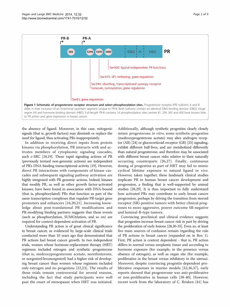
IntroductionThe mitogenic activity of estrogen is well established,but an under-studied ovarian steroid hormone, progester-one, is emerging as a primary mitogen in the breast, con-tributing significantly to genetic programming required formammary stem cell self-renewal, mammary gland develop-ment, proliferation, and hyperplasia [1]. The effects of pro-gesterone are triggered after binding of progesterone to itsintracellular receptor, the progesterone receptor (PR). ThePR exists in two primary isoforms, differing structurally bythe inclusion of an N-terminal segment unique to the full-length isoform, PR-B [2] (Figure 1). This region, termed theB-upstream segment, is missing from the shorter isoform,PR-A [3]. The two isoforms are encoded by the same gene(regulated by distinct but tandem upstream promoters) andare most often co-expressed [4]. The PR is a member of thesteroid hormone receptor subgroup of ligand-activatedtranscription factors within the large nuclear receptorsuperfamily, and is an important down-stream effector ofestrogen-receptor (ER) signaling; in most circumstances,estrogen is required for robust PR expression. PR binding
to DNA, either directly through progesterone responseelements or indirectly through tethering interactions withother transcription factors, activates transcriptionalprofiles associated with mammary gland proliferation andbreast cancer [5-9]. Additionally, PR binding interactionswith transcriptional co-activators and repressors are criticalto PR transcription factor function [10].PRs are highly post-translationally modified, primarily
through N-terminal phosphorylation (select phosphorylationsites most relevant to breast cancer biology are highlightedin Figure 1), acetylation, SUMOylation, and ubiquitination[9,11-17]. These receptor modifications dramatically alterPR function, receptor localization and turnover, and pro-moter selectivity. The PR can be phosphorylated basally inthe absence of the hormonal ligand, but is potently modi-fied after ligand treatment, in response to local growthfactors or in a cell cycle-dependent manner [12,13,15-17](G. Dressing and C. Lange, unpublished data). Mitogenicprotein kinases - such as CDK2, CK2, and MAPK - havebeen shown to phosphorylate PR and subsequently modifyPR action. Therefore, PR can be thought of as a ‘mitogenicsensor’ in the cell, with PR phosphorylation serving as areadout of kinase activity. Highly mitogenic environmentslike cancer, where kinase activities are frequently high, maybe a situation where PR is persistently phosphorylated in
* Correspondence: [email protected] of Medicine (Hematology, Oncology, and Transplantation) andthe Department of Pharmacology, University of Minnesota, Masonic CancerCenter, 420 Delaware St SE, MMC 806, Minneapolis, MN 55455, USA
© Hagan and Lange; licensee BioMed Central Ltd. This is an Open Access article distributed under the terms of theCreative Commons Attribution License (http://creativecommons.org/licenses/by/2.0), which permits unrestricted use,distribution, and reproduction in any medium, provided the original work is properly cited. The Creative Commons PublicDomain Dedication waiver (http://creativecommons.org/publicdomain/zero/1.0/) applies to the data made available in thisarticle, unless otherwise stated.
Hagan and Lange BMC Medicine
2014
2014, 12:32http://www.biomedcentral.com/1741-7015/12/32
the absence of ligand. Moreover, in this case, mitogenicsignals (that is, growth factors) may diminish or replace theneed for ligand, thus activating PRs inappropriately.In addition to receiving direct inputs from protein
kinases via phosphorylation, PR interacts with and ac-tivates members of cytoplasmic signaling cascades,such c-SRC [18,19]. These rapid signaling actions of PR(previously termed non-genomic actions) are independentof PR’s DNA-binding transcriptional activity [19]. However,direct PR interactions with components of kinase cas-cades and subsequent signaling pathway activation arehighly integrated with PR genomic actions. Indeed, kinasesthat modify PR, as well as other growth factor-activatedkinases, have been found in association with DNA-bound(that is, phosphorylated) PRs that function as part of thesame transcription complexes that regulate PR-target genepromoters and enhancers [16,20,21]. Increasing know-ledge about post-translational PR modifications andPR-modifying binding partners suggests that these events(such as phosphorylation, SUMOylation, and so on) arerequired for context-dependent activation of PR.Understanding PR action is of great clinical significance
in breast cancer, as evidenced by large-scale clinical trialsconducted more than 10 years ago that demonstrated thatPR actions fuel breast cancer growth. In two independenttrials, women whose hormone-replacement therapy (HRT)regimens included estrogen and synthetic progesterone(that is, medroxyprogesterone acetate, norethisterone,or norgestrel/levonorgestrel) had a higher risk of develop-ing breast cancer than women whose regimens includedonly estrogen and no progestins [22,23]. The results ofthese trials remain controversial for several reasons,including the fact that study participants were wellpast the onset of menopause when HRT was initiated.
Additionally, although synthetic progestins clearly closelymimic progesterone in vitro, some synthetic progestins(medroxyprogesterone acetate) may alter androgen recep-tor (AR) [24] or glucocorticoid receptor (GR) [25] signaling,exhibit different half-lives, and are metabolized differentlythan natural progesterone, and therefore may be associatedwith different breast cancer risks relative to their naturallyoccurring counterparts [26,27]. Finally, continuousdosing of progestins as part of HRT may fail to mimiccyclical lifetime exposure to natural ligand in vivo.However, taken together, these landmark clinical studiesimplicate PR in human breast cancer development andprogression, a finding that is well-supported by animalstudies [28,29]. It is thus important to fully understandhow activated PRs may contribute to early breast cancerprogression, perhaps by driving the transition from steroidreceptor (SR)-positive tumors with better clinical prog-noses to more aggressive, poorer outcome SR-negativeand luminal-B-type tumors.Convincing preclinical and clinical evidence suggests
that progestins increase breast cancer risk in part by drivingthe proliferation of early lesions [28,30-35]. Even so, at leastfive main sources of confusion remain regarding the roleof PR actions in breast cancer (expanded on in Box 1).First, PR action is context dependent - that is, PR actiondiffers in normal versus neoplastic tissue and according tohormone exposure (for example, in the presence versusabsence of estrogen), as well as organ site (for example,proliferative in the breast versus inhibitory in the uterus).Moreover, despite convincing progestin-dependent pro-liferative responses in murine models [32,36,37], earlyreports showed that progesterone was anti-proliferativeor non-proliferative in human cells [38-40]. However,recent work from the laboratory of C. Brisken [41] has
Figure 1 Schematic of progesterone receptor structure and select phosphorylation sites. Progesterone receptor (PR) isoforms A and Bdiffer in their inclusion of an N-terminal upstream segment unique to PR-B. Both isoforms contain an identical DNA binding domain (DBD), hingeregion (H) and hormone binding domain (HBD). Full-length PR-B contains 14 phosphorylation sites; serines 81, 294, 345 and 400 have known linksto PR action and gene expression in breast cancer.
Hagan and Lange BMC Medicine Page 2 of 92014, 12:32http://www.biomedcentral.com/1741-7015/12/32
shown that progesterone is proliferative in humanbreast tissue microstructures isolated from normal hu-man breast specimens. Interestingly, progesterone-dependent proliferation and signaling is preserved onlywhen the tissue architecture remains intact; human tis-sues (previously dissociated) grown in two- or three-dimensional cultures did not display this proliferativephenotype, suggestive of further context-dependent PRactions. Second, PR isoform-specific activities (PR-Aversus PR-B) overlap but can have very disparate activitieswithin a given target tissue and at selected gene pro-moters; however, despite their distinct activities, the twoPR-isoforms are not distinguished clinically. Third, ligand-independent (that is, growth factor- or kinase-dependent)activities of PR are poorly understood. Fourth, the dosing(cyclical versus continuous) and source (natural versussynthetic) of ligand are likely to be key determinants ofthe kinetics of PR action. Fifth, although anti-progestinsshowed clinical promise in early clinical trials, their usewas limited by liver toxicities (onapristone; [42]) largelyattributable to cross-reactivity with other nuclear receptors,such as GR. This review will focus on the molecular deter-minants of PR’s context-dependent actions and their clin-ical significance. These PR actions are primarily determinedby the availability of PR-binding partners and direct modifi-cations to PR that dictate promoter selection.
Post-translational modifications and molecularinteractions alter promoter selectivityMounting evidence suggests that post-translationalmodifications of PR are key determinants of promoterselectivity and, in turn, the spectrum of target genes acti-vated in response to ligand binding (reviewed in [43,44]).PR promoter preference is partially dictated by differencesin the recruitment of PR and/or its co-activators or co-repressors to specific DNA sequences. In microarrayanalyses, cells expressing wild-type PR or PRs contain-ing single point-mutations at specific phosphorylationor SUMOylation sites exhibit dramatic changes in
PR-dependent gene expression, specific to precisepost-translational modifications. For example, recentanalyses from the Lange laboratory revealed that PRphosphorylation on serine 294 favors the subsequentdeSUMOylation on PR lysine 388 [45], thereby yielding ahyperactive receptor that regulates a unique gene expres-sion signature found in high ERBB2-expressiong tumors;this unique phospho-PR gene expression signaturepredicted decreased survival in patients treated withtamoxifen [9]. By contrast, a separate gene expressionpattern is observed when PR is phosphorylated on Ser81by CK2, a kinase commonly overexpressed in breastcancers; this modification is associated with the expressionof gene sets involved in interferon and STAT5 signaling(discussed in more detail below) [8]. Therefore, in responseto ligand, growth factor-mediated PR phosphorylation(or phosphorylation-dependent alterations of other post-translational modifications such as SUMOylation) dictatesthe selective expression of specific subsets of target genesand subsequently their transcriptional programs.Target gene selectivity is achieved not only through
differential recruitment of PR [8,16], but also through asso-ciated transcriptional co-activators and repressors that arecritical to PR function [9,10,46]. For example, pioneer fac-tors are specialized subsets of transcription factors thatopen defined regions of chromatin, making it accessible forother transcription factors, like SRs (reviewed in [47,48]).These types of factors have been identified for other nuclearreceptors, such as ER and AR; however, they have yet to beidentified for PR. Preliminary data suggest that FOXA1 andSTAT5 may be putative pioneer factors for PR [8,49,50];differential binding interactions between PR and these fac-tors provide a mechanism for promoter selectivity, perhapsbased on PR post-translational modifications (that is, viaphosphorylation-specific interactions with pioneer factors).Emerging evidence suggests that interactions between
members of the SR superfamily is an additional regulatorystep in determining target-gene specificity. Interactionsbetween ER and AR have been the focus of recent in-vestigations [51,52]. Recent data from the Lanari groupdemonstrate the existence of functional cross-talk betweenER and PR; both receptors are localized together onregulatory regions of PR-target genes, such as CCND1and MYC, primarily in response to treatment with pro-gestins [53]. Moreover, work recently published from ourgroup suggests a complimentary story whereby ER and PRcooperate to regulate a subset of ER-target genes in re-sponse to estrogen, but fully independent of exogenouslyadded progestin. In this case, PR-B appears to act as a scaf-folding molecule for increased recruitment of signalingadaptors and protein kinases that phosphorylate ERwithin ER/PR-containing transcription complexes [54].Taken together, these studies suggest that context-dependent progesterone/PR action may in part depend
Box 1: Complexities of progesterone receptor actions.
• Tissue-specific effects (breast vs. reproductive tract)
• Actions in normal vs. neoplastic tissues
• Isoform-specific actions (PR-A vs. PR-B)
• Lack of clinical designation between PR isoforms
• Ligand-independent actions
• Timing of hormone delivery (continuous vs. cyclical)
• Source of hormone (synthetic vs. natural progesterone)
• PR actions are both ER-dependent and ER-independent
• Efficacy of early anti-progestins in the clinic
Hagan and Lange BMC Medicine Page 3 of 92014, 12:32http://www.biomedcentral.com/1741-7015/12/32

on the presence of other steroid hormones and theirreceptors. Detailed biochemical studies of steroid hormonereceptor cross-talk are needed to provide a framework fora better understanding of differential hormone actionsin pre- and post-menopausal conditions where endogen-ous hormone levels dramatically differ, as well as duringbreast or prostate cancer treatment with hormone-ablation therapies where closely related steroid hormonereceptors (PR, GR, AR, ER) may substitute for the blockedactivity of another (ER or AR).
Progesterone receptor phosphorylation by CK2 as aparadigm for receptor modification and regulationRecent data from our laboratory characterizing PRphosphorylation on Ser81 by CK2 exemplifies how theaforementioned modifications and signaling inputs canalter PR function. CK2 is a ubiquitously expressed kinaseoften up-regulated in many different types of cancer,including breast [55-57]. We and others have shownthat CK2 phosphorylates PR on Ser81, a site that is ba-sally phosphorylated; however, Ser81 phosphorylationlevels increase markedly in response to ligand (or whencells enter S phase in the absence of ligand) [16,58]. PR
phosphorylation at Ser81 is associated with a specificgene expression profile, which is correlated with pathwaysaltered in breast cancer, including genes implicated inmammary stem cell maintenance and renewal [8,16].Additionally, the PR target genes whose expression re-quire phosphorylation at Ser81 are significantly associ-ated with interferon/inflammation and STAT-signalingdatasets, a unique observation for SRs that representsa novel link between steroid hormone action, inflammation,and cancer [8]. A key target gene regulated by Ser81 phos-phorylation is STAT5 itself, and notably, JAK/STAT signal-ing is required for potent activation of PR Ser81-regulatedgenes, indicating a feed-forward mechanism for geneprogram activation (Figure 2). STAT5 is present, along withphosphorylated PR, on the regulatory region of WNT1, akey Ser81 target gene known to be involved in cancerand stem cell biology. Moreover, an in silico analysisof a publically available PR whole genome chromatinimmunoprecipitation dataset reveals that there is sig-nificant enrichment of STAT5 consensus sites withinPR-bound chromatin regions, indicating that STAT5may function as a pioneer factor for phosphorylated PR(perhaps specifically when PR Ser81 is phosphorylated).
Figure 2 Molecular determinants of progesterone receptor action. Co-activators/repressors: interactions between PR and knowntranscriptional co-activators (for example, SRC1) and co-repressors (for example, NCOR/SMRT) are a key determinant of promoter specificity.Pioneer factors: interactions with predicted PR pioneer factors (for example, STAT5, putatively) lead to chromatin remodeling, allowing for efficientPR recruitment and subsequent target-gene transcription. Different pioneer factors would be predicted to determine differential PR recruitment.Post-translational modifications: phosphorylation (P), acetylation (Ac), ubiquitination (Ub), and SUMOylation (Sumo) primarily on N-terminal serineand lysine residues dictate receptor localization, turnover, subcellular localization, and promoter selectivity. Steroid receptor (SR) interactions:emerging evidence suggests that interactions between members of the steroid receptor superfamily (such as ER and PR) determine PR target-genespecificity. Scaffolding interactions: PR interaction with proteins acting as scaffolds (such as DUSP6) determine receptor post-translational modifications,thereby contributing to promoter selection. Cell cycle: phosphorylation on select PR serine residues and cell cycle-dependent protein complexformation determine receptor function and recruitment of PR to specific target genes.
Hagan and Lange BMC Medicine Page 4 of 92014, 12:32http://www.biomedcentral.com/1741-7015/12/32
These data suggest that CK2-mediated Ser81 phosphory-lation of PR may activate gene expression programsinvolved in modulating inflammation related to breastcancer development and progression, including mammarystem cell maintenance and self-renewal.Recent studies have defined a new mechanism by which
CK2 and PR interact. Direct interaction between PR andDUSP6, a negative regulator of the MAPK pathway, isrequired to achieve phosphorylation on PR Ser81 [8]. Thisregulation occurs independently of DUSP6 phosphatase ac-tivity, suggesting that DUSP6 is acting as a scaffold for theinteraction between PR and the kinase that phosphor-ylates Ser81, CK2. Related to this finding, an inter-action between DUSP6 and CK2 has previously beenidentified [59]. Together, this suggests a model wherebyDUSP6 binding to CK2 brings the kinase (CK2) in closeproximity to its substrate (PR Ser81), allowing for efficientphosphorylation and subsequent selection of target geneswithin a given (that is, inflammatory, pro-growth, survival)genetic program.Cumulatively, in this vignette describing one context-
dependent scenario of PR action, there exists cross-talk be-tween mitogenic kinases (that is, CK2 phosphorylation ofPR Ser81), MAPK pathway components (that is, DUSP6interaction with PR is required for Ser81 phosphorylation),phosphorylation-dependent gene regulation (that is, Ser81phosphorylation is required for PR recruitment to specificsubsets of PR target genes), and putative phosphorylation-specific interactions with a pioneer factor/co-factor(that is, JAK/STAT-dependence of PR Ser81-regulatedgene expression). PR phosphorylation by CK2 on Ser81is an exemplary case study of how the molecular deter-minants of PR action differentially determine receptorfunction in breast cancer models (Figure 2).
Progesterone receptor clinical significance in breast cancerLuminal breast tumors are characterized by their expres-sion of ER and PR, both of which are good prognosticmarkers for predicted response to endocrine therapies.Interestingly, analysis of The Cancer Genome Atlas datafor the luminal A/B subtype of breast tumors revealsthat heterozygous loss of the PR locus occurs in 40% ofluminal tumors, while 25% of luminal tumors are alsoheterozygous for the ER locus. However, these tumorsare overwhelmingly ER-positive and largely respond wellto ER-targeted therapies [60]. Interestingly, PR and ER copynumber is often correlated in individual tumors; tumorswith altered copy numbers for ER are likely to have changesin PR copy number. Despite these genomic alterations,both PR and ER mRNA levels are similar in luminal tumorsthat are diploid versus those that have lost an allele at theseloci. Thus, gene copy number may not be a robust measureof the functional (that is, protein) readout for these steroidhormone receptors and should be interpreted with caution.
Moreover, complex intra- and inter-tumoral heterogeneitymay be reflected in analyses of genomic copy number.Because PR-positive cells release pro-proliferative factors(that is, PR target-gene products) that induce paracrinesignaling, a small percentage of PR-positive cells within anindividual tumor could have significant effects on tumorstem cell maintenance and/or tumor growth and prog-ression. This is a complex situation that makes PR loci gen-omic heterozygosity difficult to interpret. Cumulatively,these data underscore the need to gain a much better un-derstanding of PR signaling within the clinical context.HRT clinical trial data (discussed above) suggest an
important role for progestins and PR as drivers (thatis, tumor promoters) of breast cancer cell growth.Progesterone-dependent expression of secreted paracrinefactors is required for self-renewal of (PR-null) stem cells inthe normal mammary gland [32,37] (see below). PR targetgenes include soluble factors known to modify cancer stemcells (WNT1 and RANKL). However, the role of PR targetgenes in the maintenance or expansion of cancer progeni-tor or stem cells is currently unknown. While a minorityof normal (non-pregnant) breast epithelial cells containsteroid hormone receptors, the majority of luminal breastcancers express ER and PR (discussed above); heteroge-neous cells within the breast may contain both ER andPR, only ER, or only PR [61]. Interestingly, very few somaticmutations have been identified in ER [62] or PR. With re-gard to PR, isolated genetic polymorphisms linked to breastand reproductive cancers appear to increase levels of PR-Bisoform expression, rather than affect PR transcriptionalactivity [63-65]. Additionally, the PR-A promoter is morefrequently methylated (that is, silenced) relative to the PR-Bpromoter in advanced endocrine-resistant breast cancers[66]. These data imply that genetic alteration of PR itself isusually not sufficient to promote tumorigenesis. Alterna-tively, we propose that oncogenic mutations that drive sig-naling pathways provide the context for heightened ER andPR transcriptional activity. For example, high levels ofkinases, such as CK2, CDKs or MAPKs, may inducepersistent progesterone-independent phosphorylation ofPR-B on serines 81 or 294, respectively, thereby leadingto activation of phospho-isoform-specific transcriptionalprograms shown to be significantly altered in luminal breastcancer [8,9]. Therapeutic strategies that target receptor-modifying protein kinases (that is, anti-CK2, CDK2 orMAPK) and/or their transcriptional co-factors (that is,STATs, AP1, SP1, FOXO1, FOXA1) are likely to be verysuccessful at treating breast cancer and must remain adirection of robust exploration within the SR field.Historically, clinical testing of anti-progestins has been
limited [42,67-70]. The results of a clinical trial released in1999 showed promise for anti-progestins as front-linebreast cancer endocrine therapy [42]. Although patient ac-crual in this study was small (19 patients), 67% of patients
Hagan and Lange BMC Medicine Page 5 of 92014, 12:32http://www.biomedcentral.com/1741-7015/12/32
achieved tumor remission when treated with onapristone,a PR type I antagonist that blocks PR binding to DNA, asfront-line endocrine therapy for locally advanced or primarybreast cancer [42]. Liver function test abnormalities wereseen early in this trial, and for that reason new patient ac-crual was stopped. These liver-associated effects were likelydue to inhibition of GR, a closely related SR. The clinical effi-cacy of lonaprisan, a type III PR antagonist that promotesPR repression through the recruitment of transcriptional co-repressors (while maintaining DNA binding), was measuredin a phase II study as second-line therapy for PR-positivebreast cancer [70]. The results from this trial were dis-appointing, and the trial was terminated before full patientaccrual. Although a small percentage (14%) of patientsachieved stable disease, no patients achieved complete orpartial responses. This trial likely failed for a number ofreasons, including lack of patient classification, patientshaving previous exposure to endocrine therapies, anda lack of mechanistic understanding of PR inhibitoraction and isoform specificity. Notably, clinically usedanti-progestins that target the ligand-binding domain ofPR may fail to block ligand-independent actions of PR (dis-cussed above).Renewed optimism for the use of anti-progestins to
prevent or inhibit breast cancer growth is provided bymore recent preclinical studies of anti-progestins in murinemammary tumor models. In a dramatic example, treatmentof nulliparous Brca1/Trp53-deficient mice with mifepris-tone, a PR antagonist, completely inhibited the formationof mammary gland tumors normally observed in virginmice [71], perhaps via modulation of the stem cell com-partment [30,32]. Newer, highly selective anti-progestins,which are currently in development by several pharma-ceutical companies, may increase the clinical utility ofanti-progestins in breast cancer prevention and treatmentand is an area of renewed research interest. Notably, manypatients that relapse while on tamoxifen therapy retainexpression of PR, underscoring the clinical significance ofconsidering PRs as potentially acting independently of ERin the context of breast cancer progression during estrogenablation (that is, PR expression is most often used clinicallyas a measure of ER function) [72,73]. Based on our currentunderstanding of ligand-dependent and ligand-independent(kinase-induced) PR actions, classification of patients basedon gene-expression profiling could better identify the sub-population of patients that would respond well to se-lective anti-progestins. In addition, cross-talk betweenER and PR (or AR), and growth-factor signaling path-ways (discussed above) is a likely confounding compo-nent of development to endocrine-resistant disease, andshould therefore be considered (for example, via the useof pathway-specific gene biomarkers) when selecting anti-progestins as potentially beneficial front-line or second-line therapy [74-76].
As mentioned above (and in Box 1), the clinical signifi-cance of PR isoforms is likely vastly under-appreciated.In mammary tissue, PR exists as two primary isoforms,PR-A and PR-B. Although PR-B is required for mammarygland development and PR-A for uterine development,these isoforms are most often co-expressed in the sametissues, typically at a ratio of 1:1. Single isoform expressionin tissues is rare [77-79]. Interestingly, in pre-neoplasticlesions and samples from patients with breast cancer, thisbalanced A:B ratio is often altered, frequently due toapparent loss of PR-B [78,80]. Cumulative data from theLange laboratory has revealed that this imbalance may beexplained by phosphorylation-dependent turnover of tran-scriptionally active PR-B receptors relative to more stableand less active PR-A receptors. PR-B but not PR-A under-goes extensive cross-talk with mitogenic protein kinases[8,16,45,81,82]. Thus, PR-B is heavily phosphorylated inresponse to ligand or via the action of growth factors, andalthough this isoform-specific phosphorylation (on PR-BSer294) is linked to high transcriptional activity, it is alsocoupled to rapid ubiquitin-dependent turnover of the re-ceptor; regulated PR-B turnover is tightly linked to tran-scriptional activity (that is, stable non-degradable mutantsof PR are poor transcriptional activators) [83,84]. Of note,this phosphorylation event (PR-B Ser294) has been detectedin a subset of human tumors [9]. Therefore, loss of PR-B,as measured by protein levels in clinical immunohisto-chemistry tests or western blotting may actually reflect highPR-B transcriptional activity coupled with rapid proteinturnover; peak PR target-gene expression (mRNA) is coin-cident with nearly undetectable PR protein in experimentalmodels [85]. Mouse models (mammary gland) predomin-antly express PR-A prior to pregnancy. In humans, normalmammary gland function may rely upon balanced expres-sion of the two PR isoforms. Unfortunately, current immu-nohistochemistry clinical testing for PR in breast cancersamples does not differentiate between PR-A and PR-B iso-forms. Because an imbalance between the two isoforms ap-pears to be linked to cancerous phenotypes, clinical isoformdistinction may have great diagnostic potential and shouldbe considered as part of routine luminal cancer work-up.Emerging data linking progesterone regulation to the
expansion of the mammary stem cell compartment high-light the role that PR and progesterone may play in earlyevents in breast cancer. Recent seminal work in murinemodels has shown that progesterone can induce therapid expansion of mammary stem cells, a populationof SR-negative (that is, ER- and PR-negative) cells locatedin the basal epithelial compartment of the mammary gland[32,37]. Because these cells are PR negative, this expansionlikely occurs through the production of paracrine fac-tors secreted by neighboring or nearby PR-positive luminalepithelial cells. Progesterone-dependent expansion ofthe mammary stem cell population is mediated by key
Hagan and Lange BMC Medicine Page 6 of 92014, 12:32http://www.biomedcentral.com/1741-7015/12/32
PR-target genes, including RANKL and WNT4 [32,37].Brisken and colleagues have shown that progesterone-dependent control of RANKL expression in human tissuesis dependent on intact breast tissue microstructure, andhave confirmed that RANKL is required for progesterone-induced proliferation [41]; estrogen is a permissive hor-mone (for PR expression) in this context. Interestingly,PR-dependent RANKL expression requires STAT5A [50].This observation is similar to what has been published forPR regulation of WNTs [8], highlighting an emerging rolefor co-ordinate STAT5/PR regulation of select subsets ofPR-target genes related to proliferation and stem cell self-renewal (see above). Moreover, a PR-positive subpopula-tion of mammary gland progenitor cells has been recentlydiscovered [61], challenging the current dogma that mam-mary gland precursors are strictly SR-negative. Theseexciting findings suggest that this long-lived population ofcells, one that is exquisitely sensitive to mutagenic events,can expand in response to progesterone in both a paracrineand autocrine fashion [36]. Notably, these PR-positivemammary stem cells are devoid of ER protein or mRNAexpression, further underscoring the need for understand-ing PR action as independent of ER in this context.
ConclusionsRecent clinical and preclinical studies clearly demonstratethe significance of fully understanding the determinantsof context-dependent PR action. They not only challengethe current clinical diagnostic paradigm in which PR is onlyused as a marker of ER transcriptional activity, but also sup-port a renewed interest in understanding PR as a driver ofbreast tumor progression and thus a potentially very usefultarget for improved breast cancer therapy [1,86]. In this re-view, we have highlighted the concept that gene-expressionanalyses linked to PR actions suggest different transcrip-tional programs are activated in response to specific post-translational modifications (phosphorylation events) andprotein-protein interactions. Although these unique PR genesignatures highlight functional differences between modifiedPRs and their components, the overlap between these(predominantly proliferative) programs supports a strongrole for PR in early tumor progression toward more aggres-sive cancer phenotypes, and in some cases, even highlights aphospho-PR gene signature associated with poor responseto endocrine treatment [9]. Therefore, gene signatures thatdefine PR action will likely provide a useful paired diagnosticfor clinically applied selective anti-progestins. We concludethat PR function is highly dependent on the molecularcontext, which is defined by such factors as protein kinaseactivity (as a major input to receptor post-translationalmodifications), co-factor availability, and the presence ofprogesterone and other steroid hormone levels and recep-tors (Figure 2). Future therapeutic approaches should con-sider targeting receptor-modifying activities in place of or
in conjunction with anti-hormone therapies. With proges-terone emerging as the primary mitogen in the adult breast(wherein estrogen is permissive for PR expression), under-standing PR function and identifying or targeting modifiersof PR action are of critical importance to advancing thetreatment of breast cancer.
AbbreviationsAR: androgen receptor; ER: estrogen receptor; GR: glucocorticoid receptor;HRT: hormone-replacement therapy; PR: progesterone receptor; SR: steroidreceptor.
Competing interestsCAL receives consulting income from Arno Therapeutics, Inc. This interesthas been reviewed and managed by the University of Minnesota inaccordance with its Conflict of Interest policies. CRH declares that she has nocompeting interests.
Authors’ contributionsCRH and CAL together led the initial design and conception of themanuscript. CRH led the writing of the first and all subsequent drafts of themanuscript. CAL contributed significant written and editorial inputs to themanuscript at every stage. Both authors read and approved the finalmanuscript.
Authors’ informationCAL joined the University of Minnesota (Departments of Medicine andPharmacology) faculty in 1999. Her research is focused on steroid hormoneaction in breast cancer progression. Her laboratory studies the role ofcross-talk between growth factor-mediated signaling pathways and steroidhormone receptors, using the human progesterone receptor as a modelreceptor. CAL holds the Tickle Family Land Grant Endowed Chair of BreastCancer Research at the University of Minnesota. She is the Director of TheCancer Biology Training Grant (T32) and the Cell Signaling Program Leadwithin the Masonic Cancer Center. CAL is Editor-in-Chief of the journalHormones and Cancer (jointly held by The Endocrine Society and Springer).CRH is a senior post-doctoral fellow in the laboratory of CAL.
AcknowledgementsThe authors would like to thank Dr. Andrea R. Daniel (Minnesota) for hercritical review of this manuscript, and Michael Freeman (Minnesota) foreditorial assistance.
Received: 17 October 2013 Accepted: 21 January 2014Published:
References1. Brisken C: Progesterone signalling in breast cancer: a neglected hormone
coming into the limelight. Nat Rev Cancer 2013, 13:385–396.2. Kraus WL, Montano MM, Katzenellenbogen BS: Cloning of the rat
progesterone receptor gene 5′-region and identification of twofunctionally distinct promoters. Mol Endocrinol 1993, 7:1603–1616.
3. Hill KK, Roemer SC, Churchill ME, Edwards DP: Structural and functionalanalysis of domains of the progesterone receptor. Mol Cell Endocrinol2012, 348:418–429.
4. Kastner P, Krust A, Turcotte B, Stropp U, Tora L, Gronemeyer H, Chambon P:Two distinct estrogen-regulated promoters generate transcripts encodingthe two functionally different human progesterone receptor forms A andB. Embo J 1990, 9:1603–1614.
5. Owen GI, Richer JK, Tung L, Takimoto G, Horwitz KB: Progesteroneregulates transcription of the p21(WAF1) cyclin-dependent kinase inhibitorgene through Sp1 and CBP/p300. J Biol Chem 1998, 273:10696–10701.
6. Stoecklin E, Wissler M, Schaetzle D, Pfitzner E, Groner B: Interactionsin the transcriptional regulation exerted by Stat5 and by members ofthe steroid hormone receptor family. J Steroid Biochem Mol Biol 1999,69:195–204.
7. Cicatiello L, Addeo R, Sasso A, Altucci L, Petrizzi VB, Borgo R, Cancemi M,Caporali S, Caristi S, Scafoglio C, et al: Estrogens and progesterone promotepersistent CCND1 gene activation during G1 by inducing transcriptionalderepression via c-Jun/c-Fos/estrogen receptor (progesterone receptor)
Hagan and Lange BMC Medicine Page 7 of 9
20 Feb 2014
2014, 12:32http://www.biomedcentral.com/1741-7015/12/32
complex assembly to a distal regulatory element and recruitment of cyclinD1 to its own gene promoter. Mol Cell Biol 2004, 24:7260–7274.
8. Hagan CR, Knutson TP, Lange CA: A common docking domain in progesteronereceptor-B links DUSP6 and CK2 signaling to proliferative transcriptionalprograms in breast cancer cells. Nucleic Acids Res 2013, 41:8962–8942.
9. Knutson TP, Daniel AR, Fan D, Silverstein KA, Covington KR, Fuqua SA, LangeCA: Phosphorylated and sumoylation-deficient progesterone receptorsdrive proliferative gene signatures during breast cancer progression.Breast Cancer Res 2012, 14:R95.
10. McKenna NJ, Lanz RB, O’Malley BW: Nuclear receptor coregulators: cellularand molecular biology. Endocr Rev 1999, 20:321–344.
11. Daniel AR, Faivre EJ, Lange CA: Phosphorylation-dependent antagonismof sumoylation derepresses progesterone receptor action in breastcancer cells. Mol Endocrinol 2007, 21:2890–2906.
12. Lange CA, Shen T, Horwitz KB: Phosphorylation of human progesteronereceptors at serine-294 by mitogen-activated protein kinase signals theirdegradation by the 26S proteasome. Proc Natl Acad Sci U S A 2000,97:1032–1037.
13. Weigel NL, Bai W, Zhang Y, Beck CA, Edwards DP, Poletti A: Phosphorylation andprogesterone receptor function. J Steroid Biochem Mol Biol 1995, 53:509–514.
14. Daniel AR, Gaviglio AL, Czaplicki LM, Hillard CJ, Housa D, Lange CA: Theprogesterone receptor hinge region regulates the kinetics oftranscriptional responses through acetylation, phosphorylation, andnuclear retention. Mol Endocrinol 2011, 24:2126–2138.
15. Pierson-Mullany LK, Lange CA: Phosphorylation of progesterone receptorserine 400 mediates ligand-independent transcriptional activity inresponse to activation of cyclin-dependent protein kinase 2. Mol Cell Biol2004, 24:10542–10557.
16. Hagan CR, Regan TM, Dressing GE, Lange CA: CK2-dependent phosphorylationof progesterone receptors (PR) on Ser81 regulates PR-B isoform-specific targetgene expression in breast cancer cells CK2. Mol Cell Biol 2011, 31:2439–2452.
17. Faivre EJ, Daniel AR, Hillard CJ, Lange CA: Progesterone receptor rapidsignaling mediates serine 345 phosphorylation and tethering tospecificity protein 1 transcription factors. Mol Endocrinol 2008, 22:823–837.
18. Ballare C, Uhrig M, Bechtold T, Sancho E, Di Domenico M, Migliaccio A,Auricchio F, Beato M: Two domains of the progesterone receptorinteract with the estrogen receptor and are required for progesteroneactivation of the c-Src/Erk pathway in mammalian cells. Mol Cell Biol2003, 23:1994–2008.
19. Boonyaratanakornkit V, Scott MP, Ribon V, Sherman L, Anderson SM, MallerJL, Miller WT, Edwards DP: Progesterone receptor contains a proline-richmotif that directly interacts with SH3 domains and activates c-Src familytyrosine kinases. Mol Cell 2001, 8:269–280.
20. Diaz Flaque MC, Vicario R, Proietti CJ, Izzo F, Schillaci R, Elizalde PV:Progestin drives breast cancer growth by inducing p21(CIP1) expressionthrough the assembly of a transcriptional complex among Stat3,progesterone receptor and ErbB-2. Steroids 2013, 78:559–567.
21. Narayanan R, Adigun AA, Edwards DP, Weigel NL: Cyclin-dependent kinaseactivity is required for progesterone receptor function: novel role forcyclin A/Cdk2 as a progesterone receptor coactivator. Mol Cell Biol 2005,25:264–277.
22. Beral V: Breast cancer and hormone-replacement therapy in the MillionWomen Study. Lancet 2003, 362:419–427.
23. Chlebowski RT, Anderson GL, Gass M, Lane DS, Aragaki AK, Kuller LH,Manson JE, Stefanick ML, Ockene J, Sarto GE, et al: Estrogen plus progestinand breast cancer incidence and mortality in postmenopausal women.JAMA 2010, 304:1684–1692.
24. Birrell SN, Butler LM, Harris JM, Buchanan G, Tilley WD: Disruption ofandrogen receptor signaling by synthetic progestins may increase risk ofdeveloping breast cancer. FASEB J 2007, 21:2285–2293.
25. Courtin A, Communal L, Vilasco M, Cimino D, Mourra N, de Bortoli M,Taverna D, Faussat AM, Chaouat M, Forgez P, et al: Glucocorticoid receptoractivity discriminates between progesterone and medroxyprogesteroneacetate effects in breast cells. Breast Cancer Res Treat 2012, 131:49–63.
26. Fournier A, Berrino F, Riboli E, Avenel V, Clavel-Chapelon F: Breast cancerrisk in relation to different types of hormone replacement therapy in theE3N-EPIC cohort. Int J Cancer 2005, 114:448–454.
27. Lyytinen H, Pukkala E, Ylikorkala O: Breast cancer risk in postmenopausalwomen using estradiol-progestogen therapy. Obstet Gynecol 2009, 113:65–73.
28. Lanari C, Lamb CA, Fabris VT, Helguero LA, Soldati R, Bottino MC, GiulianelliS, Cerliani JP, Wargon V, Molinolo A: The MPA mouse breast cancer
model: evidence for a role of progesterone receptors in breast cancer.Endocr Relat Cancer 2009, 16:333–350.
29. Lanari C, Molinolo AA: Progesterone receptors-animal models and cellsignalling in breast cancer. Diverse activation pathways for theprogesterone receptor: possible implications for breast biology andcancer. Breast Cancer Res 2002, 4:240–243.
30. Horwitz KB, Dye WW, Harrell JC, Kabos P, Sartorius CA: Rare steroidreceptor-negative basal-like tumorigenic cells in luminal subtype humanbreast cancer xenografts. Proc Natl Acad Sci U S A 2008, 105:5774–5779.
31. Horwitz KB, Sartorius CA: Progestins in hormone replacement therapiesreactivate cancer stem cells in women with preexisting breast cancers:a hypothesis. J Clin Endocrinol Metab 2008, 93:3295–3298.
32. Joshi PA, Jackson HW, Beristain AG, Di Grappa MA, Mote PA, Clarke CL,Stingl J, Waterhouse PD, Khokha R: Progesterone induces adult mammarystem cell expansion. Nature 2010, 465:803–807.
33. Santen RJ: Risk of breast cancer with progestins: critical assessment ofcurrent data. Steroids 2003, 68:953–964.
34. Hofseth LJ, Raafat AM, Osuch JR, Pathak DR, Slomski CA, Haslam SZ:Hormone replacement therapy with estrogen or estrogen plusmedroxyprogesterone acetate is associated with increased epithelialproliferation in the normal postmenopausal breast. J Clin EndocrinolMetab 1999, 84:4559–4565.
35. Santen RJ: Menopausal hormone therapy and breast cancer. J SteroidBiochem Mol Biol 2013, 2013:2013.
36. Beleut M, Rajaram RD, Caikovski M, Ayyanan A, Germano D, Choi Y,Schneider P, Brisken C: Two distinct mechanisms underlie progesterone-induced proliferation in the mammary gland. Proc Natl Acad Sci U S A 2010,107:2989–2994.
37. Asselin-Labat ML, Shackleton M, Stingl J, Vaillant F, Forrest NC, Eaves CJ,Visvader JE, Lindeman GJ: Steroid hormone receptor status of mousemammary stem cells. J Natl Cancer Inst 2006, 98:1011–1014.
38. Groshong SD, Owen GI, Grimison B, Schauer IE, Todd MC, Langan TA,Sclafani RA, Lange CA, Horwitz KB: Biphasic regulation of breast cancercell growth by progesterone: role of the cyclin-dependent kinaseinhibitors, p21 and p27(Kip1). Mol Endocrinol 1997, 11:1593–1607.
39. Clarke RB, Howell A, Anderson E: Estrogen sensitivity of normal humanbreast tissue in vivo and implanted into athymic nude mice: analysis of therelationship between estrogen-induced proliferation and progesteronereceptor expression. Breast Cancer Res Treat 1997, 45:121–133.
40. Communal L, Vilasco M, Hugon-Rodin J, Courtin A, Mourra N, Lahlou N,Dumont S, Chaouat M, Forgez P, Gompel A: Ulipristal acetate does notimpact human normal breast tissue. Hum Reprod 2012, 27:2785–2798.
41. Tanos T, Sflomos G, Echeverria PC, Ayyanan A, Gutierrez M, Delaloye JF, RaffoulW, Fiche M, Dougall W, Schneider P, et al: Progesterone/RANKL is a majorregulatory axis in the human breast. Sci Transl Med 2013, 5:182ra155.
42. Robertson JF, Willsher PC, Winterbottom L, Blamey RW, Thorpe S: Onapristone,a progesterone receptor antagonist, as first-line therapy in primary breastcancer. Eur J Cancer 1999, 35:214–218.
43. Hagan CR, Daniel AR, Dressing GE, Lange CA: Role of phosphorylation inprogesterone receptor signaling and specificity. Mol Cell Endocrinol 2012,357:43–49.
44. Dressing GE, Hagan CR, Knutson TP, Daniel AR, Lange CA: Progesteronereceptors act as sensors for mitogenic protein kinases in breast cancermodels. Endocr Relat Cancer 2009, 16:351–361.
45. Daniel AR, Faivre EJ, Lange CA: Phosphorylation-dependent antagonismof sumoylation derepresses progesterone receptor action in breastcancer cells. Mol Endocrinol 2007, 21:2890–2906.
46. Daniel AR, Lange CA: Protein kinases mediate ligand-independentderepression of sumoylated progesterone receptors in breast cancercells. Proc Natl Acad Sci U S A 2009, 106:14287–14292.
47. Jozwik KM, Carroll JS: Pioneer factors in hormone-dependent cancers.Nat Rev Cancer 2012, 12:381–385.
48. Magnani L, Eeckhoute J, Lupien M: Pioneer factors: directingtranscriptional regulators within the chromatin environment. TrendsGenet 2011, 27:465–474.
49. Clarke CL, Graham JD: Non-overlapping progesterone receptor cistromescontribute to cell-specific transcriptional outcomes. PLoS One 2012,7:e35859.
50. Obr AE, Grimm SL, Bishop KA, Pike JW, Lydon JP, Edwards DP: Progesteronereceptor and Stat5 signaling crosstalk through RANKL in mammaryepithelial cells. Mol Endocrinol 2013, 27:1808–1824.
Hagan and Lange BMC Medicine Page 8 of 92014, 12:32http://www.biomedcentral.com/1741-7015/12/32
51. Peters AA, Buchanan G, Ricciardelli C, Bianco-Miotto T, Centenera MM, HarrisJM, Jindal S, Segara D, Jia L, Moore NL, et al: Androgen receptor inhibitsestrogen receptor-alpha activity and is prognostic in breast cancer.Cancer Res 2009, 69:6131–6140.
52. Need EF, Selth LA, Harris TJ, Birrell SN, Tilley WD, Buchanan G: Researchresource: interplay between the genomic and transcriptional networksof androgen receptor and estrogen receptor alpha in luminal breastcancer cells. Mol Endocrinol 2012, 26:1941–1952.
53. Giulianelli S, Vaque JP, Soldati R, Wargon V, Vanzulli SI, Martins R, Zeitlin E,Molinolo AA, Helguero LA, Lamb CA, et al: Estrogen receptor alphamediates progestin-induced mammary tumor growth by interactingwith progesterone receptors at the cyclin D1/MYC promoters. Cancer Res2012, 72:2416–2427.
54. Daniel AR, Gaviglio AL, Knutson TP, Ostrander JH, D'Assoro AB, RavindranathanP, Peng Y, Raj GV, Yee D, Lange CA: Progesterone receptor-B enhancesestrogen responsiveness of breast cancer cells via scaffolding PELP1- andestrogen receptor-containing transcription complexes. Oncogene 2014.doi:10.1038/onc.2013.579. [Epub ahead of print].
55. Tawfic S, Yu S, Wang H, Faust R, Davis A, Ahmed K: Protein kinase CK2signal in neoplasia. Histol Histopathol 2001, 16:573–582.
56. Guerra B, Issinger OG: Protein kinase CK2 in human diseases. Curr MedChem 2008, 15:1870–1886.
57. Meggio F, Pinna LA: One-thousand-and-one substrates of protein kinaseCK2? FASEB J 2003, 17:349–368.
58. Zhang Y, Beck CA, Poletti A, Edwards DP, Weigel NL: Identification ofphosphorylation sites unique to the B form of human progesteronereceptor. In vitro phosphorylation by casein kinase II. J Biol Chem 1994,269:31034–31040.
59. Castelli M, Camps M, Gillieron C, Leroy D, Arkinstall S, Rommel C, Nichols A:MAP kinase phosphatase 3 (MKP3) interacts with and is phosphorylatedby protein kinase CK2alpha. J Biol Chem 2004, 279:44731–44739.
60. Cancer Genome Atlas Network: Comprehensive molecular portraits ofhuman breast tumours. Nature 2012, 490:61–70.
61. Hilton HN, Graham JD, Kantimm S, Santucci N, Cloosterman D, HuschtschaLI, Mote PA, Clarke CL: Progesterone and estrogen receptors segregateinto different cell subpopulations in the normal human breast. Mol CellEndocrinol 2012, 361:191–201.
62. Fuqua SA, Wiltschke C, Zhang QX, Borg A, Castles CG, Friedrichs WE, HoppT, Hilsenbeck S, Mohsin S, O’Connell P, et al: A hypersensitive estrogenreceptor-alpha mutation in premalignant breast lesions. Cancer Res 2000,60:4026–4029.
63. Pooley KA, Healey CS, Smith PL, Pharoah PD, Thompson D, Tee L, West J,Jordan C, Easton DF, Ponder BA, et al: Association of the progesteronereceptor gene with breast cancer risk: a single-nucleotide polymorphismtagging approach. Cancer Epidemiol Biomarkers Prev 2006, 15:675–682.
64. De Vivo I, Huggins GS, Hankinson SE, Lescault PJ, Boezen M, Colditz GA,Hunter DJ: A functional polymorphism in the promoter of theprogesterone receptor gene associated with endometrial cancer risk.Proc Natl Acad Sci U S A 2002, 99:12263–12268.
65. Terry KL, De Vivo I, Titus-Ernstoff L, Sluss PM, Cramer DW: Genetic variationin the progesterone receptor gene and ovarian cancer risk. Am JEpidemiol 2005, 161:442–451.
66. Pathiraja TN, Shetty PB, Jelinek J, He R, Hartmaier R, Margossian AL, HilsenbeckSG, Issa JP, Oesterreich S: Progesterone receptor isoform-specific promotermethylation: association of PRA promoter methylation with worse outcomein breast cancer patients. Clin Cancer Res 2011, 17:4177–4186.
67. Romieu G, Maudelonde T, Ulmann A, Pujol H, Grenier J, Cavalie G, Khalaf S,Rochefort H: The antiprogestin RU486 in advanced breast cancer:preliminary clinical trial. Bull Cancer 1987, 74:455–461.
68. Klijn JG, de Jong FH, Bakker GH, Lamberts SW, Rodenburg CJ, Alexieva-Figusch J:Antiprogestins, a new form of endocrine therapy for human breast cancer.Cancer Res 1989, 49:2851–2856.
69. Perrault D, Eisenhauer EA, Pritchard KI, Panasci L, Norris B, Vandenberg T, Fisher B:Phase II study of the progesterone antagonist mifepristone in patients withuntreated metastatic breast carcinoma: a National Cancer Institute of CanadaClinical Trials Group study. J Clin Oncol 1996, 14:2709–2712.
70. Jonat W, Bachelot T, Ruhstaller T, Kuss I, Reimann U, Robertson JF: Randomizedphase II study of lonaprisan as second-line therapy for progesteronereceptor-positive breast cancer. Ann Oncol 2013, 24:2543–2548.
71. Poole AJ, Li Y, Kim Y, Lin SC, Lee WH, Lee EY: Prevention of Brca1-mediatedmammary tumorigenesis in mice by a progesterone antagonist.Science 2006, 314:1467–1470.
72. Encarnacion CA, Ciocca DR, McGuire WL, Clark GM, Fuqua SA, Osborne CK:Measurement of steroid hormone receptors in breast cancer patients ontamoxifen. Breast Cancer Res Treat 1993, 26:237–246.
73. Johnston SR, Saccani-Jotti G, Smith IE, Salter J, Newby J, Coppen M, EbbsSR, Dowsett M: Changes in estrogen receptor, progesterone receptor,and pS2 expression in tamoxifen-resistant human breast cancer.Cancer Res 1995, 55:3331–3338.
74. Hayes E, Nicholson RI, Hiscox S: Acquired endocrine resistance in breast cancer:implications for tumour metastasis. Front Biosci (Landmark Ed) 2011, 16:838–848.
75. Cleator SJ, Ahamed E, Coombes RC, Palmieri C: A 2009 update on thetreatment of patients with hormone receptor-positive breast cancer.Clin Breast Cancer 2009, 9:S6–S17.
76. Pierson-Mullany LK, Skildum A, Faivre E, Lange CA: Cross-talk betweengrowth factor and progesterone receptor signaling pathways:implications for breast cancer cell growth. Breast Dis 2003, 18:21–31.
77. Mote PA, Balleine RL, McGowan EM, Clarke CL: Colocalization ofprogesterone receptors A and B by dual immunofluorescenthistochemistry in human endometrium during the menstrual cycle. J ClinEndocrinol Metab 1999, 84:2963–2971.
78. Mote PA, Bartow S, Tran N, Clarke CL: Loss of co-ordinate expression ofprogesterone receptors A and B is an early event in breast carcinogenesis.Breast Cancer Res Treat 2002, 72:163–172.
79. Mote PA, Graham JD, Clarke CL: Progesterone receptor isoforms in normaland malignant breast. Ernst Schering Found Symp Proc 2007, 1:77–107.
80. Graham JD, Yeates C, Balleine RL, Harvey SS, Milliken JS, Bilous AM, ClarkeCL: Characterization of progesterone receptor A and B expression inhuman breast cancer. Cancer Res 1995, 55:5063–5068.
81. Boonyaratanakornkit V, McGowan E, Sherman L, Mancini MA, Cheskis BJ,Edwards DP: The role of extranuclear signaling actions of progesteronereceptor in mediating progesterone regulation of gene expression andthe cell cycle. Mol Endocrinol 2007, 21:359–375.
82. Clemm DL, Sherman L, Boonyaratanakornkit V, Schrader WT, Weigel NL,Edwards DP: Differential hormone-dependent phosphorylation ofprogesterone receptor A and B forms revealed by a phosphoserinesite-specific monoclonal antibody. Mol Endocrinol 2000, 14:52–65.
83. Qiu M, Lange CA: MAP kinases couple multiple functions of humanprogesterone receptors: degradation, transcriptional synergy, andnuclear association. J Steroid Biochem Mol Biol 2003, 85:147–157.
84. Qiu M, Olsen A, Faivre E, Horwitz KB, Lange CA: Mitogen-activated proteinkinase regulates nuclear association of human progesterone receptors.Mol Endocrinol 2003, 17:628–642.
85. Faivre EJ, Lange CA: Progesterone receptors upregulate Wnt-1 to induceepidermal growth factor receptor transactivation and c-Src-dependentsustained activation of Erk1/2 mitogen-activated protein kinase in breastcancer cells. Mol Cell Biol 2007, 27:466–480.
86. Lanari C, Wargon V, Rojas P, Molinolo AA: Antiprogestins in breast cancertreatment: are we ready? Endocr Relat Cancer 2012, 19:R35–R50.
Cite this article as: Hagan and Lange: Molecular determinants ofcontext-dependent progesterone receptor action in breast cancer. BMCMedicine
Submit your next manuscript to BioMed Centraland take full advantage of:
• Convenient online submission
• Thorough peer review
• No space constraints or color figure charges
• Immediate publication on acceptance
• Inclusion in PubMed, CAS, Scopus and Google Scholar
• Research which is freely available for redistribution
Submit your manuscript at www.biomedcentral.com/submit
Hagan and Lange BMC Medicine Page 9 of 9
10.1186/1741-7015-12-32
2014, 12:32
2014, 12:32http://www.biomedcentral.com/1741-7015/12/32