molecular cell article - uspgenoma.ib.usp.br/sites/default/files/publicacoes/mutations-in-the... ·...
TRANSCRIPT
Molecular Cell
Article
Mutations in the Intellectual DisabilityGene Ube2a Cause Neuronal Dysfunctionand Impair Parkin-Dependent MitophagyDominik M. Haddad,1,2 Sven Vilain,1,2 Melissa Vos,1,2 Giovanni Esposito,1,2 Samer Matta,1,2 Vera M. Kalscheuer,3
Katleen Craessaerts,1,2 Maarten Leyssen,1,2 Rafaella M.P. Nascimento,4 Angela M. Vianna-Morgante,4
Bart De Strooper,1,2 Hilde Van Esch,2 Vanessa A. Morais,1,2,* and Patrik Verstreken1,2,*1VIB Center for the Biology of Disease, 3000 Leuven, Belgium2KU Leuven, Center for Human Genetics and Leuven Research Institute for Neuroscience and Disease (LIND), 3000 Leuven, Belgium3Max Planck Institute for Molecular Genetics, Department of Human Molecular Genetics, Ihnestrasse 73, 14195 Berlin, Germany4Department of Genetics and Evolutionary Biology, Institute of Biosciences, University of Sao Paulo, Sao Paulo, 05508-060 Brazil
*Correspondence: [email protected] (V.A.M.), [email protected] (P.V.)http://dx.doi.org/10.1016/j.molcel.2013.04.012
SUMMARY
The prevalence of intellectual disability is around 3%;however, the etiology of the disease remains unclearin most cases. We identified a series of patients withX-linked intellectual disability presenting mutationsin the Rad6a (Ube2a) gene, which encodes for anE2 ubiquitin-conjugating enzyme. Drosophila defi-cient for dRad6 display defective synaptic functionas a consequence of mitochondrial failure. Similarly,mouse mRad6a (Ube2a) knockout and patient-derived hRad6a (Ube2a) mutant cells show defectivemitochondria. Using in vitro and in vivo ubiquitinationassays, we show that RAD6A acts as an E2 ubiquitin-conjugating enzyme that, in combination with an E3ubiquitin ligase such as Parkin, ubiquitinates mito-chondrial proteins to facilitate the clearance ofdysfunctional mitochondria in cells. Hence, we iden-tify RAD6A as a regulator of Parkin-dependentmitophagy and establish a critical role for RAD6A inmaintaining neuronal function.
INTRODUCTION
Intellectual disability (ID) represents a significant social and eco-
nomic burden. About 3%of theWestern population is diagnosed
with ID, and patients require lifelong care (Backx et al., 2010).
Numerous genetic causes of ID exist, in particular X-linked ID
(XLID); however, assessment of the molecular defects that result
in synaptic deficits as well as prognostication for therapeutic
care remain a challenge (Baker et al., 2012).
Mutations in hRad6a (Ube2a) cause XLID, but how this gene
affects neuronal function is not known (Budny et al., 2010; de
Leeuw et al., 2010; Honda et al., 2010; Nascimento et al.,
2006). RAD6A is a neuronally expressed ubiquitin conjugating
enzyme (E2) (Jentsch et al., 1987; Koken et al., 1996). RAD6-
dependent ubiquitination is best studied in DNA damage
tolerance (Karras and Jentsch, 2010; Koken et al., 1996, 1991;
M
Prakash, 1994), a process that allows specialized DNA polymer-
ases to bypass DNA lesions (Lee and Myung, 2008). Several
nuclear E3 ubiquitin ligases that act with RAD6 have been iden-
tified, including Rad18, Ubr1, and Bre1 (Game and Chernikova,
2009). Although RAD6A is rather well studied in the nucleus, it
remains unclear how mutant RAD6A translates into dysfunction
of the nervous system. Moreover, RAD6A is abundantly present
in the cytoplasm (Zenkel et al., 2007), and the non-nuclear roles
for RAD6A need further exploration.
We find that RAD6A is an E2 ubiquitin-conjugating enzyme
that, in combination with an E3 ubiquitin ligase such as Parkin
(Martin et al., 2011), controls clearance of dysfunctional mito-
chondria in mice and in human cells in vitro. Our work identifies
RAD6A as a ubiquitin-conjugating enzyme (E2) essential tomain-
tain a healthymitochondrial pool in vivo; this is critical tomaintain
normal synaptic transmission and potentially an important
element involved in the etiology of ID.
RESULTS
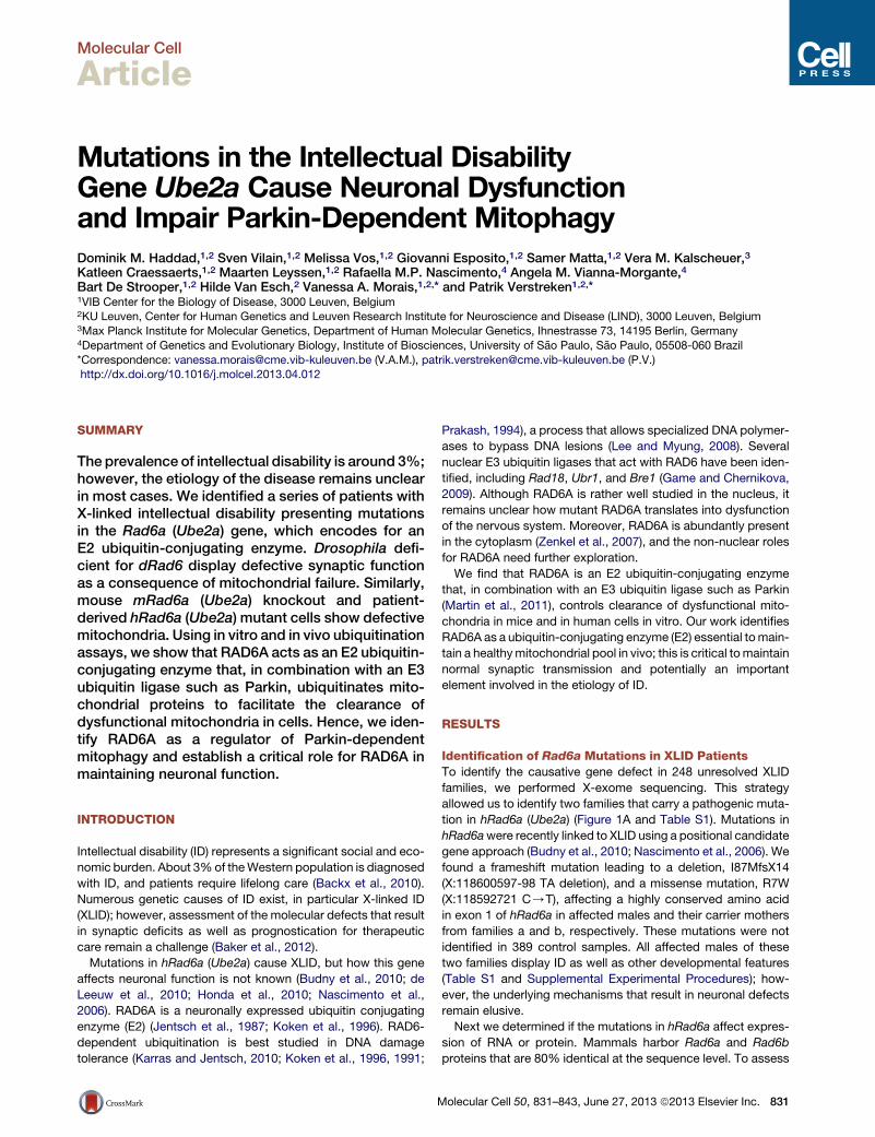
Identification of Rad6a Mutations in XLID PatientsTo identify the causative gene defect in 248 unresolved XLID
families, we performed X-exome sequencing. This strategy
allowed us to identify two families that carry a pathogenic muta-
tion in hRad6a (Ube2a) (Figure 1A and Table S1). Mutations in
hRad6awere recently linked to XLID using a positional candidate
gene approach (Budny et al., 2010; Nascimento et al., 2006). We
found a frameshift mutation leading to a deletion, I87MfsX14
(X:118600597-98 TA deletion), and a missense mutation, R7W
(X:118592721 C/T), affecting a highly conserved amino acid
in exon 1 of hRad6a in affected males and their carrier mothers
from families a and b, respectively. These mutations were not
identified in 389 control samples. All affected males of these
two families display ID as well as other developmental features
(Table S1 and Supplemental Experimental Procedures); how-
ever, the underlying mechanisms that result in neuronal defects
remain elusive.
Next we determined if the mutations in hRad6a affect expres-
sion of RNA or protein. Mammals harbor Rad6a and Rad6b
proteins that are 80% identical at the sequence level. To assess
olecular Cell 50, 831–843, June 27, 2013 ª2013 Elsevier Inc. 831
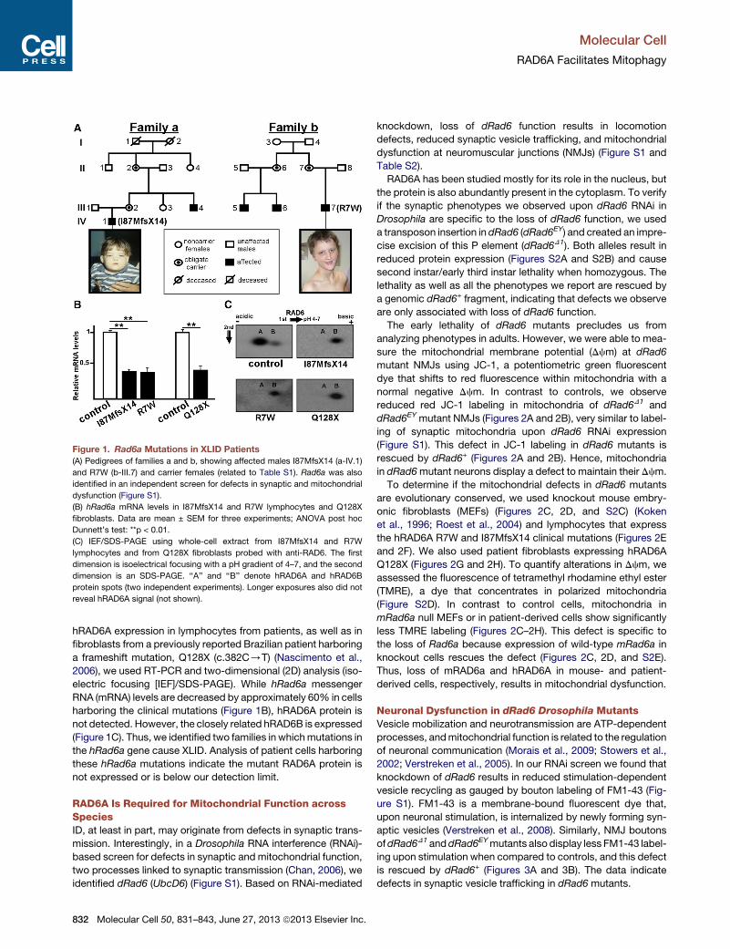
Figure 1. Rad6a Mutations in XLID Patients
(A) Pedigrees of families a and b, showing affected males I87MfsX14 (a-IV.1)
and R7W (b-III.7) and carrier females (related to Table S1). Rad6a was also
identified in an independent screen for defects in synaptic and mitochondrial
dysfunction (Figure S1).
(B) hRad6a mRNA levels in I87MfsX14 and R7W lymphocytes and Q128X
fibroblasts. Data are mean ± SEM for three experiments; ANOVA post hoc
Dunnett’s test: **p < 0.01.
(C) IEF/SDS-PAGE using whole-cell extract from I87MfsX14 and R7W
lymphocytes and from Q128X fibroblasts probed with anti-RAD6. The first
dimension is isoelectrical focusing with a pH gradient of 4–7, and the second
dimension is an SDS-PAGE. ‘‘A’’ and ‘‘B’’ denote hRAD6A and hRAD6B
protein spots (two independent experiments). Longer exposures also did not
reveal hRAD6A signal (not shown).
Molecular Cell
RAD6A Facilitates Mitophagy
hRAD6A expression in lymphocytes from patients, as well as in
fibroblasts from a previously reported Brazilian patient harboring
a frameshift mutation, Q128X (c.382C/T) (Nascimento et al.,
2006), we used RT-PCR and two-dimensional (2D) analysis (iso-
electric focusing [IEF]/SDS-PAGE). While hRad6a messenger
RNA (mRNA) levels are decreased by approximately 60% in cells
harboring the clinical mutations (Figure 1B), hRAD6A protein is
not detected. However, the closely related hRAD6B is expressed
(Figure 1C). Thus, we identified two families in whichmutations in
the hRad6a gene cause XLID. Analysis of patient cells harboring
these hRad6a mutations indicate the mutant RAD6A protein is
not expressed or is below our detection limit.
RAD6A Is Required for Mitochondrial Function acrossSpeciesID, at least in part, may originate from defects in synaptic trans-
mission. Interestingly, in a Drosophila RNA interference (RNAi)-
based screen for defects in synaptic and mitochondrial function,
two processes linked to synaptic transmission (Chan, 2006), we
identified dRad6 (UbcD6) (Figure S1). Based on RNAi-mediated
832 Molecular Cell 50, 831–843, June 27, 2013 ª2013 Elsevier Inc.
knockdown, loss of dRad6 function results in locomotion
defects, reduced synaptic vesicle trafficking, and mitochondrial
dysfunction at neuromuscular junctions (NMJs) (Figure S1 and
Table S2).
RAD6A has been studied mostly for its role in the nucleus, but
the protein is also abundantly present in the cytoplasm. To verify
if the synaptic phenotypes we observed upon dRad6 RNAi in
Drosophila are specific to the loss of dRad6 function, we used
a transposon insertion in dRad6 (dRad6EY) and created an impre-
cise excision of this P element (dRad6D1). Both alleles result in
reduced protein expression (Figures S2A and S2B) and cause
second instar/early third instar lethality when homozygous. The
lethality as well as all the phenotypes we report are rescued by
a genomic dRad6+ fragment, indicating that defects we observe
are only associated with loss of dRad6 function.
The early lethality of dRad6 mutants precludes us from
analyzing phenotypes in adults. However, we were able to mea-
sure the mitochondrial membrane potential (Dcm) at dRad6
mutant NMJs using JC-1, a potentiometric green fluorescent
dye that shifts to red fluorescence within mitochondria with a
normal negative Dcm. In contrast to controls, we observe
reduced red JC-1 labeling in mitochondria of dRad6D1 and
dRad6EY mutant NMJs (Figures 2A and 2B), very similar to label-
ing of synaptic mitochondria upon dRad6 RNAi expression
(Figure S1). This defect in JC-1 labeling in dRad6 mutants is
rescued by dRad6+ (Figures 2A and 2B). Hence, mitochondria
in dRad6mutant neurons display a defect to maintain their Dcm.
To determine if the mitochondrial defects in dRad6 mutants
are evolutionary conserved, we used knockout mouse embry-
onic fibroblasts (MEFs) (Figures 2C, 2D, and S2C) (Koken
et al., 1996; Roest et al., 2004) and lymphocytes that express
the hRAD6A R7W and I87MfsX14 clinical mutations (Figures 2E
and 2F). We also used patient fibroblasts expressing hRAD6A
Q128X (Figures 2G and 2H). To quantify alterations in Dcm, we
assessed the fluorescence of tetramethyl rhodamine ethyl ester
(TMRE), a dye that concentrates in polarized mitochondria
(Figure S2D). In contrast to control cells, mitochondria in
mRad6a null MEFs or in patient-derived cells show significantly
less TMRE labeling (Figures 2C–2H). This defect is specific to
the loss of Rad6a because expression of wild-type mRad6a in
knockout cells rescues the defect (Figures 2C, 2D, and S2E).
Thus, loss of mRAD6a and hRAD6A in mouse- and patient-
derived cells, respectively, results in mitochondrial dysfunction.
Neuronal Dysfunction in dRad6 Drosophila MutantsVesicle mobilization and neurotransmission are ATP-dependent
processes, andmitochondrial function is related to the regulation
of neuronal communication (Morais et al., 2009; Stowers et al.,
2002; Verstreken et al., 2005). In our RNAi screen we found that
knockdown of dRad6 results in reduced stimulation-dependent
vesicle recycling as gauged by bouton labeling of FM1-43 (Fig-
ure S1). FM1-43 is a membrane-bound fluorescent dye that,
upon neuronal stimulation, is internalized by newly forming syn-
aptic vesicles (Verstreken et al., 2008). Similarly, NMJ boutons
ofdRad6D1 anddRad6EYmutants also display less FM1-43 label-
ing upon stimulation when compared to controls, and this defect
is rescued by dRad6+ (Figures 3A and 3B). The data indicate
defects in synaptic vesicle trafficking in dRad6 mutants.
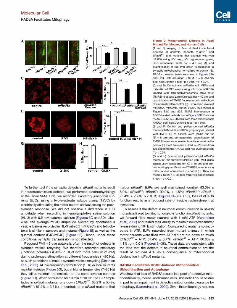
Figure 2. Mitochondrial Defects in Rad6
Mutant Fly, Mouse, and Human Cells
(A and B) Imaging of Dcm at third instar larval
boutons of controls, mutants dRad6D1 and
dRad6EY, and mutants that express wild-type
dRAD6, using JC-1 (red, JC-1 aggregates; green,
JC-1 monomers; scale bar = 4.5 mm) (A), and
quantification of red over green fluorescence in
synaptic mitochondria normalized to control (B).
RAD6 expression levels are shown in Figures S2A
and S2B. Data are mean ± SEM, n = 6; ANOVA
post hoc Dunnett’s test: *p < 0.05, **p < 0.01.
(C and D) Control and mRad6a null MEFs and
mRad6a null MEFs expressing wild-type mRAD6A
labeled with tetramethylrhodamine ethyl ester
(TMRE) to assess Dcm (C) (scale bar = 40 mm) and
quantification of TMRE fluorescence in mitochon-
dria normalized to control (D). Expression levels of
mRAD6A, mRAD6B, and mRAD6A-Myc shown in
Figures S2C and S2E. TMRE fluorescence in
FCCP-treated cells shown in Figure S2D. Data are
mean ± SEM, n = 50 cells from three experiments;
ANOVA post hoc Dunnett’s test: **p < 0.01.
(E and F) Control and patient-derived hRad6a
mutants I87MfsX14 and R7W lymphocytes labeled
with TMRE (E) to assess Dcm (scale bar for
[E] = 5 mm) and corresponding quantification of
TMRE fluorescence in mitochondria normalized to
control (F). Data are mean ± SEM; n = 20 cells from
two experiments; ANOVA post hoc Dunnett’s test:
**p < 0.01.
(G and H) Control and patient-derived hRad6a
mutant Q128X fibroblasts labeledwith TMRE (G) to
assess Dcm (scale bar for [G] = 40 mm) and cor-
responding quantification of TMRE fluorescence in
mitochondria normalized to control (H). Data are
mean ± SEM; n = 20 cells from two experiments;
t test: **p < 0.01.
Molecular Cell
RAD6A Facilitates Mitophagy
To further test if the synaptic defects in dRad6 mutants result
in neurotransmission defects, we performed electrophysiology
at the larval NMJ. First, we recorded excitatory junctional cur-
rents (EJCs) using a two-electrode voltage clamp (TEVC) by
electrically stimulating themotor neuron and assessing the post-
synaptic response. We did not observe a difference in EJC
amplitude when recording in hemolymph-like saline solution
(HL-3) with 0.5 mM external calcium (Figures 3C and 3D). Like-
wise, the average mEJC amplitude elicited by spontaneous
vesicle fusions recorded in HL-3 with 0.5 mMCaCl2 and tetrodo-
toxin is similar in controls and mutants (Figure 3E) as well as the
quantal content (EJC/mEJC) (Figure 3F). Hence, under these
conditions, synaptic transmission is not affected.
Reduced FM1-43 dye uptake is often the result of defects in
synaptic vesicle recycling. We therefore recorded excitatory
junctional potentials (EJPs) in HL-3 with more calcium (2 mM)
during prolonged stimulation at different frequencies (1–20 Hz),
as such conditions stimulate synaptic vesicle recycling (Dickman
et al., 2005). At low-frequency stimulation (1 Hz) dRad6 mutants
maintain release (Figure 3G), but at higher frequencies (7–20 Hz)
they fail to maintain transmission at the same level as controls
(Figure 3H). When stimulated for 10 min at 10 Hz, the EJP ampli-
tudes in dRad6 mutants runs down (dRad6EY: 66.2% ± 5.4%;
dRad6D1: 67.2% ± 3.5%). In controls or in dRad6 mutants that
M
harbor dRad6+, EJPs are well maintained (control: 93.0% ±
8.9%; dRad6EY; dRad6+: 90.9% ± 1.5%; dRad6D1; dRad6+:
95.4% ± 2.7%; p < 0.01) (Figures 3I–3K). Thus, loss of dRAD6
function results in a reduced rate of vesicle replenishment at
synapses.
To assess if the defect in neuronal communication in dRad6
mutants is linked tomitochondrial dysfunction in dRad6mutants,
we forward filled motor neurons with 1 mM ATP (Verstreken
et al., 2005) and tested their ability to maintain neurotransmitter
release during 10 Hz stimulation. Compared tomutants not incu-
bated in ATP, EJPs recorded from mutant animals in which
motor neurons were filled with ATP did not run down as much
(dRad6EY + ATP: 87.4% ± 8.7%; dRad6D1 + ATP: 86.6% ±
4.1%; p < 0.01) (Figures 3I–3K). These data are consistent with
the idea that the defects in neuronal communication are the
result of reduced ATP as a consequence of mitochondrial
dysfunction in dRad6 mutants.
RAD6A Facilitates CCCP-Induced MitochondrialUbiquitination and AutophagyWe show that loss of RAD6A results in a pool of defective mito-
chondria in fly,mouse, and human cells. This deficit could be due
in part to an impairment in defective mitochondria clearance by
mitophagy (Narendra et al., 2009). Given that mitophagy requires
olecular Cell 50, 831–843, June 27, 2013 ª2013 Elsevier Inc. 833
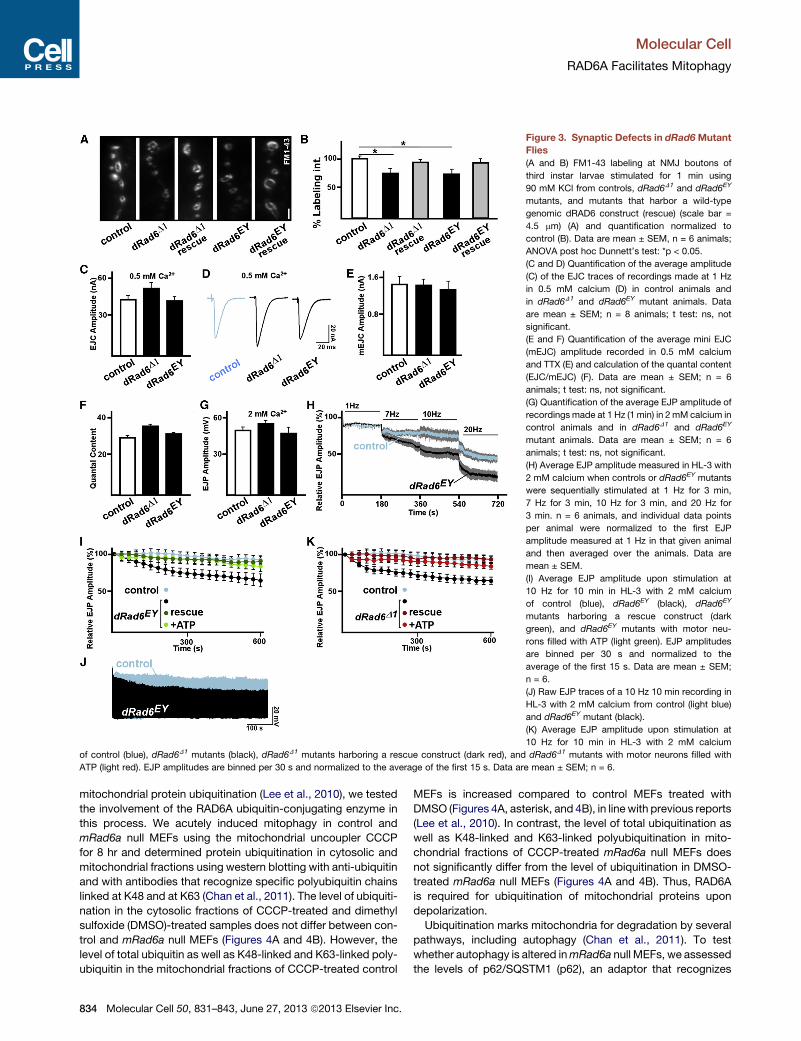
Figure 3. Synaptic Defects in dRad6Mutant
Flies
(A and B) FM1-43 labeling at NMJ boutons of
third instar larvae stimulated for 1 min using
90 mM KCl from controls, dRad6D1 and dRad6EY
mutants, and mutants that harbor a wild-type
genomic dRAD6 construct (rescue) (scale bar =
4.5 mm) (A) and quantification normalized to
control (B). Data are mean ± SEM, n = 6 animals;
ANOVA post hoc Dunnett’s test: *p < 0.05.
(C and D) Quantification of the average amplitude
(C) of the EJC traces of recordings made at 1 Hz
in 0.5 mM calcium (D) in control animals and
in dRad6D1 and dRad6EY mutant animals. Data
are mean ± SEM; n = 8 animals; t test: ns, not
significant.
(E and F) Quantification of the average mini EJC
(mEJC) amplitude recorded in 0.5 mM calcium
and TTX (E) and calculation of the quantal content
(EJC/mEJC) (F). Data are mean ± SEM; n = 6
animals; t test: ns, not significant.
(G) Quantification of the average EJP amplitude of
recordings made at 1 Hz (1 min) in 2 mM calcium in
control animals and in dRad6D1 and dRad6EY
mutant animals. Data are mean ± SEM; n = 6
animals; t test: ns, not significant.
(H) Average EJP amplitude measured in HL-3 with
2 mM calcium when controls or dRad6EY mutants
were sequentially stimulated at 1 Hz for 3 min,
7 Hz for 3 min, 10 Hz for 3 min, and 20 Hz for
3 min. n = 6 animals, and individual data points
per animal were normalized to the first EJP
amplitude measured at 1 Hz in that given animal
and then averaged over the animals. Data are
mean ± SEM.
(I) Average EJP amplitude upon stimulation at
10 Hz for 10 min in HL-3 with 2 mM calcium
of control (blue), dRad6EY (black), dRad6EY
mutants harboring a rescue construct (dark
green), and dRad6EY mutants with motor neu-
rons filled with ATP (light green). EJP amplitudes
are binned per 30 s and normalized to the
average of the first 15 s. Data are mean ± SEM;
n = 6.
(J) Raw EJP traces of a 10 Hz 10 min recording in
HL-3 with 2 mM calcium from control (light blue)
and dRad6EY mutant (black).
(K) Average EJP amplitude upon stimulation at
10 Hz for 10 min in HL-3 with 2 mM calcium
of control (blue), dRad6D1 mutants (black), dRad6D1 mutants harboring a rescue construct (dark red), and dRad6D1 mutants with motor neurons filled with
ATP (light red). EJP amplitudes are binned per 30 s and normalized to the average of the first 15 s. Data are mean ± SEM; n = 6.
Molecular Cell
RAD6A Facilitates Mitophagy
mitochondrial protein ubiquitination (Lee et al., 2010), we tested
the involvement of the RAD6A ubiquitin-conjugating enzyme in
this process. We acutely induced mitophagy in control and
mRad6a null MEFs using the mitochondrial uncoupler CCCP
for 8 hr and determined protein ubiquitination in cytosolic and
mitochondrial fractions using western blotting with anti-ubiquitin
and with antibodies that recognize specific polyubiquitin chains
linked at K48 and at K63 (Chan et al., 2011). The level of ubiquiti-
nation in the cytosolic fractions of CCCP-treated and dimethyl
sulfoxide (DMSO)-treated samples does not differ between con-
trol and mRad6a null MEFs (Figures 4A and 4B). However, the
level of total ubiquitin as well as K48-linked and K63-linked poly-
ubiquitin in the mitochondrial fractions of CCCP-treated control
834 Molecular Cell 50, 831–843, June 27, 2013 ª2013 Elsevier Inc.
MEFs is increased compared to control MEFs treated with
DMSO (Figures 4A, asterisk, and 4B), in linewith previous reports
(Lee et al., 2010). In contrast, the level of total ubiquitination as
well as K48-linked and K63-linked polyubiquitination in mito-
chondrial fractions of CCCP-treated mRad6a null MEFs does
not significantly differ from the level of ubiquitination in DMSO-
treated mRad6a null MEFs (Figures 4A and 4B). Thus, RAD6A
is required for ubiquitination of mitochondrial proteins upon
depolarization.
Ubiquitination marks mitochondria for degradation by several
pathways, including autophagy (Chan et al., 2011). To test
whether autophagy is altered inmRad6a null MEFs, we assessed
the levels of p62/SQSTM1 (p62), an adaptor that recognizes
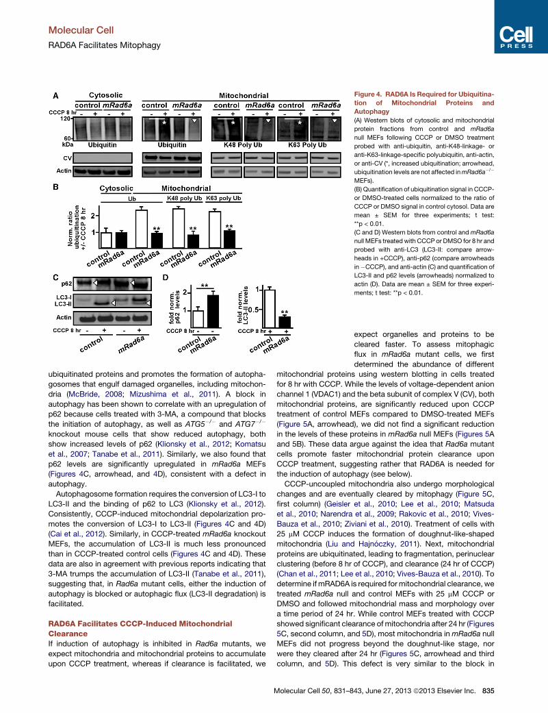
Figure 4. RAD6A Is Required for Ubiquitina-
tion of Mitochondrial Proteins and
Autophagy
(A) Western blots of cytosolic and mitochondrial
protein fractions from control and mRad6a
null MEFs following CCCP or DMSO treatment
probed with anti-ubiquitin, anti-K48-linkage- or
anti-K63-linkage-specific polyubiquitin, anti-actin,
or anti-CV (*, increased ubiquitination; arrowhead,
ubiquitination levels are not affected inmRad6a�/�
MEFs).
(B) Quantification of ubiquitination signal in CCCP-
or DMSO-treated cells normalized to the ratio of
CCCP or DMSO signal in control cytosol. Data are
mean ± SEM for three experiments; t test:
**p < 0.01.
(C and D) Western blots from control andmRad6a
null MEFs treated with CCCP or DMSO for 8 hr and
probed with anti-LC3 (LC3-II: compare arrow-
heads in +CCCP), anti-p62 (compare arrowheads
in�CCCP), and anti-actin (C) and quantification of
LC3-II and p62 levels (arrowheads) normalized to
actin (D). Data are mean ± SEM for three experi-
ments; t test: **p < 0.01.
Molecular Cell
RAD6A Facilitates Mitophagy
ubiquitinated proteins and promotes the formation of autopha-
gosomes that engulf damaged organelles, including mitochon-
dria (McBride, 2008; Mizushima et al., 2011). A block in
autophagy has been shown to correlate with an upregulation of
p62 because cells treated with 3-MA, a compound that blocks
the initiation of autophagy, as well as ATG5�/� and ATG7�/�
knockout mouse cells that show reduced autophagy, both
show increased levels of p62 (Klionsky et al., 2012; Komatsu
et al., 2007; Tanabe et al., 2011). Similarly, we also found that
p62 levels are significantly upregulated in mRad6a MEFs
(Figures 4C, arrowhead, and 4D), consistent with a defect in
autophagy.
Autophagosome formation requires the conversion of LC3-I to
LC3-II and the binding of p62 to LC3 (Klionsky et al., 2012).
Consistently, CCCP-induced mitochondrial depolarization pro-
motes the conversion of LC3-I to LC3-II (Figures 4C and 4D)
(Cai et al., 2012). Similarly, in CCCP-treated mRad6a knockout
MEFs, the accumulation of LC3-II is much less pronounced
than in CCCP-treated control cells (Figures 4C and 4D). These
data are also in agreement with previous reports indicating that
3-MA trumps the accumulation of LC3-II (Tanabe et al., 2011),
suggesting that, in Rad6a mutant cells, either the induction of
autophagy is blocked or autophagic flux (LC3-II degradation) is
facilitated.
RAD6A Facilitates CCCP-Induced MitochondrialClearanceIf induction of autophagy is inhibited in Rad6a mutants, we
expect mitochondria and mitochondrial proteins to accumulate
upon CCCP treatment, whereas if clearance is facilitated, we
Molecular Cell 50, 831–8
expect organelles and proteins to be
cleared faster. To assess mitophagic
flux in mRad6a mutant cells, we first
determined the abundance of different
mitochondrial proteins using western blotting in cells treated
for 8 hr with CCCP. While the levels of voltage-dependent anion
channel 1 (VDAC1) and the beta subunit of complex V (CV), both
mitochondrial proteins, are significantly reduced upon CCCP
treatment of control MEFs compared to DMSO-treated MEFs
(Figure 5A, arrowhead), we did not find a significant reduction
in the levels of these proteins in mRad6a null MEFs (Figures 5A
and 5B). These data argue against the idea that Rad6a mutant
cells promote faster mitochondrial protein clearance upon
CCCP treatment, suggesting rather that RAD6A is needed for
the induction of autophagy (see below).
CCCP-uncoupled mitochondria also undergo morphological
changes and are eventually cleared by mitophagy (Figure 5C,
first column) (Geisler et al., 2010; Lee et al., 2010; Matsuda
et al., 2010; Narendra et al., 2009; Rakovic et al., 2010; Vives-
Bauza et al., 2010; Ziviani et al., 2010). Treatment of cells with
25 mM CCCP induces the formation of doughnut-like-shaped
mitochondria (Liu and Hajnoczky, 2011). Next, mitochondrial
proteins are ubiquitinated, leading to fragmentation, perinuclear
clustering (before 8 hr of CCCP), and clearance (24 hr of CCCP)
(Chan et al., 2011; Lee et al., 2010; Vives-Bauza et al., 2010). To
determine if mRAD6A is required for mitochondrial clearance, we
treated mRad6a null and control MEFs with 25 mM CCCP or
DMSO and followed mitochondrial mass and morphology over
a time period of 24 hr. While control MEFs treated with CCCP
showed significant clearance of mitochondria after 24 hr (Figures
5C, second column, and 5D), most mitochondria inmRad6a null
MEFs did not progress beyond the doughnut-like stage, nor
were they cleared after 24 hr (Figures 5C, arrowhead and third
column, and 5D). This defect is very similar to the block in
43, June 27, 2013 ª2013 Elsevier Inc. 835
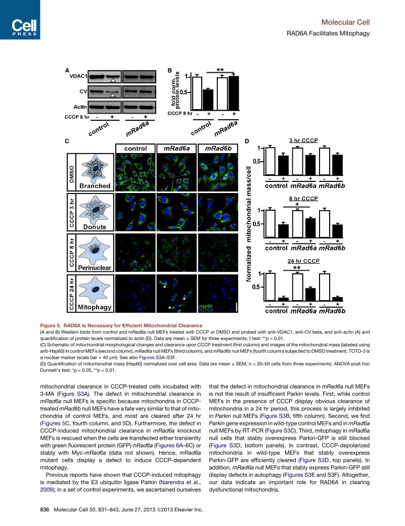
Figure 5. RAD6A Is Necessary for Efficient Mitochondrial Clearance
(A and B) Western blots from control and mRad6a null MEFs treated with CCCP or DMSO and probed with anti-VDAC1, anti-CV beta, and anti-actin (A) and
quantification of protein levels normalized to actin (D). Data are mean ± SEM for three experiments; t test: **p < 0.01.
(C) Schematic of mitochondrial morphological changes and clearance upon CCCP treatment (first column) and images of the mitochondrial mass (labeled using
anti-Hsp60) in controlMEFs (second column),mRad6a null MEFs (third column), andmRad6b null MEFs (fourth column) subjected to DMSO treatment. TOTO-3 is
a nuclear marker (scale bar = 40 mm). See also Figures S3A–S3F.
(D) Quantification of mitochondrial mass (Hsp60) normalized over cell area. Data are mean ± SEM; n = 30–50 cells from three experiments; ANOVA post hoc
Dunnett’s test: *p < 0.05, **p < 0.01.
Molecular Cell
RAD6A Facilitates Mitophagy
mitochondrial clearance in CCCP-treated cells incubated with
3-MA (Figure S3A). The defect in mitochondrial clearance in
mRad6a null MEFs is specific because mitochondria in CCCP-
treatedmRad6b null MEFs have a fate very similar to that of mito-
chondria of control MEFs, and most are cleared after 24 hr
(Figures 5C, fourth column, and 5D). Furthermore, the defect in
CCCP-induced mitochondrial clearance in mRad6a knockout
MEFs is rescued when the cells are transfected either transiently
with green fluorescent protein (GFP)-hRad6a (Figures 6A–6C) or
stably with Myc-mRad6a (data not shown). Hence, mRad6a
mutant cells display a defect to induce CCCP-dependent
mitophagy.
Previous reports have shown that CCCP-induced mitophagy
is mediated by the E3 ubiquitin ligase Parkin (Narendra et al.,
2009); in a set of control experiments, we ascertained ourselves
836 Molecular Cell 50, 831–843, June 27, 2013 ª2013 Elsevier Inc.
that the defect in mitochondrial clearance in mRad6a null MEFs
is not the result of insufficient Parkin levels. First, while control
MEFs in the presence of CCCP display obvious clearance of
mitochondria in a 24 hr period, this process is largely inhibited
in Parkin null MEFs (Figure S3B, fifth column). Second, we find
Parkin gene expression inwild-type control MEFs and inmRad6a
null MEFs by RT-PCR (Figure S3C). Third, mitophagy inmRad6a
null cells that stably overexpress Parkin-GFP is still blocked
(Figure S3D, bottom panels). In contrast, CCCP-depolarized
mitochondria in wild-type MEFs that stably overexpress
Parkin-GFP are efficiently cleared (Figure S3D, top panels). In
addition, mRad6a null MEFs that stably express Parkin-GFP still
display defects in autophagy (Figures S3E and S3F). Altogether,
our data indicate an important role for RAD6A in clearing
dysfunctional mitochondria.
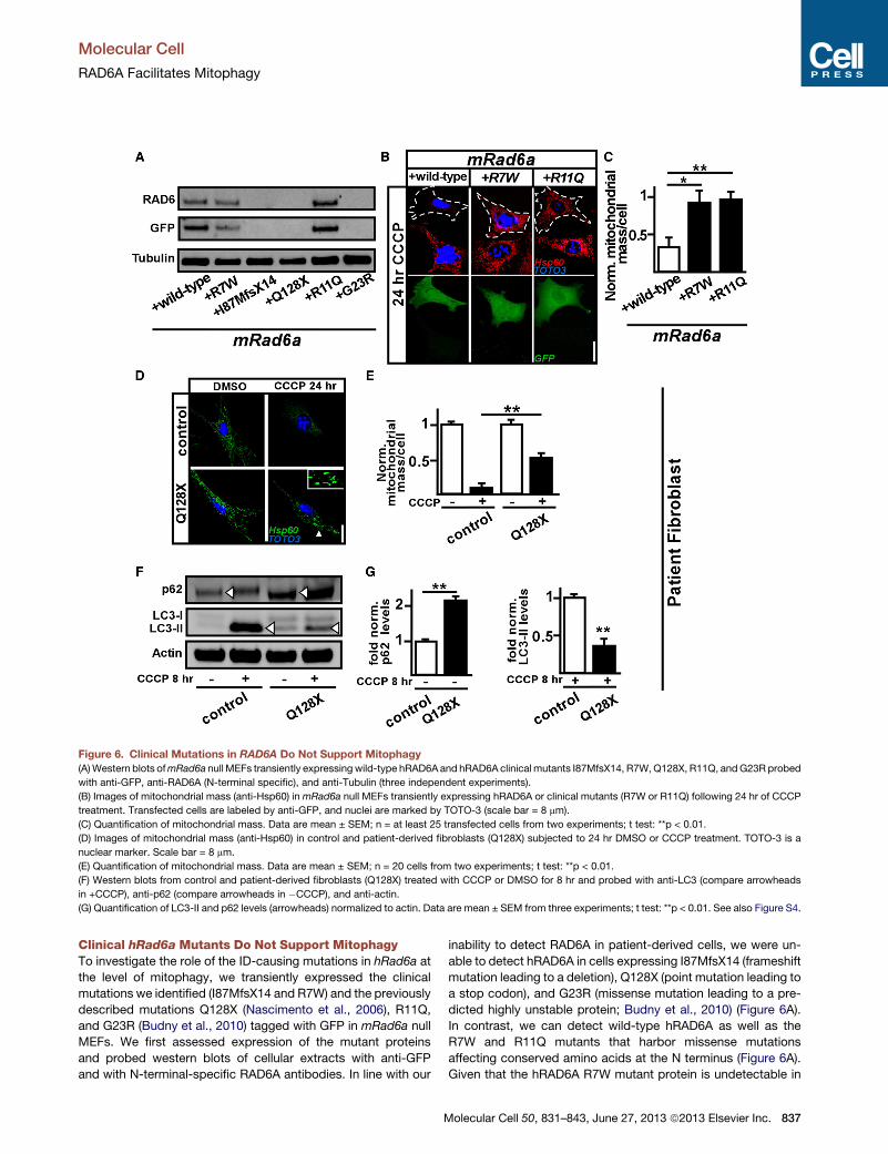
Figure 6. Clinical Mutations in RAD6A Do Not Support Mitophagy
(A)Western blots ofmRad6a null MEFs transiently expressing wild-type hRAD6A and hRAD6A clinical mutants I87MfsX14, R7W,Q128X, R11Q, andG23R probed
with anti-GFP, anti-RAD6A (N-terminal specific), and anti-Tubulin (three independent experiments).
(B) Images of mitochondrial mass (anti-Hsp60) inmRad6a null MEFs transiently expressing hRAD6A or clinical mutants (R7W or R11Q) following 24 hr of CCCP
treatment. Transfected cells are labeled by anti-GFP, and nuclei are marked by TOTO-3 (scale bar = 8 mm).
(C) Quantification of mitochondrial mass. Data are mean ± SEM; n = at least 25 transfected cells from two experiments; t test: **p < 0.01.
(D) Images of mitochondrial mass (anti-Hsp60) in control and patient-derived fibroblasts (Q128X) subjected to 24 hr DMSO or CCCP treatment. TOTO-3 is a
nuclear marker. Scale bar = 8 mm.
(E) Quantification of mitochondrial mass. Data are mean ± SEM; n = 20 cells from two experiments; t test: **p < 0.01.
(F) Western blots from control and patient-derived fibroblasts (Q128X) treated with CCCP or DMSO for 8 hr and probed with anti-LC3 (compare arrowheads
in +CCCP), anti-p62 (compare arrowheads in �CCCP), and anti-actin.
(G) Quantification of LC3-II and p62 levels (arrowheads) normalized to actin. Data are mean ± SEM from three experiments; t test: **p < 0.01. See also Figure S4.
Molecular Cell
RAD6A Facilitates Mitophagy
Clinical hRad6a Mutants Do Not Support MitophagyTo investigate the role of the ID-causing mutations in hRad6a at
the level of mitophagy, we transiently expressed the clinical
mutations we identified (I87MfsX14 and R7W) and the previously
described mutations Q128X (Nascimento et al., 2006), R11Q,
and G23R (Budny et al., 2010) tagged with GFP in mRad6a null
MEFs. We first assessed expression of the mutant proteins
and probed western blots of cellular extracts with anti-GFP
and with N-terminal-specific RAD6A antibodies. In line with our
M
inability to detect RAD6A in patient-derived cells, we were un-
able to detect hRAD6A in cells expressing I87MfsX14 (frameshift
mutation leading to a deletion), Q128X (point mutation leading to
a stop codon), and G23R (missense mutation leading to a pre-
dicted highly unstable protein; Budny et al., 2010) (Figure 6A).
In contrast, we can detect wild-type hRAD6A as well as the
R7W and R11Q mutants that harbor missense mutations
affecting conserved amino acids at the N terminus (Figure 6A).
Given that the hRAD6A R7W mutant protein is undetectable in
olecular Cell 50, 831–843, June 27, 2013 ª2013 Elsevier Inc. 837
Molecular Cell
RAD6A Facilitates Mitophagy
patient-derived cells (Figure 1), the data are consistent with the
hRAD6A R7W protein being unstable and only detectable upon
overexpression. It will be interesting to test if the R11Q mutation
is detectable in patient-derived cells.
Next, to test if hRAD6A-R7W-GFP or hRAD6A-R11Q-GFP
supports mitophagy, we incubated the mRAD6A null MEFs
that express these mutants for 24 hr in CCCP. While mRAD6A
null MEFs that express wild-type hRAD6A-GFP showed efficient
clearance of mitochondria, mRAD6A null MEFs that express
hRAD6A-R7W-GFP or hRAD6A-R11Q-GFP failed to efficiently
degrade mitochondria (Figures 6B and 6C). Hence, while
hRAD6A-R7W and hRAD6A-R11Q-GFP protein is detectable
when overexpressed in mRad6a null cells, the mutant proteins
are not able to promote CCCP-induced mitophagy.
Finally, we assessed if patient-derived fibroblasts carrying the
Q128X mutation display mitochondrial clearance when incu-
bated in CCCP. As shown in Figures 6D (arrowhead) and 6E,
mitophagy is much reduced in the patient-derived cells
compared to control cells. In line with defects in autophagy,
the patient-derived fibroblasts also display reduced LC3-II levels
upon CCCP treatment and show a concomitant accumulation of
p62 levels (Figures 6F and 6G). Hence, cells with hRAD6A clinical
mutants show a defect to promote CCCP-inducedmitochondrial
clearance.
RAD6A Is a Parkin E2 Ubiquitin-Conjugating EnzymeSeveral E3 ubiquitin ligases have been described as acting with
RAD6A in the nucleus, including Rad18, Ubr1, and Bre1 (Game
and Chernikova, 2009). To test if these E3 ubiquitin ligases are
involved in the control of mitochondrial function, we knocked
down Ubr1 and Bre1 (fruit flies do not harbor a Rad18 homolog)
using RNAi in fruit flies (Figure S4A). As a control, we also
knocked down Parkin. Knockdown of Bre1 in fruit flies results
in embryonic lethality, precluding us from analyzing JC-1 label-
ing in larval motor neurons. While knockdown of Parkin results
in reduced red JC-1 labeling, indicating a less-negative Dcm,
knockdown of Ubr1 has no effect (Figures S4B and S4C). Simi-
larly, we used GFP-labeled small hairpin RNA (shRNA) to knock
down Ubr1, Rad18, or Bre1 inmammalian cells (Figures S4D and
S4E) and tested if knockdown of these E3 ligases affected mito-
chondrial clearance upon CCCP treatment of the cells. In
contrast to Parkin mutant MEFs (Figure S3B), MEFs with
reduced levels of Rad18, Ubr1, or Bre1 efficiently cleared their
mitochondria following CCCP treatment, very similar to untrans-
fected control cells in CCCP (Figures S4F and S4G). Hence,
three of the E3 ubiquitin ligases that are known to operate with
RAD6A in the nucleus do not affect mitochondrial function.
As previously shown, Parkin is an E3 ubiquitin ligase involved
in the ubiquitination of mitochondrial proteins and mitophagy
(Lee et al., 2010) (Figure S3). In cells, loss of Parkin blocks
CCCP-induced mitophagy (Figure S3B). In flies, parkin mutant
motor neurons harbor mitochondria with a less-negative Dcm
(Figures S4B and S4C) and parkin mutant muscles harbor
enlarged and doughnut-shaped morphologically abnormal
mitochondria (Figure S4H). These phenotypes, including the
mitochondrial morphological defects in muscles, are all very
reminiscent of those seen in Rad6a mutant cells and flies (Fig-
ures 2, 5, 6, and S4H). We therefore tested the hypothesis that
838 Molecular Cell 50, 831–843, June 27, 2013 ª2013 Elsevier Inc.
RAD6A serves as an E2 ubiquitin-conjugating enzyme that can
act with Parkin. First, we performed an in vitro ubiquitination
assay (Hristova et al., 2009) using 90 nM purified recombinant
E1, 2.5 mM hRAD6A, 3 mM Parkin, and 0.2 mM ubiquitin (Fig-
ures 7A and S5A). Probing reactions that include the E1,
hRAD6A, Parkin, and ubiquitin with anti-ubiquitin reveal ubiqui-
tination that is not obvious when either protein is omitted (Fig-
ure 7A). Furthermore, we also performed a more sensitive
ELISA-based in vitro ubiquitination assay (Marblestone et al.,
2010) and incubated different concentrations of hRAD6A with
60 nM Parkin, 5 nM E1 activating enzyme, and 9 mM ubiquitin.
We then quantitatively assessed newly synthesized ubiquitin
chain formation by measuring luminescence. While hRAD6A
alone is not able to form a significant amount of ubiquitin chains
(Figure 7B), hRAD6A and Parkin together are very effective in
forming ubiquitin chains in a hRAD6A dose-dependent manner.
The in vitro ubiquitination assay reveals significant enzymatic
activity of hRAD6A toward Parkin, since adding a known Parkin
E2 ubiquitin-conjugating enzyme (UBCH7) (Tanaka et al., 2001)
is less effective at forming ubiquitin chains than hRAD6A in a
similar concentration range (Figure 7C). We fitted the data
using a Michaelis-Menten kinetics model and calculated the
apparent KM (nM) values for the Parkin-dependent ubiquitina-
tion reaction mediated by hRAD6A (Figure 7D). These data
indicate that Parkin and hRAD6A can form a functional ubiqui-
tination partnership in vitro and suggest that RAD6A can be a
cognate E2 ubiquitin-conjugating enzyme for the E3 ubiquitin
ligase Parkin.
Next, we tested if RAD6A and Parkin interact in a cell-based
assay. Parkin is implicated in mediating mitophagy upon translo-
cation to depolarized mitochondria that are destined for degra-
dation (Narendra et al., 2009). We therefore assessed if an
interaction between the two proteins would be affected by
mitochondrial depolarization. For this, we immunoprecipitated
Parkin from MEFs stably expressing Parkin-GFP (Figures S5B
and S5C) following a 6 hr treatment with CCCP. Western blots
probed with anti-RAD6A antibody revealed that Parkin coimmu-
noprecipitates with RAD6A and that RAD6A and Parkin binding
is increased upon mitochondrial uncoupling (Figure 7E, lower
panel), indicating that the two proteins interact upon mitochon-
drial depolarization.
Assessing the functionality of the RAD6A-Parkin interaction,
we tested if autoubiquitination of Parkin requires RAD6A.
Parkin-GFP autoubiquitination is dramatically induced upon
mitochondrial depolarization (Matsuda et al., 2010; Tanaka
et al., 2010). We therefore immunoprecipitated Parkin from con-
trol andmRad6a null MEFs treated for 6 hr with DMSO or CCCP
and probed the same western blots for Parkin (Figures 7E
and S5D) and ubiquitin (Figure 7F). While we observed an
obvious Parkin laddering pattern (Figures 7E and S5D) that is
also ubiquitin positive in CCCP-treated control cells (Figure 7F),
this pattern is not as prevalent in mRad6a null MEFs (Figures 7E
and 7F). Indicating specificity, immunoprecipitations (IPs) using
nonspecific immunoglobulin G (IgG) antibodies did not reveal
Parkin or ubiquitin-positive signals. Thus, RAD6A facilitates
Parkin autoubiquitination upon mitochondrial depolarization.
Finally, to test if Parkin relocalization to depolarized mitochon-
dria is RAD6A dependent, we treated Parkin-GFP expressing
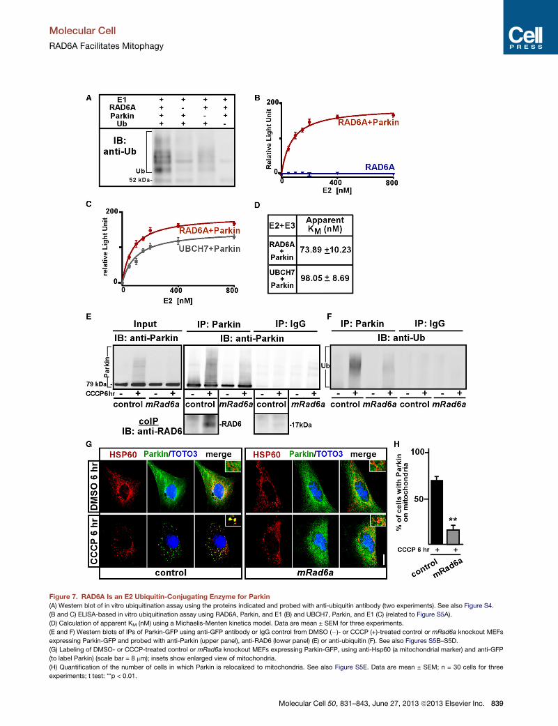
Figure 7. RAD6A Is an E2 Ubiquitin-Conjugating Enzyme for Parkin
(A) Western blot of in vitro ubiquitination assay using the proteins indicated and probed with anti-ubiquitin antibody (two experiments). See also Figure S4.
(B and C) ELISA-based in vitro ubiquitination assay using RAD6A, Parkin, and E1 (B) and UBCH7, Parkin, and E1 (C) (related to Figure S5A).
(D) Calculation of apparent KM (nM) using a Michaelis-Menten kinetics model. Data are mean ± SEM for three experiments.
(E and F) Western blots of IPs of Parkin-GFP using anti-GFP antibody or IgG control from DMSO (�)- or CCCP (+)-treated control or mRad6a knockout MEFs
expressing Parkin-GFP and probed with anti-Parkin (upper panel), anti-RAD6 (lower panel) (E) or anti-ubiquitin (F). See also Figures S5B–S5D.
(G) Labeling of DMSO- or CCCP-treated control or mRad6a knockout MEFs expressing Parkin-GFP, using anti-Hsp60 (a mitochondrial marker) and anti-GFP
(to label Parkin) (scale bar = 8 mm); insets show enlarged view of mitochondria.
(H) Quantification of the number of cells in which Parkin is relocalized to mitochondria. See also Figure S5E. Data are mean ± SEM; n = 30 cells for three
experiments; t test: **p < 0.01.
Molecular Cell
RAD6A Facilitates Mitophagy
Molecular Cell 50, 831–843, June 27, 2013 ª2013 Elsevier Inc. 839
Molecular Cell
RAD6A Facilitates Mitophagy
control andmRad6a null MEFs with CCCP or DMSO for 6 hr and
determined the subcellular localization of Parkin (Figures 7G, 7H,
and S5E). While Parkin translocates from its diffuse cytosolic
localization to mitochondria in control MEFs upon CCCP treat-
ment, it fails to do so appreciably in mRad6a null MEFs
(Figures 7G, 7H, and S5D), suggesting that Parkin transloca-
tion to depolarized mitochondria is critically dependent on
RAD6A. Taken together, our data indicate that RAD6A and Par-
kin form a functional E2/E3 ubiquitination pair that mediates
mitophagy.
DISCUSSION
In this study, we identified two families with mutations in the
XLID-associated protein RAD6A. Further investigation of the
functional implications of those mutations led to the unexpected
finding that RAD6A has a central role in triggering mitophagy
upon mitochondrial depolarization in cells and is required to
maintain mitochondrial integrity in vivo. Although a direct
involvement of mitophagy in mitochondrial quality control in vivo
is debated, disruption of the process is thought to result in the
gradual age-dependent accumulation of dysfunctional mito-
chondria (Mizushima and Komatsu, 2011). Given that a healthy
mitochondrial pool is critical not only for neuronal communica-
tion (Kang et al., 2008; Morais et al., 2009; Sheng and Cai,
2012; Verstreken et al., 2005), but also for normal spine
morphogenesis in the postsynaptic compartment (Li et al.,
2004), our findings link a cytoplasmic role of RAD6A at the level
of mitochondria to synaptic function. This conclusion is sup-
ported by electrophysiological analyses in dRad6 loss-of-func-
tion fly mutants that display reduced synaptic transmission
during intense stimulation and are largely rescued by supplying
extra ATP. Further studies are now required to assess which
neurons are most affected in the human brain. Interestingly,
loss of RAD6B, a closely related isoform, does not affect mito-
chondrial integrity, and mutations in RAD6B have never been
found in intellectually disabled patients. Without excluding addi-
tional roles for RAD6A, our work is consistent with a model in
which the cellular and neuronal dysfunction upon loss of
RAD6A function is at least in part caused by dysfunctional
mitochondria.
Disturbedmitophagy has been linked to Parkin, an E3 ubiquitin
ligase, andmutations in parkin cause juvenile Parkinsonism. Par-
kin is thought to ubiquitinate mitochondrial proteins (Chan et al.,
2011; Lee et al., 2010); however, E2 ubiquitin-conjugating
enzymes that mediate Parkin translocation to mitochondria in
response to their depolarization have not yet been identified.
Our data suggest that RAD6A is an E2 enzyme that can operate
with the E3 ligase Parkin upon mitochondrial depolarization
to induce mitochondrial ubiquitination. As ID patients do
not display overt Parkinsonism, we speculate that other E2
conjugating enzymes may be involved in Parkin-dependent
ubiquitination in the neurons of the substantia nigra, providing
compensation in the brain regions responsible for Parkinson’s
disease (PD)-related symptoms. Conversely, RAD6A can trans-
fer ubiquitin to different E3s besides Parkin (Game and Cherni-
kova, 2009); therefore, the situation in different brain regions is
more complex and will need careful scrutiny. We anticipate
840 Molecular Cell 50, 831–843, June 27, 2013 ª2013 Elsevier Inc.
that in brain regions where RAD6A is highly expressed together
with Parkin, for instance in the dorsolateral prefrontal cortex
(Kupershmidt et al., 2010), both proteins might interact. In other
brain regions, this is likely different; for instance, RAD6A is only
weakly present in the substantia nigra, a location where Parkin
is strongly expressed (Kupershmidt et al., 2010). Thus, different
neurons in the brain may be differentially dependent on RAD6A
function.
Defects in autophagy and in mitochondrial function are impli-
cated in diverse processes, such as brain development, synaptic
function, neuronal differentiation, and aging (Ishihara et al., 2009;
Kageyama et al., 2012; Le Bot, 2007; Wirawan et al., 2012),
where alterations in reactive oxygen species (ROS) homeostasis
and redox regulation, in part induced by an accumulation of
dysfunctional mitochondria, may be the culprit in a wide spec-
trum of neuronal diseases (Kirkinezos andMoraes, 2001; Oikawa
et al., 2012; Ray et al., 2012; Sabens Liedhegner et al., 2012).
Interestingly, in cortical neuron cultures, Rad6a levels are upre-
gulated in response to oxidative stress (Shalamanova et al.,
2007), particularly to hypochlorous acid (HOCl), one of the major
oxidants (Higgins et al., 2010). These findings are consistent with
a model in which RAD6A plays an important role in mitigating
oxidative stress response to maintain a healthy population of
mitochondria. In conclusion, our work links XLID-associated
defects caused by RAD6A mutations to mitochondrial deficits
and neuronal dysfunction.
EXPERIMENTAL PROCEDURES
Genetics
Identification of the Ube2a mutations identified in this study and patient
features are in the Supplemental Experimental Procedures and Table S1.
Thework in this paper was approved by the ethical review board of KU Leuven,
and patient material was obtained with the consent of the patients or their legal
guardians. UAS-RNAi lines (Dietzl et al., 2007) (Table S2) and y1 w67c23;
P{EPgy2}UbcD6EY04634 (Bellen et al., 2011) were from the Vienna Drosophila
RNAi Center (VDRC) and the Bloomington stock center. dRad6D1 was gener-
ated by P element excision (Supplemental Experimental Procedures), and
both alleles were backcrossed several times to y1 w67c23. Germline transfor-
mation of genomic dRad6 (BAC CH322-46G02) (dRad6+) in the VK37 docking
site was obtained using PhiC31-mediated integration (GenetiVision).
Cell Lines and Plasmids
Immortalized control (mRad6a/b+/+) andmRad6a null (mRad6a�/�) MEFs were
obtained from W. Baarends (EMC) (Roest et al., 2004), and human fibroblasts
harboring a hRad6aQ128Xmutation were described (Nascimento et al., 2006).
Immortalized control (mParkin+/+) and mParkin null (mParkin�/�) MEFs were
obtained from K. Winklhofer (Ludwig Maximilian University of Munich). Gener-
ation of stable and transient transfected cell lines is described in the Supple-
mental Experimental Procedures.
Fluorescence Imaging
Jm in Drosophila NMJ mitochondria was assessed using JC-1 (Molecular
Probes) (Morais et al., 2009). TMRE (Molecular Probes) labeling was adapted
from Narendra et al. (2009). FM1-43 labeling at third instar NMJs was per-
formed as described (Verstreken et al., 2008). Antibodies for immunohisto-
chemistry and imaging conditions are listed in the Supplemental Experimental
Procedures.
Electrophysiology
EJCs or EJPs in HL-3 with CaCl2 from muscle 6 in segment A2 or A3 were
performed as described (Uytterhoeven et al., 2011). Motor nerves were
Molecular Cell
RAD6A Facilitates Mitophagy
stimulated at 23 threshold, and intracellular electrodes had resistances
<10 MU. For TEVC, the holding potential was �70 mV and input resistances
were >5 MU. Data were acquired and digitized using an Axoclamp 900A
Amplifier, a Digidata 1440A, and pCLAMP 10 (Molecular devices). Motor
neurons were forward filled with 1 mM ATP as described (Verstreken et al.,
2005).
Biochemistry
Protein extracts were processed for IEF/SDS-PAGE according to the ZOOM
IPGRunner system protocol (Invitrogen). Protein extracts (25–75 mg) resus-
pended in 8 M urea, 2 M thiourea, 2% CHAPS, 20 mM dithiothreitol (DTT),
0.5% (v/v) ZOOM Carrier Ampholytes were loaded on a pH 4–7 ZOOM Strip.
Immobilized pH gradient (IPG) strips were rehydratated for 16 hr at 20�C. Thefirst dimension that consisted of isoelectrical focusing (IEF) was performed at
1 mA/strip. Each strip was equilibrated with NuPAGE LDS Sample Buffer
containing 10 mg/ml DTT for 15 min followed by buffer containing
25 mg/ml iodoacetamide for 15 min. Strips were then loaded and analyzed
on a 4%–12% Bis-Tris ZOOM Gel at 170 V for 35 min. For subcellular
fractionations, cells were collected in 0.2 M sucrose, 10 mM Tris-MOPS
(pH 7.4), 0.1 mM EGTA-Tris (pH 7.4) and then disrupted using a Teflon
homogenizer at 1,000 rpm for 30 strokes. Homogenates were centrifuged
at 800 3 g for 10 min, and supernatants were centrifuged again at
7,000 3 g for 10 min. Pellets were resuspended and proteins detected by
western blot. For autoubiquitination, the assay based on western blotting
was adapted from Hristova et al. (2009). In vitro ubiquitination based on
ELISA was performed using the E2 Profiling Kit (LifeSensors). IPs, including
the protein extraction and detection, were performed by standard protocols
(Van Humbeeck et al., 2011). Cells were lysed in 50 mM Tris-HCl (pH 7.4),
150 mM NaCl, 1% Triton X-100, and Complete protease inhibitor (Roche
Applied Science) and incubated with anti-GFP antibody for 2 hr, then
coupled to protein G beads for 1 hr. IPs were washed with lysis buffer, eluted
directly into SDS-PAGE sample buffer, and detected by western blot.
Antibodies and primers used for RT-PCR are listed in the Supplemental
Experimental Procedures.
Statistics
The statistical significance of differences between a set of two groups was
evaluated using unpaired t tests (*p < 0.05; **p < 0.01) and between more
than two groups using one-way ANOVA (p < 0.01) and Dunnett’s test
(*p < 0.05; **p < 0.01) in GraphPad Prism 5.
SUPPLEMENTAL INFORMATION
Supplemental Information includes five figures, two tables, and Supplemental
Experimental Procedures and can be found with this article online at http://dx.
doi.org/10.1016/j.molcel.2013.04.012.
ACKNOWLEDGMENTS
We thank the Bloomington and VDRC stock centers; Kyoung Sang Cho, Willy
Baarends, and Konstanze Winklhofer for reagents; and Bassem Hassan,
Wim Vandenberghe, and members of the Verstreken and De Strooper labs
for comments. V.M.K. is financed by a grant of the German Ministry of
Education and Research through the MRNET and by the Project GENCODYS
(241995), which is funded by the European Union Framework Program 7
(FP7); S.V. and M.L. are supported by an FWO postdoctoral fellowship;
and M.V. is supported by an IWT predoctoral fellowship and a PDM postdoc-
toral fellowship by the research fund KU Leuven. Support to P.V. was
provided by a Marie Curie Excellence Grant (MEXT-CT-2006-042267), an
ERC Starting Grant (260678), the Research Foundation Flanders (FWO
grants G053913, G079013, G095511, and G074709), a Methusalem grant
of the Flemish Government, the Francqui Foundation, the Hercules
Foundation (AKUL/09/037), the Instutuut voor Wetenschap en Technologie
(IWT), the Interuniversity Attraction Pole program by BELSPO (IAP P7/16
NEUROBRAINNET), the research fund KU Leuven (OT Start, GOA/13/017),
and VIB.
M
Received: September 4, 2012
Revised: February 19, 2013
Accepted: April 10, 2013
Published: May 16, 2013
REFERENCES
Backx, L., Vermeesch, J., Pijkels, E., de Ravel, T., Seuntjens, E., and Van Esch,
H. (2010). PPP2R2C, a gene disrupted in autosomal dominant intellectual
disability. Eur. J. Med. Genet. 53, 239–243.
Baker, K., Raymond, F.L., and Bass, N. (2012). Genetic investigation for adults
with intellectual disability: opportunities and challenges. Curr. Opin. Neurol.
25, 150–158.
Bellen, H.J., Levis, R.W., He, Y., Carlson, J.W., Evans-Holm, M., Bae, E., Kim,
J., Metaxakis, A., Savakis, C., Schulze, K.L., et al. (2011). The Drosophila gene
disruption project: progress using transposons with distinctive site specific-
ities. Genetics 188, 731–743.
Budny, B., Badura-Stronka, M., Materna-Kiryluk, A., Tzschach, A., Raynaud,
M., Latos-Bielenska, A., and Ropers, H.H. (2010). Novel missense mutations
in the ubiquitination-related gene UBE2A cause a recognizable X-linked
mental retardation syndrome. Clin. Genet. 77, 541–551.
Cai, Q., Zakaria, H.M., Simone, A., and Sheng, Z.H. (2012). Spatial parkin
translocation and degradation of damaged mitochondria via mitophagy in
live cortical neurons. Curr. Biol. 22, 545–552.
Chan, D.C. (2006). Mitochondria: dynamic organelles in disease, aging, and
development. Cell 125, 1241–1252.
Chan, N.C., Salazar, A.M., Pham, A.H., Sweredoski, M.J., Kolawa, N.J.,
Graham, R.L., Hess, S., and Chan, D.C. (2011). Broad activation of the ubiqui-
tin-proteasome system by Parkin is critical for mitophagy. Hum. Mol. Genet.
20, 1726–1737.
de Leeuw, N., Bulk, S., Green, A., Jaeckle-Santos, L., Baker, L.A., Zinn, A.R.,
Kleefstra, T., van der Smagt, J.J., Vianne Morgante, A.M., de Vries, B.B., et al.
(2010). UBE2A deficiency syndrome: Mild to severe intellectual disability
accompanied by seizures, absent speech, urogenital, and skin anomalies in
male patients. Am. J. Med. Genet. A. 152A, 3084–3090.
Dickman, D.K., Horne, J.A., Meinertzhagen, I.A., and Schwarz, T.L. (2005). A
slowed classical pathway rather than kiss-and-run mediates endocytosis at
synapses lacking synaptojanin and endophilin. Cell 123, 521–533.
Dietzl, G., Chen, D., Schnorrer, F., Su, K.C., Barinova, Y., Fellner, M., Gasser,
B., Kinsey, K., Oppel, S., Scheiblauer, S., et al. (2007). A genome-wide trans-
genic RNAi library for conditional gene inactivation in Drosophila. Nature 448,
151–156.
Game, J.C., and Chernikova, S.B. (2009). The role of RAD6 in recombinational
repair, checkpoints and meiosis via histone modification. DNA Repair (Amst.)
8, 470–482.
Geisler, S., Holmstrom, K.M., Skujat, D., Fiesel, F.C., Rothfuss, O.C., Kahle,
P.J., and Springer, W. (2010). PINK1/Parkin-mediated mitophagy is depen-
dent on VDAC1 and p62/SQSTM1. Nat. Cell Biol. 12, 119–131.
Higgins, G.C., Beart, P.M., Shin, Y.S., Chen, M.J., Cheung, N.S., and Nagley,
P. (2010). Oxidative stress: emerging mitochondrial and cellular themes and
variations in neuronal injury. J. Alzheimers Dis. 20(Suppl 2 ), S453–S473.
Honda, S., Orii, K.O., Kobayashi, J., Hayashi, S., Imamura, A., Imoto, I.,
Nakagawa, E., Goto, Y., and Inazawa, J. (2010). Novel deletion at Xq24
including the UBE2A gene in a patient with X-linked mental retardation.
J. Hum. Genet. 55, 244–247.
Hristova, V.A., Beasley, S.A., Rylett, R.J., and Shaw, G.S. (2009). Identification
of a novel Zn2+-binding domain in the autosomal recessive juvenile Parkinson-
related E3 ligase parkin. J. Biol. Chem. 284, 14978–14986.
Ishihara, N., Nomura, M., Jofuku, A., Kato, H., Suzuki, S.O., Masuda, K., Otera,
H., Nakanishi, Y., Nonaka, I., Goto, Y., et al. (2009). Mitochondrial fission factor
Drp1 is essential for embryonic development and synapse formation in mice.
Nat. Cell Biol. 11, 958–966.
olecular Cell 50, 831–843, June 27, 2013 ª2013 Elsevier Inc. 841
Molecular Cell
RAD6A Facilitates Mitophagy
Jentsch, S., McGrath, J.P., and Varshavsky, A. (1987). The yeast DNA repair
gene RAD6 encodes a ubiquitin-conjugating enzyme. Nature 329, 131–134.
Kageyama, Y., Zhang, Z., Roda, R., Fukaya, M., Wakabayashi, J.,
Wakabayashi, N., Kensler, T.W., Reddy, P.H., Iijima, M., and Sesaki, H.
(2012). Mitochondrial division ensures the survival of postmitotic neurons by
suppressing oxidative damage. J. Cell Biol. 197, 535–551.
Kang, J.S., Tian, J.H., Pan, P.Y., Zald, P., Li, C., Deng, C., and Sheng, Z.H.
(2008). Docking of axonal mitochondria by syntaphilin controls their mobility
and affects short-term facilitation. Cell 132, 137–148.
Karras, G.I., and Jentsch, S. (2010). The RAD6 DNA damage tolerance
pathway operates uncoupled from the replication fork and is functional beyond
S phase. Cell 141, 255–267.
Kirkinezos, I.G., and Moraes, C.T. (2001). Reactive oxygen species and
mitochondrial diseases. Semin. Cell Dev. Biol. 12, 449–457.
Klionsky, D.J., Abdalla, F.C., Abeliovich, H., Abraham, R.T., Acevedo-Arozena,
A., Adeli, K., Agholme, L., Agnello, M., Agostinis, P., Aguirre-Ghiso, J.A., et al.
(2012). Guidelines for the use and interpretation of assays for monitoring
autophagy. Autophagy 8, 445–544.
Koken, M.H., Reynolds, P., Jaspers-Dekker, I., Prakash, L., Prakash, S.,
Bootsma, D., and Hoeijmakers, J.H. (1991). Structural and functional conser-
vation of two human homologs of the yeast DNA repair gene RAD6. Proc. Natl.
Acad. Sci. USA 88, 8865–8869.
Koken, M.H., Hoogerbrugge, J.W., Jasper-Dekker, I., deWit, J.,Willemsen, R.,
Roest, H.P., Grootegoed, J.A., and Hoeijmakers, J.H. (1996). Expression of the
ubiquitin-conjugating DNA repair enzymes HHR6A and B suggests a role in
spermatogenesis and chromatin modification. Dev. Biol. 173, 119–132.
Komatsu, M., Waguri, S., Koike, M., Sou, Y.S., Ueno, T., Hara, T., Mizushima,
N., Iwata, J., Ezaki, J., Murata, S., et al. (2007). Homeostatic levels of p62
control cytoplasmic inclusion body formation in autophagy-deficient mice.
Cell 131, 1149–1163.
Kupershmidt, I., Su, Q.J., Grewal, A., Sundaresh, S., Halperin, I., Flynn, J.,
Shekar, M., Wang, H., Park, J., Cui, W., et al. (2010). Ontology-based meta-
analysis of global collections of high-throughput public data. PLoS ONE 5, 5.
Le Bot, N. (2007). Autophagy: a new regulator of development. Nat. Cell Biol.
9, 741.
Lee, K.Y., and Myung, K. (2008). PCNA modifications for regulation of post-
replication repair pathways. Mol. Cells 26, 5–11.
Lee, J.Y., Nagano, Y., Taylor, J.P., Lim, K.L., and Yao, T.P. (2010). Disease-
causing mutations in parkin impair mitochondrial ubiquitination, aggregation,
and HDAC6-dependent mitophagy. J. Cell Biol. 189, 671–679.
Li, Z., Okamoto, K., Hayashi, Y., and Sheng, M. (2004). The importance of
dendritic mitochondria in the morphogenesis and plasticity of spines and syn-
apses. Cell 119, 873–887.
Liu, X., and Hajnoczky, G. (2011). Altered fusion dynamics underlie unique
morphological changes in mitochondria during hypoxia-reoxygenation stress.
Cell Death Differ. 18, 1561–1572.
Marblestone, J.G., Suresh Kumar, K.G., Eddins, M.J., Leach, C.A., Sterner,
D.E., Mattern, M.R., and Nicholson, B. (2010). Novel approach for character-
izing ubiquitin E3 ligase function. J. Biomol. Screen. 15, 1220–1228.
Martin, I., Dawson, V.L., and Dawson, T.M. (2011). Recent advances in the
genetics of Parkinson’s disease. Annu. Rev. Genomics Hum. Genet. 12,
301–325.
Matsuda, N., Sato, S., Shiba, K., Okatsu, K., Saisho, K., Gautier, C.A., Sou,
Y.S., Saiki, S., Kawajiri, S., Sato, F., et al. (2010). PINK1 stabilized bymitochon-
drial depolarization recruits Parkin to damaged mitochondria and activates
latent Parkin for mitophagy. J. Cell Biol. 189, 211–221.
McBride, H.M. (2008). Parkin mitochondria in the autophagosome. J. Cell Biol.
183, 757–759.
Mizushima, N., and Komatsu, M. (2011). Autophagy: renovation of cells and
tissues. Cell 147, 728–741.
Mizushima, N., Yoshimori, T., and Ohsumi, Y. (2011). The role of Atg proteins in
autophagosome formation. Annu. Rev. Cell Dev. Biol. 27, 107–132.
842 Molecular Cell 50, 831–843, June 27, 2013 ª2013 Elsevier Inc.
Morais, V.A., Verstreken, P., Roethig, A., Smet, J., Snellinx, A., Vanbrabant,M.,
Haddad, D., Frezza, C., Mandemakers, W., Vogt-Weisenhorn, D., et al. (2009).
Parkinson’s disease mutations in PINK1 result in decreased Complex I activity
and deficient synaptic function. EMBO Mol Med 1, 99–111.
Narendra, D., Tanaka, A., Suen, D.F., and Youle, R.J. (2009). Parkin-
induced mitophagy in the pathogenesis of Parkinson disease. Autophagy
5, 706–708.
Nascimento, R.M., Otto, P.A., de Brouwer, A.P., and Vianna-Morgante, A.M.
(2006). UBE2A, which encodes a ubiquitin-conjugating enzyme, is mutated
in a novel X-linked mental retardation syndrome. Am. J. Hum. Genet. 79,
549–555.
Oikawa, D., Akai, R., Tokuda, M., and Iwawaki, T. (2012). A transgenic mouse
model for monitoring oxidative stress. Sci Rep 2, 229.
Prakash, L. (1994). The RAD6 gene and protein of Saccharomyces cerevisiae.
Ann. N Y Acad. Sci. 726, 267–273.
Rakovic, A., Grunewald, A., Seibler, P., Ramirez, A., Kock, N., Orolicki, S.,
Lohmann, K., and Klein, C. (2010). Effect of endogenous mutant and wild-
type PINK1 on Parkin in fibroblasts from Parkinson disease patients. Hum.
Mol. Genet. 19, 3124–3137.
Ray, P.D., Huang, B.W., and Tsuji, Y. (2012). Reactive oxygen species (ROS)
homeostasis and redox regulation in cellular signaling. Cell. Signal. 24,
981–990.
Roest, H.P., Baarends, W.M., de Wit, J., van Klaveren, J.W., Wassenaar, E.,
Hoogerbrugge, J.W., van Cappellen, W.A., Hoeijmakers, J.H., and
Grootegoed, J.A. (2004). The ubiquitin-conjugating DNA repair enzyme
HR6A is a maternal factor essential for early embryonic development in
mice. Mol. Cell. Biol. 24, 5485–5495.
Sabens Liedhegner, E.A., Gao, X.H., and Mieyal, J.J. (2012). Mechanisms of
altered redox regulation in neurodegenerative diseases—focus on S—gluta-
thionylation. Antioxid. Redox Signal. 16, 543–566.
Shalamanova, L., McArdle, F., Amara, A.B., Jackson, M.J., and Rustom, R.
(2007). Albumin overload induces adaptive responses in human proximal
tubular cells through oxidative stress but not via angiotensin II type 1 receptor.
Am. J. Physiol. Renal Physiol. 292, F1846–F1857.
Sheng, Z.H., and Cai, Q. (2012). Mitochondrial transport in neurons: impact
on synaptic homeostasis and neurodegeneration. Nat. Rev. Neurosci. 13,
77–93.
Stowers, R.S., Megeath, L.J., Gorska-Andrzejak, J., Meinertzhagen, I.A., and
Schwarz, T.L. (2002). Axonal transport of mitochondria to synapses depends
on milton, a novel Drosophila protein. Neuron 36, 1063–1077.
Tanabe, F., Yone, K., Kawabata, N., Sakakima, H., Matsuda, F., Ishidou, Y.,
Maeda, S., Abematsu, M., Komiya, S., and Setoguchi, T. (2011).
Accumulation of p62 in degenerated spinal cord under chronic mechanical
compression: functional analysis of p62 and autophagy in hypoxic neuronal
cells. Autophagy 7, 1462–1471.
Tanaka, K., Suzuki, T., Chiba, T., Shimura, H., Hattori, N., and Mizuno, Y.
(2001). Parkin is linked to the ubiquitin pathway. J. Mol. Med. 79, 482–494.
Tanaka, A., Cleland, M.M., Xu, S., Narendra, D.P., Suen, D.F., Karbowski, M.,
and Youle, R.J. (2010). Proteasome and p97mediate mitophagy and degrada-
tion of mitofusins induced by Parkin. J. Cell Biol. 191, 1367–1380.
Uytterhoeven, V., Kuenen, S., Kasprowicz, J., Miskiewicz, K., and Verstreken,
P. (2011). Loss of skywalker reveals synaptic endosomes as sorting stations
for synaptic vesicle proteins. Cell 145, 117–132.
Van Humbeeck, C., Cornelissen, T., Hofkens, H., Mandemakers, W., Gevaert,
K., De Strooper, B., and Vandenberghe, W. (2011). Parkin interacts with
Ambra1 to induce mitophagy. J. Neurosci. 31, 10249–10261.
Verstreken, P., Ly, C.V., Venken, K.J., Koh, T.W., Zhou, Y., and Bellen, H.J.
(2005). Synaptic mitochondria are critical for mobilization of reserve pool ves-
icles at Drosophila neuromuscular junctions. Neuron 47, 365–378.
Verstreken, P., Ohyama, T., and Bellen, H.J. (2008). FM 1-43 labeling of synap-
tic vesicle pools at the Drosophila neuromuscular junction. Methods Mol. Biol.
440, 349–369.
Molecular Cell
RAD6A Facilitates Mitophagy
Vives-Bauza, C., Zhou, C., Huang, Y., Cui, M., de Vries, R.L., Kim, J., May, J.,
Tocilescu, M.A., Liu, W., Ko, H.S., et al. (2010). PINK1-dependent recruitment
of Parkin to mitochondria in mitophagy. Proc. Natl. Acad. Sci. USA 107,
378–383.
Wirawan, E., Vanden Berghe, T., Lippens, S., Agostinis, P., and Vandenabeele,
P. (2012). Autophagy: for better or for worse. Cell Res. 22, 43–61.
M
Zenkel, M., Kruse, F.E., Naumann, G.O., and Schlotzer-Schrehardt, U. (2007).
Impaired cytoprotective mechanisms in eyes with pseudoexfoliation syn-
drome/glaucoma. Invest. Ophthalmol. Vis. Sci. 48, 5558–5566.
Ziviani, E., Tao, R.N., and Whitworth, A.J. (2010). Drosophila parkin requires
PINK1 for mitochondrial translocation and ubiquitinates mitofusin. Proc.
Natl. Acad. Sci. USA 107, 5018–5023.
olecular Cell 50, 831–843, June 27, 2013 ª2013 Elsevier Inc. 843