modular 5 cerebrovascular accident cva stroke · modular 5 cerebrovascular accident (cva): stroke...
TRANSCRIPT
Modular 5
Cerebrovascular Accident (CVA): Stroke
Learning Objectives:
Identify the statistical impact of CVA’s in the US.
Identify the pathophysiology of a CVA.
Differentiate between ischemic and hemorrhagic stroke.
Identify risk factors that contribute to Acute Ischemic Stroke (AIS).
List the signs and symptoms of a CVA.
List 3 different assessment tools to score the presents and severity of a stroke.
Understand the role of the healthcare provider in the emergency phase, acute phase, and long-term
care of AIS.
A cerebrovascular accident is the medical term for a stroke. A stroke occurs when blood flow to a part of the
brain is stopped either by a blockage or a rupture of a blood vessel. There are important signs of a stroke of a
CVA that if identified and treated early enough can reduce the risk of permanent damage. When a stroke goes
untreated for too long, there can be permanent brain damage.
Section 1: CVA/Strokes in the United States:
Stroke kills almost 130,000 Americans each year—that’s 1 out of every 20 deaths.
On average, one American dies from stroke every 4 minutes.
Every year, more than 795,000 people in the United States have a stroke.
About 610,000 of these are first or new strokes.
About 185,00 strokes—nearly one of four—are in people who have had a previous stroke.
About 87% of all strokes are ischemic strokes, when blood flow to the brain is blocked.
Stroke costs the United States an estimated $34 billion each year. This total includes the cost of health care
services, medications to treat stroke, and missed days of work.
Stroke is a leading cause of serious long-term disability.
Three million Americans are currently permanently disabled from stroke.
Two-thirds of strokes occur in people over age 65 but they can occur at any age.
Strokes affect men more often than women, women are more likely to die from a stroke.
Stroke is now the 5th
leading cause of death in the US.
Pathophysiology and the Basics of a Stroke
What is a Stroke/CVA?
A stroke, or cerebrovascular accident (CVA), is the rapid loss of brain function due to disturbance in the blood
supply to the brain. A stroke or "brain attack" occurs when a blood clot blocks an artery or a blood vessel breaks,
interrupting blood flow to an area of the brain. When either of these things happens, brain cells begin to die and
brain damage occurs. Lack of blood flow to the brain can cause a person to fall unconscious in as little as twelve
seconds.
What Causes a Stroke?

The leading cause of a stroke is due to thrombosis. Thrombosis is the formation of a blood clot inside a blood
vessel, obstructing the flow of blood through the circulatory system. The blood vessel over time becomes clogged
with cholesterol or clotted with blood, which could lead to stoppage of blood flow to the brain leading to a stroke.
The second leading cause of a stroke is an embolism. Embolism occurs when a clot formed in a blood vessel
somewhere in the body breaks off and becomes free-floating. This embolism then enters the brains circulatory
system and travels until it encounters an artery it can’t pass through, causing blood flow to stop and leading to a
stroke. The most common affected artery is the middle cerebral artery, located in the middle part of the brain.
The third leading cause of a stroke is caused by a hemorrhage. Hemorrhagic strokes are the most deadly, due to the
pressure they can produce on vital parts of the brain. Hemorrhages can be caused by aneurysms, weakened blood
vessels that form bubble-shaped projections and then break.
Hypotension is the fourth major cause of a stroke. Hypotension occurs when blood pressure falls to dangerously
low levels. This can lead to inadequate supply of blood to the brain. This type of stroke is different than all other
types of strokes in that all parts of the brain may lose its blood supply. This can happen in cases of certain abnormal
heart rhythms, severe heart failure, and in some heart attacks when the heart fails to adequately pump blood.
Transient ischemic attacks (TIA) also called mini-strokes are often a “red flag” warning of strokes to come. TIA is
a temporary blockage of an artery; no permanent brain tissue is damaged. Neurological symptoms go away within
twenty-four hours, depending on which artery is blocked. Not all TIAs lead to a stroke.
Types of Strokes:
The two main types of stroke include ischemic stroke and hemorrhagic stroke.
1. Ischemic stroke is the most common type of stroke and accounts for about eighty seven percent of all
strokes. Ischemic strokes are ultimately caused by a thrombus or embolus that blocks blood flow to the
brain. Blood clots (thrombus clots) usually occur in areas of the arteries that have been damaged by
atherosclerosis from a buildup of plaques. Embolus type blood clots are often caused by atrial fibrillation -
an irregular pattern of heart beat that leads to blood clot formation and poor blood flow.
Ischemic Stroke
2. Hemorrhagic stroke can be caused by uncontrolled high blood pressure, a head injury, or aneurysms.
High blood pressure is the most common cause of cerebral hemorrhage, as it causes small arteries inside
the brain to burst. This deprives brain cells of blood and dangerously increases pressure on the brain.
Subarachnoid Hemorrhage
A subarachnoid hemorrhage is a type of stroke caused by the sudden rupture of an artery. A subarachnoid
hemorrhage differs from an intracerebral hemorrhage in that the location of the rupture leads to blood filling the
space surrounding the brain rather than inside of it. Aneurysms - abnormal blood-filled pouches that balloon out
from weak spots in the wall of an artery - are the most common cause of subarachnoid hemorrhage. If an aneurysm
ruptures, blood spills into the space between the surfaces of the brain and skull, and blood vessel s in the brain may
spasm. Aneurysms are often caused or made worse by high blood pressure.
Intracerebral Hemorrhage
An intracerebral hemorrhage is a type of stroke caused by the sudden rupture of an artery within the brain. Blood is
then released into the brain compressing brain structures. Both types of stroke result in a lack of blood flow to the
brain and a buildup of blood that puts too much pressure on the brain.
The severity of a stroke depends on what part of the brain is damaged and how much of that area is affected. Some
strokes may cause some minor physical problems, such as weakness in an arm or leg and a major stroke may lead to
paralysis or death. Many stroke patients are left with weakness on one side of the body, difficulty speaking,
incontinence, and bladder problems.
Anatomy of the Brain:
The upper brain, or cerebrum, is divided into two hemispheres. The cerebrum contains the cortical area, used for
thinking, and the subcortical area, which is a complex network of relay centers and linking pathways. Each
hemisphere has four separate lobes:
1. Frontal Lobe: controls thinking, behavioral functions, body movement
2. Parietal Lobe: sensory interpretation, concrete thinking, abstract thinking, vision-space orientation, and
language
3. Temporal Lobe: emotional and memory center, language functions
4. Occipital Lobe: Visual interpretation
The back part of the brain, the cerebellum controls balance and coordination. Underneath the cerebellum is the brain
stem. The brain stem controls involuntary and automatic survival processes such as, heart rate, breathing body
temperature, sleeping cycles and regulation of some hormones.
Blood Flow to the Brain:
Blood flows from the heart to the brain through the large carotid arteries. Once inside the skull the carotid arteries
divide into front and side branches. These blood vessels supply the front two-thirds of the brain’s outer surface. The
vertebrobasilar arteries travel up and serve this region, as well as the brain stem and coordination centers. Four areas
of the brain (artery blood-flow centers) are most commonly affected by stroke. The four most common arteries
involved in a stroke are:
1. Middle Cerebral Artery
2. Anterior Cerebral Artery
3. Posterior Cerebral Artery
4. Basilar Artery
Strokes are labeled based on the area of the brain that is damaged. A stroke on the right side of the brain is referred
to as a right hemisphere stroke. A patient that suffers a right hemisphere stroke, the left side of the body will be
affected. If the stroke affects the left cerebrum, the right side of the body is affected.
Deficits caused by a stroke:
Deficiencies caused by a stroke are a result of damage to the brain and can be mental or physical deficiencies.
Common deficits include weakness or paralysis, loss of sensation, problems walking, speaking, and difficulties with
activities of daily living.
Who is at risk?
Anyone can suffer from stroke. Although many risk factors for stroke are out of our control, several can be kept in
line through proper nutrition and medical care. Those who are highest risk include:
Over age 55
Male
African American, Hispanic or Asian/Pacific Islander
A family history of stroke
Key risk factors include:
High blood pressure
High cholesterol
Smoking cigarettes
Several other medical conditions and lifestyle choices can also put people at a higher risk for stroke, including:
Diabetes
Overweight and obesity
Poor diet
Physical inactivity
Excessive alcohol use
A previous stroke or transient ischemic attack (TIA)
High blood pressure is the most important modifiable risk factor of stroke.
What are the symptoms of stroke?
Within a few minutes of having a stroke, brain cells begin to die and symptoms can become present. Stroke
symptoms typically start suddenly, over seconds to minutes. The symptoms depend on the area of the brain affected.
The more extensive the area of brain affected, the more functions that are likely to be lost.
If any of these symptoms are present, one should seek immediate medical attention. If treated within 3 hours of the
first symptom, there is an FDA-approved clot-buster medication that may reduce long-term disability for the most
common type of stroke. There are also two other types of stroke treatment available that might help reduce the
effects of stroke.
Most common symptoms include:
sudden numbness, tingling, weakness, or loss of movement in the face, arm, or leg, especially on only one
side of the body.
sudden vision changes
sudden trouble speaking
sudden problems with walking or balance
sudden confusion or trouble understanding simple statements
sudden, severe headache that is different from past headaches
Smaller strokes (or silent strokes), however, may not cause any symptoms, but can still damage brain tissue.
A possible sign that a stroke is about to occur is called a transient ischemic attack (TIA) - a temporary interruption in
blood flow to part of the brain. Symptoms of TIA are similar to stroke but last for a shorter time period and do not
leave noticeable permanent damage.
A TIA usually lasts 15 minutes or less. Because these may be signs of an impending stroke, they should be taken
seriously and medical attention should be given.
With a TIA, circulation and the vital oxygen supply are quickly restored and lasting brain and nerve damage is
avoided. With any stroke, however, the interruption of blood flow lasts long enough to kill brain cells, producing
irreversible damage.
How is stroke treated?
The primary goal in treating ischemic stroke is to restore blood flow to the brain. This will be attempted using blood
clot-busting drugs such as aspirin, heparin, or tissue plasminogen activators that must be administered within three
hours of the stroke. In addition, surgical procedures may be performed that can open up or widen arteries. These
include carotid endarterectomy (removal of plaque and widening of the carotid artery) and angioplasty (a balloon
that widens the carotid artery and is held open with a metallic mesh tube called a stent).
Some studies have found that cholesterol lowering drugs can prevent stroke recurrence.
Hemorrhagic stroke is treated differently than ischemic stroke. Surgical methods used to treat this stroke variant
include aneurysm clipping, aneurysm embolisation, and arteriovenous malformation (AVM) removal. Aneurysm
clipping consists of a small clamp placed at the base of the aneurysm that isolates it from the circulation of its
attached artery and keeps the aneurysm from bursting or re-bleeding. Aneurysm embolisation (coiling) uses a
catheter inserted into the aneurysm to deposit a tiny coil that coil fills the aneurysm, causing clotting and sealing off
the aneurysm off from arteries. AVM removal is a surgical procedure to remove usually smaller AVMs or AMVs
that are in more accessible portion of the brain in order to eliminate the risk of rupture.
Most stroke victims will require rehabilitation after the event. A person's condition is generally dependent on the
area of the brain and the amount of tissue that was damaged. It is common for the rehabilitation process to include
speech therapy, occupational therapy, physical therapy, and family education.
How can the risk of having a stroke be reduced?
One way to reduce the risk of a stroke is to notice a transient ischemic attack (TIA) or mini stroke that provides
symptoms similar to stroke. Knowing the symptoms of stroke can lead to earlier treatment and better recovery.
Much of stroke prevention is based on living a healthy lifestyle. This includes:
Knowing and controlling blood pressure
Finding out if you have atrial fibrillation
No smoking
Lowering cholesterol, sodium, and fat intake
Following a healthy diet. If you eat plenty of tomatoes, your risk of having a stroke could be reduced
significantly. Tomatoes are rich in lycopene, a powerful antioxidant. In a study published in Neurology,
October 2012, researchers found that people with high blood concentrations of lycopene had a 59% lower risk
of stroke compared to those with the lowest concentrations.
Drinking alcohol only in moderation
Treating diabetes properly
Exercising regularly. Moderate aerobic fitness can reduce stroke risk
Managing stress
Don’t use drugs
Taking preventive medications such as anti-platelet and anticoagulant drugs to prevent blood clots
Cholesterol lowering drugs can prevent stroke recurrence
Section 2: Acute Ischemic Stroke
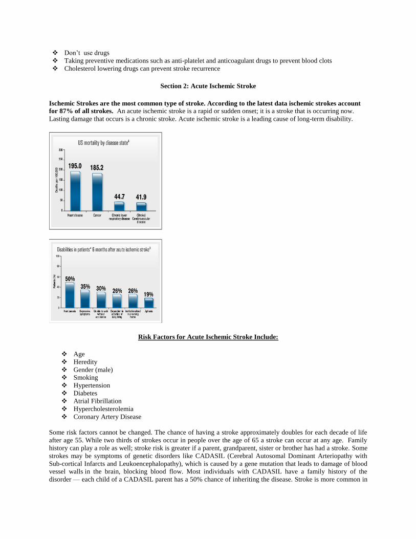
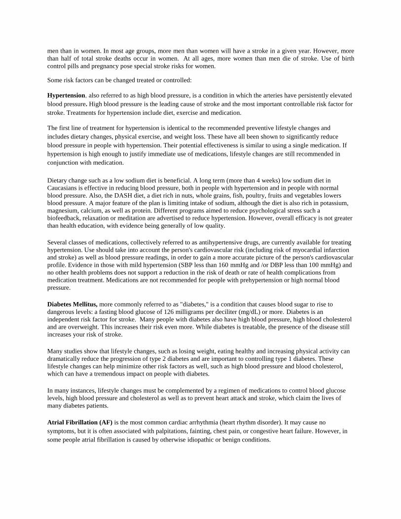
Ischemic Strokes are the most common type of stroke. According to the latest data ischemic strokes account
for 87% of all strokes. An acute ischemic stroke is a rapid or sudden onset; it is a stroke that is occurring now.
Lasting damage that occurs is a chronic stroke. Acute ischemic stroke is a leading cause of long-term disability.
Risk Factors for Acute Ischemic Stroke Include:
Age
Heredity
Gender (male)
Smoking
Hypertension
Diabetes
Atrial Fibrillation
Hypercholesterolemia
Coronary Artery Disease
Some risk factors cannot be changed. The chance of having a stroke approximately doubles for each decade of life
after age 55. While two thirds of strokes occur in people over the age of 65 a stroke can occur at any age. Family
history can play a role as well; stroke risk is greater if a parent, grandparent, sister or brother has had a stroke. Some
strokes may be symptoms of genetic disorders like CADASIL (Cerebral Autosomal Dominant Arteriopathy with
Sub-cortical Infarcts and Leukoencephalopathy), which is caused by a gene mutation that leads to damage of blood
vessel walls in the brain, blocking blood flow. Most individuals with CADASIL have a family history of the
disorder — each child of a CADASIL parent has a 50% chance of inheriting the disease. Stroke is more common in
men than in women. In most age groups, more men than women will have a stroke in a given year. However, more
than half of total stroke deaths occur in women. At all ages, more women than men die of stroke. Use of birth
control pills and pregnancy pose special stroke risks for women.
Some risk factors can be changed treated or controlled:
Hypertension, also referred to as high blood pressure, is a condition in which the arteries have persistently elevated
blood pressure. High blood pressure is the leading cause of stroke and the most important controllable risk factor for
stroke. Treatments for hypertension include diet, exercise and medication.
The first line of treatment for hypertension is identical to the recommended preventive lifestyle changes and
includes dietary changes, physical exercise, and weight loss. These have all been shown to significantly reduce
blood pressure in people with hypertension. Their potential effectiveness is similar to using a single medication. If
hypertension is high enough to justify immediate use of medications, lifestyle changes are still recommended in
conjunction with medication.
Dietary change such as a low sodium diet is beneficial. A long term (more than 4 weeks) low sodium diet in
Caucasians is effective in reducing blood pressure, both in people with hypertension and in people with normal
blood pressure. Also, the DASH diet, a diet rich in nuts, whole grains, fish, poultry, fruits and vegetables lowers
blood pressure. A major feature of the plan is limiting intake of sodium, although the diet is also rich in potassium,
magnesium, calcium, as well as protein. Different programs aimed to reduce psychological stress such a
biofeedback, relaxation or meditation are advertised to reduce hypertension. However, overall efficacy is not greater
than health education, with evidence being generally of low quality.
Several classes of medications, collectively referred to as antihypertensive drugs, are currently available for treating
hypertension. Use should take into account the person's cardiovascular risk (including risk of myocardial infarction
and stroke) as well as blood pressure readings, in order to gain a more accurate picture of the person's cardiovascular
profile. Evidence in those with mild hypertension (SBP less than 160 mmHg and /or DBP less than 100 mmHg) and
no other health problems does not support a reduction in the risk of death or rate of health complications from
medication treatment. Medications are not recommended for people with prehypertension or high normal blood
pressure.
Diabetes Mellitus, more commonly referred to as "diabetes," is a condition that causes blood sugar to rise to
dangerous levels: a fasting blood glucose of 126 milligrams per deciliter (mg/dL) or more. Diabetes is an
independent risk factor for stroke. Many people with diabetes also have high blood pressure, high blood cholesterol
and are overweight. This increases their risk even more. While diabetes is treatable, the presence of the disease still
increases your risk of stroke.
Many studies show that lifestyle changes, such as losing weight, eating healthy and increasing physical activity can
dramatically reduce the progression of type 2 diabetes and are important to controlling type 1 diabetes. These
lifestyle changes can help minimize other risk factors as well, such as high blood pressure and blood cholesterol,
which can have a tremendous impact on people with diabetes.
In many instances, lifestyle changes must be complemented by a regimen of medications to control blood glucose
levels, high blood pressure and cholesterol as well as to prevent heart attack and stroke, which claim the lives of
many diabetes patients.
Atrial Fibrillation (AF) is the most common cardiac arrhythmia (heart rhythm disorder). It may cause no
symptoms, but it is often associated with palpitations, fainting, chest pain, or congestive heart failure. However, in
some people atrial fibrillation is caused by otherwise idiopathic or benign conditions.
It may be identified clinically when taking a pulse, and the presence of AF can be confirmed with an
electrocardiogram (ECG or EKG) which demonstrates the absence of P waves together with an irregular ventricular
rate.
AF increases the risk of stroke. The degree of increase can be substantial, depending on the presence of additional
risk factors (such as high blood pressure). Atrial fibrillation may be treated with medications to either slow the heart
rate to a normal range ("rate control") or revert the heart rhythm to normal ("rhythm control"). Synchronized
electrical cardioversion can be used to convert AF to a normal heart rhythm. Surgical and catheter-based ablation
may be used to prevent recurrence of AF in certain individuals. Depending on the risk of stroke and systemic
embolism, people with AF may use anticoagulants such as warfarin, which substantially reduces the risk but may
increase the risk of major bleeding, mainly in geriatric patients. The prevalence of AF in a population increases with
age, with 8% of people over 80 having AF. Chronic AF leads to a small increase in the risk of death.
Hypercholesterolemia, is the presence of high levels of cholesterol in the blood. It is a form of "hyperlipidemia"
(elevated levels of lipids in the blood) and "hyperlipoproteinemia" (elevated levels of lipoproteins in the blood).
Cholesterol is one of three major classes of lipids which all animal cells utilize to construct their membranes and is
thus manufactured by all animal cells. Plant cells do not manufacture cholesterol. It is also the precursor of the
steroid hormones, bile acids and vitamin D.
Since cholesterol is insoluble in water, it is transported in the blood plasma within protein particles (lipoproteins).
Lipoproteins are classified by their density: very low density lipoprotein (VLDL), intermediate density lipoprotein
(IDL), low density lipoprotein (LDL) and high density lipoprotein (HDL). All the lipoproteins carry cholesterol, but
elevated levels of the lipoproteins other than HDL (termed non-HDL cholesterol), particularly LDL-cholesterol are
associated with an increased risk of atherosclerosis and disease. In contrast higher levels of HDL cholesterol are
protective. Elevated levels of non-HDL cholesterol and LDL in the blood may be a consequence of diet, obesity,
inherited (genetic) diseases (such as LDL receptor mutations in familial hypercholesterolemia), or the presence of
other diseases such as diabetes and an underactive thyroid.
Reducing dietary fat is recommended to reduce total blood cholesterol and LDL in adults. In people with very high
cholesterol (e.g. familial hypercholesterolemia) diet is often insufficient to achieve the desired lowering of LDL and
lipid lowering medications which reduce cholesterol production or absorption are usually required. If necessary
other treatments, including LDL apheresis or even surgery (for particularly severe subtypes of familial
hypercholesterolemia) are performed.
Lifestyle changes recommended for those with high cholesterol include: smoking cessation, limiting alcohol
consumption, increasing physical activity, and maintaining a healthy weight. A diet that emphasizes low-cholesterol
foods, restricts saturated fats, and avoids trans-fat is also recommended. In strictly controlled surroundings, dietary
changes can reduce cholesterol levels by 15 percent. In practice, dietary advice can provide a modest decrease in
cholesterol levels and may be sufficient in the treatment of mildly elevated cholesterol.
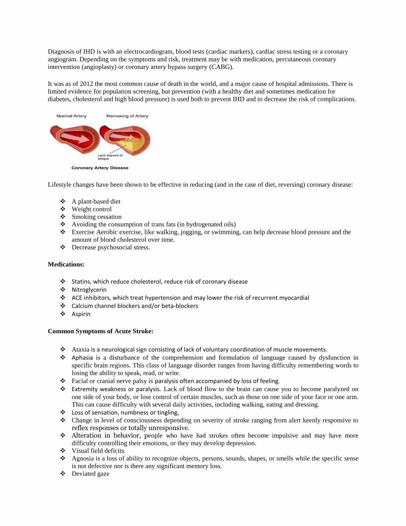
Coronary artery disease (CAD) also known as atherosclerotic heart disease, coronary heart disease, or ischemic heart
disease (IHD), is the most common type of heart disease and cause of heart attacks. The disease is caused by plaque
building up along the inner walls of the arteries of the heart, which narrows the arteries and reduces blood flow to
the heart.
While the symptoms and signs of coronary artery disease are noted in the advanced state of disease, most individuals
with coronary artery disease show no evidence of disease for decades as the disease progresses before the first onset
of symptoms, often a "sudden" heart attack, finally arises. Symptoms of stable ischaemic heart disease include
angina (characteristic chest pain on exertion) and decreased exercise tolerance. Unstable IHD presents itself as chest
pain or other symptoms at rest, or rapidly worsening angina. The risk of artery narrowing increases with age,
smoking, high blood cholesterol, diabetes, high blood pressure, and is more common in men and those who have
close relatives with CAD. Other causes include coronary vasospasm, a spasm of the blood vessels of the heart, it is
usually called Prinzmetal's angina.
Diagnosis of IHD is with an electrocardiogram, blood tests (cardiac markers), cardiac stress testing or a coronary
angiogram. Depending on the symptoms and risk, treatment may be with medication, percutaneous coronary
intervention (angioplasty) or coronary artery bypass surgery (CABG).
It was as of 2012 the most common cause of death in the world, and a major cause of hospital admissions. There is
limited evidence for population screening, but prevention (with a healthy diet and sometimes medication for
diabetes, cholesterol and high blood pressure) is used both to prevent IHD and to decrease the risk of complications.
Lifestyle changes have been shown to be effective in reducing (and in the case of diet, reversing) coronary disease:
A plant-based diet
Weight control
Smoking cessation
Avoiding the consumption of trans fats (in hydrogenated oils)
Exercise Aerobic exercise, like walking, jogging, or swimming, can help decrease blood pressure and the
amount of blood cholesterol over time.
Decrease psychosocial stress.
Medications:
Statins, which reduce cholesterol, reduce risk of coronary disease Nitroglycerin ACE inhibitors, which treat hypertension and may lower the risk of recurrent myocardial Calcium channel blockers and/or beta-blockers Aspirin
Common Symptoms of Acute Stroke:
Ataxia is a neurological sign consisting of lack of voluntary coordination of muscle movements. Aphasia is a disturbance of the comprehension and formulation of language caused by dysfunction in
specific brain regions. This class of language disorder ranges from having difficulty remembering words to
losing the ability to speak, read, or write.
Facial or cranial nerve palsy is paralysis often accompanied by loss of feeling. Extremity weakness or paralysis. Lack of blood flow to the brain can cause you to become paralyzed on
one side of your body, or lose control of certain muscles, such as those on one side of your face or one arm.
This can cause difficulty with several daily activities, including walking, eating and dressing.
Loss of sensation, numbness or tingling, Change in level of consciousness depending on severity of stroke ranging from alert keenly responsive to
reflex responses or totally unresponsive. Alteration in behavior, people who have had strokes often become impulsive and may have more
difficulty controlling their emotions, or they may develop depression.
Visual field deficits.
Agnosia is a loss of ability to recognize objects, persons, sounds, shapes, or smells while the specific sense
is not defective nor is there any significant memory loss.
Deviated gaze
Dysarthria is a motor speech disorder resulting from neurological injury of the motor component of the
motor-speech system. It is a condition in which problems occur with the muscles that help one talk; this
makes it very difficult to pronounce words.
Locked In Syndrome is a condition in which a patient is aware and awake but cannot move or communicate
verbally due to complete paralysis of nearly all voluntary muscles in the body except for the eyes.
Acute Ischemic Stroke Treatment:
Tissue plasminogen activator (abbreviated tPA or PLAT) is a protein involved in the breakdown of blood clots. It is
a serine protease found on endothelial cells, the cells that line the blood vessels. As an enzyme, it catalyzes the
conversion of plasminogen to plasmin, the major enzyme responsible for clot breakdown. Because it works on the
clotting system, tPA is used in clinical medicine to treat embolic or thrombotic stroke. Use is contraindicated in
hemorrhagic stroke and head trauma. tPA may be manufactured using recombinant biotechnology techniques. tPA
created this way may be referred to as recombinant tissue plasminogen activator (rtPA).
Eligibility:
tPA eligibility criteria for patients with acute ischemic stroke within 3 hours of symptom onset include:
An adult (≥18 years of age)
Exclusion of intracranial hemorrhage by an imaging technique sensitive for the presence of hemorrhage.
Arrives at the emergency department in time to be treated within 3 hours of symptom onset.
Contraindications:
tPA therapy with acute ischemic stroke is contraindicated in the following conditions because of an increased risk of
bleeding, which could result in significant disability or death.
Evidence of intracranial hemorrhage on pretreatment evaluation
Suspicion of subarachnoid hemorrhage on pretreatment evaluation
Recent (within 3 months) intracranial or intraspinal surgery, serious head trauma, or previous stroke
History of intracranial hemorrhage
Uncontrolled hypertension at time of treatment (eg, >185 mm Hg systolic or >110 mm Hg diastolic)
Seizure at the onset of stroke
Active internal bleeding
Intracranial neoplasm, arteriovenous malformation, or aneurysm
Known bleeding diathesis, including but not limited to:
Current use of oral anticoagulants (eg, warfarin sodium) or an international normalized ratio (INR) >1.7 or
a prothrombin time (PT) >15 seconds
Administration of heparin within 48 hours preceding the onset of stroke and an elevated activated partial
thromboplastin time (aPTT) at presentation
Platelet count <100,000/mm3
In addition to the contraindications, the risks of tPA therapy may be increased in the following conditions and
should be weighed against the anticipated benefits:
Severe neurological deficit (eg, National Institutes of Health Stroke Scale [NIHSS] >22 at presentation)
Major early infarct signs by an imaging technique (eg, substantial edema, mass effect, or midline shift)
Activase (Alteplase) is FDA-approved for treatment of acute ischemic stroke. For treatment eligible patients, the
recommended dose of Activase is 0.9 mg/kg (not to exceed 90-mg total dose) infused over 60 minutes with 10%
of the total dose administered as an initial intravenous bolus over 1 minute.
Section 3: Patient Evaluation and National Institutes of Health Stroke Scale (NIHSS)
The National Institutes of Health Stroke Scale, or NIH Stroke Scale (NIHSS) is a tool used by healthcare
providers to objectively quantify the impairment caused by a stroke. The NIHSS is composed of 11 items, each
of which scores a specific ability between a 0 and 4. For each item, a score of 0 typically indicates normal
function in that specific ability, while a higher score is indicative of some level of impairment. The individual
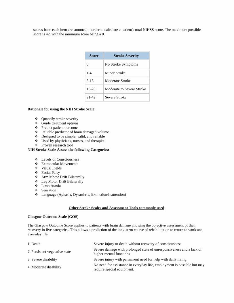
scores from each item are summed in order to calculate a patient's total NIHSS score. The maximum possible
score is 42, with the minimum score being a 0.
Score Stroke Severity
0 No Stroke Symptoms
1-4 Minor Stroke
5-15 Moderate Stroke
16-20 Moderate to Severe Stroke
21-42 Severe Stroke
Rationale for using the NIH Stroke Scale:
Quantify stroke severity
Guide treatment options
Predict patient outcome
Reliable predictor of brain damaged volume
Designed to be simple, valid, and reliable
Used by physicians, nurses, and therapist
Proven research tool
NIH Stroke Scale Assess the following Categories:
Levels of Consciousness
Extraocular Movements
Visual Fields
Facial Palsy
Arm Motor Drift Bilaterally
Leg Motor Drift Bilaterally
Limb Ataxia
Sensation
Language (Aphasia, Dysarthria, Extinction/Inattention)
Other Stroke Scales and Assessment Tools commonly used:
Glasgow Outcome Scale (GOS)
The Glasgow Outcome Score applies to patients with brain damage allowing the objective assessment of their
recovery in five categories. This allows a prediction of the long-term course of rehabilitation to return to work and
everyday life.
1. Death Severe injury or death without recovery of consciousness
2. Persistent vegetative state Severe damage with prolonged state of unresponsiveness and a lack of
higher mental functions
3. Severe disability Severe injury with permanent need for help with daily living
4. Moderate disability No need for assistance in everyday life, employment is possible but may
require special equipment.
5. Low disability Light damage with minor neurological and psychological deficits.
The GOS attempts to generalize and categorize the outcomes of patients who suffer traumatic brain injury. In
general, unlike the Glasgow Coma Scale, this scale is not used in the clinical management of the patient. Rather, it is
used often in research to quantify the level of recovery patients have achieved. Because it is a rather coarse scale,
with only 5 levels, it has been argued that this scale is not ideal for research purposes. Other, more specific, complex
and detailed grading systems have been developed for use. One of these is an adaptation of the GOS, called the
Extended GOS. The GOS is determined by a clinician at some point in the patient's recovery. Generally the time
after injury is reported along with the GOS. So, for example, at 3 months after the traumatic injury, the clinician
would note that it was a "3-month GOS" or "GOS-3". In research, common time points to evaluate the GOS include
3 months, 6 months and 12 months after injury.
The Glasgow Outcome Scale is a 5-level score:
1. Dead
2. Vegetative State (meaning the patient is unresponsive, but alive.)
3. Severely Disabled (conscious but the patient requires others for daily support due to disability)
4. Moderately Disabled (the patient is independent but disabled)
5. Good Recovery (the patient has resumed most normal activities but may have minor residual problems)
The Extended GOS, or GOS-E, has extended the scale to an 8-level score:
1. Dead
2. Vegetative State
3. Lower Severe Disability
4. Upper Severe Disability
5. Lower Moderate Disability
6. Upper Moderate Disability
7. Lower Good Recovery
8. Upper Good Recovery
Disability Rating Scale (DRS):
M. Rappaport introduced the DRS in 1982 to overcome the poor precision of the Glasgow Outcome Scale. It was
initially developed to assess individuals with TBI in the rehabilitation phase of recovery. Upon development it was
tested with older juvenile and adult individuals who had suffered from severe TBI. All tests were performed in an
inpatient rehabilitation setting. The intent of the scale was to measure the general functional changes of the patient
throughout the course of recovery.
The Disability Rating Scale (DRS) was developed as a way to track a traumatic brain injury (TBI) patient from
‘Coma to Community’. The scale is used to rate the effects of injury and decide how long recovery might take. The
rating gives insight into the impairment of the individual who suffered from the TBI.
The idea of the DRS is similar to the Glasgow Outcome Scale (GOS). However, the point of the scale is to track the
patient’s progress over time while the GOS is used to simply determine the extent of a brain injury. In many ways,
the DRS addresses many of the shortcomings of the GOS.
One advantage of the DRS is its ability to track an individual from coma to community. The maximum score a
patient can obtain on the DRS is 29 (extreme vegetative state). A person without disability would score zero. In
order to assure the reliability of this assessment tool, it must be completed on patients who are free from sedatives,
anesthesia or other mind-altering medications. The scale is intended to accurately measure general functional
changes over the course of recovery. It is an easy assessment tool to complete, and scoring is very simple and clear.
Another advantage of the DRS is that expertise in the field is not needed to complete it accurately.
Section 4: Acute Stroke Nursing Role
Nurses play a major role in all phases of care of the stroke patient. Phase 1, the emergency care phase, which
includes the prehospital setting and the emergency department (ED), and phase 2 the acute care phase, which
includes critical care units, intermediate care units, stroke units, and general medical units. Nurses are often
responsible for the coordination of care throughout the continuum, which results in improved outcomes, decrease
lengths of stay, and decreased costs.
Phase 1, the emergency care phase of stroke incudes the first 3 to 24 hours after the onset of symptoms. This phase
includes activation of the emergency medical service and response as well as the ED care protocol. The main
objectives are to:
Identify stroke symptoms
Identify infarct location
Assess patient risk of acute and long-term complications
Determine treatment options
Phase 2, the acute care 24 to 72 hours after onset of stroke. The main objectives include identifying the cause of the
stroke, preventing medical complications, preparing the patient and family for discharge, and instituting long-term
secondary prevention modalities.
Acute stroke nursing goals include:
Monitor& optimize cerebral circulation
Monitor& optimize cardiac and respiratory function
Prevent complications
Secondary stroke prevention
Preparing patient and care givers for post-acute care
Management based on ongoing assessment
Evaluate patient’s response to treatment
Early recognition of progressing neuro status
Early recognition of cardiovascular and respiratory complications
Safety and function issues
Patient/caregiver emotional response to stroke
Initial and On-going Assessment for the acute stroke patient:
Frequent monitoring of vital signs
ABCs
Temperature
Baseline assessment
Respiratory
Cardiac
Neuro
Focused medical history
Medications/allergies
Respiratory Considerations:
Hypoxemia
Respiratory Tract Infections
Pneumonia is cause of 20-40% of all deaths post stroke
Respiratory Nursing Care:
Ongoing assessment of airway and ventilation
Monitor CXRs and cultures
Positioning
Pulmonary Toilet
Pulse oximetry; maintain >PaO2
PaCO2 at 30 mmHg for ventilator patient
OOB when stable
NPO until aspiration ruled out
Cardiovascular Considerations:
Heart disease and cerbrovascular disease often coexist in same patients
Common risk factors
12-17% of stroke related deaths due to cardiac disease
Annual mortality due to MI 5-6% per year
40-70% of patients have clinical CAD
40% will have cardiac event during rehab
Cardiac Arrhythmias:
Cardiac arrhythmias account for 5% of deaths following stroke
New onset 5% to 39% following stroke
Ventricular ectopy is seen in 24-60% of hemorrhage, infarct or TIA stroke patients
Life threatening arrhythmias in the first 24h is not common
ECG Changes in Acute Stroke:
Associated with injury to the cardiovascular reflexes of the insular cortex
Sympathetic hyperactivity (norepinephrine)
QT interval prolongation (32%)
Other less often changes
ST segment changes
Abnormal U waves
More common with ICH or SAH
Cardiac Nursing Care:
24-48 hour continuous ECG monitoring
Evaluate rhythm strip every 4 hours
Serial 12 lead ECGs
Cardiac labs
Early tx of life threatening arrhythmias
Close monitoring of fluid status
Stroke patients often dehydrated
Collateral circulation augmentation
Dopamine, crystalloids, albumin
Blood Pressure Considerations:
Chronic HTN common preexisting factor
Acute HTN often seen and usually returns to normal within a few days
Change in circadian BP pattern r/t poor outcome
Loss of autoregulation in ischemic vessels
Collateral CBF dependent on systemic BP
Rapid lowering of BP will result in rapid decline of regional CBF
Blood Pressure Management:
Management is controversial and patient specific
Perfuse ischemic penumbra
MAP 60-150mmHg in normal patients
MAP 120-150mmHg in HTN patients
Close monitoring of BP
Cautious reduction when indicated (rare)
BP >230/120 requires treatment
TX HTN with Labetolol, Enalapril, Nitroprusside
Non-pharmacologic interventions are effective
Venous Thrombosis and Pulmonary Embolism Considerations:
Incidence 10-20% in patients not receiving prophylaxis are at risk
5% of patients who are receiving prophylaxis are still at risk
May account for 2-13% of deaths following stroke
Venous Thrombosis and Pulmonary Embolism Care:
DVT prophylaxis
Subcutaneous heparin or LMWH
Pneumatic compressions stockings
Early mobilization and ambulation
Assess for sinus tach, hypoxia, tachypnea
Monitor coagulation levels
Patient education for anticoagulant therapy
Hyperglycemia Considerations:
Seen in 50% of stroke patients
Most without diabetes mellitus
Return to prestroke levels over first week
Predictor of poor outcome
Hyperglycemia managed as other with patients with hyperglycemia
Unknown how fast to reduce serum glucose and target serum glucose specific to stroke
Unknown if treatment improves outcome
Hyperthermia Considerations:
Moderate hyperthermia markedly increases nervous system injury
Multiple mechanisms (Ischemic Cascade)
Intracerebral Temp. exceeds body Temp by 1o
Fever associated with worse outcome
Hypothermia therapy is promising
Results of clinical trials pending
Target 36.7 C for first few days after stroke
Possible Nursing Diagnoses:
Ineffective Airway
Impaired Gas Exchange
Altered Breathing Pattern
Altered Tissue Perfusion (cerebral)
Immobility
Potential for Impaired Skin Integrity
Potential for Injury
Altered Nutrition
Ineffective Individual/Family Coping
Knowledge Deficit
Depression/Anxiety/Isolation
Altered Body Image
Possible Causes for Neurological Decline:
Infarct extension
Recurrent infarction (2%)
Hemorrhagic conversion (11%)
Edema and IICP (10%-20%)
Hydrocephalus
Metabolic disturbances
Hypoxia
Hypoglycemia
Hyponatremia
Medication effects
Seizures (4% to 15%)
Post Stroke Complications:
Mobility/disuse injury
Falls
Contractures
Decubitus
Sensory/neglect
Communication
Psych/Social issues
Depression
Pharmcologic therapy
Caregiver Strain
Social Isolation
Focused Patient Education:
Knowledge of stroke symptoms and to call 911 (most important)
Medication administration, rationale, interactions (OTCs and herbs)
When to call doctor
Secondary Stroke Prevention
Knowledge of treatment for stroke type
Risk factor analysis
Identification of life style changes
Community resources
Approaches to Rehabilitation post-Stroke include:
Prevention of secondary complications
Remediation, or treatment to reduce neurological deficits
Compensation to offset and adapt to residual disabilities
Maintenance of function over the long term
Section 5: Caregivers/Family Members:
Caregivers/family members play a major role throughout the post-stroke recovery process. Caregivers/family
members are essential to successful home care and too often over looked and ignored. Caring for stroke survivors at
home can cause high levels of emotional, mental and physical stress. In addition to distress, disruption of
employment and family life makes caregiving very challenging. Family caregivers can promote positive post-stroke
recovery outcomes; however, they need to care for themselves as well. Post-stroke recovery varies for each person;
even if the stroke survivor returns to work and maintains a large amount of autonomy, family members may play a
bigger role in the stroke survivor’s live than before the stroke.
It is important that the caregivers/family members of stroke survivors know that they are not alone. Programs such
as the National Stroke Association is dedicated to providing educational programs and resources to help
caregivers/family members navigate the challenging post-stroke recovery journey.
The role of Caregivers/Family members may include the following:
Assist with doctor’s appointments, medications, and exercises.
Manage financial matters and transportation.
Provide the stroke survivor with physical, mental and emotional support.
Assist the stroke survivor with daily activities such as personal care and hygiene.
Plan out the stroke survivor’s care, including setting routines and managing the care team.
Assess stroke survivor’s medical needs, communicate with healthcare professionals and advocate
(medically) for the stroke survivor.
Keys to coping with the emotional challenges of a Caregiver:
Join a support group, can help reduce anxiety and stress.
Recognize depression and get help if needed.
Accept help from others (community, friends, and other family members).
Find things to be grateful for daily and humor in everyday life can help reduce stress.
Take care of yourself; find time for exercise, meditation, etc.
Learn to accept a “new normal” life may never be the same again depending on the stroke survivors’
disability.
Getting help with Caregiving:
At some point caregivers may need help. Many caregivers try to shoulder all the emotional and physical burdens by
themselves. For a variety of reasons, even the best caregiver may no longer be able to continue caring for the loved
one alone. Asking for help can reduce the everyday burden of the emotional and physical stress of being primary
caregiver.
Friends and family: Can give you a break by taking the stroke survivor/loved one out or staying with them.
Place of worship: Religious communities can be invaluable sources of support. Religious communities can
offer everything from prayer support and home-cooked meals to full- fledged senior programs and adult
day care.

Meals on Wheels: If cooking meals becomes difficult on top of all other responsibilities, social-service
programs such as Meals on Wheels will deliver hot meals to the home at little or no expense.
Community services: Most states and local communities offer assistance and programs.
Home-care services: These provide either skilled nursing care prescribed by a physician or respite/support
services for personal care and household chores.
Post Test: CVA/Stroke
1. Stroke kills almost 130,000 Americans each year—that’s 1 out of every 20 deaths.
A. True b. False
2. Two-thirds of strokes occur in people over age 65 but they can occur at any age.
A. True B. False
3. A stroke, or cerebrovascular accident (CVA), is the rapid loss of brain function due to disturbance in the blood supply
to the brain.
A. True B. False
4. Ischemic stroke is a rare type of stroke and accounts for about 10% percent of all strokes.
A. True B. False
5. Most common symptoms include: sudden numbness, tingling, weakness, or loss of movement in the face, arm, or leg,
especially on only one side of the body, sudden vision changes, sudden trouble speaking, sudden problems with
walking or balance, sudden confusion or trouble understanding simple statements and sudden, severe headache that is
different from past headaches.
A. True B. False
6. Risk Factors for Acute Ischemic Stroke Include: Age, Heredity, Gender (male), Smoking, Hypertension, Diabetes,
Atrial Fibrillation, Hypercholesterolemia, and Coronary Artery Disease.
A. True B. False
7. Risk of having a stroke can be reduced by living a healthy life style, controlling diet, exercising, proper management of
medical conditions such as hypertension and diabetes.
A. True B. False
8. Tissue plasminogen activator (tPA) which is a thrombolytic drug can be used with any type of stroke as long as it is
administered within the first 3 hours of symptoms onset.
A. True B. False
9. The Glasgow Outcome Score applies to patients with brain damage allowing the objective assessment of their recovery
in five categories.
A. True B. False
10. Acute Stroke Nursing objectives include: Monitoring cerebral circulation, cardiovascular system and respiratory
function as well evaluate patient’s response to treatment.
A. True B. False