models and synergizes with targeted drugs...2015/01/24 · models and synergizes with targeted...
TRANSCRIPT
1
The BET Bromodomain inhibitor OTX015 affects pathogenetic pathways in pre-clinical B-cell tumor
models and synergizes with targeted drugs
Michela Boi 1*
, Eugenio Gaudio 1*
, Paola Bonetti 1*
, Ivo Kwee 1,2,3
, Elena Bernasconi 1, Chiara Tarantelli
1,
Andrea Rinaldi 1, Monica Testoni
1, Luciano Cascione
1, Maurilio Ponzoni
4, Afua Adjeiwaa Mensah
1,
Anastasios Stathis 5, Georg Stussi
5, Maria E Riveiro
6, Patrice Herait
7, Giorgio Inghirami
8,9,10, Esteban
Cvitkovic 6,7
, Emanuele Zucca 5, Francesco Bertoni
1,5
1Lymphoma and Genomics Research Program, IOR Institute of Oncology Research, Bellinzona, Switzerland;
2
Dalle Molle Institute for Artificial Intelligence (IDSIA), Manno, Switzerland; 3 Swiss Institute of Bioinformatics
(SIB), Lausanne, Switzerland; 4 Unit of Lymphoid Malignancies, Department of Onco-Haematology, San
Raffaele Scientific Institute, Milan, Italy; 5, IOSI Oncology Institute of Southern Switzerland, Bellinzona,
Switzerland; 6, OTD Oncology Therapeutic Development, Clichy, France;
7, Oncoethix, Lausanne, Switzerland;
8,
Department of Pathology and Center for Experimental Research and Medical Studies (CeRMS), University of
Turin, Turin, Italy; 9 Department of Pathology and Laboratory Medicine, Weill Cornell Medical College, New
York, USA and 10
Department of Pathology, and NYU Cancer Center, New York University School of Medicine,
New York, NY, USA.
*equally contributed
Running Title: OTX015 in pre-clinical models of lymphomas
Key-words: Lymphoma, Epigenetics, JQ1, MYC, MYD88
Financial Support: Research funds from Oncoethix (Lausanne, Switzerland) (to FB); Nelia et Amadeo Barletta
Foundation (Lausanne, Switzerland) (to FB); AIRC 5x1000 No. 10007 (to GI).
Corresponding author: Dr Francesco Bertoni, Lymphoma and Genomics Research Program, IOR Institute of
Oncology Research, via Vincenzo Vela 6, 6500 Bellinzona, Switzerland. Phone: +41 91 8200 367; fax: +41 91
8200 305; e-mail: [email protected].
Disclosure of Potential Conflicts of Interest
FB and GI have received research funds from Oncoethix and Celgene. PH is a Founder, Chief Medical Officer
and shareholder of Oncoethix SA, holder of OTX015 license. EC is a Founder, Chief Scientific Officer,
shareholder of Oncoethix SA, holder of OTX015 license. EZ has received research funds from Celgene,
Novartis, Mundipharma, Roche, Pharmacyclics Inc., Johnson & Johnson's Janssen Pharmaceutical, Gilead. The
remaining Authors have no conflict of interest.
Title: 120/120 characters
Text: 4677/5000 words
References: 51/50
Figures and Tables: 6/6
Abstract: 240/250 words
Research. on May 31, 2020. © 2015 American Association for Cancerclincancerres.aacrjournals.org Downloaded from
Author manuscripts have been peer reviewed and accepted for publication but have not yet been edited. Author Manuscript Published OnlineFirst on January 26, 2015; DOI: 10.1158/1078-0432.CCR-14-1561

2
STATEMENT OF TRANSLATIONAL RELEVANCE
In cancer cells, the epigenome is often deregulated, and inhibition of the Bromodomain and extra-terminal
(BET) family of bromodomain-containing proteins is a novel epigenetic therapeutic approach. OTX015, a
new oral BET Bromodomain inhibitor that is now in it early clinical development, shows a wide pre-clinical
activity in lymphoma models and it affects important biologic pathways, such as MYC, NFKB, TLR and
JAK/STAT pathways. The observed synergism with different compounds provides the basis for the future
clinical development of OTX015 in combination.
Research. on May 31, 2020. © 2015 American Association for Cancerclincancerres.aacrjournals.org Downloaded from
Author manuscripts have been peer reviewed and accepted for publication but have not yet been edited. Author Manuscript Published OnlineFirst on January 26, 2015; DOI: 10.1158/1078-0432.CCR-14-1561
3
ABSTRACT
Purpose: In cancer cells, the epigenome is often deregulated, and inhibition of the Bromodomain and extra-
terminal (BET) family of bromodomain-containing proteins is a novel epigenetic therapeutic approach.
Preliminary results of an on-going phase I trial have reported promising activity and tolerability with the new BET
Bromodomain inhibitor OTX015.
Experimental Design: We assessed the pre-clinical activity of OTX015 as single agent and in combination in
mature B-cell lymphoma models and performed in vitro and in vivo experiments to identify the mechanism of
action and the genetic features associated with sensitivity to the compound.
Results: OTX015 showed anti-proliferative activity in a large panel of cell lines derived from mature B-cell
lymphoid tumors with median IC50 of 240 nM, without significant differences among the different histotypes. In
vitro and in vivo experiments showed that OTX015 targeted NFKB/TLR/JAK/STAT signaling pathways, MYC
and E2F1-regulated genes, cell cycle regulation and chromatin structure. OTX015 presented in-vitro synergism
with several anti-cancer agents, especially with mTOR and BTK inhibitors. Gene expression signatures
associated with different degrees of sensitivity to OTX015 were identified. Although OTX015 was mostly
cytostatic, the compound induced apoptosis in a genetically defined subgroup of cells, derived from activated B-
cell like DLBCL, bearing wtTP53, mutations in MYD88, and CD79B or CARD11.
Conclusions: together with the data coming from the on-going phase I study, the in vitro and in vivo data
presented here provide the basis for further clinical investigation of OTX015 as single agent and in combination
therapies.
Research. on May 31, 2020. © 2015 American Association for Cancerclincancerres.aacrjournals.org Downloaded from
Author manuscripts have been peer reviewed and accepted for publication but have not yet been edited. Author Manuscript Published OnlineFirst on January 26, 2015; DOI: 10.1158/1078-0432.CCR-14-1561
4
INTRODUCTION
The epigenome is in a highly dynamic condition due to precise temporal and spatial chromatin modifications,
and proper chromatin regulation is fundamental in controlling gene expression and critical for fundamental
cellular processes, including self-renewal, differentiation and proliferation (1, 2). In cancer cells, the
epigenome is very often deregulated due to aberrant changes in histone modifications, DNA methylation and
noncoding RNA expression levels (1). The contribution to the assembly and the positioning of the
transcriptional machinery represents one of the most important functions of chromatin remodeling that is
largely mediated by a variety of histone-modifying enzymes that write and read the “histone code” (1, 2).
Epigenetic writers are enzymes that chemically modify DNA or histones, erasers remove such chemical
modifications, and, finally, readers recognize specific histone acetylated lysine residues and facilitate
transcriptional activation by recruiting transcription factors and other elements of the transcription machinery.
Importantly, chromatin modifications can be manipulated and reversed (3), providing the rational to
pharmacologically target the epigenome. In the lymphoma field, the epigenetic erasers histone deacetylases
(HDAC) represent the currently most explored therapeutic targets (3), with HDAC inhibitors (HDACIs),
vorinostat and romidepsin, approved by the US Food and Drug Administration for cutaneous T-cell
lymphomas. Inhibition of the Bromodomain and extra-terminal (BET) family of bromodomain-containing
proteins (BRD2, BRD3, BRD4 and the testis-specific BRDT) is a novel and promising epigenetic therapeutic
approach (4, 5). The BET Bromodomain proteins mainly act as epigenetic readers (2, 6), and their important
role in transcription regulation is demonstrated by their enrichment in super-enhancers, clusters of enhancers
incorporating high amounts of transcription factors and co-activators that modulate the expression of key
genes controlling cell identity in normal cells and of oncogenes (6, 7). Different evidence supports the direct
involvement of BET Bromodomain proteins in cancer (2), including the observation that Eμ-BRD2 transgenic
mice, over-expressing BRD2 in the B-cell compartment, develop aggressive B-cell leukemias and
lymphomas resembling diffuse large B-cell lymphomas (DLBCL) (8). Furthermore, BET Bromodomain
inhibitors have shown anti-tumor activity in different pre-clinical models derived from hematological or solid
tumors (4, 5, 9-20). The compounds induce cell cycle arrest in G1 and, depending on the type of tumor
model, apoptosis or cell differentiation (4, 5, 9, 11, 21). BET Bromodomain inhibitors highly down-regulate
the transcription of genes regulated by super-enhancers, such as MYC and other genes fundamental for
neoplastic cells (4-6, 10, 14, 15). At least four BET Bromodomain inhibitors are in on-going
oncology/hematology phase I clinical studies (CPI-0610, NCT01949883; GSK525762, NCT01587703;
OTX015, NCT01713582; TEN-010, NCT01987362). The first results from the phase I with the orally
available BET Bromodomain inhibitor OTX015 (17) have been reported with clinical responses in both
leukemia and lymphoma patients in the absence of major toxicities (22). Here, we show the pre-clinical
activity of OTX015, as a single agent and in combination, in mature B-cell lymphomas, and we report data on
the possible mechanism of action and on genetic features associated with sensitivity to the compound.
Material and methods
Cell lines and molecules
Established human cell lines derived from DLBCL, mantle cell lymphomas (MCL), multiple myeloma (MM),
splenic marginal zone lymphoma (splenic MZL) and prolymphocytic leukemia (PLL) were cultured in the culture
media listed in Supplemental Table S1. The cell lines were not authenticated independently. OTX015 was
provided by Oncoethix SA (Lausanne, Switzerland). Other compounds used were: everolimus, doxorubicin,
ibrutinib, lenalidomide, bendamustine, decitabine, idelalisib, vorinostat, romidepsin (Selleckchem, Houston, TX,
USA), and rituximab (Roche, Basel, Switzerland).
Research. on May 31, 2020. © 2015 American Association for Cancerclincancerres.aacrjournals.org Downloaded from
Author manuscripts have been peer reviewed and accepted for publication but have not yet been edited. Author Manuscript Published OnlineFirst on January 26, 2015; DOI: 10.1158/1078-0432.CCR-14-1561
5
Cell proliferation, cell death, cell cycle
The effect on cell proliferation, cell growth, apoptosis and cell cycle were assessed as previously described (23,
24).
Senescence
Cells were treated with DMSO or different doses of OTX015 and stained using a β-Galactosidase Staining Kit
(Calbiochem, La Jolla, CA, USA). Cells were fixed with 4% formalin and the nuclei stained with fast-red dye.
Images were acquired using a Zeiss light microscope. Senescence-associated β-galactosidase (SA-βgal)
activity was also assessed by a fluorescence-based assay using flow cytometry. Cells were seeded and treated
with OTX015 or the equivalent amount of DMSO and, after the treatment, were incubated with 100nM
Bafilomycin A1 (Sigma-Aldrich, St. Louis, MO, USA) for 1 hour to alkalize the lysosomes. Cells were then
incubated for 2 hours with 33µL of 2mM 5-dodecanoylaminofluorescein-di-β-D-galactopyranoside (C12FDG,
Invitrogen, Carlsbad, CA, USA), washed thoroughly and analyzed with a FACSCan flow cytometer (Becton
Dickinson AG, Allschwil, CH). Data were analyzed with FlowJo 7.6.3 software (Tree Star, Ashland, OR, USA).
Western blotting analysis
Protein extraction, separation and immunoblotting were performed as previously described (23). The following
antibodies were used: anti-BRD2 (ab37633), anti-BRD3 (ab56342, AbCam), anti-BRD4 (ab75898) (AbCam,
Cambridge, UK), anti-MYC (9E10, Becton Dickinson AG, or Cell Signaling Technology, Danvers, MA, USA),
anti--GAPDH (MAB374, Millipore, Zug, Switzerland), anti-STAT3 (9139), anti-phospho-STAT3 (Ser727) (9134),
and anti-phospho-STAT3 (Tyr705) (9131) (Cell Signaling Technology).
Immunohistochemistry
Monoclonal antibodies against phospho-STAT, p50 (Cell Signaling) were applied. In all instances, antigen
retrieval was performed with Tris-EDTA at pH9, 30’ at 98°C. Reactions were developed with Ultravision Quanto
Detection System (TL-125-QHL, Thermo Scientific, Lausanne, Switzerland).
Real-time Polymerase chain reaction (PCR)
RNA was extracted using the RNeasy kit (Qiagen AG, Hombrechtikon, CH). Real-time polymerase chain
reaction (PCR) was performed as previously described (23) (sequences available upon request).
Gene Expression Profiling
Gene Expression Profiling (GEP) was done using the HumanHT-12-v4 Expression BeadChip (Illumina, San
Diego, CA, USA). Data processing and statistical analysis was performed using R/Bioconductor (25). Transcript
mapping was based on HG19 using manufacturer supplied annotation. Data were quantile normalized and
subsequently batch corrected using ComBat (26). Differential expression analysis was performed using LIMMA
(27). Functional annotation was performed using the Gene Set Enrichment Analysis (28) and Metacore
(Thomson Reuters, New York, NY, USA) tools. The top significantly differentially expressed genes between
treated and untreated cells were analyzed in MetaCore by building a network consisting of the shortest paths
(that is, having the smallest possible number of directed one-step interactions) between pairs of the transcripts
in each direction, using standard Dijkstra’s shortest paths algorithm, maximum path length of two, and filtering
by limiting the interactions type to transcription regulation, influence on expression and miRNA binding. Raw
data will be available at the National Center for Biotechnology Information Gene Expression Omnibus
(http://www.ncbi.nlm.nih.gov/geo) database.
Evaluation of interleukins production
Cells treated with OTX015 or DMSO were harvested, washed and resuspended in fix-perm buffer (Becton
Dickinson AG) and left for 30’ at 4°C. After a wash with perm buffer (Becton Dickinson AG), cells were re-
Research. on May 31, 2020. © 2015 American Association for Cancerclincancerres.aacrjournals.org Downloaded from
Author manuscripts have been peer reviewed and accepted for publication but have not yet been edited. Author Manuscript Published OnlineFirst on January 26, 2015; DOI: 10.1158/1078-0432.CCR-14-1561
6
suspended in perm buffer and the antibodies anti-IL4-PE and anti-IL10-PE (eBioscience, San Diego, CA, USA).
Samples were left for 30’ in the dark. After the incubation, cells were washed in PBS, resuspended in PBS,
analyzed using the FACScan flow cytometer. Data were analyzed with FlowJo 7.6.3 software.
Chromatin immunoprecipitation (ChIP) followed by high-throughput DNA sequencing (ChIP-Seq)
We analyzed the publicly available ChIP-Seq dataset from the Short Read Archive under accession number
SRP043524 (20). The 36-bp sequence reads were aligned to human reference genome build GRCh37 (hg19)
using Bowtie (29). Redundant reads were removed and reads uniquely mapping to reference genome were
used for further analysis. A maximum of one mismatch was allowed for each read. The detection of genomic
regions enriched by ChIP versus the negative control immunoprecipitation (IP) experiment done with an anti-
Flag antibody was carried out using HOMER v2.6 (30). Specific peaks were defined as having at least a four-
fold difference in enrichment within a 200bp region between the two SRP043524 experimental conditions
(DMSO vs JQ1) and applying the HOMER default thresholds for statistical significance (FDR = 0.001 and
Poisson p-value cutoff = 1e-04). All discovered putative peaks were ranked by their Normalized Tag Counts
(number of tags found at the peak, normalized to 10 million total mapped tags) and annotated with
annotatePeaks.pl using the GCRh37 (hg19) dataset.
Chromatin Immunoprecipitation
Cells were cross-linked with 1% formaldehyde. Crosslinking was quenched with 125 mM glycine. Cells were
washed with ice-cold PBS containing 1X HALT protease inhibitor (Thermo Scientific) and resuspended in SDS
lysis buffer (ChIP Assay Kit, Millipore) before sonication. For each immunoprecipitation reaction, 1X10⁶
chromatin cell equivalents were incubated overnight with 5μg anti-BRD4 antibody (catalog no. A301-985A;
Bethyl Laboratories, Montgomery, TX, USA) or 3μg of the negative control antibody, anti-IgG (Millipore).
Immune complexes were collected by incubation with 20μL magnetic protein G beads (4°C, 1.5 hours). Protein
G-bound complexes were sequentially washed with Low Salt Wash Buffer, High Salt Wash Buffer, LiCl Wash
Buffer and twice with TE Buffer (ChIP Assay Kit, Millipore). Elution of protein/DNA complexes was performed
using 1% SDS and 0.1M NaHCO3. Following reversal of crosslinks (65°C for 4 hours), samples were treated
with RNAse A and then Proteinase K. DNA samples purification was performed with QIAquick PCR purification
kit (Qiagen). Chromatin samples to which no antibody had been added were processed in parallel as input
references. Quantitative real-time PCR of ChIP-DNA was performed using primers specific for a locus of BRD4
binding in the upstream regulatory region of MYD88 (chr3:38179682-38179840) as determined by analysing the
ChIP-Seq dataset SRP043524 (20); primers specific for human alpha-satellite were used as a negative control
(sequences available upon request).
In vivo experiments
At day 1, tumors were established by injecting SU-DHL-2 lymphoma cells (200µL of PBS, 15X106 cells/mouse)
into the left flanks of female NOD-SCID mice (approximately 20g of body weight; The Harlan Laboratory, S.
Pietro al Natisone, Udine, Italy). Tumor size was measured on regular basis. Treatments were conducted daily,
twice a day. Mice maintenance and animal experiments were performed with study protocols approved by the
local Cantonal Veterinary Authority (No. 5/2011). Differences in tumor volumes were calculated using the
Wilcoxon rank-sum test (Stata/SE 12.1 for Mac, Stata Corporation, College Station, TX, USA). The P-value for
significance was < 0.05.
Drug combinations and evaluation of synergism
Cells were seeded in 96-well plates at 104 cells per well. Molecules were serially diluted in tissue culture media
and added to cells (in five replicates) and incubated for 72 hours at 37oC, 5% CO2. MTT assay was then
Research. on May 31, 2020. © 2015 American Association for Cancerclincancerres.aacrjournals.org Downloaded from
Author manuscripts have been peer reviewed and accepted for publication but have not yet been edited. Author Manuscript Published OnlineFirst on January 26, 2015; DOI: 10.1158/1078-0432.CCR-14-1561
7
performed as described (31). The combinations were evaluated using the Chou-Talalay Combination Index (CI),
calculated with the Synergy R package (32). The effect of the combinations was defined synergistic (CI < 0.9),
additive (CI, 0-9-1.1) or antagonist (CI>1.1).
RESULTS
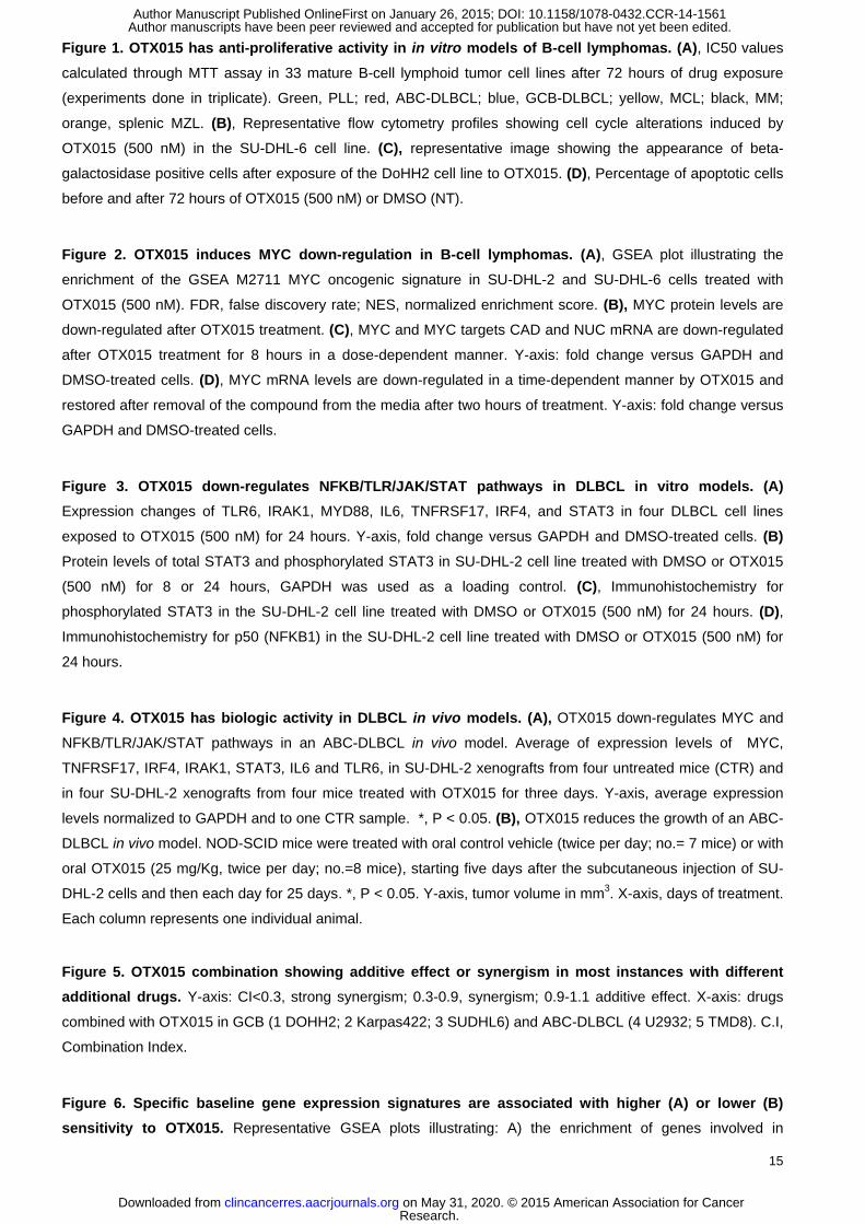
OTX015 has anti-proliferative activity in in vitro models of B-cell lymphomas
We first evaluated the anti-proliferative activity of the BET Bromodomain-inhibitor OTX015 in a panel of 33 cell
lines derived from mature B-cell lymphoid tumors. As assessed by MTT assays performed after 72 hours of
drug exposure, OTX015 was active in a dose-dependent manner in almost all the cell lines (Fig. 1A), at
concentrations achievable in the clinical setting (33). The median IC50 value for the whole series was 240 nM
(range, 70 nM – 15 μM). The median values for the individual lymphoma entities were 195 nM (70 nM - 1.5 μM)
in DLBCL, 470 nM (340 nM - 15 μM) in MCL, 170 nM (105 - 240 nM) in splenic MZL, 450 nM (60 - 700 nM) in
MM and 90 nM in the PLL cell line. The anti-proliferative effect of OTX015 did not significantly differ among the
different the histotypes, or between germinal-center type (GCB) DLBCL (190 nM; 80 nM - 1.5 μM) and activated
B-cell like (ABC) DLBCL (200 nM; 70 nM - 2.28 μM).
In accordance with the observed anti-proliferative activity, OTX015 treatment of DLBCL cells inhibited cell
growth and induced cell cycle arrest with G1 accumulation and decreased S-phase (Fig. 1B, Supplemental Fig.
S1). OTX015 also induced a time-dependent increase of beta-galactosidase positive cells (Fig. 1C).
OTX015 induces apoptosis in a genetically defined subgroup of DLBCL
We then assessed the induction of apoptosis after OTX015 exposure in DLBCL cells. Dose and time-dependent
apoptosis was observed in 3/22 (14%) cell lines treated for 72 hours with a dose of 500 nM (Fig. 1D,
Supplemental Fig. S2). A massive apoptotic induction was obtained in SU-DHL-2 and TMD8, while OCI-Ly3 had
a lower, although significant, induction of apoptosis. All these three cell lines presented common genetic and
biologic features: derivation from ABC-DLBCL, mutated MYD88 gene, wild type TP53, mutations in CD79B (SU-
DHL-2, TMD8) or in CARD11 (OCI-Ly3). The presence of mutations in genes coding for MYD88 and for
components of the BCR signaling was significantly associated with apoptosis induction (P = 0.027).
Transcriptional signature of OTX015 in DLBCL cell lines
To obtain a global view of the transcriptional changes after OTX015 treatment, we performed GEP on two
sensitive cell lines, one derived from GCB-DLBCL (SU-DHL-6) and one from ABC-DLBCL (SU-DHL-2), treated
with DMSO or with OTX015 (500 nM) for 1, 2, 4, 8 and 12 hours. OTX015 affected, in a time-dependent manner,
important biological processes: nuclear factor-kB (NFKB), Toll-like receptor (TLR), Janus kinase (JAK) / signal
transducers and activators of transcription (STAT) signaling pathways, MYC and E2F1-regulated genes, cell
cycle regulation and chromatin structure (Fig. 2, Supplemental Fig. S3 and S4, Supplemental Table S2). The
up-regulated genes were mainly represented by transcripts coding for histones, overlapping with those up-
regulated by HDAC-i, while the down-regulated transcripts comprised MYC and E2F1 targets or genes involved
in NFKB/TLR/JAK/STAT pathways. Supplemental Table S3 lists the top most differentially expressed probes
(adjusted-P<0.01 and absolute fold change >1.5), with MYC as the most down-regulated transcript. The
OTX015 GEP signature appeared similar to that reported following exposure to another BET Bromodomain
inhibitor, JQ1, in different tumor models (4, 5, 14, 15, 21) (Supplemental Fig. S5). OTX015 appeared similar to
JQ1 also in terms of activity on cell viability and induction of apoptosis in SU-DHL-2 and DOHH2 DLBCL cell
lines (data not shown), as also recently reported by Chapuy et al. (10).
Research. on May 31, 2020. © 2015 American Association for Cancerclincancerres.aacrjournals.org Downloaded from
Author manuscripts have been peer reviewed and accepted for publication but have not yet been edited. Author Manuscript Published OnlineFirst on January 26, 2015; DOI: 10.1158/1078-0432.CCR-14-1561
8
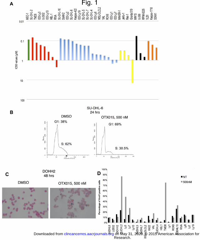
OTX015 induces MYC down-regulation in DLBCL
Similarly to that reported for other BET Bromodomain inhibitors (4, 5, 21), treatment with OTX015 led to an
important negative regulation of MYC and its target genes. MYC was the central hub connecting almost all the
most significantly OTX015 regulated genes (Supplemental Fig. S4), and MYC target genes were significantly
enriched among OTX015-regulated transcripts (Fig. 2A). MYC changes were validated at RNA and protein level
(Fig. 2B-C). The effect of OTX015 on MYC and its target genes was both time and dose dependent (Fig. 2C-D).
The effect on MYC appeared reversible since the mRNA levels started to be restored in a time-dependent
manner, albeit with different kinetics among the individual DLBCL cell lines, after replacing drug-containing
medium with fresh medium (Fig. 2D). The level and the kinetics of MYC down-regulation after drug exposure did
not appear associated with the sensitivity to OTX015 (data not shown).
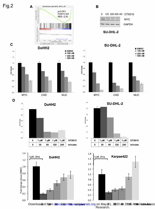
OTX015 affects the NFKB, TLR and JAK/STAT signaling pathways in DLBCL cells
OTX015 negatively regulated transcripts encoding members of the NFKB, TLR and JAK/STAT signaling
pathways, such as MYD88, IRAK1, TLR6, TNFRSF17, IL6 and IRF4 in both GCB- and ABC-DLBCL cells (Fig.
3). The inhibitory effect of OTX015 on the pathways was further confirmed at protein level. Both immunoblotting
and immunohistochemistry showed a reduction of the transcriptionally active phospho-STAT3 in SU-DHL-2 and
TMD8, two ABC-DLBCL cell lines, as well a reduction of the nuclear localization of p50 (NFKB1), indicating a
clear inhibitory effect of OTX015 on the canonical NFKB pathway (Fig. 3B-D). In ABC-DLBCL cell lines, we
observed a reduced production of IL10 and IL4 after 24 hours of OTX015 treatment (Supplemental Fig. 6), and
a dose-dependent down-regulation of additional transcripts suggestive of NFKB activation (BIRC3, TNFAIP3)
(data not shown).
Based on the hypothesis that MYD88 may play a central role in the mechanism of action of OTX015, we sought
to understand whether BET Bromodomain inhibitors had a direct effect on the gene regulation. We first
analysed the publicly available ChIP-Seq data obtained for HBL1 ABC-DLBCL cells treated with the BET
Bromodomain inhibitor JQ1 (20). Treatment with JQ1 (500 nM) for 3 hours reduced the binding of BRD4 to the
upstream regulatory region of MYD88 (FDR < 0.001) (Supplemental Fig. 7A). We then performed a ChIP
experiment in SU-DHL-2 ABC-DLBCL cells exposed to DMSO or to OTX015 (500 nM) for 3 hours. The drug
appeared to reduce the binding of BRD4 to the upstream regulatory regions of the MYD88 gene (Supplemental
Fig. 7B). These data suggest that BET Bromodomain inhibitors dislocate BRD4 from MYD88 regulatory regions.
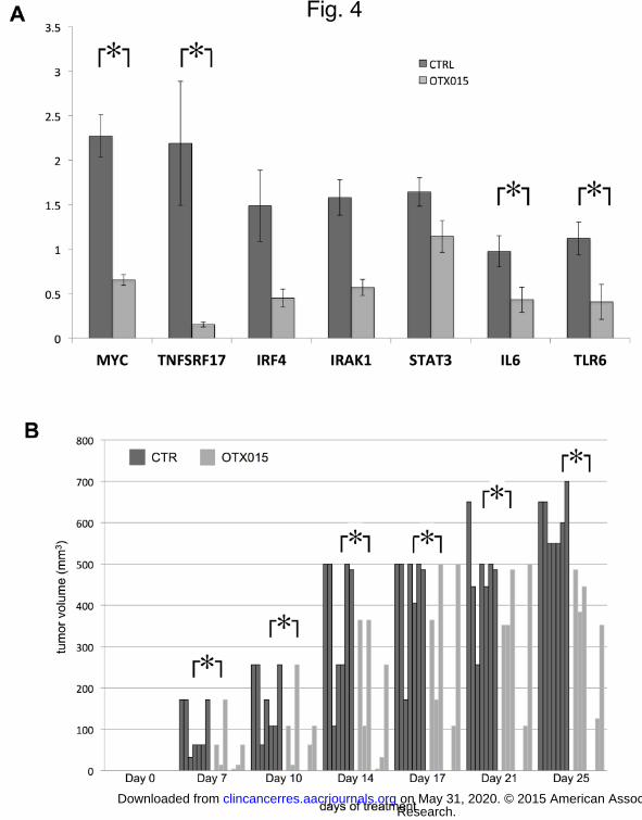
OTX015 has biological activity in a DLBCL in vivo model
We then assessed the ability of OTX015 to down-regulate MYC, the NFKB, TLR and JAK/STAT pathway also in
an in vivo model. We treated SU-DHL-2 xenografts grown subcutaneously in NOD-SCID mice with OTX015 (50
mg/Kg, orally, twice per day; no.= 4 mice) or with control (vehicle orally, twice per day; no.= 4 mice) for three
days, starting when the tumors had reached the volume of 500 mm3. No body weight losses were registered
during the three days of treatment. Real-time PCR showed that there was a significant down-regulation of MYC,
IL6, TLR6, TNFSRF17 and, although not reaching the statistical significance, of IRAK1, IRF4 and STAT3 (Fig.
4A).
We then assessed the in vivo anti-lymphoma activity of OTX015. NOD-SCID mice were treated with control
vehicle (per os, twice per day; no.= 7 mice) or with OTX015 (25 mg/Kg, orally, twice per day; no.=8 mice),
starting five days after the subcutaneous injection of SU-DHL-2 cells and then each day for 25 days. No loss in
body weight was observed. OTX015 induced a reduced growth of the lymphoma xenografts at each analyzed
Research. on May 31, 2020. © 2015 American Association for Cancerclincancerres.aacrjournals.org Downloaded from
Author manuscripts have been peer reviewed and accepted for publication but have not yet been edited. Author Manuscript Published OnlineFirst on January 26, 2015; DOI: 10.1158/1078-0432.CCR-14-1561
9
time point (days 7, 10, 14, 17, 21 and 25) (Figure 4B). At the end of the experiment (day 25), the median tumor
volumes for the control and for the experimental arm were 600 mm3 (95% C.I., 550-684) and 239 mm
3 (95% C.I.,
0-582), respectively (P = 0.001).
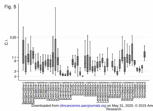
OTX015 shows in-vitro synergism with several anti-cancer agents
We evaluated the combination of OTX015 with a series of conventional and targeted anti-lymphoma agents in a
panel of five DLBCL cell lines (Fig. 5). Strong synergism was observed, in all the cell lines, when OTX015 was
combined with the mammalian target of rapamycin (mTOR) inhibitor everolimus (median CI, 0.11; range 0.1-
0.17) and, in ABC-cells, with the BTK-inhibitor ibrutinib (CI=0.04; 0.02-0.1). Synergism was also observed with
OTX015 plus the PI3K-delta inhibitor idelalisib (CI=0.5; 0.04-2.4), the class I and II HDACi vorinostat (CI=0.5;
0.3-0.6), anti-CD20 moAb rituximab (CI=0.5; 0.4-0.5), the hypomethylating agent decitabine (CI=0.6; 0.6-0.7),
the immunomodulant lenalidomide (CI=0.7; 0.6-0.7). OTX015 combinations with the class I HDACi romidepsin
(CI=1.08; 1-1.22) and with the chemotherapy agents bendamustine (CI=0.63; 0.1-3.97) and doxorubicin
(CI=0.83; 0.71-0.96) presented a moderate additive effect. GCB- and ABC-DLBCL cells showed a different
sensitivity to the combinations: a stronger synergism was observed in ABC than in GCB DLBCL cells for
ibrutinib (P<0.0001), for idelalisib (P<0.0001), lenalidomide (P=0.0001), and rituximab (P=0.007).
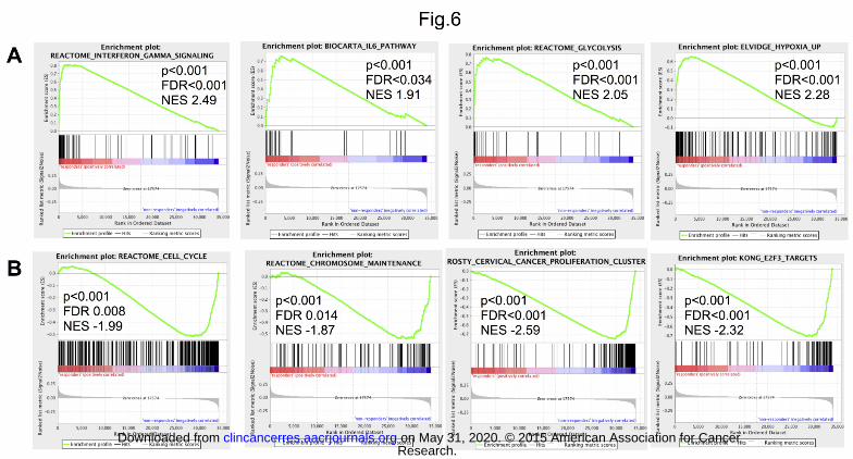
Baseline gene expression profile is associated with response to OTX015
To identify genes and pathways that might predict sensitivity to OTX015 in DLBCL we integrated the sensitivity
data with the baseline gene expression profile in 14 cell lines with an IC50 lower than 500 nM and eight with a
higher IC50.
Transcripts positively associated with OTX015 sensitivity were significantly enriched of genes involved in
interferon, IL6 and IL10 signaling genes, TLR and JAK/STAT signaling, STAT3 targets, genes involved in
glucose metabolism, and hypoxia-regulated genes (Fig. 6A; Supplemental Table S4A). The leading edge genes
(the top differentially ranked transcripts based on sensitivity to OTX015) comprised STAT1, STAT3, STAT4,
STAT6, IL6, JAK1, JAK2, TNF, IRAK1, TLR9, MYD88, TLR8, TRAF3, and AKT1.
Transcripts associated with lower sensitivity to OTX015 were significantly enriched of E2F target genes, genes
involved in cell cycle regulation, DNA repair, P53 signaling, chromatin structure, and apoptosis (Fig. 6B;
Supplemental Table S4B). BCL2L1, HDAC1, HDAC2, HDAC5, HDAC8, CHEK1, CHEK2, TP53, ATM, BRCA1,
CDKN1A, CDKN2A, BIRC5, MGMT were among the leading edge genes. MYC or MYC targets did not appear
associated with sensitivity to OTX015.
Discussion
We have evaluated the activity and the mechanism of action of a new BET Bromodomain inhibitor, OTX015, in
pre-clinical models of mature B-cell lymphoid tumors. We have shown that: 1) OTX015 has in vitro and in vivo
anti-proliferative activity; 2) OTX015 effect is largely cytostatic, with induction of apoptosis in only a genetically
defined subset of cell lines; 3) OTX015 inhibits MYC, NFKB, TLR and JAK/STAT pathways; 4) OTX015 shows
synergistic or additive effects when combined with several anti-lymphoma agents; 5) specific baseline gene
expression features are associated with a different sensitivity to OTX015.
At clinically achievable concentrations (33), OTX015 had an anti-proliferative activity in the most of the
lymphoma cell lines tested, with no differences in sensitivity among the histologic subtypes. The effect was
largely cytostatic, with apoptosis limited to a few cell lines. OTX015 appeared to induce a senescence-like
phenotype, possibly compatible with the recently described “senescence with incomplete growth arrest”, also
Research. on May 31, 2020. © 2015 American Association for Cancerclincancerres.aacrjournals.org Downloaded from
Author manuscripts have been peer reviewed and accepted for publication but have not yet been edited. Author Manuscript Published OnlineFirst on January 26, 2015; DOI: 10.1158/1078-0432.CCR-14-1561
10
observed after treatment of DLBCL cells with demethylating agents (34). The anti-tumor activity of OTX015 was
validated in an in vivo model of ABC-DLBCL with demonstration of a reduced tumor growth.
Similarly to other BRD-inhibitors (4, 5, 10, 21, 35), OTX015 determined a down-regulation of MYC and of MYC
target genes in DLBCL cell lines carrying a wild-type MYC and in cells in which the oncogene is translocated or
amplified. This is a remarkable achievement since MYC is a difficult therapeutic target and it plays a major role
in the pathogenesis and progression of lymphomas, as exemplified by the very poor outcome of DLBCL cases
over-expressing both MYC and BCL2 proteins and of “double hit” lymphomas usually bearing concomitant
translocations of MYC and BCL2 oncogenes (36).
An important biologic effect we observed with OTX015, both in the in vitro and in vivo setting, was the down-
regulation of the NFKB, TLR and JAK/STAT3 signaling pathways, important in both the pathogenesis and the
chemo-resistance of lymphomas, and particularly of ABC-DLBCL. This observation is in accordance with the
strong suppression of the TLR signaling-mediated lipopolysaccharide-induced inflammatory response, reported
with BET Bromodomain inhibitors (37). A similar effect has been reported for JQ1 in different DLBCL cell lines
(10). Additional observations indicated that the down-regulation of the TLR and JAK/STAT3 signaling pathways
is a relevant mechanism of action of OTX015 in DLBCL. First, high expression of genes involved in interferon,
TLR and JAK/STAT signaling and STAT3 targets were highly associated with the sensitivity to the compound.
Second, OTX015-induced apoptosis was limited to cell lines, bearing a functional TP53, derived from ABC-
DLBCL with somatic mutations activating both BCR signaling and MYD88. Mutations activating the BCR and
TLR/MYD88 signaling are common in ABC-DLBCL, in which they represent driver events (38), and their
prevalence is especially high in two aggressive extranodal forms of DLBCL, primary central nervous system
lymphoma and primary testis DLBCL (39, 40). CD79A and CD79B are upstream of BTK. BTK binds to MYD88,
especially if the latter is encoded by the L265P somatic variant (41). OTX015 down-regulated the expression of
MYD88 and of additional members of the TLR pathway, and both OTX015 and JQ1 reduced the BRD4 binding
to MYD88 upstream regulatory region. These events could inhibit both BCR and TLR signaling, particularly
affecting cells dependent on these pathways. Similarly, the BTK-inhibitor ibrutinib is more active in ABC-DLBCL
cases with both CD79B/CD79A and MYD88 mutated genes (38, 42). Importantly, the combination of OTX015
and ibrutinib was strongly synergistic in ABC-DLBCL cells. The down-regulation of IRF4 might contribute to the
synergism, as reported for the combination of ibrutinib and lenalidomide (43). Notably, OTX015 was active also
in ABC-DLBCL cells carrying somatic mutations of CARD11, coding for a protein downstream of BTK, and,
accordingly, representing a marker of resistance to ibrutinib (38, 42).
Besides ibrutinib, additional anti-lymphoma agents presented an increased activity when combined with
OTX015. The strongest synergism was obtained with the combination with the mTOR inhibitor everolimus at
doses that can be reached in patients (44). The mTOR is central to a signaling cascade leading to cell growth
and proliferation, and mTOR inhibitors are approved for treatment of relapsed mantle cell lymphoma (45).
OTX015 was also synergistic with the PI3K-delta inhibitor idelalisib, which has shown promising clinical
responses in B-cell lymphomas (46), and with the lenalidomide, as also recently reported for another BET
Bromodomain inhibitor in mantle cell lymphoma (47).
The synergism observed for OTX015 combined with clinically achievable doses of rituximab is reinforced by
recent reports with another BET bromodomain inhibitor (13). The down-regulation of CCDC86, coding for the
nuclear factor cyclon, by both JQ1 (13) and OTX015, might represent the mechanism of action of the synergism.
The combination appeared more active in ABC-DLBCL cell lines, maybe due the common targeting of the IL10
and STAT3 pathway by both OTX015 and rituximab (48).
OTX015 presented synergism with the demethylating agent decitabine and the HDACi vorinostat at
concentrations pharmacologically achievable in clinical use (49, 50), in accordance with the similarities here
Research. on May 31, 2020. © 2015 American Association for Cancerclincancerres.aacrjournals.org Downloaded from
Author manuscripts have been peer reviewed and accepted for publication but have not yet been edited. Author Manuscript Published OnlineFirst on January 26, 2015; DOI: 10.1158/1078-0432.CCR-14-1561
11
observed at the level of gene expression signatures, and also with published data obtained with other BET
bromodomain inhibitors (11). HDACs of both class I and II were associated with a lower sensitivity to OTX015
as single agent and, indeed the synergism appeared stronger with the class I and IIa/b HDACi vorinostat than
that seen with the class I HDACi romidepsin suggesting that the synergism might be class dependent.
The integration of baseline GEP and sensitivity in a large panel of DLBCL cell lines allowed the identification of
functional pathways that might predict the response to the BET Bromodomain inhibitor. The cells with the
highest sensitivity to OTX015 had not only high expression levels of genes involved in the TLR/JAK/STAT
pathway, but also of transcripts that code for proteins implicated in glucose metabolism and in hypoxia.
Although the latter two features can be associated with an active MYC program, it is important to underline that
we did not detect any association between expression of MYC or MYC targets and sensitivity to OTX015. Based
on our data, it will be important to correlate the demonstration of an active TLR/JAK/STAT pathway (for example
detecting pSTAT3) and/or the presence of somatic mutations of MYD88, CD79A/B, CARD11 and TP53 with the
response to BET Bromodomain inhibitors in the on-going clinical trials.
A strong E2F gene expression signature was associated with lower sensitivity to OTX015, although not with a
clear resistance to the compound since all our cell lines were much more sensitive to the BET Bromodomain
inhibitor than other large series of cancer cell lines comprising solid tumors models (5, 14, 15). In accordance
with the cell cycle arrest observed at 24-48 hours, E2F1 target genes, but not E2F1 itself, are down-regulated
by both OTX015 and JQ1 (10), at a later time-point than the effect on MYC or on TLR/JAK/STAT pathways. The
high synergism reported in leukemia pre-clinical models with the combination of the BET Bromodomain inhibitor
PFI-1 with a pan-aurora kinase inhibitor (12) suggests that the combination of BET Bromodomain inhibitors with
drugs targeting the cell cycle might overcome the observed lower sensitivity.
The gene expression signatures associated with a lower sensitivity to OTX015 comprised transcripts (BCL2L1,
BIRC5, MGMT, CHEK1, CHEK2), which represent potential molecules to be inhibited in combination with BET
Bromodomain inhibitors.
There are now different BET Bromodomain inhibitors under development with some of them, including OTX015,
already in phase I clinical studies. Based on our data of an OTX015 gene expression signature highly
overlapping with that reported with JQ1, and on the literature (10, 12, 16, 51) all the molecules, so far all pan-
BET Bromodomain inhibitors, have similar pre-clinical activity data and mechanism of action. Alongside the
different drug delivery modalities (for example, OTX015 and GSK525762 are given orally, TEN-010
subcutaneously), the toxicity and pharmacokinetics data that from the on-going clinical studies will be most
relevant for the further clinical development of this class of compounds.
In conclusion, the BET Bromodomain inhibitor OTX015 appears as a promising new anti-lymphoma agent with
anti-proliferative activity in the vast majority of the examined pre-clinical models, capable of down-regulating
important signaling pathways and of synergizing with other anti-cancer molecules. Particular genetic lesions and
gene expression signatures are associated with high sensitivity to OTX015 anti-tumor activity. Additional studies
are needed to elucidate the mechanism of action of OTX015, particularly when combined with other targeted
agents. Together with the early report of clinical responses in both leukemia and lymphoma patients treated with
OTX015 in the absence of major toxicities (22), the data presented here provide the basis for further clinical
investigation of OTX015 in combination therapies.
Acknowledgements
We would like to thank Diletta Di Mitri for her help in the evaluation of interleukins production.
Research. on May 31, 2020. © 2015 American Association for Cancerclincancerres.aacrjournals.org Downloaded from
Author manuscripts have been peer reviewed and accepted for publication but have not yet been edited. Author Manuscript Published OnlineFirst on January 26, 2015; DOI: 10.1158/1078-0432.CCR-14-1561
12
Authorship
Contribution: M.B. designed and performed in vitro experiments, interpreted data, drafted the manuscript; E.G.
designed and performed in vitro and in vivo experiments, interpreted data; P.B. designed and performed in vitro
experiments, interpreted data; E.B., C.T., A.A.M. and M.T. performed experiments; A.R., performed gene
expression profiling; I.K. and L.C., performed data mining; M.P., performed immunohistochemistry; G.S.,
performed morphology; A.S., E.Z., P.H., G.I, E.C. provided advice; E.R. provided advice and designed
experiments; F.B. designed the study, interpreted data, and wrote the manuscript. M.B., E.G., P.B., equally
contributed. All authors have approved the final manuscript.
The current affiliation for M.B. and G.I. is Weill Cornell Medical College, New York, N.Y., USA. The current
affiliation for P.B. is Italian Institute of Technology, Center for Genomic Sciences, Milan, Italy.
References
1. Jiang Y, Hatzi K, Shaknovich R. Mechanisms of epigenetic deregulation in lymphoid neoplasms. Blood. 2013;121:4271-9. 2. Dawson MA, Kouzarides T, Huntly BJ. Targeting epigenetic readers in cancer. N Engl J Med. 2012;367:647-57. 3. Geutjes EJ, Bajpe PK, Bernards R. Targeting the epigenome for treatment of cancer. Oncogene. 2012;31:3827-44. 4. Delmore JE, Issa GC, Lemieux ME, Rahl PB, Shi J, Jacobs HM, et al. BET bromodomain inhibition as a therapeutic strategy to target c-Myc. Cell. 2011;146:904-17. 5. Mertz JA, Conery AR, Bryant BM, Sandy P, Balasubramanian S, Mele DA, et al. Targeting MYC dependence in cancer by inhibiting BET bromodomains. Proc Natl Acad Sci U S A. 2011;108:16669-74. 6. Loven J, Hoke HA, Lin CY, Lau A, Orlando DA, Vakoc CR, et al. Selective inhibition of tumor oncogenes by disruption of super-enhancers. Cell. 2013;153:320-34. 7. Hnisz D, Abraham BJ, Lee TI, Lau A, Saint-Andre V, Sigova AA, et al. Super-Enhancers in the Control of Cell Identity and Disease. Cell. 2013. 8. Greenwald RJ, Tumang JR, Sinha A, Currier N, Cardiff RD, Rothstein TL, et al. E mu-BRD2 transgenic mice develop B-cell lymphoma and leukemia. Blood. 2004;103:1475-84. 9. Dawson MA, Prinjha RK, Dittmann A, Giotopoulos G, Bantscheff M, Chan WI, et al. Inhibition of BET recruitment to chromatin as an effective treatment for MLL-fusion leukaemia. Nature. 2011;478:529-33. 10. Chapuy B, McKeown MR, Lin CY, Monti S, Roemer MG, Qi J, et al. Discovery and characterization of super-enhancer-associated dependencies in diffuse large B cell lymphoma. Cancer Cell. 2013;24:777-90. 11. Bhadury J, Nilsson LM, Muralidharan SV, Green LC, Li Z, Gesner EM, et al. BET and HDAC inhibitors induce similar genes and biological effects and synergize to kill in Myc-induced murine lymphoma. Proc Natl Acad Sci U S A. 2014;111:E2721-30. 12. Picaud S, Da Costa D, Thanasopoulou A, Filippakopoulos P, Fish PV, Philpott M, et al. PFI-1, a highly selective protein interaction inhibitor, targeting BET Bromodomains. Cancer Res. 2013;73:3336-46. 13. Emadali A, Rousseaux S, Bruder-Costa J, Rome C, Duley S, Hamaidia S, et al. Identification of a novel BET bromodomain inhibitor-sensitive, gene regulatory circuit that controls Rituximab response and tumour growth in aggressive lymphoid cancers. EMBO molecular medicine. 2013;5:1180-95. 14. Lockwood WW, Zejnullahu K, Bradner JE, Varmus H. Sensitivity of human lung adenocarcinoma cell lines to targeted inhibition of BET epigenetic signaling proteins. Proc Natl Acad Sci U S A. 2012;109:19408-13. 15. Puissant A, Frumm SM, Alexe G, Bassil CF, Qi J, Chanthery YH, et al. Targeting MYCN in neuroblastoma by BET bromodomain inhibition. Cancer Discov. 2013;3:308-23. 16. Cheng Z, Gong Y, Ma Y, Lu K, Lu X, Pierce LA, et al. Inhibition of BET bromodomain targets genetically diverse glioblastoma. Clin Cancer Res. 2013;19:1748-59. 17. Noel JK, Iwata K, Ooike S, Sugahara K, Nakamura H, Daibata M. Development of the BET bromodomain inhibitor OTX015. Proceeding of the 2013 AACR- NCI-EORTC Molecular Targets and Cancer Therapeutics Conference. 2013:414-. 18. Bandopadhayay P, Bergthold G, Nguyen B, Schubert S, Gholamin S, Tang Y, et al. BET Bromodomain Inhibition of MYC-Amplified Medulloblastoma. Clin Cancer Res. 2014;20:912-25. 19. Trabucco SE, Gerstein RM, Evens AM, Bradner JE, Shultz LD, Greiner DL, et al. Inhibition of bromodomain proteins for the treatment of human diffuse large B-cell lymphoma. Clin Cancer Res. 2014. 20. Ceribelli M, Kelly PN, Shaffer AL, Wright GW, Xiao W, Yang Y, et al. Blockade of oncogenic IkappaB kinase activity in diffuse large B-cell lymphoma by bromodomain and extraterminal domain protein inhibitors. Proc Natl Acad Sci U S A. 2014;111:11365-70. 21. Ott CJ, Kopp N, Bird L, Paranal RM, Qi J, Bowman T, et al. BET bromodomain inhibition targets both c-Myc and IL7R in high-risk acute lymphoblastic leukemia. Blood. 2012;120:2843-52.
Research. on May 31, 2020. © 2015 American Association for Cancerclincancerres.aacrjournals.org Downloaded from
Author manuscripts have been peer reviewed and accepted for publication but have not yet been edited. Author Manuscript Published OnlineFirst on January 26, 2015; DOI: 10.1158/1078-0432.CCR-14-1561
13
22. Stathis A, Quesnel B, Amorim S, Thieblemont C, Zucca E, Raffoux E, et al. 5LBA Results of a first-in-man phase I trial assessing OTX015, an orally available BET-bromodomain (BRD) inhibitor, in advanced hematologic malignancies. European Journal of Cancer. 2014;50:196. 23. Boi M, Rinaldi A, Kwee I, Bonetti P, Todaro M, Tabbo F, et al. PRDM1/BLIMP1 is commonly inactivated in anaplastic large T-cell lymphoma. Blood. 2013;122:2683-93. 24. Mensah AA, Kwee I, Gaudio E, Rinaldi A, Ponzoni A, Cascione L, et al. Novel HDAC inhibitors exhibit pre-clinical efficacy in lymphoma models and point to the importance of CDKN1A expression levels in mediating their anti-tumor response. Oncotarget. 2014. 25. Gentleman R, Carey V, Dudoit S, Irizarry R, Huber W, editors. Bioinformatics and Computational Biology Solutions using R and Bioconductor. New York, NY, USA: Springer; 2005. 26. Johnson WE, Li C, Rabinovic A. Adjusting batch effects in microarray expression data using empirical Bayes methods. Biostatistics. 2007;8:118-27. 27. Smyth GK. Linear models and empirical Bayes methods for assessing differential expression in microarray experiments. Statistical applications in genetics and molecular biology. 2004;3:Article3. 28. Subramanian A, Tamayo P, Mootha VK, Mukherjee S, Ebert BL, Gillette MA, et al. Gene set enrichment analysis: a knowledge-based approach for interpreting genome-wide expression profiles. Proc Natl Acad Sci U S A. 2005;102:15545-50. 29. Langmead B, Trapnell C, Pop M, Salzberg SL. Ultrafast and memory-efficient alignment of short DNA sequences to the human genome. Genome Biol. 2009;10:R25. 30. Heinz S, Benner C, Spann N, Bertolino E, Lin YC, Laslo P, et al. Simple combinations of lineage-determining transcription factors prime cis-regulatory elements required for macrophage and B cell identities. Mol Cell. 2010;38:576-89. 31. Lacrima K, Valentini A, Lambertini C, Taborelli M, Rinaldi A, Zucca E, et al. In vitro activity of cyclin-dependent kinase inhibitor CYC202 (Seliciclib, R-roscovitine) in mantle cell lymphomas. Annals of Oncology. 2005;16:1169-76. 32. Lee JJ, Kong M, Ayers GD, Lotan R. Interaction index and different methods for determining drug interaction in combination therapy. Journal of biopharmaceutical statistics. 2007;17:461-80. 33. Odore E, Rezai K, Riveiro E, Bourdel F, Herait P, Cvitkovic E, et al. Abstract LB-231: A phase I pharmacokinetic study of OTX015 for the treatment of patients with hematologic malignancies. Cancer Research. 2014;74:LB-231. 34. Clozel T, Yang S, Elstrom RL, Tam W, Martin P, Kormaksson M, et al. Mechanism-based epigenetic chemosensitization therapy of diffuse large B-cell lymphoma. Cancer Discov. 2013;3:1002-19. 35. Zhao X, Lwin T, Zhang X, Huang A, Wang J, Marquez VE, et al. Disruption of the MYC-miRNA-EZH2 loop to suppress aggressive B-cell lymphoma survival and clonogenicity. Leukemia. 2013;27:2341-50. 36. Thieblemont C, Briere J. MYC, BCL2, BCL6 in DLBCL: impact for clinics in the future? Blood. 2013;121:2165-6. 37. Nicodeme E, Jeffrey KL, Schaefer U, Beinke S, Dewell S, Chung CW, et al. Suppression of inflammation by a synthetic histone mimic. Nature. 2010;468:1119-23. 38. Staudt LM. II. Therapy of DLBCL based on genomics. Hematol Oncol. 2013;31 Suppl 1:26-8. 39. Gonzalez-Aguilar A, Idbaih A, Boisselier B, Habbita N, Rossetto M, Laurenge A, et al. Recurrent mutations of MYD88 and TBL1XR1 in primary central nervous system lymphomas. Clin Cancer Res. 2012;18:5203-11. 40. Kraan W, Horlings HM, van Keimpema M, Schilder-Tol EJ, Oud ME, Scheepstra C, et al. High prevalence of oncogenic MYD88 and CD79B mutations in diffuse large B-cell lymphomas presenting at immune-privileged sites. Blood cancer journal. 2013;3:e139. 41. Yang G, Zhou Y, Liu X, Xu L, Cao Y, Manning RJ, et al. A mutation in MYD88 (L265P) supports the survival of lymphoplasmacytic cells by activation of Bruton's tyrosine kinase in Waldenstrom's macroglobulinemia. Blood. 2013;122:1222-32. 42. Honigberg LA, Smith AM, Sirisawad M, Verner E, Loury D, Chang B, et al. The Bruton tyrosine kinase inhibitor PCI-32765 blocks B-cell activation and is efficacious in models of autoimmune disease and B-cell malignancy. Proc Natl Acad Sci U S A. 2010;107:13075-80. 43. Yang Y, Shaffer AL, 3rd, Emre NC, Ceribelli M, Zhang M, Wright G, et al. Exploiting Synthetic Lethality for the Therapy of ABC Diffuse Large B Cell Lymphoma. Cancer Cell. 2012;21:723-37. 44. Tabernero J, Rojo F, Calvo E, Burris H, Judson I, Hazell K, et al. Dose- and schedule-dependent inhibition of the mammalian target of rapamycin pathway with everolimus: a phase I tumor pharmacodynamic study in patients with advanced solid tumors. J Clin Oncol. 2008;26:1603-10. 45. Reeder CB, Ansell SM. Novel therapeutic agents for B-cell lymphoma: developing rational combinations. Blood. 2011;117:1453-62. 46. Gopal AK, Kahl BS, de Vos S, Wagner-Johnston ND, Schuster SJ, Jurczak WJ, et al. PI3Kdelta Inhibition by Idelalisib in Patients with Relapsed Indolent Lymphoma. N Engl J Med. 2014. 47. Moros A, Rodriguez V, Saborit-Villarroya I, Montraveta A, Balsas P, Sandy P, et al. Synergistic antitumor activity of lenalidomide with the BET bromodomain inhibitor CPI203 in bortezomib-resistant mantle cell lymphoma. Leukemia. 2014. 48. Jazirehi AR, Bonavida B. Cellular and molecular signal transduction pathways modulated by rituximab (rituxan, anti-CD20 mAb) in non-Hodgkin's lymphoma: implications in chemosensitization and therapeutic intervention. Oncogene. 2005;24:2121-43.
Research. on May 31, 2020. © 2015 American Association for Cancerclincancerres.aacrjournals.org Downloaded from
Author manuscripts have been peer reviewed and accepted for publication but have not yet been edited. Author Manuscript Published OnlineFirst on January 26, 2015; DOI: 10.1158/1078-0432.CCR-14-1561

14
49. Momparler RL. Pharmacology of 5-Aza-2'-deoxycytidine (decitabine). Semin Hematol. 2005;42:S9-16. 50. Smith MA, Houghton P. A proposal regarding reporting of in vitro testing results. Clin Cancer Res. 2013;19:2828-33. 51. Gupta SS, Maetzig T, Maertens GN, Sharif A, Rothe M, Weidner-Glunde M, et al. Bromo- and extraterminal domain chromatin regulators serve as cofactors for murine leukemia virus integration. J Virol. 2013;87:12721-36.
Research. on May 31, 2020. © 2015 American Association for Cancerclincancerres.aacrjournals.org Downloaded from
Author manuscripts have been peer reviewed and accepted for publication but have not yet been edited. Author Manuscript Published OnlineFirst on January 26, 2015; DOI: 10.1158/1078-0432.CCR-14-1561
15
Figure 1. OTX015 has anti-proliferative activity in in vitro models of B-cell lymphomas. (A), IC50 values
calculated through MTT assay in 33 mature B-cell lymphoid tumor cell lines after 72 hours of drug exposure
(experiments done in triplicate). Green, PLL; red, ABC-DLBCL; blue, GCB-DLBCL; yellow, MCL; black, MM;
orange, splenic MZL. (B), Representative flow cytometry profiles showing cell cycle alterations induced by
OTX015 (500 nM) in the SU-DHL-6 cell line. (C), representative image showing the appearance of beta-
galactosidase positive cells after exposure of the DoHH2 cell line to OTX015. (D), Percentage of apoptotic cells
before and after 72 hours of OTX015 (500 nM) or DMSO (NT).
Figure 2. OTX015 induces MYC down-regulation in B-cell lymphomas. (A), GSEA plot illustrating the
enrichment of the GSEA M2711 MYC oncogenic signature in SU-DHL-2 and SU-DHL-6 cells treated with
OTX015 (500 nM). FDR, false discovery rate; NES, normalized enrichment score. (B), MYC protein levels are
down-regulated after OTX015 treatment. (C), MYC and MYC targets CAD and NUC mRNA are down-regulated
after OTX015 treatment for 8 hours in a dose-dependent manner. Y-axis: fold change versus GAPDH and
DMSO-treated cells. (D), MYC mRNA levels are down-regulated in a time-dependent manner by OTX015 and
restored after removal of the compound from the media after two hours of treatment. Y-axis: fold change versus
GAPDH and DMSO-treated cells.
Figure 3. OTX015 down-regulates NFKB/TLR/JAK/STAT pathways in DLBCL in vitro models. (A)
Expression changes of TLR6, IRAK1, MYD88, IL6, TNFRSF17, IRF4, and STAT3 in four DLBCL cell lines
exposed to OTX015 (500 nM) for 24 hours. Y-axis, fold change versus GAPDH and DMSO-treated cells. (B)
Protein levels of total STAT3 and phosphorylated STAT3 in SU-DHL-2 cell line treated with DMSO or OTX015
(500 nM) for 8 or 24 hours, GAPDH was used as a loading control. (C), Immunohistochemistry for
phosphorylated STAT3 in the SU-DHL-2 cell line treated with DMSO or OTX015 (500 nM) for 24 hours. (D),
Immunohistochemistry for p50 (NFKB1) in the SU-DHL-2 cell line treated with DMSO or OTX015 (500 nM) for
24 hours.
Figure 4. OTX015 has biologic activity in DLBCL in vivo models. (A), OTX015 down-regulates MYC and
NFKB/TLR/JAK/STAT pathways in an ABC-DLBCL in vivo model. Average of expression levels of MYC,
TNFRSF17, IRF4, IRAK1, STAT3, IL6 and TLR6, in SU-DHL-2 xenografts from four untreated mice (CTR) and
in four SU-DHL-2 xenografts from four mice treated with OTX015 for three days. Y-axis, average expression
levels normalized to GAPDH and to one CTR sample. *, P < 0.05. (B), OTX015 reduces the growth of an ABC-
DLBCL in vivo model. NOD-SCID mice were treated with oral control vehicle (twice per day; no.= 7 mice) or with
oral OTX015 (25 mg/Kg, twice per day; no.=8 mice), starting five days after the subcutaneous injection of SU-
DHL-2 cells and then each day for 25 days. *, P < 0.05. Y-axis, tumor volume in mm3. X-axis, days of treatment.
Each column represents one individual animal.
Figure 5. OTX015 combination showing additive effect or synergism in most instances with different
additional drugs. Y-axis: CI<0.3, strong synergism; 0.3-0.9, synergism; 0.9-1.1 additive effect. X-axis: drugs
combined with OTX015 in GCB (1 DOHH2; 2 Karpas422; 3 SUDHL6) and ABC-DLBCL (4 U2932; 5 TMD8). C.I,
Combination Index.
Figure 6. Specific baseline gene expression signatures are associated with higher (A) or lower (B)
sensitivity to OTX015. Representative GSEA plots illustrating: A) the enrichment of genes involved in
Research. on May 31, 2020. © 2015 American Association for Cancerclincancerres.aacrjournals.org Downloaded from
Author manuscripts have been peer reviewed and accepted for publication but have not yet been edited. Author Manuscript Published OnlineFirst on January 26, 2015; DOI: 10.1158/1078-0432.CCR-14-1561

16
interferon and IL6 signaling, in glucose metabolism, and in hypoxia among the transcripts associated with a
higher OTX015 sensitivity; B) the enrichment of genes involved in cell cycle regulation and chromatin structure
and of E2F targets among the transcripts associated with a lower sensitivity to OTX015. FDR, false discovery
rate; NES, normalized enrichment score.
Research. on May 31, 2020. © 2015 American Association for Cancerclincancerres.aacrjournals.org Downloaded from
Author manuscripts have been peer reviewed and accepted for publication but have not yet been edited. Author Manuscript Published OnlineFirst on January 26, 2015; DOI: 10.1158/1078-0432.CCR-14-1561
Published OnlineFirst January 26, 2015.Clin Cancer Res Michela Boi, Eugenio Gaudio, Paola Bonetti, et al. with targeted drugspathways in pre-clinical B-cell tumor models and synergizes The BET Bromodomain inhibitor OTX015 affects pathogenetic
Updated version
10.1158/1078-0432.CCR-14-1561doi:
Access the most recent version of this article at:
Material
Supplementary
http://clincancerres.aacrjournals.org/content/suppl/2015/01/27/1078-0432.CCR-14-1561.DC1
Access the most recent supplemental material at:
Manuscript
Authorbeen edited. Author manuscripts have been peer reviewed and accepted for publication but have not yet
E-mail alerts related to this article or journal.Sign up to receive free email-alerts
Subscriptions
Reprints and
To order reprints of this article or to subscribe to the journal, contact the AACR Publications
Permissions
Rightslink site. Click on "Request Permissions" which will take you to the Copyright Clearance Center's (CCC)
.http://clincancerres.aacrjournals.org/content/early/2015/01/24/1078-0432.CCR-14-1561To request permission to re-use all or part of this article, use this link
Research. on May 31, 2020. © 2015 American Association for Cancerclincancerres.aacrjournals.org Downloaded from
Author manuscripts have been peer reviewed and accepted for publication but have not yet been edited. Author Manuscript Published OnlineFirst on January 26, 2015; DOI: 10.1158/1078-0432.CCR-14-1561