mitochondrial functions in mood disorderspsych.lf1.cuni.cz/zf/publikace/b005.pdf · chapter 5...
TRANSCRIPT
Chapter 5
Mitochondrial Functions in Mood Disorders
Jana Hroudová, Zdeněk Fišar and Jiří Raboch
Additional information is available at the end of the chapter
http://dx.doi.org/10.5772/53254
1. Introduction
Depression is a serious mental disorder manifested by depressed mood, pessimisticthoughts, feelings of worthlessness, feelings of guilt, tearfulness, reduced or increased sleep,appetite loss or appetite disturbance, weight loss or weight gain, social restlessness, loss ofinterest, difficulty concentrating. Mania is characterized by abnormally elevated or irritablemood, arousal, and/or energy levels. Bipolar disorder features intermittent episodes of ma‐nia or hypomania and depressive episodes; rapid cycling, mixed states, and psychotic symp‐toms occuring in some cases. Depression and mania are thought to be heterogeneousillnesses that can result from dysfunction of several neurotransmitters or metabolic systems.
The predisposition to the disease is determined by genetic, psychosocial and biological fac‐tors; individual sensitivity to depressogenic effects during stressful life events is also a con‐tributing factor. Pathophysiology of mood disorders is not sufficiently elucidated and about1/3 of patients do not response to pharmacotherapy sufficiently. The exact molecular siteand the primary cause of signal transduction disturbance associated with the symptoms ofdepression or mania are still unknown.
Recently, attention in the research of biological basis of mood disorders has been devoted to anoverlapping set of molecular and cellular mechanisms of mood disorders, antidepressant re‐sponse, neuroplasticity, and chronic stress [1], e.g. to changes in neuroprogression, inflamma‐tory and cell-mediated immune response, antioxidant capacity, oxidative and nitrosativestress, and mitochondrial functions [2]. Therefore, changes in the activities of compounds ofthese intracellular signalling pathways are studied with the aim of discovering new biologicalmarkers of mood disorders or predictors of response to antidepressant treatment [3-4]. Mito‐chondrial dysfunctions are assuming an increasingly important role in hypotheses of mooddisorders, bipolar disorder mainly. Recently discussed biological hypotheses of mood disor‐ders include the neurotrophic and neuroplasticity hypothesis of depression [1,5-8] and the mi‐tochondrial hypothesis [9-11].
© 2013 Hroudová et al.; licensee InTech. This is an open access article distributed under the terms of theCreative Commons Attribution License (http://creativecommons.org/licenses/by/3.0), which permitsunrestricted use, distribution, and reproduction in any medium, provided the original work is properly cited.
It is well-known that mitochondria strongly affect many intracellular processes coupled tosignal transduction, neuron survival and plasticity. Impaired mitochondrial functions mani‐fest themselves in various ways, they may be related to many psychiatric and neurodege‐nerative diseases, including bipolar disorder, major depressive disorder, schizophrenia,psychosis and anxiety [12-16]. Impaired functions of mitochondria can be assessed both inisolated mitochondria and in intact or permeabilized cells. Better insight into molecularmechanisms of cellular respiration, control of oxidative phosphorylation (OXPHOS) and ef‐fects of antidepressants and mood stabilizers on these processes is likely to lead to a betterunderstanding of pathophysiology of neuropsychiatric disorders.
2. Mitochondria
Mitochondria are small cellular structures consisting of an outer and inner membrane, an in‐termembrane space and an intracellular matrix. The outer membrane covers the organelle,the inner membrane folds and forms cristae. This settlement extends the surface and enablesplenty of chemical reactions. In the mitochondrial matrix, the enzymes of the tricarboxylicacid cycle (TCA, also called citric acid cycle or Krebs cycle) are localized. It is the centralpathway of metabolism; its main function is oxidation of acetyl-CoA derived from carbohy‐drates, amino acids and fatty acids (FAs). The TCA is organized into a supramolecular com‐plex that enables interaction with mitochondrial membranes and the electron transportchain (ETC) in OXPHOS [17]. Most of the TCA enzymes provide other additional “moon‐lighting” functions, e.g. they stabilize the mitochondrial DNA (mtDNA) or are associatedwith mitochondrial RNA (mtRNA) translation, oxidative stress, iron metabolism and tu‐mour suppression [18].
In addition to their crucial role in generation of adenosine-5’- triphosphate (ATP), mitochon‐dria are involved in other important processes, such as regulation of free radicals, neuro‐transmitters, calcium, and apoptosis. They are also involved in neuronal development -synaptogenesis, synaptic development and plasticity. Impaired function of mitochondrialeads to impaired bioenergetics, decrease of ATP production, impaired calcium homeostasis,increased production of free radicals and oxidative stress [19-20]. Furthermore, monoamineoxidase (MAO), the enzyme responsible for the metabolism of monoamine neurotransmit‐ters, is localized in the outer mitochondrial membrane.
Mitochondrial proteins are encoded by both nuclear and mitochondrial DNA. All 13 poly‐peptides encoded by mtDNA form subunits of respiratory chain complexes I, III, IV and V[21-22]. Furthermore, the mitochondrial genome encodes transfer RNA (tRNA) and riboso‐mal RNA (rRNA) used for RNA translation [23]. Complex II is encoded only by nuclearDNA (nDNA). OXPHOS is under the control of the nuclear genome as well as the mitochon‐drial genome, which is only maternally inherited. Nevertheless, the dominant role in theregulation of mitochondrial activity has a nucleus; nuclear-encoded transcript factors con‐trol the activity of the mitochondrial genome and coordinate the expression of nuclear andmitochondrial genes to mitochondrial proteins [23-24].
Mood Disorders102
Genetic defects or stress can cause mitochondrial dysfunctions, which leads to increased oxi‐dative stress and/or altered calcium homeostasis [25]. An excess of glutamate in the synapse[26] leads to an excess of cytosolic calcium, which produces overactivity of calcium-depend‐ent enzymes and an overload of mitochondria by calcium; it leads to cytoskeletal degrada‐tion, protein malformation, decrease of ATP production, and increase of oxygen radicalgeneration. These processes can lead to atrophy or death of neurons [27-28]. Different stimu‐li, such as hypoxia-ischemia, seizure and hypoglycemia, all activate this pathway. Thus, en‐hancing mitochondrial function may represent a critical component for the optimaltreatment of stress-related diseases [11].
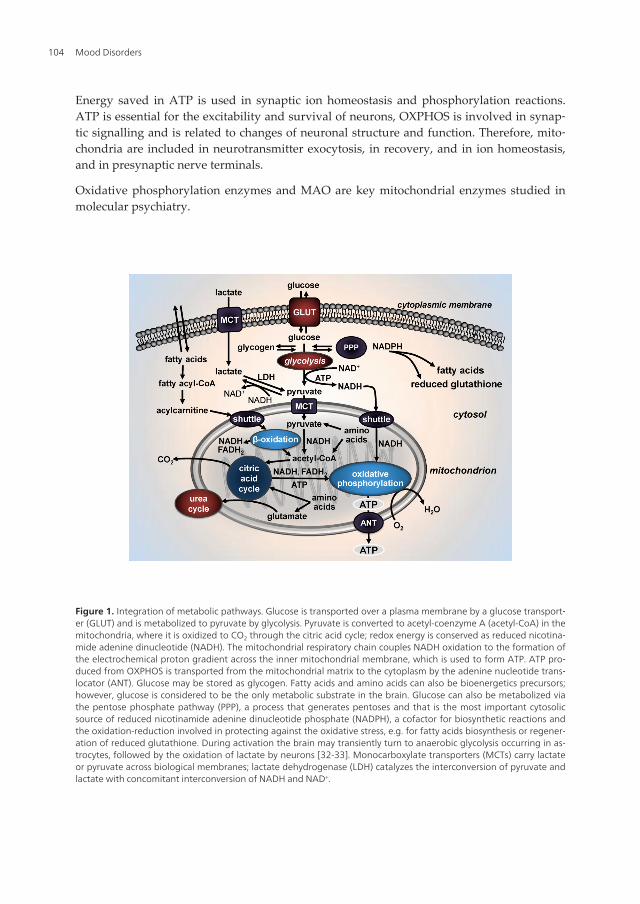
Eukaryotes synthetize ATP mainly by glycolysis in the cytosol and by OXPHOS in the mito‐chondria; i.e. the majority of cellular ATP is generated by glycolytic degradation of glucoseto pyruvate in cytosol followed by aerobic cellular respiration. When pyruvate is convertedto acetyl coenzyme A (acetyl-CoA), acetyl-CoA enters the TCA cycle and the result of thisprocess is ATP production by OXPHOS in mitochondria [29]. OXPHOS yields about 17times more ATP than glycolysis. Therefore, it is considered as the main energy source and akey element of bioenergetics [30-31]. Integration of main metabolic pathways coupled toOXPHOS is illustrated in Figure 1.
The highest number of mitochondria is present in organs demanding the most energy -brain, liver and muscles. Neurons usually utilize glucose as a source of energy. Since thebrain stores only a very small amount of glycogen, it needs a steady supply of glucose.Neurons are known to have a lower glycolytic rate than astrocytes and when stressedthey are unable to upregulate glycolysis. Following inhibition of mitochondrial respira‐tion, neurons die rapidly, whereas astrocytes utilize glycolytically generated ATP. Glucosemetabolism in neurons is directed mainly to the pentose phosphate pathway, leading toregeneration of reduced glutathione, which probably supports antioxidant controlled neu‐ron survival [32]. The regulative processes of OXPHOS are tightly related to reactive oxy‐gen species (ROS) production, integrity of mitochondrial membranes, apoptosis, andintramitochondrial Ca2+ levels. Although this is known, the control mechanisms have notyet been sufficiently investigated.
2.1. Physiology of oxidative phosphorylation
The respiratory chain is localized in cristae, structures formed by the inner mitochondrialmembrane and extending to the surface [34]. ETC consists of complexes with supramolecu‐lar organization, where mitochondrial proton pumps (complexes I, III and IV) transport pro‐tons and generate a proton gradient [31,35]. Continuously, electrons are transported tocomplex III and finally complex IV enables the conversion of O2 to H2O. Most of the ATPsynthesis comes from the electrochemical gradient across the inner membranes of mitochon‐dria by ATP synthase (complex V). The CoQ cofactor is responsible for transferring elec‐trons from complexes I and II to complex III; the second important cofactor is cytochrome c(cyt c), which transfers electrons from complex III to complex IV [36]. Both cofactors modu‐late energy and free radical production [37-38]. Processes in the inner mitochondrial mem‐brane are depicted in Figure 2.
Mitochondrial Functions in Mood Disordershttp://dx.doi.org/10.5772/53254
103
Energy saved in ATP is used in synaptic ion homeostasis and phosphorylation reactions.ATP is essential for the excitability and survival of neurons, OXPHOS is involved in synap‐tic signalling and is related to changes of neuronal structure and function. Therefore, mito‐chondria are included in neurotransmitter exocytosis, in recovery, and in ion homeostasis,and in presynaptic nerve terminals.
Oxidative phosphorylation enzymes and MAO are key mitochondrial enzymes studied inmolecular psychiatry.
Figure 1. Integration of metabolic pathways. Glucose is transported over a plasma membrane by a glucose transport‐er (GLUT) and is metabolized to pyruvate by glycolysis. Pyruvate is converted to acetyl-coenzyme A (acetyl-CoA) in themitochondria, where it is oxidized to CO2 through the citric acid cycle; redox energy is conserved as reduced nicotina‐mide adenine dinucleotide (NADH). The mitochondrial respiratory chain couples NADH oxidation to the formation ofthe electrochemical proton gradient across the inner mitochondrial membrane, which is used to form ATP. ATP pro‐duced from OXPHOS is transported from the mitochondrial matrix to the cytoplasm by the adenine nucleotide trans‐locator (ANT). Glucose may be stored as glycogen. Fatty acids and amino acids can also be bioenergetics precursors;however, glucose is considered to be the only metabolic substrate in the brain. Glucose can also be metabolized viathe pentose phosphate pathway (PPP), a process that generates pentoses and that is the most important cytosolicsource of reduced nicotinamide adenine dinucleotide phosphate (NADPH), a cofactor for biosynthetic reactions andthe oxidation-reduction involved in protecting against the oxidative stress, e.g. for fatty acids biosynthesis or regener‐ation of reduced glutathione. During activation the brain may transiently turn to anaerobic glycolysis occurring in as‐trocytes, followed by the oxidation of lactate by neurons [32-33]. Monocarboxylate transporters (MCTs) carry lactateor pyruvate across biological membranes; lactate dehydrogenase (LDH) catalyzes the interconversion of pyruvate andlactate with concomitant interconversion of NADH and NAD+.
Mood Disorders104
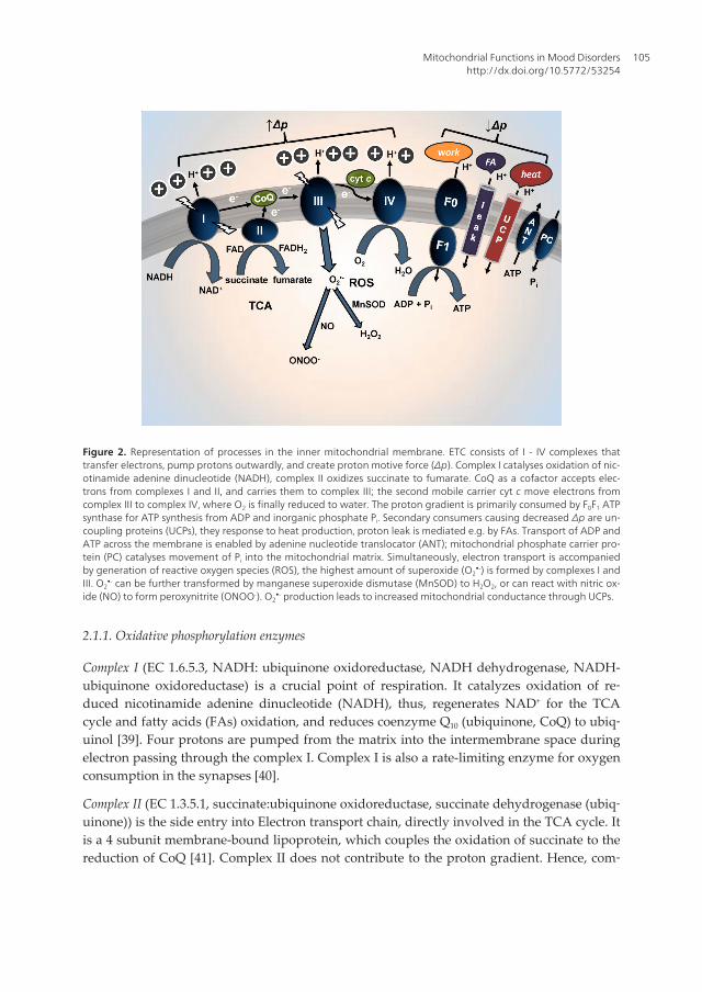
Figure 2. Representation of processes in the inner mitochondrial membrane. ETC consists of I - IV complexes thattransfer electrons, pump protons outwardly, and create proton motive force (Δp). Complex I catalyses oxidation of nic‐otinamide adenine dinucleotide (NADH), complex II oxidizes succinate to fumarate. CoQ as a cofactor accepts elec‐trons from complexes I and II, and carries them to complex III; the second mobile carrier cyt c move electrons fromcomplex III to complex IV, where O2 is finally reduced to water. The proton gradient is primarily consumed by F0F1 ATPsynthase for ATP synthesis from ADP and inorganic phosphate Pi. Secondary consumers causing decreased Δp are un‐coupling proteins (UCPs), they response to heat production, proton leak is mediated e.g. by FAs. Transport of ADP andATP across the membrane is enabled by adenine nucleotide translocator (ANT); mitochondrial phosphate carrier pro‐tein (PC) catalyses movement of Pi into the mitochondrial matrix. Simultaneously, electron transport is accompaniedby generation of reactive oxygen species (ROS), the highest amount of superoxide (O2
•-) is formed by complexes I andIII. O2
•- can be further transformed by manganese superoxide dismutase (MnSOD) to H2O2, or can react with nitric ox‐ide (NO) to form peroxynitrite (ONOO-). O2
•- production leads to increased mitochondrial conductance through UCPs.
2.1.1. Oxidative phosphorylation enzymes
Complex I (EC 1.6.5.3, NADH: ubiquinone oxidoreductase, NADH dehydrogenase, NADH-ubiquinone oxidoreductase) is a crucial point of respiration. It catalyzes oxidation of re‐duced nicotinamide adenine dinucleotide (NADH), thus, regenerates NAD+ for the TCAcycle and fatty acids (FAs) oxidation, and reduces coenzyme Q10 (ubiquinone, CoQ) to ubiq‐uinol [39]. Four protons are pumped from the matrix into the intermembrane space duringelectron passing through the complex I. Complex I is also a rate-limiting enzyme for oxygenconsumption in the synapses [40].
Complex II (EC 1.3.5.1, succinate:ubiquinone oxidoreductase, succinate dehydrogenase (ubiq‐uinone)) is the side entry into Electron transport chain, directly involved in the TCA cycle. Itis a 4 subunit membrane-bound lipoprotein, which couples the oxidation of succinate to thereduction of CoQ [41]. Complex II does not contribute to the proton gradient. Hence, com‐
Mitochondrial Functions in Mood Disordershttp://dx.doi.org/10.5772/53254
105
plex II subunits are encoded only by nDNA, complex II is suspected to normalize the activi‐ty of ETC, when mtDNA defects are suspected [42].
Complex III (EC 1.10.2.2, ubiquinol:ferricytochrome-c oxidoreductase, CoQ-cytochrome c re‐ductase) consists of two centers, Qi center - facing to matrix; and Qo center - oriented to in‐termembrane space [43]. Complex III catalyses the oxidation of one molecule of ubiquinoland the reduction of two molecules of cytochrome c. Reaction mechanism of complex III oc‐curs in two steps called the Q cycle [44]. In the process of Q cycle four protons are releasedinto the inter membrane space.
Complex IV (EC 1.9.3.1, ferrocytochrome-c:oxygen oxidoreductase, cytochrome c oxidase,COX) enables the terminal reduction of O2 to H2O, retains all partially reduced intermedi‐ates until full reduction is achieved [45]. The complex IV mediates pumping of 4 protonsacross the membrane. Previously, it was suggested as an endogenous metabolic marker forneuronal activity [46].
Complex V (EC 3.6.3.14, ATP synthase, FoF1-ATPase) consists of two regions: 1. F1 portion issoluble domain with three nucleotide binding sites, it is localized above the inner side of themembrane and stably connected with Fo domain; 2. Fo portion is proton pore embedded inthe membrane, it consists of three subunits and spans the membrane from the inner to theouter side [47-48]. This formation enables the conversion of electrochemical potential energyto chemical energy - a portion of the Fo rotates as the protons pass through the membraneand forces F1 as motor to synthetize ATP [47,49].
2.1.2. Monoamine oxidase
Monoamine oxidase (MAO, EC 1.4.3.4) is located in the outer mitochondrial membrane andcatalyses the oxidative deamination of amine neurotransmitters as well as xenobioticamines. It regulates the metabolic degradation of catecholamines and serotonin (5-hydroxy‐tryptamin, 5-HT) in neural and other target tissues. A major physiological role of intra‐neuronal MAO is to keep cytosolic monoamine concentrations very low. This membrane-bound enzyme is a flavoprotein, which use FAD as cofactor. The cofactor was identified asthe site, where irreversible inhibitors of MAO are covalently linked [50-51]. It exists in twoisoforms MAO-A and MAO-B, they differ in substrate preference, inhibitory specificity, tis‐sue and cell distribution, and in immunological properties [52]. MAO-A metabolizes 5-HTand is sensitive to inhibition by low concentrations of clorgyline, whereas MAO-B prefersbenzylamine or 2-phenylethylamine (PEA) as substrate and is sensitive to inhibition by lowconcentrations of l-deprenyl. Tyramine, tryptamine, dopamine, norepinephrine (NE) andepinephrine are equally well oxidized by both isoforms of MAO [50]. The high levels of bothforms are found in the brain; MAO-B is found in dopamine-secreting neurons in the brain.
Monoamine metabolism by MAO involves oxidative deamination to corresponding alde‐hyde and free amine. Catalysis in MAO depends on the transfer of electrons to FAD, andmechanism-based inhibitors, such as the irreversible antidepressants, modify flavin [53].The aldehyde is rapidly metabolized by aldehyde dehydrogenase to acidic metabolites. Me‐tabolism of monoamines by MAO is a major source of hydrogen peroxide (H2O2) in the
Mood Disorders106
brain. Normally the H2O2 is then inactivated by glutathione peroxidase but it can be convert‐ed, chemically, by Fe2+ ions (Fenton reaction) into the highly reactive hydroxyl radical. Thisradical has widespread deleterious effects which can cause neuronal damage and death andmay account for associated health-related problems [51,54].
MAOs have important role in brain development and function, and MAO inhibitors(MAOIs) have a range of potential therapeutic uses [53]. Generally, selective inhibitors ofMAO-A and nonselective MAOIs seem to be effective in the treatment of patients with de‐pression, panic disorder, and other anxiety disorders [55]. It is supposed that MAO-B inhibi‐tion may slow the course of various neurodegenerative disorders; so, selective inhibitors ofMAO-B may be efficacious in treating of Parkinson’s disease [56] and possibly Alzheimer'sdisease [57]. MAO-B is the sole type in human platelets and the amino acid sequences ofMAO-B in both platelets and brain are identical [58]; thus, platelet MAO can be adopted as auseful surrogate model for the study of aspects of central neuronal function related to mono‐aminergic neurotransmission [3].
2.2. Regulation of OXPHOS
There are five levels of OXPHOS regulation: 1. direct modulation of ETC kinetic parameters,2. regulation of intrinsic efficiency of OXPHOS (by changes in proton conductance, in theP/O ratio or in the channelling of ECT intermediate substrates), 3. mitochondrial networkdynamics (fusion, fission, motility, membrane lipid composition, swelling), 4. mitochondrialbiogenesis and degradation, 5. cellular and mitochondrial microenvironment [59].
OXPHOS efficiency is dependent on delivery of reducing equivalents into ETC and on ac‐tivities of participating enzymes or enzyme complexes. The optimal efficiency and flowratios are determined by control of complex I (reflects integrated cellular pathway) andcomplex II (TCA cycle precedes) [60]. Depletion of TCA cycle intermediates plays an im‐portant role in the OXPHOS flux control. In respirometry assays, supplies of complex I aswell as complex II are required. Convergent electron input and reconstitution of the TCAcycle are needed to achieve maximal respiration [30]. It is controlled also by the availabili‐ty of adenosine 5´-diphosphate (ADP) for the adenine nucleotide transporter in the innermitochondrial membrane [61].
Complex I is suggested to be responsible for adaptive changes and physiological set up ofOXPHOS efficiency [62]. The stoichiometric efficiency of OXPHOS is defined by the P/Oratio, or the amount of inorganic phosphate (Pi) incorporated into ATP per amount of con‐sumed oxygen. P/O ratio was analysed in rat brain, liver and heart mitochondria. Therewere found tissue-specific differences and dependency of the P/O ratio on the respiratoryrates with complex I, but not with complex II substrates [62]. Metabolic control analysis,which compared ETC activities and oxygen consumption rates, determined the role ofcomplex I in rat brain synaptosomes. Results of the study suggest complex I as rate-limit‐ing for oxygen consumption and responsible for high level of control over mitochondrialbioenergetics [40].
Mitochondrial Functions in Mood Disordershttp://dx.doi.org/10.5772/53254
107
As mentioned above, mitochondria exhibit transmembrane potential across the inner mem‐brane that is necessary for OXPHOS. Protons are transported outwardly and create protonmotive force (Δp), which consists of electrical part Δψm (negative inside) and chemical partΔpH [63-64]. In mitochondria, the Δp is made up of the Δψm mainly. The Δψm controls theability of the mitochondria to generate ATP, generate ROS and sequester Ca2+ entering thecell. The Δψm and ATP synthesis express a degree of coupling; optimal ATP synthesis re‐quires Δψm values between the range -100 mV and -150 mV. These values are reached pri‐marily by Δψm, which maintain at higher values (about -200 mV) and by secondary controlmechanisms, which decrease the Δψm to lower levels [49]. Changes of Δψm influence perme‐ability of biological membranes and ROS production, more negative Δψm (< -150 mV) leadsto exponentially increased permeability as well as O2
•-and H2O2 production [31]. Similarly,mitochondrial membranes increase exponentially their permeability for protons [49]. On theother hand, lower mitochondrial Δp and Δψm (e.g. caused by inhibition of respiratory chain)can result in hydrolysis of cytoplasmic ATP and slightly lower potential than that generatedby the respiratory chain [65]. Therefore, Δψm is precisely controlled and can be regulated byvarious parameters.
ATP production is controlled by different mechanisms, depending on energy demands,thermogenesis, etc. [49]. First mechanism of OXPHOS control has been called as “respiratorycontrol”, and is based on feedback mechanisms controlling the rate of ATP synthesis, first ofall by Δp and Δψm. Higher levels of ADP in mitochondria lead to stimulation of ATP syn‐thase together with decrease of Δp. Originally, pilot studies of OXPHOS dynamics used theterminology of respiratory steady states, described by Chance and Williams. Respirationwas characterized by respiratory states (Table 1), by active state 3 (ADP stimulated) and fol‐lowed by controlled state 4 (decrease after conversion of ADP to ATP) [66-67]. DecreasedP/O ratio (caused mostly by increased Δp) leads to energy waste - proton leak (slip in COX),the decrease in the coupling, and increased thermogenesis [68]. However, conception ofstates had limited applicability in intact cells and in isolated mitochondria, did not includefor instance COX, adenine nucleotide transporter, and extramitochondrial ATP/ADP ratio.
Recently, primary control has been implemented by secondary control mechanisms that are Δpindependent [49,70]. Mitochondrial Ca2+ levels have been included [31]. Ca2+ transport was pre‐sumed to be important only in buffering of cytosolic Ca2+ by acting as sink under conditions ofCa2+ overload. When the cytoplasmic Ca2+ level was overloaded, Ca2+ accumulated in mito‐chondrial matrix and utilized Δψm [65,72-73]. Nowadays it is considered that Ca2+ regulates ofactivities of dehydrogenases via phosphorylation; ATP synthesis is switched on by cAMP-de‐pendent phosphorylation and switched-off by calcium induced dephosphorylation [29,74].
In the TCA cycle, glycerophosphate dehydrogenase, pyruvate dehydrogenase, isocitrate de‐hydrogenase, and α-ketoglutarate dehydrogenase are influenced by Ca2+ levels and theirphosphorylation lead to increased ATP production, production of glycogen, and glucose ox‐idation [73]. Reversible phosphorylation of pyruvate dehydrogenase complex mediated bycalcium partly regulates the supply of reducing equivalents (NADH/NAD+ ratio). Activationof the TCA cycle enhances the NADH production that triggers movement of electrons downcomplexes I through to complex IV by initially donating of complex I [75].
Mood Disorders108
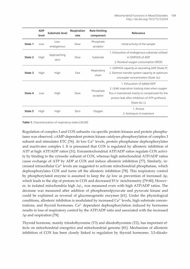
ADP
levelSubstrate level
Respiration
rate
Rate-limiting
componentRelevance
State 1 LowLow-
endogenousSlow
Phosphate
acceptorInitial activity of the sample
State 2 HighApproaching
zeroSlow Substrate
1. Exhaustion of endogenous substrate utilized
in OXPHOS of ADP
2. Residual oxygen consumption (ROX)
State 3 High High FastRespiratory
chain
1. OXPHOS capacity at saturating ADP (State P)
2. Electron transfer system capacity at optimum
uncoupler concentration (State 3u)
State 4 Low High SlowPhosphate
acceptor
1. Exhaustion of added ADP
2. LEAK respiration (resting state when oxygen
flux is maintained mainly to compensate for the
proton leak after inhibition of ATP synthesis)
(State 4o, L)
State 5 High High Zero Oxygen1. Anoxia
2. Antimycin A treatment
Table 1. Characterization of respiratory states [30,69]
Regulation of complex I and COX subunits via specific protein kinases and protein phospha‐tases was observed. cAMP-dependent protein kinase catalyses phosphorylation of complex Isubunit and stimulates ETC [76]. At low Ca2+ levels, protein phosphatase dephosphorylatesand inactivates complex I. It is presumed that COX is regulated by allosteric inhibition ofATP at high ATP/ADP ratios [31]. Extramitochondrial ATP/ADP ratios regulate COX activi‐ty by binding to the cytosolic subunit of COX, whereas high mitochondrial ATP/ADP ratioscause exchange of ATP by ADP at COX and induce allosteric inhibition [77]. Similarly, in‐creased intracellular Ca2+ levels are suggested to activate mitochondrial phosphatase, whichdephosphorylates COX and turns off the allosteric inhibition [78]. This respiratory controlby phosphorylated enzyme is assumed to keep the Δp low as prevention of increased Δp,which leads to the slip of protons in COX and decreased H+/e- stoichiometry [79-80]. Howev‐er, in isolated mitochondria high Δψm was measured even with high ATP/ADP ratios. Thedecrease was measured after addition of phosphoenolpyruvate and pyruvate kinase andcould be explained as reversal of gluconeogenetic enzymes [61]. Under the physiologicalconditions, allosteric inhibition is modulated by increased Ca2+ levels, high substrate concen‐trations, and thyroid hormones. Ca2+ dependent dephosphorylation induced by hormonesresults in loss of respiratory control by the ATP/ADP ratio and associated with the increasedΔp and respiration [79].
Thyroid hormone, mainly triiodothyronine (T3) and diiodothyronine (T2), has important ef‐fects on mitochondrial energetics and mitochondrial genome [81]. Mechanism of allostericinhibition of COX has been closely linked to regulation by thyroid hormones. 3,5-diiodo‐
Mitochondrial Functions in Mood Disordershttp://dx.doi.org/10.5772/53254
109
thyronine (T2) mediates short term effects of thyroid hormones and increases immediatelybasal metabolic rate. T2 is formed by intracellular deiodination of T3 and binds to specificT2 binding sites, which were identified in the inner mitochondrial membrane [82]. Thisbinding to subunit Va of COX abolishes the allosteric inhibition of respiration by ATP [83]that could result in partial uncoupling of OXPHOS via increased Δψm, and continue to in‐trinsic uncoupling of COX by higher membrane potentials [49]. Therefore, thyroid hor‐mones enhance the proton permeability; hyperthyroidism stimulated mitochondrial protonleak and ATP turnover in rat hepatocytes, where non-mitochondrial oxygen consumptionremained unchanged [84-85]. Oppositely, in rat hypothyroid cells significant decrease ofnon-mitochondrial oxygen consumption and proton leak were observed, ATP turnover wasunaffected [86].
2.3. Proton permeability of membranes
OXPHOS in cells is not fully efficient. Decrease of the proton gradient across the inner mito‐chondrial membrane by “proton leak” causes uncoupling of fuel oxidation from ATP gener‐ation, and some energy is lost as heat. The mechanism of the basal proton conductance ofmitochondria (insensitive to known activators and inhibitors) is not understood. There iscorrelation between mitochondrial proton conductance and composition of inner mem‐brane: phospholipid fatty acyl polyunsaturation correlates positively and monounsaturationcorrelates negatively with proton conductance [87].
Uncoupling proteins (UCPs) and adenine nucleotide translocator (ANT) are two types ofmitochondrial carrier, which cause inhibitor-sensitive inducible proton conductance. UCPsthemselves do not contribute to the basal proton conductance of mitochondria; however,they are important metabolic regulators in permitting fat oxidation and in attenuating freeradical production [88]. The amount of ANT present in the mitochondrial inner membranestrongly affects the basal proton conductance of the membrane and suggests that ANT is amajor catalyst of the basal FA-independent proton leak in mitochondria [89].
2.3.1. Fatty acids
Long-chain fatty acids (FAs) are weak acids that can cross the membrane in both protonatedand deprotonated forms. Effects of FAs are interrelated to 1. increase uncoupling, 2. increaseROS production, 3. opening mitochondrial permeability transition pores (MPTP) [90]. Fur‐ther, they can modulate effects of thyroid hormones as well as sex steroid hormones [84].FAs can act as like classic OXPHOS uncouplers with protonophoric action on the inner mito‐chondrial membrane and/or interactions of FAs with ADP carrier, COX and ATP synthaseare presumed [91]. Recent study suggests that FAs are not only inducers of uncoupling, butthey also regulate this process. It supposes that transport of FA anions participates in bothADP/ATP antiport and aspartate/glutamate antiport, at the same time [92]. On the otherhand, studies using lipid membranes suppose that FAs are capable of spontaneous flip-flop[93]. Since FAs move across the membrane spontaneously and rapidly, no protein transport‐ers are necessary. Further, coupling/uncoupling effects depend on their concentrations pHgradient across the membranes [94-95].
Mood Disorders110

2.3.2. Uncoupling proteins
Uncoupling diverts a significant proportion of energy to thermogenesis. UCPs are mito‐chondrial carriers catalysing a regulated proton leak across the inner membrane [96-97].There are five types of UCP in mammals. UCP1 is presented exclusively in the inner mito‐chondrial membrane of brown adipose tissue, and its main function is to catalyse adaptivethermogenesis [98]. It can be stimulated by FA and has synergic action of norepinephrineand thyroid hormones [49,99]. Concentrations of UCP2 and UCP3 in tissues are much lowerthan of UCP1, and their functions are not exactly known. They probably minimally contrib‐ute to basal metabolic rate, control of adaptive thermogenesis, preventive action against oxi‐dative stress and ROS control, control of cellular energy balance, regulation of Ca2+
homeostasis, regulation of FA oxidation and ATP synthesis [100-103]. UCP2, UCP4 andUCP5 are present in the central nervous system (CNS); they have been suggested to haveeffects protecting neurons from the Ca2+ overload and/or oxidative stress [104-105].
UCP activities can be positively or negatively regulated by different factors. UCP are stimu‐lated by FA and by ROS, generated by as a side reaction between CoQ and oxygen [106].UCP mediate the FA dependent proton influx that leads to uncoupled ATP synthesis andheat production [107]. It is supposed that UCP and FA decrease Δψm if it is sufficiently high.
2.4. Reactive oxygen species production
Reduction of O2 to water by aerobic respiration is accompanied by reactive intermediate for‐mation. Generally, complex I and complex III are considered as the major O2
•- sources [108].
Complex I releases O2•- to matrix, complex III can release O2
•- to both sides of the inner mito‐chondrial membrane [109]. Additionally, other ROS sources, e.g. MAO, present in the outermitochondrial membrane, and α-ketoglutarate dehydrogenase (α-KGDH), the TCA cycleenzyme complex, are able to generate H2O2. MAO catalyses the oxidative deamination ofbiogenic and xenobiotic monoamines and increases the amount of ROS in mitochondria.H2O2 production by α-KGDH is dependent on NADH/NAD+ ratio. Higher NADH leads tohigher H2O2 production, therefore, α-KGDH could significantly contribute to oxidativestress in mitochondria [110].
Physiologically generated H2O2 and O2•- from ETC are dependent on the magnitude of Δp
and the respiratory state of mitochondria [111]. State 4 is characterized with high rate ofROS production, contrary to state 3 with high rate of oxygen uptake and slow ROS produc‐tion. State 5, described as anoxic, with limited oxygen supply and lack of respiration pro‐duce minimum ROS [98,112]. In isolated rat liver mitochondria ROS production and Δψm
were studied in state 3 and state 4. These states attenuate Δψm and ROS, correlation of ROSwith Δψm was observed [113]. However, this correlation with respiratory states was not ob‐served in the study using isolated mitochondria, ROS production correlated directly withΔψm [114].
Complex I is considered to be the primary source of ROS in brain under physiological condi‐tions, as well as in pathological processes (e.g. neurodegenerative disorders). ROS seem tobe the key factors in brain aging processes and mitochondrial respiration with ROS produc‐
Mitochondrial Functions in Mood Disordershttp://dx.doi.org/10.5772/53254
111
tion significantly contributes to functional changes in brain during aging. Study in isolatedrat mitochondria found significantly increased H2O2 production and 30 % reduction of com‐plex I activity in aged rats [115]. Defective mitochondria release large amounts of ROS, simi‐larly, decline of antioxidative enzyme activities (e.g. in elderly) enhances ROS production[116]. Negative results of ROS can affect respiratory chain: complexes I, III and IV seem to bethe most affected, whereas function of complex II appears to be unchanged [117].
2.5. Apoptosis
Mitochondrial dysfunctions may accompany the clinical picture of neuropsychiatric disor‐ders and contribute to neural apoptosis [118]; mitochondria play a pivotal role in intrinsicpathway of apoptosis [38]. Several interrelated mitochondrial pathways facilitate cell death:mitochondrial permeability transition (MPT) and the release of apoptotic cell death promot‐ing factors, cytochrome c release by proapoptotic members of the Bcl-2 (B-cell lymphoma 2)family of proteins, disruption of ATP production, and alteration of the cell’s redox statusand overproduction of ROS [114]. If they are activated, change their conformations and in‐duce formation of oligomers to form mitochondrial outer membrane pores, resulting toMPT. In apoptotic cells rapid loss of mitochondrial Δψm is accompanied by ROS production.Consequently, other proapoptotic proteins cytochrome c and Smac are released and triggerthe caspase cascade leading to apoptosis [119]. Released cytochrome c in cytosol binds toapoptotic protease-activating factor-1 (Apaf-1) and induces formation of apoptosome [120].MPT means alteration of permeability properties of membranes, originally was defined asincrease of the inner mitochondrial membrane permeability to solutes of molecular massless than 1500 Da [121]. Decreased MPT and activities of respiratory chain complexes, andincreased ROS production were observed in cultured fibroblasts obtained from patientswith CoQ deficiency [37]. MPT results from formation and opening of a channel known asMPTP. MPTP is dynamic multiprotein complex that span both the outer and inner mito‐chondrial membrane and contain the adenine nucleotide translocator (ANT) in the innermembrane, and the voltage-dependent anion channels (VDAC) in the outer membrane andcyclophilin D in the matrix [122]. Once open, MPTP allows the release of pro-apoptotic fac‐tors, such as cyt c and apoptosis inducing factor (AIF), into the cytoplasm.
2.6. Specific inhibitors of complexes of ETC
Rotenone is a specific complex I inhibitor, thenoyltrifluoroacetone (TTFA) specifically inhib‐its complex II. Both substances induce O2
•- production that may result to major ROS produc‐tion [45,123-124]. Pyrrolnitrin inhibits both complex I as well as complex II. It affects electrontransport among NADH, CoQ and succinate, whereas COX remains unaffected [125].
Complex III inhibitors antimycin, myxothiazol and stigmatellin differ in their mechanism ofaction. Antimycin A inhibits the transfer of electrons from cytochrome b to CoQ, blocks theQi side of complex III. Oppositely, myxothiazol or stigmatellin block electron transfer fromreduced CoQ at Qo side [75]. Stigmatellin inhibits transfer of electrons and recycling of CoQ;myxothiazol inhibits electron transfer from reduced CoQ to cytochrome c [126].
Mood Disorders112
Complex IV inhibitors KCN and sodium azide decrease COX activity [127]. Azide specifical‐ly blocks crossover between cytochrome a and cytochrome a3. Further, it inhibits succinateoxidase activity specific for active respiration (state 3), but without any significant inhibitionof state 4 [128]. Inhibition of COX by KCN is reversible, cyanide inhibits both electron andproton transport of COX [129]. Complex V is inhibited by oligomycin, which blocks its pro‐ton channel (Fo subunit). This inhibitor increases Δψm and is used to prevent state 3 of respi‐ration. Oligomycin induces artificially state 4, i.e. state of respiration independent of ADPphosphorylation or resting state (LEAK) [130].
During the oxidation of complex I substrates (pyruvate, malate, glutamate), rotenone inhibi‐tion did not increase H2O2; contrary, oxidation of complex I and II substrates in the presenceof antimycin A increased H2O2. Both myxothiazol and stigmatellin inhibited O2
•- productionand/or should inhibit the effect of antimycin [126,131]. The maximum of O2
•- production hasbeen observed in human skin fibroblasts with the prolonged treatment of rotenone, but notwith antimycin A [132]. Interestingly, rotenone prevented antimycin A to induce ROS pro‐duction in complex I, but not in complex II [43]. Qo side of complex III was found as thesource of increased O2
•- after transient exposure to hydrogen peroxide [75]. KCN and so‐dium azide increase ROS formation [126]. Oligomycin induces hyperpolarization of innermitochondrial membrane and can increase O2
•- levels [133].
2.7. Mitochondria and neuroplasticity
Mitochondrial distribution and activity are key factors in neuronal morphogenesis - synap‐togenesis, developmental and synaptic plasticity and axogenesis. During the development,neuronal stem cells proliferate and differentiate into neurons; subsequently axons and den‐drites form synapses [134-135]. The role of mitochondria in neuroplasticity is illustrated inFigure 3 [20]. Due to ATP production and importance of mitochondria in synaptic ion ho‐meostasis and phosphorylation reactions, mitochondria would be accumulated at siteswhere ATP consumption and Ca2+ concentration are higher. It was reported that mitochon‐dria are more abundant in the regions of growing axons than in the non-growing axons. Mi‐tochondrial net movement is anterograde in growing axons and is retrograde in non-growing axons. Shortly before axogenesis mitochondria congregate at the base of the neuritethat is destined to become the axon. Nerve growth factor (NGF) was found as one of the sig‐nals inducing accumulation of mitochondria in the active growing cone [136]. Interestingly,when the ATP production is impaired and cells provide alternative source of energy, axo‐genesis is abolished although growth of dendrites remains relatively unaffected [134].
There are changes in mitochondrial energy metabolism occurring in brain cells during CNSdevelopment. During embryonic and early postnatal development fats are primarily used,later on, glucose becomes as fuel. This fact supports the role of mitochondria in biochemicalrequirements of highly proliferative neuronal stem cells and post-mitotic neurons. Duringneuronal differentiation the number of mitochondria per cell increases, but the velocity atwhich individual mitochondria move decreases as neurite outgrowth slows and synapto‐genesis occurs [20,137].
Mitochondrial Functions in Mood Disordershttp://dx.doi.org/10.5772/53254
113
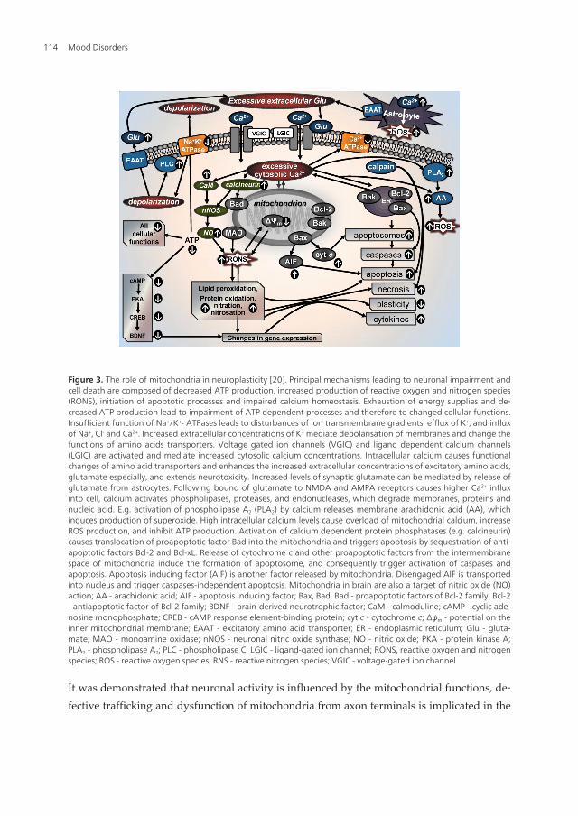
Figure 3. The role of mitochondria in neuroplasticity [20]. Principal mechanisms leading to neuronal impairment andcell death are composed of decreased ATP production, increased production of reactive oxygen and nitrogen species(RONS), initiation of apoptotic processes and impaired calcium homeostasis. Exhaustion of energy supplies and de‐creased ATP production lead to impairment of ATP dependent processes and therefore to changed cellular functions.Insufficient function of Na+/K+- ATPases leads to disturbances of ion transmembrane gradients, efflux of K+, and influxof Na+, Cl- and Ca2+. Increased extracellular concentrations of K+ mediate depolarisation of membranes and change thefunctions of amino acids transporters. Voltage gated ion channels (VGIC) and ligand dependent calcium channels(LGIC) are activated and mediate increased cytosolic calcium concentrations. Intracellular calcium causes functionalchanges of amino acid transporters and enhances the increased extracellular concentrations of excitatory amino acids,glutamate especially, and extends neurotoxicity. Increased levels of synaptic glutamate can be mediated by release ofglutamate from astrocytes. Following bound of glutamate to NMDA and AMPA receptors causes higher Ca2+ influxinto cell, calcium activates phospholipases, proteases, and endonucleases, which degrade membranes, proteins andnucleic acid. E.g. activation of phospholipase A2 (PLA2) by calcium releases membrane arachidonic acid (AA), whichinduces production of superoxide. High intracellular calcium levels cause overload of mitochondrial calcium, increaseROS production, and inhibit ATP production. Activation of calcium dependent protein phosphatases (e.g. calcineurin)causes translocation of proapoptotic factor Bad into the mitochondria and triggers apoptosis by sequestration of anti‐apoptotic factors Bcl-2 and Bcl-xL. Release of cytochrome c and other proapoptotic factors from the intermembranespace of mitochondria induce the formation of apoptosome, and consequently trigger activation of caspases andapoptosis. Apoptosis inducing factor (AIF) is another factor released by mitochondria. Disengaged AIF is transportedinto nucleus and trigger caspases-independent apoptosis. Mitochondria in brain are also a target of nitric oxide (NO)action; AA - arachidonic acid; AIF - apoptosis inducing factor; Bax, Bad, Bad - proapoptotic factors of Bcl-2 family; Bcl-2- antiapoptotic factor of Bcl-2 family; BDNF - brain-derived neurotrophic factor; CaM - calmoduline; cAMP - cyclic ade‐nosine monophosphate; CREB - cAMP response element-binding protein; cyt c - cytochrome c; Δψm - potential on theinner mitochondrial membrane; EAAT - excitatory amino acid transporter; ER - endoplasmic reticulum; Glu - gluta‐mate; MAO - monoamine oxidase; nNOS - neuronal nitric oxide synthase; NO - nitric oxide; PKA - protein kinase A;PLA2 - phospholipase A2; PLC - phospholipase C; LGIC - ligand-gated ion channel; RONS, reactive oxygen and nitrogenspecies; ROS - reactive oxygen species; RNS - reactive nitrogen species; VGIC - voltage-gated ion channel
It was demonstrated that neuronal activity is influenced by the mitochondrial functions, de‐fective trafficking and dysfunction of mitochondria from axon terminals is implicated in the
Mood Disorders114
pathogenesis of axonal degeneration [138-140]. In addition, dendritic mitochondria are es‐sential in the morphogenesis and plasticity of spines and synapses [141]. Recent findingssuggest roles for mitochondria as mediators of at least some effects of glutamate and BDNFon synaptic plasticity [136]. BDNF promotes synaptic plasticity, in part, by enhancing mito‐chondrial energy production. It increases glucose utilization and increases mitochondrialrespiratory coupling at complex [62,142].
Mitochondria are dynamic organelles; their function is modulated by fission, fusion andmoving within the axons and dendrites [38]. Their structure, functions and properties differin axons and dendrites [141,143]. Transport and positioning of mitochondria are essential forneuronal homeostasis and the mitochondrial movement is a part of regulation by intracellu‐lar signals.
3. Advances in biological hypotheses of mood disorders
Findings about intracellular processes associated with mood disorders and long-term effectsof antidepressants demonstrate an important role of signalling pathways primarily regulat‐ed by monoamine neurotransmitters; this was settled as the basis of many biochemical hy‐potheses [144-145]. While dysfunctions within monoaminergic neurotransmitter systems arelikely to play an important role in pathophysiology of mood disorders, it probably repre‐sents the downstream effects of more primary abnormalities in signal transduction. Thus,new theories about the pathophysiology of depression and the action of antidepressanttreatment proposes that mood disorders are caused by structural or functional changes inparticular molecules and signalling pathways in the brain, and that antidepressants functionby counteracting these molecular changes. It is supposed that structural and functional brainabnormalities in patients with depressive disorder may be associated with low levels ofbrain-derived neurotrophic factor (BDNF), abnormal function of hypothalamic-pituitary-adrenal (HPA) axis, glutamatergic toxicity, activation of inflammatory and cell-mediatedimmune response, decreased antioxidant capacity and increased oxidative and nitrosativestress, disturbed chronobiological rythms, and mitochondrial dysfunctions [2,146-148].
Research on the biological basis of mood disorders emphasises the changes of neural net‐works and synaptic plasticity. Evidence exists for impairment of neuroplasticity in major de‐pression. Chronic stress is known to contribute both to development of major depression invulnerable persons and to reduction of synaptic plasticity, induction of structural changes indendrites, and impairment of neurogenesis [1]. Mitochondria may be primary regulators ofthese processes, as they regulate not only neuronal survival and death, but also plasticity.There is mounting evidence for the role of mitochondrial dysfunction in the pathophysiolo‐gy and treatment of bipolar disorder [11].
3.1. Monoamine hypothesis
Discovery of the first effective antidepressants, MAOIs and tricyclic antidepressants, im‐plied hypothesis about significant role for the biogenic amine, particularly NE and 5-HT in
Mitochondrial Functions in Mood Disordershttp://dx.doi.org/10.5772/53254
115
the ethiopathogenesis of affective disorders. Classic monoamine hypothesis is an earlymilestone in the field of depression. It proposed that depression might be produced by a 5-HT or NE deficiency at functionally important receptor sites in the brain, i.e. that brainmonoamine systems have a primary direct role in depression [149-150]. Soon it became evi‐dent that the monoamine hypothesis in its original form could not explain all of the effectsof antidepressants [151-152]. In order to test this hypothesis, a series of studies was con‐ducted to evaluate effects of monoamine depletion on depressive symptoms in depressedpatients and in healthy controls. Relapse to 5-HT depletion or to catecholamine depletionwas found to be specific to the type of antidepressant treatment and type of depletion. 5-HT or NE/dopamine depletion did not decrease mood in healthy controls and slightly low‐ered mood in healthy controls with a family history of major depressive disorder. In drug-free patients with major depressive disorder in remission, a moderate mood decrease wasfound for acute tryptophan depletion only. However, acute tryptophan depletion inducedrelapse in patients in remission who used serotonergic antidepressants [153]. Depletionstudies failed to demonstrate a causal relation between 5-HT and NE with depressive dis‐order [154-155]. The effects of acute tryptophan depletion on cognition in non-vulnerableparticipants are independent of mood changes [155]. Even simultaneous disruption of 5-HT and catecholamine systems didn’t significantly alter mood in unmedicated depressedsubjects [156]. These findings forced a major revision of the classic monoamine hypothesisof depression. According to this revised monoamine theory of depression [148,157] monoa‐mine systems are only modulating other brain neurobiological systems that have more pri‐mary role in depression.
3.2. Neurotrophic hypothesis
The neurotrophic hypothesis of depression [5-6,8] supposed that vulnerability to depressioncan arise as a result of neuronal damage, e.g. after chronic stress, long-term increased levelsof glucocorticoids, hypoglycemia, ischemia, effects of neurotoxins or certain viral infections,etc. The therapeutic effects of antidepressants consist in the increased function of the nora‐drenergic or serotonergic system, leading to increased activity of transcription factor CREB(cAMP response element binding protein), higher expression of neurotrophin BDNF and itsreceptor trkB, and consequently to increased neuronal plasticity and resumption of cellularfunctions.
According to neurogenic hypothesis [158-159], depression may develop due to the de‐creased neurogenesis in hippocampus, and antidepressants takes effect through the stimula‐tion of neurogenesis. Hypothesis of cellular plasticity [160] relate the neurotrophic and theneurogenic hypothesis to the statement that depression can be generally caused by damagedcellular plasticity leading to inadequate relations between structure and function. Molecularmechanisms leading to a disturbance of neuroplasticity are not known. The bioenergetic andneurochemical model of bipolar disorder attempts to identify these mechanisms and focusesattention on mitochondrial dysfunctions [9,161].
Mood Disorders116
3.3. Inflammatory and neurodegenerative hypothesis
The central nervous system, endocrine and immune systems use neurotransmitters, cyto‐kines and hormones to communicate among them [162]. Now there is evidence that the acti‐vation of the immune system is associated with the symptoms of depression [163-164]. Theinflammatory and neurodegenerative hypothesis of depression [165] supposes that depres‐sion is associated with both inflammatory processes, as well as with neurodegeneration andreduced neurogenesis. According to this hypothesis, enhanced neurodegeneration and im‐paired neurogenesis in depression are caused by inflammatory processes, related to the pro‐duction of oxidative and nitrosative stress, tryptophan catabolites along theindoleamine-2,3-dioxygenase pathway, proinflammatory cytokines and lowered ω-3 poly‐unsaturated fatty acid status. Anti-inflammatory compounds should be able to counteract atleast partly the enhanced neurodegeneration and decreased neurogenesis.
3.4. Mitochondrial hypothesis
Mitochondrial dysfunctions (leading to decreased ATP production, oxidative stress, and in‐duction of apoptosis) occur in the early stages of different neurodegenerative diseases, asso‐ciated often with mood disorders.
The role of mitochondrial dysfunction during bipolar disorder is supported both by obser‐vation of the changes of brain metabolism and by effects of mood stabilizers (lithium andvalproate) on mitochondrial functions. Metabolic changes in brain were observed in bipolardisorder by magnetic resonance spectroscopy (MRS). It suggests the presumptions that mi‐tochondrial dysfunctions include impaired OXPHOS, final shift to glycolytic production ofenergy, general decrease of energy (decreased ATP production), changed concentrations ofphosphomonoesters and changed lipid metabolism [9].
mtDNA mutations in the brain, associations of mtDNA polymorphisms and bipolar disor‐der and changes in gene expression related to mitochondria in the brain were observed[10,166]. Mitochondrial dysfunction hypothesis of bipolar disorder is based on these obser‐vations. According to this hypothesis, mtDNA polymorphisms/mutations or mtRNA dele‐tions caused by nuclear gene mutations can cause mitochondrial dysregulation of calciumleading to symptoms of bipolar disorder [10,161,167]. Mitochondrial hypothesis correspondsto, above mentioned, neurotrophic hypothesis because of an important role of calcium sig‐nalling pathway in synaptic plasticity regulation.
3.5. Biological markers of mood disorders
Biological markers are defined as a characteristic that is objectively measured and evaluatedas an indicator of normal biologic processes, pathogenic processes, or pharmacologic re‐sponses to a therapeutic intervention. In medicine, a biomarker is an indicator of a particulardisease state or a particular state of an organism.
Identification of biologic markers of mood disorders and factors capable of predicting theresponse to treatment with antidepressants has not been sufficiently successful [3,168-169].
Mitochondrial Functions in Mood Disordershttp://dx.doi.org/10.5772/53254
117
In accordance to actual neurochemical hypotheses of mood disorders, biological markershave been primarily found at the level of neurotransmitter concentrations, their metabolitesor precursors. Subsequently, attention was shifted to the receptor systems, and since the1990´s, intracellular processes have become main interest. The chance to find sensitive andspecific biological predictors of antidepressant treatment has been increased, because of in‐troduction of new methods of molecular biology. These methods enable us better observa‐tion of cellular processes connected with the transduction of nervous signals in the brain.The choice of parameters, which should be studied as perspective biological markers ofmood disorders, have been derived first of all from new findings of signalling pathways in‐volved in neurotransmission and from above mentioned neurochemical hypotheses ofmood disorders. From the view of intracellular processes, energetic metabolism, activities ofPKC, CREB, BDNF, Bcl-2, glycogen synthase kinase-3, caspases or calcium could play aprincipal role in findings of biological markers of mood disorders. According to the com‐plexity and connectivity of signalling pathways involved in etiopathogenesis of mood disor‐ders, number of chosen parameters is not final.
4. Antidepressants, mood stabilizers and mitochondrial functions
Antidepressants are used mainly to alleviate mood disorders, such as major depression anddysthymia and anxiety disorders. Mood stabilizers are psychiatric medication used in treat‐ment of mood disorders, which are characterized by intense and sustained mood shifts (e.g.bipolar disorder).
The antidepressant activity of the first generation of antidepressants, tricyclic antidepres‐sants and MAOIs, was explained by their effects on availability of monoamine neurotrans‐mitters. The next generations of antidepressants included selective serotonin reuptakeinhibitors (SSRIs), norepinephrine reuptake inhibitors (NRIs), serotonin-norepinephrine re‐uptake inhibitors (SNRI), noradrenergic and specific serotonergic antidepressants (NaSSAs),norepinephrine-dopamine reuptake inhibitors (NDRIs), serotonin antagonist and reuptakeinhibitors (SARIs), selective serotonin reuptake enhancer (SSRE), melatonergic agonists(MASSA), sigma receptor agonists etc. The therapeutic response to antidepressants occursafter long-term treatment; therefore, effects of antidepressants are linked to cellular adapta‐tions including density and/or sensitivity of neurotransmitter receptors and transporters,regulation of signal transduction cascades, and changes in gene expression [170].
Most of mood stabilizers are anticonvulsants (valproate, carbamazepine, and lamotrigine),with an important exception of lithium, which is the oldest and the best known mood stabi‐lizing drug. Some atypical antipsychotics (olanzapine, quetiapine, aripiprazole, risperidone,ziprasidone) have mood stabilizing effects, as well.
Although a wide range of pharmacologically different antidepressants and mood stabiliz‐ers is available, molecular mechanisms of their therapeutic effects haven’t yet been suffi‐ciently clarified. Relatively little information is known about the association amongtherapeutic and/or adverse effects of drugs and mitochondrial enzyme activities. Incom‐
Mood Disorders118
plete data exist on the effect of pharmacologically selective antidepressants and mood sta‐bilizers on MAO activity. Measurement of both mitochondrial respiration and membranepotential during action of appropriate endogenous and exogenous substances enables theidentification of the primary sites of effectors and the distribution of control, allowingdeeper quantitative analyses [171].
4.1. Inhibition of MAO
MAO inhibition is the best known direct action of some antidepressants on mitochondrialenzymes. The antidepressant effect of MAOIs has been established more than 50 years ago.Iproniazid became the first MAO inhibitor to be used successfully in the treatment of de‐pression; it is an irreversible and nonselective MAO inhibitor [172]. It is known to act as apro-drug and can be converted into isopropyl hydrazine which binds covalently to MAO[173]. Clorgyline is an irreversible inhibitor preferential for MAO-A, structurally related topargyline (MAO-B inhibitor). It has antidepressant activity, and may potentially be useful inthe treatment of Parkinson's disease. Selegiline (l-deprenyl) is an irreversible inhibitor pref‐erential for MAO-B; it is used for the treatment of Parkinson's disease, depression and seniledementia. Inhibitors of MAO lose its selectivity at high doses. Moreover, there are feedbacksand interconnections of intracellular signalling pathways which lead to mutual interactionsof monoaminergic and other systems [4]. So, inhibiting of MAO-B should influence process‐es mediated primarily by substrates for MAO-A, and vice versa. The major disadvantagewas the incidence of the cheese reaction with those early inhibitors [51].
The selective reversible MAO-A inhibitors such as moclobemide increase the content of 5-HT, NE and dopamine in the brain [174] but did not provoke the cheese reaction. Moclo‐bemide has been extensively evaluated in the treatment of a wide spectrum of depressivedisorders and social phobia. Overall, moclobemide appears to be safe and devoid of ma‐jor side effects, although it is considered as a mild antidepressant, better tolerated by old‐er patients [175-181]. Moclobemide undergoes extensive metabolism with less than 1 % ofthe dose being excreted unchanged. Metabolic pathways of moclobemide include mainlyoxidative attack on the morpholine moiety [182]. However, major metabolites in plasmawere found to be less effective MAO-A inhibitors than moclobemide or pharmacologicallyinactive [183-184].
MAO inhibitors were developed as antidepressants but many drugs, including the oxazoli‐dinone antibacterial agents, share similar molecular properties and have MAO inhibitory ac‐tivity. These compounds were of interest as potential antidepressants because they could beselective inhibitors of either the A or B isoforms and were usually reversible [53].
Antidepressants which act primarily as 5-HT and/or NE reuptake inhibitors show inhibitoryactivity towards MAO also. It has been suggested that tricyclic antidepressants exert someof their therapeutic effect by inhibiting MAO [185]. They are able to inhibit MAO-B both invitro [186-187] and in vivo [188-189]. However, in vivo inhibition of the human platelet MAO-B in the patients taking tricyclic antidepressants was not confirmed by others [190-191]. Fivetricyclic antidepressants, amitriptyline, clomipramine, desipramine, imipramine and iprin‐dole, have comparable potencies as inhibitors of MAO in rodent brain and liver [192]. These
Mitochondrial Functions in Mood Disordershttp://dx.doi.org/10.5772/53254
119
antidepressants have been shown to partially protect mouse brain MAO in vivo from the ir‐reversible enzyme inhibition produced by subsequent injection of phenelzine [193]. Concen‐trations of tricyclic antidepressants, which showed a pronounced inhibitory effect on theMAOs activity, were significantly higher than plasma levels of the drug found under thera‐peutic conditions [194-195]. MAO activity was inhibited after long-term administration ofviloxazine, nomifensine, zimelidine, maprotiline, imipramine, amitriptyline, and nortripty‐line in systematic studies of Egashira [196-197]. Competitive inhibition of MAO-A and non‐competitive inhibition of MAO-B was found for these drugs. Similar results were obtainedwhen different tricyclic antidepressants and SSRIs were examined with isolated rat brainmitochondria [198]. Fluoxetine and norfluoxetine showed affinities both for MAO-A [199]and MAO-B [200]. Fluoxetine and norfluoxetine also significantly inhibited the binding ofthe specific radioligands to MAO in vivo. These results support a potential role of MAO in‐hibition in the therapeutic effects of fluoxetine.
4.2. Effects of antidepressants on mitochondrial functions
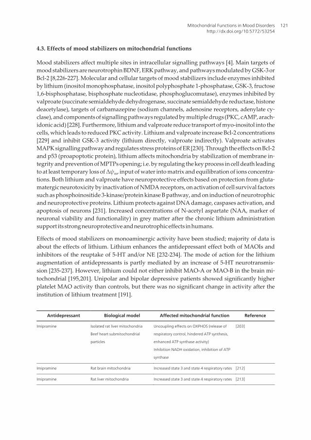
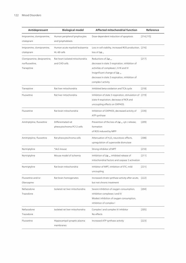
There is relatively little data about effects of antidepressants on mitochondrial functions as sum‐marized in the Table 2. In vitro study examined influence of pharmacologically different antide‐pressants and mood stabilizers on activity both mitochondrial MAO [201] and respiratory chaincomplexes; imipramine, desipramine, amitriptyline, citalopram, and mirtazapine were foundas complex I inhibitors in isolated pig brain mitochondria [202]. In isolated rat liver mitochon‐dria effects of imipramine and clomipramine were compared to classic uncouplers, drugs en‐hanced ATP synthase activity, hindered ATP synthesis and released respiratory control [203]. Inisolated rat liver mitochondria, nefazodone was found as inhibitor of mitochondrial complexes Iand IV; buspirone inhibited complex I but had no effect on complex IV. Trazodone did not affecton both complex I and complex IV [204], but decreased oxygen consumption and reduced Na+, K+-ATPase activity. Trazodone acts also as uncoupler of OXPHOS [205].
Effects of antidepressants on apoptotic markers, e.g. cytochrome c release and DNA frag‐mentation, seem to be different. Various antidepressants exhibited potential anticancerproperties and caused cytotoxic effects. Paroxetine, fluoxetine and clomipramine increasedlevels of apoptotic markers leading to apoptosis in glioma and neuroblastoma cells, whereasimipramine and mianserin do not [206]. Desipramine induced apoptosis in rat glioma cellsby activation of caspases, without any change of mitochondrial membrane potential Δψm
[207]. Fluoxetine and amitriptyline protected PC12 cells from cell death induced by hydro‐gen peroxide [208]. Amitriptyline and tranylcypromine prevented the loss of mitochondrialΔψm, over expression of Bax, reduction in Bcl-2 level, cytochrome c release, caspase-3 activa‐tion, and formation of ROS. In contrast, fluoxetine seemed to have additive toxic effect to 1-methyl-4-phenylpyridinium (MPP+) against neuronal cell damage by increasingmitochondrial damage and oxidative stress [209]. Nortriptyline was identified as strong in‐hibitor of MPT and was observed as potential inhibitor of neuronal cell death; it protectedisolated mitochondria against programmed cell death, inhibited release of apoptotic mito‐chondrial factors and caspases, increased Ca2+ retention in mitochondria and delayed theCa2+ induced loss of Δψm, further leading to neuronal cell death [210-211].
Mood Disorders120
4.3. Effects of mood stabilizers on mitochondrial functions
Mood stabilizers affect multiple sites in intracellular signalling pathways [4]. Main targets ofmood stabilizers are neurotrophin BDNF, ERK pathway, and pathways modulated by GSK-3 orBcl-2 [8,226-227]. Molecular and cellular targets of mood stabilizers include enzymes inhibitedby lithium (inositol monophosphatase, inositol polyphosphate 1-phosphatase, GSK-3, fructose1,6-bisphosphatase, bisphosphate nucleotidase, phosphoglucomutase), enzymes inhibited byvalproate (succinate semialdehyde dehydrogenase, succinate semialdehyde reductase, histonedeacetylase), targets of carbamazepine (sodium channels, adenosine receptors, adenylate cy‐clase), and components of signalling pathways regulated by multiple drugs (PKC, cAMP, arach‐idonic acid) [228]. Furthermore, lithium and valproate reduce transport of myo-inositol into thecells, which leads to reduced PKC activity. Lithium and valproate increase Bcl-2 concentrations[229] and inhibit GSK-3 activity (lithium directly, valproate indirectly). Valproate activatesMAPK signalling pathway and regulates stress proteins of ER [230]. Through the effects on Bcl-2and p53 (proapoptotic protein), lithium affects mitochondria by stabilization of membrane in‐tegrity and prevention of MPTPs opening; i.e. by regulating the key process in cell death leadingto at least temporary loss of Δψm, input of water into matrix and equilibration of ions concentra‐tions. Both lithium and valproate have neuroprotective effects based on protection from gluta‐matergic neurotoxicity by inactivation of NMDA receptors, on activation of cell survival factorssuch as phosphoinositide 3-kinase/protein kinase B pathway, and on induction of neurotrophicand neuroprotective proteins. Lithium protects against DNA damage, caspases activation, andapoptosis of neurons [231]. Increased concentrations of N-acetyl aspartate (NAA, marker ofneuronal viability and functionality) in grey matter after the chronic lithium administrationsupport its strong neuroprotective and neurotrophic effects in humans.
Effects of mood stabilizers on monoaminergic activity have been studied; majority of data isabout the effects of lithium. Lithium enhances the antidepressant effect both of MAOIs andinhibitors of the reuptake of 5-HT and/or NE [232-234]. The mode of action for the lithiumaugmentation of antidepressants is partly mediated by an increase of 5-HT neurotransmis‐sion [235-237]. However, lithium could not either inhibit MAO-A or MAO-B in the brain mi‐tochondrial [195,201]. Unipolar and bipolar depressive patients showed significantly higherplatelet MAO activity than controls, but there was no significant change in activity after theinstitution of lithium treatment [191].
Antidepressant Biological model Affected mitochondrial function Reference
Imipramine Isolated rat liver mitochondria
Beef heart submitochondrial
particles
Uncoupling effects on OXPHOS (release of
respiratory control, hindered ATP synthesis,
enhanced ATP synthase activity)
Inhibition NADH oxidation, inhibition of ATP
synthase
[203]
Imipramine Rat brain mitochondria Increased state 3 and state 4 respiratory rates [212]
Imipramine Rat liver mitochondria Increased state 3 and state 4 respiratory rates [213]
Mitochondrial Functions in Mood Disordershttp://dx.doi.org/10.5772/53254
121
Antidepressant Biological model Affected mitochondrial function Reference
Imipramine, clomipramine,
citalopram
Human peripheral lymphocytes
and lymphoblasts
Dose-dependent induction of apoptosis [214,215]
Imipramine, clomipramine,
citalopram
Human acute myeloid leukaemia
HL-60 cells
Loss in cell viability, increased ROS production,
loss of Δψ m
[216]
Clomipramine, desipramine,
norfluoxetine,
Tianeptine
Rat heart isolated mitochondria
and CHO cells
Reductions of Δψ m,
decrease in state 3 respiration, inhibition of
activities of complexes I, II/III and IV
Insignificant change of Δψ m,
decrease in state 3 respiration, inhibition of
complex I activity
[217]
Tianeptine Rat liver mitochondria Inhibited beta-oxidation and TCA cycle [218]
Fluoxetine Rat liver mitochondria Inhibition of state 3 respiration, stimulation of
state 4 respiration, decrease of RCR and
uncoupling effects on OXPHOS
[219]
Fluoxetine Rat brain mitochondria Inhibition of OXPHOS, decreased activity of
ATP synthase
[220]
Amitriptyline, fluoxetine Differentiated rat
pheocytochroma PC12 cells
Prevention of the loss of Δψ m, cyt c release,
formation
of ROS induced by MPP+
[209]
Amitriptyline, fluoxetine Rat pheocytochroma cells Attenuation of H2O2 neurotoxic effects,
upregulation of superoxide dismutase
[208]
Nortriptyline *ALS mouse Strong inhibitor of MPT [210]
Nortriptyline Mouse model of ischemia Inhibition of Δψ m, inhibited release of
mitochondrial factors and caspase 3 activation
[211]
Nortriptyline Rat brain mitochondria Inhibitor of MPT, inhibition of ETC, mild
uncoupling
[221]
Fluoxetine and/or
Olanzapine
Rat brain homogenates Increased citrate synthase activity after acute,
but not chronic treatment
[222]
Nefazodone
Trazodone
Isolated rat liver mitochondria Severe inhibition of oxygen consumption,
inhibition complexes I and IV
Modest inhibition of oxygen consumption,
inhibition of complex I
[204]
Nefazodone
Trazodone
Isolated rat liver mitochondria Complex I and complex IV inhibitor
No effects
[205]
Fluoxetine Hippocampal synaptic plasma
membranes
Increased ATP synthase activity [223]
Mood Disorders122
Antidepressant Biological model Affected mitochondrial function Reference
Sertraline Isolated rat liver mitochondria Uncoupling effects on OXPHOS, inhibition of
complex I and complex V activities, induction
of Ca2+ mediated MPT
[224]
Venlafaxine, paroxetine,
nortriptiline
Rat brain homogenates (after 15
days of drug administration)
Differences in brain areas: increased or
unchanged citrate synthase and SDH activities
[225]
Paroxetine, fluoxetine,
klomipramine
Rat glioma and human
neuroblastoma cell lines
Increased cyt c release, caspase-3-like activity,
induction of apoptosis
[206]
Desipramine Rat glioma cells Activation of caspases 3 and 9, no changes of
Δψ m
[207]
ALS mouse – model of neurodegeneration
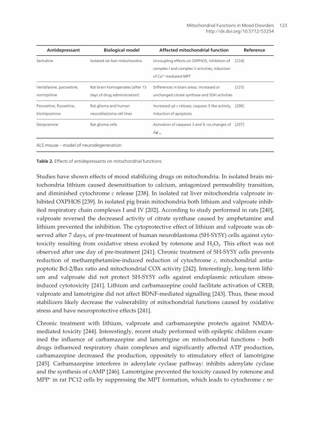
Table 2. Effects of antidepressants on mitochondrial functions
Studies have shown effects of mood stabilizing drugs on mitochondria. In isolated brain mi‐tochondria lithium caused desensitisation to calcium, antagonized permeability transition,and diminished cytochrome c release [238]. In isolated rat liver mitochondria valproate in‐hibited OXPHOS [239]. In isolated pig brain mitochondria both lithium and valproate inhib‐ited respiratory chain complexes I and IV [202]. According to study performed in rats [240],valproate reversed the decreased activity of citrate synthase caused by amphetamine andlithium prevented the inhibition. The cytoprotective effect of lithium and valproate was ob‐served after 7 days, of pre-treatment of human neuroblastoma (SH-SY5Y) cells against cyto‐toxicity resulting from oxidative stress evoked by rotenone and H2O2. This effect was notobserved after one day of pre-treatment [241]. Chronic treatment of SH-SY5Y cells preventsreduction of methamphetamine-induced reduction of cytochrome c, mitochondrial antia‐poptotic Bcl-2/Bax ratio and mitochondrial COX activity [242]. Interestingly, long-term lithi‐um and valproate did not protect SH-SY5Y cells against endoplasmic reticulum stress-induced cytotoxicity [241]. Lithium and carbamazepine could facilitate activation of CREB,valproate and lamotrigine did not affect BDNF-mediated signalling [243]. Thus, these moodstabilizers likely decrease the vulnerability of mitochondrial functions caused by oxidativestress and have neuroprotective effects [241].
Chronic treatment with lithium, valproate and carbamazepine protects against NMDA-mediated toxicity [244]. Interestingly, recent study performed with epileptic children exam‐ined the influence of carbamazepine and lamotrigine on mitochondrial functions - bothdrugs influenced respiratory chain complexes and significantly affected ATP production,carbamazepine decreased the production, oppositely to stimulatory effect of lamotrigine[245]. Carbamazepine interferes in adenylate cyclase pathway: inhibits adenylate cyclaseand the synthesis of cAMP [246]. Lamotrigine prevented the toxicity caused by rotenone andMPP+ in rat PC12 cells by suppressing the MPT formation, which leads to cytochrome c re‐
Mitochondrial Functions in Mood Disordershttp://dx.doi.org/10.5772/53254
123
lease and subsequent apoptosis. Though, lamotrigine seems to have neuroprotective effectdue to the mitochondrial respiratory complex I inhibition [247].
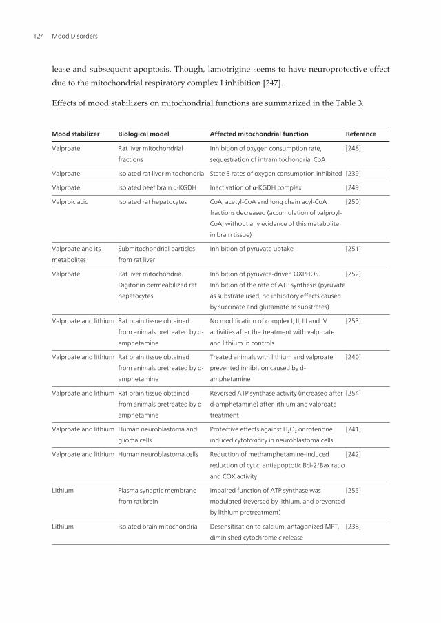
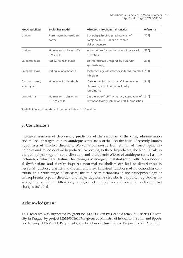
Effects of mood stabilizers on mitochondrial functions are summarized in the Table 3.
Mood stabilizer Biological model Affected mitochondrial function Reference
Valproate Rat liver mitochondrial
fractions
Inhibition of oxygen consumption rate,
sequestration of intramitochondrial CoA
[248]
Valproate Isolated rat liver mitochondria State 3 rates of oxygen consumption inhibited [239]
Valproate Isolated beef brain α-KGDH Inactivation of α-KGDH complex [249]
Valproic acid Isolated rat hepatocytes CoA, acetyl-CoA and long chain acyl-CoA
fractions decreased (accumulation of valproyl-
CoA; without any evidence of this metabolite
in brain tissue)
[250]
Valproate and its
metabolites
Submitochondrial particles
from rat liver
Inhibition of pyruvate uptake [251]
Valproate Rat liver mitochondria.
Digitonin permeabilized rat
hepatocytes
Inhibition of pyruvate-driven OXPHOS.
Inhibition of the rate of ATP synthesis (pyruvate
as substrate used, no inhibitory effects caused
by succinate and glutamate as substrates)
[252]
Valproate and lithium Rat brain tissue obtained
from animals pretreated by d-
amphetamine
No modification of complex I, II, III and IV
activities after the treatment with valproate
and lithium in controls
[253]
Valproate and lithium Rat brain tissue obtained
from animals pretreated by d-
amphetamine
Treated animals with lithium and valproate
prevented inhibition caused by d-
amphetamine
[240]
Valproate and lithium Rat brain tissue obtained
from animals pretreated by d-
amphetamine
Reversed ATP synthase activity (increased after
d-amphetamine) after lithium and valproate
treatment
[254]
Valproate and lithium Human neuroblastoma and
glioma cells
Protective effects against H2O2 or rotenone
induced cytotoxicity in neuroblastoma cells
[241]
Valproate and lithium Human neuroblastoma cells Reduction of methamphetamine-induced
reduction of cyt c, antiapoptotic Bcl-2/Bax ratio
and COX activity
[242]
Lithium Plasma synaptic membrane
from rat brain
Impaired function of ATP synthase was
modulated (reversed by lithium, and prevented
by lithium pretreatment)
[255]
Lithium Isolated brain mitochondria Desensitisation to calcium, antagonized MPT,
diminished cytochrome c release
[238]
Mood Disorders124
Mood stabilizer Biological model Affected mitochondrial function Reference
Lithium Postmortem human brain
cortex
Dose-depedent increased activities of
complexes I+III, II+III and succinate
dehydrogenase
[256]
Lithium Human neuroblastoma SH-
SY5Y cells
Attenuation of rotenone-induced caspase-3
activation
[257]
Carbamazepine Rat liver mitochondria Decreased state 3 respiration, RCR, ATP
synthesis, Δψ m
[258]
Carbamazepine Rat brain mitochondria Protection against rotenone induced complex I
inhibition
[259]
Carbamazepine,
lamotrigine
Human white blood cells Carbamazepine decreased ATP production,
stimulatory effect on production by
lamotrigine
[245]
Lamotrigine Human neuroblastoma
SH-SY5Y cells
Suppression of MPT formation, attenuation of
rotenone-toxicity, inhibition of ROS production
[247]
Table 3. Effects of mood stabilizers on mitochondrial functions
5. Conclusions
Biological markers of depression, predictors of the response to the drug administrationand molecular targets of new antidepressants are searched on the basis of recently knownhypotheses of affective disorders. We come out mostly from stimuli of neurotrophic hy‐pothesis and mitochondrial hypothesis. According to these hypotheses, the leading role inthe pathophysiology of mood disorders and therapeutic effects of antidepressants has mi‐tochondria, which are destined for changes in energetic metabolism of cells. Mitochondri‐al dysfunctions and thereby impaired neuronal metabolism can lead to disturbances inneuronal function, plasticity and brain circuitry. Impaired functions of mitochondria con‐tribute to a wide range of diseases; the role of mitochondria in the pathophysiology ofschizophrenia, bipolar disorder, and major depressive disorder is supported by studies in‐vestigating genomic differences, changes of energy metabolism and mitochondrialchanges included.
Acknowledgment
This. research was supported by grant no. 41310 given by Grant Agency of Charles Univer‐sity in Prague, by project MSM0021620849 given by Ministry of Education, Youth and Sportsand by project PRVOUK-P26/LF1/4 given by Charles University in Prague, Czech Republic.
Mitochondrial Functions in Mood Disordershttp://dx.doi.org/10.5772/53254
125
Author details
Jana Hroudová, Zdeněk Fišar and Jiří Raboch
*Address all correspondence to: [email protected]
Department of Psychiatry, First Faculty of Medicine, Charles University in Prague andGeneral University Hospital in Prague, Prague, Czech Republic
References
[1] Pittenger C, Duman RS. Stress, depression, and neuroplasticity: a convergence ofmechanisms. Neuropsychopharmacology 2008;33(1): 88-109.
[2] Maes M, Fišar Z, Medina M, Scapagnini G, Nowak G, Berk M. New drug targets indepression: inflammatory, cell-mediated immune, oxidative and nitrosative stress,mitochondrial, antioxidant, and neuroprogressive pathways. And new drug candi‐dates-Nrf2 activators and GSK-3 inhibitors. Inflammopharmacology 2012;20(3):127-150.
[3] Fišar Z, Raboch J. Depression, antidepressants, and peripheral blood components.Neuro Endocrinol Lett 2008;29(1): 17-28.
[4] Fišar Z, Hroudová J. Intracellular signalling pathways and mood disorders. Folia Bi‐ol 2010b;56(4): 135-148.
[5] Duman RS, Heninger GR, Nestler, EJ. A molecular and cellular theory of depression.Arch Gen Psychiatry 1997;54(7): 597-606.
[6] Duman RS. Synaptic plasticity and mood disorders. Mol Psychiatry 2002;7(Suppl 1):S29-S34.
[7] Zarate CA Jr, Singh J, Manji HK. Cellular plasticity cascades: targets for the develop‐ment of novel therapeutics for bipolar disorder. Biol Psychiatry 2006;59(11):1006-1020.
[8] Einat, H, Manji HK. Cellular plasticity cascades: genes-to-behavior pathways in ani‐mal models of bipolar disorder. Biol Psychiatry 2006;59(12): 1160-1171.
[9] Stork C, Renshaw RF. Mitochondrial dysfunction in bipolar disorder: evidence frommagnetic resonance spectroscopy research. Mol Psychiatry 2005;10(10): 900-919.
[10] Kato T. Role of mitochondrial DNA in calcium signaling abnormality in bipolar dis‐order. Cell Calcium 2008;44(1): 92-102.
[11] Quiroz JA, Gray NA, Kato T, Manji HK. Mitochondrially mediated plasticity in thepathophysiology and treatment of bipolar disorder. Neuropsychopharmacology2008;33(11): 2551-2565.
Mood Disorders126
[12] Shao L, Martin MV, Watson SJ, Schatzberg A, Akil H, Myers RM, Jones EG, BunneyWE, Vawter MP. Mitochondrial involvement in psychiatric disorders. Ann Med2008;40(4): 281-295.
[13] Rezin GT, Amboni G, Zugno AI, Quevedo J, Streck EL. Mitochondrial dysfunctionand psychiatric disorders. Neurochem Res 2009;34(6): 1021-1029.
[14] Jou SH, Chiu NY, Liu CS. Mitochondrial dysfunction and psychiatric disorders.Chang Gung Med J 2009;32(4): 370-379.
[15] Orth M, Schapira AH. Mitochondria and degenerative disorders. Am J Med Genet2001;106(1): 27-36.
[16] Schapira AHV. Mitochondrial diseases. Lancet 2012;379(9828): 1825-1834.
[17] Vélot C, Srere PA. Reversible transdominant inhibition of a metabolic pathway. Invivo evidence of interaction between two sequential tricarboxylic acid cycle enzymesin yeast. J Biol Chem 2000;275(17): 12926-12933.
[18] Sriram G, Martinez JA, McCabe ER, Liao JC, Dipple KM. Single-gene disorders: whatrole could moonlighting enzymes play? Am J Hum Genet 2005;76(6): 911-924.
[19] Fišar Z, Hroudová J. Common aspects of neuroplasticity, mood disorders and mito‐chondrial functions. Act Nerv Super Rediviva 2010a;52(1): 3-20.
[20] Hroudova J, Fisar Z. Connectivity between mitochondrial functions and psychiatricdisorders. Psychiatry Clin Neurosci 2011;65(2): 130-141.
[21] Scheffler IE. Molecular genetics of succinate: quinone oxidoreductase in eukaryotes.Prog Nucleic Acid Res Mol Biol 1998;60: 267-315.
[22] Müller U, Troidl C, Niemann S. SDHC mutations in hederitary paraganglioma/pheo‐chromocytoma. Fam Cancer 2005;4(1): 9-12.
[23] Reinecke F, Smeitink JA, van der Westhuizen FH. OXPHOS gene expression and con‐trol in mitochondrial disorders. Biochim Biophys Acta 2009;1792(12): 1113-1121.
[24] Cannino G, Di Liegro CM, Rinaldi AM. Nuclear-mitochondrial interaction. Mito‐chondrion 2007;7(6): 359-366.
[25] Hovatta I, Juhila J, Donner J. Oxidative stress in anxiety and comorbid disorders.Neurosci Res 2010;68(4): 261-275.
[26] Moghaddam B. Stress activation of glutamate neurotransmission in the prefrontalcortex: implications for dopamine-associated psychiatric disorders. Biol Psychiatry2002;51(10): 775-787.
[27] Atlante A, Calissano P, Bobba A, Giannattasio S, Marra E, Passarella S. Glutamateneurotoxicity, oxidative stress and mitochondria. FEBS Lett 2001;497(1): 1-5.
[28] Lipton P. Ischemic cell death in brain neurons. Physiol Rev 1999;79(4): 1431-1568.
Mitochondrial Functions in Mood Disordershttp://dx.doi.org/10.5772/53254
127
[29] Ludwig B, Bender E, Arnold S, Hüttemann M, Lee I, Kadenbach B. Cytochrome C ox‐idase and the regulation of oxidative phosphorylation. Chembiochem 2001;2(6):392-403.
[30] Gnaiger E. Capacity of oxidative phosphorylation in human skeletal muscle: newperspectives of mitochondrial physiology. Int J Biochem Cell Biol 2009;41(10):1837-1845.
[31] Kadenbach B, Ramzan R, Wen L, Vogt S. New extension of the Mitchell Theory foroxidative phosphorylation in mitochondria of living organisms. Biochim Biophys Ac‐ta 2010;1800(3): 205-212.
[32] Bolaños JP, Almeida A, Moncada S. Glycolysis: a bioenergetic or a survival pathway?Trends Biochem Sci 2010;35(3): 145-149.
[33] Magistretti PJ, Pellerin L. Cellular mechanisms of brain energy metabolism and theirrelevance to functional brain imaging. Philos Trans R Soc Lond B Biol Sci1999;354(1387): 1155-1163.
[34] Vonck J, Schäfer E. Supramolecular organization of protein complexes in the mito‐chondrial inner membrane. Biochim Biophys Acta 2009;1793(1): 117-124.
[35] Bunoust O, Devin A, Avéret N, Camougrand N, Rigoulet M. Competition of elec‐trons to enter the respiratory chain: a new regulatory mechanism of oxidative metab‐olism in Saccharomyces cerevisiae. J Biol Chem 2005;280(5): 3407-3413.
[36] Solmaz SR, Hunte C. Structure of complex III with bound cytochrome c in reducedstate and definition of a minimal core interface for electron transfer. J Biol Chem2008;283(25): 17542-17549.
[37] Rodríguez-Hernández A, Cordero MD, Salviati L, Artuch R, Pineda M, Briones P,Gómez Izquierdo L, Cotán D, Navas P, Sánchez-Alcázar JA. Coenzyme Q deficiencytriggers mitochondria degradation by mitophagy. Autophagy 2009;5(1): 19-32.
[38] Mattson MP, Gleichmann M, Cheng A. Mitochondria in neuroplasticity and neuro‐logical disorders. Neuron 2008;60(5): 748-766.
[39] Hirst J. Towards the molecular mechanism of respiratory complex I. Biochem J2009;425(2): 327-339.
[40] Telford JE, Kilbride SM, Davey GP. Complex I is rate-limiting for oxygen consump‐tion in the nerve terminal. J Biol Chem 2009;284(14): 9109-9114.
[41] Tomitsuka E, Kita K, Esumi H. Regulation of succinate-ubiquinone reductase and fu‐marate reductase activities in human complex II by phosphorylation of its flavopro‐tein subunit. Proc Jpn Acad Ser B Phys Biol Sci 2009;85(7): 258-265.
[42] Saada A, Shaag A, Mandel H, Nevo Y, Eriksson S, Elpeleg O. Mutant mitochondrialthymidine kinase in mitochondrial DNA depletion myopathy. Nat Genet 2001;29(3):342-344.
Mood Disorders128
[43] Chen Q, Vazquez EJ, Moghaddas S, Hoppel CL, Lesnefsky EJ. Production of reactiveoxygen species by mitochondria. Central role of complex III. J Biol Chem2003;278(38): 36027-36031.
[44] Trumpower BL. The protonmotive Q cycle. Energy transduction by coupling of pro‐ton translocation to electron transfer by the cytochrome bc1 complex. J Biol Chem1990;265(20): 11409-11412.
[45] Turrens JF. Mitochondrial formation of reactive oxygen species. J Physiol 2003;552(Pt2): 335-344.
[46] Wong-Riley MT. Cytochrome oxidase: an endogenous metabolic marker for neuronalactivity. Trends Neurosci 1989;12(3): 94-101.
[47] Noji H, Yoshida M. The rotary machine in the cell, ATP synthase.J Biol Chem2001;276(3): 1665-1668.
[48] Zanotti F, Gnoni A, Mangiullo R, Papa S. Effect of the ATPase inhibitor protein IF1 onH+ translocation in the mitochondrial ATP synthase complex. Biochem Biophys ResCommun 2009;384(1): 43-48.
[49] Kadenbach B. Intrinsic and extrinsic uncoupling of oxidative phosphorylation. Bio‐chim Biophys Acta 2003;1604(2): 77-94.
[50] Youdim MB, Bakhle YS. Monoamine oxidase: isoforms and inhibitors in Parkinson'sdisease and depressive illness. Br J Pharmacol 2006;147(Suppl 1): S287-S296.
[51] Youdim MB, Edmondson D, Tipton KF. The therapeutic potential of monoamine oxi‐dase inhibitors. Nat Rev Neurosci 2006;7(4): 295-309.
[52] Bach AWJ, Lan NC, Johnson DL, Abell CW, Bembenek ME, Kwan S-W, Seeburg PH,Shih JC. cDNA cloning of human liver monoamine oxidase A and B: molecular basisof differences in enzymatic properties. Proc Natl Acad Sci U S A 1988;85(13):4934-4938.
[53] Ramsay RR, Gravestock MB. Monoamine oxidases: to inhibit or not to inhibit. MiniRev Med Chem 2003;3(2): 129-136.
[54] Naoi M, Maruyama W, Akao Y, Yi H, Yamaoka Y. Involvement of type A monoa‐mine oxidase in neurodegeneration: regulation of mitochondrial signaling leading tocell death or neuroprotection. J Neural Transm Suppl 2006;(71): 67-77.
[55] Stahl SM, Felker A. Monoamine oxidase inhibitors: a modern guide to an unrequitedclass of antidepressants. CNS Spectr 2008;13(10): 855-870.
[56] Horstink M, Tolosa E, Bonuccelli U, Deuschl G, Friedman A, Kanovsky P, Larsen JP,Lees A, Oertel W, Poewe W, Rascol O, Sampaio C, European Federation of Neurolog‐ical Societies, Movement Disorder Society-European Section. Review of the therapeu‐tic management of Parkinson's disease. Report of a joint task force of the EuropeanFederation of Neurological Societies and the Movement Disorder Society-European
Mitochondrial Functions in Mood Disordershttp://dx.doi.org/10.5772/53254
129
Section. Part I: early (uncomplicated) Parkinson's disease. Eur J Neurol 2006;13(11):1170-1185.
[57] Riederer P, Danielczyk W, Grünblatt E. Monoamine oxidase-B inhibition in Alzheim‐er's disease. NeuroToxicology 2004;25(1-2): 271-277.
[58] Chen K, Wu HF, Shih JC. The deduced amino acid sequences of human platelet andfrontal cortex monoamine oxidase B are identical. J Neurochem 1993;61(1): 187-190.
[59] Benard G, Bellance N, Jose C, Melser S, Nouette-Gaulain K, Rossignol R. Multi-sitecontrol and regulation of mitochondrial energy production. Biochim Biophys Acta2010;1797(6-7): 698-709.
[60] Cairns CB, Walther J, Harken AH, Banerjee A. Mitochondrial oxidative phosphoryla‐tion thermodynamic efficiencies reflect physiological organ roles. Am J Physiol1998;274(5 Pt 2): R1376-R1383.
[61] Ramzan R, Staniek K, Kadenbach B, Vogt S. Mitochondrial respiration and mem‐brane potential are regulated by the allosteric ATP-inhibition of cytochrome c oxi‐dase. Biochim Biophys Acta 2010;1797(9): 1672-1680.
[62] Cocco T, Pacelli C, Sgobbo P, Villani G. Control of OXPHOS efficiency by complex Iin brain mitochondria. Neurobiol Aging 2009;30(4): 622-629.
[63] Mitchell P. Coupling of phosphorylation to electron and hydrogen transfer by achemi-osmotic type of mechanism. Nature 1961;191: 144-148.
[64] Mitchell P. Chemiosmotic coupling in oxidative and photosynthetic phosphorylation,Biol Rev Camb Philos Soc 1966;41(3): 445-502.
[65] Nicholls DG, Vesce S, Kirk L, Chalmers S. Interactions between mitochondrial bioen‐ergetics and cytoplasmic calcium in cultured cerebellar granule cells. Cell Calcium2003;34(4-5): 407-424.
[66] Chance B, Willams GR. The respiratory chain and oxidative phosphorylation. AdvEnzymol Relat Subj Biochem 1956;17: 65-134.
[67] Tager JM, Wanders RJ, Groen AK, Kunz W, Bohnensack R, Küster U, Letko G,Böhme G, Duszynski J, Wojtczak L. Control of mitochondrial respiration. FEBS Lett1983;151(1): 1-9.
[68] Mourier A, Devin A, Rigoulet M. Active proton leak in mitochondria: a new way toregulate substrate oxidation. Biochim Biophys Acta 2010;1797:255-261.
[69] Chance B, Williams GR. Respiratory enzymes in oxidative phosphorylation. III. Thesteady state. J Biol Chem 1955;217(1): 409-427.
[70] Walsh C, Barrow S, Voronina S, Chvanov M, Petersen OH, Tepikin A. Modulation ofcalcium signalling by mitochondria. Biochim Biophys Acta 2009;1787(11): 1374-1382.
Mood Disorders130
[71] Kadenbach B, Hüttemann M, Arnold S, Lee I, Bender E. Mitochondrial energy me‐tabolism is regulated via nuclear-coded subunits of cytochrome c oxidase. Free RadicBiol Med 2000;29(3-4): 211-221.
[72] McCormack JG, Halestrap AP, Denton RM. Role of calcium ions in regulation ofmammalian intramitochondrial metabolism. Physiol Rev 1990;70(2): 391-425.
[73] Rizzuto R, Bernardi P, Pozzan T. Mitochondria as all-round players of the calciumgame. J Physiol 2000;529 Pt 1:37-47.
[74] Lee I, Bender E, Arnold S, Kadenbach B. New control of mitochondrial membranepotential and ROS formation--a hypothesis. Biol Chem 2001;382(12): 1629-1636.
[75] Viola HM, Hool LC. Qo site of mitochondrial complex III is the source of increasedsuperoxide after transient exposure to hydrogen peroxide. J Mol Cell Cardiol2010;49(5): 875-885.
[76] Papa S, Scacco S, Sardanelli AM, Petruzzella V, Vergari R, Signorile A, Technikova-Dobrovova Z. Complex I and the cAMP cascade in human physiopathology. BiosciRep 2002;22(1): 3-16.
[77] Napiwotzki J, Kadenbach B: Extramitochondrial ATP/ADP-ratios regulate cyto‐chrome c oxidase activity via binding to the cytosolic domain of subunit IV. BiolChem 1998;379(3): 335-339.
[78] Lee I, Bender E, Kadenbach B. Control of mitochondrial membrane potential andROS formation by reversible phosphorylation of cytochrome c oxidase. Mol Cell Bio‐chem 2002;234-235(1-2): 63-70.
[79] Bender E, Kadenbach B. The allosteric ATP-inhibition of cytochrome c oxidase activi‐ty is reversibly switched on by cAMP-dependent phosphorylation. FEBS Lett2000;466(1):130-134.
[80] Arnold S, Kadenbach B. Cell respiration is controlled by ATP, an allosteric inhibitorof cytochrome-c oxidase. Eur J Biochem 1997;249(1): 350-354.
[81] Cheng SY, Leonard JL, Davis PJ. Molecular aspects of thyroid hormone actions. En‐docr Rev 2010;31(2): 139-170.
[82] Goglia F, Moreno M, Lanni A. Action of thyroid hormones at the cellular level: themitochondrial target. FEBS Lett 1999;452(3): 115-120.
[83] Arnold S, Goglia F, Kadenbach B. 3,5-Diiodothyronine binds to subunit Va of cyto‐chrome-c oxidase and abolishes the allosteric inhibition of respiration by ATP. Eur JBiochem 1998;252(2): 325-330.
[84] Starkov AA. "Mild" uncoupling of mitochondria. Biosci Rep 1997;17(3): 273-279.
[85] Harper ME, Brand MD. Hyperthyroidism stimulates mitochondrial proton leak andATP turnover in rat hepatocytes but does not change the overall kinetics of substrateoxidation reactions. Can J Physiol Pharmacol 1994;72(8): 899-908.
Mitochondrial Functions in Mood Disordershttp://dx.doi.org/10.5772/53254
131
[86] Harper ME, Brand MD. The quantitative contributions of mitochondrial proton leakand ATP turnover reactions to the changed respiration rates of hepatocytes from ratsof different thyroid status. J Biol Chem 1993;268(20): 14850-14860.
[87] Brand MD, Turner N, Ocloo A, Else PL, Hulbert AJ. Proton conductance and fatty ac‐yl composition of liver mitochondria correlates with body mass in birds. Biochem J2003;376(Pt 3): 741-748.
[88] Azzu V, Jastroch M, Divakaruni AS, Brand MD. The regulation and turnover of mito‐chondrial uncoupling proteins. Biochim Biophys Acta 2010;1797(6-7): 785-791.
[89] Brand MD, Pakay JL, Ocloo A, Kokoszka J, Wallace DC, Brookes PS, Cornwall EJ.The basal proton conductance of mitochondria depends on adenine nucleotide trans‐locase content. Biochem J 2005;392(Pt 2): 353-362.
[90] Rial E, Rodríguez-Sánchez L, Gallardo-Vara E, Zaragoza P, Moyano E, González-Bar‐roso MM. Lipotoxicity, fatty acid uncoupling and mitochondrial carrier function. Bio‐chim Biophys Acta 2010;1797(6-7): 800-806.
[91] Wojtczak L, Schönfeld P. Effect of fatty acids on energy coupling processes in mito‐chondria. Biochim Biophys Acta 1993;1183(1): 41-57.
[92] Samartsev VN, Marchik EI, Shamagulova LV. Free fatty acids as inducers and regu‐lators of uncoupling of oxidative phosphorylation in liver mitochondria with partici‐pation of ADP/ATP- and aspartate/glutamate-antiporter. Biochemistry (Mosc)2011;76(2): 217-224.
[93] Kleinfeld AM, Chu P, Romero C. Transport of long-chain native fatty acids acrosslipid bilayer membranes indicates that transbilayer flip-flop is rate limiting. Bio‐chemistry 1997;36(46): 14146-14158.
[94] Di Paola M, Lorusso M. Interaction of free fatty acids with mitochondria: coupling,uncoupling and permeability transition. Biochim Biophys Acta 2006;1757(9-10):1330-1337.
[95] Kamp F, Hamilton JA. pH gradients across phospholipid membranes caused by fastflip-flop of un-ionized fatty acids. Proc Natl Acad Sci U S A 1992;89(23): 11367-11370.
[96] Hagen T, Vidal-Puig A. Mitochondrial uncoupling proteins in human physiologyand disease. Minerva Med 2002;93(1): 41-57.
[97] Echtay KS, Roussel D, St-Pierre J, Jekabsons MB, Cadenas S, Stuart JA, Harper JA,Roebuck SJ, Morrison A, Pickering S, Clapham JC, Brand MD. Superoxide activatesmitochondrial uncoupling proteins. Nature 2002;415(6867): 96-99.
[98] Brand MD, Affourtit C, Esteves TC, Green K, Lambert AJ, Miwa S, Pakay JL, ParkerN. Mitochondrial superoxide: production, biological effects, and activation of uncou‐pling proteins. Free Radic Biol Med 2004;37(6): 755-767.
Mood Disorders132
[99] Zaninovich AA. Role of uncoupling proteins UCP1, UCP2 and UCP3 in energy bal‐ance, type 2 diabetes and obesity. Synergism with the thyroid]. Medicina (B Aires)2005;65(2): 163-169.
[100] Douette P, Sluse FE. Mitochondrial uncoupling proteins: new insights from function‐al and proteomic studies. Free Radic Biol Med 2006;40(7): 1097-1107.
[101] Boss O, Hagen T, Lowell BB. Uncoupling proteins 2 and 3: potential regulators of mi‐tochondrial energy metabolism. Diabetes 2000;49(2): 143-156.
[102] Trenker M, Malli R, Fertschai I, Levak-Frank S, Graier WF. Uncoupling proteins 2and 3 are fundamental for mitochondrial Ca2+ uniport. Nat Cell Biol 2007;9(4):445-452.
[103] De Marchi U, Castelbou C, Demaurex N. Uncoupling protein 3 (UCP3) modulatesthe activity of Sarco/endoplasmic reticulum Ca2+-ATPase (SERCA) by decreasing mi‐tochondrial ATP production. J Biol Chem 2011;286(37): 32533-32541.
[104] Liu D, Chan SL, de Souza-Pinto NC, Slevin JR, Wersto RP, Zhan M, Mustafa K, deCabo R, Mattson MP. Mitochondrial UCP4 mediates an adaptive shift in energy me‐tabolism and increases the resistance of neurons to metabolic and oxidative stress.Neuromolecular Med 2006;8(3): 389-414.
[105] Kwok KH, Ho PW, Chu AC, Ho JW, Liu HF, Yiu DC, Chan KH, Kung MH, RamsdenDB, Ho SL. Mitochondrial UCP5 is neuroprotective by preserving mitochondrialmembrane potential, ATP levels, and reducing oxidative stress in MPP+ and dopa‐mine toxicity. Free Radic Biol Med 2010;49(6): 1023-1035.
[106] Wolkow CA, Iser WB. Uncoupling protein homologs may provide a link betweenmitochondria, metabolism and lifespan. Ageing Res Rev 2006;5(2): 196-208.
[107] Beck V, Jabůrek M, Demina T, Rupprecht A, Porter RK, Jezek P, Pohl EE. Polyunsatu‐rated fatty acids activate human uncoupling proteins 1 and 2 in planar lipid bilayers.FASEB J 2007;21(4): 1137-1144.
[108] Ježek P, Hlavatá L. Mitochondria in homeostasis of reactive oxygen species in cell,tissues, and organism. Int J Biochem Cell Biol 2005;37(12): 2478-2503.
[109] Muller FL, Liu Y, Van Remmen H. Complex III releases superoxide to both sides ofthe inner mitochondrial membrane. J Biol Chem 2004;279(47): 49064-49073.
[110] Tretter L, Adam-Vizi V. Generation of reactive oxygen species in the reaction cata‐lyzed by alpha-ketoglutarate dehydrogenase. J Neurosci 2004;24(36): 7771-7778.
[111] Murphy MP. How mitochondria produce reactive oxygen species. Biochem J2009;417(1): 1-13.
[112] Cadenas E, Davies KL. Mitochondrial free radical generation, oxidative stress, andaging. Free Radic Biol Med 2000;29: 222-230.
Mitochondrial Functions in Mood Disordershttp://dx.doi.org/10.5772/53254
133
[113] Tirosh O, Aronis A, Melendez JA. Mitochondrial state 3 to 4 respiration transitionduring Fas-mediated apoptosis controls cellular redox balance and rate of cell death.Biochem Pharmacol 2003;66(8): 1331-1334.
[114] Aronis A, Melendez JA, Golan O, Shilo S, Dicter N, Tirosh O. Potentiation of Fas-mediated apoptosis by attenuated production of mitochondria-derived reactive oxy‐gen species. Cell Death Differ 2003;10(3): 335-344.
[115] Petrosillo G, Matera M, Casanova G, Ruggiero FM, Paradies G. Mitochondrial dys‐function in rat brain with aging Involvement of complex I, reactive oxygen speciesand cardiolipin. Neurochem Int 2008;53(5): 126-131.
[116] Schönfeld P, Wojtczak L. Fatty acids as modulators of the cellular production of reac‐tive oxygen species. Free Radic Biol Med 2008;45(3): 231-241.
[117] Boffoli D, Scacco SC, Vergari R, Solarino G, Santacroce G, Papa S. Decline with age ofthe respiratory chain activity in human skeletal muscle. Biochim Biophys Acta1994;1226(1): 73-82.
[118] Marazziti D, Baroni S, Picchetti M, Landi P, Silvestri S, Vatteroni E, Catena Dell'OssoM. Psychiatric disorders and mitochondrial dysfunctions. Eur Rev Med PharmacolSci 2012;16(2): 270-275.
[119] Bolaños JP, Moro MA, Lizasoain I, Almeida A. Mitochondria and reactive oxygenand nitrogen species in neurological disorders and stroke: Therapeutic implications.Adv Drug Deliv Rev 2009;61(14): 1299-1315.
[120] Jiang X, Wang X. Cytochrome c promotes caspase-9 activation by inducing nucleo‐tide binding to Apaf-1. J Biol Chem 2000;275(40): 31199-31203.
[121] Tsujimoto Y, Shimizu S. Role of the mitochondrial membrane permeability transitionin cell death. Apoptosis 2007;12(5): 835-840.
[122] Baines CP, Kaiser RA, Sheiko T, Craigen WJ, Molkentin JD. Voltage-dependent anionchannels are dispensable for mitochondrial-dependent cell death. Nat Cell Biol2007;9(5): 550-555.
[123] Chen Y, McMillan-Ward E, Kong J, Israels SJ, Gibson SB. Mitochondrial electron-transport-chain inhibitors of complexes I and II induce autophagic cell death mediat‐ed by reactive oxygen species. J Cell Sci 2007;120(Pt 23): 4155-4166.
[124] Foster KA, Galeffi F, Gerich FJ, Turner DA, Müller M. Optical and pharmacologicaltools to investigate the role of mitochondria during oxidative stress and neurodegen‐eration. Prog Neurobiol 2006;79(3): 136-171.
[125] Wong DT, Horng JS, Gordee RS. Respiratory chain of a pathogenic fungus, Micro‐sporum gypseum: effect of the antifungal agent pyrrolnitrin. J Bacteriol 1971;106(1):168-73.
Mood Disorders134
[126] Raha S, McEachern GE, Myint AT, Robinson BH. Superoxides from mitochondrialcomplex III: the role of manganese superoxide dismutase. Free Radic Biol Med2000;29(2): 170-180.
[127] Ferguson M, Mockett RJ, Shen Y, Orr WC, Sohal RS. Age-associated decline in mito‐chondrial respiration and electron transport in Drosophila melanogaster. Biochem J2005;390(Pt 2): 501-511.
[128] Wilson DF, Chance B. Azide inhibition of mitochondrial electron transport. I. Theaerobic steady state of succinate oxidation. Biochim Biophys Acta 1967;131(3):421-430.
[129] Nůsková H, Vrbacký M, Drahota Z, Houštěk J. Cyanide inhibition and pyruvate-in‐duced recovery of cytochrome c oxidase. J Bioenerg Biomembr 2010;42(5): 395-403.
[130] Sjövall F, Morota S, Hansson MJ, Friberg H, Gnaiger E, Elmér E. Temporal increase ofplatelet mitochondrial respiration is negatively associated with clinical outcome inpatients with sepsis. Crit Care 2010;14(6): R214.
[131] Konstantinov AA, Peskin AV, Popova EYu, Khomutov GB, Ruuge EK. Superoxidegeneration by the respiratory chain of tumor mitochondria. Biochim Biophys Acta1987;894(1): 1-10.
[132] Koopman WJ, Nijtmans LG, Dieteren CE, Roestenberg P, Valsecchi F, Smeitink JA,Willems PH. Mammalian mitochondrial complex I: biogenesis, regulation, and reac‐tive oxygen species generation. Antioxid Redox Signal 2010;12(12): 1431-1470.
[133] Kirkland RA, Franklin JL. Bax affects production of reactive oxygen by the mitochon‐dria of non-apoptotic neurons. Exp Neurol 2007;204(1): 458-461.
[134] Mattson MP, Partin J. Evidence for mitochondrial control of neuronal polarity. J Neu‐rosci Res 1999;56(1): 8-20.
[135] Erecinska M, Cherian S, Silver IA. Energy metabolism in mammalian brain duringdevelopment. Prog Neurobiol 2004;73(6): 397-445.
[136] Chada SR, Hollenberck PJ. Nerve growth factor signaling regulates motility anddocking of axonal mitochondria. Curr Biol 2004;14(14): 1272-1276.
[137] Chang DT, Reynolds IJ. Differences in mitochondrial movement and morphology inyoung and mature primary cortical neurons in culture. Neuroscience 2006;141(2):727-736.
[138] Cai Q, Sheng ZH. Mitochondrial transport and docking in axons. Exp Neurol2009;218(2): 257-267.
[139] Stamer K, Vogel R, Thies E, Mandelkow E, Mandelkow EM. Tau blocks traffic of or‐ganelles, neurofilaments, and APP vesicles in neurons and enhances oxidative stress.J Cell Biol 2002;156(6): 1051-1063.
Mitochondrial Functions in Mood Disordershttp://dx.doi.org/10.5772/53254
135
[140] Konradi C, Eaton M, MacDonald ML, Walsh J, Benes FM, Heckers S. Molecular evi‐dence for mitochondrial dysfunction in bipolar disorder. Arch Gen Psychiatry2004;61(3): 300-308.
[141] Overly CC, Rieff HI, Hollenberck PJ. Organelle motility and metabolism in axons vsdendrites of cultured hippocampal neurons. J Cell Sci 1996;109(Pt 5): 971-980.
[142] Markham A, Cameron I, Franklin P, Spedding M. BDNF increases rat brain mito‐chondrial respiratory coupling at complex I, but not complex II. Eur. J Neurosci2004;20(5): 1189-1196.
[143] Ruthel G, Hollenberck PJ. Response of mitochondrial traffic to axon determinationand differential branch growth. J Neurosci 2003;23(24): 8618-8624.
[144] Fišar Z, Jirák R, Bob P, Papežová H. Vybrané kapitoly z biologické psychiatrie. Pra‐ha: Grada Publishing, a.s.; 2009.
[145] Fišar Z. Biochemické hypotézy afektivních poruch. Praha: Galén; 1998.
[146] Krishnan V, Nestler EJ. The molecular neurobiology of depression. Nature2008;455(7215): 894-902.
[147] Mathew SJ, Manji HK, Charney DS. Novel drugs and therapeutic targets for severemood disorders. Neuropsychopharmacology 2008;33(9): 2080-2092.
[148] aan het Rot M, Mathew SJ, Charney DS. Neurobiological mechanisms in major de‐pressive disorder. CMAJ 2009;180(3): 305-313.
[149] Coppen A. The biochemistry of affective disorders. Br J Psychiatry 1967;113(504):1237-1264.
[150] Schildkraut JJ. The catecholamine hypothesis of affective disorders: a review of sup‐porting evidence. Am J Psychiatry 1965;122(5): 509-522.
[151] Nestler EJ, Barrot M, DiLeone RJ, Eisch AJ, Gold SJ, Monteggia LM. Neurobiology ofdepression. Neuron 2002;34(1): 13-25.
[152] Delgado PL, Miller HL, Salomon RM, Licinio J, Krystal JH, Moreno FA, HeningerGR, Charney DS. Tryptophan-depletion challenge in depressed patients treated withdesipramine or fluoxetine: implications for the role of serotonin in the mechanism ofantidepressant action. Biol Psychiatry 1999;46(2): 212-220.
[153] Ruhé HG, Mason NS, Schene AH. Mood is indirectly related to serotonin, norepi‐nephrine and dopamine levels in humans: a meta-analysis of monoamine depletionstudies. Mol Psychiatry 2007;12(4): 331-359.
[154] Cowen PJ.Serotonin and depression: pathophysiological mechanism or marketingmyth? Trends Pharmacol Sci 2008;29(9): 433-436.
[155] Mendelsohn D, Riedel WJ, Sambeth A. Effects of acute tryptophan depletion onmemory, attention and executive functions: a systematic review. Neurosci BiobehavRev 2009;33(6): 926-952.
Mood Disorders136
[156] Berman RM, Sanacora G, Anand A, Roach LM, Fasula MK, Finkelstein CO, WachenRM, Oren DA, Heninger GR, Charney DS. Monoamine depletion in unmedicated de‐pressed subjects. Biol Psychiatry 2002;51(6): 469-473.
[157] Heninger GR, Delgado PL, Charney DS. The revised monoamine theory of depres‐sion: a modulatory role for monoamines, based on new findings from monoaminedepletion experiments in humans. Pharmacopsychiatry 1996;29(1): 2-11.
[158] Jacobs BL. Adult brain neurogenesis and depression. Brain Behav Immun 2002;16(5):602-609
[159] Jacobs BL, van Praag H, Gage FH. Adult brain neurogenesis and psychiatry: a noveltheory of depression. Mol Psychiatry 2000;5(3): 262-269.
[160] Kempermann G, Kronenberg G. Depressed new neurons--adult hippocampal neuro‐genesis and a cellular plasticity hypothesis of major depression. Biol Psychiatry2003;54(5): 499-503.
[161] Kato T, Kato N. Mitochondrial dysfunction in bipolar disorder. Bipolar Disord2000;2(3 Pt 1): 180-190.
[162] Haddad JJ, Saadé NE, Safieh-Garabedian B. Cytokines and neuro-immune-endocrineinteractions: a role for the hypothalamic-pituitary-adrenal revolving axis. J Neuroim‐munol 2002;133(1-2): 1-19.
[163] Leonard BE, Myint A. The psychoneuroimmunology of depression. Hum Psycho‐pharmacol 2009;24(3): 165-175.
[164] Catena-Dell'Osso M, Bellantuono C, Consoli G, Baroni S, Rotella F, Marazziti D. In‐flammatory and neurodegenerative pathways in depression: a new avenue for anti‐depressant development? Curr Med Chem 2011;18(2): 245-255.
[165] Maes M, Yirmyia R, Noraberg J, Brene S, Hibbeln J, Perini G, Kubera M, Bob P, LererB, Maj M. The inflammatory & neurodegenerative (I&ND) hypothesis of depression:leads for future research and new drug developments in depression. Metab Brain Dis2009;24(1): 27-53.
[166] Iwamoto K, Bundo M, Kato T. Altered expression of mitochondria-related genes inpostmortem brains of patients with bipolar disorder or schizophrenia, as revealed bylarge-scale DNA microarray analysis. Hum Mol Genet 2005;14(2): 241-253.
[167] Kato T. Mitochondrial dysfunction as the molecular basis of bipolar disorder: thera‐peutic implications. CNS Drugs 2007;21(1): 1-11.
[168] Balon R. Biological predictors of antidepressant treatment outcome. Clin Neurophar‐macol 1989;12(3): 195-214.
[169] Joyce PR, Paykel ES. Predictors of drug response in depression. Arch Gen Psychiatry1989;46(1): 89-99.
Mitochondrial Functions in Mood Disordershttp://dx.doi.org/10.5772/53254
137
[170] Duman RS. Depression: a case of neuronal life and death? Biol Psychiatry 2004;56(3):140-145.
[171] Brand MD, Nicholls DG. Assessing mitochondrial dysfunction in cells. Biochem J2011;435(2): 297-312.
[172] Fagervall I, Ross SB. Inhibition of monoamine oxidase in monoaminergic neurones inthe rat brain by irreversible inhibitors. Biochem Pharmacol 1986;35(8): 1381-1387.
[173] Smith TE, Weisssbach H, Udenfriend S. Studies on monoamine oxidase: the mecha‐nism of inhibition of monoamine oxidase by iproniazid. Biochemistry 1963;2(4):746-751.
[174] Haefely W, Burkard WP, Cesura AM, Kettler R, Lorez HP, Martin JR, Richards JG,Scherschlicht R, Da Prada M.: Biochemistry and pharmacology of moclobemide, aprototype RIMA. Psychopharmacology (Berl) 1992;106(Suppl): S6-S14.
[175] Lecrubier Y, Guelfi JD. Efficacy of reversible inhibitors of monoamine oxidase-A invarious forms of depression. Acta Psychiatr Scand Suppl 1990;360: 18-23.
[176] Lecrubier Y. Risk-benefit assessment of newer versus older monoamine oxidase(MAO) inhibitors. Drug Saf 1994;10(4): 292-300.
[177] Iwersen S, Schmoldt A. Three suicide attempts with moclobemide. J Toxicol ClinToxicol 1996;34(2): 223-225.
[178] Lotufo-Neto F, Trivedi M, Thase ME. Meta-analysis of the reversible inhibitors ofmonoamine oxidase type A moclobemide and brofaromine for the treatment of de‐pression. Neuropsychopharmacology 1999;20(3): 226-247.
[179] Magder DM, Aleksic I, Kennedy SH. Tolerability and efficacy of high-dose moclobe‐mide alone and in combination with lithium and trazodone. J Clin Psychopharmacol2000;20(3): 394-395.
[180] Bonnet U. Moclobemide: therapeutic use and clinical studies. CNS Drug Rev2003;9(1): 97-140.
[181] Praško J, Dockery C, Horáček J, Houbová P, Kosová J, Klaschka J, Pašková B, Praško‐vá H, Seifertová D, Záleský R, Höschl C. Moclobemide and cognitive behavioral ther‐apy in the treatment of social phobia. A six-month controlled study and 24 monthsfollow up. Neuro Endocrinol Lett 2006;27(4): 473-481.
[182] Jauch R, Griesser E, Oesterhelt G, Arnold W, Meister W, Ziegler WH, Guentert TW.Biotransformation of moclobemide in humans. Acta Psychiatr Scand Suppl 1990;360:87-90.
[183] Da Prada M, Kettler R, Keller HH, Burkard WP, Muggli-Maniglio D, Haefely WE.Neurochemical profile of moclobemide, a short-acting and reversible inhibitor ofmonoamine oxidase type A. J Pharmacol Exp Ther 1989;248(1): 400-414.
Mood Disorders138
[184] Baker GB, Urichuk LJ, McKenna KF, Kennedy SH. Metabolism of monoamine oxi‐dase inhibitors. Cell Mol Neurobiol 1999;19(3): 411-426.
[185] Reyes E, Lisansky J. Effects of tricyclic antidepressants on platelet monoamine oxi‐dase activity. Clin Pharmacol Ther 1984;35(4): 531-534.
[186] Roth JA, Gillis CN. Inhibition of lung, liver and brain monoamine oxidase by imipra‐mine and desipramine. Biochem Pharmacol 1974;23(6): 1138-1140.
[187] Edwards DJ, Burns MO. Effects of tricyclic antidepressants upon human plateletmonoamine oxidase. Life Sci 1974;15(12): 2045-2058.
[188] Sullivan JL, Dackis C, Stanfield C. In vivo inhibition of platelet MAO activity by tri‐cyclic antidepressants. Am J Psychiatry 1977;134(2): 188-190.
[189] Sullivan JL, Zung WW, Stanfield CN, Cavenar JO Jr. Clinical correlates of tricyclicantidepressant-mediated inhibition of platelet monoamine oxidase. Biol Psychiatry1978;13(3): 399-407.
[190] Davidson J, Mcleod MN, Linnoila M, Kurland AA, White HL. Platelet MAO inhibi‐tion following tricyclic antidepressant therapy. Am J Psychiatry 1978;135(5): 603-605.
[191] Reveley MA, Glover V, Slander M, Coppen A. Absence of platelet monamine oxidaseinhibition during amitriptyline or zimelidine treatment. Br J Clin Pharmacol1979;8(4): 375-378.
[192] Green AL, McGachy HA. The inhibition of monoamine oxidase by tricyclic antide‐pressants: the influence of the nature of the substrate and the source of the enzyme. JPharm Pharmacol 1987;39(5): 392-394.
[193] Green AL, O'Grady JE, Vass M. The effect of some tricyclic antidepressants on the in‐hibition of mouse brain monoamine oxidase in-vivo by phenelzine. J Pharm Pharma‐col 1989;41(1): 50-51.
[194] Honecker H, Christ W, Müller-Oerlinghausen B, Coper H. Critical evaluation ofmeasurement of platelet monoamine oxidase in man. J Clin Chem Clin Biochem1976;14(9): 453-458.
[195] Nag M. Effect of chlorpromazine, imipramine and lithium on MAO-A and MAO-Bactivity in rat brain mitochondria. Indian J Exp Biol 2004;42(9): 941-944.
[196] Egashira T, Takayama F, Yamanaka Y. Effects of long-term treatment with dicyclic,tricyclic, tetracyclic, and noncyclic antidepressant drugs on monoamine oxidase ac‐tivity in mouse brain. Gen Pharmacol 1996;27(5): 773-778.
[197] Egashira T, Takayama F, Yamanaka Y. The inhibition of monoamine oxidase activityby various antidepressants: differences found in various mammalian species. Jpn JPharmacol 1999;81(1): 115-121.
Mitochondrial Functions in Mood Disordershttp://dx.doi.org/10.5772/53254
139
[198] Gnerre C, Kosel M, Baumann P, Carrupt PA, Testa B. Interaction of psychotropicdrugs with monoamine oxidase in rat brain. J Pharm Pharmacol 2001;53(8):1125-1130.
[199] Mukherjee J, Yang Z-Y.: Monoamine oxidase A inhibition by fluoxetine: an in vitroand in vivo study. Synapse 1999;31(4): 285-289.
[200] Mukherjee J, Yang ZY. Evaluation of monoamine oxidase B inhibition by fluoxetine(Prozac): an in vitro and in vivo study. Eur J Pharmacol 1997;337(1): 111-114.
[201] Fišar Z, Hroudová J, Raboch J. Inhibition of monoamine oxidase activity by antide‐pressants and mood stabilizers. Neuro Endocrinol Lett 2010;31(5): 645-656.
[202] Hroudova J, Fisar Z. Activities of respiratory chain complexes and citrate synthaseinfluenced by pharmacologically different antidepressants and mood stabilizers.Neuro Endocrinol. Lett 2010;31(3): 336-342.
[203] Weinbach EC, Costa JL, Nelson BD, Claggett CE, Hundal T, Bradley D, Morris SJ. Ef‐fects of tricyclic antidepressant drugs on energy-linked reactions in mitochondria. Bi‐ochem Pharmacol 1986;35(9): 1445-1451.
[204] Dykens JA, Jamieson JD, Marroquin LD, Nadanaciva S, Xu JJ, Dunn MC, Smith AR,Will Y. In vitro assessment of mitochondrial dysfunction and cytotoxicity of nefazo‐done, trazodone, and buspirone. Toxicol Sci 2008;103(2): 335-345.
[205] Velasco A, González-Calvo V, Alvarez FJ, Dueñas A, García-Roldán JL. Effect of tra‐zodone on oxidative metabolism of rat brain in vitro. Rev Esp Fisiol 1985;41(2):201-205.
[206] Levkovitz, Y, Gil-Ad I, Zeldich E, Dayag M, Weizman A. Differential induction ofapoptosis by antidepressants in glioma and neuroblastoma cell lines: evidence for p-c-Jun, cytochrome c, and caspase-3 involvement. J Mol Neurosci 2005;27(1): 29-42.
[207] Ma J, Qiu Y, Yang L, Peng L, Xia Z, Hou LN, Fang C, Qi H, Chen HZ. Desipramineinduces apoptosis in rat glioma cells via endoplasmic reticulum stress-dependentCHOP pathway. J Neurooncol 2010;101(1): 41-48.
[208] Kolla N, Wei Z, Richardson JS, Li XM. Amitriptyline and fluoxetine protect PC12cells from cell death induced by hydrogen peroxide. J Psychiatry Neurosci 2005;30(3):196-201.
[209] Han YS, Lee CS. Antidepressants reveal differential effect against 1-methyl-4-phenyl‐pyridinium toxocity in differentiated PC12 cells. Eur J Pharmacol 2009;604(1-3):36-44.
[210] Wang H, Guan Y, Wang X, Smith K Cormier K, Zhu S, Stavrovskaya IG, Huo C, Fer‐rante RJ, Kristal BS, Friedlander RM. Nortriptyline delays disease onset in models ofchronic neurodegeneration. Eur J Neurosci 2007;26(3): 633-641.
Mood Disorders140
[211] Zhang WH, Wang H, Wang X, Narayanan MV, Stavrovskaya IG, Kristal BS, Fried‐lander RM. Nortriptyline protects mitochondria and reduces cerebral ischemia/hypo‐xia injury. Stroke 2008;39(2): 455-462.
[212] Katyare SS, Rajan RR. Effect of long-term in vivo treatment with imipramine on theoxidative energy metabolism in rat brain mitochondria. Comp Biochem Psysiol CPharmacol Toxicol Endocrinol 1995;112(3): 353-357.
[213] Katyare SS, Rajan RR. Enhanced oxidative phosphorylation in rat liver mitochondriafollowing prolonged in vivo treatment with imipramine. Br J Pharmacol 1988;95(3):914-922.
[214] Xia Z, Karlsson H, DePierre JW, Nässberger L. Tricyclic antidepressants induceapoptosis in human T lymphocytes. Int J Immunopharmacol 1997;19(11-12): 645-654.
[215] Karlsson H, Gu Y, DePierre J, Nässberger L. Induction of apoptosis in proliferatinglymphocytes by tricyclic antidepressants. Apoptosis 1998;3(4): 255-260.
[216] Xia Z, Lundgren B, Bergstrand A, DePierre JW, Nässberger L. Changes in the genera‐tion of reactive oxygen species and in mitochondrial membrane potential duringapoptosis induced by the antidepressants imipramine, clomipramine, and citalopramand the effects on these changes by Bcl-2 and Bcl-X(L). Biochem Pharmacol1999;57(10): 1199-1208.
[217] Abdel-Razaq W, Kendall DA, Bates TE. The effects of antidepressants on mitochon‐drial function in a model cell system and isolated mitochondria. Neurochem Res2011;36(2): 327-338.
[218] Fromenty B, Freneaux E, Labbe G, Deschamps D, Larrey D, Letteron P, Pessayre D.Tianeptine, a new tricyclic antidepressant metabolized by beta-oxidation of its hepta‐noic side chain, inhibits the mitochondrial oxidation of medium and short chain fattyacids in mice. Biochem Pharmacol 1989;38(21): 3743-3751.
[219] Souza ME, Polizello AC, Uyemura SA, Castro-Silva O, Curti C. Effect of fluoxetineon rat liver mitochondria. Biochem Pharmacol 1994;48(3): 535-541.
[220] Curti C, Mingatto FE, Polizello AC, Galastri LO, Uyemura SA, Santos AC. Fluoxetineinteracts with the lipid bilayer of the inner membrane in isolated rat brain mitochon‐dria, inhibiting electron transport and F1F0-ATPase activity. Mol Cell Biochem1999;199(1-2): 103-109.
[221] Morota S, Månsson R, Hansson MJ, Kasuya K, Shimazu M, Hasegawa E, Yanagi S,Omi A, Uchino H, Elmér E. Evaluation of putative inhibitors of mitochondrial per‐meability transition for brain disorders--specificity vs. toxicity. Exp Neurol2009;218(2): 353-362.
[222] Agostinho FR, Réus GZ, Stringari RB, Ribeiro KF, Ferraro AK, Benedet J, Rochi N,Scaini G, Streck EL, Quevedo J. Treatment with olanzapine, fluoxetine and olanza‐pine/fluoxetine alters citrate synthase activity in rat brain. Neurosci Lett 2011;487(3):278-281.
Mitochondrial Functions in Mood Disordershttp://dx.doi.org/10.5772/53254
141
[223] Gamaro GD, Streck EL, Matté C, Prediger ME, Wyse AT, Dalmaz C. Reduction ofhippocampal Na+, K+-ATPase activity in rats subjected to an experimental model ofdepression. Neurochem Res 2003;28(9): 1339-1344.
[224] Li Y, Couch L, Higuchi M, Fang JL, Guo L. Mitochondrial dysfunction induced bysertraline, an antidepressant agent. Toxicol Sci 2012;127(2): 582-591.
[225] Scaini G, Santos PM, Benedet J, Rochi N, Gomes LM, Borges LS, Rezin GT, PezenteDP, Quevedo J, Streck EL. Evaluation of Krebs cycle enzymes in the brain of rats af‐ter chronic administration of antidepressants. Brain Res Bull 2010;82(3-4): 224-227.
[226] Gould TD, Manji HK. Glycogen synthase kinase-3: a putative molecular target forlithium mimetic drugs. Neuropsychopharmacology 2005;30(7): 1223-1237.
[227] Shaltiel G, Chen G, Manji HK. Neurotrophic signaling cascades in the pathophysiolo‐gy and treatment of bipolar disorder. Curr Opin Pharmacol 2007;7(1): 22-26.
[228] Gould TD, Quiroz JA, Singh J, Zarate CA, Manji HK. Emerging experimental thera‐peutics for bipolar disorder: insights from the molecular and cellular actions of cur‐rent mood stabilizers. Mol Psychiatry 2004;9(8): 734-755.
[229] Chen G, Zeng WZ, Yuan PX, Huang LD, Jiang YM, Zhao ZH, Manji HK. The mood-stabilizing agents lithium and valproate robustly increase the levels of the neuropro‐tective protein bcl-2 in the CNS. J Neurochem 1999;72(2): 879-882.
[230] Bown CD, Wang JF, Chen B, Young LT. Regulation of ER stress proteins by val‐proate: therapeutic implications. Bipolar Disord 2002;4(2): 145-151.
[231] Chuang DM. The antiapoptotic actions of mood stabilizers: molecular mechanismsand therapeutic potentials. Ann N Y Acad Sci 2005;1053: 195-204.
[232] Magder DM, Aleksic I, Kennedy SH. Tolerability and efficacy of high-dose moclobe‐mide alone and in combination with lithium and trazodone. J Clin Psychopharmacol2000;20(3): 394-395.
[233] Nierenberg AA, Fava M, Trivedi MH, Wisniewski SR, Thase ME, McGrath PJ, AlpertJE, Warden D, Luther JF, Niederehe G, Lebowitz B, Shores-Wilson K, Rush AJ. Acomparison of lithium and T3 augmentation following two failed medication treat‐ments for depression: a STAR*D report. Am J Psychiatry 2006;163(9): 1519-1530.
[234] Kitaichi Y, Inoue T, Nakagawa S, Izumi T, Koyama T. Effect of co-administration ofsubchronic lithium pretreatment and acute MAO inhibitors on extracellular monoa‐mine levels and the expression of contextual conditioned fear in rats. Eur J Pharma‐col 2006;532(3): 236-245.
[235] Redrobe JP, Bourin M. Evidence of the activity of lithium on 5-HT1B receptors in themouse forced swimming test: comparison with carbamazepine and sodium val‐proate. Psychopharmacology (Berl) 1999;141(4): 370-377.
Mood Disorders142
[236] Haddjeri N, Szabo ST, de Montigny C, Blier P. Increased tonic activation of rat fore‐brain 5-HT(1A) receptors by lithium addition to antidepressant treatments Neuro‐psychopharmacology. 2000;22(4): 346-356.
[237] Muraki I, Inoue T, Hashimoto S, Izumi T, Ito K, Koyama T. Effect of subchronic lithi‐um treatment on citalopram-induced increases in extracellular concentrations of se‐rotonin in the medial prefrontal cortex. J Neurochem 2001;76(2): 490-497.
[238] Shalbuyeva N, Brustovetsky T, Brustovetsky N. Lithium desensitizes brain mito‐chondria to calcium, antagonizes permeability transition, and diminishes cytochromeC release. J Biol Chem 2007;282(25): 18057-18068.
[239] Haas R, Stumpf DA, Parks JK, Eguren L. Inhibitory effects of sodium valproate onoxidative phosphorylation. Neurology 1981;31(11): 1473-1476.
[240] Corrêa C, Amboni G, Assis LC, Martins MR, Kapczinski F, Streck EL, Quevedo J. Ef‐fects of lithium and valproate on hippocampus citrate synthase activity in an animalmodel of mania. Prog Neuropsychopharmacol Biol Psychiatry 2007;31(4): 887-891.
[241] Lai JS, Zhao C, Warsh JJ, Li PP. Cytoprotection by lithium and valproate varies be‐tween cell types and cellular stresses. Eur J Pharmacol 2006;539(1-2): 18-26.
[242] Bachmann RF, Wang Y, Yuan P, Zhou R, Li X, Alesci S, Du J, Manji HK. Commoneffects of lithium and valproate on mitochondrial functions: protection against meth‐amphetamine-induced mitochondrial damage. Int J Neuropsychopharmacol2009;12(6): 805-822.
[243] Mai L, Jope RS, Li X. BDNF-mediated signal transduction is modulated by GSK3betaand mood stabilizing agents. J Neurochem 2002;82(1): 75-83.
[244] Bown CD, Wang JF, Young LT. Attenuation of N-methyl-D-aspartate-mediated cyto‐plasmic vacuolization in primary rat hippocampal neurons by mood stabilizers. Neu‐roscience 2003;117(4): 949-955.
[245] Berger I, Segal I, Shmueli D, Saada A. The effect of antiepileptic drugs on mitochon‐drial activity: a pilot study. J Child Neurol 2010;25(5): 541-545.
[246] Chen G, Pan B, Hawver DB, Wright CB, Potter WZ, Manji HK. Attenuation of cyclicAMP production by carbamazepine. J Neurochem 1996;67(5): 2079-2086.
[247] Kim YJ, Ko HH, Han ES, Lee CS. Lamotrigine inhibition of rotenone- or 1-methyl-4-phenylpyridinium-induced mitochondrial damage and cell death. Brain Res Bull2007;71(6): 633-640.
[248] Ponchaut S, van Hoof F, Veitch K. In vitro effects of valproate and valproate metabo‐lites on mitochondrial oxidations. Relevance of CoA sequestration to the observed in‐hibitions. Biochem Pharmacol 1992;43(11): 2435-2442.
[249] Luder AS, Parks JK, Frerman F, Parker WD Jr. Inactivation of beef brain alpha-keto‐glutarate dehydrogenase complex by valproic acid and valproic acid metabolites. JClin Invest 1990;86(5): 1574-1581.
Mitochondrial Functions in Mood Disordershttp://dx.doi.org/10.5772/53254
143
[250] Becker CM, Harris RA. Influence of valproic acid on hepatic carbohydrate and lipidmetabolism. Arch Biochem Biophys 1983;223(2): 381-392.
[251] Aires CC, Soveral G, Luís PB, ten Brink HJ, de Almeida IT, Duran M, Wanders RJ,Silva MF. Pyruvate uptake is inhibited by valproic acid and metabolites in mitochon‐drial membranes. FEBS Lett 2008;582(23-24): 3359-3366.
[252] Silva MF, Ruiter JP, Illst L, Jakobs C, Duran M, de Almeida IT, Wanders RJ. Val‐proate inhibits the mitochondrial pyruvate-driven oxidative phosphorylation in vi‐tro. J Inherit Metab Dis 1997;20(3): 397-400.
[253] Valvassori SS, Rezin GT, Ferreira CL, Moretti M, Gonçalves CL, Cardoso MR, StreckEL, Kapczinski F, Quevedo J. Effects of mood stabilizers on mitochondrial respirato‐ry chain activity in brain of rats treated with d-amphetamine. J Psychiatr Res2010;44(14): 903-909.
[254] Zugno AI, Valvassori SS, Scherer EB, Mattos C, Matté C, Ferreira CL, Rezin GT,Wyse AT, Quevedo J, Streck EL. Na+,K+-ATPase activity in an animal model of ma‐nia. J Neural Transm 2009;116(4): 431-436.
[255] de Vasconcellos AP, Zugno AI, Dos Santos AH, Nietto FB, Crema LM, Gonçalves M,Franzon R, de Souza Wyse AT, da Rocha ER, Dalmaz C. Na+,K(+)-ATPase activity isreduced in hippocampus of rats submitted to an experimental model of depression:effect of chronic lithium treatment and possible involvement in learning deficits.Neurobiol Learn Mem 2005;84(2): 102-110.
[256] Maurer IC, Schippel P, Volz HP. Lithium-induced enhancement of mitochondrial ox‐idative phosphorylation in human brain tissue. Bipolar Disord 2009;11(5): 515-522.
[257] King TD, Bijur GN, Jope RS. Caspase-3 activation induced by inhibition of mitochon‐drial complex I is facilitated by glycogen synthase kinase-3beta and attenuated bylithium. Brain Res 2001;919(1): 106-114.
[258] Santos NA, Medina WS, Martins NM, Mingatto FE, Curti C, Santos AC. Aromatic an‐tiepileptic drugs and mitochondrial toxicity: effects on mitochondria isolated fromrat liver. Toxicol In Vitro 2008;22(5): 1143-1152.
[259] Costa C, Belcastro V, Tozzi A, Di Filippo M, Tantucci M, Siliquini S, Autuori A, Pic‐coni B, Spillantini MG, Fedele E, Pittaluga A, Raiteri M, Calabresi P. Electrophysiolo‐gy and pharmacology of striatal neuronal dysfunction induced by mitochondrialcomplex I inhibition. J Neurosci 2008;28(32): 8040-8052.
Mood Disorders144