mir-17-92 facilitates neuronal differentiation of transplanted … · research open access...
TRANSCRIPT
RESEARCH Open Access
miR-17-92 facilitates neuronaldifferentiation of transplantedneural stem/precursor cells underneuroinflammatory conditionsSusu Mao1,2, Xiuhua Li1, Jin Wang1, Xin Ding1, Chenyu Zhang1 and Liang Li1*
Abstract
Background: Neural stem/precursor cells (NSCs) are of particular interest because of their potential application incell therapy for brain damage. However, most brain injury cases are followed with neuroinflammatory stress, whichaffects the lineage selection of grafted NSCs by promoting astrocytogenesis, thus hampering the potential forneural replacement. The present study investigated the role of miR-17-92 in protecting against detrimental effectsof neuroinflammation on NSC differentiation in cell therapy.
Methods: NSCs were treated with conditioned medium from lesioned astrocytes with/without neutralizingantibodies of leukemia inhibitory factor (LIF) or/and ciliary neurotrophic factor (CNTF), respectively. Afterward, thelevels of p-STAT3 and p-JAK2 were determined by western blotting while expression of glial fibrillary acidic protein(GFAP) and β-tubulin III was assessed by immunostaining. The activation of JAK-STAT pathway and celldifferentiation were also evaluated after we overexpressed miR-17-92 in NSCs under different neuroinflammatoryconditions. After the transplantation of miR-17-92-overexpressing NSCs into injured mouse cortex, PH3, nestin,GFAP, and NeuN were analyzed by immunostaining. In addition, motor coordination of mice was evaluated byrotarod test.
Results: Conditioned medium from lesioned astrocytes activated JAK-STAT pathway and facilitated astrocyticdifferentiation in NSCs while neutralizing antibodies of LIF and CNTF remarkably attenuated such effects. miR-17-92cluster repressed the expression of multiple proteins including GP130, CNTFR, JAK2, and STAT3 in JAK-STATpathway. Overexpression of miR-17-92 in NSCs systematically blocked the activation of JAK-STAT pathway mediatedby LIF and CNTF, which facilitated neuronal differentiation in vitro. Furthermore, miR-17-92 increased neuronalgeneration of grafted NSCs and reduced astrogliosis, which resulted in the improvement of motor coordination ofbrain-injured mice.
Conclusions: Our results suggest that miR-17-92 promotes neuronal differentiation of grafted NSCs underneuroinflammatory condition via inhibition of multiple proteins in JAK-STAT pathway.
Keywords: miR-17-92, Neural stem/precursor cells, Transplantation, Neuroinflammation, JAK-STAT pathway,Differentiation
(Continued on next page)
* Correspondence: [email protected] Key Laboratory of Pharmaceutical Biotechnology, CollaborativeInnovation Center of Chemistry for Life Sciences, Jiangsu EngineeringResearch Center for MicroRNA Biology and Biotechnology, NJU AdvancedInstitute for Life Sciences (NAILS), School of Life Sciences, Nanjing University,163 Xianlin Road, Nanjing, Jiangsu 210023, ChinaFull list of author information is available at the end of the article
© 2016 The Author(s). Open Access This article is distributed under the terms of the Creative Commons Attribution 4.0International License (http://creativecommons.org/licenses/by/4.0/), which permits unrestricted use, distribution, andreproduction in any medium, provided you give appropriate credit to the original author(s) and the source, provide a link tothe Creative Commons license, and indicate if changes were made. The Creative Commons Public Domain Dedication waiver(http://creativecommons.org/publicdomain/zero/1.0/) applies to the data made available in this article, unless otherwise stated.
Mao et al. Journal of Neuroinflammation (2016) 13:208 DOI 10.1186/s12974-016-0685-5
(Continued from previous page)
Abbreviations: NSCs, Neural stem/precursor cells; LIF, Leukemia inhibitory factor; CNTF, Ciliary neurotrophic factor;GP130, Glycoprotein 130; CNTFR, Ciliary neurotrophic factor receptor; JAK2, Janus kinases 2; STAT3, Signal transducerand activator of transcription 3; CNS, Central nervous system; BMP, Bone morphogenetic protein; IL-6, Interleukin 6;3′UTR, 3′ Untranslated region; bFGF, Bovine fibroblast growth factor; hEGF, Human epidermal growth factor;GFAP, Glial fibrillary acidic protein; OSP, Oligodendrocyte-specific protein; CM, Conditioned medium;NeuN, Neuronal nuclei; PH3, Phospho-histone H3; DMEM/F12, Dulbecco’s modified Eagle’s medium: nutrientmixture F-12; DAPI, 4′,6-Diamidino-2-phenylindole; GAPDH, Glyceraldehyde 3-phosphate dehydrogenase;EGFP, Enhanced green fluorescent protein
BackgroundNeural stem/precursor cells (NSCs) consist of a het-erogeneous population of self-renewing progenitor/precursor cells that can be expanded in vitro, whichfurther give rise to neurons, astrocytes, and oligodendro-cytes after transplanting into the central nervous system(CNS) [1, 2]. Recent studies suggest that transplantationof NSCs has become a promising therapy for variousneurological disorders including ischemia, traumatic braininjury, and several neurodegenerative diseases of CNS[3, 4]. The tightly regulated cellular processes, espe-cially the neural differentiation, are essential for graftedNSCs to produce enough neurons to restore neuraldamage in CNS [5]. The differentiation of NSCs is theresult of changes in stem-cell properties that are con-trolled by both extrinsic and intrinsic cues [6]. Pro-neural genes are intrinsic determinants that controlneurogenesis while astrocytogenesis can be initiated byseveral extrinsic gliogenic signals, such as ciliary neuro-trophic factor (CNTF), leukemia inhibitory factor (LIF),and bone morphogenetic proteins (BMPs) [6, 7]. Sub-stantial evidences suggest that neurological disease orinjury induces a neuroinflammatory response in theCNS, which has great impact on endogenous neurogen-esis as well as differentiation of grafted NSCs [8, 9].Ideguchi et al. showed that inflammatory response bythe host brain suppresses neuronal differentiation oftransplanted ES cell-derived neural precursor cells [10].The low yield of NSCs-derived neurons limits the po-tential clinical application for neuronal replacement.In the condition of neuroinflammatory response, glia
cells are activated which have been proved to affectneuronal survival [8]. In addition, it is also suggestedthat activated astrocyte can modulate NSCs differentiation[11]. As one of the key players mediating inflammatory re-sponse, astrocytes may produce several cytokines includ-ing LIF, CNTF, and interleukin 6 (IL-6) that have beenproved to modulate neural differentiation in vitro [12].Particular attention has been paid to LIF and CNTF be-cause of their roles in the developmental switch fromneurogenesis to astrocytogenesis [13]. LIF and CNTF pro-mote premature generation of astrocytes in vitro via acti-vation of the Janus kinase (JAK)-signal transducer and
activator of transcription (STAT) pathway [14]. Further-more, cytokine-induced STAT signaling directly activatesJAK-STAT pathway in a positive, autoregulatory loop,resulting a potentiation of JAK-STAT-induced astrocyto-genesis [15]. Therefore, we believe that the inhibition ofJAK-STAT pathway in grafted NSCs under neuroinflam-matory condition caused by brain injury may repressastrocytogenesis while enhancing the number of generatedneurons, which may further exert beneficial effects on tis-sue regeneration and functional recovery.MicroRNAs are approximately 21–22 nucleotides
which modulate genes expression by targeting 3′ un-translated region (3′ UTR) of mRNAs [16]. Substantialevidences suggest that miRNAs are enriched in CNS in-dicating their significant role in neural development andphysiology [17, 18]. Our previous work demonstratedthat miR-17 modulates neuron-astrocyte transition viainhibiting BMPR2 expression [19]. Additionally, thereare evidences that a cluster of miRNAs repress multipletargets coordinated in the same pathway, which showthat those small RNAs have great potential in modulat-ing cell signals [20–22].In the present study, we demonstrated that LIF and
CNTF secreted by astrocyte in neuroinflammatorycondition facilitated astrocytogenesis of NSCs throughactivation of JAK-STAT pathway. We also showedthat miR-17-92 cluster effectively blocked the activa-tion of LIF/CNTF-JAK-STAT signal by repressingmultiple protein targets existed in this pathway, lead-ing to the increase of neurogenesis of grafted NSCsin vivo. Such improvement also benefited motor activ-ity in injured mice after NSC transplantation.
MethodsEmbryonic neurosphere culture and lentiviraltransductionEmbryonic neurospheres (NSCs) were derived from thecortex of E14.5 wild-type C57BL/6J mice as previouslydescribed [19]. The cortex was dissected and mechanic-ally dissociated, and the resulting single-cell suspensionwas placed in Dulbecco’s modified Eagle’s medium: nu-trient mixture F-12 (DMEM/F12, Life Technologies,Grand Island, NY, USA) supplemented with N2 plus
Mao et al. Journal of Neuroinflammation (2016) 13:208 Page 2 of 14
media supplement (R&D Systems, Minneapolis, MN,USA), 10 ng/ml recombinant bovine fibroblast growthfactor (bFGF; R&D Systems), and recombinant humanepidermal growth factor (hEGF; R&D Systems). After 5 daysin culture, free-floating neurospheres were redissociatedand allowed to reform spheres at least three times beforefurther use. For differentiation studies, whole-mount or dis-sociated neurospheres were plated onto ploy-L-ornithine(Sigma-Aldrich, St. Louis, MO, USA) and human fibronec-tin (R&D Systems)-coated coverslips and further culturedin the absence of bFGF/hEGF.The lentiviral vector lenti-17-92 used for miR-17-92
cluster overexpression under the control of the elong-ation factor 1-alpha promoter and the respective controlvector driving EGFP expression were generated byGenePharma Inc. (Shanghai, China). For lentiviral tran-duction, fourth-passage neurospheres were single-celldissociated, grown in suspension for 24 h, and then ex-posed to lenti-17-92 or control lentivirus for 10 h.Transduction efficiency was estimated by quantitativereal-time PCR (QPCR).
Quantitative real-time PCRTotal RNA was isolated using TRIzol reagent (LifeTechnologies) according to the manufacturer’s instruc-tions. Quantitative real-time PCR of mature miRNAs wasperformed using TaqMan microRNA probes (AppliedBiosystems, Foster City, CA, USA) according to themanufacturer’s instructions. U6 snRNA was used fornormalization in miRNA expression studies. All ofthe reactions were run in triplicates. A relative foldchange in expression of miRNA was calculated withthe equation 2−ΔCT.
Astrocyte culture and preparation of conditioned mediumPrimary astrocytes were purified from neonatal P0mice cortices and plated in DMEM/F12/10 % FBS inpoly-L-lysine (Sigma-Aldrich)-coated 75 cm2 cultureflasks, as previously described [23]. Medium waschanged after 3 days in culture and thereafter threetimes per week. After incubating for 2 weeks, to ob-tain pure astrocytes, the mixed cells were orbitallyshaken at 200 rpm for 6 h, and the supernatant wasremoved. The attached cells were washed twice,digested by trypsin and then replated. When the as-trocytes have reached 90 % of confluence at passage3, cells were washed and a defined serum-freemedium was added, containing DMEM/F12 and 1 % N2plus supplement. After 6 h of equilibration in theserum-free medium, the cultures were mechanicallylesioned with a pipette tip in a 2-cm-gird frame. Con-ditioned medium (CM) was collected 48 h after theinjury, filtered at 0.22 μm and immediately stored at−80 °C until later use.
Immunocytochemical and immunohistochemical proceduresWhole mount neurospheres and dissociated cells werefixed for 20 min in 4 % paraformaldehyde (PFA)followed by 1 h blocking with 5 % normal horse serumin phosphate-buffered saline (PBS)/0.3 % triton X-100.Incubation with primary antibodies was performed in2 % BSA overnight at 4 °C. Cell nuclei were visualizedwith DAPI. For immunohistochemistry on tissue sec-tions, mice were perfused transcardially with 50 ml PBSfollowed by 50 ml 4 % PFA. The brains were removed,post-fixed in the same fixative overnight at 4 °C, andcryoprotected in 15 % sucrose overnight followed by30 % sucrose overnight. Cryostat sections (30 μm thick)were cut and processed for immunohistochemistry.Free-floating sections were incubated for 1 h in PBS/0.1 % triton X-100/5 % horse serum. The primary anti-bodies used on cells or tissue sections for multiple labelimmunofluorescence were as follows: rabbit polyclonalto glial fibrillary acidic protein (GFAP) (ab7779; 1:1000;Abcam, Hong Kong, China), chicken polyclonal anti-nestin (ab81755; 1:1000; Abcam), rabbit polyclonal anti-Sox2 (ab97959; 1:500; Abcam), mouse monoclonal toMAP2 (ab11267; 1:1000; Abcam), mouse monoclonal toCNPase (ab6319; 1:1000; Abcam), rabbit polyclonalanti-oligodendrocyte specific protein (ab53041; 1:1000;abcam), mouse monoclonal to neuron specific β-tubulinIII (ab7751; 1:500; Abcam), mouse monoclonal anti-neuronal nuclei (NeuN) (MAB377; 1:100; Millipore,Billerica, MA, USA), rabbit polyclonal anti-p-histoneH3 (sc-8656-R; 1:1000; Santa Cruz Biotechnology,Santa Cruz, CA), and rabbit polyclonal anti-Doublecortin (ab18723; 1:1000; Abcam). Following pri-mary antibodies, the appropriate secondary antibodieswere used: Alexa Fluor® 488-conjugated AffiniPure don-key anti-chicken IgY (IgG) (H+L) (703-545-155; 1:1000;Jackson ImmunoResearch, West Grove, PA, USA), AlexaFluor® 594-conjugated AffiniPure donkey anti-chicken IgY(IgG) (H+L) (703-585-155; 1:1000; Jackson ImmunoRe-search), Alexa Fluor® 488-conjugated donkey anti-mouseIgG (H+L) (A-21202; 1:1000; Life Technologies, GrandIsland, NY, USA), Alexa Fluor® 594-conjugated donkeyanti-rabbit IgG (H+L) (A-21207; 1:1000; Life Technolo-gies), Alexa Fluor® 594-conjugated donkey anti-mouse IgG(H+L) (A-21203; 1:1000; Life Technologies). Finally, thecells or brain sections were stained with DAPI. For quanti-tative analysis, the numbers of each type of cell scored infour random fields were averaged, utilizing the Image-ProPlus image analysis software.
Western blot analysisThe cultured cells were lysed using RIPA buffer (ThermoScientific, Rockford, IL, USA), and the protein was ex-tracted according to the manufacturer’s instructions. Totalproteins were determined using a Bicinchoninic Acid
Mao et al. Journal of Neuroinflammation (2016) 13:208 Page 3 of 14

Protein Assay Kit (Thermo Scientific, Rockford, IL, USA).The samples were loaded onto 10 % SDS-polyacrylamidegel electrophoresis separating gel and transferred ontopolyvinylidene difluoride membranes (Roche Diagnostics,Indianapolis, IN, USA). Primary antibodies used were asfollows: goat polyclonal anti-p-JAK2 (sc-21870; 1:500;Santa Cruz), rabbit polyclonal anti-JAK2 (SC-294; 1:500;Santa Cruz), rabbit monoclonal anti-STAT3 (ab109085;1:1000; Abcam), rabbit monoclonal anti-p-STAT3 (ab76315;1:1000; Abcam), mouse monoclonal anti-CNTFR (sc-393214; 1:500; santa cruz), rabbit polyclonal anti-GP130(3732; 1:1000; Cell Signaling Technology, Beverly, MA,USA), rabbit monoclonal to GFAP (ab68428, 1:2000;Abcam), and rabbit monoclonal anti-GAPDH (ab181602;1:2000; Abcam). Horseradish peroxidase-conjugated anti-mouse, anti-rabbit, and anti-goat secondary antibodies(1:1000; Santa Cruz Biotechnology) were used. Colordevelopment was achieved by using the ECL WesternBlotting Detection Kit (Thermo Scientific). Quantificationof protein expression level was performed using theImageJ analysis software.
Luciferase assayThe CNTFR or GP130 3′ UTR containing the pre-dicted target sequence was cloned and inserted intothe pMIR-REPORT™ luciferase vector (Ambion, Austin,TX), respectively. The primers used were as follows. ForCNTFR, forward primer: 5′CTAGACTAGTCACGAGGACATGCCAGAGC3′, reverse primer: 5′ CCCAAGCTTCCATTGAGTCAGACTAGAAGGGAC3′; for GP130,forward primer: 5′CTAGACTAGTCCTCACTCCCTGAAGATAGGC3′, reverse primer: 5′CCCAAGCTTGCCACCCTTCAACAAACACC3′. Three mutated vec-tors were generated by Life Technologies by replacingthe predicted target region with its reverse sequence(mut CNTFR, from TTTGCAC to AAACGTG; mut1GP130, from GCACTTT to CGTGAAA; mut2 GP130,from TTTGCAC to AAACGTG). HEK 293T cells wereseeded onto 24-well plates for 12 h. Afterwards,0.2 μg of firefly luciferase reporter plasmid; 0.2 μg ofβ-galactosidase expression vector (Ambion); 0.2 μg ofexpression vector pcDNA3.1-overexpressing miR-17-92cluster; empty vector pcDNA3.1; or 10-, 20-, 50-nM miR-17, miR-20a, miR-19a, and miR-19b mimics were trans-fected into the cells. Cells were harvested for the luciferaseassay (Promega, Madison, WI, USA) 24 h later, and the lu-ciferase activity was normalized to the β-galactosidaseactivity.
Traumatic brain injury and transplantationAll of the animal care and experimental procedures wereperformed in accordance with the Laboratory AnimalCare Guidelines approved by the Model Animal ResearchCenter of Nanjing University. Traumatic brain injury was
performed as previously described [3]. Female C57BL/6mice 10–12 weeks old were deeply anesthetized and posi-tioned in a stereotaxic frame. A burr hole was drilled atcoordinates AP 1.0, L 1.8 (relative to Bregma = 0) using adental drill, and stab wound injury was caused to the righthemisphere of the cerebral cortex by inserting a 26-gaugeneedle 1.0 mm deep from the brain surface (DV 1.0 rela-tive to Bregma = 0). The needle was then retracted andreinserted five times. Immediately after injury, 1 μl offreshly dissociated NSCs (105 cells), either transduced withmiR-17-92-overexpressing lentivirus or control lentivirus,was injected 0.3 mm ventrally to the injury site using a 1-μlsyringe with a 26-gauge needle (SGE; Syringe perfection).Cells were injected slowly over 5 min, the syringe was leftin place for an extra 5 min and then withdrawn gently, andthe skin was sutured. After surgery, mice were held on aheated cushion before being returned to their home cages.
Rotarod testThe rotarod test described by Hamm et al. was modifiedfor use in mice [24]. Briefly, prior to surgeries, micewere trained on the rotarod for three consecutive days(three trials per day) at an accelerating speed increasingfrom 4 to 40 rpm over 5 min. To test for motor coordin-ation, three trials for fast speed (32 rpm) were performedup to 5 min with 30-min intervals between trials and thebest performance value for each animal was recorded.Mice that failed to stay on the rotarod for >30 s at 32 rpmwere excluded from the experiment. A trial was termi-nated if the animal fell off the rotarod or gripped the de-vice and spun around past the lowest point. Post-injurytesting was monitored at 1, 4, 6, and 12 weeks. Three dif-ferent groups of mice were tested: (1) mice with brain in-jury without cell transplantation (n = 8 mice), (2) micewith brain injury that received control grafts of CON-NSC (n = 8 mice), and (3) mice with brain injury that re-ceived grafts of miR-17-92-NSC (n = 8 mice). Mice werecared for in accordance with institutional guidelines.
Statistical analysisData are presented as means ± SEM of at least three in-dependent experiments. Direct comparisons were madeusing Student’s t tests, and multiple group comparisonswere made using one-way analysis of variance (ANOVA)followed by Tukey’s test. Statistical significance was de-fined as P < 0.05, 0.01, or 0.001 (indicated as *, **, or ***,respectively). PRISM 5.0 software (GraphPad Inc., LaJolla, CA) was used for data analysis.
ResultsInhibition of LIF and CNTF secreted by reactive astrocytereduced astrocytogenesis in neuroinflammatory conditionTo investigate the effects of neuroinflammation onembryonic-derived NSCs differentiation, we first isolated
Mao et al. Journal of Neuroinflammation (2016) 13:208 Page 4 of 14
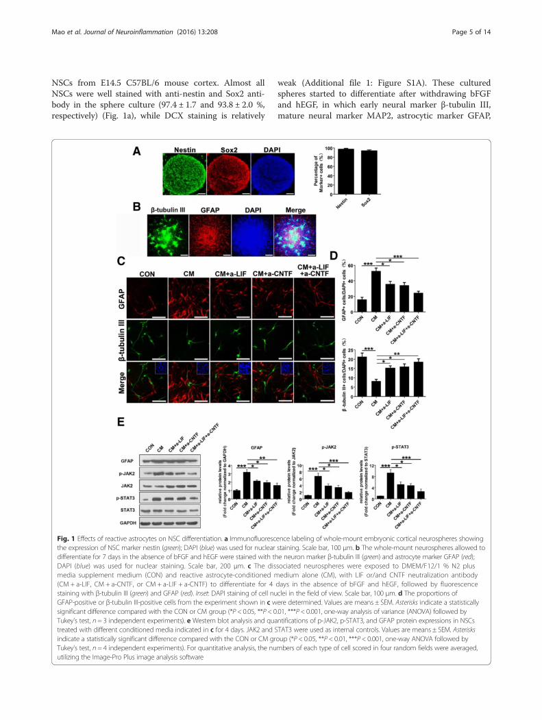
NSCs from E14.5 C57BL/6 mouse cortex. Almost allNSCs were well stained with anti-nestin and Sox2 anti-body in the sphere culture (97.4 ± 1.7 and 93.8 ± 2.0 %,respectively) (Fig. 1a), while DCX staining is relatively
weak (Additional file 1: Figure S1A). These culturedspheres started to differentiate after withdrawing bFGFand hEGF, in which early neural marker β-tubulin III,mature neural marker MAP2, astrocytic marker GFAP,
Fig. 1 Effects of reactive astrocytes on NSC differentiation. a Immunofluorescence labeling of whole-mount embryonic cortical neurospheres showingthe expression of NSC marker nestin (green); DAPI (blue) was used for nuclear staining. Scale bar, 100 μm. b The whole-mount neurospheres allowed todifferentiate for 7 days in the absence of bFGF and hEGF were stained with the neuron marker β-tubulin III (green) and astrocyte marker GFAP (red);DAPI (blue) was used for nuclear staining. Scale bar, 200 μm. c The dissociated neurospheres were exposed to DMEM/F12/1 % N2 plusmedia supplement medium (CON) and reactive astrocyte-conditioned medium alone (CM), with LIF or/and CNTF neutralization antibody(CM + a-LIF, CM + a-CNTF, or CM + a-LIF + a-CNTF) to differentiate for 4 days in the absence of bFGF and hEGF, followed by fluorescencestaining with β-tubulin III (green) and GFAP (red). Inset: DAPI staining of cell nuclei in the field of view. Scale bar, 100 μm. d The proportions ofGFAP-positive or β-tubulin III-positive cells from the experiment shown in c were determined. Values are means ± SEM. Asterisks indicate a statisticallysignificant difference compared with the CON or CM group (*P < 0.05, **P < 0.01, ***P < 0.001, one-way analysis of variance (ANOVA) followed byTukey’s test, n = 3 independent experiments). e Western blot analysis and quantifications of p-JAK2, p-STAT3, and GFAP protein expressions in NSCstreated with different conditioned media indicated in c for 4 days. JAK2 and STAT3 were used as internal controls. Values are means ± SEM. Asterisksindicate a statistically significant difference compared with the CON or CM group (*P < 0.05, **P < 0.01, ***P < 0.001, one-way ANOVA followed byTukey’s test, n = 4 independent experiments). For quantitative analysis, the numbers of each type of cell scored in four random fields were averaged,utilizing the Image-Pro Plus image analysis software
Mao et al. Journal of Neuroinflammation (2016) 13:208 Page 5 of 14
oligodendrocyte marker CNPase and oligodendrocyte-specific protein (OSP) were observed (Fig. 1b, Additionalfile 1: Figure S1B, C). Next, NSCs were cultured inastrocytic-conditioned medium (hereafter refers to asCM) from lesioned astrocytes which mimic neuroinflam-matory condition in vitro. Compared to control group,NSCs are subjected to CM treatment differentiated intomore astrocytes and less neurons (Fig. 1c, d). Since pre-vious study has reported that LIF and CNTF are upregu-lated in conditioned medium from reactive astrocytes[25], we first confirmed that LIF and CNTF stimulationactivated JAK-STAT pathway and significantly increasedthe ratio of GFAP-positive to β-tubulin III-positive cellsin the NSC population under differentiation conditions(Additional file 1: Figure S2). To investigate the role ofendogenous LIF and CNTF in CM on NSCs differen-tiation, we employed LIF or/and CNTF-neutralizingantibodies in CM, which remarkably decreased theproportion of GFAP-positive cells, while increasingthat of β-tubulin III-positive cells compared to theCM group (Fig. 1c, d). Additionally, we further evalu-ated the activation status of JAK2/STAT3 signalingpathway by western blot analysis. We found that CMstimulation significantly increased the levels of p-JAK2, p-STAT3, and GFAP in NSCs, while LIF or/andCNTF-neutralizing antibody treatment blocked suchupregulation in those cells (Fig. 1e). These findingssuggest that LIF and CNTF secreted by reactive astro-cytes activate JAK2/STAT3 pathway, which further in-duce astrocytogenesis and inhibit neurogenesis ofNSCs while inhibiting these signals can significantlyreduce astrocytogenesis of NSCs in neuroinflamma-tory condition.
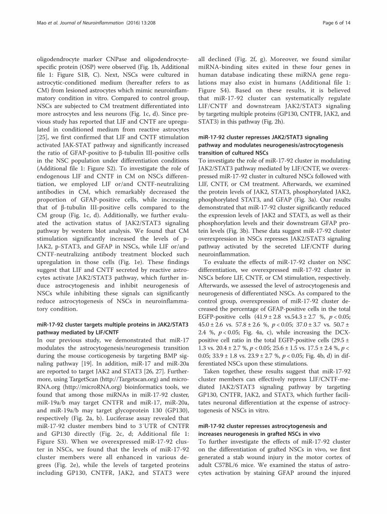
miR-17-92 cluster targets multiple proteins in JAK2/STAT3pathway mediated by LIF/CNTFIn our previous study, we demonstrated that miR-17modulates the astrocytogenesis/neurogenesis transitionduring the mouse corticogenesis by targeting BMP sig-naling pathway [19]. In addition, miR-17 and miR-20aare reported to target JAK2 and STAT3 [26, 27]. Further-more, using TargetScan (http://Targetscan.org) and micro-RNA.org (http://microRNA.org) bioinformatics tools, wefound that among those miRNAs in miR-17-92 cluster,miR-19a/b may target CNTFR and miR-17, miR-20a,and miR-19a/b may target glycoprotein 130 (GP130),respectively (Fig. 2a, b). Luciferase assay revealed thatmiR-17-92 cluster members bind to 3′UTR of CNTFRand GP130 directly (Fig. 2c, d; Additional file 1:Figure S3). When we overexpressed miR-17-92 clus-ter in NSCs, we found that the levels of miR-17-92cluster members were all enhanced in various de-grees (Fig. 2e), while the levels of targeted proteinsincluding GP130, CNTFR, JAK2, and STAT3 were
all declined (Fig. 2f, g). Moreover, we found similarmiRNA-binding sites exited in these four genes inhuman database indicating these miRNA gene regu-lations may also exist in humans (Additional file 1:Figure S4). Based on these results, it is believedthat miR-17-92 cluster can systematically regulateLIF/CNTF and downstream JAK2/STAT3 signalingby targeting multiple proteins (GP130, CNTFR, JAK2, andSTAT3) in this pathway (Fig. 2h).
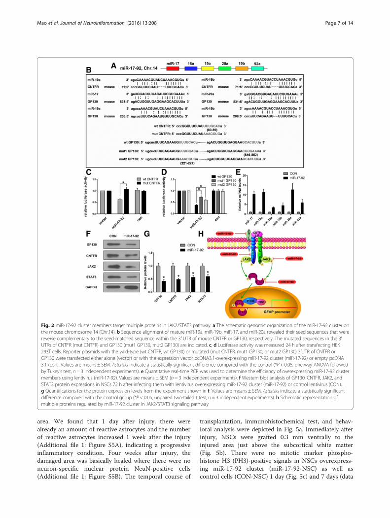
miR-17-92 cluster represses JAK2/STAT3 signalingpathway and modulates neurogenesis/astrocytogenesistransition of cultured NSCsTo investigate the role of miR-17-92 cluster in modulatingJAK2/STAT3 pathway mediated by LIF/CNTF, we overex-pressed miR-17-92 cluster in cultured NSCs followed withLIF, CNTF, or CM treatment. Afterwards, we examinedthe protein levels of JAK2, STAT3, phosphorylated JAK2,phosphorylated STAT3, and GFAP (Fig. 3a). Our resultsdemonstrated that miR-17-92 cluster significantly reducedthe expression levels of JAK2 and STAT3, as well as theirphosphorylation levels and their downstream GFAP pro-tein levels (Fig. 3b). These data suggest miR-17-92 clusteroverexpression in NSCs represses JAK2/STAT3 signalingpathway activated by the secreted LIF/CNTF duringneuroinflammation.To evaluate the effects of miR-17-92 cluster on NSC
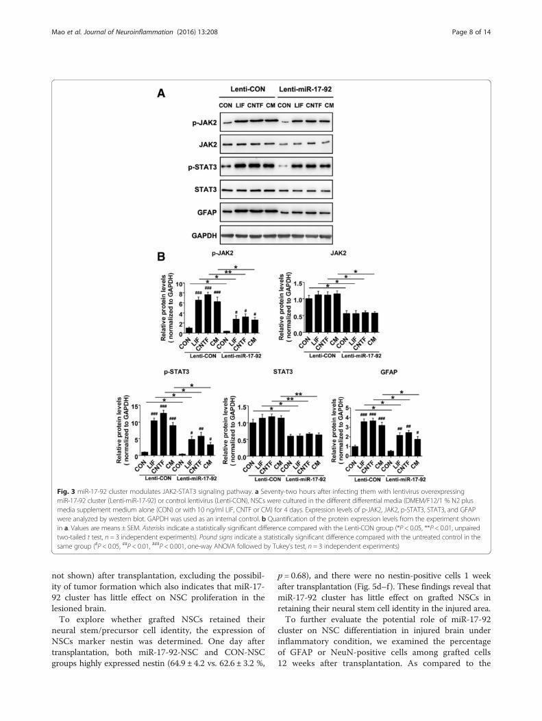
differentiation, we overexpressed miR-17-92 cluster inNSCs before LIF, CNTF, or CM stimulation, respectively.Afterwards, we assessed the level of astrocytogenesis andneurogenesis of differentiated NSCs. As compared to thecontrol group, overexpression of miR-17-92 cluster de-creased the percentage of GFAP-positive cells in the totalEGFP-positive cells (41.9 ± 2.8 vs.54.3 ± 2.7 %, p < 0.05;45.0 ± 2.6 vs. 57.8 ± 2.6 %, p < 0.05; 37.0 ± 3.7 vs. 50.7 ±2.4 %, p < 0.05; Fig. 4a, c), while increasing the DCX-positive cell ratio in the total EGFP-positive cells (29.5 ±1.3 vs. 20.4 ± 2.7 %, p < 0.05; 25.6 ± 1.5 vs. 17.5 ± 2.4 %, p <0.05; 33.9 ± 1.8 vs. 23.9 ± 2.7 %, p < 0.05; Fig. 4b, d) in dif-ferentiated NSCs upon these stimulations.Taken together, these results suggest that miR-17-92
cluster members can effectively repress LIF/CNTF-me-diated JAK2/STAT3 signaling pathway by targetingGP130, CNTFR, JAK2, and STAT3, which further facili-tates neuronal differentiation at the expense of astrocy-togenesis of NSCs in vitro.
miR-17-92 cluster represses astrocytogenesis andincreases neurogenesis in grafted NSCs in vivoTo further investigate the effects of miR-17-92 clusteron the differentiation of grafted NSCs in vivo, we firstgenerated a stab wound injury in the motor cortex ofadult C57BL/6 mice. We examined the status of astro-cytes activation by staining GFAP around the injured
Mao et al. Journal of Neuroinflammation (2016) 13:208 Page 6 of 14
area. We found that 1 day after injury, there werealready an amount of reactive astrocytes and the numberof reactive astrocytes increased 1 week after the injury(Additional file 1: Figure S5A), indicating a progressiveinflammatory condition. Four weeks after injury, thedamaged area was basically healed where there were noneuron-specific nuclear protein NeuN-positive cells(Additional file 1: Figure S5B). The temporal course of
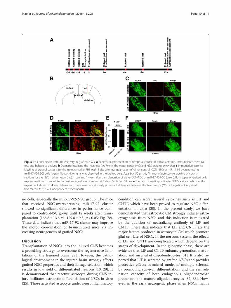
transplantation, immunohistochemical test, and behav-ioral analysis were depicted in Fig. 5a. Immediately afterinjury, NSCs were grafted 0.3 mm ventrally to theinjured area just above the subcortical white matter(Fig. 5b). There were no mitotic marker phospho-histone H3 (PH3)-positive signals in NSCs overexpress-ing miR-17-92 cluster (miR-17-92-NSC) as well ascontrol cells (CON-NSC) 1 day (Fig. 5c) and 7 days (data
Fig. 2 miR-17-92 cluster members target multiple proteins in JAK2/STAT3 pathway. a The schematic genomic organization of the miR-17-92 cluster onthe mouse chromosome 14 (Chr.14). b Sequence alignment of mature miR-19a, miR-19b, miR-17, and miR-20a revealed their seed sequences that werereverse complementary to the seed-matched sequence within the 3′ UTR of mouse CNTFR or GP130, respectively. The mutated sequences in the 3′UTRs of CNTFR (mut CNTFR) and GP130 (mut1 GP130, mut2 GP130) are indicated. c, d Luciferase activity was measured 24 h after transfecting HEK293T cells. Reporter plasmids with the wild-type (wt CNTFR, wt GP130) or mutated (mut CNTFR, mut1 GP130, or mut2 GP130) 3′UTR of CNTFR orGP130 were transfected either alone (vector) or with the expression vector pcDNA3.1-overexpressing miR-17-92 cluster (miR-17-92) or empty pcDNA3.1 (con). Values are means ± SEM. Asterisks indicate a statistically significant difference compared with the control (*P < 0.05, one-way ANOVA followedby Tukey’s test, n = 3 independent experiments). e Quantitative real-time PCR was used to determine the efficiency of overexpressing miR-17-92 clustermembers using lentivirus (miR-17-92). Values are means ± SEM (n = 3 independent experiments). f Western blot analysis of GP130, CNTFR, JAK2, andSTAT3 protein expressions in NSCs 72 h after infecting them with lentivirus overexpressing miR-17-92 cluster (miR-17-92) or control lentivirus (CON).g Quantifications for the protein expression levels from the experiment shown in f. Values are means ± SEM. Asterisks indicate a statistically significantdifference compared with the control group (*P < 0.05, unpaired two-tailed t test, n = 3 independent experiments). h Schematic representation ofmultiple proteins regulated by miR-17-92 cluster in JAK2/STAT3 signaling pathway
Mao et al. Journal of Neuroinflammation (2016) 13:208 Page 7 of 14
not shown) after transplantation, excluding the possibil-ity of tumor formation which also indicates that miR-17-92 cluster has little effect on NSC proliferation in thelesioned brain.To explore whether grafted NSCs retained their
neural stem/precursor cell identity, the expression ofNSCs marker nestin was determined. One day aftertransplantation, both miR-17-92-NSC and CON-NSCgroups highly expressed nestin (64.9 ± 4.2 vs. 62.6 ± 3.2 %,
p = 0.68), and there were no nestin-positive cells 1 weekafter transplantation (Fig. 5d–f ). These findings reveal thatmiR-17-92 cluster has little effect on grafted NSCs inretaining their neural stem cell identity in the injured area.To further evaluate the potential role of miR-17-92
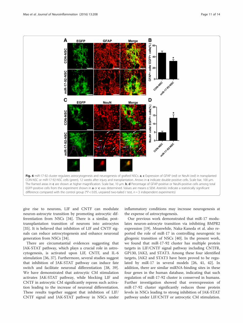
cluster on NSC differentiation in injured brain underinflammatory condition, we examined the percentageof GFAP or NeuN-positive cells among grafted cells12 weeks after transplantation. As compared to the
Fig. 3 miR-17-92 cluster modulates JAK2-STAT3 signaling pathway. a Seventy-two hours after infecting them with lentivirus overexpressingmiR-17-92 cluster (Lenti-miR-17-92) or control lentivirus (Lenti-CON), NSCs were cultured in the different differential media (DMEM/F12/1 % N2 plusmedia supplement medium alone (CON) or with 10 ng/ml LIF, CNTF or CM) for 4 days. Expression levels of p-JAK2, JAK2, p-STAT3, STAT3, and GFAPwere analyzed by western blot. GAPDH was used as an internal control. b Quantification of the protein expression levels from the experiment shownin a. Values are means ± SEM. Asterisks indicate a statistically significant difference compared with the Lenti-CON group (*P < 0.05, **P < 0.01, unpairedtwo-tailed t test, n = 3 independent experiments). Pound signs indicate a statistically significant difference compared with the untreated control in thesame group (#P < 0.05, ##P < 0.01, ###P < 0.001, one-way ANOVA followed by Tukey’s test, n = 3 independent experiments)
Mao et al. Journal of Neuroinflammation (2016) 13:208 Page 8 of 14
control group, miR-17-92 cluster significantly decreasedthe proportion of astrocytes (32.1 ± 2.9 vs. 46.8 ±3.5 %, p < 0.05; Fig. 6a, b), while that of neurons wasremarkably increased (13.4 ± 2.0 vs. 4.8 ± 0.9 %, p < 0.05;Fig. 6c, d).
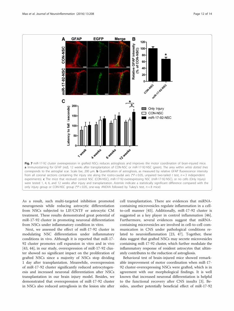
miR-17-92 cluster overexpression reduces astrogliosis andimproves the motor coordination of brain-injured miceWe observed that there are large amount of reactive as-trocytes gathering around the lesion site, which normallyreflects the recovery situation after brain injury. There-fore, we assessed the potential effect of miR-17-92 clus-ter overexpression on astrogliosis 12 weeks after NSCtransplantation by immunostaining of GFAP. Our resultsshowed that miR-17-92 cluster overexpression in NSCs
significantly decreased astrogliosis by 32.2 % at the le-sion site (Fig. 7a, b). This is partly because of the re-duced astrocytogenesis of grafted NSCs, which directlydiminished the number of reactive astrocytes. Mean-while, the result also points to a potential non-cell au-tonomous effect of grafted NSCs on host astrocytes thatcontributes to the reduction of astrogliosis.Due to the mild injury in the motor cortex, no obvious
motor defects related to walking, climbing, or feedingabilities were observed among almost all the operatedanimals. Therefore, we checked the potential effect ofmiR-17-92-NSC transplantation using a rotarod test, amore sophisticated task of motor coordination. Weshowed that mice that received cell grafts of either typeappeared to be doing better than the group that received
Fig. 4 miR-17-92 cluster regulates astrocytogenesis and neurogenesis of NSCs in vitro. a, b Immunofluorescence labeling of NSCs showing theexpression of astrocyte marker GFAP or neuron marker doublecortin (DCX) after infecting with lentivirus overexpressing miR-17-92 cluster(miR-17-92) or lentivirus expressing EGFP alone (CON) in the different differential media. Arrow heads indicate double positive cells. Scale bar,50 μm. c, d Percentage of GFAP-positive or DCX-positive among total EGFP-positive cells from the experiment shown in a or b was determined.Values are means ± SEM. Asterisks indicate a statistically significant difference compared with the control group (*P < 0.05, unpaired two-tailedt test, n = 3 independent experiments)
Mao et al. Journal of Neuroinflammation (2016) 13:208 Page 9 of 14
no cells, especially the miR-17-92-NSC group. The micethat received NSC-overexpressing miR-17-92 clustershowed no significant differences in performance com-pared to control-NSC group until 12 weeks after trans-plantation (168.8 ± 13.6 vs. 129.8 ± 9.5, p < 0.05; Fig. 7c).These data indicate that miR-17-92 cluster may improvethe motor coordination of brain-injured mice via in-creasing neurogenesis of grafted NSCs.
DiscussionTransplantation of NSCs into the injured CNS becomesa promising strategy to overcome the regenerative limi-tations of the lesioned brain [28]. However, the patho-logical environment in the injured brain strongly affectsgrafted NSC properties and their lineage selection, whichresults in low yield of differentiated neurons [10, 29]. Itis demonstrated that reactive astrocyte during CNS in-jury facilitates astrocytic differentiation of NSCs in vitro[25]. Those activated astrocyte under neuroinflammatory
condition can secret several cytokines such as LIF andCNTF, which have been proved to regulate NSC differ-entiation in vitro [30]. In the present study, we havedemonstrated that astrocytic CM strongly induces astro-cytogenesis from NSCs and this induction is mitigatedby the addition of neutralizing antibody of LIF andCNTF. These data indicate that LIF and CNTF are themajor factors produced in astrocytic CM which promoteglial cell fate of NSCs. In the nervous system, the effectsof LIF and CNTF are complicated which depend on thestages of development. In the gliogenic phase, there areevidence that LIF and CNTF enhance generation, matur-ation, and survival of oligodendrocytes [31]. It is also re-ported that LIF is secreted by grafted NSCs and providesprotective effects in animal model of multiple sclerosisby promoting survival, differentiation, and the remyeli-nation capacity of both endogenous oligodendrocyteprecursors and mature oligodendrocytes [32, 33]. How-ever, in the early neurogenic phase when NSCs mainly
Fig. 5 PH3 and nestin immunoreactivity in grafted NSCs. a Schematic presentation of temporal course of transplantation, immunohistochemicaltest, and behavioral analysis. b Diagram illustrating the injury site (red line) in the motor cortex (MC) and NSC grafting (green dot). c Immunofluorescencelabeling of coronal sections for the mitotic marker PH3 (red), 1 day after transplantation of either control (CON-NSC) or miR-17-92-overexpressing(miR-17-92-NSC) cells (green). No positive signal was observed in the grafted cells. Scale bar, 50 μm. d, f Immunofluorescence labeling of coronalsections for the NSC marker nestin (red), 1 day and 1 week after transplantation of either CON-NSC or miR-17-92-NSC (green). Both types of grafted cellsexpress nestin at 1 day, while no positive signal was observed at 7 days. Scale bar, 50 μm. e The ratio of nestin-positive to EGFP-positive cells from theexperiment shown in d was determined. There was no statistically significant difference between the two groups (N.S. not significant, unpairedtwo-tailed t test, n = 3 independent experiments)
Mao et al. Journal of Neuroinflammation (2016) 13:208 Page 10 of 14
give rise to neurons, LIF and CNTF can modulateneuron-astrocyte transition by promoting astrocytic dif-ferentiation from NSCs [34]. There is a similar, post-transplantation transition of neurons into astrocytes[35]. It is believed that inhibition of LIF and CNTF sig-nals can reduce astrocytogenesis and enhance neuronalgeneration from NSCs [34].There are circumstantial evidences suggesting that
JAK-STAT pathway, which plays a crucial role in astro-cytogenesis, is activated upon LIF, CNTF, and IL-6stimulation [36, 37]. Furthermore, several studies suggestthat inhibition of JAK-STAT pathway can induce fateswitch and facilitate neuronal differentiation [38, 39].We have demonstrated that astrocytic CM stimulationactivates JAK-STAT pathway, while blocking LIF andCNTF in astrocytic CM significantly repress such activa-tion leading to the increase of neuronal differentiation.These results together suggest that inhibition of LIF/CNTF signal and JAK-STAT pathway in NSCs under
inflammatory conditions may increase neurogenesis atthe expense of astrocytogenesis.Our previous work demonstrated that miR-17 modu-
lates neuron-astrocyte transition via inhibiting BMPR2expression [19]. Meanwhile, Naka-Kaneda et al. also re-ported the role of miR-17 in controlling neurogenic togliogenic transition of NSCs [40]. In the present work,we found that miR-17-92 cluster has multiple proteintargets in LIF/CNTF signal pathway including CNTFR,GP130, JAK2, and STAT3. Among these four identifiedtargets, JAK2 and STAT3 have been proved to be regu-lated by miR-17 in several models [26, 41, 42]. Inaddition, there are similar miRNA-binding sites in thesefour genes in the human database, indicating that suchregulation of miR-17-92 cluster is conserved in humans.Further investigation showed that overexpression ofmiR-17-92 cluster significantly reduces those proteinlevels in NSCs leading to strong inhibition of JAK-STATpathway under LIF/CNTF or astrocytic CM stimulation.
Fig. 6 miR-17-92 cluster regulates astrocytogenesis and neurogenesis of grafted NSCs. a, c Expression of GFAP (red) or NeuN (red) in transplantedCON-NSC or miR-17-92-NSC cells (green), 12 weeks after injury and transplantation. Arrows in c indicate double positive cells. Scale bar, 100 μm.The framed areas in c are shown at higher magnification. Scale bar, 10 μm. b, d Percentage of GFAP-positive or NeuN-positive cells among totalEGFP-positive cells from the experiment shown in a or c was determined. Values are means ± SEM. Asterisks indicate a statistically significantdifference compared with the control group (*P < 0.05, unpaired two-tailed t test, n = 3 independent experiments)
Mao et al. Journal of Neuroinflammation (2016) 13:208 Page 11 of 14
As a result, such multi-targeted inhibition promotedneurogenesis while reducing astrocytic differentiationfrom NSCs subjected to LIF/CNTF or astrocytic CMtreatment. These results demonstrated great potential ofmiR-17-92 cluster in promoting neuronal differentiationfrom NSCs under inflammatory condition in vitro.Next, we assessed the effect of miR-17-92 cluster in
modulating NSC differentiation under inflammatoryconditions in vivo. Although it is reported that miR-17-92 cluster promotes cell expansion in vitro and in vivo[43, 44], in our study, overexpression of miR-17-92 clus-ter showed no significant impact on the proliferation ofgrafted NSCs since a majority of NSCs stop dividing1 day after transplantation. Meanwhile, overexpressionof miR-17-92 cluster significantly reduced astrocytogen-esis and increased neuronal differentiation after NSCstransplantation in our brain injury model. Besides, wedemonstrated that overexpression of miR-17-92 clusterin NSCs also reduced astrogliosis in the lesion site after
cell transplantation. There are evidences that miRNA-containing microvesicles regulate inflammation in a cell-to-cell manner [45]. Additionally, miR-17-92 cluster issuggested as a key player in control inflammation [46].Furthermore, several evidences suggest that miRNA-containing microvesicles are involved in cell-to-cell com-munication in CNS under pathological conditions re-lated to neuroinflammation [23, 47]. Together, thesedata suggest that grafted NSCs may secrete microvesiclescontaining miR-17-92 cluster, which further modulate theinflammatory response of resident astrocytes that ultim-ately contributes to the reduction of astrogliosis.Behavioral test of brain-injured mice showed remark-
able improvement of motor coordination when miR-17-92 cluster-overexpressing NSCs were grafted, which is inagreement with our morphological findings. It is wellknown that increased neuronal differentiation is helpfulto the functional recovery after CNS insults [3]. Be-sides, another potentially beneficial effect of miR-17-92
Fig. 7 miR-17-92 cluster overexpression in grafted NSCs reduces astrogliosis and improves the motor coordination of brain-injured mice.a Immunostaining for GFAP (red), 12 weeks after transplantation of CON-NSC or miR-17-92-NSC (green). The area within white dotted linescorresponds to the astroglial scar. Scale bar, 200 μm. b Quantification of astrogliosis, as measured by relative GFAP fluorescence intensityfrom all coronal sections containing the injury site along the rostro-caudal axis (*P < 0.05, unpaired two-tailed t test, n = 3 independentexperiments). c The mice that received control NSC (CON-NSC), miR-17-92-overexpressing NSC (miR-17-92-NSC), or no cells (Only Injury)were tested 1, 4, 6, and 12 weeks after injury and transplantation. Asterisks indicate a statistically significant difference compared with theonly injury group or CON-NSC group (*P < 0.05, one-way ANOVA followed by Tukey’s test, n = 8 mice)
Mao et al. Journal of Neuroinflammation (2016) 13:208 Page 12 of 14
cluster-overexpressing NSCs is the significant inhibitionof astrogliosis since reactive astrocyte as well as glialscar may have detrimental consequences after CNSdamage [48, 49].It is undoubted that during brain lesion, both astrocyte
and microglia are activated, which result in a neuroin-flammatory condition [50]. Cytokines secreted by bothcell types may affect the cell lineage selection of graftedNSCs. In addition to the LIF and CNTF secreted by acti-vated astrocyte, it is demonstrated that activated micro-glia also facilitates astrocytogenesis via producing IL-6and LIF [8, 10]. Notably, all these cytokines affect NSCsdifferentiation through the modulation of JAK-STATpathway [51, 52]. Furthermore, one of the targeted pro-teins of miR-17-92 cluster, GP130, controls IL-6, anddownstream JAK-STAT pathway that regulates NSClineage selection [31, 51]. Therefore, we believe thatoverexpression of miR-17-92 cluster in grafted NSCs canremarkably block the activation of JAK-STAT pathwaymediated by LIF/CNTF/IL-6 under neuroinflammatorycondition in vivo.
ConclusionsTaken together, our results suggest that LIF and CNTFproduced by reactive astrocyte activate JAK-STAT path-way in NSCs. miR-17-92 cluster effectively inhibits the ac-tivation of JAK-STAT pathway under neuroinflammatorycondition via targeting multiple proteins, leading to the in-crease of neuronal differentiation from grafted NSCs afterbrain injury. These findings not only provide a better un-derstanding of NSC differentiation regulated by neuroin-flammation but also potentiate the clinical application ofneural cell replacement.
Additional file
Additional file 1: Figures S1–S5. Figure S1. Characterization of theNSCs. Figure S2. Effect of LIF and CNTF on differentiation of NSCs.Figure S3. miR-17-92 members directly target the 3′UTR of CNTFR orGP130. Figure S4. Bioinformatics analysis of miR-17-92 cluster membersbinding sites within CNTFR, GP130, JAK2, and STAT3. Figure S5.Traumatic brain injury. (PDF 558 kb)
AcknowledgementsWe thank Qipeng Zhang and Ke Zen for helpful comments. Technicalassistance by Hanqin Li and Qun Chen is gratefully acknowledged.
FundingThis work was supported by grants from the National Natural ScienceFoundation of China (31471019).
Availability of data and materialsAll raw data used in this manuscript are available on request.
Authors’ contributionsSM and LL designed the study. SM, XL, JW, and XD performed theexperiments. CZ provided space and equipment for the project. SM wrotethe draft of the manuscript. LL supervised the study and edited themanuscript. All authors read and approved the final manuscript.
Competing interestsThe authors declare that they have no competing interests.
Consent for publicationNot applicable.
Ethics approval and consent to participateOur study does not include any human participants. All of the animal careand experimental procedures were performed in accordance with theLaboratory Animal Care Guidelines approved by the Model Animal ResearchCenter of Nanjing University.
Author details1State Key Laboratory of Pharmaceutical Biotechnology, CollaborativeInnovation Center of Chemistry for Life Sciences, Jiangsu EngineeringResearch Center for MicroRNA Biology and Biotechnology, NJU AdvancedInstitute for Life Sciences (NAILS), School of Life Sciences, Nanjing University,163 Xianlin Road, Nanjing, Jiangsu 210023, China. 2Jiangsu Key Laboratory ofNeuroregeneration, Co-Innovation Center of Neuroregeneration, NantongUniversity, Nantong 226001, China.
Received: 19 March 2016 Accepted: 18 August 2016
References1. Kim DY, Hwang I, Muller FL, Paik JH. Functional regulation of FoxO1 in
neural stem cell differentiation. Cell Death Differ. 2015;22:2034–45.2. Kokaia Z, Martino G, Schwartz M, Lindvall O. Cross-talk between neural stem
cells and immune cells: the key to better brain repair? Nat Neurosci.2012;15:1078–87.
3. Makri G, Lavdas AA, Katsimpardi L, Charneau P, Thomaidou D, Matsas R.Transplantation of embryonic neural stem/precursor cells overexpressingBM88/Cend1 enhances the generation of neuronal cells in the injuredmouse cortex. Stem Cells. 2010;28:127–39.
4. Mosher KI, Andres RH, Fukuhara T, Bieri G, Hasegawa-Moriyama M, He Y,Guzman R, Wyss-Coray T. Neural progenitor cells regulate microgliafunctions and activity. Nat Neurosci. 2012;15:1485–7.
5. Lukovic D, Stojkovic M, Moreno-Manzano V, Jendelova P, Sykova E,Bhattacharya SS, Erceg S. Concise review: reactive astrocytes and stem cellsin spinal cord injury: good guys or bad guys? Stem Cells. 2015;33:1036–41.
6. Bertrand N, Castro DS, Guillemot F. Proneural genes and the specification ofneural cell types. Nat Rev Neurosci. 2002;3:517–30.
7. Li H, Grumet M. BMP and LIF signaling coordinately regulate lineagerestriction of radial glia in the developing forebrain. Glia. 2007;55:24–35.
8. Covacu R, Brundin L. Effects of neuroinflammation on neural stem cells.Neuroscientist. 2015. doi:10.1177/1073858415616559.
9. Liu Q, Sanai N, Jin WN, La Cava A, Van Kaer L, Shi FD. Neural stem cellssustain natural killer cells that dictate recovery from brain inflammation. NatNeurosci. 2016;19:243–52.
10. Ideguchi M, Shinoyama M, Gomi M, Hayashi H, Hashimoto N, Takahashi J.Immune or inflammatory response by the host brain suppresses neuronaldifferentiation of transplanted ES cell-derived neural precursor cells. JNeurosci Res. 2008;86:1936–43.
11. Wang FW, Hao HB, Zhao SD, Zhang YM, Liu Q, Liu HJ, Liu SM, Yuan QH,Bing LJ, Ling EA, Hao AJ. Roles of activated astrocyte in neural stem cellproliferation and differentiation. Stem Cell Res. 2011;7:41–53.
12. Kang SS, Keasey MP, Arnold SA, Reid R, Geralds J, Hagg T. EndogenousCNTF mediates stroke-induced adult CNS neurogenesis in mice. NeurobiolDis. 2013;49:68–78.
13. Molyneaux BJ, Arlotta P, Menezes JR, Macklis JD. Neuronal subtypespecification in the cerebral cortex. Nat Rev Neurosci. 2007;8:427–37.
14. Seo H, Lee K. Epac2 contributes to PACAP-induced astrocyticdifferentiation through calcium ion influx in neural precursor cells.BMB Rep. 2016;49:128–33.
15. He F, Ge W, Martinowich K, Becker-Catania S, Coskun V, Zhu W, Wu H,Castro D, Guillemot F, Fan G, et al. A positive autoregulatory loop ofJak-STAT signaling controls the onset of astrogliogenesis. Nat Neurosci.2005;8:616–25.
16. Le MTN, Xie HM, Zhou BY, Chia PH, Rizk P, Um M, Udolph G, Yang H, Lim B,Lodish HF. MicroRNA-125b promotes neuronal differentiation in humancells by repressing multiple targets. Mol Cell Biol. 2009;29:5290–305.
Mao et al. Journal of Neuroinflammation (2016) 13:208 Page 13 of 14
17. Fan Z, Lu M, Qiao C, Zhou Y, Ding JH, Hu G. MicroRNA-7 enhancessubventricular zone neurogenesis by inhibiting NLRP3/caspase-1 axis inadult neural stem cells. Mol Neurobiol. 2015. doi:10.1007/s12035-015-9620-5.
18. Zhang JF, Shi LL, Zhang L, Zhao ZH, Liang F, Xu X, Zhao LY, Yang PB, ZhangJS, Tian YF. MicroRNA-25 negatively regulates cerebral ischemia/reperfusioninjury-induced cell apoptosis through Fas/FasL pathway. J Mol Neurosci.2016;58:507–16.
19. Mao S, Li H, Sun Q, Zen K, Zhang CY, Li L. miR-17 regulates the proliferationand differentiation of the neural precursor cells during mousecorticogenesis. FEBS J. 2014;281:1144–58.
20. Ma S, Liu M, Xu Z, Li Y, Guo H, Ge Y, Liu Y, Zheng D, Shi J. A doublefeedback loop mediated by microRNA-23a/27a/24-2 regulates M1 versusM2 macrophage polarization and thus regulates cancer progression.Oncotarget. 2016;7:13502–19.
21. Sun Q, Mao S, Li H, Zen K, Zhang CY, Li L. Role of miR-17 family in thenegative feedback loop of bone morphogenetic protein signaling inneuron. PLoS One. 2013;8:e83067.
22. Wu W, Takanashi M, Borjigin N, Ohno SI, Fujita K, Hoshino S, Osaka Y,Tsuchida A, Kuroda M. MicroRNA-18a modulates STAT3 activity throughnegative regulation of PIAS3 during gastric adenocarcinogenesis. Br JCancer. 2013;108:653–61.
23. Mao S, Sun Q, Xiao H, Zhang C, Li L. Secreted miR-34a in astrocyticshedding vesicles enhanced the vulnerability of dopaminergic neurons toneurotoxins by targeting Bcl-2. Protein Cell. 2015;6:529–40.
24. Hamm RJ, Pike BR, O’Dell DM, Lyeth BG, Jenkins LW. The rotarod test: anevaluation of its effectiveness in assessing motor deficits followingtraumatic brain injury. J Neurotrauma. 1994;11:187–96.
25. Faijerson J, Tinsley RB, Aprico K, Thorsell A, Nodin C, Nilsson M, BlomstrandF, Eriksson PS. Reactive astrogliosis induces astrocytic differentiation of adultneural stem/progenitor cells in vitro. J Neurosci Res. 2006;84:1415–24.
26. Li YM, Vecchiarelli-Federico LM, Li YJ, Egan SE, Spaner D, Hough MR,Ben-David Y. The miR-17-92 cluster expands multipotent hematopoieticprogenitors whereas imbalanced expression of its individual oncogenicmiRNAs promotes leukemia in mice. Blood. 2012;119:4486–98.
27. Zhang MM, Liu QF, Mi SP, Liang X, Zhang ZQ, Su XM, Liu JY, Chen YY,Wang MM, Zhang YA, et al. Both miR-17-5p and miR-20a alleviatesuppressive potential of myeloid-derived suppressor cells by modulatingSTAT3 expression. J Immunol. 2011;186:4716–24.
28. Chau MJ, Deveau TC, Song M, Gu X, Chen D, Wei L. iPSC transplantationincreases regeneration and functional recovery after ischemic stroke inneonatal rats. Stem Cells. 2014;32:3075–87.
29. Lozano D, Gonzales-Portillo GS, Acosta S, de la Pena I, Tajiri N, Kaneko Y,Borlongan CV. Neuroinflammatory responses to traumatic brain injury:etiology, clinical consequences, and therapeutic opportunities.Neuropsychiatr Dis Treat. 2015;11:97–106.
30. Bauer S, Kerr BJ, Patterson PH. The neuropoietic cytokine family indevelopment, plasticity, disease and injury. Nat Rev Neurosci. 2007;8:221–32.
31. Mayer M, Bhakoo K, Noble M. Ciliary neurotrophic factor and leukemiainhibitory factor promote the generation, maturation and survival ofoligodendrocytes in vitro. Development. 1994;120:143–53.
32. Laterza C, Merlini A, De Feo D, Ruffini F, Menon R, Onorati M, Fredrickx E,Muzio L, Lombardo A, Comi G, et al. iPSC-derived neural precursors exert aneuroprotective role in immune-mediated demyelination via the secretionof LIF. Nat Commun. 2013;4:2597.
33. Butzkueven H, Emery B, Cipriani T, Marriott MP, Kilpatrick TJ. Endogenousleukemia inhibitory factor production limits autoimmune demyelination andoligodendrocyte loss. Glia. 2006;53:696–703.
34. Hirabayashi Y, Gotoh Y. Stage-dependent fate determination of neuralprecursor cells in mouse forebrain. Neurosci Res. 2005;51:331–6.
35. Laywell ED, Kearns SM, Zheng T, Chen KA, Deng J, Chen HX, Roper SN,Steindler DA. Neuron-to-astrocyte transition: phenotypic fluidity and theformation of hybrid asterons in differentiating neurospheres. J CompNeurol. 2005;493:321–33.
36. Jhaveri K, Teplinsky E, Silvera D, Valeta-Magara A, Arju R, Giashuddin S,Sarfraz Y, Alexander M, Darvishian F, Levine PH, et al. Hyperactivated mTORand JAK2/STAT3 pathways: molecular drivers and potential therapeutictargets of inflammatory and invasive ductal breast cancers afterneoadjuvant chemotherapy. Clin Breast Cancer. 2016;16:113–22.
37. Lee J, Son MJ, Woolard K, Donin NM, Li A, Cheng CH, Kotliarova S, Kotliarov Y,Walling J, Ahn S, et al. Epigenetic-mediated dysfunction of the bone
morphogenetic protein pathway inhibits differentiation ofglioblastoma-initiating cells. Cancer Cell. 2008;13:69–80.
38. DeVito WJ, Stone S. Ethanol inhibits prolactin-induced activation of theJAK/STAT pathway in cultured astrocytes. J Cell Biochem. 1999;74:278–91.
39. Zhang YL, Zhou Z, Han WW, Zhang LL, Song WS, Huang JH, Liu S. Oleanolicacid inhibiting the differentiation of neural stem cells into astrocyte bydown-regulating JAK/STAT signaling pathway. Am J Chin Med.2016;44:103–17.
40. Naka-Kaneda H, Nakamura S, Igarashi M, Aoi H, Kanki H, Tsuyama J,Tsutsumi S, Aburatani H, Shimazaki T, Okano H. The miR-17/106-p38 axis is akey regulator of the neurogenic-to-gliogenic transition in developing neuralstem/progenitor cells. Proc Natl Acad Sci U S A. 2014;111:1604–9.
41. Carraro G, El-Hashash A, Guidolin D, Tiozzo C, Turcatel G, Young BM, DeLanghe SP, Bellusci S, Shi W, Parnigotto PP, Warburton D. miR-17 family ofmicroRNAs controls FGF10-mediated embryonic lung epithelial branchingmorphogenesis through MAPK14 and STAT3 regulation of E-cadherindistribution. Dev Biol. 2009;333:238–50.
42. Foshay KM, Gallicano GI. miR-17 family miRNAs are expressed during earlymammalian development and regulate stem cell differentiation. Dev Biol.2009;326:431–43.
43. Li J, Chen L, Qiuqin T, Wu W, Hao G, Lou L, Jie W, Hua J, Hongjuan D, Xia Y, etal. The role, mechanism and potentially novel biomarker of microRNA-17-92cluster in macrosomia. Sci Rep. 2015;5:17212.
44. Zhou Y, Zhang L, Ji H, Lu X, Xia J, Li L, Chen F, Bu H, Shi Y. MiR-17~92ablation impairs liver regeneration in an estrogen-dependent manner. J CellMol Med. 2016;20:939–48.
45. Hulsmans M, Holvoet P. MicroRNA-containing microvesicles regulatinginflammation in association with atherosclerotic disease. Cardiovasc Res.2013;100:7–18.
46. Philippe L, Alsaleh G, Bahram S, Pfeffer S, Georgel P. The miR-17approximately 92 cluster: a key player in the control of inflammation duringrheumatoid arthritis. Front Immunol. 2013;4:70.
47. Brites D, Fernandes A. Neuroinflammation and depression: microgliaactivation, extracellular microvesicles and microRNA dysregulation. FrontCell Neurosci. 2015;9:476.
48. Fan X, Luo G, Yang D, Ming M, Liu H, Pu P, Le W. Critical role of lysosomeand its associated protein cathepsin D in manganese-induced toxicity incultured midbrain astrocyte. Neurochem Int. 2010;56:291–300.
49. Zhang X, Li L, Zhang X, Xie W, Li L, Yang D, Heng X, Du Y, Doody RS, Le W.Prenatal hypoxia may aggravate the cognitive impairment and Alzheimer’sdisease neuropathology in APPSwe/PS1A246E transgenic mice. NeurobiolAging. 2013;34:663–78.
50. Pekny M, Wilhelmsson U, Pekna M. The dual role of astrocyte activation andreactive gliosis. Neurosci Lett. 2014;565:30–8.
51. Coorey NJ, Shen W, Zhu L, Gillies MC. Differential expression of IL-6/gp130cytokines, Jak-STAT signaling and neuroprotection after Muller cell ablationin a transgenic mouse model. Invest Ophthalmol Vis Sci. 2015;56:2151–61.
52. Nicola NA, Babon JJ. Leukemia inhibitory factor (LIF). Cytokine GrowthFactor Rev. 2015;26:533–44.
• We accept pre-submission inquiries
• Our selector tool helps you to find the most relevant journal
• We provide round the clock customer support
• Convenient online submission
• Thorough peer review
• Inclusion in PubMed and all major indexing services
• Maximum visibility for your research
Submit your manuscript atwww.biomedcentral.com/submit
Submit your next manuscript to BioMed Central and we will help you at every step:
Mao et al. Journal of Neuroinflammation (2016) 13:208 Page 14 of 14