microrna-126 regulates endothelial expression of vascular ... · pdf filemicrorna-126...
TRANSCRIPT
MicroRNA-126 regulates endothelial expressionof vascular cell adhesion molecule 1Tamia A. Harris*, Munekazu Yamakuchi†, Marcella Ferlito†, Joshua T. Mendell‡§¶, and Charles J. Lowenstein*†�**
*The Cellular and Molecular Medicine Program and Departments of †Medicine, ‡Pediatrics, �Pathology, and ¶Molecular Biology and Genetics, and §Instituteof Genetic Medicine, The Johns Hopkins University School of Medicine, Baltimore, MD 21205
Edited by Richard A. Flavell, Yale University School of Medicine, New Haven, CT, and approved December 12, 2007 (received for review August 8, 2007)
Adhesion molecules expressed by activated endothelial cells playa key role in regulating leukocyte trafficking to sites of inflamma-tion. Resting endothelial cells normally do not express adhesionmolecules, but cytokines activate endothelial cells to expressadhesion molecules such as vascular cell adhesion molecule 1(VCAM-1), which mediate leukocyte adherence to endothelial cells.We now show that endothelial cells express microRNA 126 (miR-126), which inhibits VCAM-1 expression. Transfection of endothe-lial cells with an oligonucleotide that decreases miR-126 permits anincrease in TNF-�-stimulated VCAM-1 expression. Conversely,overexpression of the precursor to miR-126 increases miR-126levels and decreases VCAM-1 expression. Additionally, decreasingendogenous miR-126 levels increases leukocyte adherence to en-dothelial cells. These data suggest that microRNA can regulateadhesion molecule expression and may provide additional controlof vascular inflammation.
inflammation � nitric oxide � leukocyte � atherosclerosis
Adhesion molecules play a critical role in leukocyte traffick-ing. Selectins and their glycoprotein ligands mediate leu-
kocyte rolling along endothelial cells, the first step in leukocytetrafficking (1–4). Subsequently, cytokines and chemokines ac-tivate leukocytes and endothelial cells to express intercellularadhesion molecules and their integrin ligands, which in turnmediate leukocyte adhesion to endothelial cells (5, 6). Leuko-cytes then transmigrate across the endothelial cell barrier andmigrate through tissue to the site of injury.
Vascular cell adhesion molecule 1 (VCAM-1) is an intercel-lular adhesion molecule expressed by endothelial cells (7).VCAM-1 on the endothelial surface mediates leukocyte adhe-sion by interacting with its �4�1 integrin ligand, very late antigen4 (VLA-4; CD49d/CD29), which is expressed on most leukocytesand on stimulated neutrophils (8). Although resting endothelialcells do not express VCAM-1, treatment with a variety ofcytokines or bacterial products induce endothelial cells to ex-press VCAM-1 within 4–12 h. VCAM-1 expression is thought tobe regulated primarily at the level of transcription (9–11).Elegant cellular studies have shown that a set of transcriptionfactors interact with each other and with the 5� f lanking regionof the VCAM-1 gene to regulate VCAM-1 expression (9). Thesetranscription factors include NF-�B, IFN regulatory factor-1(IRF-1), Sp1, and scaffold factors such as high mobility groupI(Y) [HMG I(Y)] (10, 11). Although VCAM-1 expression can beregulated at the posttranscriptional level, the mechanisms bywhich this occurs are not completely defined (12, 13).
We hypothesized that an endogenous microRNA (miRNA)molecule regulates VCAM-1 expression and vascular inflamma-tion. miRNA are small noncoding RNA that regulate targetgenes posttranscriptionally. These endogenously expressedmiRNA molecules are transcribed in the nucleus as long primarytranscripts (pri-miRNA) that are further processed by the nu-clear enzyme Drosha into shorter precursor species(premiRNA), which can then be exported from the nucleus bythe Ran-GTP-dependent nuclear export factor, exportin-5 (14–16). Once in the cytoplasm, the premiRNA is processed by the
cytosolic enzyme Dicer into the mature 20- to 24-nt miRNAspecies, which forms a complex with the RNA-induced silencingcomplex (RISC) and represses the expression of target tran-scripts via binding to complementary sequences in the 3�-UTR(17). miRNA regulate expression of genes by two distinctmechanisms: if miRNA precisely match their target sequence,then the mRNA is degraded by Ago2-RISC; however, if themiRNA contains mismatches to its target, then the mRNAtranslation is blocked by Ago1-RISC (18). Accordingly, wesearched for miRNA expressed by endothelial cells that mightregulate adhesion molecules.
We found that a particular miRNA, miRNA 126 (miR-126), isselectively expressed in endothelial cells. In silico analysis shows thatVCAM-1 is a potential target of miR-126. We now show thatendogenous miR-126 suppresses VCAM-1 expression, therebydecreasing leukocyte interactions with endothelial cells. Our datasuggest that miRNA can regulate vascular inflammation.
ResultsWe measured the expression of miRNA in endothelial cells byusing a microarray (see Materials and Methods). In brief, totalRNA from human umbilical vein endothelial cells (HUVEC)was size fractionated, labeled with a fluorescent dye, and hy-bridized to a microarray chip (n � 3). Of the 500 miRNA probeson the microarray chip, the 26 miRNA with the highest level ofexpression in HUVEC were identified (Table 1). The mostfrequently expressed miRNA is miR-126.
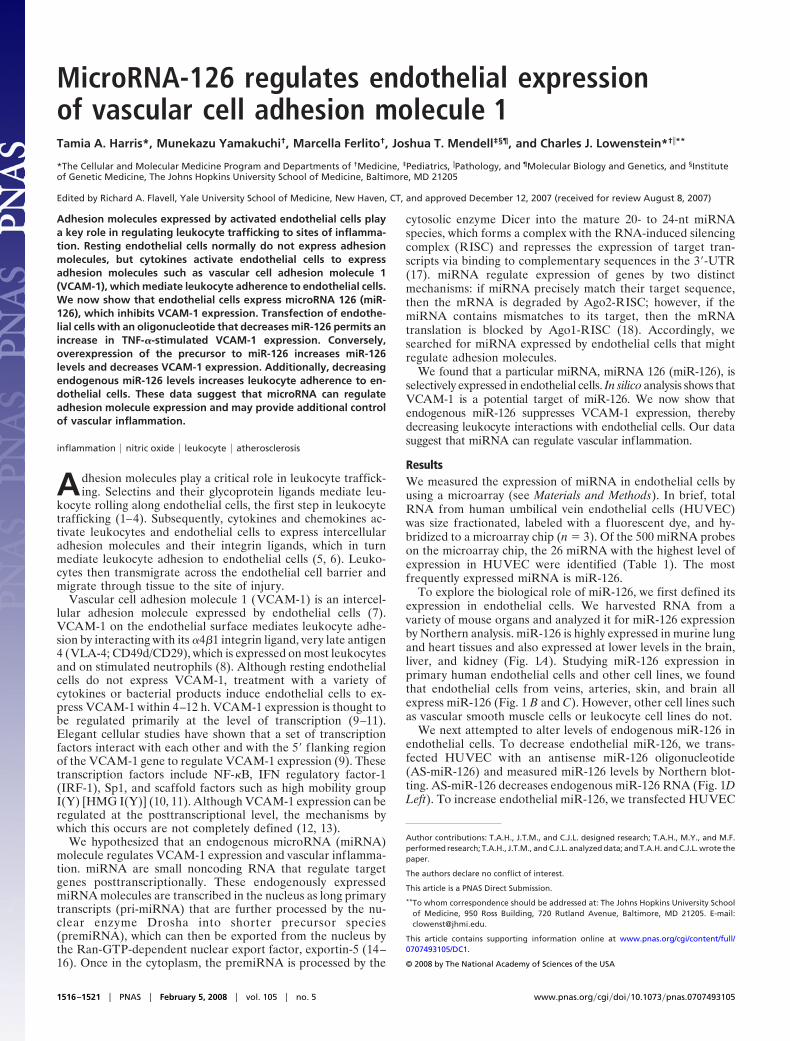
To explore the biological role of miR-126, we first defined itsexpression in endothelial cells. We harvested RNA from avariety of mouse organs and analyzed it for miR-126 expressionby Northern analysis. miR-126 is highly expressed in murine lungand heart tissues and also expressed at lower levels in the brain,liver, and kidney (Fig. 1A). Studying miR-126 expression inprimary human endothelial cells and other cell lines, we foundthat endothelial cells from veins, arteries, skin, and brain allexpress miR-126 (Fig. 1 B and C). However, other cell lines suchas vascular smooth muscle cells or leukocyte cell lines do not.
We next attempted to alter levels of endogenous miR-126 inendothelial cells. To decrease endothelial miR-126, we trans-fected HUVEC with an antisense miR-126 oligonucleotide(AS-miR-126) and measured miR-126 levels by Northern blot-ting. AS-miR-126 decreases endogenous miR-126 RNA (Fig. 1DLeft). To increase endothelial miR-126, we transfected HUVEC
Author contributions: T.A.H., J.T.M., and C.J.L. designed research; T.A.H., M.Y., and M.F.performed research; T.A.H., J.T.M., and C.J.L. analyzed data; and T.A.H. and C.J.L. wrote thepaper.
The authors declare no conflict of interest.
This article is a PNAS Direct Submission.
**To whom correspondence should be addressed at: The Johns Hopkins University Schoolof Medicine, 950 Ross Building, 720 Rutland Avenue, Baltimore, MD 21205. E-mail:[email protected].
This article contains supporting information online at www.pnas.org/cgi/content/full/0707493105/DC1.
© 2008 by The National Academy of Sciences of the USA
1516–1521 � PNAS � February 5, 2008 � vol. 105 � no. 5 www.pnas.org�cgi�doi�10.1073�pnas.0707493105
with an RNA precursor to miR-126 (premiR-126). premiR-126dramatically increases miR-126 RNA (Fig. 1D Right).
Computer analysis suggested that miR-126 may be a negativeregulator of VCAM-1 expression (see Materials and Methods).To identify potential mRNA transcripts that are regulated bymiR-126, we searched through a computer database (Human
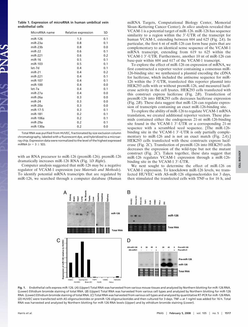
miRNA Targets, Computational Biology Center, MemorialSloan–Kettering Cancer Center). In silico analysis revealed thatVCAM-1 is a potential target of miR-126. miR-126 has sequencesimilarity to a region within the 3�-UTR of the transcript forhuman VCAM-1, extending between 604 and 625 (Fig. 2A). Inparticular, the first 6 nt of miR-126 can form base pairs that arecomplementary to an identical sense sequence of the VCAM-1mRNA transcript, extending from 619 to 625 within theVCAM-1 3�-UTR. Furthermore, another 10 nt of miR-126 canbase-pair within 604 and 617 of the VCAM-1 transcript.
To explore the effect of miR-126 on expression of mRNA, wefirst constructed a reporter vector containing a consensus miR-126-binding site: we synthesized a plasmid encoding the cDNAfor luciferase, which included the antisense sequence for miR-126 within the 3�-UTR, transfected this reporter plasmid intoHEK293 cells with or without premiR-126, and measured lucif-erase activity in the cell lysates. HEK293 cells transfected withthis construct express luciferase (Fig. 2B). Transfection ofpremiR-126 into HEK293 cells decreases luciferase expression(Fig. 2B). These data suggest that miR-126 can regulate expres-sion of transcripts containing an exact miR-126-binding site.
To explore the ability of miR-126 to regulate VCAM-1 mRNAtranslation, we created additional reporter vectors. These plas-mids contained either the endogenous 21-nt miR-126-bindingsite found in the VCAM-1 3�-UTR or a corresponding 21-ntsequence with a scrambled seed sequence. [The miR-126-binding site in the VCAM-1 3�-UTR is only partially comple-mentary to miR-126 and is not an exact match (Fig. 2 A).]HEK293 cells transfected with these constructs express lucif-erase (Fig. 2C). Transfection of premiR-126 into HEK293 cellsdecreases the expression of the wild-type but not the mutantconstruct (Fig. 2C). Taken together, these data suggest thatmiR-126 regulates VCAM-1 expression through a miR-126-binding site in the VCAM-1 3�-UTR.
We next sought to determine the effect of miR-126 onVCAM-1 expression. To knockdown miR-126 levels, we trans-fected HUVEC with AS-miR-126 oligonucleotides for 3 days,then stimulated the transfected cells with TNF-� for 16 h, and
Table 1. Expression of microRNA in human umbilical veinendothelial cells
MicroRNA name Relative expression SD
miR-126 1.3 0.1miR-23a 0.8 0.1miR-23b 0.8 0.0miR-22 0.6 0.1miR-221 0.6 0.2miR-16 0.5 0.1miR-103 0.5 0.1let-7d 0.4 0.1miR-21 0.4 0.2miR-221 0.4 0.1miR-107 0.4 0.1miR-100 0.4 0.0let-7a 0.4 0.1miR-27a 0.4 0.0miR-26a 0.3 0.0miR-24 0.3 0.0miR-20a 0.3 0.0miR-17-5 0.3 0.1miR-181 0.2 0.1miR-106a 0.2 0.1miR-29a 0.2 0.1miR-130a 0.2 0.0
Total RNA was purified from HUVEC, fractionated by size exclusion columnchromatography, labeled with a fluorescent dye, and hybridized to a microar-ray chip. Expression data were normalized to the level of the highest expressedmiRNA (n � 3 � SD).
A B
C D
Fig. 1. Endothelial cells express miR-126. (A) (Upper) Total RNA was harvested from various mouse tissues and analyzed by Northern blotting for miR-126 RNA.(Lower) Ethidium bromide staining of total RNA. (B) (Upper) Total RNA was harvested from various cell types and analyzed by Northern blotting for miR-126RNA. (Lower) Ethidium bromide staining of total RNA. (C) Total RNA was harvested from various cell types and analyzed by quantitative RT-PCR for miR-126 RNA.(D) HUVEC were transfected with AS oligonucleotides or premiR-126 oligonucleotides and then cultured for 3 days. TNF-� at 1 ng/ml was added for 16 h. TotalRNA was harvested and analyzed by Northern blotting for miR-126 RNA levels (Upper) and by ethidium bromide staining (Lower).
Harris et al. PNAS � February 5, 2008 � vol. 105 � no. 5 � 1517
CELL
BIO
LOG
Y
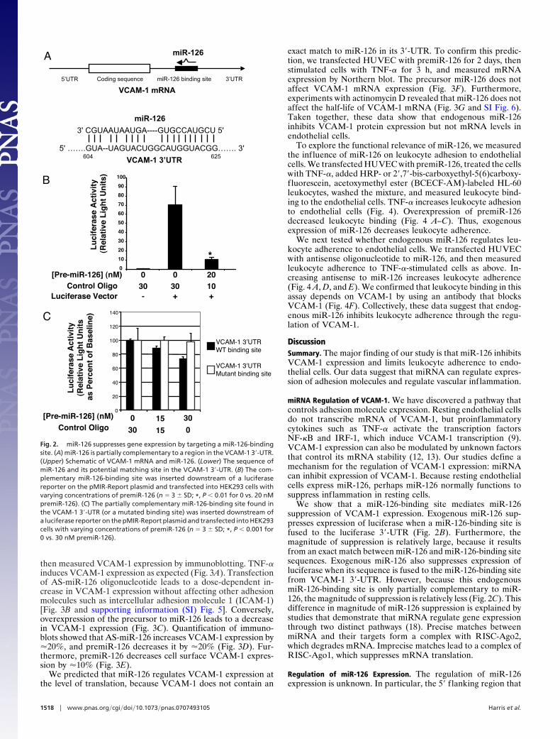
then measured VCAM-1 expression by immunoblotting. TNF-�induces VCAM-1 expression as expected (Fig. 3A). Transfectionof AS-miR-126 oligonucleotide leads to a dose-dependent in-crease in VCAM-1 expression without affecting other adhesionmolecules such as intercellular adhesion molecule 1 (ICAM-1)[Fig. 3B and supporting information (SI) Fig. 5]. Conversely,overexpression of the precursor to miR-126 leads to a decreasein VCAM-1 expression (Fig. 3C). Quantification of immuno-blots showed that AS-miR-126 increases VCAM-1 expression by�20%, and premiR-126 decreases it by �20% (Fig. 3D). Fur-thermore, premiR-126 decreases cell surface VCAM-1 expres-sion by �10% (Fig. 3E).
We predicted that miR-126 regulates VCAM-1 expression atthe level of translation, because VCAM-1 does not contain an
exact match to miR-126 in its 3�-UTR. To confirm this predic-tion, we transfected HUVEC with premiR-126 for 2 days, thenstimulated cells with TNF-� for 3 h, and measured mRNAexpression by Northern blot. The precursor miR-126 does notaffect VCAM-1 mRNA expression (Fig. 3F). Furthermore,experiments with actinomycin D revealed that miR-126 does notaffect the half-life of VCAM-1 mRNA (Fig. 3G and SI Fig. 6).Taken together, these data show that endogenous miR-126inhibits VCAM-1 protein expression but not mRNA levels inendothelial cells.
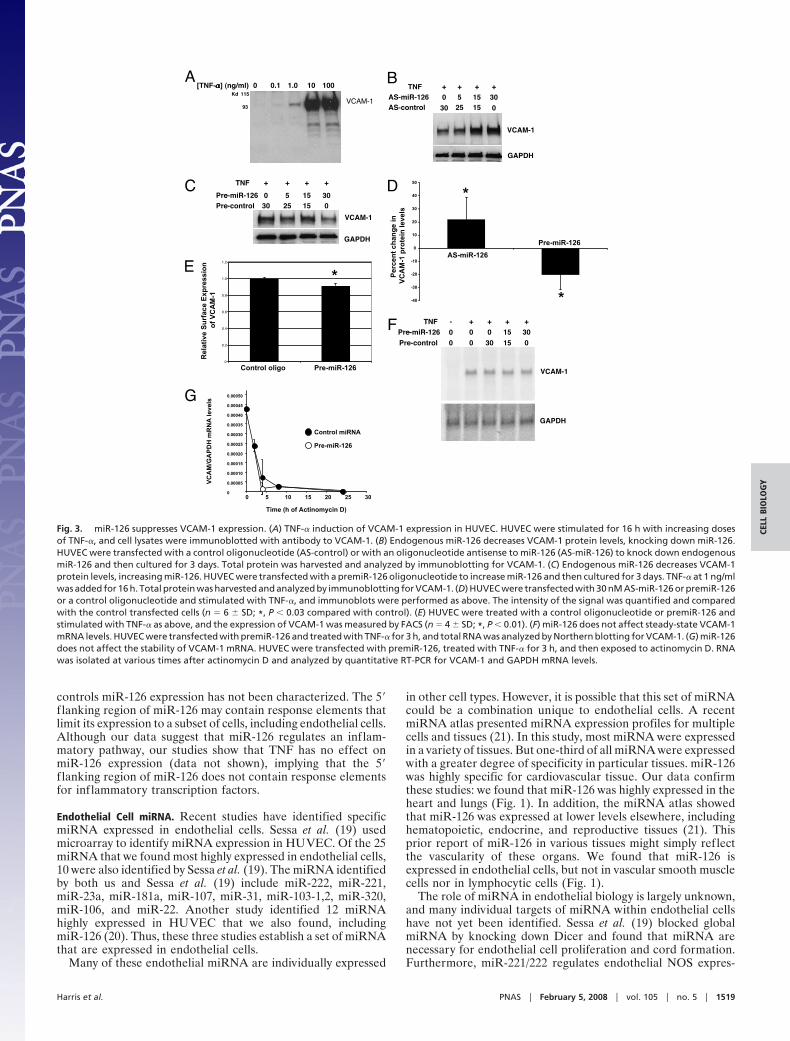
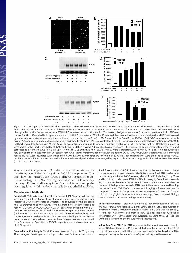
To explore the functional relevance of miR-126, we measuredthe influence of miR-126 on leukocyte adhesion to endothelialcells. We transfected HUVEC with premiR-126, treated the cellswith TNF-�, added HRP- or 2�,7�-bis-carboxyethyl-5(6)carboxy-fluorescein, acetoxymethyl ester (BCECF-AM)-labeled HL-60leukocytes, washed the mixture, and measured leukocyte bind-ing to the endothelial cells. TNF-� increases leukocyte adhesionto endothelial cells (Fig. 4). Overexpression of premiR-126decreased leukocyte binding (Fig. 4 A–C). Thus, exogenousexpression of miR-126 decreases leukocyte adherence.
We next tested whether endogenous miR-126 regulates leu-kocyte adherence to endothelial cells. We transfected HUVECwith antisense oligonucleotide to miR-126, and then measuredleukocyte adherence to TNF-�-stimulated cells as above. In-creasing antisense to miR-126 increases leukocyte adherence(Fig. 4 A, D, and E). We confirmed that leukocyte binding in thisassay depends on VCAM-1 by using an antibody that blocksVCAM-1 (Fig. 4F). Collectively, these data suggest that endog-enous miR-126 inhibits leukocyte adherence through the regu-lation of VCAM-1.
DiscussionSummary. The major finding of our study is that miR-126 inhibitsVCAM-1 expression and limits leukocyte adherence to endo-thelial cells. Our data suggest that miRNA can regulate expres-sion of adhesion molecules and regulate vascular inflammation.
miRNA Regulation of VCAM-1. We have discovered a pathway thatcontrols adhesion molecule expression. Resting endothelial cellsdo not transcribe mRNA of VCAM-1, but proinflammatorycytokines such as TNF-� activate the transcription factorsNF-�B and IRF-1, which induce VCAM-1 transcription (9).VCAM-1 expression can also be modulated by unknown factorsthat control its mRNA stability (12, 13). Our studies define amechanism for the regulation of VCAM-1 expression: miRNAcan inhibit expression of VCAM-1. Because resting endothelialcells express miR-126, perhaps miR-126 normally functions tosuppress inflammation in resting cells.
We show that a miR-126-binding site mediates miR-126suppression of VCAM-1 expression. Exogenous miR-126 sup-presses expression of luciferase when a miR-126-binding site isfused to the luciferase 3�-UTR (Fig. 2B). Furthermore, themagnitude of suppression is relatively large, because it resultsfrom an exact match between miR-126 and miR-126-binding sitesequences. Exogenous miR-126 also suppresses expression ofluciferase when its sequence is fused to the miR-126-binding sitefrom VCAM-1 3�-UTR. However, because this endogenousmiR-126-binding site is only partially complementary to miR-126, the magnitude of suppression is relatively less (Fig. 2C). Thisdifference in magnitude of miR-126 suppression is explained bystudies that demonstrate that miRNA regulate gene expressionthrough two distinct pathways (18). Precise matches betweenmiRNA and their targets form a complex with RISC-Ago2,which degrades mRNA. Imprecise matches lead to a complex ofRISC-Ago1, which suppresses mRNA translation.
Regulation of miR-126 Expression. The regulation of miR-126expression is unknown. In particular, the 5� f lanking region that
A
B
C
5’UTR Coding sequence 3’UTR
VCAM-1 mRNA
miR-126
5' …….GUA--UAGUACUGGCAUGGUACGG……. 3'
3' CGUAAUAAUGA----GUGCCAUGCU 5'
VCAM-1 3’UTR
miR-126
604 625
miR-126 binding site
Fig. 2. miR-126 suppresses gene expression by targeting a miR-126-bindingsite. (A) miR-126 is partially complementary to a region in the VCAM-1 3�-UTR.(Upper) Schematic of VCAM-1 mRNA and miR-126. (Lower) The sequence ofmiR-126 and its potential matching site in the VCAM-1 3�-UTR. (B) The com-plementary miR-126-binding site was inserted downstream of a luciferasereporter on the pMIR-Report plasmid and transfected into HEK293 cells withvarying concentrations of premiR-126 (n � 3 � SD; *, P � 0.01 for 0 vs. 20 nMpremiR-126). (C) The partially complementary miR-126-binding site found inthe VCAM-1 3�-UTR (or a mutated binding site) was inserted downstream ofa luciferase reporter on the pMIR-Report plasmid and transfected into HEK293cells with varying concentrations of premiR-126 (n � 3 � SD; *, P � 0.001 for0 vs. 30 nM premiR-126).
1518 � www.pnas.org�cgi�doi�10.1073�pnas.0707493105 Harris et al.
controls miR-126 expression has not been characterized. The 5�f lanking region of miR-126 may contain response elements thatlimit its expression to a subset of cells, including endothelial cells.Although our data suggest that miR-126 regulates an inflam-matory pathway, our studies show that TNF has no effect onmiR-126 expression (data not shown), implying that the 5�f lanking region of miR-126 does not contain response elementsfor inflammatory transcription factors.
Endothelial Cell miRNA. Recent studies have identified specificmiRNA expressed in endothelial cells. Sessa et al. (19) usedmicroarray to identify miRNA expression in HUVEC. Of the 25miRNA that we found most highly expressed in endothelial cells,10 were also identified by Sessa et al. (19). The miRNA identifiedby both us and Sessa et al. (19) include miR-222, miR-221,miR-23a, miR-181a, miR-107, miR-31, miR-103-1,2, miR-320,miR-106, and miR-22. Another study identified 12 miRNAhighly expressed in HUVEC that we also found, includingmiR-126 (20). Thus, these three studies establish a set of miRNAthat are expressed in endothelial cells.
Many of these endothelial miRNA are individually expressed
in other cell types. However, it is possible that this set of miRNAcould be a combination unique to endothelial cells. A recentmiRNA atlas presented miRNA expression profiles for multiplecells and tissues (21). In this study, most miRNA were expressedin a variety of tissues. But one-third of all miRNA were expressedwith a greater degree of specificity in particular tissues. miR-126was highly specific for cardiovascular tissue. Our data confirmthese studies: we found that miR-126 was highly expressed in theheart and lungs (Fig. 1). In addition, the miRNA atlas showedthat miR-126 was expressed at lower levels elsewhere, includinghematopoietic, endocrine, and reproductive tissues (21). Thisprior report of miR-126 in various tissues might simply reflectthe vascularity of these organs. We found that miR-126 isexpressed in endothelial cells, but not in vascular smooth musclecells nor in lymphocytic cells (Fig. 1).
The role of miRNA in endothelial biology is largely unknown,and many individual targets of miRNA within endothelial cellshave not yet been identified. Sessa et al. (19) blocked globalmiRNA by knocking down Dicer and found that miRNA arenecessary for endothelial cell proliferation and cord formation.Furthermore, miR-221/222 regulates endothelial NOS expres-
A B
C D
E
G
F
Fig. 3. miR-126 suppresses VCAM-1 expression. (A) TNF-� induction of VCAM-1 expression in HUVEC. HUVEC were stimulated for 16 h with increasing dosesof TNF-�, and cell lysates were immunoblotted with antibody to VCAM-1. (B) Endogenous miR-126 decreases VCAM-1 protein levels, knocking down miR-126.HUVEC were transfected with a control oligonucleotide (AS-control) or with an oligonucleotide antisense to miR-126 (AS-miR-126) to knock down endogenousmiR-126 and then cultured for 3 days. Total protein was harvested and analyzed by immunoblotting for VCAM-1. (C) Endogenous miR-126 decreases VCAM-1protein levels, increasing miR-126. HUVEC were transfected with a premiR-126 oligonucleotide to increase miR-126 and then cultured for 3 days. TNF-� at 1 ng/mlwas added for 16 h. Total protein was harvested and analyzed by immunoblotting for VCAM-1. (D) HUVEC were transfected with 30 nM AS-miR-126 or premiR-126or a control oligonucleotide and stimulated with TNF-�, and immunoblots were performed as above. The intensity of the signal was quantified and comparedwith the control transfected cells (n � 6 � SD; *, P � 0.03 compared with control). (E) HUVEC were treated with a control oligonucleotide or premiR-126 andstimulated with TNF-� as above, and the expression of VCAM-1 was measured by FACS (n � 4 � SD; *, P � 0.01). (F) miR-126 does not affect steady-state VCAM-1mRNA levels. HUVEC were transfected with premiR-126 and treated with TNF-� for 3 h, and total RNA was analyzed by Northern blotting for VCAM-1. (G) miR-126does not affect the stability of VCAM-1 mRNA. HUVEC were transfected with premiR-126, treated with TNF-� for 3 h, and then exposed to actinomycin D. RNAwas isolated at various times after actinomycin D and analyzed by quantitative RT-PCR for VCAM-1 and GAPDH mRNA levels.
Harris et al. PNAS � February 5, 2008 � vol. 105 � no. 5 � 1519
CELL
BIO
LOG
Y
sion and c-Kit expression. Our data extend these studies byidentifying a miRNA that regulates VCAM-1 expression. Wealso show that miRNA can target a different aspect of endo-thelial biology: miRNA can regulate vascular inflammatorypathways. Future studies may identify sets of targets and path-ways regulated within endothelial cells by endothelial miRNA.
Materials and MethodsReagents. HUVEC and endothelial cell basal media (EBM-2) and growth factorswere purchased from Lonza. RNA oligonucleotides were purchased fromIntegrated DNA Technologies or Ambion. The sequence of the antisensemiR-126 (referred to as miR-126AS) from Integrated DNA Technologies is asfollows: GCAUUAUUACUCACGGUACGA. All bases were modified with a 2�-OMe. HUVEC were transfected with siPort NeoFX reagent or Amine reagent(Ambion). VCAM-1 monoclonal antibody, ICAM-1 monoclonal antibody, andcontrol IgG were purchased from Santa Cruz Biotechnology. Luciferase Re-porter plasmid was purchased from Ambion. Microarrays were purchasedfrom Combimatrix. Quantitative RT-PCR reagents were purchased from Ap-plied Biosystems.
Endothelial miRNA Analysis. Total RNA was harvested from HUVEC by usingTRIzol reagent (Invitrogen) according to the manufacturer’s instructions.
Small RNA species, �20–30 nt, were fractionated by size-exclusion columnchromatography by using Microcon YM-100 (Amicon). Small RNA species werefluorescently labeled with Cy3 by using a Label IT miRNA labeling kit by Mirusand hybridized to a human miRNA 4 � 2K microarray by Combimatrix accord-ing to the manufacturer’s instructions. Expression data were normalized tothe level of the highest expressed miRNA (n � 3). Data were visualized by usingthe Axon GenePixTM 4200AL scanner and imaging software. We used acomputer to search for potential mRNA targets of miR-126. (http://cbio.mskcc.org/cgi-bin/mirnaviewer/mirnaviewer.pl, Computational BiologyCenter, Memorial Sloan–Kettering Cancer Center).
Northern Blot Analysis. Total RNA harvested as above were ran on a 15% TBE(90 mM Tris/64.6 mM boric acid/2.5 mM EDTA, pH 8.3)–urea gel (Invitrogen)and transferred to a Nytran nylon transfer membrane (Schleicher and Schuell).A 32P-probe was synthesized from miRNA-126 antisense oligonucleotides(Integrated DNA Technologies) and hybridized by using UltraHyb reagents(Ambion) according to the manufacturer’s instructions.
miRNA Real-Time PCR. Total RNA was harvested from various mouse tissue byusing RNA Later (Ambion). RNA was isolated from tissue by using the TRIzolreagent (Invitrogen). miR-126 expression was analyzed by TaqMan miRNAassays according to the manufacturer’s protocol (Applied Biosystems).
A B
C D
E F
Fig. 4. miR-126 suppresses leukocyte adhesion ex vivo. (A) HUVEC were transfected with premiR-126 or a control oligonucleotide for 2 days and then treatedwith TNF-� or control for 6 h. BCECF-AM-labeled leukocytes were added to the HUVEC, incubated at 37°C for 45 min, and then washed. Adherent cells werephotographed with a fluorescent camera. (B) HUVEC were transfected with premiR-126 or a control oligonucleotide for 2 days and then treated with TNF-� orcontrol for 6 h. HRP-labeled leukocytes were added to HUVEC, incubated at 37°C for 45 min, and then washed. Adherent cells were lysed, and HRP was assayedby a spectrophotometer at A450, and then calibrated to a standard curve (n � 2 � SD; P � 0.1 for 0 vs. 30 nM premiR-126). (C) HUVEC were transfected withpremiR-126 or a control oligonucleotide for 2 days and then treated with TNF-� or control for 6 h. Cell lysates were immunoblotted with antibody to VCAM-1.(D) HUVEC were transfected with AS-miR-126 or an AS-control oligonucleotide for 2 days and then treated with TNF-� or control for 6 h. HRP-labeled leukocyteswere added to the HUVEC, incubated at 37°C for 45 min, and then washed. Adherent cells were lysed, and HRP was assayed by a spectrophotometer at A450 andcalibrated to a standard curve (n � 3 � SD; *, P � 0.02 for 0 vs. 30 nM AS-miR-126). (E) HUVEC were transfected with AS-miR-126 or a control oligonucleotidefor 2 days and then treated with TNF-� or control for 6 h. Cell lysates were immunoblotted with antibody to VCAM-1. (F) HUVEC were treated with TNF-� or controlfor 6 h. HUVEC were incubated with antibody to VCAM-1, ICAM-1, or control IgG for 30 min at 37°C. HRP-labeled leukocytes were then added to the HUVEC,incubated at 37°C for 45 min, and washed. Adherent cells were lysed, and HRP was assayed by a spectrophotometer at A450 and calibrated to a standard curve(n � 3 � SD; *, P � 0.02).
1520 � www.pnas.org�cgi�doi�10.1073�pnas.0707493105 Harris et al.
Cell Culture and miRNA Transfection. HUVEC were obtained from Cambrex andgrown in EBM-2 media supplemented with essential growth factors. By using1–3 �l siPort NeoFx (Ambion) reagent, 0–40 nM of the precursor miR-126oligonucleotide (Ambion) or a 2�-OMe-modified antisense miR-126 oligonu-cleotide (Integrated DNA Technologies) were transfected into HUVEC for 3days.
Western Blotting. Western blotting was performed as described previously(22). In brief, HUVEC were lysed with Laemmli sample buffer (Bio-Rad),sonicated, boiled, fractionated on a 7.5% Tris�HCl gel (Bio-Rad), and trans-ferred to a nitrocellulose membrane, which was hybridized with an antibodyto VCAM-1, ICAM-1, or GAPDH (Santa Cruz Biotechnology).
Generation of Luciferase Reporter Construct. A miR-126 consensus responseelement, GCATTATTACTCACGGTACGA, VCAM-1 wild-type 3�-UTR responseelement, GTATAGTACTGGCATGGTACGG, and a VCAM-1 mutant 3�-UTR re-sponse element, GTATAGTACTGGCATGGCGATG, were inserted into the mul-tiple cloning site of the miRNA expression vector pMIR-REPORT downstreamof the cDNA for luciferase (Ambion). Each vector, along with varying doses ofprecursor miR-126 oligonucleotide (Ambion), was transfected into HEK293cells by using the Amine reagent (Ambion) or Lipofectamine 2000 reagent(Invitrogen) according to the manufacturer’s instructions. Cells were culturedfor 2 days and assayed by using the Dual-Luciferase Reporter Assay System(Promega).
VCAM-1 Real-Time PCR. HUVEC were cultured and transfected as above for 2days and stimulated with 1 ng/ml TNF-� for 3 h. HUVEC were then treated with
5 �g/ml actinomycin D (Sigma) for 0, 2, 4, 8, and 24 h. RNA was isolated fromcells by using the TRIzol reagent (Invitrogen). VCAM-1 and GAPDH mRNAexpression were analyzed by the TaqMan gene expression assay according tothe manufacturer’s instructions (Applied Biosystems).
HL-60 Cell Binding to HUVEC. The human promyelocytic cell line HL-60 wasobtained from American Type Culture Collection. HUVEC were transfected for2 days with varying doses of premiR-126 or antisense miR-126 oligonucleo-tides. Transfected HUVEC were treated with TNF-� for 6–8 h. HL-60 cells werelabeled with HRP and incubated with transfected HUVEC for 45 min. Theculture wells were washed several times with serum-free media and lysed with1% Triton solution. 3,3�,5,5�-Tetramethylbenzidine (TMB) substrate wasadded, and the concentration of HRP was measured at A450. To quantitate theprecise number of adherent HL-60 cells, a standard curve was constructed byusing known amounts of HRP-labeled HL-60 cells.
Statistical Analyses. Data are expressed as the mean � SD. Statistical compar-isons were made between two groups with the t test and between multiplegroups by ANOVA. A value of P � 0.05 was considered significant.
ACKNOWLEDGMENTS. We thank Dr. Craig Fletcher (The Johns Hopkins Univer-sity School of Medicine) for assistance with the leukocyte adherence assay. Thiswork was supported by National Institutes of Health Grants R01 HL63706-04, R01HL074061,P01HL65608,andP01HL56091;AmericanHeartAssociationGrantEIG0140210N; The Ciccarone Center; The John and Cora H. Davis Foundation; TheClarence P. Doodeman Professorship (C.J.L.); National Institutes of Health GrantR01 CA120185 and the Rita Allen Foundation (J.T.M.); and by a training grantfrom the Cellular and Molecular Medicine Program (to T.A.H.).
1. Bevilacqua MP (1993) Annu Rev Immunol 11:767–804.2. Choi J, Enis DR, Koh KP, Shiao SL, Pober JS (2004) Annu Rev Immunol 22:683–709.3. Smalley DM, Ley K (2005) J Cell Mol Med 9:255–266.4. Luo BH, Carman CV, Springer TA (2007) Annu Rev Immunol 25:619–647.5. Weber C (2003) J Mol Med 81:4–19.6. Salmi M, Jalkanen S (2005) Nat Rev Immunol 5:760–771.7. Osborn L, Hession C, Tizard R, Vassallo C, Luhowskyj S, Chi-Rosso G, Lobb R (1989) Cell
59:1203–1211.8. Alon R, Kassner PD, Carr MW, Finger EB, Hemler ME, Springer TA (1995) J Cell Biol
128:1243–1253.9. Collins T, Read MA, Neish AS, Whitley MZ, Thanos D, Maniatis T (1995) FASEB J
9:899–909.10. Neish AS, Read MA, Thanos D, Pine R, Maniatis T, Collins T (1995) Mol Cell Biol
15:2558–2569.11. Essani NA, Bajt ML, Farhood A, Vonderfecht SL, Jaeschke H (1997) J Immunol 158:5941–
5948.
12. Croft D, McIntyre P, Wibulswas A, Kramer I (1999) Am J Pathol 154:1149–1158.13. Pietersma A, Tilly BC, Gaestel M, de Jong N, Lee JC, Koster JF, Sluiter W (1997) Biochem
Biophys Res Commun 230:44–48.14. Kim VN (2005) Nat Rev Mol Cell Biol 6:376–385.15. Yi R, Qin Y, Macara IG, Cullen BR (2003) Genes Dev 17:3011–3016.16. Bohnsack MT, Czaplinski K, Gorlich D (2004) RNA 10:185–191.17. Bartel DP (2004) Cell 116:281–297.18. Forstemann K, Horwich MD, Wee L, Tomari Y, Zamore PD (2007) Cell 130:287–297.19. Suarez Y, Fernandez-Hernando C, Pober JS, Sessa WC (2007) Circ Res 100:1164 –
1173.20. Poliseno L, Tuccoli A, Mariani L, Evangelista M, Citti L, Woods K, Mercatanti A,
Hammond S, Rainaldi G (2006) Blood 108:3068–3071.21. Landgraf P, Rusu M, Sheridan R, Sewer A, Iovino N, Aravin A, Tuschl T (2007) Cell
129:1401–1414.22. Matsushita K, Morrell CN, Cambien B, Yang SX, Yamakuchi M, Bao C, Hara MR, Quick
RA, Cao W, O’Rourke B, et al. (2003) Cell 115:139–150.
Harris et al. PNAS � February 5, 2008 � vol. 105 � no. 5 � 1521
CELL
BIO
LOG
Y