micro bacteriology (5) - mt. sac 1/pdf micro lectures/micro...bacteriology bacteriology by dr....
TRANSCRIPT

Bacteriology Bacteriology Bacteriology
ByByDr. Carmen RexachDr. Carmen Rexach
Mt San Antonio CollegeMt San Antonio CollegeMicrobiologyMicrobiology

Bergey’s manual4 divisions based on cell wall structure: divided into sections
• Spirochetes• Aerobic-microaerophilic• Gram neg rods & cocci• Facultative anaerobic
gram negative rods• Anaerobic gram negative
helical rods• Dissimilatory sulfide
reducing• Anaerobic gram neg cocci• Rickettsias and Chlamydias• Mycoplasmas• Gram positive cocci
• Endospore forming gram positive rods & cocci
• Regular nonsporing gram positive rods
• Irregular nonsporing gram positive rods
• Mycobacteria• Nocardioforms• Gliding, sheathed, budding
bacteria• Chemoautotrophic bacteria• Archaeobacteria• Phototrophic bacteria• actinomycetes
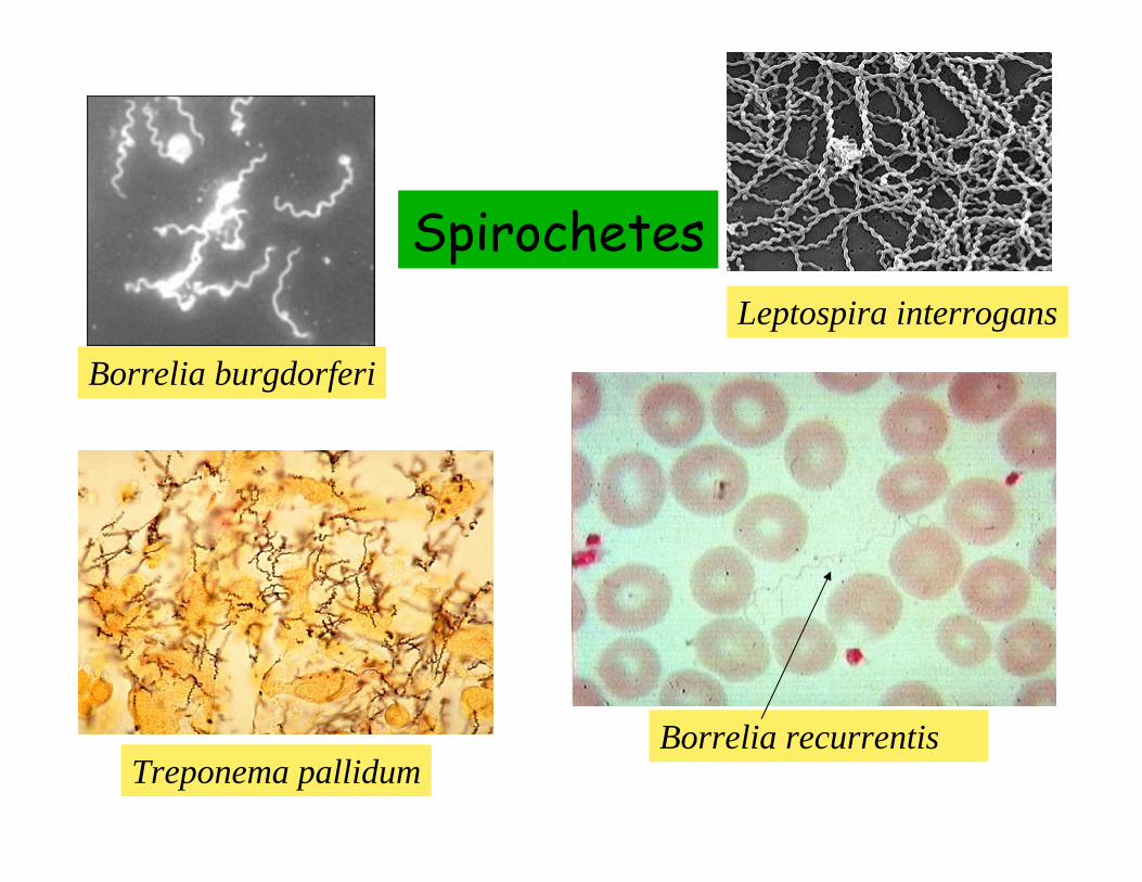
Spirochetes
Borrelia burgdorferi
Treponema pallidumBorrelia recurrentis
Leptospira interrogans

General characteristics• Habitat
– soil, contaminated water, animal parasites• Motility
– Axial filament generates quick corkscrew like motion
• Shape– Can be tightly coiled or curved like R. rubrum
• Examples– Treponema pallidum– Borrelia recurrentis– Borrelia burgdorferi– Leptospira interrogans
Treponema pallidum: causative agent of syphilis
• 5-20μm long, 0.2μm wide• Cannot be cultured in medium
– Humans only natural host– Rabbit testes used for diagnostic or
experimental work• Very fragile
– Not viable for long outside of the body– Easily killed by disinfectants– Very susceptible to antibiotics, including
penicillin

Pathogenesis and clinical dz
• STD• Second only to gonorrhea in frequency of
reporting• Very dangerous because of four stages
– 1o, 2o, latent, 3o
• Congenital transmission• Clinical symptoms largely due to immune
response• Transmission is primarily sexual

Primary syphilis• Lesion at site of entry forms between 10-70
days post infection = chancre• Relatively painless• Especially problematic for females • Disappears spontaneously within 3-6 weeks
giving illusion of a cure

Primary syphilis

Secondary syphilis• 2wks to 6 months later• Systematic spread of spirochetes• Generalized rash and lesions (highly infective)• Fever, headaches, sore throat• Symptoms gradually subside over many years
with three possible outcomes– 1) Complete remission (30-40% untreated)– 2) recurrence 2o lesions at 3-12 month
intervals– 3) progression into latent and then 3o
• Latent = high serum titers• No clinical symptoms• 50% of these individuals develop no further
symptoms

papulosquamousrash
Secondary syphilis

Tertiary syphilis• Occurs 3 to 30 years after secondary• Develops rapidly in immunocompromised
individuals• Gummas = lesions develop due to hypersensitivity
to spirochetes in many body tissues• CNS lesions = neurosyphilis
– Mental changes, major cause of insanity• Aortic aneurysms• Number of tertiary syphilis cases has doubled
since 1986

Tertiary syphilis

Late syphilis:ulcerating gumma


Borrelia recurrentisrelapsing fever
• Tick borne disease• Can be cultured in vitro• Also called epidemic relapsing fever = campers• Symptoms
– Sudden fever, headache, muscle pain, sometimes rash, then nausea, vomiting, prostration
– Patient appears to recover, then relapses to initial symptoms 4-10 times
• Treat with broad spectrum antibiotics, such as tetracyclines

Borrelia burgdorferiLyme disease
• First American case 1975, reported in Europe in 1900’s
• Reported from most states• Vector = Ixodes• Seasonal occurrence due to increased
outdoor activity• Can be cultured in vitro

Lyme disease• Two distinct clinical stages
– Early• Flu-like symptoms• Swollen lymph nodes, fever, headache, stiff neck,
arthralgias, myalgias, fatigue• Erythema migrans
– Site of inoculation 2-4 wks post– Late
• One month to several years post early symptoms• Neurologic, cardiac, arthritic symptoms• 6% develop chronic arthritis

Lyme disease
Erythema migransIxodes scapularis

Leptospira interrogansLeptospirosis, “rat fever”
• Water contaminated with urine from infected animals
• Common in vets, abattoir workers, farmers, dog owners
• Increased prevalence summer months• Entry: breaks in skin and mucosa• Spread through blood leading to kidney infection,
flu-like symptoms• <100 cases annually in US• Vaccine available for high risk individuals• Antibiotics such as penicillin, streptomycin,
tetracycline not effective >4days post-infection

Leptospirosis
Weil’s syndrome: severe leptospirosis

Leptospira: favelas in Brazil

Aerobic/microaerophilic, motile, helical/vibrioid gram negative bacteria
• Not included in spirochetes because no axial filament
• Contain single flagellum at one or both poles, sometimes tufts, rigid
• Habitat– Most aquatic (Spirillum volutans)– Soil
• Nitrogen fixing bacteria such as Asospirillumassociated with tropical grasses, corn, sugar cane
Helical bacteria

Helical pathogens• Campylobacter: microaerophilic normal inhabitants
of intestinal flora of domestic animals and poultry– Campylobacter fetus
• Abortion in domestic animals• Acute gastritis in immunocompromised
– Campylobacter jejuni• Acquired from food, milk, contact with infected
animals• Symptoms: bloody diarrhea, fever, headache,
abdominal pain, self limiting (6-10 days)– Helicobacter pylori
• Culture on chocolate agar under microaerophilicconditions
• Gastritis, stomach and duodenal ulcers• Chronic infections associated with stomach cancer

Vibrioids• Bdellovibrio
– Preys on gram negative bacteria
– Reproduces in periplasmic space by fragmentation
• Vampirovibrio– Parasitic bacterium
Bdellovibrio attacking S. serpens

Pseudomonads• Common soil inhabitants• Can also be pathogenic• Large variety of enzymes
– Important decomposers of unusual substances including pesticides
– Some can grow in antiseptics– Some show tendency toward plasmid mediated
antibiotic resistance– Aerobic, but may also use N as final electron
acceptor– Can cause N depletion of the soil
Gram negative aerobic rods and Gram negative aerobic rods and coccicocci

Medically important pseudomonads
• Pseudomonas pseudomallei– Extremely virulent, endemic in Southeast Asia
• Pseudomonas aeruginosa *• Legionella *• Neisseria gonorrhoeae• Moraxella• Francisella tularensis *• Bordetella pertussis *• Brucella *

Pseudomonas aeruginosa• Obligate aerobe can be
cultured in vitro• Produce blue green water
soluble pigment called pycocyanin that diffuses into culture medium
• Produces exotoxin which prevents protein synthesis in infected host cells
• Ubiquitous in the environment, found on plants

Pseudomonas aeruginosa• First observed on
bandages covering infected wounds = sweet grape-like odor
• Nosocomial infections, especially in patients with CF, burns, urinary catheters, cancer chemo
• 60-70% mortality, very resistant to therapy
• Pseudomonas skin infections in people using contaminated hot tub
P. aeruginosa in a biofilm from catheter

Pseudomonas aeruginosa• Mariana Bridi da Costa, 20 yo
Brazilian model and participant in Miss World
• Septicemia due to Ps aeruginosaUT infection
• Misdiagnosed as kidney stones on late Dec, 2008
• Admitted to hospital on Jan 3, 2009 in septic shock– Septicemia decreases O2 supply
to extremities = necrosis of tissue
– BT = O neg, difficulty in finding donors
• Both arms and both legs amputated, stomach removed, both kidneys removed
• Died Feb 24, 2009 at 3:30am

Legionella pneumophila:Legionnaire’s disease
• Pleomorphic, cultured in guinea pigs, embryonatedeggs, nutrient media
• Natural habitat: water and soil, intracellular parasite of protozoa
• Initial occurrence– Summer 1976 American
Legion convention in Philadelphia with 5000 attendees
– 2 week incubation period– 221 cases, 170
hospitalized, 41 deaths

Legionnaire’s disease• Two forms of disease
– 1) mild flu-like with year round occurrence called Pontiac Fever
– 2) Legionnaire’s—more severe, most common in immunocompromised individuals (age, respiratory illness)
• Symptoms– Signs of lobar pneumonia 2-10 days post-
exposure– Diarrhea, weakness, headache, muscle aches,
malaise, anorexia, dry cough, increase temperature lasting 5-16 days
– Evades immune system in phagocytic monocytes

Legionellosis outbreaks in US– 1985
• 14 cases, 3 fatal, church banquet in Michigan– 1989
• 34 cases, 2 fatal, contaminated mist machine in Louisiana– 1998
• 45 cases of Pontiac fever, whirlpool in Wisconsin hotel– 1999
• 22 cases of Pontiac fever, 2 Legionnaire’s, Georgia hotel – 2000
• Potting soil in California, Oregon, Washington• 20 cases of Pontiac fever, whirlpool at Wisconsin hotel• 15 cases of Legionnaire’s, exposure to plant lagoon in Minnesota
– 2001• 10 cases, 1 fatal, contaminated cooling tower in Cleveland auto
plant– 2002
• 16 cases in prison in Connecticut• 117 cases of Pontiac fever, restaurant patrons in Tennessee
– 2004• 66 cases Pontiac fever, hotel in Oklahoma

Francisella tularensis:tularemia
• Zoonoses with wild animal reservoir• Organism
– Gram negative coccobacilllus– Requires cysteine and blood in culture medium
for growth• Transmission
– Direct contact, ingestion of contaminated food/water, insect vector (ticks with transovarial passage and deerflies)
– Associated with hunters, usual source rabbits or rodents

tularemia• Pathogenesis
– Ulcer-like lesion at site of inoculation or bite– Spreads to regional lymph nodes = swelling, pain
• Systemic symptoms– Dizziness, headache, chills, fever, sweating,
prostration, gi symptoms, pneumonia• Course
– Heals in 4-6 weeks– Complete resolution in 3-6 months– Some relapse (sequestered in host cells)– Mortality = 10% in untreated cases

Tularemia clinical images
Tularemia ulcerTularemia pneumoniaWith bilateral infiltrates

Bordetella pertussiswhooping cough
• Frequent cause of death in children before vaccine
• Organism– Strict aerobe– Produces exotoxin
• Induces accumulation of mucus, destruction of cilia, excessive coughing
• Transmission– aerosol

Whooping Cough

Whooping cough• Pathogenesis and symptoms
– Attaches to ciliated epithelial cells of respiratory tract
– 10 day incubation period– Prodrome similar to URI last 2 weeks
• Sneezing, runny nose, coughing– Stage 2 (1-6 weeks)
• Paroxysms = uncontrollable coughing spasms• Can’t breath between coughs, anoxia, vomiting
• Distribution– Worldwide, no seasonality– 45% in children under 1 year old

Brucella• 6 species, cause abortion in animals, 4
cause human disease (3 most common)– B. abortus = disease in cattle– B. melitensis – sheep and goats– B. suis= swine
• Organism– Gram negative– Pleomorphic coccobacillus
• Transmission– Humans in contact with contaminated animal
products, unpasteurized milk or milk products
– Disease = brucellosis

brucellosis• Pathogenesis
– Enter body via lesions, cuts, ingestion, inhalation– Associated with intracellular survival: phagocytosed
by WBC and survive inside PMN’s and macrophage– Distributed through lymphatic system to liver
• Symptoms– Hepatosplenomegaly– Generalized clinical symptoms over weeks or
months– Undulant fever, weakness, malaise, body ache,
headache,sweating – Prolonged recovery– Most common among people who work with animals

Brucellosis• “ brucellosis bacteria can remain living in
refrigerated contaminated milk for up to 10 days, in Roquefort cheese for 2 months, and in refrigerated butter for 4 months. In the environment the bacteria can survive in water for 10 to 70 days and in the dust or soil for up to 10 weeks. “
From: BRUCELLOSIS, UNPASTEURIZED DAIRY PRODUCTS - MEXICO (02): (GUANAJUATO) Aug 2009by Gaby Barcenas

Brucellosis
CT scan: human brucelloma of spleen

Agricultural importance• Rhizobium/Bradyrhizobium
– Formation of nodules in roots of legumes– Nitrogen-fixing symbionts
• Agrobacterium tumefaciens– Plant pathogen– Causes crown gall– Developed by inserting plasmid into
plant’s chromosomal DNA

Crown Gall

Facultatively Anaerobic Gram-negative Rods
• Medically important group = Enterbacteriaceae
• Vibrionaceae = 20 species of vibrio, 11 pathogenic to humans– V. cholerae– V. vulnificus
• Pasteurellaceae– Pasteurella– Haemophilus
• Haemophilus influenza

Enterbacteriaceae• In general:
– Normal flora and pathogens of large intestines of humans and animals
– 100,000 annual deaths in US– Also found in soil, water, decaying matter– Distinguish by carbohydrate fermentation and
serology– Produce lytic enzymes= bacteriocins which lyse
closely related bacteria– Cannot destroy by freezing, often resistant to
antibiotics• Examples
– Escherichia coli– Salmonella– Shigella– Yersinia

Escherichia coli
• Roles– Normal microbial flora– Pathogenic when in “wrong place” or in
immunocompromised host• Some produce enterotoxins (exotoxins) resulting in fluid loss
and diarrhea– Important in biotechnology
• Four categories– Enterotoxigenic (ETEC)– Enteropathogenic (EPEC)– Enterohemorrhagic (EHEC)– Enteroinvasive (EIEC)

EHEC
• Produces toxin similar to shiga toxin of S. dysentareiae
• Prominent serotype = E. coli 0157:H7– Transmission
• Contaminated raw milk, undercooked meat, fast food, apple juice, sprouts, etc.
– Pathogenesis and symptoms• Begins with watery diarrhea and abdominal pain
x4days post exposure• Turns into bloody diarrhea usually with complete
recovery in 10 days• 10% children < 5 = HUS (life threatening-direct
kidney damage from toxin)

E. coli O111• Less common EHEC• 291 cases reported among patrons of
Country Cottage Restaurant in Oklahoma between Aug 15-17, 2008
• 67 pts hospitalized, 16 on dialysis for HUS
• Source has not been identified

Salmonellatyphoid fever, bacteremia,
enterocolitis
• 5 major species, 1500 serotypes• Salmonella enterocolitica
– Common microbial flora of animals, birds– Transmitted to humans– 18-36 hours after infection
• Food poisoning, diarrhea, fever, abdominal pain, nausea• Self limiting• Dangerous in very young or old due to dehydration
• Salmonella typhi = typhoid fever

Typhoid fever• Pathogenesis
– Attaches to & penetrates epithelial lining of small intestines
– Multiplies intracellulary after phagocytosis by macrophage, carried throughout body
• Clinical symptoms– Initially fever, malaise, lethargy, aches & pains– Infection of gallbladder & shedding into intestinal lumen– Lesions of Peyer’s patches– Constant high fever, tenderness, diarrhea or
constipation, vomiting• Transmission
– Typhoid Mary– Sewage system, contaminated water, etc.
• Treatment: chloramphenicol (resistant strains), smx/tmp, ampicillin

Mary Malon• Article appeared in
NY Times January 20, 1909
• First “healthy carrier” of typhoid fever in US– Occurs in 3-5% of
typhoid cases– Recent studies have
implicated typhoid coated gall stones

Shigellabacillary dysentery/shigellosis
• Primarily human pathogen• 20,000 cases annually in US• Four species
– S. dysenteriae• Most dangerous due to shiga toxin• Can develop immunity if exposed, but remain a
carrier– S. flexneri– S. boydii– S. sonneii

Dysentery• Transmission
– Fecal/oral (5 f’s)– Ability to maintain personal hygeine
• Pathogenesis– Ingested, penetrate large intestines,
phagocytosed, intracellular multiplication– Penetrate only to submucosa & cause
inflammation and sloughing of epthelium = lesions
– Incubation period = 1-3 days• Symptoms
– Abdominal cramps, fever, diarrhea(mucus/blood), dehydration, loss of electrolytes – can be fatal
– Self limiting with 3-7 days to recovery

Yersinia pestis = plague• Endemic in Asia, occasional US case• Organism
– Non-motile, gram negative coccobacillus• 3 Transmission cycles
– 1) natural cycle = sylvatic plague• Wild rodent reservoir• Transmission among rodents by fleas, usually
carried by squirrels, mice, prairie dogs, chipmunks
• Some infections subclinical, others apparent with death
– 2) urban/domestic cycle• Spread to urban rodents living in close
proximity to humans– 3) human plague
• Human bitten by infected flea

Plague
• Bubonic plague – Flea bite usually in leg with spread to lymph nodes– Multiplication and swelling in lymph node = buboes– Fever, chills, nausea, malaise, pain, bacteremia
• Septicemic plague– Spreads into blood resulting in purple lesions due to
leaking blood (black death)– Bacterial emboli trapped in lungs
• Pneumonic plague– Aerosolized from emboli– Death within 2 to 3 days
• Mortality– Untreated bubonic and septicemic = 50-75%– Pneumonic = 100%

Plague
Characteristic safety pin appearance

Plague: history
• AD550– 100 million deaths over 60 year period
• 14th century– 25% of population of Europe died
• Decline since 1800’s• India 1994
– Over 5000 persons infected• US 1944-1993
– 362 cases of human plague– Very susceptible to tetracycline, streptomycin,
chloramphenicol

VibrionaceaeV. cholerae = cholera
• In general– Can be grown in
vitro– Over 100
serotypes, with 01 responsible for epidemic cholera
• Transmission– Fecal/oral– Contaminated food
and water

Cholera
• Attach to & proliferate in small intestines
• Produces enterotoxin– Adenylate cyclase via signal transduction
• Result:– Huge water and electrolyte loss from
cells– Acidosis, dehydration, shock and death

Cholera
• Incubation period– 1-2 days with abrupt onset
• Initial symptoms– Vomiting and diarrhea
• Mortality– 60% without treatment
• Occurrence– India, Asia, Africa– US due to improperly cooked shellfish

Images of cholera
Cholera dehydration
Cholera cot

Oral Rehydration Therapy
• Recipe– ½ tsp salt (NaCl)– ¼ tsp sodium bicarbonate (NaHCO3
-)– ¼ tsp KCl– 4 T sugar (sucrose)
– Dissolve in 1 Liter of boiled water

PasteurellaceaeH. influenza
• Organism– Requires special
growth factors (hemin and NAD) only found in blood
– Major disease producing organism, only in humans
– Small gram negative coccobacillus
– Many subtypes based on metabolic reactions
• Virulence factors– Ability to produce
capsule– Produces enzymes
which degrade IgA

H. Influenzapathogenesis
• Colonizes respiratory tract of >50% children (asymptomatic carriers)
• Most common in young children
• Causes: nose & throat infections, sinus, middle ear, pneumonia, rarely meningitis, Hib epiglotittis (obstruction of trachea), infectious conjunctivitis
• Leading cause of invasive bacterial disease in children in US in 1980’s (8-10,000 cases)
• Transmission:– Respiratory droplets
• Immunity– Passive infant from
mother– Vaccine = 95%
decrease in dz in US
Empyema due to H. influenza

Anaerobic, gram-negative, straight, curved, helical rods
• Bacteroides (B. fragilis)– Inhabitants of human gi tract, oral cavity,
genital tract, respiratory tract– Involved in polymocrobic disease– Usually results from injury, surgery,
appendicitis, cancer allowing fecal material entry into abdominal cavity
– Abscesses responsible or PID secondary to abortion, surgery, cancer, prolonged labor, IUD’s, gonorrhea
– Also liver abscesses, respiratory tract infections, infections associated with human bite, septicemia, brain abscesses

Rickettsias and Chlamydias
• General– Obligate intracellular parasites– Halfway between virus and bacteria– Pleomorphic, divide by binary fission
• Rickettsias– Coxiella burnetti– Rickettsia prowazeckii, typhii, ricketsii
• Chlamydias– Chlamydia trachomatis

Coxiella burnettiQ fever
• Transmission– To animals by insect bites = inapparent infection– Shed in urine, feces, milk, other body fluids– To humans via contaminated milk, aerosols– Especially common among cattle farmers where
spread among cattle by ticks• Requires increased time and temperature to
pasteurize because organisms are thermotolerant!
• Symptoms– Flu-like, prolonged fever, chest pains, pneumonia– Rarely fatal
• Sequelae: endocarditis much later because organism sequesters in liver

Rickettsia prowazekiiepidemic typhus
• Named after two investigators who died from lab infections of typhus
• Mortality rate– 70% in untreated
• Transmission– Conditions of poor hygiene and
overcrowding– Body louse feeds on infected humans,
moves to next host and excretes rickettsiae when feeding
– Causes itching, scratch forces contaminated feces into wound

Epidemic typhus
• Pathogenesis– Multiply in endothelial cells of blood vessels– Spread throughout body causing
inflammation/swelling of blood vessels– Blockage of bv, reduced blood flow and leakage
leading to rashes and spots• Symptoms
– Headaches, chills, fever, stupor, delirium, shock– Can recrudesce as Brill-Zinser disease

Epidemic typhus
Pediculus corporis

Epidemic typhus and history• Determine the course of many military campaigns
– 1489 Spanish siege of Granada• 3000 Moorish troops killed in battle, 17,000 died
of typhus– 1528 French army in Naples
• Verge of victory when 30,000 French soldiers died of typhus—they lost!
– Napolean in Moscow• Started with 500,000 troops, 300,000 killed by
typhus– WWI
• 30 million people infected in Russia, over 3 million died
• Italian delousing stations and DDT

ChlamydiasChlamydia trachomatis
• Intracellular parasite seqeusteredwithout disease symptoms until later time
• Three categories of disease– 1) trachoma– 2) sexually transmitted diseases– 3) lymphogranuloma venereum

Trachoma • Infection of eyelid can cause inturn of eyelashes and ulceration of cornea (entropion)
• Can result in blindness
• Poverty, overcrowding, unsanitary conditions
• Spread by direct or indirect contact
• Humans only host

Lymphogranulomavenereum
• STD• Initial symptoms include lesions on
skin/mucous tissues of genitals• Enlarged and painful buboes in
inguinal lymph nodes which suppurate and drain
• Fever, nausea, headache, conjunctivitis or skin rash, can lead to permanent lymphatic or rectal blockage

Lymphogranulomavenereum

Chronic lymphogranuloma venereum in female
Genital elephantiasis

Mycoplasmas• No cell wall• Smallest living free bacteria
(300nm), some parasitic• Natural flora in mouth, throat,
genitourinary tract of mammals and birds
• Pleomorphic, facultative anaerobes or aerobes
• Divide by binary fission or fragmentation

Mycoplasmas
• Mycoplasma pneumonia– Primary atypical pneumonia– Chest pain, fever, pharyngitis, bronchitis
• Mycoplasma hominis– Microbial flora of genitourinary tract– Postpartum septicemia
• Ureaplasma urelyticum– Std, common cause of urethritis, postpartum
fever, spontaneous abortions, low birthweightinfants
• Thermoplasma– Harmless, found in hot water systems

Gram-positive cocciStaphylococcus aureus
• Found on skin, nose, around rectum of healthy persons
• Organism– Facultative anaerobe, non-
spore forming, grown on blood agar, capsule former
• Difficult to treat– 1) Survives for months
dried pus/bodily fluids– 2) More resistant to
disinfectants than most vegetative bacteria
– 3) plasmids are readily transferred, many resistant strains

S. aureus: pathogenesis & virulence factors
• Causes infections when gets into deeper tissues
• Characterized by suppuration, localized inflammation
• Produces toxins and enzymes– Exfoliatin
• Causes superficial layers of skin to peel– Enterotoxins
• Acute intestinal symptoms• Toxin associated with toxic shock
syndrome

S. aureus: superficial & systemic infections
• Superficial infections– Most common– Boils, carbuncles, impetigo, infections of
surgical/accidental wounds or burns– Characterized by abscess formation
• Systemic infections– Squeeze a boil = carries staphylococci into
blood– Result: abscess in deep organs = liver, lung,
brain tissue– Bacteremia leads to high mortality

S. aureus: toxic shock & scalded skin syndrome• TSS
– High fever, vomiting, diarrhea, sometimes sore throat, muscle aches, kidney failure, red rash
– Skin sheds from feet and hands– Associated with Rely tampons
• SSS– Children and infants infected with exfoliatin
producing strains– Usually benign, can be fatal
• Food poisoning– Ingest food containing preformed toxin (cured
meat)– Nausea, vomiting, diarrhea, abdominal pain,
prostration x2 days, usually complete recovery

Scalded skin syndrome

Gram-positive cocciStreptococcus
• Susceptible to disinfectants and chemotherapeutic agents
• Normal microbial flora of mouth, skin, nose, mucosal surfaces of animals and humans
• Many hemolytic• Classified into groups using
Lancefield system = 20 groups– Distinguishes between
carbohydrate antigens in cell wall
• Most important human pathogen = S. pyogenes

Streptococcus pyogenesdefense mechanisms
• Capsule• M-protein
– antiphagocytic• Erythrogenic
toxins– Produces fever,
rash (scarlet fever)• Streptolysin O & S
– hemolytic– Hyaluronidase
• Hyaluronidase– Break down
hyaluronic acid holding together neighboring cells
• Streptokinase– Digests fibrin in
inflammatory barrier
• Protein F– Promotes adhesion
to cells in pharynx and others

Streptococcus pyogenespathogenesis & sequelae
• Pharyngitis– Strep throat, caused by group A strep– Different strain each time with different
virulence levels = immunity– Must seek treatment
• Sequelae– Scarlet fever– Streptococcal toxic shock-like syndrome– Puerperal fever– Skin infection– Poststreptococcal disease

Streptococcal toxic shock-like syndrome
• Group α, β hemolytic strep• Symptoms
– High fever, erythema, pupuric skin rash– May become gangrenous– Necrotizing fasciitis
• Can begin as cellulitis or pharyngitis and develop to serious stage in 24 hours
• Renal failure, pneumonia, cardiac failure• Bacteria produce large amounts of streptococcal
pyrogenic exotoxin A• Mortality rate
– 30%– If patient develops pneumonia, 50%

Necrotizing fasciitis

Endospore forming gram-positive rods and cocci
• Bacillus anthracis• Bacillus thuringiensis• Clostridium botulinum• Clostridium tetani• Clostridium difficile

Bacillus anthracisanthrax
• Primarily disease of cattle and sheep• Model for study of infectious disease
because relatively large (5-10μm)• Very resistant
– Can persist in soil and animal products for years
• Pathogenesis– Enters through skin abrasions, inhalation,
ingestion– Contains unusual capsule composed of D-
glutamic acid related to signs and symptoms

Anthraxthree clinical forms
• Cutaneous– Most common, Spores enter tissues through
abrasions or lesions– Infection on exposed skin surfaces– Swollen lesions develop and form black scab =eschar– Low mortality if localize,If spreads to blood (5%)
often fatal• Pulmonary
– Inhalation of spores usually by person handling contaminated animals (wool sorters disease)
– High fever, respiratory distress, pneumonia, sepsis,death in untreated cases
• Intestinal– Eating contaminated meat– Severe enteritis, high mortality– Very rare

Anthrax
Cutaneous anthrax
Pulmonary anthrax

Bacillus thuringiensismicrobial larvicide
• Insect pathogen causes intoxication in larva resulting in paralysis so they cannot feed
• Used to control growth of insect larvae in dairy ponds (even in Turlock!)– Killed 90% of larva in pond by end of 2nd week!
• Different strains specific to different species of insect– Dipterans, lepidopterans, coleopterans
• Toxin extracted and introduced into plants to induce insect resistance
Clostridium botulinumbotulism
• Organism– Large, gram+, anaerobic bacillus – Produces endotoxin causing food poisoning
• 1-2μg produces illness/death in humans– Very heat resistant spores– Can survive in temperatures >100oC for several
hours• Pathogenesis
– Ingestion of preformed toxin produced during growth in food
– Toxin absorbed in small intestines, into blood, into peripheral nerves
– Prevents release of ACh from motor neurons– Paralysis of respiratory functions

botulism
• Symptoms– 12-36 hours post ingestion up to 8 days– Weakness,dizziness, diplopia, dysphonia, dysphagia,
dilated pupils– Muscle weakness, paralysis
• Mortality– 20-70% depending on amount and serotype
• Infant botulism– Discovered in 1976– Bacteria grows in intestines due to ingestion of
spores (honey)– Not all infants susceptible – Can be mistaken for SIDS

botulism
• Grows in animal feed, in sediments in lakes and ponds– Responsible for death of thousands of
waterfowl & other wild birds• Transmission
– Spores may be present on food– Toxin produced when microbe in anaerobic
environment in foods stored at room temperature, especially alkaline foods (ph<4.6 inhibitory)
– Home canned foods, commercially canned foods (rarely)
– Used cosmetically to prevent brow wrinkles (botox)

botox

Clostridium tetani: tetanus• Organism
– Large, gram-positive, spore former
– Obligate anaerobe– Produces exotoxin called
tetanospasmin– Found in soils and manures
• Pathogenesis– Contamination of wounds with
spores (usually no disease)– Must have anaerobic conditions
& some dead tissue– Spores germinate, bacteria
proliferate producing toxin– Signs and symptoms appear
within 4-10 days– Puncture wounds, umbilical cord
infections in infants

tetanus
• Signs and symptoms– Toxin spreads to spinal cord– Effects synapse so that signal is continuous
= tetany of muscles– Muscle stiffness, spasms of jaw = lockjaw– Exhaustion, then spasms so strong that
bone breaks– Respiratory complications– High death rate– Nonfatal cases recovery completely in a
few weeks

Tetanus: history
• Most frequent source of infection = soil contaminated wounds
• Civil War = hundreds of cases• World War II = only 12 cases due to
availability of tetanus toxoid• Tetanus toxoid
– Infants at 6wks/3mo (DPT), 3 doses followed by boosters every 10 years

tetanus
risus sardonicus
Neonatal tetanus

Regular, nonsporing, gram-positive rods
• Lactobacillus– Produces lactic acid from carbohydrates– Normal inhabitant of human
gastrointestinal tract, oral, vaginal areas
– Used to produce buttermilk (L. delbrueckii), acidophilus (L. acidophilus), and pickles
• Listeria monocytogenes– listeriosis

listeriosis• Occurrence
– Primary infection in animals
– Can occur in human fetuses, newborns, pregnant females, immunosuppressed individuals
• Pathogenesis– Enters tissues,
phagocytosis by monocytes & macrophage
– Intracellular development
– Spread to adjacent cells– Can be controlled by CMI

listeriosis
• Fetus and newborn– Into blood from infected mother– Spontaneous abortion or stillbirth
• Survivors– Septicemia – Lesions of the legs and trunk– Meningitis most common manifestation
• Adults– From contaminated milk products, water,
domestic animals, human carriers– Colonize throat & gi tract– Spread to lymph and bloodstream

listeriosis
• Clinical symptoms– Pharyngitis– Gastroenteritis– Speticemia– Meningitis– Endocarditis– Pneumonia– Abscesses in organs
• Prevention– Drink pasteurized milk!

Irregular non-sporing gram-positive rods
• Corynebacteria– In general
• Narrow rods up to 5μm in length• Often form a “V” or “L” shaped arrangement
= pallisades• Pleomorphic, club shaped with metachromatic
granules– C. diphtheriae
• Actinomyces– In general
• Anaerobes found in mouth,throat of humans and animals
– A. bovis– A. israelii

Corynebacterium diphtheriaediphtheria
• Occurrence– Humans only natural host– Transmitted by airborne
droplets• Organism
– More resistant to drying than other vegetative organisms (3/4 month in dried respiratory exudate)
– Must be infected with lysogenic β-corynephage to produce exotoxin
• Pathogenesis– Colonize mucosal tissue of
upper respiratory tact– Toxin prevents protein
synthesis

Diphtheria
“bull neck”Pseudomembrane formation

Mycobacteria
• In general– Aerobic, non-sporeforming non-motile, rod-
shaped, acid fast (most pathogenic species), many native in soil
• M. tuberculosis• M. leprae• M. bovis
– Tuberculosis of cows– May be transmitted to people through
contaminated milk

M. tuberculosistuberculosis
• Organism– Slow growing,
obligate aerobe• Pathogenesis
– May be dormant for years
– Stages• Primary• Secondary

Primary tb• Inhalation of aerosolized bacteria from infected person• Bacilli multiply in alveoli and alveolar macrophage to
lymph nodes, initiating CMI (scar formation around bacilli)
• Gives rise to positive skin test, but no other symptoms• Three options
– Bacteria may be completely destroyed = no further symptoms
– Disseminated tb• Young children = seeded throughout body = fatal• Miliary tuberculosis
– Latent-dormant tb• Continuation of primary• May result in reoccurrence

Complications of tuberculosis
TB peritonitisSlide showing AFB in situ

Secondary tb• 90% of these individuals harbor tb in
latent/dormant stage for more than one year
• Usually immunocompromised• Develop of caseous necrosis• Erosion of bronchial tubes, expulsion of
bacilli through airway• Healing or treatment difficult• Consumption = can result in death• Treatment
– Dots– MDRTB– XDRTB

M. LepraeHansen’s disease = leprosy
• In general– Armadillos only natural non-human reservoir– Transfer person to person via respiratory
tract, skin lesions– Incubation period averages > 20 years– Major growth in low temperature body tissues
(nose, ears, skin of extremities)– Some phagocytized and grow inside
macrophage– Early symptoms include anesthesia of body
parts = disfiguring disease

leprosy• Mechanisms of tissue destruction
– Uncertain– Probably a combination of neurological damage,
accumulation of bacilli, immune reactions and secondary infection
• Two forms– Lepromatous
• Most severe• Large nodular lesions• Impaired immune response limits formation of
granulation tissue– Tuberculoid
• Less severe because of normal immune response• Granulation type lesions with few bacilli• Less tissue damage, better response to therapy

13 year old boy and 30 year old man with lepromatousleprosy in Molokai, Hawaii in 1931

Images of leprosy

Mycobacterium lepromatosis
• Found by DNA analysis in 4 patients who died from severe form of leprosy
• Geographically diverse distribution– 2 pts from Singapore, 2 from Mexico– Resulted in cases of diffuse
lepromatous leprosy called DLL “diffuse leprocy of Lucio and Latapi”
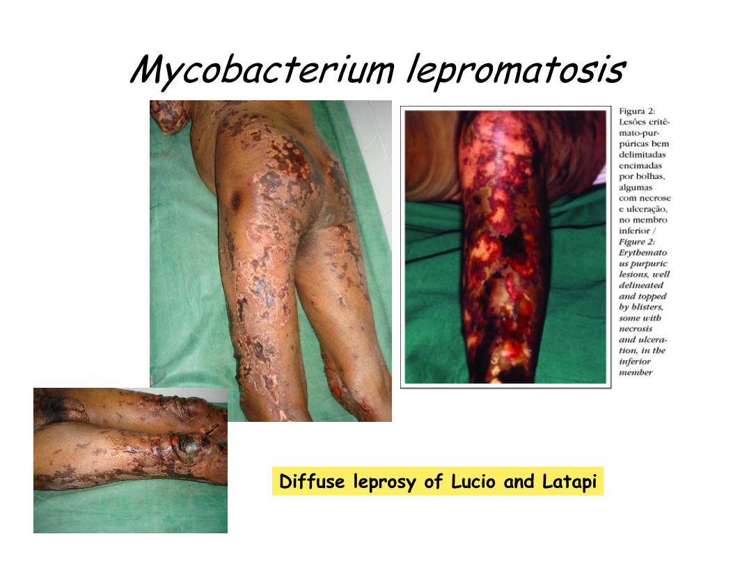
Mycobacterium lepromatosis
Diffuse leprosy of Lucio and Latapi

Nocardioforms
• In general– Look like the Actinomycetes, but aerobic– Reproduce by fragmentation– Often acid-fast– Common soil organisms
• N. asteroides– Opportunist of immunocompromised individuals– Lung infection, often misdiagnosed as tb– Can be systemic– Penetrating dermal lesions with connecting
sinuses (Madura foot)

Madura foot

Archaeobacteria• Halobacterium
– Hypersalineenvironments
• Solar salt evaporation ponds
• Natural salt lakes• Surfaces of heavily
salted foods

Phototrophic bacteria
• Purple and green phototrophic bacteria– Anoxygenic photosynthesis– Uses H2S and other reduced sulfur compounds
to produce granules– Purple and green nonsulfur bacteria
• Use organic compounds for photosynthesis• Cyanobacteria
– Aerobic– Produce oxygen photosynthetically– Can also use anoxygenic photosynthesis of
sulfur– Some fix nitrogen– Varied morphology

Actinomycetes
• In general– Filamentous bacteria– Found in soil
• Streptomyces– Form conidiospores
(like fungi)– Each can produce a
new colony– Strict aerobes– Used for antibiotics