metin n. gurcan, ph.d. - ohio state university college of …€¦ · · 2015-10-23metin n....
TRANSCRIPT

METIN N. GURCAN, Ph.D. Associate Professor
Department of Biomedical Informatics The Ohio State University, College of Medicine
310 G Lincoln Tower Phone: (614) 688-9857 1800 Cannon Drive Fax: (614) 688-6600 Columbus, Ohio 43210 [email protected]
Employment 2010-present Associate Professor, Department of Biomedical Informatics,
The Ohio State University 2007-2010 Assistant Professor, Department of Biomedical Informatics,
The Ohio State University 2006 Research Assistant Professor, Department of Biomedical Informatics,
The Ohio State University 2005-2006 Research Scientist, Department of Biomedical Informatics,
The Ohio State University 2004 – 2005 Senior Algorithmic Engineer, iCAD Inc., Beavercreek, OH
2002 – 2003 CT Product Director, CADx Systems, Inc., Beavercreek, OH
2001 Senior Engineer, Qualia Computing, Inc., Beavercreek, OH
2001 Research Investigator, University of Michigan, Department of Radiology, Ann Arbor, MI
1999 – 2001 Research Fellow, University of Michigan Department of Radiology, Ann Arbor, MI
1998 Instructor, Department of Computer Engineering, Ankara, Turkey
1991 – 1999 Research and Teaching Assistant, Department of Electrical and Electronics Engineering, Ankara, Turkey
Education Postdoctoral Training, Radiology, University of Michigan, 2001 Ph.D. Electrical and Electronics Engineering, Bilkent University, Ankara, Turkey, 1999 M.Sc. Digital Systems Engineering, UMIST, Manchester, England, 1994 B.Sc. Electrical and Electronics Engineering, Bilkent University, Ankara, Turkey, 1991 Honors and Awards 2009-2013 The Ohio State University Comprehensive Cancer Center REAP Award
2013 Senior Member, SPIE
2008 Young Investigator Award, The Children’s Neuroblastoma Cancer Foundation
2007 caBIG™ Embodying the Vision Award, The National Cancer Institute
2006 Senior Member, IEEE
1992 Foreign and Commonwealth Organization Award, British Foreign Office
1987 Bilkent University Foundation Scholarship
1987 Is Bank Scholar Achievement Award

Grants, Contracts: Ongoing: Computer-based assessment of tumor microenvironment (TME) 2014-2017 in Follicular Lymphoma Funding Agency: NIH/National Cancer Institute (R01 CA134451) Role: Principal Investigator Pathology Image Informatics Platform for visualization, analysis and management 2015-2020 Funding Agency:NIH/National Cancer Institute (U24CA199374) Role: Principal Investigator Mesenchynmal stem cells as a protective niche for latent M.tb 2014-2016 Funding Agency: NIAID (R56 AI111823) Role: Consortium PD/PI at OSU Defining molecular events for targeted therapy of glioblastoma 2013-2015 using digital image analysis Funding Agency: OSUCCC Intramural Research Program - 2014 Pelotonia Idea Grant Role: Principal Investigator A low-cost alternative automated segmentation Acne REcognition System (ARES) for acne image analysis, and clinical trial scoring 2015-2016 Funding Agency: American Acne and Rosacea Society Role: Co-investigator Genetic-based susceptibility to pulmonary tuberculosis 2015-2017 Funding Agency: NIH/National Institute of Allergy and Infectious Diseases (NIAID) (R21 AI115038) Role: Co-investigator CellMarker 2013-2015 Funding Agency: Ohio Development Services Agency Role: Principal Investigator Task-Specific Compression for Biomedical Big Data 2015-2018 Funding Agency: NIH/National Cancer Institute (U01 CA198945) Role: Consortium PD/PI at OSU
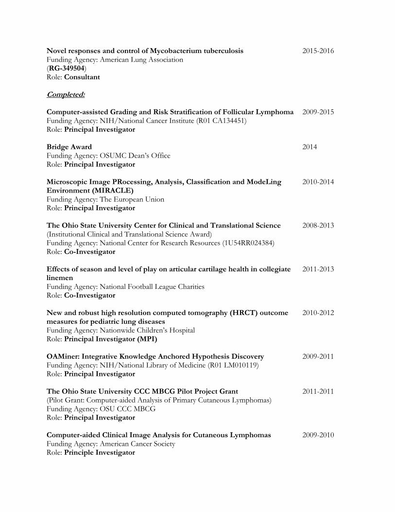
Novel responses and control of Mycobacterium tuberculosis 2015-2016 Funding Agency: American Lung Association (RG-349504) Role: Consultant Completed: Computer-assisted Grading and Risk Stratification of Follicular Lymphoma 2009-2015 Funding Agency: NIH/National Cancer Institute (R01 CA134451) Role: Principal Investigator Bridge Award 2014 Funding Agency: OSUMC Dean’s Office Role: Principal Investigator Microscopic Image PRocessing, Analysis, Classification and ModeLing 2010-2014 Environment (MIRACLE) Funding Agency: The European Union Role: Principal Investigator The Ohio State University Center for Clinical and Translational Science 2008-2013 (Institutional Clinical and Translational Science Award) Funding Agency: National Center for Research Resources (1U54RR024384) Role: Co-Investigator Effects of season and level of play on articular cartilage health in collegiate 2011-2013 linemen Funding Agency: National Football League Charities Role: Co-Investigator New and robust high resolution computed tomography (HRCT) outcome 2010-2012 measures for pediatric lung diseases Funding Agency: Nationwide Children’s Hospital Role: Principal Investigator (MPI) OAMiner: Integrative Knowledge Anchored Hypothesis Discovery 2009-2011 Funding Agency: NIH/National Library of Medicine (R01 LM010119) Role: Principal Investigator The Ohio State University CCC MBCG Pilot Project Grant 2011-2011 (Pilot Grant: Computer-aided Analysis of Primary Cutaneous Lymphomas) Funding Agency: OSU CCC MBCG Role: Principal Investigator Computer-aided Clinical Image Analysis for Cutaneous Lymphomas 2009-2010 Funding Agency: American Cancer Society Role: Principle Investigator

Role of PTEN in the Tumor Microenvironment 2007-2010 Funding Agency: Department of Defense (W81XWH-07-1-0402) Role: Co-Investigator Computer-aided Prognosis of Neuroblastoma 2008-2010 Funding Agency: The Children’s Neuroblastoma Cancer Foundation Role: Principal Investigator caBIG in vivo imaging workspace Core Middleware Development 2007-2008 Funding Agency: NIH/National Cancer Institute (caBIG-IMG-22-04132006) Role: Co-Investigator
Membership and Offices in Professional Organizations
2013 – Present Senior Member, The International Society for Optical Engineering (SPIE)
2001 – 2013 Member, The International Society for Optical Engineering (SPIE)
2000 – Present Member, Radiological Society of North America (RSNA)
1998 – 1999 Electronics Communication Development Officer, IEEE Region 8
1994 – Present Member, IEEE Signal Processing Society
1994 – Present Member, IEEE Engineering in Medicine and Biology Society
2006 – Present Senior Member, Institute of Electrical and Electronics Engineering (IEEE)
1990 – 2006 Member, Institute of Electrical and Electronics Engineering (IEEE)
Professional and Educational Activities
PhD Thesis Supervised:
Olcay Sertel: “Image Analysis for Computer-aided Histopathology,” 2010
Jeffrey Prescott: “Computer-assisted Discovery and Characterization of Imaging Biomarkers for Disease Diagnosis and Treatment Planning,” 2010.
Postdoctoral Researcher Mentorship
Ashish Sharma, PhD
Berkant Barla Cambazoglu, PhD
Kamel Boussaid, PhD
Sufyan Ababneh, PhD
Furqan Haq, PhD
Myriam Oger, PhD
Hatice Cinar Akakin, PhD
Hui Kong, PhD
Khalid Niazi, PhD
Mohammad Faizal Ahmad Fauzi, PhD
Keluo Yao, MD
Fazly Salleh Abas, PhD

Pelin Kus, PhD
Evgin Goceri, PhD Faculty Mentorship
Gary Tozbikian, MD
David Liebner, MD Medical Student Supervision
Mark Swanson
Michael Priddy
John Bernot
Salma Shaikhouni Journal Special Issue Editor:
IEEE Transactions on Medical Imaging: Special Issue on Multivariate Microscopy Image Analysis, 2010
IEEE TBME Letters: Special Issue on Emerging Technologies in Multi-parameter Biomedical Optical Imaging and Image Analysis, 2010.
Computerized Medical Imaging and Graphics: Special Issue on Whole Slide Microscopic Image Analysis, 2011.
Journal Editorial Board /Associate Editor:
Journal of Pathology Informatics, 2010 – present
Journal of Medical Imaging, 2013- present
Signal, Image and Video Processing, 2013 – 2014 Conference Chair:
SPIE Medical Imaging Digital Pathology, 2013-present Conference Program Committee:
IEEE-EURASIP Workshop on Nonlinear Signal and Image Processing (NSIP), 1999
IEEE International Symposium on Computer and Information Sciences (ISCIS), 2009
International Symposium on Visual Computing (ISVC), 2009, 2010
Pathology Informatics, HIMA Workshop, 2010 - present
SPIE Medical Imaging, Computer-aided Diagnosis 2011 – 2012
SPIE Medical Imaging, Chair, Digital Pathology 2012 - present

Grant Reviewing Committees
Maryland Industrial Partnership Program, 2007
The National Cancer Institute, Network for Translational Research: Optical Imaging, 2008
The National Institutes of Health, Special Emphasis Panel ZRG1 – SBIBV, 2009
The National Institutes of Health, BMIT-A, 2011
National Science Foundation, 2012
The National Institutes of Health, BCHI, 2014
The National Institutes of Health, BDMA, 2014
The National Institutes of Health, ZRG1 BST-N (51) R, 2014
National Science Foundation, 2014
The National Institutes of Health, ZCA1 SRB-J (M2) S, 2015
Book Reviewing
Wiley-IEEE Press, 2009-2010 Journal Reviewing
IEEE Transactions on Image Processing
IEEE Transactions on Medical Imaging
IEEE Transactions on Systems, Man and Cybernetics – Part B
IEEE Signal Processing
IEEE Selected Topics on Signal Processing
IEEE Transactions on Biomedical Engineering
IEEE Transactions on Information Technology in Biomedicine
IEEE TBME Letters
AAPM Medical Physics
Academic Radiology
Analytical and Quantitative Cytology and Histology
Journal of Digital Imaging
Journal of Electronic Imaging
SPIE Optical Engineering
SPIE Machine Vision and Applications
Analytical and Quantitative Cytology and Histology
International Journal of Computer-assisted Radiology and Surgery
Computer Methods and Programs in Medicine
Computers in Biology and Medicine
Journal of Pathology Informatics
Micron: The International Research and Review Journal for Microscopy
Medical Image Analysis

Committees:
OSUMC Medical Student Research Faculty Advisory Council, 2011-
OSUMC Department of Biomedical Informatics, Scientific Review Committee, Chair, 2011-2014
OSUMC Department of Biomedical Informatics, Promotion and Tenure Committee, 2011-
OSUMC Department of Biomedical Informatics Clinical Faculty Search Committee, 2012- Tutorial Presentations (peer-reviewed and competitively selected)
“Image Processing Techniques in Computer-aided Detection and Diagnosis,” IEEE International Conference on Image Processing (ICIP), Atlanta, GA, October 8, 2006.
“Microscopic Image Processing and Analysis Techniques,” IEEE International Conference on Image Processing (ICIP), San Diego, CA, October 12, 2008.
“Biomedical Image Processing and Analysis Techniques,” IEEE International Conference on Acoustics, Speech and Signal Processing (ICASSP), Dallas, TX, March 14, 2010.
“Clinical Image Analysis: Challenges, Techniques and Opportunities,” International Conference of the IEEE Engineering in Medicine and Biology Society (EMBC ’11), August 30, Boston, MA, 2011
Scientific Meeting Organization
“Computational Histopathology: Advances and New Challenges,” IEEE International Symposium on Biomedical Imaging: From Nano to Macro (ISBI), Paris, France, May 15, 2008
“Histopathology Imaging: Clinical Challenges and Quantitative Image Analysis Solutions,” MICCAI 2008, New York, NY, September 6, 2008.
“Histopathology Image Analysis: Now and Future,” Columbus, OH, July 6-7, Columbus, OH
“Optical Tissue Image Analysis in Microscopy, Histopathology and Endoscopy,” MICCAI 2009, London, UK
IEEE International Symposium on Biomedical Imaging (ISBI), “Multi-parameter Biomedical Optical Imaging,” with Atam Dhawan, Rotterdam, the Netherlands, 2010
“Pattern Recognition in Histopathological Image Analysis – Contest,” ICPR 2010, Istanbul, Turkey
“Histology Image Analysis (HIMA),” September 19, 2010, Westin Copley Place Hotel, Boston, MA.
“Histopathology Image Analysis (HIMA),” September 18, 2011, MICCAI 2011, Toronto, Canada.
“Histology Image Analysis (HIMA) Workshop,” October 4, 2011, Pittsburgh, PA
“Histopathology Image Analysis (HIMA): Image Computing in Digital Pathology,” October 5, 2012, MICCAI 2012, Nice, France

“Histology Image Analysis (HIMA) Workshop,” October 9, 2012, Chicago, IL
“Mitosis Detection in Breast Cancer Histological Images An ICPR 2012 Contest,” ICPR 2012, November 11, 2012, Tsukuba Science City, Japan
“Histology Image Analysis (HIMA) Workshop,” May 13, 2014, Pittsburgh, PA
“Histology Image Analysis (HIMA) Workshop,” May 5, 2015, Pittsburgh, PA
Conference Session Chair:
IEEE-EURASIP Workshop on Nonlinear Signal and Image Processing (NSIP), Antalya, Turkey 1999
IEEE International Symposium on Biomedical Imaging: From Nano to Macro (ISBI), Paris, France, May 15, 2008
Advancing Practice, Instruction and Innovation Through Informatics (APIII), Pittsburg, PA, 2008
IEEE Engineering in Medicine and Biology Conference (EMBC), Minneapolis, MN, 2009
IEEE International Symposium on Biomedical Imaging (ISBI), Rotterdam, the Netherlands, 2010
International Conference on Pattern Recognition (ICPR), Istanbul, Turkey, 2010
“Histology Image Analysis (HIMA) Workshop,” September 19, 2010, Westin Copley Place Hotel, Boston, MA.
“Histology Image Analysis (HIMA) Workshop,” October 4, 2011, Pittsburgh, PA.
“Digital Pathology – Special Session,” SPIE Medical Imaging, Feb. 9, 2012, San Diego, CA.
“Histopathology Image Analysis (HIMA): Image Computing Digital Pathology,” Oct. 5, 2012, Nice, France.
“Histology Image Analysis (HIMA) Workshop,” Pathology Informatics, October 9, 2012, Chicago, IL
“Digital Pathology – Keynote and New Trends,” SPIE Medical Imaging, Feb. 10, 2013, Lake Bueno Vista, FL.
“Digital Pathology – Keynote and New Trends,” SPIE Medical Imaging, Feb. 16, 2014, San Diego, CA.
“Digital Pathology – Keynote and New Trends,” SPIE Medical Imaging, Feb. 25, 2015, Orlando, FL.
Conference Reviewing
IEEE ICIP
IEEE ICASSP
IEEE ISBI
IEEE EMBC
SPIE VCIP
EUSIPCO
APIII
ISCIS
MICCAI Competition Judge:
The Ohio State University Medical Center Roessler Scholarship 2008 - present
The Ohio State University Medical Center Research Day 2007 - present Articles in Refereed Journals
1. Gerek ON, Gurcan MN, Cetin AE, “Frequency band characteristics of tree-structured filter
banks,'' Electronics Letters, 32(8): 724-726, August, 1996.
2. Gurcan MN, Yardimci Y, Cetin AE, Ansari R, ``Detection of Microcalcifications in Mammograms Using Higher Order Statistics,'' IEEE Signal Processing Letters, 4(8): 213-216, August 1997.
3. Gurcan MN, Gerek ON, Cetin AE, “Nonlinear Subband Decomposition Structures in GF-(N) Arithmetic,” Signal Processing, 64(2): 209-213, February 1998.
4. Gurcan MN, Koyuturk M, Yildiz HS, Cetin-Atalay R, Cetin AE, “Identification of Relative Protein Bands in Polyacrylamide Gel Electrophoresis (PAGE) Using Multiresolution Snake Algorithm,” Biotechniques, 26(6): 1162-1169, June 1999.
5. Gurcan MN, Sahiner B, Chan H-P, Hadjiiski LM, Petrick N, “Selection of an Optimal Neural Network Architecture for Computer-aided Detection of Microcalcifications – Comparison of Automated Optimization Techniques,” Medical Physics, 28(9):1937-1948, September 2001.
6. Hadjiiski L, Sahiner B, Chan H-P, Petrick N, Helvie MA, Gurcan MN, “Analysis of Temporal Change of Mammographic Features: Computer-Aided Classification of Malignant and Benign Breast Masses,” Medical Physics, 28(11): 2309-2317, November 2001
7. Sahiner B, Petrick N, Chan H-P, Hadjiiski LM, Paramagul C, Helvie, MA, Gurcan MN, “Computer-Aided Characterization of Mammographic Masses: Accuracy of Mass Segmentation and its Effects on Characterization,” IEEE Transactions on Medical Imaging, 20(12):1275-1284, 2001.
8. Gurcan MN, Chan H-P, Sahiner B, Hadjiiski L, Petrick N, Helvie MA, "Optimal neural network architecture selection: Improvement in computerized detection of microcalcifications," Academic Radiology, 9(4):420-429, 2002.
9. Gurcan MN, Sahiner B, Petrick N, Chan H-P, Kazerooni EA, Cascade PN, Hadjiiski LM, “Lung nodule detection on thoracic computed tomography images: Preliminary evaluation of a computer-aided diagnosis system,” Medical Physics, 29(11):2552-2558, 2002.
10. Gurcan MN, Pan T, Sharma A, Kurc T, Oster S, Langella S, Hastings S, Siddiqui K, Siegel EL, Saltz J, “GridIMAGE: A Novel use of grid computing to support interactive human and computer-assisted detection decision support,” Journal of Digital Imaging, 20(2):160-171, 2007.

11. Pan T, Gurcan MN, Langella S, Oster S, Hastings S, Sharma A, Rutt B, Ervin D, Kurc T, Siddiqui K, Saltz J, Siegel EL, “GridCAD: A grid-based computer-aided detection system,” Radiographics, 27:889-897, 2007.
12. Kong J, Sertel O, Shimada H, Boyer K, Saltz J, Gurcan MN, “Computer-assisted grading of neuroblastic differentiation,” Archives of Pathology & Laboratory Medicine, vol. 132, no. 6, pp. 903-904, 2008.
13. Kumar, VS, Narayanan S, Kurc T, Kong J, Gurcan MN, Saltz J, “Analysis and semantic querying in large biomedical image datasets,” IEEE Computer, vol. 41, no. 4, pp. 52-59, 2008.
14. J. Saltz, T. Kurc, S. Hastings, S. Langella, S. Oster, D. Ervin, A. Sharma, T. Pan, M. Gurcan, J. Permar, R. Ferreira, P. Payne, U. Catalyurek, E. Caserta, G. Leone, M. C. Ostrowski, R. Madduri, I. Foster, S. Madhavan, K. H. Buetow, K. Shanbhag, and E. Siegel, "e-Science, caGrid, and Translational Biomedical Research," Computer, vol. 41, pp. 58-66, 2008.
15. Sertel O, Kong J, Shimada H, Catalyurek U, Saltz JH, Gurcan MN, “Computer-aided prognosis of neuroblastoma on whole-slide images: Classification of stromal development,” Pattern Recognition, vol. 42, no. 6, pp. 1093-1103, 2009.
16. Kong J, Sertel O, Shimada H, Boyer KL, Saltz JH, Gurcan MN, “Computer-aided evaluation of neuroblastoma on whole-slide histology images: Classifying grade of neuroblastic differentiation,” Pattern Recognition, vol. 42, no. 6, pp. 1080-1092, 2009.
17. Sharma A, Pan T, Cambazoglu BB, Hastings S, Gurcan MN, Kurc T, Saltz J, “VirtualPACS – A Federating gateway to access remote image data resources over the grid,” Journal of Digital Imaging, vol. 22, no. 1, pp. 1-10, 2009.
18. Erdal S, Catalyurek U, Payne P, Kamal J, Saltz J, Gurcan MN, “A Knowledge-Anchored Integrative Image Search and Retrieval System,” Journal of Digital Imaging, vol. 22, no. 2, pp. 166-182, 2009.
19. Sertel O, Kong J, Catalyurek UV, Lozanski G, Saltz J, Gurcan MN, “Histopathological image analysis using model-based intermediate representations and color texture: Follicular lymphoma grading,” The Journal of Signal Processing Systems, vol. 55, pp. 169-183, 2009.
20. Ruiz A, Sertel O, Ujaldon M, Catalyurek U, Saltz J, Gurcan MN, “Stroma classification for Neuroblastoma on graphic processors,” International Journal of Data Mining and Bioinformatics, vol. 3, no. 3, pp. 280-298, 2009.
21. Cooper L, Sertel O, Kong J, Lozanski G, Huang K, Gurcan MN, “Feature-Based Registration of Histopathology Images with Different Stains: An Application for Computerized Follicular Lymphoma Prognosis,” Computer Methods and Programs in Biomedicine, 96(3), pp. 182-192, 2009.
22. Trimboli A, Cantemir-Stone CZ, Li F, Wallace JA, Merchant A, Creasap N, Thompson JC, Caserta E, Wang H, Chong J-L, Naidu S, Wei G, Sharma SM, Stephens JA, Fernandez SA, Gurcan MN, Weinstein MB, Barsky SH, Yee L, Rosol TJ, Stromberg PC, Robinson ML, Pepin F, Hallett M, Park M, Ostrowski MC, Leone G, Pten in Stromal Fibroblasts Suppresses Mammary Epithelial Tumors, Nature, 461, pp. 1084-1091, 22 October 2009.
23. Gurcan MN, Boucheron L, Can A, Madabhushi A, Rajpoot N, Yener B, “Histopathological Image Analysis: A review,” IEEE Reviews in Biomedical Engineering, vol. 2, pp 147-171, 2009.

24. Swanson M, Prescott J, Best TM, Powell K, Jackson RD, Haq F, Gurcan MN, “Semi-automated segmentation to assess the lateral meniscus in normal and osteoarthritis knees,” , vol. 18, no. 3, pp. 344-353, 2010.
25. Prescott J, Zhang D, Wang JZ, Mayr NA, Yuh WTC, Saltz J, Gurcan MN, “Temporal Analysis of Tumor Heterogeneity and Volume for Cervical Cancer Treatment Outcome Prediction: Preliminary Evaluation,” Journal of Digital Imaging, vol. 23, no. 3, pp. 342-357, 2010.
26. Belkacem-Boussaid K, Pennell M, Lozanski G, Shana’ah A, Gurcan MN, “Computer-aided classification of centroblast cells in follicular lymphoma,” Analytical and Quantitative Cytology and Histology, vol. 32, no. 5, pp 254-260, 2010.
27. Sertel O, Lozanski G, Shana’ah A, Gurcan MN, “Computer-aided Detection of Centroblasts for Follicular Lymphoma Grading using Adaptive Likelihood based Cell Segmentation,” IEEE Transactions on Biomedical Imaging, vol. 57, no. 10, pp. 2613-2616, 2010.
28. Samsi S, Lozanski G, Shana’ah A, Krishanmurthy A, Gurcan MN, “Detection of follicles from IHC stained slides of follicular lymphoma using iterative watershed,” IEEE Transactions on Biomedical Imaging, vol. 57, no. 10, pp. 2609-2612, 2010.
29. Gurcan MN, Roysam B, Dhawan AP, Wang LV, “Editorial to Special Letters Issue on Emerging Technologies in Multiparameter Biomedical Optical Imaging and Image Analysis,” IEEE Transactions on Biomedical Imaging, vol. 57, no. 10, pp. 2551-2554, 2010.
30. Erter J, Alinari L, Darabi K, Gurcan MN, Garzon R, Marcucci G, Bechtel MA, Wong H, Porcu P, "New Targets of Therapy in T-Cell Lymphomas," Current Drug Targets, vol. 11, pp. 482-493, 2010.
31. Patterson E, Rayo M, Gill C, Gurcan MN, “Barriers and facilitators to adoption of soft copy interpretation from the user perspective: Lessons learned from filmless radiology for slideless pathology,” Journal of Pathology Informatics, vol. 2, no. 1, 2011.
32. Prescott J, Best T, Swanson M, Haq F, Jackson RD, Gurcan MN, “Anatomically-anchored template-based level set segmentation: Application to quadriceps muscles in MR images from the Osteoarthritis Initiative,” Journal of Digital Imaging, vol. 24, no. 1, pp. 28-43, 2011.
33. Dundar M, Badve S, Bilgin G, Jain R, Sertel O, Gurcan MN, “Computerized classification of intraductal breast lesions using histopathological images,” IEEE Transactions on Biomedical Imaging (TBME), vol. 58, no. 7, pp. 1977-1984, 2011.
34. Ababneh S, Prescott J, Gurcan MN, “Automatic graph-cut based segmentation of bones from knee magnetic resonance images for osteoarthritis research,” Medical Image Analysis, vol. 15, no. 4, pp. 438-448, 2011.
35. Suhre A, Kose K, Cetin AE, Gurcan MN, “Content-Adaptive Color Transform For Image Compression,” Optical Engineering, vol. 50, no. 5, 2011.
36. Sertel O, Dogtas B, Chiu CS, Gurcan MN, “Microscopic image analysis for quantitative characterization of muscle fiber type composition,” Computerized Medical Imaging and Graphics, vol. 35, no. 7-8, pp. 616-628, 2011.
37. Belkacem-Boussaid K, Samsi S, Lozanski G, Gurcan MN, “Automatic detection of follicular regions in H&E images using iterative shape index,” Computerized Medical Imaging and Graphics, vol. 35, no. 7-8, pp. 592-602, 2011.

38. Kong H, Gurcan MN, Belkacem-Boussaid K, “Partitioning Histopathological Images: An Integrated Framework for Supervised Color-Texture Segmentation and Cell Splitting,” IEEE Transactions on Medical Imaging, vol. 30, no. 9, pp. 1661-1677, 2011.
39. Samsi S, Krishnamurthy A, Gurcan MN, “An Efficient Computational Framework for the Analysis of Whole Slide Images: Application to Follicular Lymphoma Immunohistochemistry,” Journal of Computational Science, vol. 3, no. 5 pp. 269-279, 2012.
40. Akakin, HC, Gurcan MN, “Content-based microscopic image retrieval system for multi-image queries,” IEEE Transactions on Information Technology in Biomedicine, vol. 16, no. 4, pp. 758-769, 2012.
41. Mumcuoglu E, Hassanpour R, Tasel S, Perkins G, Martone M, Gurcan MN, “Computerized detection and segmentation of mitochondria on electron microscope images,” Journal of Microscopy, vol. 246, no. 3, pp. 248-265, 2012.
42. Oger M, Belhomme P, Gurcan MN, “A General Framework for the Segmentation of Follicular Lymphoma Virtual Slides,” Computerized Medical Imaging and Graphics, vol. 36, pp. 442-451, 2012.
43. Mahoney E, Lucas D, Gupta S, Wagner A, Herman S, Smith L, Yeh Y, Andritsos L, Jones J, Flynn J, Blum K, Zhang X, Lehman A, Kong H, Gurcan MN, Greever M, Johnson A, Byrd J, "ER stress and autophagy: new players in the mechanism of action and drug resistance of the cyclin-dependent kinase inhibitor flavopiridol," Blood, vol. 120, no. 6, pp. 1262-1273, 2012.
44. Payne P, Jackson RD, Best T, Borlawsky TB, Lai AM, James S, Gurcan MN, “Applying knowledge-anchored hypothesis discovery methods to advance clinical and translational research: The OAMiner Project,” Journal of the American Medical Informatics Association, vol. 19, pp. 1110-1114, 2012.
45. Mumcuoglu E, Long F, Castile R, Gurcan MN, “Image Analysis for Cystic Fibrosis: Computer-Assisted Airway Wall and Vessel Measurements from Low-Dose, Limited Scan Lung CT Images,” Journal of Digital Imaging, vol. 26, no. 1, pp. 82-96, 2013.
46. Das H, Wang Z, Niazi K, Aggarwal R, Lu J, Kanji S, Das M, Joseph M, Gurcan MN, Cristini V, “Impact of Diffusion Barriers to Small Cytotoxic Molecules on the Efficacy of Immunotherapy in Breast Cancer,” PLOS ONE, vol. 8, no. 4, e61398, 2013.
47. Roux L, Racoceanu D, Lomenie N, Kulikova M, Irshad H, Klossa J, Capron F, Genestie C, Le Naour G, Gurcan MN, “Mitosis Detection in Breast Cancer Histological Images An ICPR 2012 Contest”, Journal of Pathology Informatics, 4:8, 2013.
48. Lozanski G, Pennell M, Shana’ah A, Zhao W, Gewirtz A, Racke F, Hsi E, Simpson S, Mosse C, Alam S, Swierczynski S, Hasserjian, RP, Gurcan MN, “Inter-reader variability in follicular lymphoma grading: Conventional and digital reading,” Journal of Pathology Informatics, 4:30, pp 1-9, 2013.
49. Niazi K, Beamer G, Gurcan MN, “Detecting and characterizing cellular responses to Mycobacterium tuberculosis from histology slides,” Cytometry: Part A, Volume 85, Issue 2, pp. 151–161, 2014.

50. Kornaropoulos E, Niazi M, Lozanski G, Gurcan MN, “Histopathological image analysis for centroblasts classification through dimensionality reduction approaches,” Cytometry: Part A, vol. 5, issue 85, pp. 242-255, 2014.
51. Goceri E, Gurcan MN, Dicle O, “Fully automated liver segmentation from SPIR image series,” Computers in Biology and Medicine, vol. 53, pp. 265-268, 2014.
52. Bokhari S, Catalyurek U, Gurcan MN, “Massively multi-threaded maxflow for image segmentation on the Cray XMT-2,” Journal of Concurrency and Computation: Practice and Experience, vol. 26, pp. 2836-2855, 2014.
53. Niazi, K, Yearsley M, Zhou X, Frankel W, Gurcan MN, “Perceptual clustering for automatic hotspot detection from Ki-67 stained neuroendocrine tumour images,” Journal of Microscopy, vol. 256, issue 3, pp.213-225, 2014.
54. Belhomme P, Toralba S, Plancoulaine B, Oger M, Gurcan MN, Bor-Angelier C, “Heterogeneity Assessment of Histological Tissue Sections in Whole Slide Images,” Computerized Medical Imaging and Graphics, vol. 42, pp. 51-55, 2015.
55. Fauzi M, Khansa I, Catignani K, Gordillo G, Sen C, Gurcan MN, “Computerized Segmentation and Measurement of Chronic Wound Images,” Computers in Biology and Medicine, vol. 60, pp. 74-85, 2015.
56. Smith B, Arabandi S, Brochhausen M, Calhoun M, Ciccarese P, Doyle S, Gibaud B, Goldberg I, Kahn CE Jr, Overton J, Tomaszewski J, Gurcan MN, “Biomedical Imaging Ontologies: A Survey and Proposal for Future Work,” Journal of Pathology Informatics, 6:37, 2015.
57. Niazi M, Dhulekar N, Schmidt D, Major S, Cooper R, Abeijon C, Gatti D, Kramnik I, Yener B, Gurcan MN and Beamer M, “Lung necrosis and neutrophils reflect common pathways of susceptibility to Mycobacterium tuberculosis in genetically diverse, immune competent mice,” Disease Models and Mechanisms, vol. 8, no.9, pp. 1141-1153, 2015.
58. Fauzi M, Gokozan HN, Elder B, Puduvalli VK, Pierson CR, Otero JJ, Gurcan MN, “A multi-resolution textural approach to diagnostic neuropathology reporting,” Journal of Neurooncology, Accepted.
Patents 1. Gurcan MN, “Computer-aided detection methods in volumetric imagery,” issued Jun 26,
2007, Patent no. US# 7, 236, 620.
2. Gurcan, MN, Hardie, RC, Rogers, SK, “Shape estimates and temporal registration of lesions and nodules,” issued February 3, 2009, Patent. No, US# 7,486,812.
Chapters in Books, Theses, Edited Books 1. Gurcan MN, “An Optical Fibre Link Employing Digital Filtering,” Master’s Thesis,
University of Manchester Institute of Science and Technology, October 1993, Manchester, UK.

2. Gurcan MN, Yardimci Y, Cetin AE, “Microcalcification Detection Using Adaptive Filtering and Gaussianity Tests,” in Digital Mammography (Karssemeijer N, Thijssen N, Hendriks J,van Erning L, eds.) Kluwer Academic Publishers, Dordrecht, The Netherlands, pp. 157-164, 1998.
3. Gurcan MN, “Computer-aided Diagnosis in Radiology,” Ph.D. Thesis, Bilkent University, March 1999, Ankara, Turkey.
4. Cetin, AE, Akarun L, Ertuzun A, Gurcan MN, Yardimci Y, Proceedings of the IEEE-EURASIP Workshop on Nonlinear Signal and Image Processing (NSIP'99), Bogazici Univ, Istanbul, Turkey, 1999.
5. Catalyurek UV, Narayanan S, Sertel O, Kong J, Cambazoglu BB, Pan T, Sharma A, Hastings S, Langella S, Oster S, Kurc T, Gurcan MN, Saltz J, “Service-based access to and processing of large scientific datasets,” High Performance Computing (HPC) and Grids in Action, Vol. 16, Advances in Parallel Computing, Editor: L. Grandinetti, IOS Press, Amsterdam, 2008.
6. Catalyurek UV, Hartley T, Sertel O,Ujaldón M. Ruiz A, Saltz J, and Gurcan MN "Processing of Large-Scale Biomedical Images on a Cluster of Multi-Core CPUs and GPUs." In Trends in High Performance and Large Scale Computing. Amsterdam, Netherlands: IOS Press, Editors: Wolfgang Gentzsch, Lucio Grandinetti, Gerhard Joubert, vol. 18, Amsterdam, 2009.
7. Gurcan MN, Madabhushi, A, Medical Imaging 2013: Digital Pathology (Proceedings Volume), Proceedings of SPIE Volume: 8676, ISBN: 9780819494504, 2013.
8. Gurcan MN, Madabhushi, A, Medical Imaging 2014: Digital Pathology (Proceedings Volume), Proceedings of SPIE Volume: 9041, ISBN: 9780819498342, 2014.
9. Gurcan MN, Madabhushi, A, Medical Imaging 2015: Digital Pathology (Proceedings Volume), Proceedings of SPIE Volume: 9420, ISBN: 9780819498342, 2015.
Scientific Articles in Refereed Proceedings 1. Gurcan MN, Gerek ON, Cetin AE, “Binary Morphological Subband Decomposition For
Image Coding,” Proceedings of IEEE Int. Symp. on Time-Frequency and Time Scale Analysis, pp. 357-360, 17-21 June 1996, Paris, France.
2. Gurcan MN, Gerek ON, Cetin AE, “A Morphological Subband Decomposition Structure Using GF(n) Arithmetic,” Proceedings of IEEE International Conference On Image Processing, vol I, pp. 253-256, 16-19 September 1996, Lausanne, Switzerland.
3. Gurcan MN, Yardimci Y, Cetin AE, Ansari R, “Detection of microcalcifications in mammograms using nonlinear subband decomposition and outlier labeling,” Proceedings of SPIE Visual Communications and Image Processing Conference, vol. 3024, pp. 909-918, 8-14 February, 1997, San Jose, CA.
4. Gurcan MN, Yardimci Y, Cetin AE, Ansari R, “Automated Detection and Enhancement of Microcalcifications in Mammograms Using Nonlinear Subband Decomposition,” Proceedings of IEEE ICASSP97, International Conference on Acoustics, Speech, and Signal Processing, vol. 4, pp. 3069-3072, April 20-24, 1997, Munich, Germany.
5. Gurcan MN, Yardimci Y, Cetin AE, “2-D Adaptive Filtering Based Gaussianity Tests in Microcalcification Detection,” Proceedings of SPIE Visual Communications and Image Processing Conference, vol. 3309, pp. 625-633, 24-30 January, 1998, San Jose, CA.
6. Gurcan MN, Koyuturk M, Yildiz HS, Cetin-Atalay R, Cetin AE, “Identification of Relative Protein Bands in Polyacrylamide Gel Electrophoresis (PAGE) Using Multiresolution Snake Algorithm,” Proceedings of IEEE-SP International Symposium on Time-Frequency/Time-Scale Analysis, pp. 277-280, October 7-9, 1998, Pittsburgh, Pennsylvania, USA.
7. Gurcan MN, Yardimci Y, Cetin AE, “Microcalcification Segmentation and Mammogram Image Enhancement Using Nonlinear Filtering,” Proceedings of 1999 IEEE-Eurasip Workshop on Nonlinear Signal and Image Processing (NSIP'99), vol. 2, pp. 588-592, June 20-23, 1999, Antalya, Turkey.
8. Gurcan MN, Yardimci Y, Cetin AE, “Influence Function Based Gaussianity Tests for Detection of Microcalcifications in Mammogram Images,” Proceedings of IEEE International Conference on Image Processing (ICIP’99), vol. 3, pp. 407-411, October 24-28, 1999, Kobe, Japan.
9. Chan H-P, Hadjiiski L, Petrick N, Helvie MA, Sahiner B, Paramagul C, Gurcan MN, Lo S-C B, Freedman MT, Dorfman DD, Berbaum KS, “Pilot Clinical Study of a Computer-Aided Diagnostic Workstation for Mammography,” Proceedings of Era of Hope: Department of Defense Breast Cancer Research Program Meeting, Vol. I, p. 241, June 8-11, 2000, Atlanta, GA.
10. Paquerault S, Chan H-P, Sahiner B, Petrick N, Hadjiiski LM, Gurcan MN, Zhou C, Helvie, MA, “Prediction of object location in different views using geometrical models,” Proceedings of the 5th International Workshop on Digital Mammography, pp. 748-755 June 11-14, 2000, Toronto, Canada.
11. Gurcan MN, Sahiner B, Chan H-P, Hadjiiski L, Petrick N, “Optimal Selection of Neural Network Architecture for CAD using Simulated Annealing,” Proceedings of the 22nd Annual International Conference of the IEEE Engineering in Medicine and Biology Society, 2000, vol. 4, pp. 3052–3055, July 23-28, 2000, Chicago, Illinois.
12. Gurcan MN, Petrick N, Sahiner B, Chan H-P, Cascade, PN, Kazerooni, EA, Hadjiiski LM, Computerized lung nodule detection on thoracic CT images: combined rule-based and statistical classifier for false positive reduction,” Proceedings of SPIE Medical Imaging 2001, vol. 4322, February 17-22, 2001, San Diego, California.
13. Hadjiiski L, Sahiner B, Chan H-P, Petrick N, Helvie MA, Gurcan MN, “Analysis of temporal change of mammographic features for computer-aided characterization of malignant and benign masses,” Proceedings of SPIE Medical Imaging 2001, vol. 4322, February 17-22, 2001, San Diego, CA.
14. Gurcan MN, Chan H-P, Sahiner B, Hadjiiski L, Petrick N, Helvie MA, "Optimal neural network architecture selection: Effects on computer-aided detection of mammographic microcalcifications," Proceedings of SPIE Medical Imaging 2002, vol. 4684, pp. 325-1328, 23 - 28 February 2002 San Diego, California.
15. Sahiner B, Gurcan MN, Chan H-P, Hadjiiski L, Petrick N, Helvie MA, "The use of joint two-view information for computerized lesion detection on mammograms: Improvement of microcalcification detection accuracy," Proceedings of SPIE Medical Imaging 2002, vol. 4684, pp. 754-761, 23 - 28 February 2002 San Diego, California.

16. Hadjiiski L, Chan H-P, Sahiner B, Petrick N, Helvie MA, Roubidoux M, Gurcan MN, "Computer-Aided Characterization of Malignant and Benign Microcalcification Clusters Based on the Analysis of Temporal Change of Mammographic Features," Proceedings of SPIE Medical Imaging 2002, vol. 4684, pp.749-753, 23 - 28 February 2002 San Diego, California.
17. Gurcan MN, Hardie RC, Rogers SK, Dozer DE, Allen BH, Burns RV, Hoffmeister JW, “Automated global matching of temporal thoracic helical CT studies: Feasibility Study,” vol. 1256, pp. 1031-1036, Proceedings of CARS 2003, June 2003, London, U.K.
18. Gurcan MN, Hardie RC, Allen BH, Rogers SK, Dozer DE, Burns RV, Hoffmeister JW, “Automated nodule volume estimation from CT images: Minimax-based estimation,” PACS 2004, March 2004, St Antonio, Texas.
19. Gurcan MN, Stein A, Hoffmeister JW, “Polyp size measurements: Comparison of size estimation methods for axial supine and prone views.” International Symposium on Virtual Colonoscopy, October 17-18, 2005, Boston, Massachussetts.
20. Gurcan MN, Ernst R, Oto A, Worrell S, Hoffmeister JW, Rogers S, “Measurement of colonic polyp size from virtual colonoscopic studies: Comparison of manual and automated methods,” Proceedings of SPIE Medical Imaging 2006, vol. 6144, 11 - 16 February 2006, San Diego, California.
21. Gurcan MN, Pan T, Shimada H, Saltz J, “Image analysis for neuroblastoma classification: Segmentation of Cell Nuclei,” 28th Annual International Conference of the IEEE Engineering in Medicine and Biology Society Conference, 30 August – 3 September 2006, New York City, New York.
22. Erdal S, Catalyurek U, Kamal J, Gurcan MN, “Flexible patient information search and retrieval framework: Pilot Implementation,” PACS and Imaging Informatics, SPIE Symposium on Medical Imaging, 17-22 February, 2007, San Diego, California.
23. Kong J, Shimada H, Boyer K, Saltz J, Gurcan MN, “Image analysis for automated assessment of grade of neuroblastic differentiation,” IEEE ISBI2007: International Symposium on Biomedical Imaging: From nano to macro, April 12-15, 2007, Metro Washington, D.C.
24. Cambazoglu BB, Sertel O, Kong J, Saltz J, Gurcan MN, Catalyurek U, “Efficient processing of pathological images using the grid: Computer-aided prognosis of neuroblastoma,” Challenges of Large Scale Applications in Distributed Environments (CLADE 2007), June 25, 2007, Monterey Bay, CA.
25. Kong J, Sertel O, Shimada H, Boyer K, Saltz J, Gurcan MN, “Computer-aided grading of neuroblastic differentiation: Multi-resolution and multi-classifier approach,” IEEE ICIP 2007: International Conference on Image Processing, September 16-19, 2007, San Antonio, TX.
26. Kumar VS, Kurc T, Kong J, Catalyurek U, Gurcan MN, Saltz J, “Performance vs. accuracy tradeoffs for large-scale image analysis applications,” IEEE Cluster 2007, September 17-20, 2007, Austin, TX.
27. Ruiz A, Sertel O, Ujaldon M, Catalyurek U, Saltz J, Gurcan MN, “Pathological Image Analysis Using the GPU: Stroma Classification for Neuroblastoma,” IEEE BIBM’07, November 2-4, 2007, Silicon Valley, CA.

28. Gurcan MN, Kong J, Sertel O, Cambazoglu BB, Saltz J, Catalyurek U, “Computerized pathological image analysis for neuroblastoma prognosis,” AMIA 2007, November 10-14, 2007, Chicago, IL.
29. Prescott J, Donqing Z, Wang J, Mayr N, Yuh W, Saltz J, Gurcan MN, “Cancer treatment outcome prediction by assessing temporal change: Application to cervical cancer,” SPIE Medical Imaging 2008, 16 - 21 February 2008, San Diego, California.
30. Sertel O, Kong J, Lozanski G, Catalyurek U, Saltz J, Gurcan MN, “Computerized microscopic image analysis of follicular lymphoma,” SPIE Medical Imaging 2008, 16 - 21 February 2008, San Diego, California.
31. Sertel O, Kong J, Shimada H, Catalyurek U, Saltz J, Gurcan MN, “Computer-aided prognosis of neuroblastoma: classification of stromal development on whole-slide images,” SPIE Medical Imaging 2008, 16 - 21 February 2008, San Diego, California.
32. Kong J, Sertel O, Shimada H, Boyer K, Saltz J, Gurcan MN, “A multi-resolution image analysis system for computer-assisted grading of neuroblastoma differentiation,” SPIE Medical Imaging 2008, 16 - 21 February 2008, San Diego, California.
33. Prescott J, Zhang D, Wang J, Mayr N, Saltz J, Gurcan MN, “Outcome Prediction for Radiation Treatment of Cervical Cancer by Assessing Tumor Heterogeneity and Temporal Change,” SIIM 2008, May 15-18, 2008, Seattle, WA.
34. Sertel O, Kong J, Catalyurek U, Lozanski G, Shanaah A, Saltz J, Gurcan MN, “Texture classification using nonlinear color quantization: Application to histopathological image analysis,” IEEE ICASSP 2008, March 30-April 4, 2008, Las Vegas, NV.
35. Ruiz A, Kong J, Ujaldon M, Boyer K, Saltz J, Gurcan MN, “Pathological image segmentation for neuroblastoma using the GPU,” IEEE ISBI 2008, May 14-17, 2008, Paris, France.
36. Qureshi H, Sertel O, Rajpoot N, Wilson R, Gurcan MN, “Adaptive discriminant wavelet packet transform and local binary patterns for meningioma subtype classification,” MICCAI 2008, September 6-10, 2008, New York, NY.
37. Prescott JW, Swanson MS, Haq F, Best TM, Powell K, Jackson R, Gurcan MN, "An Automated Method for Femur Segmentation for Osteoarthritis Research." IEEE EMBC 2009, September 2-6, 2009, Minneapolis, MN.
38. Belkacem-Boussaid K, Sertel O, Lozanski G, Shana’aah A, Gurcan MN, "Extraction of color features in the spectral domain to recognize centroblasts in histopathology," IEEE EMBC 2009, September 2-6, 2009, Minneapolis, MN.
39. Samsi S, Krishnamurthy AK, Groseclose M, Caprioli RM, Lozanski G, Gurcan MN, "Imaging Mass Spectrometry Analysis for Follicular Lymphoma Grading," IEEE EMBC 2009, September 2-6, 2009, Minneapolis, MN.
40. Mumcuoglu EU, Prescott J, Baker BN, Clifford B, Long F, Castile R, Gurcan MN, "Image Analysis for Cystic Fibrosis: Automatic Lung Airway Wall and Vessel Measurement on CT Images," IEEE EMBC 2009, September 2-6, 2009, Minneapolis, MN.
41. Prescott JW, Priddy M, Swanson MS, Haq F, Best TM, Powell K, Jackson R, Gurcan MN, "An Automated Method for Intramuscular Fat Detection in Quadriceps Muscles for Osteoarthritis Diagnosis," IEEE EMBC 2009, September 2-6, 2009, Minneapolis, MN.

42. Oger M, Belhomme P, Gurcan MN, “Classification of low-resolution virtual slides from breast tumor sections: Comparison between global and local analysis,” IEEE EMBC 2009, September 2-6, 2009, Minneapolis, MN.
43. Sertel O, Catalyurek U, Shimada H, Gurcan MN, “Computer-aided prognosis of neuroblastoma: Detection of Mitosis and Karyorrhexis cells in digitized histological images,” IEEE EMBC 2009, September 2-6, 2009, Minneapolis, MN.
44. Teodoro G, Sachetto R, Sertel O, Gurcan MN, Meira W, Catalyurek U, Ferreira R, “Coordinating the use of GPU and CPU for improving performance of compute intensive applications,” IEEE Cluster 2009, August 31 – September 4, 2009, New Orleans, LA.
45. Prescott, JW, Swanson M, Powell K, Haq F, Best T, Jackson R, Gurcan MN, “Template-based level-set segmentation using anatomical information,” IEEE ISCIS 2009, September 14-16, 2009, Guzelyurt, Northern Cyprus.
46. Sertel O, Catalyurek U, Shimada H, Gurcan MN, “A combined computerized-system for classifying digitized whole-slide neuroblastoma histology: Model-based structural features,” MICCAI 2009, 20-24 September, 2009, London, United Kingdom.
47. Prescott, JW, Haq F, Best T, Jackson R, Gurcan MN, “An analysis of methods for the selection of atlases for use in medical image segmentation,” SPIE Medical Imaging 2010, 13-18 February 2010, San Diego, California.
48. Belkacem-Boussaid K, Prescott J, Lozanski G, Gurcan MN, “Segmentation of follicular regions on H&E slides using matching filter and active contour models,” SPIE Medical Imaging 2010, 13-18 February 2010, San Diego, California.
49. Belkacem-Boussaid K, Pennell M, Lozanski G, Shana’ah A, Gurcan MN, “Effect of pathologist agreement on evaluating a computer-assisted system: Recognizing centroblasts in follicular lymphoma cases,” IEEE ISBI 2010, pp.1411-1414, 14-17 April 2010, Rotterdam, The Netherlands.
50. Sertel O, Dogtas B, Chiu CS, Gurcan MN, “Muscle histology image analysis for sarcopenia: Registration of successive sessions with distinctive atpase activity,” IEEE ISBI 2010, pp. 1423-1426, 14-17 April 2010, Rotterdam, The Netherlands.
51. Ababneh S, Gurcan MN, “An Efficient Graph-Cut Segmentation for Knee Bone Osteoarthritis Medical Images,” 2010 IEEE International Conference on Electro/Information Technology (EIT 2010), Illinois State University, Normal, Illinois, USA, May 20-22, 2010.
52. Ababneh S, Gurcan MN, “An Automated Content-Based Segmentation Framework: Application to MR Images of Knee for Osteoarthritis Research,” 2010 IEEE International Conference on Electro/Information Technology (EIT 2010), Illinois State University, Normal, Illinois, USA, May 20-22, 2010.
53. Dundar M, Badve S, Raykar V, Jain R, Sertel O, Gurcan MN, “A multiple instance learning approach toward optimal classification of pathology slides: Classifying intraductal breast lesions as a case study,” International Conference on Pattern Recognition (ICPR) 2010, pp. 2732-2735, Istanbul, Turkey, August 23-26, 2010 (Best Paper Award).
54. Sertel O, Catalyurek U, Lozanski G, Shana’ah A, Gurcan MN, “An image analysis approach for detecting malignant cells in digitized H&E-stained histology images of follicular
lymphoma,” International Conference on Pattern Recognition (ICPR) 2010, pp.273-276, Istanbul, Turkey, August 23-26, 2010.
55. Gurcan MN, Madabhushi A, Rajpoot N, “Pattern Recognition in Histopathological Images: An ICPR 2010 Contest,” D. Ünay, Z. Çataltepe, and S. Aksoy (Eds.): International Conference on Pattern Recognition (ICPR) 2010, LNCS 6388, pp. 226–234, 2010, Istanbul, Turkey, August 23-26, 2010.
56. Kong H, Boussaid K, Gurcan MN, “Cell segmentation and splitting for histopathological image analysis,” SPIE Medical Imaging 2011, 12-17 February 2011, Orlando, Florida.
57. Kong H, Boussaid K, Gurcan MN, “Splitting touching cell-clusters on histopathological images,” IEEE ISBI 2011, 30 March – 2 April 2011, Chicago, Illinois.
58. Cinar-Akakin H, Kong H, Elkins C, Hemminger J, Miller B, Ming J, Plocharczyk, Roth R, Weinberg M, Ziegler R, Lozanski G, Gurcan MN, “Automated detection of cells from immunohistochemically-stained tissues: Application to Ki-67 nuclei staining,” SPIE Medical Imaging 2012, 4-9 February 2012, San Diego, CA.
59. Oztan B, Kong H, Gurcan MN, Yener B, “Follicular lymphoma grading using cell-graphs and multi-scale feature analysis,” SPIE Medical Imaging 2012, 4-9 February 2012, San Diego, CA.
60. Acar E, Lozanski G, Gurcan MN, “Tensor-based computation and modeling in multi-resolution digital pathology imaging: Application to Follicular Lymphoma Grading,” –SPIE Medical Imaging 2013, 9-14 February 2013, Orlando, FL.
61. Niazi K, Pennel M, Elkins C, Hemminger J, Jin M, Kirby S, Kurt H, Miller B, Plocharczyk E, Roth R, Ziegler R, Sha’anah A, Racke F, Lozanski G, Gurcan MN, “Entropy based quantification of Ki-67 positive cell images and its evaluation by a reader study,” SPIE Medical Imaging 2013, 9-14 February, 2013, Orlando, FL.
62. Niazi K, Satoskar A, Gurcan MN, “An automated method for counting cytotoxic T-cells from CD8 stained images of renal biopsies,” SPIE Medical Imaging 2013, 9-14 February, 2013, Orlando, FL.
63. Fauzi M, Gokozan H, Elder B, Puduvalli V, Otero J, Gurcan MN, “Classification of glioblastoma and metastasis for neuropathology intraoperative diagnosis: A Multi-resolution textural approach to model the background, SPIE Medical Imaging 2014, 15-20 February, 2014, San Diego, CA.
64. Fauzi M, Khansa I, Catignani K, Gordillo G, Sen C, Gurcan MN, “Segmentation and automated measurement of chronic wound images: Probability map approach,” SPIE Medical Imaging 2014, 15-20 February, 2014, San Diego, CA.
65. Niazi K, Downs-Kelly E, Gurcan MN, “Hot spot detection for breast cancer in Ki-67 stained slides: Image dependent filtering approach,” SPIE Medical Imaging 2014, 15-20 February 2014, San Diego, CA.
66. Niazi K, Hemminger J, Kurt H, Lozanski G, Gurcan MN, “Grading Vascularity from Histopathological Images based on Traveling Salesman Distance and Vessel Size,” SPIE Medical Imaging 2014, 15-20 February, 2014, San Diego, CA.
67. Akakin H, Gozokan H, Otero J, Gurcan MN, “An Adaptive Algorithm for Detection of Multiple-Type, Positively Stained Nuclei in IHC images with minimal Prior Information:

Application to OLIG2 Staining Gliomas,” SPIE Medical Imaging 2015, 21-26 February, 2015, Orlando, FL.
68. Niazi K, Weiser D, Pawel B, Gurcan MN, “Characterizing primary refractory neuroblastoma: Prediction of outcome by microscopic image analysis,” SPIE Medical Imaging 2015, 21-26 February 2015, Orlando, FL.
69. Fauzi M, Gozokan H, Pierson C, Otero J, Gurcan MN, “Prognostic Reporting of P53 Expression by Image Analysis in Glioblastoma Patients: Detection and Classification,” HIS Conference, February 2015, Melbourne, Australia.
Invited Presentations 1. “Image Processing Applications in Molecular Biology and Genetics,” Department Seminar
Series, Department of Molecular Biology and Genetics, Ankara, Turkey, April 14, 1999.
2. “Comparison of Simulated Annealing and Genetic Algorithm for Optimization of Neural Network Architectures,” Food and Drug Administration, Washington, DC, September 18, 2000.
3. “Computerized Lung Nodule Detection on Thoracic CT Images: Combined Rule-Based and Statistical Classifier for False Positive Reduction,” Food and Drug Administration, Washington, DC, September 18, 2000.
4. “Computer-aided Analysis of Virtual Slides for Neuroblastoma,” Advances of Neuroblastoma Research (ANR) Workshop: Pathology in the 21st Century, Los Angeles, CA, May 20, 2006.
5. “Computer-assisted Microscopic Image Analysis for Clinical Research,” The Mass Spectroscopy Research Center, Vanderbilt University, Nashville, TN, August 4, 2008.
6. “Clinical Image Analysis,” Ohio Collaborative Conference on Bioinformatics (OCCBIO) 2009, Case Western Reserve University, Cleveland, June 16, 2009.
7. “Computer-aided Prognosis of Neuroblastoma,” Children’s Hospital of Philadelphia, University of Pennsylvania, Philadelphia, January 13, 2010.
8. “Computer-assisted Medical Image Analysis,” Center for Advanced Imaging, Radiology Department, West Virginia University, May 6, 2010.
9. “Computer-assisted Medical Image Analysis,” Stockholm Medical Imaging Laboratory, Karolinska Institute, June 21, 2010.
10. “Computer-assisted Image Analysis for Pathology,” Grand Rounds, Department of Pathology, The Ohio State University, November 9, 2010.
11. “Computer-assisted Biomedical Image Analysis,” Department of Computer and Mathematical Sciences, California Institute of Technology, March 28, 2011.
12. “Computer-Assisted Histopathological Image Analysis: Expectations and Challenges,” Trends in Experimental Pathology: Imaging of Model Organisms/Experimental Trends - "Brave New World", ASIP Annual Meeting at Experimental Biology 2011, April 9, 2011 – Washington, DC.

13. “Multi-scale analysis of histopathological images: Experience with follicular lymphoma and neuroblastoma,” NSF-RPI Workshop, Rennslear Polytechnic Institute, Troy, NY, September 19, 2011.
14. “Computer-aided Diagnosis and Prognosis in Pathology,” Department of Veterinary Biosciences, College of Veterinary Medicine, The Ohio State University, October 17, 2011.
15. “Computer-aided Diagnosis and Prognosis in Pathology,” Ground Rounds Talk, Children’s Hospital Los Angeles/Saban Research Institute, Los Angeles, CA, January 18, 2012.
16. “Computer-aided Image Analysis: A new paradigm for biology and medicine” Battelle, Columbus, OH, February 13, 2012.
17. “An Engineer’s Quest for Better Diagnosis,” Isik University, Istanbul, Turkey, June 22, 2012.
18. “Microscopic Image Analysis for Pathology and Biology,” Bilkent University, Ankara, Turkey, June 25, 2012.
19. “The Six Million Dollar Doctor: How to Enhance Speed and Vision of Doctors?” Case Western Reserve University, Cleveland, OH September 20, 2012.
20. “Information-based Visual Decomposition and Modeling of Medical Images,” Emory University, Department of Biomedical Informatics, Atlanta, GA, January 29, 2013
21. “Medical Image Analysis: Information-based Visual Decomposition and Modeling,” Indiana University Purdue University Indianapolis, Indianapolis, IN, April 5, 2013.
22. “Clinical Image Analysis Laboratory at The Ohio State University,” Battelle, Columbus, OH July 26, 2013
23. “Medical Image Analysis: Visual Decomposition and Modeling,” International Workshop on Computational Intelligence for Multimedia Understanding Antalya, Turkey, October 3 - 4, 2013
24. “Image analysis algorithms: What can they do for pathology?” Cambridge Healthtech Institute’s Second Annual Digital Pathology Meeting: Transforming Medicine in a Digital World, February 10-12, 2014, Moscone North Convention Center, San Francisco, CA
25. “Medical Image Analysis: Visual Decomposition and Modeling,” UCSF Radiology, February 14 2014, China Basin, San Francisco, CA
26. “Microscopic Image Analysis: Closing the loop,” Bioimage Informatics, October 10, 2014, Leuven, Belgium.
27. “Histopathological Image Analysis: Now and Future,” OSU Neurology Ground Rounds, October 21, 2014, Columbus, OH.
28. “Clinical Image Analysis: Closing the loop,” 2nd Nordic Digital Pathology Symposium 2014, November 5, 2014, Linköping, Sweden.
29. “Clinical Image Analysis: Affecting Decisions,” CIM Workshop on Mathematics and Medicine, November 7, 2014, Uppsala, Sweden.
30. “Hot spot detection for histopathological images,” MMTC Digital Pathology, February 19, 2015, San Francisco, CA.

31. “Histopathological Image Analysis: Closing the Loop,” Food and Drug Administration, March 25, 2015, Silver Springs, MD.
32. “How ontologies can help in addressing the big data challenges of pathology imaging: perspectives from a collaborating pathologist and image scientist,” The Role of Ontology in Big Cancer Data, National Center for Ontological Research, May 12-13, 2015, Rockville, MD.
33. “Digital Image Analysis,” Digital Pathology Congress USA, June 22-23, 2015, San Diego, CA.
Abstracts and Presentations 1. Gurcan MN, Yardimci Y, Cetin AE, “Microcalcification Segmentation and Mammogram
Image Enhancement Using Nonlinear Filtering,” Presented at 1999 IEEE-Eurasip Workshop on Nonlinear Signal and Image Processing (NSIP'99), vol. 2, pp. 588-592, June 20-23, 1999, Antalya, Turkey.
2. Chan H-P, Hadjiiski L, Petrick N, Helvie MA, Sahiner B, Paramagul C, Gurcan MN, Lo S-C B, Freedman MT, Dorfman DD, Berbaum KS, “Pilot Clinical Study of a Computer-Aided Diagnostic Workstation for Mammography,” Proceedings of Era of Hope: Department of Defense Breast Cancer Research Program Meeting, Vol. I, p. 241, June 8-11, 2000, Atlanta, GA.
3. Gurcan MN, Sahiner B, Chan H-P, Hadjiiski L, Petrick N, “Optimal Selection of Neural Network Architecture for CAD using Simulated Annealing,” Presented at 22nd Annual International Conference of the IEEE Engineering in Medicine and Biology Society, 2000, July 23-28, 2000, Chicago, Illinois.
4. Hadjiiski, LM, Petrick N, Chan H-P, Sahiner B, Helvie MA, Zhou C, Gurcan MN, Paquerault S, “Regional registration of masses on current and prior mammograms using DWCE segmentation,” Presented at The World Congress on Medical Physics and Biomedical Engineering, July 23-28, 2000, Chicago, Illinois.
5. Gurcan MN, Sahiner B, Petrick N, Chan H-P, Cascade PN, Kazerooni EA, Hadjiiski LM, “Computer-aided diagnosis: Computerized lung nodule detection on thoracic CT images,” Poster Presentation at the University of Michigan Comprehensive Cancer Center, Cancer Research Symposium, Ann Arbor, MI, October 11, 2000.
6. Chan H-P, Hadjiiski, LM, Petrick N, Helvie MA, Sahiner B, Paramagul C, Gurcan MN, Lo SCB, Freedman MT, Dorfman DD, K. S. Berbaum, “Pilot clinical study of a computer-aided diagnosis workstation for mammography,” Poster presentation at the Era of Hope Meeting, U. S. Army Medical Research and Materiel Command, Department of Defense, Breast Cancer Research Program, Poster E-67, Atlanta, Georgia, June 8-12, 2000.
7. Gurcan MN, Sahiner B, Chan H-P, Hadjiiski L, Petrick N, “Selection of an Optimal Neural Network Architecture for Computer-aided Diagnosis: Comparison of Automated Optimization Techniques”, Presented at 86th Scientific Assembly and Annual Meeting of Radiological Society of North America (RSNA), November 26-December 1, 2000, Chicago, Illinois.
8. Hadjiiski L, Chan H-P, Sahiner B, Petrick N, Helvie MA, Gurcan MN, “Computer-aided Classification of Malignant and Benign Breast Masses by Analysis of Interval Change of

Features in Temporal Pairs of Mammograms,” Presented at 86th Scientific Assembly and Annual Meeting of Radiological Society of North America (RSNA), November 26-December 1, 2000, Chicago, Illinois.
9. Gurcan MN, Chan H-P, Sahiner B, Hadjiiski LM, Petrick N, Helvie M, “Improvement of computerized detection of microcalcifications using a convolution neural network architecture selected by an automated optimization algorithm,” Medical Image Perception Conference IX, Sept. 20-23, 2001, Warrenton, Virginia.
10. Sahiner B, Chan HP, Petrick N, Hadjiiski LM, Paquerault S, Gurcan MN, “Resampling schemes for estimating the accuracy of a classifier designed with a limited data set,” Medical Image Perception Conference IX, Sept. 20-23, 2001,Warrenton, Virginia.
11. Gurcan MN, Sahiner B, Petrick N, Chan H-P, Kazerooni EA, Cascade PN, Hadjiiski LM, “Automated Lung Nodule Detection in Helical CT Images – False Positive Reduction Strategies,” 87th Scientific Assembly and Annual Meeting of Radiological Society of North America (RSNA), November 25-30, 2001, Chicago, Illinois.
12. Hadjiiski, LM, Chan H-P, Petrick N, Sahiner B, Gurcan MN, Helvie M, Paramagul C, Roubidoux M, “Computerized Regional Registration of Corresponding Microcalcification Clusters on Temporal Pairs of Mammograms for Interval Change Analysis,” 87th Scientific Assembly and Annual Meeting of Radiological Society of North America (RSNA), November 25-30, 2001, Chicago, Illinois.
13. Gurcan MN, Allen BH, Rogers, SK, Dozer D, Burns R, Hoffmeister J, “Accurate nodule volume estimation from helical CT images: Comparison of slice-based and volume-based methods,” 88th Scientific Assembly and Annual Meeting of Radiological Society of North America (RSNA), December 1-6, 2002, Chicago, Illinois.
14. Gurcan, MN, Wormanns D., Hardie, RC, Allen, BH, Rogers, SK, Hoffmeister, JW, “Accurate nodule volume estimation from helical CT images: Effect of reconstruction filter, slice thickness, and volume estimation method,” 89th Scientific Assembly and Annual Meeting of Radiological Society of North America (RSNA), November 30-December 5, 2003, Chicago, Illinois.
15. Ernst, RD, Hardie, RC, Gurcan, MN, Oto, A, Walser, EM, Allen, BH, “CAD performance analysis for pulmonary nodule detection in standard-dose thick-slice helical CT images,” 89th Scientific Assembly and Annual Meeting of Radiological Society of North America (RSNA), November 30-December 5, 2003, Chicago, Illinois.
16. Koroglu, M, Ernst, R, Oto, A, Hardie, RC, Gurcan, MN, Allen, BH, “Computer aided detection for pulmonary nodules,” Annual Meeting of American Roentgen Ray Society (ARRS), May 2-7, 2004, Miami Beach, Florida.
17. Ernst, RD, Hardie, RC, Gurcan, MN, Oto, A, Rogers, SK, Hoffmeister, JW, “CAD Performance analysis for pulmonary nodule detection: comparison of thick- and thin-slice helical CT scans,” 90th Scientific Assembly and Annual Meeting of Radiological Society of North America (RSNA), November 28-December 3, 2004, Chicago, Illinois.
18. Saltz J, Kurc T, Langella S, Hastings S, Oster S, Pan T, Sharma A, Gurcan MN, “gridImage-Grid Computing to Middleware Support Human Markup and Multiple CAD Systems,” 92nd Scientific Assembly and Annual Meeting of Radiological Society of North America (RSNA), November 26-December 1, 2006, Chicago, Illinois.

19. Saltz J, Kurc T, Langella S, Hastings S, Oster S, Pan T, Sharma A, Gurcan MN, “caGrid (Cancer Grid) Middleware- Connecting Healthcare to the Grid,” 92nd Scientific Assembly and Annual Meeting of Radiological Society of North America (RSNA), November 26-December 1, 2006, Chicago, Illinois.
20. Erberich S, Gurcan MN, Bhandekar M, Pan T, Sharma A, Nelson M, Shimada H, Saltz J, “Image Quantification for Neuroblastoma Assessment using DCE-MRI: A caBIG Analytical Service, 92nd Scientific Assembly and Annual Meeting of Radiological Society of North America (RSNA), November 26-December 1, 2006, Chicago, Illinois.
21. Gurcan MN, Pan T, Shimada H, Saltz J, “Image analysis for neuroblastoma classification: Hysteresis thresholding for nuclei segmentation,” Proceedings of APIII 2006, 15-18 August 2006, Vancouver, British Columbia.
22. Sharma A, Huang K, Pan T, Gurcan MN, Saltz J, “A parallel image registration framework for terabyte sized microscopy datasets,” Proceedings of APIII 2006, 15-18 August 2006, Vancouver, British Columbia.
23. Huang K, Sharma A, Cooper L, Pan T, Gurcan MN, Saltz J, “A novel image registration pipeline for 3-D reconstruction from microscopy images,” Proceedings of APIII 2006, 15-18 August 2006, Vancouver, British Columbia.
24. Pan T, Sharma A, Gurcan MN, Huang K, Leone G, Saltz J, “GridCAD Microscopy: A caBIG based system for image processing and quantitative analysis,” Proceedings of APIII 2006, 15-18 August 2006, Vancouver, British Columbia.
25. Gurcan MN, Pan T, Rutt B, Sharma A, Saltz J, “gridIMAGE: A Grid computing platform for radiological and pathological image analysis,” SIIM 2007 Annual Meeting, June 7-10, 2007, Providence, Rhode Island.
26. Hartley, TDR, Sertel O, Khan M, Catalyurek U, Saltz J, Gurcan MN, “Neuroblastoma stroma classification on the Sony Playstation 3,” APIII 2007, September 9-12, 2007, Pittsburg, PA.
27. Kong J, Sertel O, Shimada H, Boyer K, Saltz J, Gurcan MN, “Computer-assisted prognosis of neuroblastoma: Grading of differentiation,” APIII 2007, September 9-12, 2007, Pittsburg, PA.
28. Kong J, Sertel O, Lozanski G, Boyer K, Saltz J, Gurcan MN, “Automated detection of follicular centers for follicular lymphoma grading,” APIII 2007, September 9-12, 2007, Pittsburg, PA.
29. Sertel O, Ruiz A, Catalyurek U, Ujaldon M, Saltz J, Gurcan MN, “Computationally efficient pathological image analysis: Use of GPUs for classification of stromal density,” APIII 2007, September 9-12, 2007, Pittsburg, PA.
30. Sertel O, Kong J, Lozanski G, Shimada H, Catalyurek U, Saltz J, Gurcan MN, “Texture characterization for whole-slide histopathological image analysis: Applications to neuroblastoma and follicular lymphoma,” APIII 2007, September 9-12, 2007, Pittsburg, PA.
31. Cambazoglu BB, Pan T, Sharma A, Kumar V, Kong J, Sertel O, Gurcan MN, Catalyurek UV, Saltz J, “A grid-enabled image processing infrastructure for large pathology images,” APIII 2007, September 9-12, 2007, Pittsburg, PA.

32. Kong J, Sertel O, Gewirtz A, Shana’ah A, Racke F, Zhao J, Boyer K, Catalyurek U, Gurcan MN, Lozanski G, “Development of computer based system to aid pathologists in histological grading of follicular lymphomas,” ASH 2007, December 8-11, 2007, Atlanta, GA.
33. Gurcan MN, Sertel O, Kong J, Ruiz A, Ujaldon M, Catalyurek U, Lozanski G, Shimada H, Saltz J, “Computer-assisted histopathology: Experience with neuroblastoma and follicular lymphoma,” Workshop on Bio-image Informatics: Biological Imaging, Computer Vision and Data Mining 2008, January 17-18, 2008, Santa Barbara, CA.
34. Sertel O, Kong J, Lozanski G, Shana’ah A, Gewirtz A, Racke F, Zhao J, Catalyurek U, Saltz J, Gurcan MN, “Computer-assisted grading of follicular lymphoma: High grade differentiation,” USCAP 2008, March 1-7, 2008, Denver, CO.
35. Pan T, Lander J, Waterhouse M, Wright A, Kauskal S, Harmer T, Sharma A, Saltz J, Quirke P, Gurcan MN, Treanor D, “Utilizing grid infrastructure for pathological image analysis,” Biomedical Informatics Without Borders: Enabling Collaboration To Strengthen Research and Care, September 2-3, 2008, Bethesda, MD.
36. Prescott J, Haq F, Best T, Pennell M, Jackson R, Gurcan MN, “Relationship between meniscus volume and cartilage morphology in MR images of osteoarthritis patients,” World Congress on Osteoarthritis, September 10-13, 2009, Montreal, Canada.
37. Haq F, Gurcan MN, Powell K, Jackson R, Payne P, Prescott J, Swanson M, Best T, “Knee strength predicts loss of function and decline in physical activity in participants with osteoarthritis: A 2-year followup of the OAI study,” World Congress on Osteoarthritis, September 10-13, 2009, Montreal, Canada.
38. Prescott J, Haq F, Best T, Pennell M, Jackson R, Gurcan MN, “Imaging biomarkers for osteoarthritis research,” Toward Quantitative Imaging: Reading Room of the Future, RSNA 2009, Nov. 29 – December 4, 2009, Chicago IL.
39. Gurcan MN, Sertel O, Shimada H, “Computer vision in neuroblastoma: computer-aided prognosis,” Advances in Neuroblastoma Research (ANR 2010), June 21-24, 2010, Stockholm, Sweden.
40. Boussaid K, Porcu P, Wong HK, Gurcan MN, “Computer-assisted identification of boundaries of lesions in cutaneous lymphomas,” First World Congress for Cutaneous Lymphomas, September 22-25, 2010, Chicago, IL.
41. Prescott J, Best T, Haq F, Pennell M, Jackson R, Gurcan MN, “Vastus intermedius cross-sectional area is associated with radiographic severity of knee osteoarthritis,” 58th American College of Sports Medicine (ACSM) Annual Meeting, May 31-June 4, 2011, Denver, CO.
42. Cinar-Akakin H, Lozanski G, Gurcan MN, “Application of Multi-scale Filtering to Cell Nucleus Detection with Automated Scale Selection,” Congress of the International Academy of Digital Pathology, Quebec, Canada from August 3-5, 2011.
43. Gurcan MN, “Introduction to Histopathological Image Analysis (HIMA),” Pathology Informatics, Chicago, IL, Oct. 9, 2012.
44. Lustberg MB, Ruppert AS, Carothers S, Bingman A, McCarthy B, Raman S, Das M, Kanji S, Lu J, Das H, Cinar-Akakin H, Gurcan MN, Berger MJ, Wesolowski R, Olson EM, Ramaswamy B, Mrozek E, Layman RM, Binkley P, Shapiro CL, “Endothelial progenitor

cells as novel markers of anthracycline induced cardiac injury,” Thirty-Fifth Annual CTRC-AACR San Antonio Breast Cancer Symposium-- Dec 4-8, 2012; San Antonio, TX.
45. Belhomme P, Toralba S, Plancoulaine B, Oger M, Gurcan MN, Bor-Angelier C, “Heterogeneity Assessment of Histological Tissue Sections in Whole Slide Images,” 12th European Congress on Digital Pathology, June 18-21 2014, Paris, France.
46. Thompson J, Chaudhari A, Schmitt L, Wei W, Gurcan MN, Best T, Siston R, “Sensitivity of Model Predictions of Muscle Forces to Subject-Specific Quadriceps Muscle Parameters: Pilot Study in Osteoarthritic Gait,” 7th World Congress on Biomechanics, July 6-11, 2014, Boston, MA.
47. Xu J, Welker AM, Otero J, Elder JB, Beattie C, Gurcan MN, Calhoun M, Miller B, Winter JO, Puduvalli VK, “Characterization of PolyDots, a Novel Nanomicele Drug Delivery System, for Targeted Therapy of Neurological Malignancies,” American Association for Cancer Research, April 2015, Philadelphia, Pennsylvania.
48. Gokozan H, Fauzi M, Elder B, Winter J, Puduvalli V, Pierson C, Gurcan MN, Otero J, “Towards Single Cell Pathway Component Analysis in Diagnostic Pathology: Digitized Image Analysis,” FASEB Journal, April 2015.
49. Yao K, Niazi K, Zynger D, Chen J, Clinton S, Koyuturk M, LaFramboise T, Gurcan MN, “Novel Computational Image Features For Prostate Cancer Grading,” Pathology Informatics Summit 2015 (WCPI), May 2015, Pittsburgh, Pennsylvania.