methods for subtyping and molecular comparison of...
TRANSCRIPT
CLINICAL MICROBIOLOGY REVIEWS,0893-8512/99/$04.0010
Oct. 1999, p. 612–626 Vol. 12, No. 4
Copyright © 1999, American Society for Microbiology. All Rights Reserved.
Methods for Subtyping and Molecular Comparisonof Human Viral Genomes
MAX ARENS*
Edward Mallinckrodt Department of Pediatrics, Washington UniversitySchool of Medicine, St. Louis, Missouri
INTRODUCTION .......................................................................................................................................................612NUCLEOTIDE SEQUENCING ................................................................................................................................613
Introduction and General Approach....................................................................................................................613Application to Human Immunodeficiency Virus ................................................................................................613Molecular Forensics of HIV ..................................................................................................................................614Use with Dengue, JC, and Influenza Viruses......................................................................................................616
RESTRICTION FRAGMENT LENGTH POLYMORPHISM ANALYSIS .........................................................616Review of the Method.............................................................................................................................................616Examples Involving DNA Viruses: CMV, HSV, and Adenovirus .....................................................................616RT-PCR for RFLP Analysis of RNA Viruses: Enteroviruses and Measles Virus..........................................618
SOUTHERN BLOT ANALYSIS................................................................................................................................618Description of Method ...........................................................................................................................................618Use with CMV .........................................................................................................................................................618
OLIGONUCLEOTIDE FINGERPRINT ANALYSIS .............................................................................................618Method .....................................................................................................................................................................618Application to Enteroviruses.................................................................................................................................618
REVERSE HYBRIDIZATION...................................................................................................................................619Technique.................................................................................................................................................................619Application to HCV ................................................................................................................................................619
DNA ENZYME IMMUNOASSAY ............................................................................................................................619Description of the Commercial Assay..................................................................................................................619Use for HCV and Comparison to RH Assay.......................................................................................................619
RNASE PROTECTION ANALYSIS.........................................................................................................................619Development of the Method and Application to Viruses ..................................................................................619Influenza Virus and RSV Analysis .......................................................................................................................619
SINGLE-STRAND CONFORMATION POLYMORPHISM ANALYSIS............................................................619Development of the Assay and Application to Viruses......................................................................................619Analysis of Parvovirus, Hepatitis B Virus, and HCV ........................................................................................621
HETERODUPLEX MOBILITY ASSAY AND HETERODUPLEX TRACKING ASSAY..................................621HIV Heteroduplex Mobility Assay........................................................................................................................621HIV Heteroduplex Tracking ..................................................................................................................................623Use of HMA To Type and Subtype Influenza Virus ..........................................................................................623
GENOME SEGMENT LENGTH POLYMORPHISM ANALYSIS (ELECTROPHEROTYPING) .................623Application to Viruses with Segmented Genomes..............................................................................................623
CONCLUDING REMARKS......................................................................................................................................624REFERENCES ............................................................................................................................................................625
INTRODUCTION
The major methods for detection of viruses in the clinicallaboratory today include (i) identification of cytopathic effects(CPE) in cell cultures, (ii) use of fluorescent antibodies directlyon specimen material, (iii) enzyme immunoassays for antigendetection, and (iv) amplification techniques with viral genomesas targets. These methods are used routinely as the first meansof detection. In most cases, presumptive identification of thevirus is made in conjunction with detection. Thus, if a viralculture from a nasopharyngeal swab shows positive CPE whichis characteristic of respiratory syncytial virus (RSV), then a
virus is detected and identified at the same time. The above-mentioned methods allow identification of the virus. Typingand subtyping require additional investigation employing ei-ther serologic techniques to identify unique antigenic epitopesor molecular techniques to dissect the genome of the virus andcompare, directly or indirectly, the nucleotide compositions ofdifferent isolates. Today’s molecular techniques have been in-strumental in viral subtype analysis that has gone well beyondthe realm of antigen-antibody interaction. Many of the meth-ods discussed here are capable of identifying a single basechange in a viral genome of several hundred kilobases. Molec-ular characterization for the purpose of subtyping is not rele-vant to treatment (except in the case of hepatitis C virus[HCV]) but is useful mainly for epidemiologic purposes andfor investigations into pathogenesis and disease progression.
Two decades ago, the major method of identification ofviruses was growth in cell cultures and observation for CPE.
* Mailing address: Edward Mallinckrodt Department of Pediatrics,Washington University School of Medicine, One Children’s Pl., St.Louis, MO 63110. Phone: (314) 454-8035. Fax: (314) 454-8020. E-mail:[email protected].
612
on July 11, 2018 by guesthttp://cm
r.asm.org/
Dow
nloaded from
The use of fluorescent antibodies (initially polyclonals, latermonoclonals, and, still later, mixtures of monoclonals) was justmaking its way into the clinical virology lab. Restriction endo-nucleases had recently been discovered and were being put touse in various techniques for characterization of viral genomesat the molecular level. Nucleic acid probe technology was be-ing developed for use in the clinical laboratory. However,probes could not be radioactively labeled to a high enoughspecific activity to be used as devices for sensitive direct de-tection of viral genomes in clinical specimens. The reality oftheir use fell far short of the expectations at the time. Probeswould later find their greatest utility in research laboratorieswith such techniques as Southern blotting and RNase protec-tion assays.
The middle 1980s brought the PCR to the virology labora-tory and with it the ability to amplify femtogram amounts ofDNA or RNA to levels that could be easily detected inethidium bromide-stained agarose gels or by enzyme-linkedimmunosorbent assay or hybrid capture assays. PCR has argu-ably been the single most important development in the pasttwo decades with regard to the ability to characterize andcompare the genomes of viruses. It has not only been at thecenter of numerous assays that have been aimed at the detec-tion of minute levels of virus has but also allowed detection ofviruses that had previously been very difficult to detect (e.g.,those causing cerebrospinal fluid infections), thus enabling adiagnosis in cases that would otherwise have gone undiag-nosed. In addition to sensitivity and specificity, another majoradvantage that PCR provides is the ability to sample (amplify)a relatively small portion of the viral genome (a few hundred toa few thousand base pairs) that may have been subjected toevolutionary pressures and to enter that portion into a com-parison or subtype analysis while excluding the remainder ofthe genome. Thus, relatively small variable regions that actu-ally confer the subtype can be compared without the back-ground clutter of the much larger genome. Numerous tech-niques for identification and comparison of viruses, many ofwhich are discussed here, have been spun off of PCR.
The classical method for typing and subtyping viruses isserotyping. Long before molecular methods were available,identifying differences among viruses was accomplished by theuse of antibodies that could define antigenic differences. Somevirus species, such as human cytomegalovirus (CMV) and mea-sles virus, cannot be divided into different types or subtypesbecause significant antigenic differences do not exist. On theother hand, serologic methods have been used to define majorviral groups (i.e., types), such as influenza virus types A to C,parainfluenza virus types 1 to 4, poliovirus types 1 to 3, andherpes simplex virus types 1 and 2 (HSV-1 and -2, respective-ly). In some cases, and with the use of monoclonal antibodies,finer distinctions can be made. In general, serotyping has beenuseful for making relatively large distinctions among virusesbut does not have the ability to distinguish individual isolateswithin a serotype simply because the major antigenic epitopesthat define the serotype are highly resistant to change. Theneed for this capability has provided the impetus for the de-velopment of molecular methods described in this review.
Viral genomes can vary somewhat (i.e., mutate) at the nu-cleotide level and yet maintain their essential characteristics atthe protein and virion levels. This variability is the basis formolecular characterization and subtype classification of vi-ruses. Some regions of the viral genome may be extremelystable and resistant to mutation, while other regions may behypervariable. If less than the whole genome is being studied,the investigator must choose the region for analysis with careso as to observe a reasonable number of mutations. The
changes at the nucleotide level may or may not result in aminoacid changes, but it is irrelevant for these genotypic methods.Nucleotide changes in a viral isolate from an infected patientmay allow that isolate to be uniquely identified or grouped withsimilar isolates of the same virus by molecular techniques. It isalso noteworthy that RNA viruses have much higher rates ofspontaneous mutation than DNA viruses, primarily due to thefact that viral RNA polymerases do not have 39-to-59 exonu-clease activity and thus cannot edit mistakes made during rep-lication of the genome.
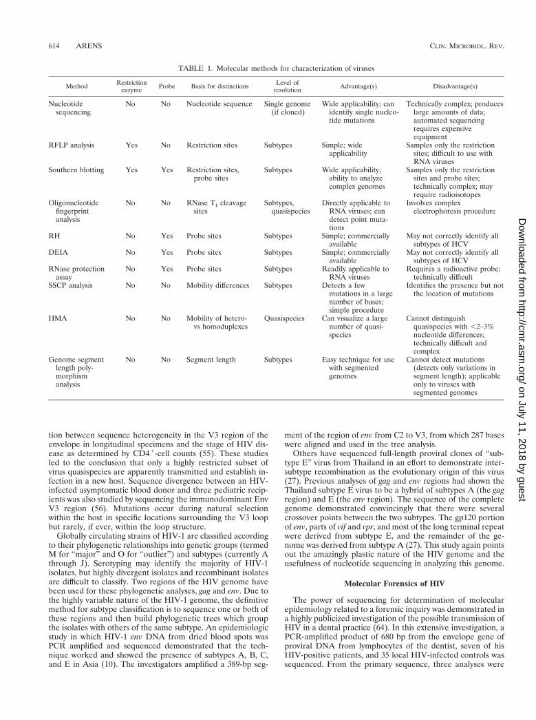
Tables 1 and 2 summarize the techniques and applicationsdiscussed in this review. Table 1 describes the methods andtheir requirements, such as the use of restriction enzymes,other enzymes, and probes, as well as the ability of the tech-nique to sample a large number of bases and detect pointmutations. One must consider these attributes of a particularassay when deciding which to use for an application. Table 2identifies the practical uses of the methods and gives examplesof viruses that have been studied by a particular method asdiscussed in this review.
NUCLEOTIDE SEQUENCING
Introduction and General Approach
The experimental determination of the linear arrangementof bases in a viral genome is the ultimate form of subtyping.Sequencing of the genome has the potential to distinguish evenbetween parent and progeny if a single mutation has occurredin the replicative process. Sequencing generally requires acommitment of time and resources beyond those of the othermethods discussed herein, and it produces a volume of datathat may exceed what the investigator had hoped for and iscapable of dealing with. However, if only small portions orspecific variable regions of a viral genome are sequenced forthe purpose of subtyping, the volume of sequencing data canbe kept to a minimum. Generally this is the case. Only theportions of the genome that confer the subtype need be com-pared, but in the beginning this information may not be known.In most cases the comparison will include only the regions thatare subjected to immunological pressure by the host (i.e., themajor antigenic epitopes of the surface proteins). However, inother cases the untranslated regions of the genomes are uniqueto a subtype because they contain random mutations that maypersist indefinitely in the complete absence of immunologicpressure. The use of PCR amplification in conjunction withsequencing and recent developments in automated sequencing(with cycle labeling of the oligonucleotides) have removedmuch of the tedium from this process and streamlined it to thepoint that one can obtain a fully analyzed sequence of a small(e.g., 1-kb) portion of a viral genome within about 3 days afterobtaining a clinical isolate or, in some cases, after receiving aclinical specimen. Of course, all RNA genomes must first beconverted to DNA prior to sequencing, so the use of reversetranscriptase PCR (RT-PCR) is a natural fit for this processand leads directly to a sequencable product (Fig. 1).
Application to Human Immunodeficiency Virus
The complete sequence of the human immunodeficiencyvirus type 1 (HIV-1) genome was published in 1985 (67). Sincethat time, there have been numerous reports of the use ofsequencing to demonstrate the genetic variability of the virus.Early reports showed a high degree of variation over time inspecific genes within an individual patient (28). Other investi-gators using HIV sequencing attempted to establish a correla-
VOL. 12, 1999 SUBTYPING AND MOLECULAR COMPARISON OF HUMAN VIRUSES 613
on July 11, 2018 by guesthttp://cm
r.asm.org/
Dow
nloaded from
tion between sequence heterogeneity in the V3 region of theenvelope in longitudinal specimens and the stage of HIV dis-ease as determined by CD41-cell counts (55). These studiesled to the conclusion that only a highly restricted subset ofvirus quasispecies are apparently transmitted and establish in-fection in a new host. Sequence divergence between an HIV-infected asymptomatic blood donor and three pediatric recip-ients was also studied by sequencing the immunodominant EnvV3 region (56). Mutations occur during natural selectionwithin the host in specific locations surrounding the V3 loopbut rarely, if ever, within the loop structure.
Globally circulating strains of HIV-1 are classified accordingto their phylogenetic relationships into genetic groups (termedM for “major” and O for “outlier”) and subtypes (currently Athrough J). Serotyping may identify the majority of HIV-1isolates, but highly divergent isolates and recombinant isolatesare difficult to classify. Two regions of the HIV genome havebeen used for these phylogenetic analyses, gag and env. Due tothe highly variable nature of the HIV-1 genome, the definitivemethod for subtype classification is to sequence one or both ofthese regions and then build phylogenetic trees which groupthe isolates with others of the same subtype. An epidemiologicstudy in which HIV-1 env DNA from dried blood spots wasPCR amplified and sequenced demonstrated that the tech-nique worked and showed the presence of subtypes A, B, C,and E in Asia (10). The investigators amplified a 389-bp seg-
ment of the region of env from C2 to V3, from which 287 baseswere aligned and used in the tree analysis.
Others have sequenced full-length proviral clones of “sub-type E” virus from Thailand in an effort to demonstrate inter-subtype recombination as the evolutionary origin of this virus(27). Previous analyses of gag and env regions had shown theThailand subtype E virus to be a hybrid of subtypes A (the gagregion) and E (the env region). The sequence of the completegenome demonstrated convincingly that there were severalcrossover points between the two subtypes. The gp120 portionof env, parts of vif and vpr, and most of the long terminal repeatwere derived from subtype E, and the remainder of the ge-nome was derived from subtype A (27). This study again pointsout the amazingly plastic nature of the HIV genome and theusefulness of nucleotide sequencing in analyzing this genome.
Molecular Forensics of HIV
The power of sequencing for determination of molecularepidemiology related to a forensic inquiry was demonstrated ina highly publicized investigation of the possible transmission ofHIV in a dental practice (64). In this extensive investigation, aPCR-amplified product of 680 bp from the envelope gene ofproviral DNA from lymphocytes of the dentist, seven of hisHIV-positive patients, and 35 local HIV-infected controls wassequenced. From the primary sequence, three analyses were
TABLE 1. Molecular methods for characterization of viruses
Method Restrictionenzyme Probe Basis for distinctions Level of
resolution Advantage(s) Disadvantage(s)
Nucleotidesequencing
No No Nucleotide sequence Single genome(if cloned)
Wide applicability; canidentify single nucleo-tide mutations
Technically complex; produceslarge amounts of data;automated sequencingrequires expensiveequipment
RFLP analysis Yes No Restriction sites Subtypes Simple; wideapplicability
Samples only the restrictionsites; difficult to use withRNA viruses
Southern blotting Yes Yes Restriction sites,probe sites
Subtypes Wide applicability;ability to analyzecomplex genomes
Samples only the restrictionsites and probe sites;technically complex; mayrequire radioisotopes
Oligonucleotidefingerprintanalysis
No No RNase T1 cleavagesites
Subtypes,quasispecies
Directly applicable toRNA viruses; candetect point muta-tions
Involves complexelectrophoresis procedure
RH No Yes Probe sites Subtypes Simple; commerciallyavailable
May not correctly identify allsubtypes of HCV
DEIA No Yes Probe sites Subtypes Simple; commerciallyavailable
May not correctly identify allsubtypes of HCV
RNase protectionassay
No Yes Probe sites Subtypes Readily applicable toRNA viruses
Requires a radioactive probe;technically difficult
SSCP analysis No No Mobility differences Subtypes Detects a fewmutations in a largenumber of bases;simple procedure
Identifies the presence but notthe location of mutations
HMA No No Mobility of hetero-vs homoduplexes
Quasispecies Can visualize a largenumber of quasi-species
Cannot distinguishquasispecies with ,2–3%nucleotide differences;technically difficult andcomplex
Genome segmentlength poly-morphismanalysis
No No Segment length Subtypes Easy technique for usewith segmentedgenomes
Cannot detect mutations(detects only variations insegment length); applicableonly to viruses withsegmented genomes
614 ARENS CLIN. MICROBIOL. REV.
on July 11, 2018 by guesthttp://cm
r.asm.org/
Dow
nloaded from
performed: analysis of DNA distance, construction of phylo-genetic trees, and analysis of amino acid signature patterns.The results showed that the viruses from the dentist and five ofthe patients were closely related, and taken with the epidemi-ologic results, it was concluded that these patients had been
infected (via an unknown mechanism) while receiving care atthe dentist’s office. Obviously the nature of this particular in-vestigation necessitated a highly sophisticated analysis of theviral genomes. Investigators using the various techniques dis-cussed in this review must determine the ability of a particular
FIG. 1. Automated cycle sequencing as a rapid and convenient method for sequence analysis of PCR products. The target DNA was extracted and amplified andthe PCR products were purified away from unincorporated nucleotides and primers. The sequencing template was “amplified” in another PCR mixture containing thesequencing primers, a mixture of deoxy- and dideoxynucleotides, and modified Taq polymerase. Dideoxynucleotides were coupled to fluorescent dyes, a different colorfor each of the four bases; these act as terminators of the growing oligonucleotides. Thus, the color of the dye is the key to which dideoxynucleotide is at the 39 terminusof each oligonucleotide. The large array of oligonucleotides was separated in a polyacrylamide gel which was continuously scanned by a laser during the run, and thecolors were detected as they passed the scan point. Each oligonucleotide in the array, as they move through the gel, is only one base shorter than the oligonucleotideabove it in the gel. The detection information was fed directly into a computer for analysis and construction of the four-color electropherogram, which was translatedinto the linear sequence by commercially available software.
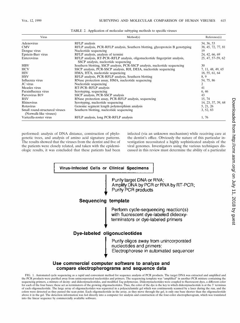
TABLE 2. Application of molecular subtyping methods to specific viruses
Virus Method(s) Reference(s)
Adenovirus RFLP analysis 34, 36, 53CMV RFLP analysis, PCR-RFLP analysis, Southern blotting, glycoprotein B genotyping 38, 45, 72, 77, 81Dengue virus Nucleotide sequencing 19Epstein-Barr virus RFLP analysis, analysis of termini 24, 42, 66, 69Enterovirus RFLP analysis, RT-PCR–RFLP analysis, oligonucleotide fingerprint analysis,
SSCP analysis, nucleotide sequencing25, 47, 57–59, 62
HBV Southern blotting, SSCP analysis, PCR-SSCP analysis, nucleotide sequencing 30HCV SSCP analysis, PCR-SSCP analysis, RH, DEIA, nucleotide sequencing 7, 11, 48, 49, 65HIV HMA, HTA, nucleotide sequencing 18, 55, 61, 64HSV RFLP analysis, PCR-RFLP analysis, Southern blotting 8, 9Influenza virus RNase protection assay, HMA, nucleotide sequencing 54, 75, 86JC virus Nucleotide sequencing 2Measles virus RT-PCR–RFLP analysis 41Parainfluenza virus Serotyping, sequencing 6, 46Parvovirus B19 SSCP analysis, PCR-SSCP analysis 43RSV RNase protection assay, PCR-RFLP analysis, sequencing 15, 74Rhinovirus Serotyping, nucleotide sequencing 14, 23, 37, 39, 68Rotavirus Genome segment length polymorphism analysis 5, 21, 26Small round-structured viruses
(Norwalk-like viruses)Southern blotting, nucleotide sequencing 3, 52, 83
Varicella-zoster virus RFLP analysis, long PCR-RFLP analysis 1, 76
VOL. 12, 1999 SUBTYPING AND MOLECULAR COMPARISON OF HUMAN VIRUSES 615
on July 11, 2018 by guesthttp://cm
r.asm.org/
Dow
nloaded from
method to provide the level of sophistication appropriate forthe application at hand.
Use with Dengue, JC, and Influenza Viruses
Dengue virus is an RNA virus of the flavivirus genus thatcauses a mild flu-like illness in millions of people each year andsevere to life-threatening hemorrhagic fever and shock syn-drome in some individuals. A molecular epidemiologic studywhich was performed by direct sequencing of PCR productsrevealed six genotypic groups among 28 dengue virus type 2isolates (19). The investigation was undertaken because of thesuggestion that certain topotypes (i.e., genetically related vi-ruses that circulate in a particular geographic region) might bemore likely to cause severe disease than others. The authorswere unable to demonstrate this possibility because of manyconfounding variables but nonetheless generated surveillancedata that may be useful in vaccine development.
Another investigation with a similar hypothesis was reportedfor JC virus (2). JC virus is found in four different genotypes in70 to 90% of the adult population worldwide and is the causeof progressive multifocal leukoencephalopathy (PML) inabout 5% of autopsied AIDS patients. By direct sequencing ofspecific regions of the genome, type determination was madeand compared among 50 PML patients and 103 control sub-jects. Brain tissues from the PML patients had a significantlyhigher proportion of JC virus type 2 than urine isolates fromthe controls, thus indicating a biologic difference between theJC virus genotypes and perhaps demonstrating a propensity fortype 2 to cause PML.
A very recent application of sequencing for the purpose ofmolecular characterization is the case of avian influenza A(H5N1) virus isolated from a child in Hong Kong with fatalinfluenza (75). The isolate was obtained from a tracheal aspi-rate specimen. The hemagglutinin (HA) and neuraminidasegenes were amplified by RT-PCR and sequenced to confirmthe H5N1 genotype. The sequence analysis revealed the pres-ence of a multiple basic amino acid insertion upstream fromthe trypsin cleavage site (75). This insertion has previouslybeen found in highly pathogenic avian influenza virus strainsand is thought to extend the tissue range of the virus by allow-ing proteases other than trypsin to cleave the HA protein intoHA1 and HA2 domains and thus enable systemic spread of thevirus.
RESTRICTION FRAGMENT LENGTHPOLYMORPHISM ANALYSIS
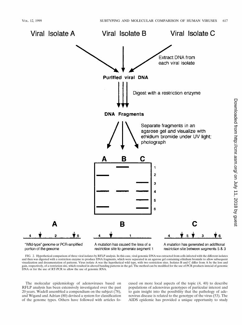
Review of the Method
The site of cleavage of DNA by a restriction endonuclease issequence dependent. The presence of mutations at potentialcleavage sites in some viral strains results in different patternsof fragments when they are separated in an agarose gel, aphenomenon termed restriction fragment length polymor-phism (RFLP) (Fig. 2). The technique requires fairly largeamounts of purified or partially purified viral DNA, a set ofrestriction enzymes to enzymatically cleave the DNA, the abil-ity to separate the resulting DNA fragments by electrophore-sis, and a method of documenting the results. The results aredisplayed as patterns of bands in an ethidium bromide-stainedagarose gel. Viruses with large genomes (e.g., CMV) may have20 to 50 bands, while viruses with smaller genomes (e.g., ad-enoviruses) have only 5 to 10 bands. Of course, the restrictionenzyme used for the digestion is an important factor in theresultant number of bands, and care must be taken to ensure
that digestion is complete so that partial digestion products donot obscure the final analysis of bands. Restriction endonucle-ases have recognition sites usually consisting of either four orsix bases in a specific palindromic sequence. If a mutationoccurs in the DNA genome to either eliminate or create arestriction site, polymorphisms occur in the pattern of bands inthe gel (Fig. 2). There is no easy way, nor is there generally aneed, to correlate the pattern (i.e., a missing band or an extraband) with mutations at specific locations in the genome with-out extensive molecular hybridization studies or even sequenc-ing of the genomes being compared. A distinct limitation of themethod is that the presence of a mutation cannot be detectedunless that mutation happens to fall within the recognitionsequence of the restriction endonuclease being used for diges-tion of the DNA. The use of different restriction enzymes willoptimize the probability of detecting mutations in a particulargenome or portion of a genome. Also, the absence of differingrestriction patterns may simply be the result of testing with toofew restriction enzymes.
Examples Involving DNA Viruses: CMV,HSV, and Adenovirus
CMV was the first virus to be subjected to RFLP analysis ofviral genomic DNA pursuant to epidemiologic studies. Kil-patrick et al. (45) demonstrated the usefulness of this methodby restriction enzyme digestion of 11 human strains of CMV.Each strain had a unique pattern of fragments when separatedby electrophoresis in an agarose gel. Subsequent studies havedocumented a number of interesting observations with regardto the epidemiology of CMV. Five of six congenitally infectedbabies had the same strain of CMV as their mothers, thusleading to the obvious conclusion that endogenous CMV is themost frequent cause of intrauterine transmission (38). Spectorand Spector (72) used the RFLP technique to study the epi-demiologic relationships of three CMV isolates from newborntwins and their mother. The infants were infected with twodifferent strains of virus during an extended hospitalizationafter birth (twin A was infected at 9 weeks of age and twin Bwas infected at 6 weeks). The mother, who was CMV seroneg-ative prior to and shortly after giving birth, became infected at6 months postpartum with the strain carried by twin A. Inanother study among infants in a nursery, RFLP analysis ofCMV strains isolated from the urine of eight babies demon-strated that one of the babies had transmitted CMV to twoother babies, apparently via fomites. The two babies beganshedding CMV on the same day, 22 days after the index patientwas transferred to the intensive care nursery.
Other viral genomes have been subjected to RFLP analysis.The method was originally referred to as restriction endonu-clease fingerprinting as it was applied to HSV in groundbreak-ing studies demonstrating exogenous reinfection (9) and nos-ocomial outbreaks (8). These and other studies found HSV-1to have 19 of 57 total restriction enzyme cleavage sites thatwere variable, with four restriction enzymes among the isolatesin the studies (8, 9). From this information it was estimated (9)that the smallest number of potential differentiable strains ofHSV-1 is 524,288, thus lending theoretical support to the ob-servation that epidemiologically unlinked isolates have differ-ent RFLP patterns. An RFLP analysis of the isolates from theearlier study (8) led to the conclusion that there had been twoindependent introductions of HSV-1 into a pediatric intensivecare unit. An outbreak of HSV encephalitis in Boston, Mass.,engendered an RFLP analysis of the isolates, and all seven haddifferent restriction patterns, thus proving that they were notepidemiologically related (29).
616 ARENS CLIN. MICROBIOL. REV.
on July 11, 2018 by guesthttp://cm
r.asm.org/
Dow
nloaded from
The molecular epidemiology of adenoviruses based onRFLP analysis has been extensively investigated over the past20 years. Wadell assembled a compendium on the subject (78),and Wigand and Adrian (80) devised a system for classificationof the genome types. Others have followed with articles fo-
cused on more local aspects of the topic (4, 40) to describepopulations of adenovirus genotypes of particular interest andto gain insight into the possibility that the pathology of ade-novirus disease is related to the genotype of the virus (53). TheAIDS epidemic has provided a unique opportunity to study
FIG. 2. Hypothetical comparison of three viral isolates by RFLP analysis. In this case, viral genomic DNA was extracted from cells infected with the different isolatesand then was digested with a restriction enzyme to produce DNA fragments, which were separated in an agarose gel containing ethidium bromide to allow subsequentvisualization and documentation of patterns. Virus isolate A was the hypothetical wild type, with two restriction sites. Isolates B and C differ from A by the loss andgain, respectively, of a restriction site, which resulted in altered banding patterns in the gel. The method can be modified for the use of PCR products instead of genomicDNA or for the use of RT-PCR to allow the use of genomic RNA.
VOL. 12, 1999 SUBTYPING AND MOLECULAR COMPARISON OF HUMAN VIRUSES 617
on July 11, 2018 by guesthttp://cm
r.asm.org/
Dow
nloaded from
adenoviruses. Because they are spread easily and persist forlong periods of time in immunocompromised patients, theconditions are good for widespread exchange of genetic mate-rial among and within serotypes through recombination. Hier-holzer et al. (33–36) found numerous antigenically intermedi-ate strains and DNA variants. They concluded that restrictionenzyme analysis of adenoviruses from AIDS patients thus hadonly limited usefulness for typing but was helpful in identifyinggroups of isolates with similar properties (34).
RT-PCR for RFLP Analysis of RNA Viruses:Enteroviruses and Measles Virus
Restriction endonucleases are capable of cleaving onlyDNA, but with the help of RT-PCR, portions of the genomesof RNA viruses can be converted to DNA, amplified, andsubjected to RFLP analysis. Many of the enteroviruses havebeen analyzed by this method for the purposes of both detec-tion and differentiation of the numerous serotypes by Kuan(47). In this study, a 297-bp amplicon from the 59 untranslatedregion (UTR) of the virus was cleaved by restriction enzymesand the fragments were analyzed on an agarose gel. Fragmentpatterns were characteristic of the serotype, as shown by com-parison with prototype strains. Molecular epidemiology ofmeasles virus, another RNA virus, has recently been used todocument the changing distribution of measles virus genotypesin Japan. This investigation was accomplished by amplifyingthe HA and nucleoprotein genes and then subjecting the am-plified product to digestion by restriction enzymes, which al-lowed strain typing of the isolate (41). Measles virus type 2represented 80% of the isolates in western Japan in 1985, butby 1990 the majority were type 1 and by 1995 all strains studiedwere type 1 as determined by RFLP analysis.
RFLP analysis is a simple yet powerful technique. It hasbeen widely used because of its ability to detect point muta-tions if they occur in a restriction site. It can be used tocompare a large number of isolates in epidemiologic studiesand can even be adapted for use with RNA viruses after anRT-PCR step.
SOUTHERN BLOT ANALYSIS
Description of Method
Classical Southern blotting (70) is a modification of RFLPanalysis in which the viral DNA is cut with a restriction enzymeand then fragments are subjected to electrophoresis in an aga-rose or polyacrylamide gel, transferred onto a nitrocellulosesheet, and then hybridized with a labeled probe from the entiregenome or against a specific region of the genome.
Use with CMV
This method (22) and RFLP analysis (71) have been used todemonstrate that an AIDS patient can be infected with multi-ple strains of CMV. In a landmark study on the transmission ofCMV in cadaveric renal transplantation (12), 15 distinct strains(genotypes) of CMV were isolated from 19 organ recipients(i.e., four pairs [8 patients] of the 19 had paired isolates withthe same genotype). In all four pairs of patients who hadreceived kidneys from the same cadaver, both recipients shedthe same strain of CMV, suggesting that both had acquired itfrom the donor and that seropositive recipients can be rein-fected by a new strain of CMV after transplantation (12).
OLIGONUCLEOTIDE FINGERPRINT ANALYSIS
Method
Oligonucleotide fingerprint analysis, a technically complexbut powerful method of analyzing randomly distributed non-contiguous segments of an RNA molecule or RNA virus ge-nome, was first described by DeWachter and Fiers (20). Themethod employs digestion of radioactively labeled genomicRNA with RNase T1, an endonuclease purified from Aspergillusoryzae that cleaves single-stranded RNA on the 39 side of Gresidues, and two-dimensional electrophoresis. The electro-phoresis procedure is performed on 8% polyacrylamide in 6 Murea at 4°C in the first direction and then on 22% acrylamidein Tris-borate buffer at room temperature in the second direc-tion after a 90° rotation from the first direction. 32P-labeledRNA oligonucleotides are detected by autoradiography as afan-shaped array of spots and are distributed according to sizeand composition (20). The method allows comparison of thelarger T1-resistant oligonucleotides present in the RNA beingtested (representing about 10% of the genome of certainenteroviruses [44]). This procedure was considered to be themost sensitive method available, short of nucleotide se-quencing, for analysis of RNA genetic relatedness.
Application to Enteroviruses
The oligonucleotide fingerprint method was first applied topoliovirus RNA by Lee et al. (50) and Lee and Wimmer (51)and soon thereafter was used in the analysis of other RNAviruses, including influenza virus (84), vesicular stomatitis virus(13), and enteroviruses (31, 57, 62). The most widely usedapplication has been in the study of molecular variation amongthe enteroviruses, particularly polioviruses. T1 fingerprintingwas a powerful tool in the early epidemiologic studies of thedistribution of poliovirus subpopulations among infected hu-man communities and in studies undertaken to clarify themolecular evolution of poliovirus during infection and passagethrough the human intestine. Using T1 fingerprinting, it wasobserved that the poliovirus genome was quite plastic, sincethe genome underwent continual change during natural epi-demic transmission. Within a single epidemic in a confinedgeographic area, small differences between patterns were de-tected and no two patterns were exactly alike (62). New spotsappeared on the autoradiograms and others disappeared; eventhe patterns among infected family members were different(62).
Other practical applications were also discovered. Isolates ofvaccine origin could easily be distinguished from circulating“wild” strains of poliovirus by this method, proving it a timelymethod for detection of wild-type infections and for trackingpossible vaccine revertants of the type 1 oral polio vaccine (62).
Oligonucleotide fingerprinting showed that in contrast to theextreme variability of the poliovirus genome, the genome ofenterovirus 70 (EV 70), the cause of epidemic acute hemor-rhagic conjunctivitis around the world, is quite stable (44).Only one basic genotype of EV 70 appears to be in circulationworldwide, and isolates from one year to the next and from onegeographic region to the next have very similar patterns. Nu-merous intervening infections that linked different isolates didnot result in major changes in the fingerprint patterns of EV 70(44).
618 ARENS CLIN. MICROBIOL. REV.
on July 11, 2018 by guesthttp://cm
r.asm.org/
Dow
nloaded from
REVERSE HYBRIDIZATION
Technique
The reverse hybridization (RH) method is based on a systemin which PCR products amplified from test specimens arehybridized to probes bound in parallel lines to membranestrips. An assay employing this technology for type and subtypeanalysis of HCV is commercially available (Inno-LiPA HCV;Innogenetics, Zwijnaarde, Belgium, and, in the United States,Norcross, Ga.). HCV RNA is extracted from serum and am-plified by a nested RT-PCR with biotinylated primers directedagainst the HCV 59 noncoding (NC) region or 59 UTR. Theseproducts are hybridized to probes on the strips and hybrids aredetected by alkaline phosphatase-conjugated streptavidin. Re-action of the amplified fragment with specific probes results inthe formation of visible lines on the strip, thus presumablyallowing identification of at least six of the nine currentlyknown major HCV genotypes and also the respective subtype.
Application to HCV
This assay has been evaluated for its usefulness in genotyp-ing of HCV. HCV RNA isolated from the sera of 61 HCV-positive patients was analyzed by sequencing of the 59 NCregion and the less well conserved nonstructural-5 region andby RH (85). The RH assay was able to correctly identify all ofthe HCV genotypes (only types 1, 2, and 3 were found in thesepatients). However, the RH assay incorrectly identified thesubtype of 1 of 11 HCV subtype 1a genotypes as subtype 1band identified 3 of 31 subtype 1b genotypes as subtype 1a, andit also identified 4 of 4 subtype 2c genotypes as subtype 2a. AllHCV subtype 2a, 2b, and 3a genotypes in this study werecorrectly identified by the RH assay. A subsequent evaluationby the same group (49) and by another group (65) led to theconclusion that this particular RH assay misinterpreted 2 to10% of the subtype 1a and 1b genotypes and also that themethod could not be used for differentiation of HCV subtypes2a and 2c. The shortcomings of this assay are apparently theresult of the choice of probes. The distinction between sub-types 1a and 1b is dependent on a single nucleotide differenceat position 299 in the 59 UTR, and subtypes 2a and 2c areidentical in the region targeted by the Inno-LiPA probes.
DNA ENZYME IMMUNOASSAY
Description of the Commercial Assay
A commercial DNA enzyme immunoassay (DEIA) productis available for identification of HCV types and subtypes. RNAfrom serum is extracted and amplified by RT-PCR with nestedprimers directed against the HCV core region. Heat-denaturedPCR products are hybridized to probes bound to multiwellplates. Hybrids are detected with a murine monoclonal anti-body against double-stranded DNA followed by rabbit anti-mouse antibody conjugated to horseradish peroxidase. A sub-strate is then added. The DEIA reagents contain six differentprobes directed against the HCV core region.
Use for HCV and Comparison to RH Assay
In a recent evaluation and direct comparison of the RHassay (Inno-LiPA HCV II) and the DEIA (65), 112 of 120evaluable samples (93.3%) yielded concordant results. Of theeight subtyping discrepancies, all involved subtype 1 and 2viruses that were misassigned by the RH assay apparentlybecause of the suboptimal probes discussed above.
Thus, both the RH assay and the DEIA appear to be excel-lent choices for the determination of HCV type and reasonablechoices for the determination of subtype. However, the DEIAhas a higher accuracy for subtyping, especially for identifyingthe subtypes of HCV-1 and -2.
RNASE PROTECTION ANALYSIS
Development of the Method and Application to Viruses
RNase protection analysis was developed by Myers et al.(60) for the purpose of demonstrating the presence and ap-proximate locations of point mutations in DNA. The techniqueinvolves the use of a relatively short, in vitro-synthesized, ra-dioactive RNA probe that is transcribed from wild-typegenomic DNA (or cloned DNA) from a standard strain or thetype strain of an organism. In the original method, this RNAprobe was then hybridized to DNA from test strains prior todigestion of the heteroduplex with RNase A. The resultingfragments were separated in a polyacrylamide gel containing 8M urea (Fig. 3). RNase A cleaves the radioactive RNA probeat positions of mismatch with the DNA. Test genomes withmutations that are not present in the probe or with mutationsat different positions than the probe will have altered bandingpatterns in the gel. The method is able to detect about half ofall single-base mutations (60). It is not clear why all mutationsare not detected, but this is perhaps related to the secondarystructure of the probe-test heteroduplex.
Influenza Virus and RSV Analysis
A modification of ribonuclease protection, sometimes calledthe RNase A mismatch cleavage method, has allowed its usewith the RNA genomes of viruses, including influenza virusand RSV. By using a technique that had been developed todemonstrate the presence of point mutations in RNA tran-scripts from the c-ras gene (82), the genetic relatedness andevolution of field isolates of influenza virus were investigated(54). This method was also employed in an epidemiologic studyof RSV in which it was found, using a probe made from the Gglycoprotein of the A2 strain of RSV, that all epidemiologicallylinked isolates (from coinfected twins, infants infected during anosocomial outbreak, and institutionalized adults infected dur-ing an outbreak) had identical band patterns (74). The samemethod was used to study the genetic variability and evolutionof the different subtypes of RSV circulating in Spain (15).Mutations which appeared in early isolates were retained inlater isolates, and viruses isolated during a short time spanshowed highly similar band patterns. Additionally, differentisolates from the same winter outbreak of RSV showed con-siderable heterogeneity and the RSV G gene accumulatedmutations at a faster rate than other genes. Although themethod is technically difficult, it has considerable power todefine epidemiologically related isolates in the same way thatRFLP analysis has for DNA viruses.
SINGLE-STRAND CONFORMATIONPOLYMORPHISM ANALYSIS
Development of the Assay and Application to Viruses
Single-strand conformation polymorphism (SSCP) analysisis the term applied to the method developed by Orita et al. (63)with which they demonstrated that a single nucleotide substi-tution was sufficient to cause a mobility shift of a fragment ofsingle-stranded DNA in a neutral polyacrylamide gel. Initially,
VOL. 12, 1999 SUBTYPING AND MOLECULAR COMPARISON OF HUMAN VIRUSES 619
on July 11, 2018 by guesthttp://cm
r.asm.org/
Dow
nloaded from
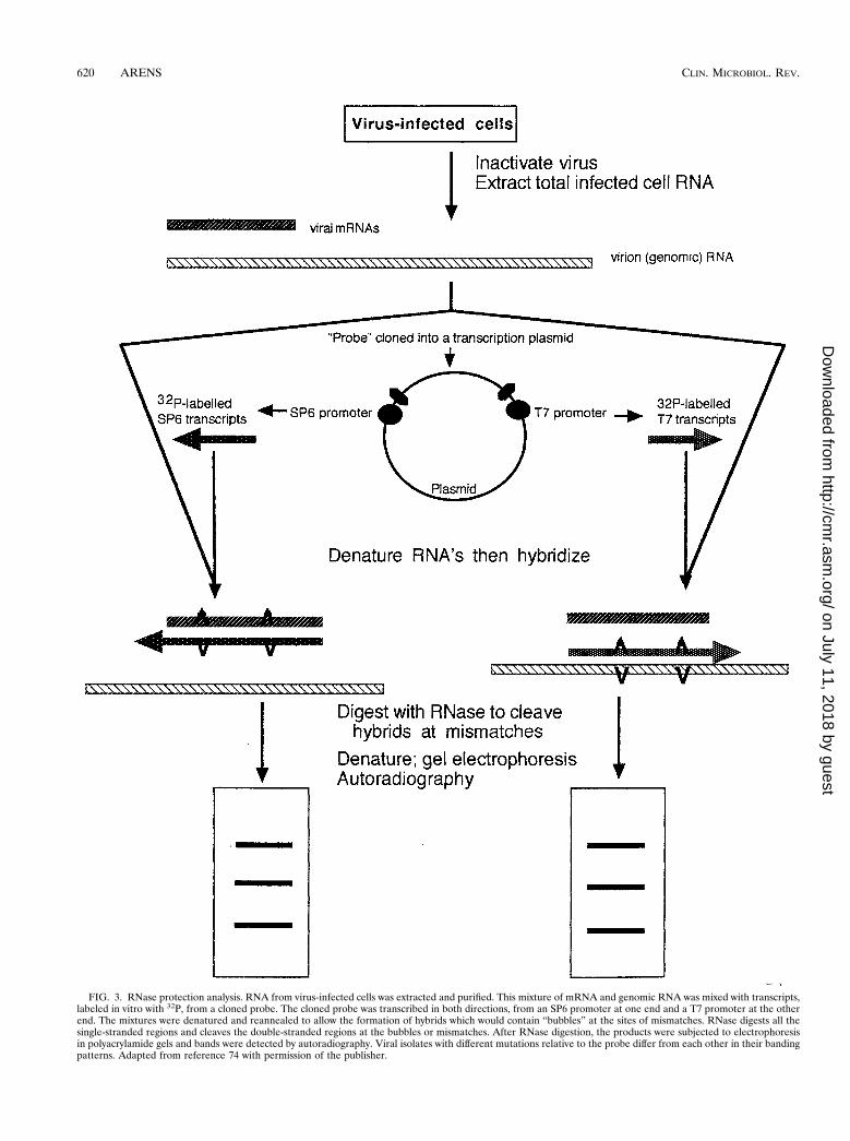
FIG. 3. RNase protection analysis. RNA from virus-infected cells was extracted and purified. This mixture of mRNA and genomic RNA was mixed with transcripts,labeled in vitro with 32P, from a cloned probe. The cloned probe was transcribed in both directions, from an SP6 promoter at one end and a T7 promoter at the otherend. The mixtures were denatured and reannealed to allow the formation of hybrids which would contain “bubbles” at the sites of mismatches. RNase digests all thesingle-stranded regions and cleaves the double-stranded regions at the bubbles or mismatches. After RNase digestion, the products were subjected to electrophoresisin polyacrylamide gels and bands were detected by autoradiography. Viral isolates with different mutations relative to the probe differ from each other in their bandingpatterns. Adapted from reference 74 with permission of the publisher.
620 ARENS CLIN. MICROBIOL. REV.
on July 11, 2018 by guesthttp://cm
r.asm.org/
Dow
nloaded from
the general procedure was to use RFLP fragments fromgenomic DNA (one from the wild-type genome and one froma possible mutant), denature the fragments by alkali treatment,subject them to electrophoresis in a neutral polyacrylamide gel,and compare their mobilities. More recently, modificationshave been made to accommodate PCR amplification of a spe-cific region of wild-type or mutant genomes prior to denatur-ation and separation on a neutral gel (32). In either case, ifmutations are present in the segment of the mutant genomebeing tested, that segment will likely run at a different positionin the gel than the same segment from the wild-type genome.The altered electrophoresis pattern is apparently due to themutation-altered secondary structure of the restriction frag-ment or the PCR amplicon (63). The separation of the wild-type and mutant fragments is dependent on several environ-mental factors, including the temperature of the gel duringelectrophoresis, the concentration and composition of the elec-trophoresis buffer, and the presence of denaturing agents inthe gel. Several sets of conditions should be tried empirically tooptimize mutation detection. One major advantage of thismethod is that it can “sample” the genetic makeup of severalhundred base pairs of DNA, whereas RFLP analysis can sam-ple only a few bases (the restriction sites).
Analysis of Parvovirus, Hepatitis B Virus, and HCV
An analysis of the genetic variability in the nonstructuralgene of human parvovirus B19 by PCR-SSCP analysis revealedthe presence of six genotypes among 50 samples of virus fromseveral countries (43). Sequencing of this region confirmed thepresence of mutations in the different genotypes, and all weresilent mutations. There was a good correlation between theSSCP type and the country from which the virus was obtained.Within Japan, genotypes 1, 2, 3, and 4 circulated between 1981and 1987 in about equal numbers. However, between 1990 and1994, 90% of the samples tested were type 3.
An epidemiologic investigation into an outbreak of hepatitisB virus (HBV) infection in a pediatric oncology unit was basedon the ability to distinguish genotypes of the virus by PCR-SSCP analysis (30). PCR was used to amplify a 189-bp productfrom the hypervariable (pre-S1) region of the genome, and thisproduct was denatured and subjected to electrophoresis inneutral polyacrylamide gels. Forty unrelated controls all haddistinct patterns in gels, and all but 6 of 58 oncology patientshad patterns that fell into five different groups: one shared by16 patients, one shared by 19 patients, one shared by 9 patients,one shared by 5 patients, and one shared by 3 patients. Thus,there had been several independent introductions of HBV intothe unit, and some of these isolates had also been spreadextensively within the unit.
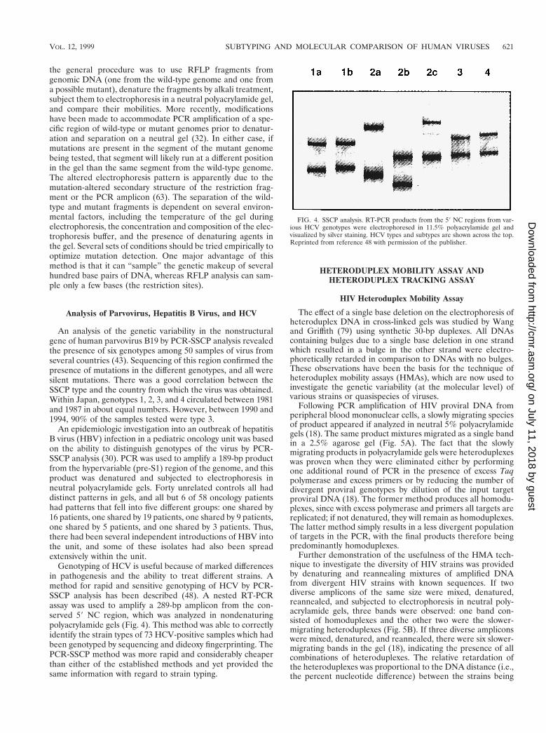
Genotyping of HCV is useful because of marked differencesin pathogenesis and the ability to treat different strains. Amethod for rapid and sensitive genotyping of HCV by PCR-SSCP analysis has been described (48). A nested RT-PCRassay was used to amplify a 289-bp amplicon from the con-served 59 NC region, which was analyzed in nondenaturingpolyacrylamide gels (Fig. 4). This method was able to correctlyidentify the strain types of 73 HCV-positive samples which hadbeen genotyped by sequencing and dideoxy fingerprinting. ThePCR-SSCP method was more rapid and considerably cheaperthan either of the established methods and yet provided thesame information with regard to strain typing.
HETERODUPLEX MOBILITY ASSAY ANDHETERODUPLEX TRACKING ASSAY
HIV Heteroduplex Mobility Assay
The effect of a single base deletion on the electrophoresis ofheteroduplex DNA in cross-linked gels was studied by Wangand Griffith (79) using synthetic 30-bp duplexes. All DNAscontaining bulges due to a single base deletion in one strandwhich resulted in a bulge in the other strand were electro-phoretically retarded in comparison to DNAs with no bulges.These observations have been the basis for the technique ofheteroduplex mobility assays (HMAs), which are now used toinvestigate the genetic variability (at the molecular level) ofvarious strains or quasispecies of viruses.
Following PCR amplification of HIV proviral DNA fromperipheral blood mononuclear cells, a slowly migrating speciesof product appeared if analyzed in neutral 5% polyacrylamidegels (18). The same product mixtures migrated as a single bandin a 2.5% agarose gel (Fig. 5A). The fact that the slowlymigrating products in polyacrylamide gels were heteroduplexeswas proven when they were eliminated either by performingone additional round of PCR in the presence of excess Taqpolymerase and excess primers or by reducing the number ofdivergent proviral genotypes by dilution of the input targetproviral DNA (18). The former method produces all homodu-plexes, since with excess polymerase and primers all targets arereplicated; if not denatured, they will remain as homoduplexes.The latter method simply results in a less divergent populationof targets in the PCR, with the final products therefore beingpredominantly homoduplexes.
Further demonstration of the usefulness of the HMA tech-nique to investigate the diversity of HIV strains was providedby denaturing and reannealing mixtures of amplified DNAfrom divergent HIV strains with known sequences. If twodiverse amplicons of the same size were mixed, denatured,reannealed, and subjected to electrophoresis in neutral poly-acrylamide gels, three bands were observed: one band con-sisted of homoduplexes and the other two were the slower-migrating heteroduplexes (Fig. 5B). If three diverse ampliconswere mixed, denatured, and reannealed, there were six slower-migrating bands in the gel (18), indicating the presence of allcombinations of heteroduplexes. The relative retardation ofthe heteroduplexes was proportional to the DNA distance (i.e.,the percent nucleotide difference) between the strains being
FIG. 4. SSCP analysis. RT-PCR products from the 59 NC regions from var-ious HCV genotypes were electrophoresed in 11.5% polyacrylamide gel andvisualized by silver staining. HCV types and subtypes are shown across the top.Reprinted from reference 48 with permission of the publisher.
VOL. 12, 1999 SUBTYPING AND MOLECULAR COMPARISON OF HUMAN VIRUSES 621
on July 11, 2018 by guesthttp://cm
r.asm.org/
Dow
nloaded from
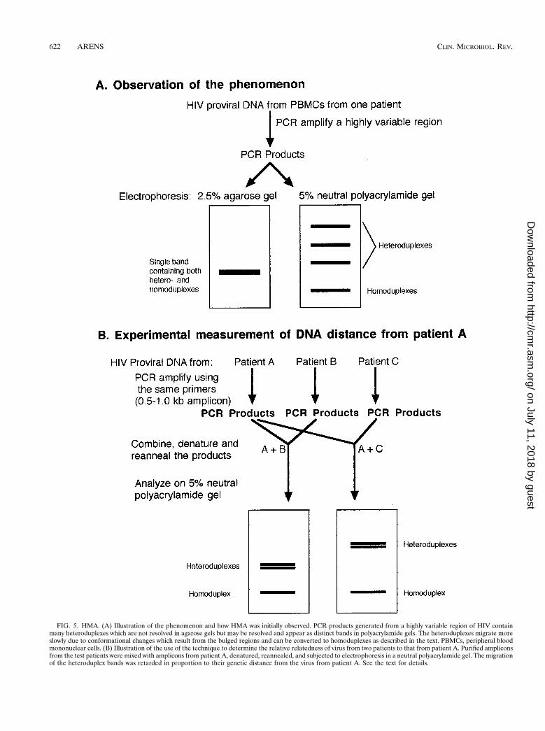
FIG. 5. HMA. (A) Illustration of the phenomenon and how HMA was initially observed. PCR products generated from a highly variable region of HIV containmany heteroduplexes which are not resolved in agarose gels but may be resolved and appear as distinct bands in polyacrylamide gels. The heteroduplexes migrate moreslowly due to conformational changes which result from the bulged regions and can be converted to homoduplexes as described in the text. PBMCs, peripheral bloodmononuclear cells. (B) Illustration of the use of the technique to determine the relative relatedness of virus from two patients to that from patient A. Purified ampliconsfrom the test patients were mixed with amplicons from patient A, denatured, reannealed, and subjected to electrophoresis in a neutral polyacrylamide gel. The migrationof the heteroduplex bands was retarded in proportion to their genetic distance from the virus from patient A. See the text for details.
622 ARENS CLIN. MICROBIOL. REV.
on July 11, 2018 by guesthttp://cm
r.asm.org/
Dow
nloaded from
tested (Fig. 5B). In Fig. 5B, strain C is more divergent from Athan strain B is divergent from A since the heteroduplex bandsare further from the homoduplex band, and the relative distancescan be calculated (18) (see below). The technique can detectgenetic differences of as little as 2% in an amplicon of severalhundred base pairs but is not reliable at detection of differencesless than that. The major advantage of this technique is that it canbe used to screen isolates and determine genetic relatedness with-out the laborious task of DNA sequencing.
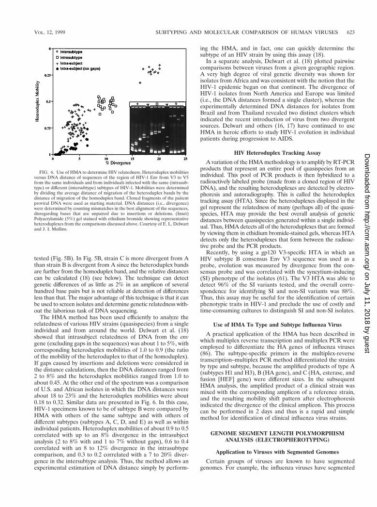
The HMA method has been used efficiently to analyze therelatedness of various HIV strains (quasispecies) from a singleindividual and from around the world. Delwart et al. (18)showed that intrasubject relatedness of DNA from the envgene (excluding gaps in the sequences) was about 1 to 5%, withcorresponding heteroduplex mobilities of 1.0 to 0.9 (the ratioof the mobility of the heteroduplex to that of the homoduplex).If gaps caused by insertions and deletions were considered inthe distance calculations, then the DNA distances ranged from2 to 8% and the heteroduplex mobilities ranged from 1.0 toabout 0.45. At the other end of the spectrum was a comparisonof U.S. and African isolates in which the DNA distances wereabout 18 to 23% and the heteroduplex mobilities were about0.18 to 0.32. Similar data are presented in Fig. 6. In this case,HIV-1 specimens known to be of subtype B were compared byHMA with others of the same subtype and with others ofdifferent subtypes (subtypes A, C, D, and E) as well as withinindividual patients. Heteroduplex mobilities of about 0.9 to 0.5correlated with up to an 8% divergence in the intrasubjectanalysis (2 to 8% with and 1 to 7% without gaps), 0.6 to 0.4correlated with an 8 to 12% divergence in the intrasubtypecomparison, and 0.3 to 0.2 correlated with a 7 to 20% diver-gence in the intersubtype analysis. Thus, the method allows anexperimental estimation of DNA distance simply by perform-
ing the HMA, and in fact, one can quickly determine thesubtype of an HIV strain by using this assay (18).
In a separate analysis, Delwart et al. (18) plotted pairwisecomparisons between viruses from a given geographic region.A very high degree of viral genetic diversity was shown forisolates from Africa and was consistent with the notion that theHIV-1 epidemic began on that continent. The divergence ofHIV-1 isolates from North America and Europe was limited(i.e., the DNA distances formed a single cluster), whereas theexperimentally determined DNA distances for isolates fromBrazil and from Thailand revealed two distinct clusters whichindicated the recent introduction of virus from two divergentsources. Delwart and others (16, 17) have continued to useHMA in heroic efforts to study HIV-1 evolution in individualpatients during progression to AIDS.
HIV Heteroduplex Tracking Assay
A variation of the HMA methodology is to amplify by RT-PCRproducts that represent an entire pool of quasispecies from anindividual. This pool of PCR products is then hybridized to aradioactively labeled probe (made from a cloned region of HIVDNA), and the resulting heteroduplexes are detected by electro-phoresis and autoradiography. This is called the heteroduplextracking assay (HTA). Since the heteroduplexes displayed in thegel represent the relatedness of many (perhaps all) of the quasi-species, HTA may provide the best overall analysis of geneticdistances between quasispecies generated within a single individ-ual. Thus, HMA detects all of the heteroduplexes that are formedby viewing them in ethidium bromide-stained gels, whereas HTAdetects only the heteroduplexes that form between the radioac-tive probe and the PCR products.
Recently, by using a gp120 V3-specific HTA in which anHIV subtype B consensus Env V3 sequence was used as aprobe, evolution was measured by divergence from the con-sensus probe and was correlated with the syncytium-inducing(SI) phenotype of the isolates (61). The V3 HTA was able todetect 96% of the SI variants tested, and the overall corre-spondence for identifying SI and non-SI variants was 88%.Thus, this assay may be useful for the identification of certainphenotypic traits in HIV-1 and preclude the use of costly andtime-consuming cultures to distinguish SI and non-SI isolates.
Use of HMA To Type and Subtype Influenza Virus
A practical application of the HMA has been described inwhich multiplex reverse transcription and multiplex PCR wereemployed to differentiate the HA genes of influenza viruses(86). The subtype-specific primers in the multiplex-reversetranscription–multiplex PCR method differentiated the strainsby type and subtype, because the amplified products of type A(subtypes H1 and H3), B (HA gene), and C (HA, esterase, andfusion [HEF] gene) were different sizes. In the subsequentHMA analysis, the amplified product of a clinical strain wasmixed with the corresponding amplicon of a reference strain,and the resulting mobility shift pattern after electrophoresisindicated the divergence of the clinical amplicon. This processcan be performed in 2 days and thus is a rapid and simplemethod for identification of clinical influenza virus strains.
GENOME SEGMENT LENGTH POLYMORPHISMANALYSIS (ELECTROPHEROTYPING)
Application to Viruses with Segmented Genomes
Certain groups of viruses are known to have segmentedgenomes. For example, the influenza viruses have segmented
FIG. 6. Use of HMA to determine HIV relatedness. Heteroduplex mobilitiesversus DNA distance of sequences of the region of HIV-1 Env from V3 to V5from the same individuals and from individuals infected with the same (intrasub-type) or different (intersubtype) subtypes of HIV-1. Mobilities were determinedby dividing the average distance of migration of the heteroduplex bands by thedistance of migration of the homoduplex band. Cloned fragments of the patientproviral DNA were used as starting material. DNA distances (i.e., divergence)were determined by counting mismatches in the best alignment of the sequences,disregarding bases that are unpaired due to insertions or deletions. (Inset)Polyacrylamide (5%) gel stained with ethidium bromide showing representativeheteroduplexes from the comparisons discussed above. Courtesy of E. L. Delwartand J. I. Mullins.
VOL. 12, 1999 SUBTYPING AND MOLECULAR COMPARISON OF HUMAN VIRUSES 623
on July 11, 2018 by guesthttp://cm
r.asm.org/
Dow
nloaded from
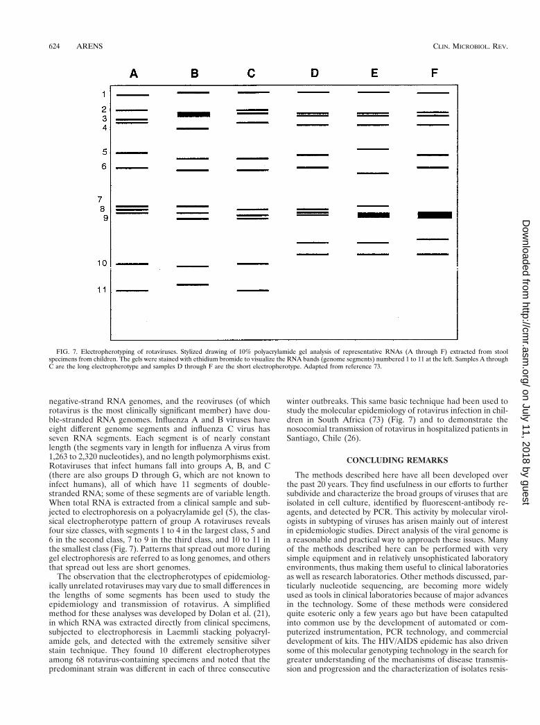
negative-strand RNA genomes, and the reoviruses (of whichrotavirus is the most clinically significant member) have dou-ble-stranded RNA genomes. Influenza A and B viruses haveeight different genome segments and influenza C virus hasseven RNA segments. Each segment is of nearly constantlength (the segments vary in length for influenza A virus from1,263 to 2,320 nucleotides), and no length polymorphisms exist.Rotaviruses that infect humans fall into groups A, B, and C(there are also groups D through G, which are not known toinfect humans), all of which have 11 segments of double-stranded RNA; some of these segments are of variable length.When total RNA is extracted from a clinical sample and sub-jected to electrophoresis on a polyacrylamide gel (5), the clas-sical electropherotype pattern of group A rotaviruses revealsfour size classes, with segments 1 to 4 in the largest class, 5 and6 in the second class, 7 to 9 in the third class, and 10 to 11 inthe smallest class (Fig. 7). Patterns that spread out more duringgel electrophoresis are referred to as long genomes, and othersthat spread out less are short genomes.
The observation that the electropherotypes of epidemiolog-ically unrelated rotaviruses may vary due to small differences inthe lengths of some segments has been used to study theepidemiology and transmission of rotavirus. A simplifiedmethod for these analyses was developed by Dolan et al. (21),in which RNA was extracted directly from clinical specimens,subjected to electrophoresis in Laemmli stacking polyacryl-amide gels, and detected with the extremely sensitive silverstain technique. They found 10 different electropherotypesamong 68 rotavirus-containing specimens and noted that thepredominant strain was different in each of three consecutive
winter outbreaks. This same basic technique had been used tostudy the molecular epidemiology of rotavirus infection in chil-dren in South Africa (73) (Fig. 7) and to demonstrate thenosocomial transmission of rotavirus in hospitalized patients inSantiago, Chile (26).
CONCLUDING REMARKS
The methods described here have all been developed overthe past 20 years. They find usefulness in our efforts to furthersubdivide and characterize the broad groups of viruses that areisolated in cell culture, identified by fluorescent-antibody re-agents, and detected by PCR. This activity by molecular virol-ogists in subtyping of viruses has arisen mainly out of interestin epidemiologic studies. Direct analysis of the viral genome isa reasonable and practical way to approach these issues. Manyof the methods described here can be performed with verysimple equipment and in relatively unsophisticated laboratoryenvironments, thus making them useful to clinical laboratoriesas well as research laboratories. Other methods discussed, par-ticularly nucleotide sequencing, are becoming more widelyused as tools in clinical laboratories because of major advancesin the technology. Some of these methods were consideredquite esoteric only a few years ago but have been catapultedinto common use by the development of automated or com-puterized instrumentation, PCR technology, and commercialdevelopment of kits. The HIV/AIDS epidemic has also drivensome of this molecular genotyping technology in the search forgreater understanding of the mechanisms of disease transmis-sion and progression and the characterization of isolates resis-
FIG. 7. Electropherotyping of rotaviruses. Stylized drawing of 10% polyacrylamide gel analysis of representative RNAs (A through F) extracted from stoolspecimens from children. The gels were stained with ethidium bromide to visualize the RNA bands (genome segments) numbered 1 to 11 at the left. Samples A throughC are the long electropherotype and samples D through F are the short electropherotype. Adapted from reference 73.
624 ARENS CLIN. MICROBIOL. REV.
on July 11, 2018 by guesthttp://cm
r.asm.org/
Dow
nloaded from
tant to antiretroviral agents. In the future, these methods forthe molecular analysis of viral genomes will become morewidely available and will be used more frequently in practicalapplications in clinical laboratories.
REFERENCES
1. Adams, S. G., D. E. Dohner, and L. D. Gelb. 1989. Restriction fragmentdifferences between the genomes of the Oka varicella vaccine virus andAmerican wild-type varicella-zoster virus. J. Med. Virol. 29:38–45.
2. Agostini, H. T., C. F. Ryschkewitsch, R. Mory, E. J. Singer, and G. L. Stoner.1997. JC virus (JCV) genotypes in brain tissue from patients with progressivemultifocal leukoencephalopathy (PML) and in urine from controls withoutPML: increased frequency of JCV type 2 in PML. J. Infect. Dis. 176:1–8.
3. Ando, T., Q. Jin, J. R. Gentsch, S. S. Monroe, J. S. Noel, S. F. Dowell, H. G.Cicirello, M. A. Kohn, and R. I. Glass. 1995. Epidemiologic applications ofnovel molecular methods to detect and differentiate small round structuredviruses (Norwalk-like viruses). J. Med. Virol. 47:145–152.
4. Arens, M., and V. Dilworth. 1988. Remarkably homogeneous population ofadenovirus type 3 and 7 genome types. J. Clin. Microbiol. 26:1604–1608.
5. Arens, M., and E. M. Swierkosz. 1989. Detection of rotavirus by hybridiza-tion with a nonradioactive synthetic DNA probe and comparison with com-mercial enzyme immunoassays and silver-stained polyacrylamide gels. J. Clin.Microbiol. 27:1277–1279.
6. Beraud, F., N. Kessler, and M. Aymard. 1984. Contribution of monoclonalantibodies to the study of parainfluenza virus antigens. Dev. Biol. Stand.57:257–267.
7. Biasin, M. R., G. Fiordalisi, I. Zanella, A. Cavicchini, G. Marchelle, and D.Infantolino. 1997. A DNA hybridization method for typing hepatitis C virusgenotype 2c. J. Virol. Methods 65:307–315.
8. Buchman, T. G., B. Roizman, G. Adams, and B. H. Stover. 1978. Restrictionendonuclease fingerprinting of herpes simplex virus DNA: a novel epidemi-ological tool applied to a nosocomial outbreak. J. Infect. Dis. 138:488–498.
9. Buchman, T. G., B. Roizman, and A. J. Nahmias. 1979. Demonstration ofexogenous genital reinfection with herpes simplex virus type 2 by restrictionendonuclease fingerprinting of viral DNA. J. Infect. Dis. 140:295–304.
10. Cassol, S., B. G. Weniger, G. Babu, M. O. Salminen, X. Zheng, M. T. Htoon,A. Delaney, M. O’Shaughnessy, and C.-Y. Ou. 1996. Detection of HIV type1 env subtypes A, B, C, and E in Asia using dried blood spots: a newsurveillance tool for molecular epidemiology. AIDS Res. Hum. Retroviruses70:7013–7029.
11. Castelain, S., H. Khorsi, P. Zawadzki, J.-M. Sueur, J.-P. Capron, F. Eb, andG. Duverlie. 1997. Direct blotting electrophoresis for sequencing and geno-typing hepatitis C virus. J. Virol. Methods 65:237–243.
12. Chou, S. 1986. Acquisition of donor strains of cytomegalovirus by renal-transplant recipients. N. Engl. J. Med. 314:1418–1423.
13. Clewley, J. P., D. H. L. Bishop, C.-Y. Kang, J. Coffin, W. M. Schnitzlein,M. E. Reichmann, and R. E. Shope. 1977. Oligonucleotide fingerprints ofRNA species obtained from rhabdoviruses belonging to the vesicular stoma-titis virus subgroup. J. Virol. 23:152–166.
14. Cooney, M. K., J. P. Fox, and G. E. Kenny. 1982. Antigenic groupings of 90rhinovirus serotypes. Infect. Immun. 37:642–647.
15. Cristina, J., J. A. Lopez, C. Albo, B. Garcıa-Barreno, J. Garcıa, J. A. Melero,and A. Portela. 1990. Analysis of genetic variability in human respiratorysyncytial virus by the RNase A mismatch cleavage method: subtype diver-gence and heterogeneity. Virology 174:126–134.
16. Delwart, E. L., H. Pan, H. W. Sheppard, D. Wolpert, A. U. Neumann, B.Korber, and J. I. Mullins. 1997. Slower evolution of human immunodefi-ciency virus type 1 quasispecies during progression to AIDS. J. Virol. 71:7498–7508.
17. Delwart, E. L., H. W. Sheppard, B. D. Walker, J. Goudsmit, and J. I.Mullins. 1994. Human immunodeficiency virus type 1 evolution in vivotracked by DNA heteroduplex mobility assays. J. Virol. 68:6672–6683.
18. Delwart, E. L., E. G. Shpaer, J. Louwagie, F. E. McCutchan, M. Grez, H.Rubsamen-Waigmann, and J. I. Mullins. 1993. Genetic relationships deter-mined by a DNA heteroduplex mobility assay: analysis of HIV-1 env genes.Science 262:1257–1261.
19. Deubel, B., R. M. Nogueira, M. T. Drouet, H. Zeller, J. M. Reynes, and D. Q.Ha. 1993. Direct sequencing of genomic cDNA fragments amplified by thepolymerase chain reaction for molecular epidemiology of dengue-2 viruses.Arch. Virol. 129:197–210.
20. De Wachter, R., and W. Fiers. 1972. Preparative two-dimensional polyacryl-amide gel electrophoresis of 32P-labeled RNA. Anal. Biochem. 49:184–197.
21. Dolan, K. T., E. M. Twist, P. Horton-Slight, C. Forrer, L. M. Bell, S. A.Plotkin, and H. F. Clark. 1985. Epidemiology of rotavirus electropherotypesdetermined by a simplified diagnostic technique with RNA analysis. J. Clin.Microbiol. 21:753–758.
22. Drew, W. L., E. S. Sweet, R. C. Miner, and E. S. Mocarski. 1984. Multipleinfections by cytomegalovirus in patients with acquired immunodeficiencysyndrome: documentation by Southern blot hybridization. J. Infect. Dis.150:952–953.
23. Duechler, M., T. Skern, W. Sommergruber, C. Neubauer, P. Gruendler, I.
Fogy, D. Blaas, and E. Kuechler. 1987. Evolutionary relationships within thehuman rhinovirus genus: comparison of serotypes 89, 2, and 14. Proc. Natl.Acad. Sci. USA 84:2605–2609.
24. Falk, K., J. W. Gratama, M. Rowe, J. Z. Zou, F. Khanim, L. S. Young, M. A.Oosterveer, and I. Ernberg. 1995. The role of repetitive DNA sequences inthe size variation of Epstein-Barr virus (EBV) nuclear antigens, and theidentification of different EBV isolates using RFLP and PCR analysis.J. Gen. Virol. 76:779–790.
25. Fujioka, S., H. Koide, Y. Kitaura, H. Duguchi, and K. Kawamura. 1995.Analysis of enterovirus genotypes using single-strand conformation polymor-phisms of polymerase chain reaction products. J. Virol. Methods 51:253–258.
26. Gaggero, A., L. F. Avendano, J. Fernandez, and E. Spencer. 1992. Nosoco-mial transmission of rotavirus from patients admitted with diarrhea. J. Clin.Microbiol. 30:3294–3297.
27. Gao, F., D. L. Robertson, S. G. Morrison, H. Hui, S. Craig, J. Decker, P. N.Fultz, M. Girard, G. M. Shaw, B. H. Hahn, and P. M. Sharp. 1996. Theheterosexual human immunodeficiency virus type 1 epidemic in Thailand iscaused by an intersubtype (A/E) recombinant of African origin. J. Virol.70:7013–7029.
28. Hahn, B. H., G. M. Shaw, M. E. Taylor, R. R. Redfield, P. D. Markham, S. Z.Salahuddin, F. Wong-Staal, R. C. Gallo, E. S. Parks, and W. P. Parks. 1986.Genetic variation in HTLV-III/LAV over time in patients with AIDS or atrisk for AIDS. Science 232:1548–1553.
29. Hammer, S. M., T. G. Buchman, L. J. D’Angelo, A. W. Karchmer, B. Roiz-man, and M. S. Hirsch. 1980. Temporal cluster of herpes simplex enceph-alitis: investigation by restriction endonuclease cleavage of viral DNA. J.Infect. Dis. 141:436–440.
30. Hardie, D. R., J. Kannemeyer, and L. M. Stannard. 1996. DNA single strandconformation polymorphism identifies five defined strains of hepatitis B virus(HBV) during an outbreak of HBV infection in an oncology unit. J. Med.Virol. 49:49–54.
31. Harris, T. J. R., K. J. H. Robson, and F. Brown. 1980. A study of the levelof nucleotide sequence conservation between the RNAs of two serotypes offoot and mouth disease virus. J. Gen. Virol. 50:403–418.
32. Hayashi, K. 1991. PCR-SSCP: a simple and sensitive method for detection ofmutations in the genomic DNA. Genome Res. 1:34–38.
33. Hierholzer, J. C. 1992. Adenoviruses in the immunocompromised host. Clin.Microbiol. Rev. 5:262–274.
34. Hierholzer, J. C., T. Adrian, L. J. Anderson, R. Wigand, and J. W. Gold.1988. Analysis of antigenically intermediate strains of subgenus B and Dadenoviruses from AIDS patients. Arch. Virol. 103:99–115.
35. Hierholzer, J. C., and M. Barme. 1974. Counterimmunoelectrophoresis withadenovirus type-specific anti-hemagglutinin sera as a rapid diagnosticmethod. J. Immunol. 112:987–995.
36. Hierholzer, J. C., R. Wigand, L. J. Anderson, T. Adrian, and J. W. Gold.1988. Adenoviruses from patients with AIDS: a plethora of serotypes and adescription of five new serotypes of subgenus D (types 43–47). J. Infect. Dis.158:804–813.
37. Horsnell, C., R. E. Gama, P. J. Hughes, and G. Stanway. 1995. Molecularrelationships between 21 human rhinovirus serotypes. J. Gen. Virol. 76:2549–2555.
38. Huang, E.-S., C. A. Alford, D. W. Reynolds, S. Stagno, and R. F. Pass. 1980.Molecular epidemiology of cytomegalovirus infections in women and theirinfants. N. Engl. J. Med. 303:958–962.
39. Hughes, P. J., C. North, C. H. Jellis, P. D. Minor, and G. Stanway. 1988. Thenucleotide sequence of human rhinovirus 1B: molecular relationships withinthe rhinovirus genus. J. Gen. Virol. 69:49–58.
40. Johansson, M. E., M. A. Andersson, and P. Å. Thorner. 1994. Adenovirusesisolated in the Stockholm area during 1987–1992: restriction endonucleaseanalysis and molecular epidemiology. Arch. Virol. 137:101–115.
41. Katayama, Y., K. Shibahara, T. Kohama, M. Homma, and H. Hotta. 1997.Molecular epidemiology and changing distribution of genotypes of measlesvirus field strains in Japan. J. Clin. Microbiol. 35:2651–2653.
42. Katz, B. Z., J. C. Niederman, B. A. Olson, and G. Miller. 1988. Fragmentlength polymorphisms among independent isolates of Epstein-Barr virusfrom immunocompromised and normal hosts. J. Infect. Dis. 157:299–308.
43. Kerr, J. R., M. D. Curran, J. E. Moore, D. D. Erdman, P. V. Coyle, T.Nunoue, D. Middleton, and W. P. Ferguson. 1995. Genetic diversity in thenon-structural gene of parvovirus B19 detected by single-stranded confor-mational polymorphism assay (SSCP) and partial nucleotide sequencing.J. Virol. Methods 53:213–222.
44. Kew, O. M., B. K. Nottay, M. H. Hatch, J. C. Hierholzer, and J. F. Obijeski.1983. Oligonucleotide fingerprint analysis of enterovirus 70 isolates from the1980 to 1981 pandemic of acute hemorrhagic conjunctivitis: evidence for aclose genetic relationship among Asian and American strains. Infect. Im-mun. 41:631–635.
45. Kilpatrick, B. A., E.-S. Huang, and J. S. Pagano. 1976. Analysis of cytomeg-alovirus genomes with restriction endonucleases HinD III and EcoR-1. J. Vi-rol. 18:1095–1105.
46. Komada, H., E. Klippmark, C. Orvell, R. E. Randall, Y. Ito, and E. Norrby.1991. Immunological relationships between parainfluenza virus type 4 and
VOL. 12, 1999 SUBTYPING AND MOLECULAR COMPARISON OF HUMAN VIRUSES 625
on July 11, 2018 by guesthttp://cm
r.asm.org/
Dow
nloaded from
other paramyxoviruses studied by use of monoclonal antibodies. Arch. Virol.116:277–283.
47. Kuan, M. M. 1997. Detection and rapid differentiation of human enterovi-ruses following genomic amplification. J. Clin. Microbiol. 35:2598–2601.
48. Lareu, R. R., N. R. Swanson, and S. A. Fox. 1997. Rapid and sensitivegenotyping of hepatitis C virus by single-strand conformation polymorphism.J. Virol. Methods 64:11–18.
49. Lee, J.-H., W. K. Roth, and S. Zeuzem. 1997. Evaluation and comparison ofdifferent hepatitis C virus genotyping and serotyping assays. J. Hepatol.26:1001–1009.
50. Lee, Y. F., N. Kitamura, A. Nomoto, and E. Wimmer. 1979. Sequence studiesof poliovirus RNA. IV. Nucleotide sequence complexities of poliovirus type1, type 2 and two type 1 defective interfering particles RNAs, and fingerprintof the poliovirus type 3 genome. J. Gen. Virol. 44:311–322.
51. Lee, Y. F., and E. Wimmer. 1976. Fingerprinting high molecular weight RNAby two-dimensional electrophoresis: application to poliovirus RNA. NucleicAcids Res. 3:1647–1658.
52. Levett, P. N., M. Gu, B. Luan, M. Fearon, J. Stubberfield, F. Jamieson, andM. Petric. 1996. Longitudinal study of molecular epidemiology of smallround-structured viruses in a pediatric population. J. Clin. Microbiol. 34:1497–1501.
53. Li, Q., Q. Zheng, Y. Liu, and G. Wadell. 1996. Molecular epidemiology ofadenovirus types 3 and 7 isolated from children with pneumonia in Beijing.J. Med. Virol. 49:170–177.
54. Lopez-Galindez, C., J. A. Lopez, J. A. Melero, L. De La Fuente, C. Martinez,J. Ortin, and M. Perucho. 1988. Analysis of genetic variability and mappingof point mutations in influenza virus by the RNase mismatch cleavagemethod. Proc. Natl. Acad. Sci. USA 85:3522–3526.
55. McNearney, T., Z. Hornickova, R. Markham, A. Birdwell, M. Arens, A. Saah,and L. Ratner. 1992. Relationship of human immunodeficiency virus type 1sequence heterogeneity to stage of disease. Proc. Natl. Acad. Sci. USA89:10247–10251.
56. McNearney, T., Z. Hornickova, B. Kloster, A. Birdwell, G. A. Storch, S. H.Polmar, M. Arens, and L. Ratner. 1993. Evolution of sequence divergenceamong human immunodeficiency virus type 1 isolates derived from a blooddonor and a recipient. Pediatr. Res. 33:36–42.
57. Minor, P. D., G. C. Schild, M. Ferguson, A. Mackay, D. I. Magrath, A. John,P. J. Yates, and M. Spitz. 1982. Genetic and antigenic variation in type 3polioviruses: characterization of strains by monoclonal antibodies and T1oligonucleotide mapping. J. Gen. Virol. 61:167–176.
58. Mulders, M. N., G. Y. Lipskaya, H. G. A. M. van der Avoort, M. P. G.Koopmans, O. M. Kew, and A. M. van Loon. 1995. Molecular epidemiologyof wild poliovirus type 1 in Europe, the Middle East, and the Indian sub-continent. J. Infect. Dis. 171:1399–1405.
59. Mulders, M. N., A. M. van Loon, H. G. A. M. van der Avoort, J. H. J.Reimerink, A. Ras, T. M. Bestebroer, M. A. Drebot, O. M. Kew, and M. P. G.Koopmans. 1995. Molecular characterization of a wild poliovirus type 3epidemic in The Netherlands (1992 and 1993). J. Clin. Microbiol. 33:3252–3256.
60. Myers, R. M., Z. Larin, and T. Maniatis. 1985. Detection of single basesubstitutions by ribonuclease cleavage at mismatches in RNA:DNA du-plexes. Science 230:1242–1246.
61. Nelson, J. A. E., S. A. Fiscus, and R. Swanstrom. 1997. Evolutionary variantsof the human immunodeficiency virus type 1 V3 region characterized byusing a heteroduplex tracking assay. J. Virol. 71:8750–8758.
62. Nottay, B. K., O. M. Kew, M. H. Hatch, J. T. Heyward, and J. F. Obijeski.1981. Molecular variation of type 1 vaccine-related and wild poliovirusesduring replication in humans. Virology 108:405–423.
63. Orita, M., H. Iwahana, H. Kanazawa, K. Hayashi, and T. Sekiya. 1989.Detection of polymorphisms of human DNA by gel electrophoresis as single-strand conformation polymorphisms. Proc. Natl. Acad. Sci. USA 86:2766–2770.
64. Ou, C.-Y., C. A. Ciesielski, G. Myers, C. I. Bandea, C.-C. Luo, B. T. M.Korber, J. I. Mullins, G. Schochetman, R. L. Berkelman, A. N. Economou,J. J. Witte, L. J. Furman, G. A. Satten, K. A. MacInnes, J. W. Curran, andH. W. Jaffe. 1992. Laboratory Investigation Group, Epidemiologic Investi-gation Group: molecular epidemiology of HIV transmission in a dentalpractice. Science 256:1165–1171.
65. Pogam, S. L., F. Dubois, R. Christen, C. Raby, A. Cavicchini, and A.
Goudeau. 1998. Comparison of DNA enzyme immunoassay and line probeassays (Inno-LiPA HCV I and II) for hepatitis C virus genotyping. J. Clin.Microbiol. 36:1461–1463.
66. Rabb-Traub, N., and K. Flynn. 1986. The structure of the termini of theEpstein-Barr virus as a marker of clonal cellular proliferation. Cell 47:883–889.
67. Ratner, L., W. Haseltine, R. Patarca, K. J. Livak, B. Starcich, S. F. Josephs,E. R. Doran, F. A. Rafalski, E. A. Whitehorn, K. Baumeister, L. Ivanoff, S. R.Petteway, M. L. Pearson, J. A. Lautenberger, T. S. Papas, J. Ghrayeb, N. T.Chang, R. C. Gallo, and F. Wong-Staal. 1985. Complete nucleotide sequenceof the AIDS virus, HTLV-III. Nature 313:277–284.
68. Rivera, V. M., J. D. Welsh, and J. V. Maizel. 1988. Comparative sequenceanalysis of the 59 noncoding region of the enteroviruses and rhinoviruses.Virology 165:42–50.
69. Sidagis, J., K. Ueno, M. Tokunaga, M. Ohyama, and Y. Eizuru. 1997. Mo-lecular epidemiology of Epstein-Barr virus (EBV) in EBV-related malignan-cies. Int. J. Cancer 72:72–76.
70. Southern, E. M. 1975. Detection of specific sequences among DNA frag-ments separated by gel electrophoresis. J. Mol. Biol. 98:503–517.
71. Spector, S. A., K. K. Hirata, and T. R. Neuman. 1984. Identification ofmultiple cytomegalovirus strains in homosexual men with acquired immu-nodeficiency syndrome. J. Infect. Dis. 150:953–955.
72. Spector, S. A., and D. H. Spector. 1982. Molecular epidemiology of cyto-megalovirus infections in premature twin infants and their mother. Pediatr.Infect. Dis. 1:405–409.
73. Steele, A. D. 1991. Shift in genomic RNA patterns of human rotavirusesisolated from white children in South Africa. SAMJ 79:143–145.
74. Storch, G. A., C. S. Park, and D. E. Dohner. 1989. RNA fingerprinting ofrespiratory syncytial virus using ribonuclease protection. J. Clin. Investig.83:1894–1902.
75. Subbarao, K., A. Klimov, J. Katz, H. Regnery, W. Lim, H. Hall, M. Perdue,D. Swayne, C. Bender, J. Huang, M. Hemphill, T. Rowe, M. Shaw, X. Xu, K.Fukuda, and N. Cox. 1998. Characterization of an avian influenza A (H5N1)virus isolated from a child with a fatal respiratory illness. Science 279:393–396.
76. Takayama, M., N. Takayama, N. Inoue, and Y. Kameoka. 1996. Applicationof long PCR method of identification of variations in nucleotide sequencesamong varicella-zoster virus isolates. J. Clin. Microbiol. 34:2869–2874.
77. Tolpin, M. D., J. A. Stewart, D. Warren, B. A. Mojica, M. A. Collins, S. A.Doveikis, C. Cabradilla, V. Schauf, T. N. K. Raju, and K. Nelson. 1985.Transfusion transmission of cytomegalovirus confirmed by restriction endo-nuclease analysis. J. Pediatr. 107:953–956.
78. Wadell, G. 1984. Molecular epidemiology of human adenoviruses. Curr. Top.Microbiol. Immunol. 110:191–220.
79. Wang, Y.-H., and J. Griffith. 1991. Effects of bulge composition and flankingsequence on the kinking of DNA by bulged bases. Biochemistry 30:1358–1363.
80. Wigand, R., and T. Adrian. 1991. A rational system for classifying anddenominating adenovirus genome types. Virus Res. 142:47–56.
81. Winston, D. J., E.-S. Huang, M. J. Miller, C.-H. Lin, W. G. Ho, R. P. Gale,and R. E. Champlin. 1985. Molecular epidemiology of cytomegalovirus in-fections associated with bone marrow transplantation. Ann. Intern. Med.102:16–20.
82. Winter, E., F. Yamamoto, C. Almoguera, and M. Perucho. 1985. A methodto detect and characterize point mutations in transcribed genes: amplifica-tion and overexpression of the mutant c-Ki-ras allele in human tumor cells.Proc. Natl. Acad. Sci. USA 82:7575–7579.
83. Yamazaki, K., M. Oseto, Y. Seto, E. Utagawa, T. Kimoto, Y. Minekawa, S.Inouye, S. Yamazaki, Y. Okuno, and I. Oishi. 1996. Reverse transcription-polymerase chain reaction detection and sequence analysis of small round-structured viruses in Japan. Arch. Virol. Suppl. 12:271–276.
84. Young, J. F., U. Desselberger, and P. Palese. 1979. Evolution of humaninfluenza A viruses in nature: sequential mutations in the genomes of newH1N1 isolates. Cell 18:73–83.
85. Zeuzem, S., B. Ruster, J.-H. Lee, T. Stripf, and W. K. Roth. 1995. Evaluationof a reverse hybridization assay for genotyping of hepatitis C virus. J. Hepa-tol. 23:654–661.
86. Zou, S. 1997. A practical approach to genetic screening for influenza virusvariants. J. Clin. Microbiol. 35:2623–2627.
626 ARENS CLIN. MICROBIOL. REV.
on July 11, 2018 by guesthttp://cm
r.asm.org/
Dow
nloaded from