membrane transport and cell signaling
TRANSCRIPT
CAMPBELL BIOLOGY IN FOCUS
© 2016 Pearson Education, Inc.
URRY • CAIN • WASSERMAN • MINORSKY • REECE
Lecture Presentations by
Kathleen Fitzpatrick and
Nicole Tunbridge,
Simon Fraser University
SECOND EDITION
5Membrane
Transport and
Cell Signaling
Overview: Life at the Edge
The plasma membrane separates the living cell
from its surroundings
The plasma membrane exhibits selective
permeability, allowing some substances to cross it
more easily than others
© 2016 Pearson Education, Inc.

Figure 5.1
© 2016 Pearson Education, Inc.
Concept 5.1: Cellular membranes are fluid mosaics of lipids and proteins
Phospholipids are the most abundant lipid in most
membranes
Phospholipids are amphipathic molecules,
containing hydrophobic and hydrophilic regions
A phospholipid bilayer can exist as a stable
boundary between two aqueous compartments
© 2016 Pearson Education, Inc.
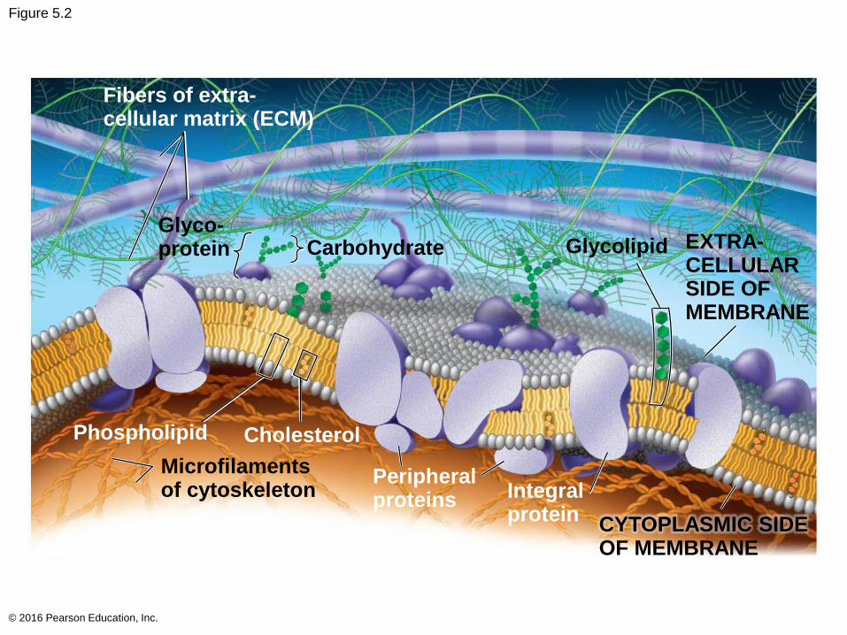
Figure 5.2
© 2016 Pearson Education, Inc.
Fibers of extra-cellular matrix (ECM)
Glyco-protein Carbohydrate Glycolipid EXTRA-
CELLULARSIDE OFMEMBRANE
Microfilamentsof cytoskeleton
Peripheralproteins Integral
proteinCYTOPLASMIC SIDEOF MEMBRANE
CholesterolPhospholipid
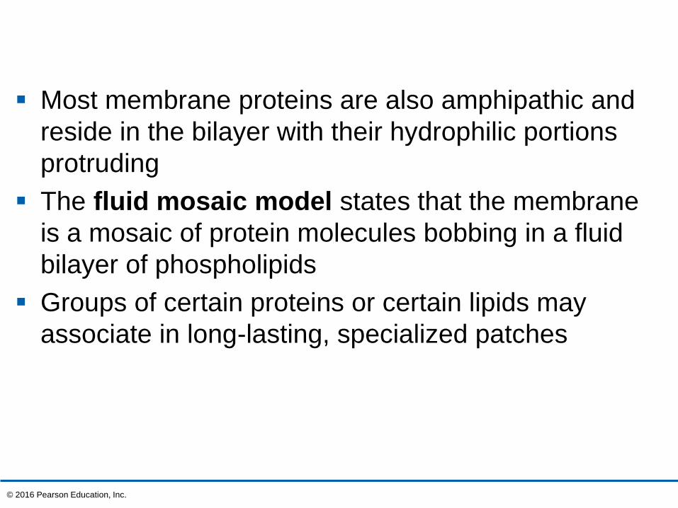
Most membrane proteins are also amphipathic and
reside in the bilayer with their hydrophilic portions
protruding
The fluid mosaic model states that the membrane
is a mosaic of protein molecules bobbing in a fluid
bilayer of phospholipids
Groups of certain proteins or certain lipids may
associate in long-lasting, specialized patches
© 2016 Pearson Education, Inc.
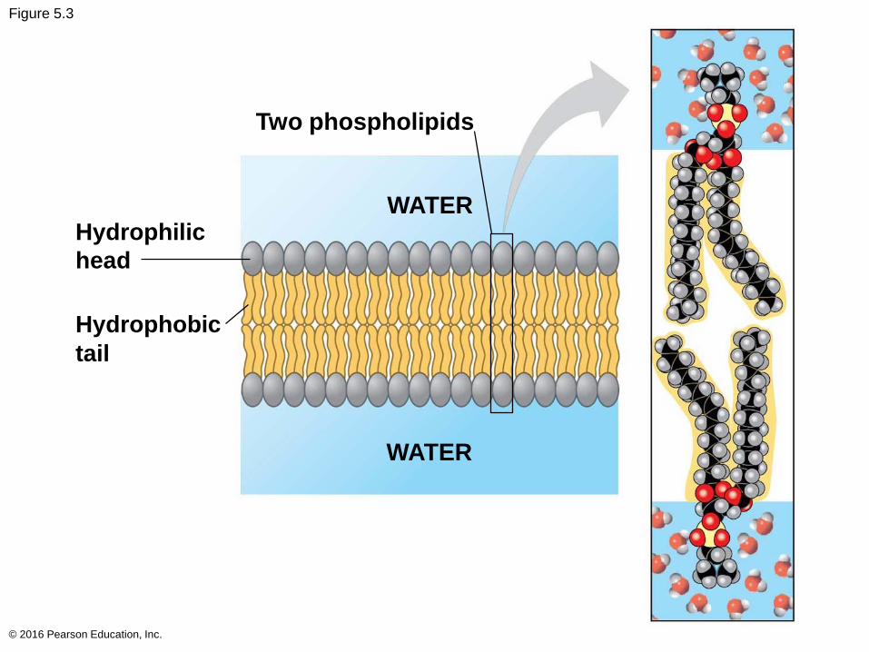
Figure 5.3
© 2016 Pearson Education, Inc.
Two phospholipids
Hydrophilic
head
WATER
WATER
Hydrophobic
tail
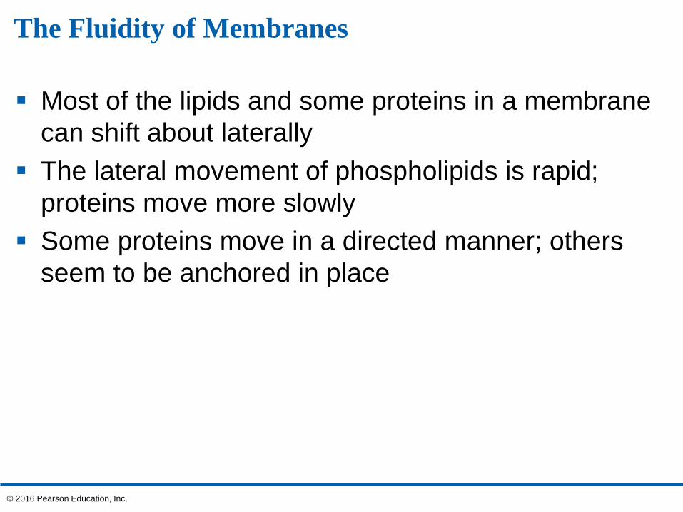
The Fluidity of Membranes
Most of the lipids and some proteins in a membrane
can shift about laterally
The lateral movement of phospholipids is rapid;
proteins move more slowly
Some proteins move in a directed manner; others
seem to be anchored in place
© 2016 Pearson Education, Inc.
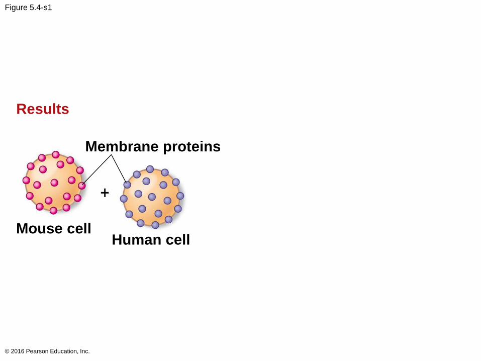
Figure 5.4-s1
© 2016 Pearson Education, Inc.
Results
Membrane proteins
Mouse cellHuman cell
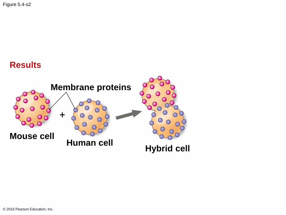
Figure 5.4-s2
© 2016 Pearson Education, Inc.
Results
Membrane proteins
Mouse cellHuman cell
Hybrid cell
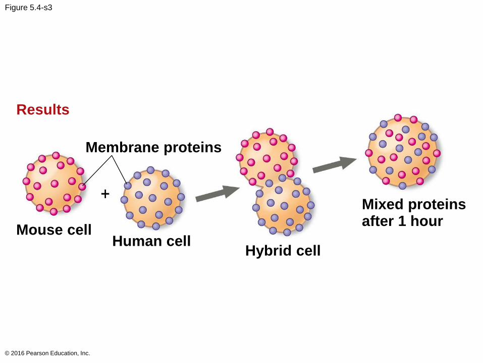
Figure 5.4-s3
© 2016 Pearson Education, Inc.
Results
Membrane proteins
Mouse cellHuman cell
Hybrid cell
Mixed proteinsafter 1 hour
As temperatures cool, membranes switch from a
fluid state to a solid state
The temperature at which a membrane solidifies
depends on the types of lipids
A membrane remains fluid to a lower temperature if
it is rich in phospholipids with unsaturated
hydrocarbon tails
Membranes must be fluid to work properly; they are
usually about as fluid as salad oil
© 2016 Pearson Education, Inc.
The steroid cholesterol has different effects on
membrane fluidity at different temperatures
At warm temperatures (such as 37°C), cholesterol
restrains movement of phospholipids
At cool temperatures, it maintains fluidity by
preventing tight packing
© 2016 Pearson Education, Inc.
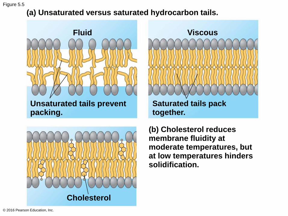
Figure 5.5
© 2016 Pearson Education, Inc.
(b) Cholesterol reducesmembrane fluidity atmoderate temperatures, butat low temperatures hinderssolidification.
Saturated tails packtogether.
Fluid Viscous
Unsaturated tails preventpacking.
Cholesterol
(a) Unsaturated versus saturated hydrocarbon tails.
Evolution of Differences in Membrane Lipid Composition
Variations in lipid composition of cell membranes of
many species appear to be adaptations to specific
environmental conditions
Ability to change the lipid compositions in response
to temperature changes has evolved in organisms
that live where temperatures vary
© 2016 Pearson Education, Inc.
Membrane Proteins and Their Functions
A membrane is a collage of different proteins
embedded in the fluid matrix of the lipid bilayer
Proteins determine most of the membrane’s specific
functions
© 2016 Pearson Education, Inc.

Integral proteins penetrate the hydrophobic interior
of the lipid bilayer
Integral proteins that span the membrane are called
transmembrane proteins
The hydrophobic regions of an integral protein
consist of one or more stretches of nonpolar amino
acids, often coiled into helices
Peripheral proteins are loosely bound to the
surface of the membrane
© 2016 Pearson Education, Inc.
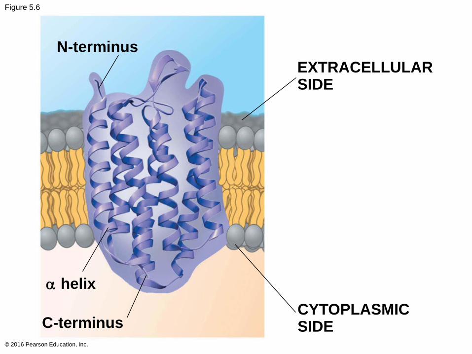
Figure 5.6
© 2016 Pearson Education, Inc.
N-terminus
EXTRACELLULARSIDE
C-terminusCYTOPLASMICSIDE
helix
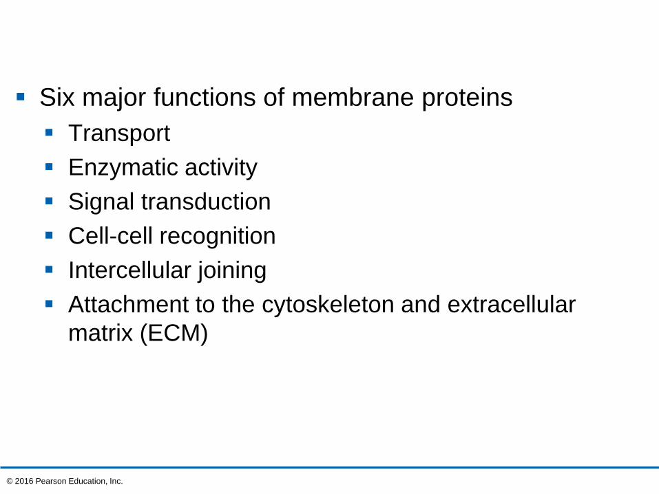
Six major functions of membrane proteins
Transport
Enzymatic activity
Signal transduction
Cell-cell recognition
Intercellular joining
Attachment to the cytoskeleton and extracellular
matrix (ECM)
© 2016 Pearson Education, Inc.
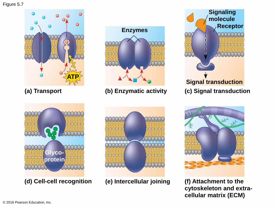
Figure 5.7
© 2016 Pearson Education, Inc.
(a) Transport (b) Enzymatic activity (c) Signal transduction
(d) Cell-cell recognition (e) Intercellular joining (f) Attachment to thecytoskeleton and extra-cellular matrix (ECM)
Signal transduction
Enzymes
Signalingmolecule
Receptor
Glyco-protein
ATP
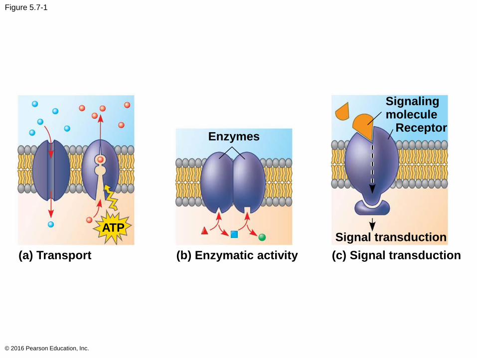
Figure 5.7-1
© 2016 Pearson Education, Inc.
Enzymes
Signalingmolecule
Receptor
ATPSignal transduction
(a) Transport (b) Enzymatic activity (c) Signal transduction
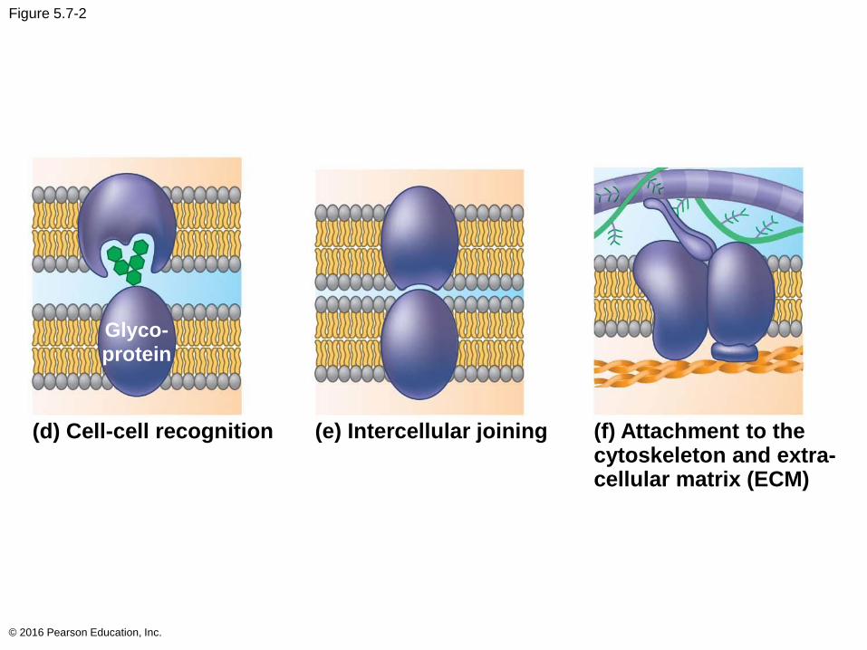
Figure 5.7-2
© 2016 Pearson Education, Inc.
(d) Cell-cell recognition (e) Intercellular joining (f) Attachment to thecytoskeleton and extra-cellular matrix (ECM)
Glyco-
protein
The Role of Membrane Carbohydrates in Cell-Cell Recognition
Cells recognize each other by binding to surface
molecules, often containing carbohydrates, on the
extracellular surface of the plasma membrane
Membrane carbohydrates may be covalently
bonded to lipids (forming glycolipids) or, more
commonly, to proteins (forming glycoproteins)
Carbohydrates on the external side of the plasma
membrane vary among species, individuals, and
even cell types in an individual
© 2016 Pearson Education, Inc.
Synthesis and Sidedness of Membranes
Membranes have distinct inside and outside faces
The asymmetrical arrangement of proteins, lipids,
and associated carbohydrates in the plasma
membrane is determined as the membrane is built
by the ER and Golgi apparatus
© 2016 Pearson Education, Inc.
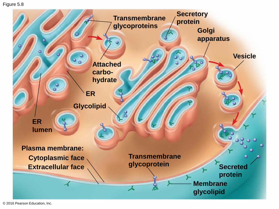
Figure 5.8
© 2016 Pearson Education, Inc.
Transmembraneglycoproteins
Secretoryprotein
Vesicle
ER
Plasma membrane:
Transmembraneglycoprotein Secreted
protein
Membrane
glycolipid
ER
lumen
Golgi
apparatus
Attachedcarbo-hydrate
Glycolipid
Cytoplasmic face
Extracellular face
Concept 5.2: Membrane structure results in selective permeability
A cell must regulate transport of substances across
cellular boundaries
Plasma membranes are selectively permeable,
regulating the cell’s molecular traffic
© 2016 Pearson Education, Inc.
The Permeability of the Lipid Bilayer
Hydrophobic (nonpolar) molecules, such as
hydrocarbons, can dissolve in the lipid bilayer of the
membrane and cross it easily
Polar molecules, such as sugars, do not cross the
membrane easily
© 2016 Pearson Education, Inc.
Transport Proteins
Transport proteins allow passage of hydrophilic
substances across the membrane
Some transport proteins, called channel proteins,
have a hydrophilic channel that certain molecules or
ions can use as a tunnel
Channel proteins called aquaporins facilitate the
passage of water
© 2016 Pearson Education, Inc.
Other transport proteins, called carrier proteins,
bind to molecules and change shape to shuttle
them across the membrane
A transport protein is specific for the substance it
moves
© 2016 Pearson Education, Inc.
Concept 5.3: Passive transport is diffusion of a substance across a membrane with no energy investment
Diffusion is the tendency for molecules to spread
out evenly into the available space
Although each molecule moves randomly, diffusion
of a population of molecules may be directional
At dynamic equilibrium, as many molecules cross
the membrane in one direction as in the other
© 2016 Pearson Education, Inc.
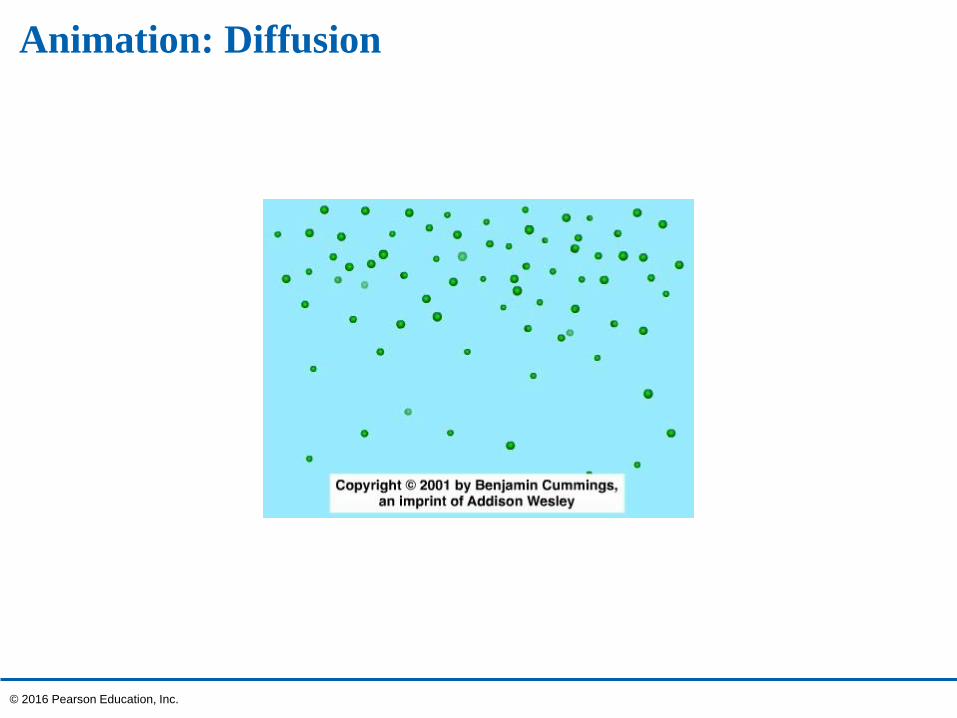
Animation: Diffusion
© 2016 Pearson Education, Inc.
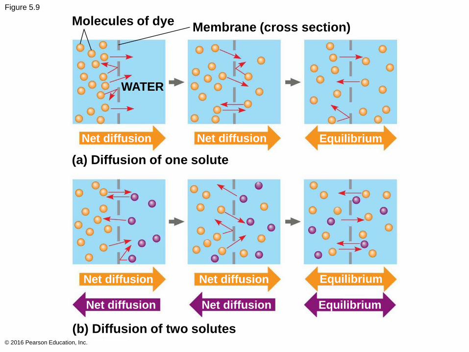
Figure 5.9
© 2016 Pearson Education, Inc.
Molecules of dyeMembrane (cross section)
Net diffusion Net diffusion Equilibrium
(a) Diffusion of one solute
Net diffusion Net diffusion Equilibrium
(b) Diffusion of two solutes
WATER
Net diffusion Net diffusion Equilibrium
Substances diffuse down their concentration
gradient, from where it is more concentrated to
where it is less concentrated
No work must be done to move substances down
the concentration gradient
The diffusion of a substance across a biological
membrane is passive transport because no
energy is expended by the cell to make it happen
© 2016 Pearson Education, Inc.
Effects of Osmosis on Water Balance
Osmosis is the diffusion of free water across a
selectively permeable membrane
Water diffuses across a membrane from the region
of lower solute concentration to the region of higher
solute concentration until the solute concentration is
equal on both sides
© 2016 Pearson Education, Inc.
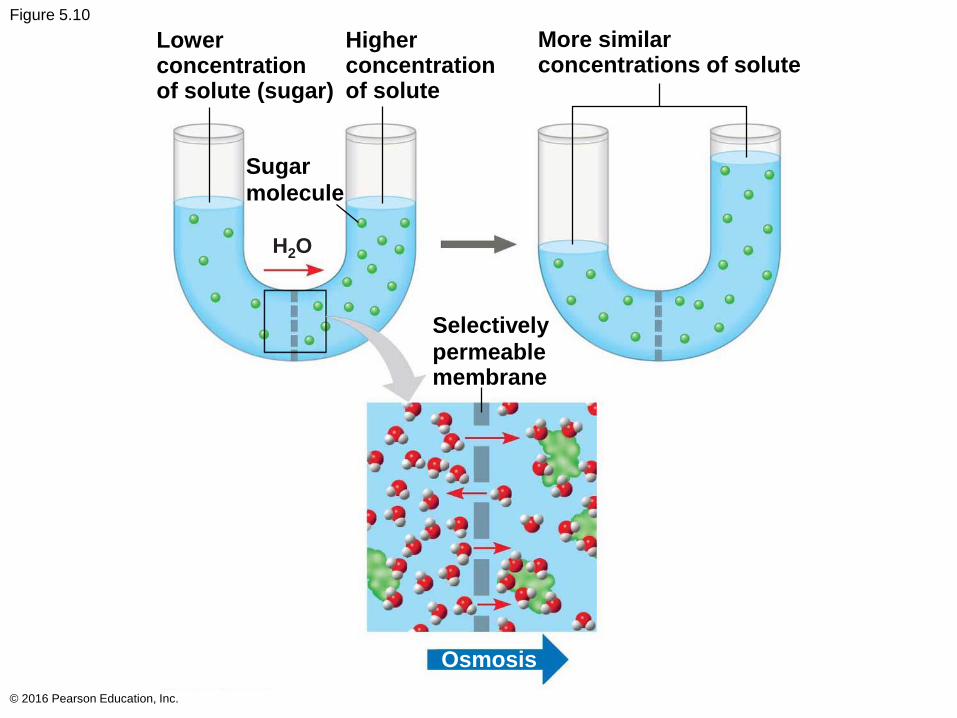
Figure 5.10
© 2016 Pearson Education, Inc.
Higherconcentrationof solute
More similarconcentrations of solute
Selectively
permeablemembrane
Osmosis
Lowerconcentrationof solute (sugar)
Sugar
molecule
H2O
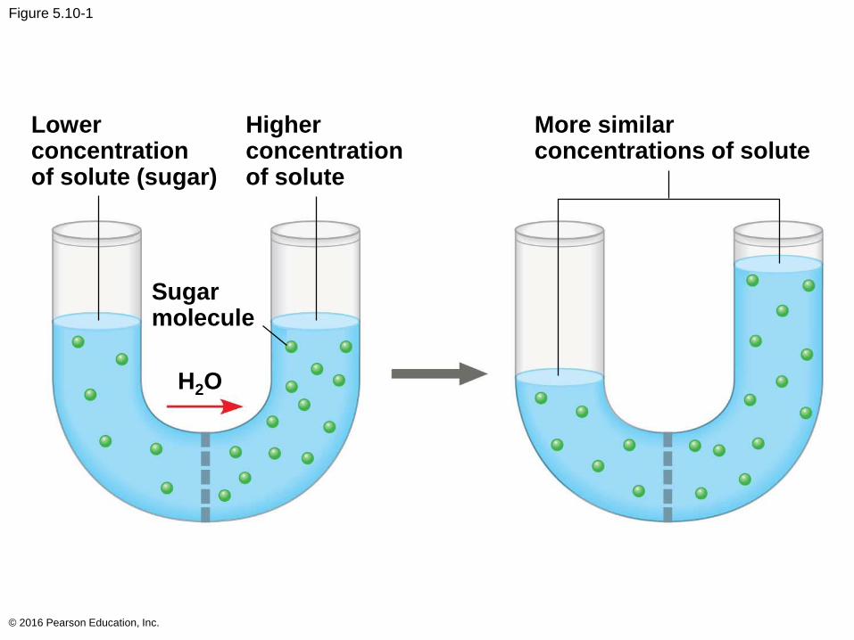
Figure 5.10-1
© 2016 Pearson Education, Inc.
Lowerconcentrationof solute (sugar)
Higherconcentrationof solute
More similarconcentrations of solute
Sugarmolecule
H2O
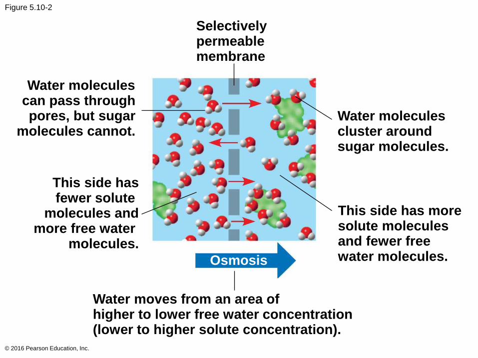
Figure 5.10-2
© 2016 Pearson Education, Inc.
Water moleculescan pass throughpores, but sugar
molecules cannot.
This side hasfewer solute
molecules andmore free water
molecules.
Water moves from an area ofhigher to lower free water concentration(lower to higher solute concentration).
This side has moresolute moleculesand fewer freewater molecules.
Water moleculescluster aroundsugar molecules.
Selectivelypermeablemembrane
Osmosis
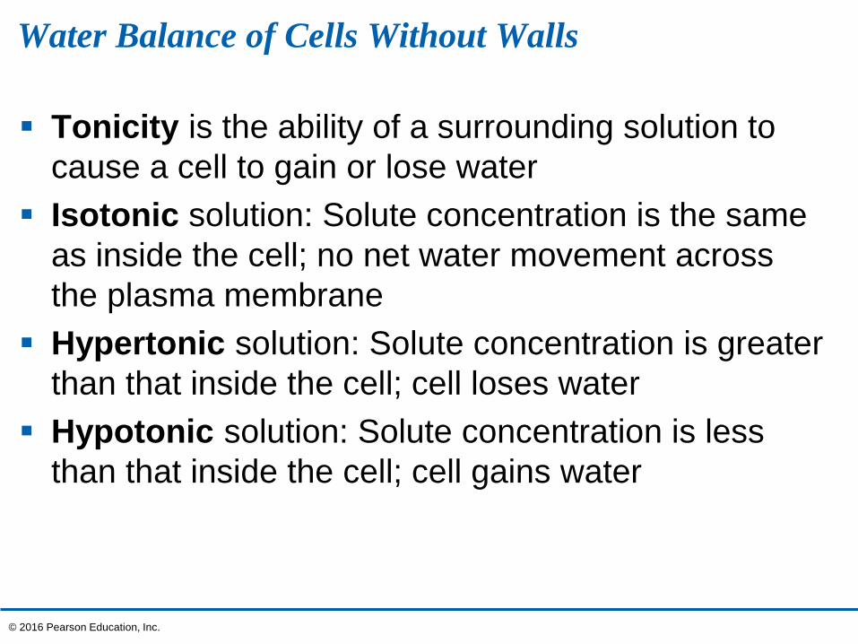
Water Balance of Cells Without Walls
Tonicity is the ability of a surrounding solution to
cause a cell to gain or lose water
Isotonic solution: Solute concentration is the same
as inside the cell; no net water movement across
the plasma membrane
Hypertonic solution: Solute concentration is greater
than that inside the cell; cell loses water
Hypotonic solution: Solute concentration is less
than that inside the cell; cell gains water
© 2016 Pearson Education, Inc.
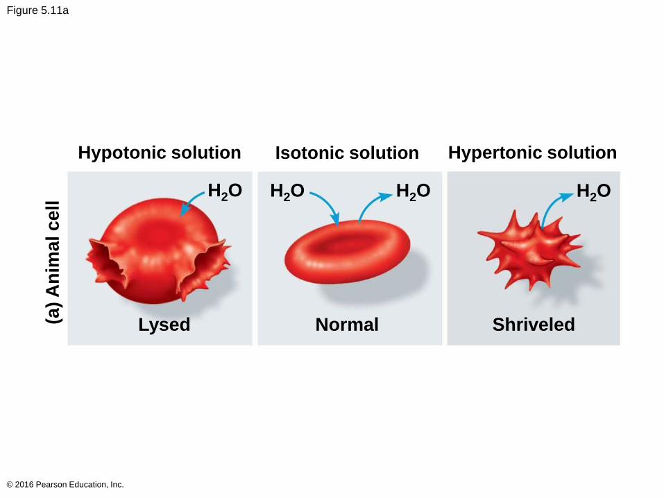
Figure 5.11a
© 2016 Pearson Education, Inc.
Hypotonic solution Isotonic solution Hypertonic solution
Lysed Normal Shriveled
H2O H2O H2O H2O
(a)
An
ima
l c
ell
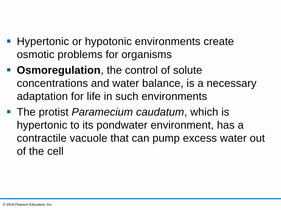
Hypertonic or hypotonic environments create
osmotic problems for organisms
Osmoregulation, the control of solute
concentrations and water balance, is a necessary
adaptation for life in such environments
The protist Paramecium caudatum, which is
hypertonic to its pondwater environment, has a
contractile vacuole that can pump excess water out
of the cell
© 2016 Pearson Education, Inc.
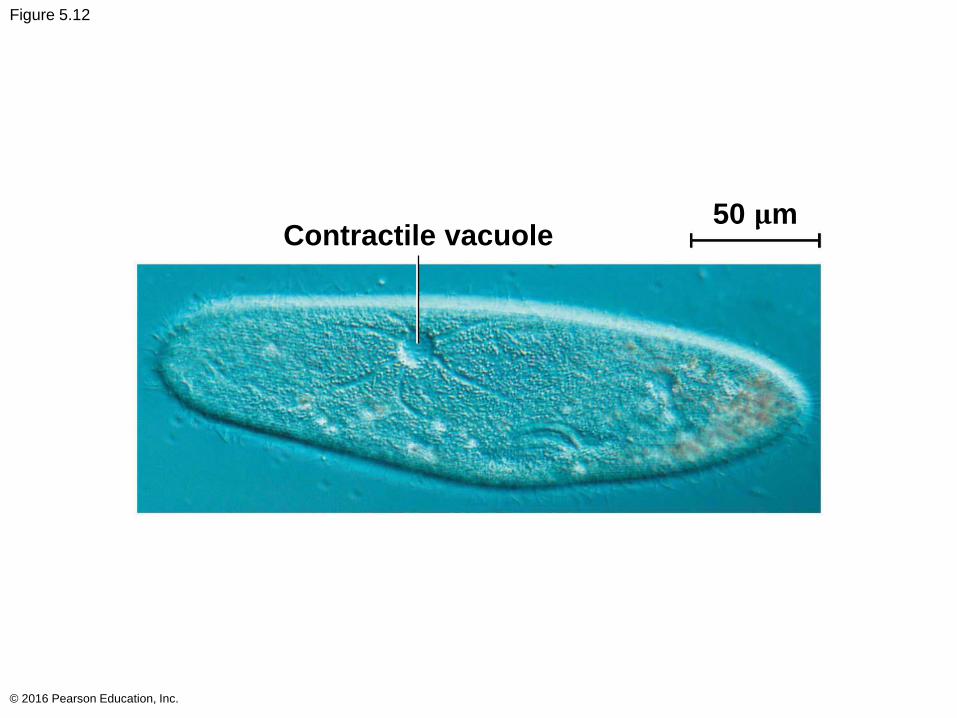
Figure 5.12
© 2016 Pearson Education, Inc.
Contractile vacuole50 𝛍m
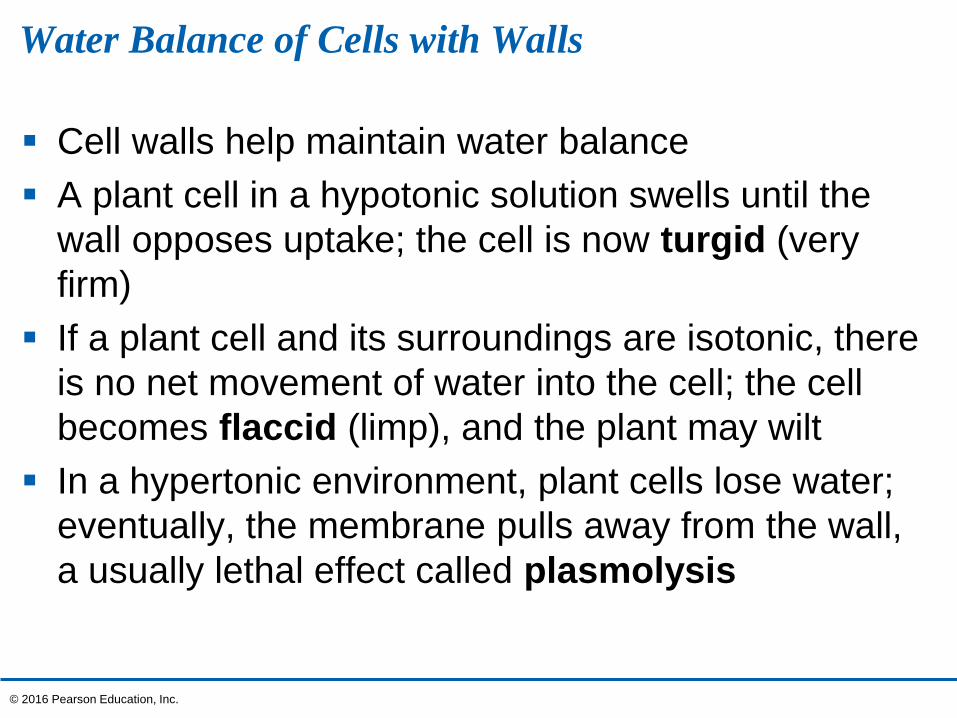
Water Balance of Cells with Walls
Cell walls help maintain water balance
A plant cell in a hypotonic solution swells until the
wall opposes uptake; the cell is now turgid (very
firm)
If a plant cell and its surroundings are isotonic, there
is no net movement of water into the cell; the cell
becomes flaccid (limp), and the plant may wilt
In a hypertonic environment, plant cells lose water;
eventually, the membrane pulls away from the wall,
a usually lethal effect called plasmolysis
© 2016 Pearson Education, Inc.
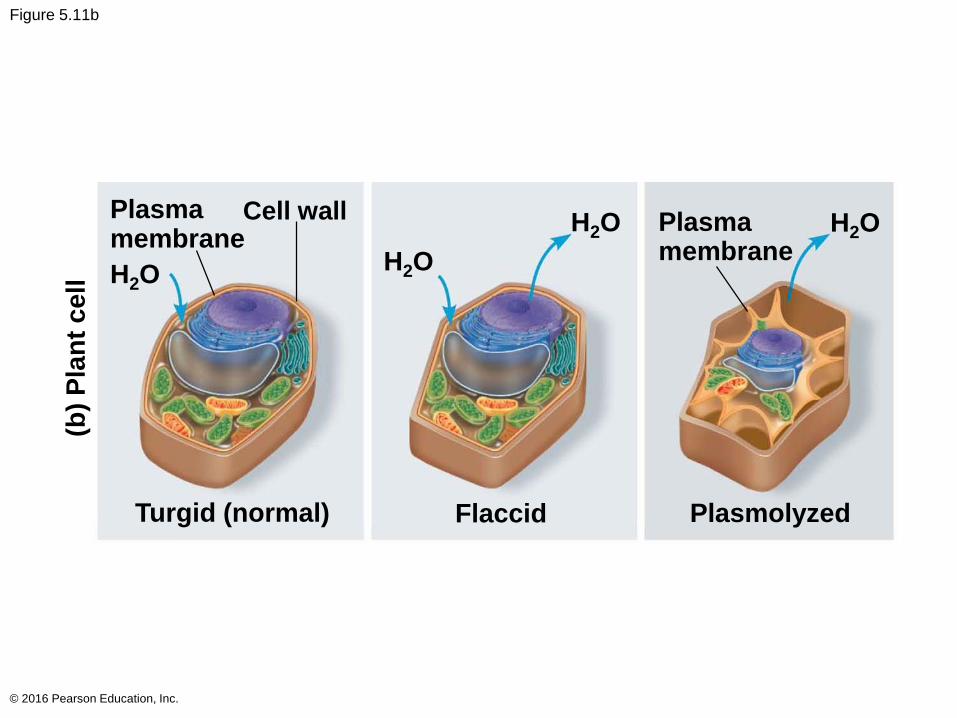
Figure 5.11b
© 2016 Pearson Education, Inc.
Plasmamembrane
H2O
Flaccid Plasmolyzed
H2O
H2OH2O
Turgid (normal)
Plasmamembrane
Cell wall
(b)
Pla
nt
ce
ll
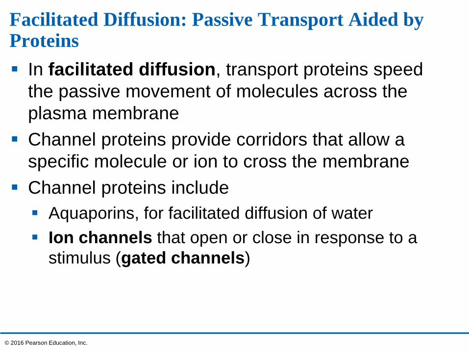
Facilitated Diffusion: Passive Transport Aided by Proteins
In facilitated diffusion, transport proteins speed
the passive movement of molecules across the
plasma membrane
Channel proteins provide corridors that allow a
specific molecule or ion to cross the membrane
Channel proteins include
Aquaporins, for facilitated diffusion of water
Ion channels that open or close in response to a
stimulus (gated channels)
© 2016 Pearson Education, Inc.
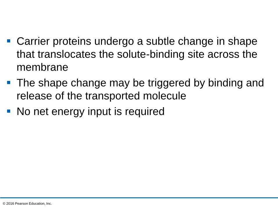
Carrier proteins undergo a subtle change in shape
that translocates the solute-binding site across the
membrane
The shape change may be triggered by binding and
release of the transported molecule
No net energy input is required
© 2016 Pearson Education, Inc.
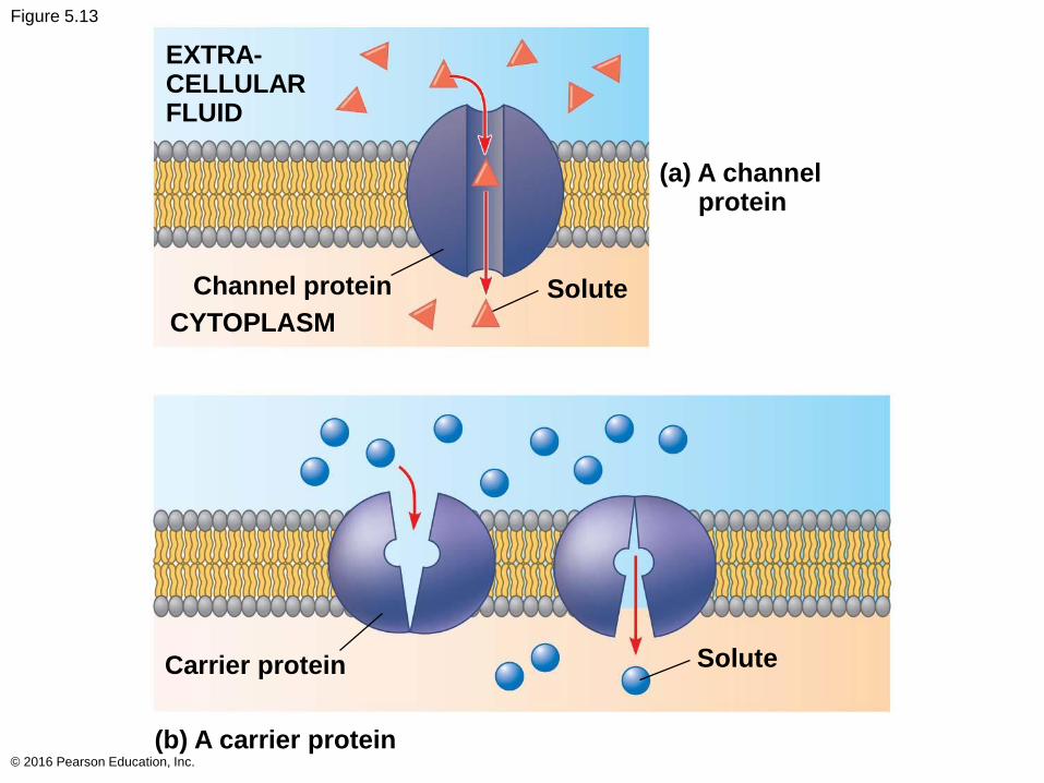
Figure 5.13
© 2016 Pearson Education, Inc.
(a) A channelprotein
EXTRA-CELLULARFLUID
Channel protein
CYTOPLASM
Solute
SoluteCarrier protein
(b) A carrier protein
Concept 5.4: Active transport uses energy to move solutes against their gradients
Facilitated diffusion speeds transport of a solute by
providing efficient passage through the membrane
but does not alter the direction of transport
Some transport proteins, however, can move
solutes against their concentration gradients
© 2016 Pearson Education, Inc.
The Need for Energy in Active Transport
Active transport moves substances against their
concentration gradients
Active transport requires energy, usually in the form
of ATP
© 2016 Pearson Education, Inc.

Active transport allows cells to maintain
concentration gradients that differ from their
surroundings
The sodium-potassium pump is one type of active
transport system
© 2016 Pearson Education, Inc.
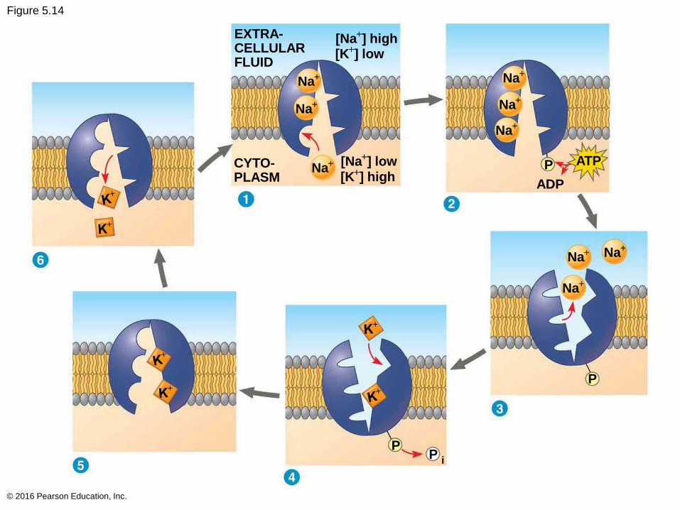
Figure 5.14
© 2016 Pearson Education, Inc.
EXTRA-CELLULARFLUID
[Na+] high
[K+] low
Na+
Na+
Na+ [Na
+] low
[K+] high
CYTO-PLASM
Na+
Na+ Na
+
Na+
Na+
Na+
P
ADP
ATP
P
PP i
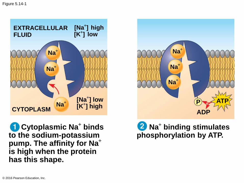
Figure 5.14-1
© 2016 Pearson Education, Inc.
Cytoplasmic Na+
bindsto the sodium-potassiumpump. The affinity for Na
+
is high when the proteinhas this shape.
Na+
binding stimulatesphosphorylation by ATP.
Na+
Na+
Na+
Na+
Na+
Na+
[Na+] low ATP
ADP
EXTRACELLULARFLUID
[K+] high
[Na+] high
[K+] low
PCYTOPLASM
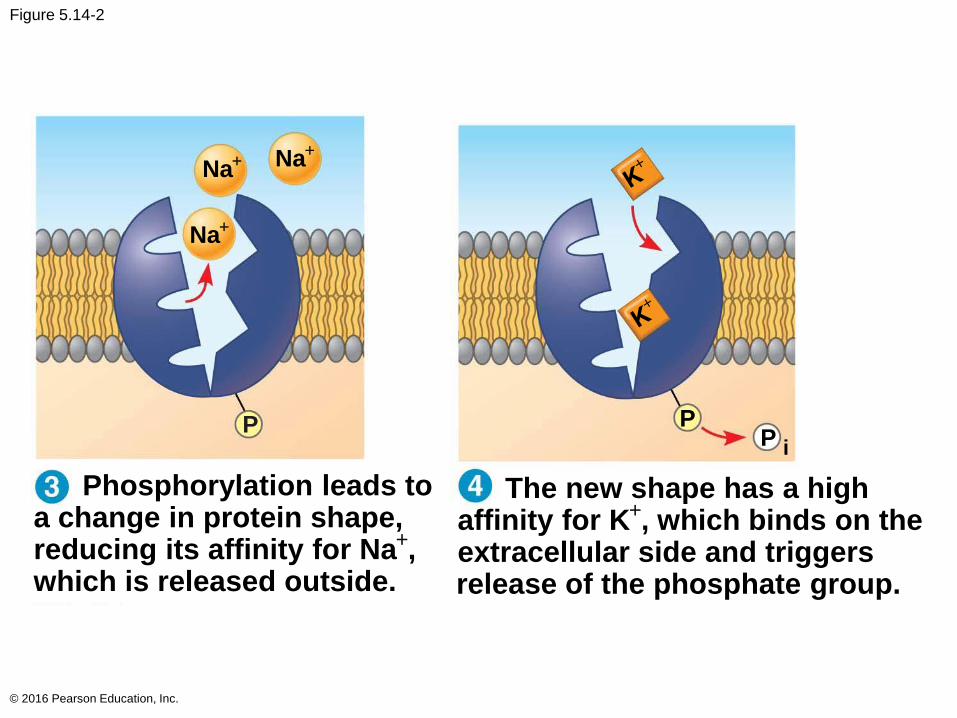
Figure 5.14-2
© 2016 Pearson Education, Inc.
The new shape has a highaffinity for K
+, which binds on the
extracellular side and triggersrelease of the phosphate group.
Phosphorylation leads toa change in protein shape,reducing its affinity for Na
+,
which is released outside.
Na+
Na+ Na
+
P PP i

Figure 5.14-3
© 2016 Pearson Education, Inc.
Loss of the phosphategroup restores the protein’soriginal shape, which has alower affinity for K+.
K+ is released; affinityfor Na+ is high again, andthe cycle repeats.
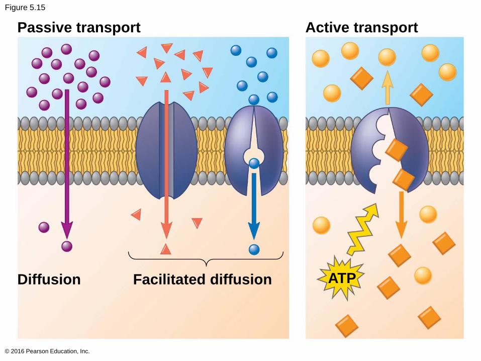
Figure 5.15
© 2016 Pearson Education, Inc.
Passive transport Active transport
Diffusion Facilitated diffusion ATP
How Ion Pumps Maintain Membrane Potential
Membrane potential is the voltage across a
membrane
Voltage is created by differences in the distribution
of positive and negative ions across a membrane
© 2016 Pearson Education, Inc.
Two combined forces, collectively called the
electrochemical gradient, drive the diffusion of
ions across a membrane
A chemical force (the ion’s concentration gradient)
An electrical force (the effect of the membrane
potential on the ion’s movement)
© 2016 Pearson Education, Inc.
An electrogenic pump is a transport protein that
generates voltage across a membrane
The sodium-potassium pump is the major
electrogenic pump of animal cells
The main electrogenic pump of plants, fungi, and
bacteria is a proton pump
Electrogenic pumps help store energy that can be
used for cellular work
© 2016 Pearson Education, Inc.
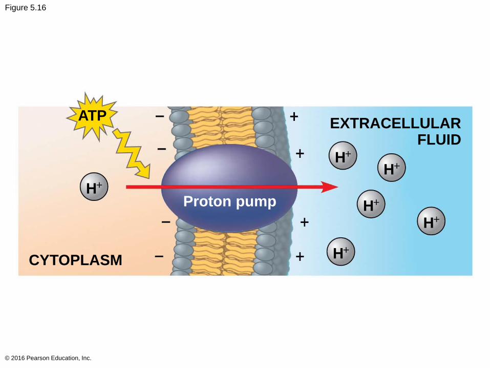
Figure 5.16
© 2016 Pearson Education, Inc.
ATPEXTRACELLULAR
H+
H+
FLUID
H+
Proton pump H+
H+
CYTOPLASM H+
Cotransport: Coupled Transport by a Membrane Protein
Cotransport occurs when active transport of a
solute indirectly drives transport of other solutes
Plant cells use the gradient of hydrogen ions
generated by proton pumps to drive active transport
of nutrients into the cell
© 2016 Pearson Education, Inc.
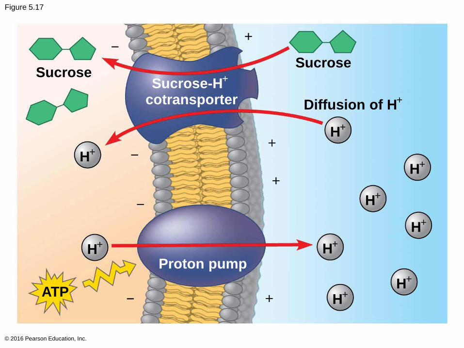
Figure 5.17
© 2016 Pearson Education, Inc.
SucroseSucrose-H
+
cotransporter Diffusion of H+
H+
H+
H+
H+
H+
H+ H
+
Proton pump
ATP H+
H+
Sucrose
+
+
+
+
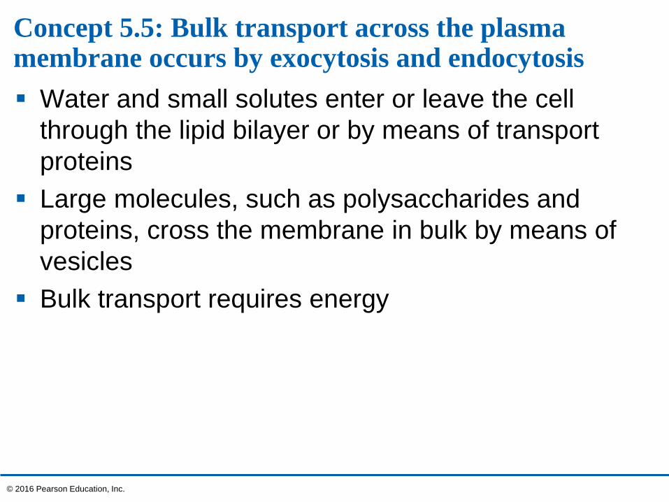
Concept 5.5: Bulk transport across the plasma membrane occurs by exocytosis and endocytosis
Water and small solutes enter or leave the cell
through the lipid bilayer or by means of transport
proteins
Large molecules, such as polysaccharides and
proteins, cross the membrane in bulk by means of
vesicles
Bulk transport requires energy
© 2016 Pearson Education, Inc.
Exocytosis
In exocytosis, transport vesicles migrate to the
membrane, fuse with it, and release their contents
Many secretory cells use exocytosis to export
products
© 2016 Pearson Education, Inc.
Endocytosis
In endocytosis, the cell takes in molecules and
particulate matter by forming new vesicles from the
plasma membrane
Endocytosis is a reversal of exocytosis, involving
different proteins
There are three types of endocytosis
Phagocytosis (“cellular eating”)
Pinocytosis (“cellular drinking”)
Receptor-mediated endocytosis
© 2016 Pearson Education, Inc.

Figure 5.18
© 2016 Pearson Education, Inc.
CYTOPLASM
Coatedvesicle
Coatprotein
Coatedpit
Phagocytosis Pinocytosis
Pseudopodium
Solutes
Receptor
Food orotherparticle
Foodvacuole
Receptor-MediatedEndocytosis
Plasmamembrane
EXTRACELLULARFLUID
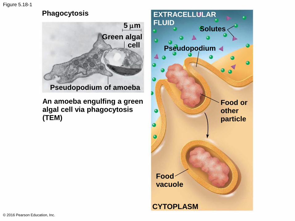
Figure 5.18-1
© 2016 Pearson Education, Inc.
Phagocytosis
Green algalcell
Solutes
Pseudopodium of amoeba
Pseudopodium
An amoeba engulfing a greenalgal cell via phagocytosis(TEM)
Food orotherparticle
Foodvacuole
CYTOPLASM
EXTRACELLULARFLUID5 mm

Figure 5.18-2
© 2016 Pearson Education, Inc.
Pinocytosis
Pinocytotic vesicles forming(TEMs) Coated
pit
Coatprotein
Plasmamembrane
Coatedvesicle
0.2
5 m
m
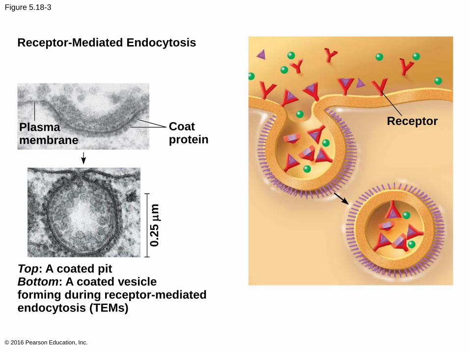
Figure 5.18-3
© 2016 Pearson Education, Inc.
Receptor-Mediated Endocytosis
Plasmamembrane
Coatprotein
Top: A coated pitBottom: A coated vesicleforming during receptor-mediatedendocytosis (TEMs)
Receptor
0.2
5 m
m
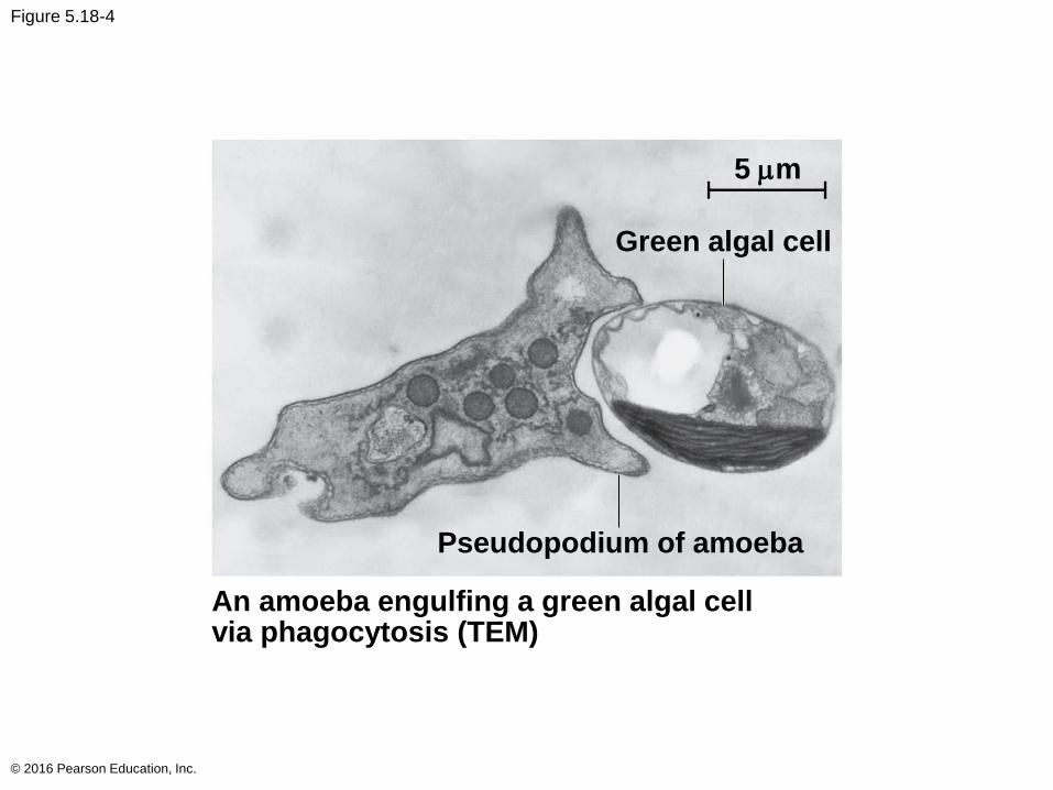
Figure 5.18-4
© 2016 Pearson Education, Inc.
Green algal cell
Pseudopodium of amoeba
An amoeba engulfing a green algal cellvia phagocytosis (TEM)
5 mm
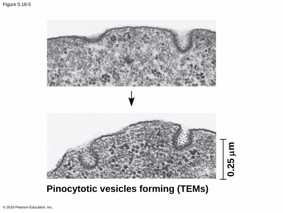
Figure 5.18-5
© 2016 Pearson Education, Inc.
Pinocytotic vesicles forming (TEMs)
0.2
5 m
m
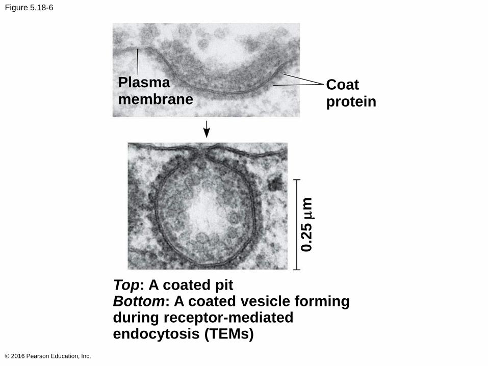
Figure 5.18-6
© 2016 Pearson Education, Inc.
Plasmamembrane
Coatprotein
Top: A coated pitBottom: A coated vesicle formingduring receptor-mediatedendocytosis (TEMs)
0.2
5 m
m
Concept 5.6: The plasma membrane plays a key role in most cell signaling
In multicellular organisms, cell-to-cell communication
allows the cells of the body to coordinate their
activities
Communication between cells is also essential for
many unicellular organisms
© 2016 Pearson Education, Inc.

Local and Long-Distance Signaling
Eukaryotic cells may communicate by direct contact
Animal and plant cells have junctions that directly
connect the cytoplasm of adjacent cells
These are called gap junctions (animal cells) and
plasmodesmata (plant cells)
The free passage of substances in the cytosol from
one cell to another is a type of local signaling
© 2016 Pearson Education, Inc.
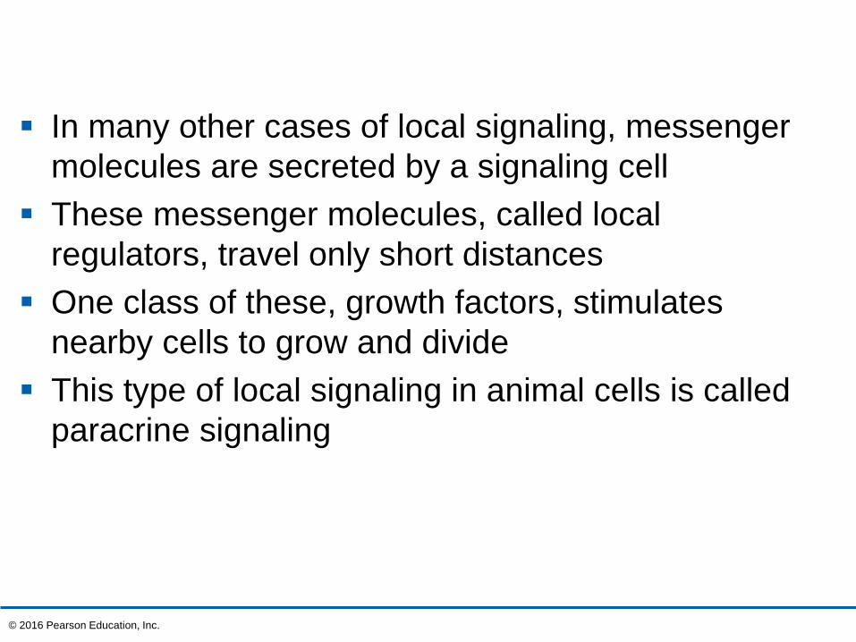
In many other cases of local signaling, messenger
molecules are secreted by a signaling cell
These messenger molecules, called local
regulators, travel only short distances
One class of these, growth factors, stimulates
nearby cells to grow and divide
This type of local signaling in animal cells is called
paracrine signaling
© 2016 Pearson Education, Inc.
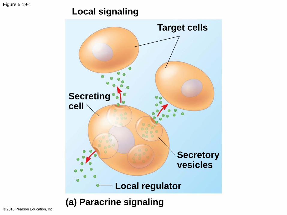
Figure 5.19-1
© 2016 Pearson Education, Inc.
Local signaling
Target cells
Secretingcell
Secretoryvesicles
Local regulator
(a) Paracrine signaling
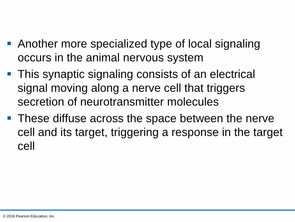
Another more specialized type of local signaling
occurs in the animal nervous system
This synaptic signaling consists of an electrical
signal moving along a nerve cell that triggers
secretion of neurotransmitter molecules
These diffuse across the space between the nerve
cell and its target, triggering a response in the target
cell
© 2016 Pearson Education, Inc.
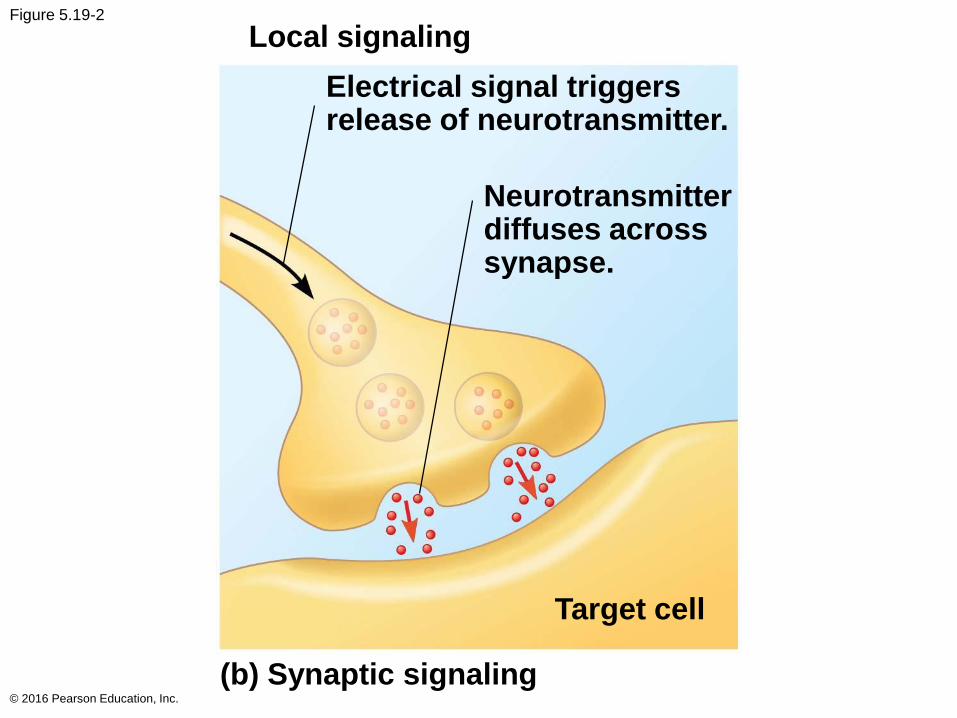
Figure 5.19-2
© 2016 Pearson Education, Inc.
Local signaling
Electrical signal triggersrelease of neurotransmitter.
Neurotransmitterdiffuses acrosssynapse.
Target cell
(b) Synaptic signaling
In long-distance signaling, plants and animals use
chemicals called hormones
In hormonal signaling in animals (called endocrine
signaling), specialized cells release hormone
molecules that travel via the circulatory system
Hormones vary widely in size and shape
© 2016 Pearson Education, Inc.
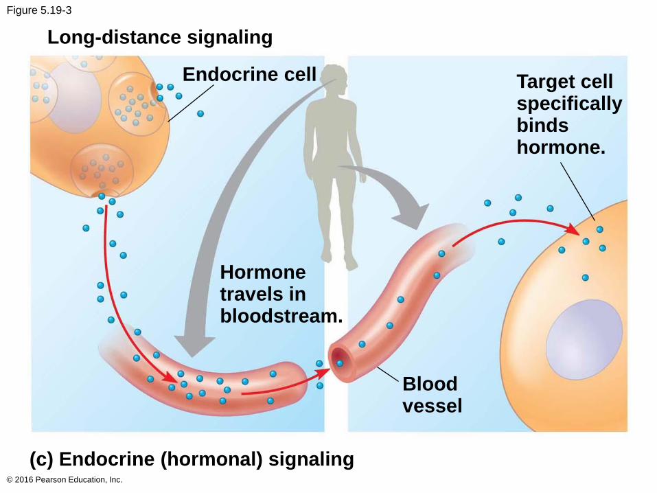
Figure 5.19-3
© 2016 Pearson Education, Inc.
Long-distance signaling
Endocrine cell Target cellspecificallybindshormone.
Hormonetravels inbloodstream.
Bloodvessel
(c) Endocrine (hormonal) signaling
The Three Stages of Cell Signaling: A Preview
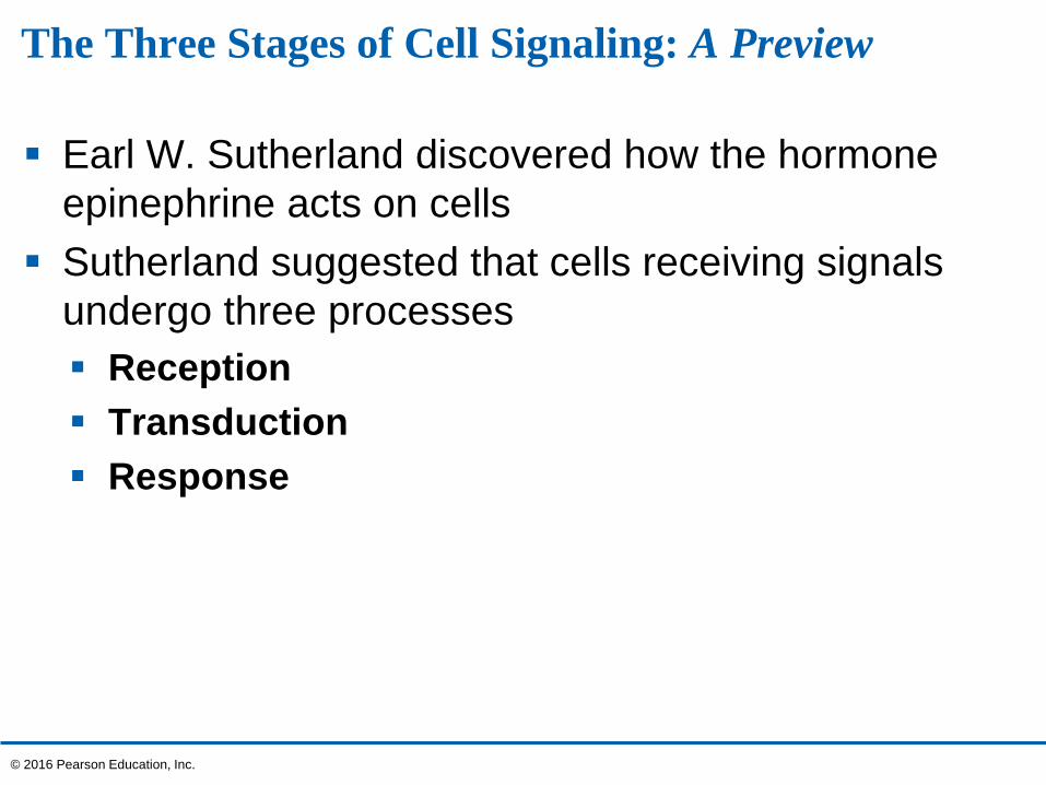
Earl W. Sutherland discovered how the hormone
epinephrine acts on cells
Sutherland suggested that cells receiving signals
undergo three processes
Reception
Transduction
Response
© 2016 Pearson Education, Inc.
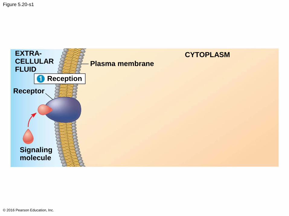
Figure 5.20-s1
© 2016 Pearson Education, Inc.
EXTRA-CELLULARFLUID
Plasma membrane
CYTOPLASM
Reception
Receptor
Signalingmolecule
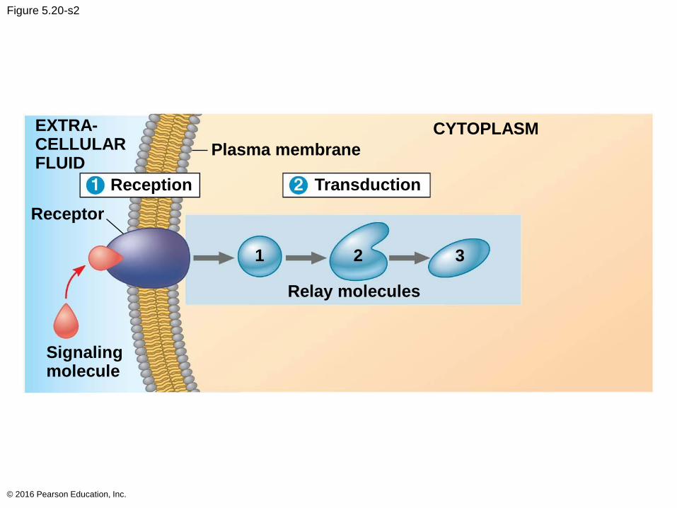
Figure 5.20-s2
© 2016 Pearson Education, Inc.
EXTRA-CELLULARFLUID
Plasma membrane
CYTOPLASM
Reception
Receptor
Signalingmolecule
Relay molecules
Transduction
1 2 3
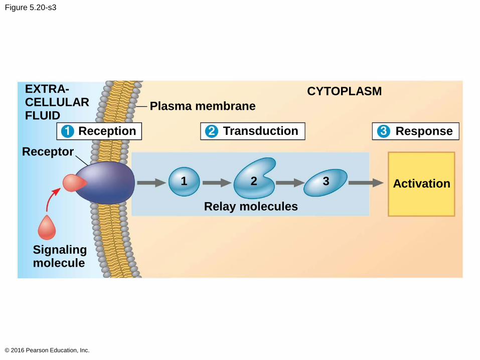
Figure 5.20-s3
© 2016 Pearson Education, Inc.
EXTRA-CELLULARFLUID
Plasma membrane
CYTOPLASM
Reception
Receptor
Signalingmolecule
Relay molecules
Transduction Response
Activation1 2 3
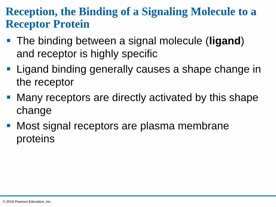
Reception, the Binding of a Signaling Molecule to a Receptor Protein
The binding between a signal molecule (ligand)
and receptor is highly specific
Ligand binding generally causes a shape change in
the receptor
Many receptors are directly activated by this shape
change
Most signal receptors are plasma membrane
proteins
© 2016 Pearson Education, Inc.
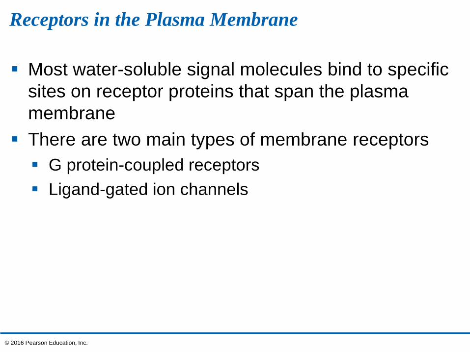
Receptors in the Plasma Membrane
Most water-soluble signal molecules bind to specific
sites on receptor proteins that span the plasma
membrane
There are two main types of membrane receptors
G protein-coupled receptors
Ligand-gated ion channels
© 2016 Pearson Education, Inc.
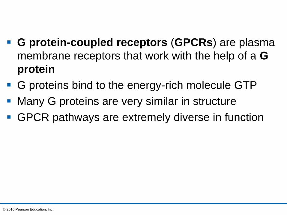
G protein-coupled receptors (GPCRs) are plasma
membrane receptors that work with the help of a G
protein
G proteins bind to the energy-rich molecule GTP
Many G proteins are very similar in structure
GPCR pathways are extremely diverse in function
© 2016 Pearson Education, Inc.
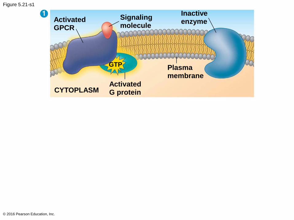
Figure 5.21-s1
© 2016 Pearson Education, Inc.
ActivatedGPCR
Signalingmolecule
Inactiveenzyme
Plasmamembrane
ActivatedG proteinCYTOPLASM
GTP
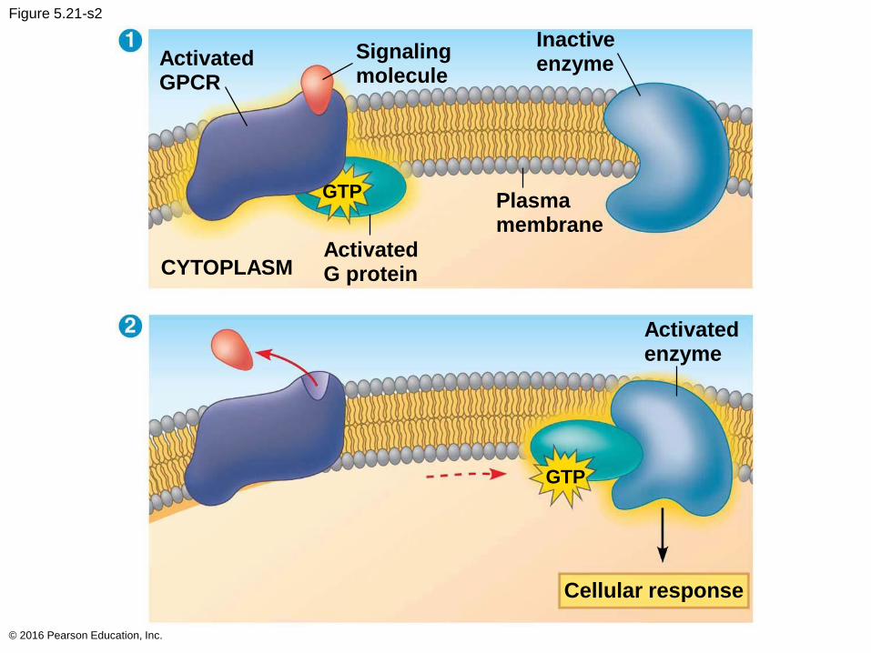
Figure 5.21-s2
© 2016 Pearson Education, Inc.
ActivatedGPCR
Signalingmolecule
Inactiveenzyme
Plasmamembrane
ActivatedG proteinCYTOPLASM
GTP
Activatedenzyme
GTP
Cellular response
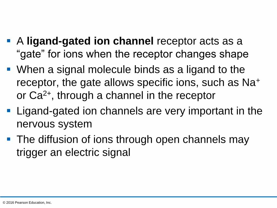
A ligand-gated ion channel receptor acts as a
“gate” for ions when the receptor changes shape
When a signal molecule binds as a ligand to the
receptor, the gate allows specific ions, such as Na+
or Ca2+, through a channel in the receptor
Ligand-gated ion channels are very important in the
nervous system
The diffusion of ions through open channels may
trigger an electric signal
© 2016 Pearson Education, Inc.
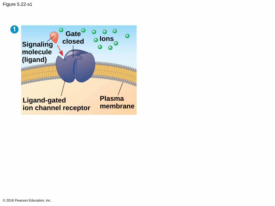
Figure 5.22-s1
© 2016 Pearson Education, Inc.
Signalingmolecule(ligand)
Gateclosed Ions
Ligand-gatedion channel receptor
Plasmamembrane
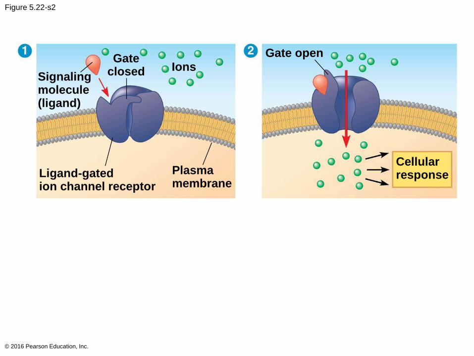
Figure 5.22-s2
© 2016 Pearson Education, Inc.
Signalingmolecule(ligand)
Gateclosed Ions
Ligand-gatedion channel receptor
Plasmamembrane
Gate open
Cellularresponse
Figure 5.22-s3
© 2016 Pearson Education, Inc.
Signalingmolecule(ligand)
Gateclosed Ions
Ligand-gatedion channel receptor
Plasmamembrane
Gate open
Cellularresponse
Gate closed
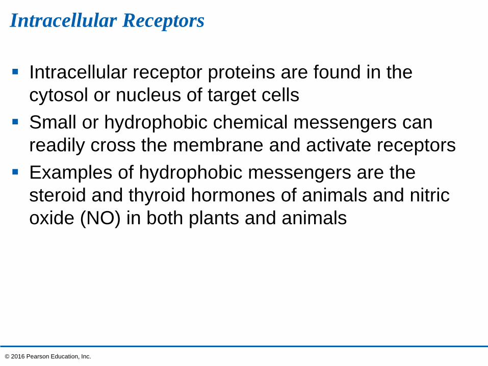
Intracellular Receptors
Intracellular receptor proteins are found in the
cytosol or nucleus of target cells
Small or hydrophobic chemical messengers can
readily cross the membrane and activate receptors
Examples of hydrophobic messengers are the
steroid and thyroid hormones of animals and nitric
oxide (NO) in both plants and animals
© 2016 Pearson Education, Inc.
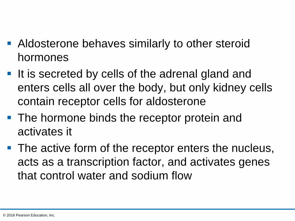
Aldosterone behaves similarly to other steroid
hormones
It is secreted by cells of the adrenal gland and
enters cells all over the body, but only kidney cells
contain receptor cells for aldosterone
The hormone binds the receptor protein and
activates it
The active form of the receptor enters the nucleus,
acts as a transcription factor, and activates genes
that control water and sodium flow
© 2016 Pearson Education, Inc.
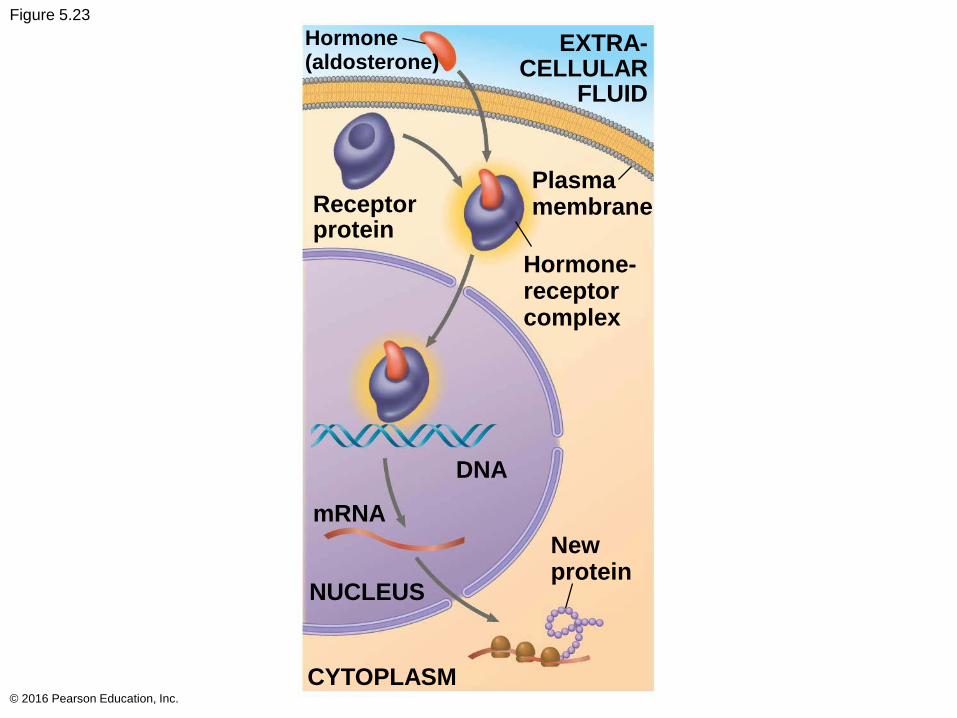
Figure 5.23
© 2016 Pearson Education, Inc.
Hormone(aldosterone)
EXTRA-CELLULAR
FLUID
Receptorprotein
Plasmamembrane
Hormone-receptorcomplex
DNA
mRNA
NUCLEUS
CYTOPLASM
Newprotein
Transduction by Cascades of Molecular Interactions
Signal transduction usually involves multiple steps
Multistep pathways can amplify a signal: A few
molecules can produce a large cellular response
Multistep pathways provide more opportunities for
coordination and regulation of the cellular response
than simpler systems do
© 2016 Pearson Education, Inc.
The molecules that relay a signal from receptor to
response are often proteins
Like falling dominoes, the activated receptor
activates another protein, which activates another,
and so on, until the protein producing the response
is activated
At each step, the signal is transduced into a
different form, commonly a shape change in a
protein
© 2016 Pearson Education, Inc.
Protein Phosphorylation and Dephosphorylation
Phosphorylation and dephosphorylation are a
widespread cellular mechanism for regulating
protein activity
Protein kinases transfer phosphates from ATP to
protein, a process called phosphorylation
A signaling pathway involving phosphorylation and
dephosphorylation can be referred to as a
phosphorylation cascade
The addition of phosphate groups often changes
the form of a protein from inactive to active
© 2016 Pearson Education, Inc.
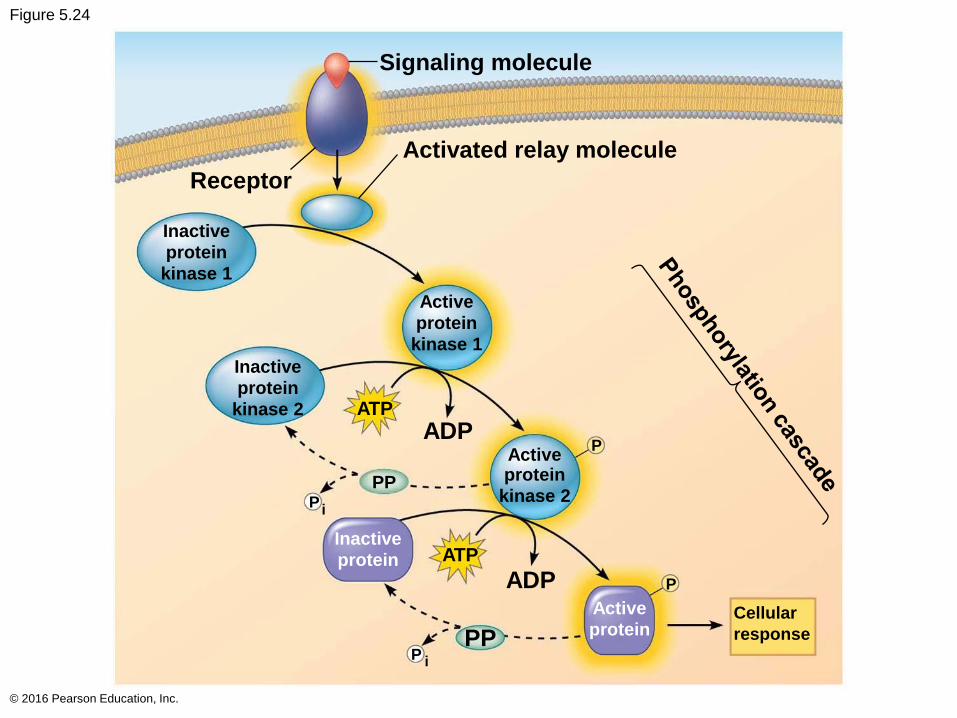
Figure 5.24
© 2016 Pearson Education, Inc.
Signaling molecule
Activated relay molecule
Receptor
Inactive
protein
kinase 1
Inactive
protein
kinase 2
Active
protein
kinase 1
Activeprotein
kinase 2
Inactive
protein
Active
proteinCellular
response
ATP
ADP
PPP
i
ATP
ADP
PPP i
P
P

Figure 5.24-1
© 2016 Pearson Education, Inc.
Signaling molecule
Activated relay molecule
Receptor
Inactiveproteinkinase 1
Activeproteinkinase 1
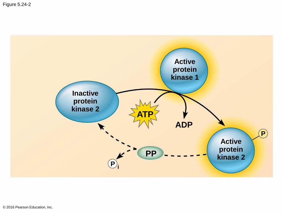
Figure 5.24-2
© 2016 Pearson Education, Inc.
Inactiveproteinkinase 2
Activeproteinkinase 1
Activeproteinkinase 2
ATP
ADP
PP
P
Pi
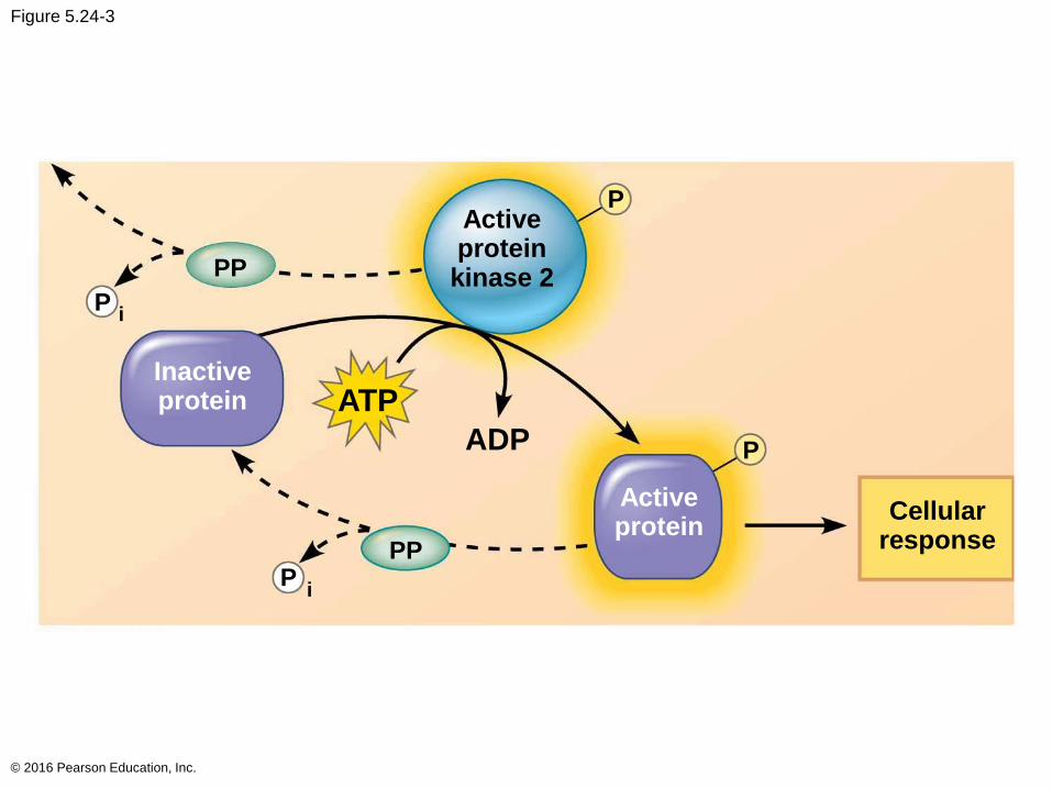
Figure 5.24-3
© 2016 Pearson Education, Inc.
Inactiveprotein
Activeproteinkinase 2
Activeprotein
ATP
ADP
PP
P
P
P
P
PP
Cellularresponse
i
i
Protein phosphatases remove the phosphates
from proteins, a process called dephosphorylation
Phosphatases provide a mechanism for turning off
the signal transduction pathway
They also make protein kinases available for reuse,
enabling the cell to respond to the signal again
© 2016 Pearson Education, Inc.
Small Molecules and Ions as Second Messengers
The extracellular signal molecule (ligand) that binds
to the receptor is a pathway’s “first messenger”
Second messengers are small, nonprotein, water-
soluble molecules or ions that spread throughout a
cell by diffusion
Cyclic AMP and calcium ions are common second
messengers
© 2016 Pearson Education, Inc.
Cyclic AMP (cAMP) is one of the most widely used
second messengers
Adenylyl cyclase, an enzyme in the plasma
membrane, rapidly converts ATP to cAMP in
response to a number of extracellular signals
The immediate effect of cAMP is usually the
activation of protein kinase A, which then
phosphorylates a variety of other proteins
© 2016 Pearson Education, Inc.
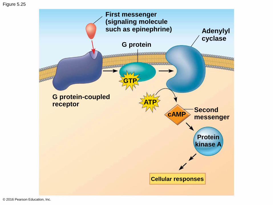
Figure 5.25
© 2016 Pearson Education, Inc.
First messenger(signaling moleculesuch as epinephrine)
G protein
GTP
ATP
cAMP
G protein-coupledreceptor
Secondmessenger
Proteinkinase A
Cellular responses
Adenylylcyclase
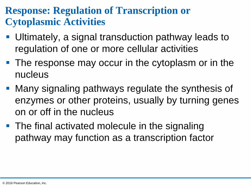
Response: Regulation of Transcription or Cytoplasmic Activities
Ultimately, a signal transduction pathway leads to
regulation of one or more cellular activities
The response may occur in the cytoplasm or in the
nucleus
Many signaling pathways regulate the synthesis of
enzymes or other proteins, usually by turning genes
on or off in the nucleus
The final activated molecule in the signaling
pathway may function as a transcription factor
© 2016 Pearson Education, Inc.
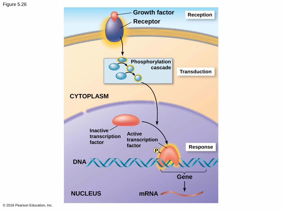
Figure 5.26
© 2016 Pearson Education, Inc.
Growth factor
ReceptorReception
Transduction
Phosphorylationcascade
CYTOPLASM
Response
DNA
P
NUCLEUS mRNA
Gene
Activetranscriptionfactor
Inactivetranscriptionfactor
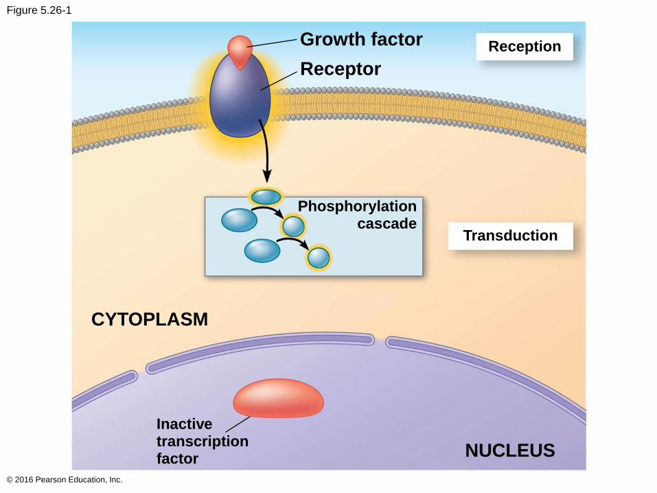
Figure 5.26-1
© 2016 Pearson Education, Inc.
Receptor
Reception
Transduction
Phosphorylationcascade
CYTOPLASM
Growth factor
Inactivetranscriptionfactor NUCLEUS
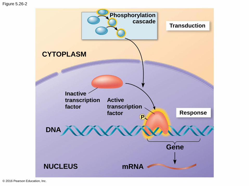
Figure 5.26-2
© 2016 Pearson Education, Inc.
Transduction
Phosphorylationcascade
CYTOPLASM
Response
DNA
P
NUCLEUS mRNA
Gene
Activetranscriptionfactor
Inactivetranscriptionfactor
Other pathways regulate the activity of enzymes
rather than their synthesis, such as the opening of
an ion channel or a change in cell metabolism
© 2016 Pearson Education, Inc.
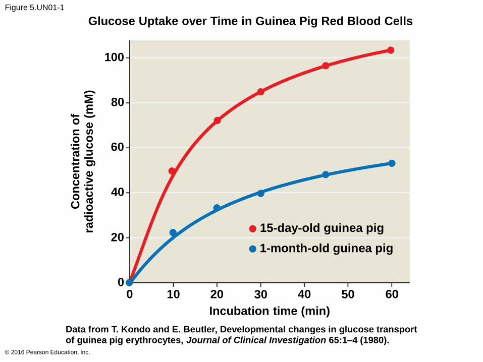
Figure 5.UN01-1
© 2016 Pearson Education, Inc.
Glucose Uptake over Time in Guinea Pig Red Blood Cells
15-day-old guinea pig
1-month-old guinea pig
Incubation time (min)
Data from T. Kondo and E. Beutler, Developmental changes in glucose transportof guinea pig erythrocytes, Journal of Clinical Investigation 65:1–4 (1980).
Co
ncen
trati
on
of
rad
ioacti
ve g
luco
se (
mM
)
60504030201000
20
40
60
80
100
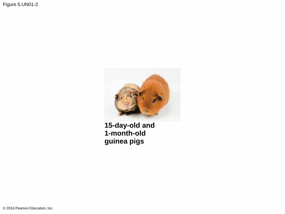
Figure 5.UN01-2
© 2016 Pearson Education, Inc.
15-day-old and1-month-oldguinea pigs
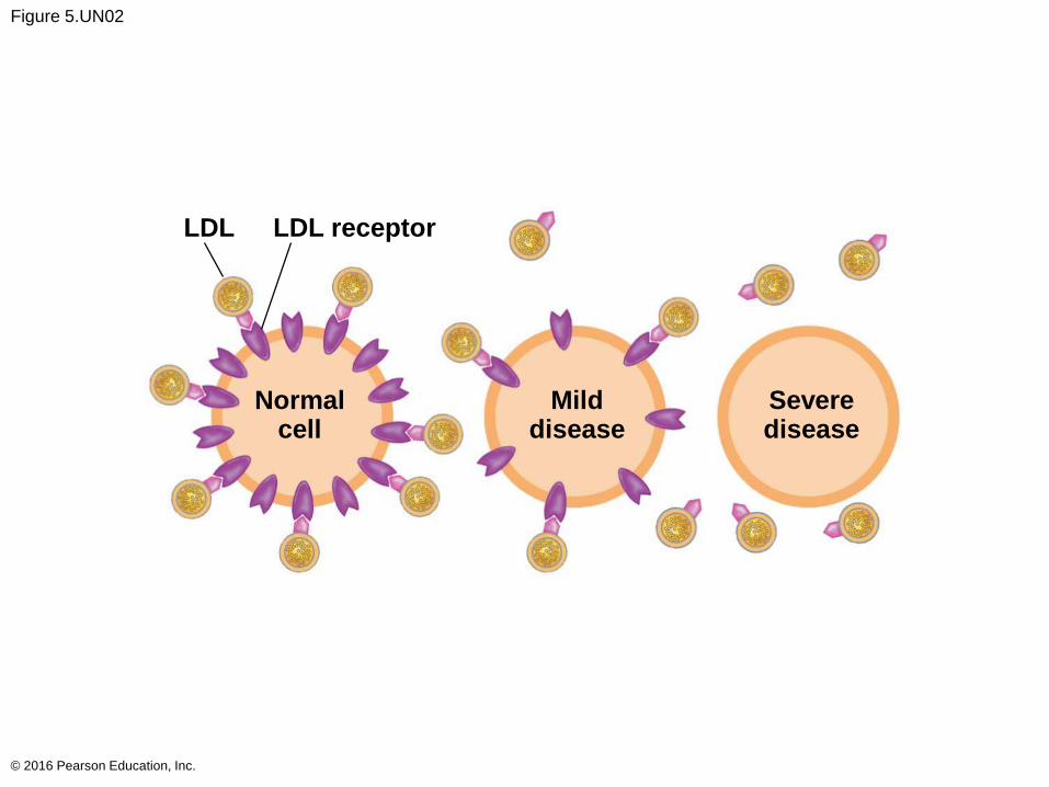
Figure 5.UN02
© 2016 Pearson Education, Inc.
LDL receptorLDL
Normalcell
Milddisease
Severedisease
Figure 5.UN03
© 2016 Pearson Education, Inc.
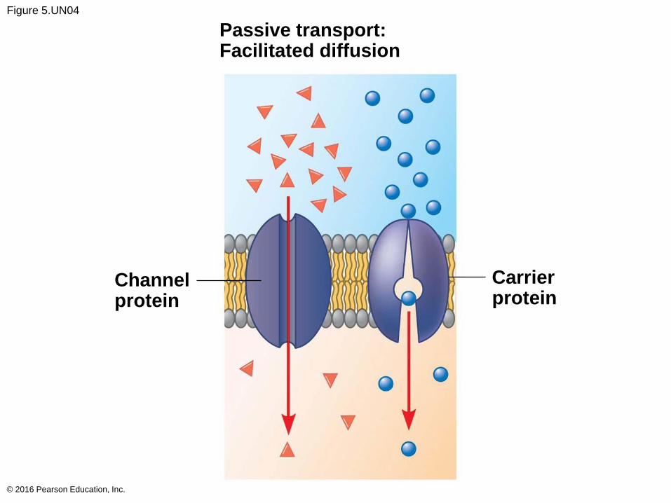
Figure 5.UN04
© 2016 Pearson Education, Inc.
Passive transport:Facilitated diffusion
Channelprotein
Carrierprotein
Figure 5.UN05
© 2016 Pearson Education, Inc.
Active transport
ATP
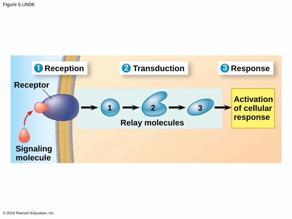
Figure 5.UN06
© 2016 Pearson Education, Inc.
Reception
Receptor
ResponseTransduction
1 2 3Activationof cellularresponse
Relay molecules
Signalingmolecule