medical ultrasound: imaging of soft tissue strain and...
TRANSCRIPT
J. R. Soc. Interface
on July 10, 2018http://rsif.royalsocietypublishing.org/Downloaded from
doi:10.1098/rsif.2011.0054Published online
REVIEW
*Author for c
Received 29 JAccepted 23 M
Medical ultrasound: imaging of softtissue strain and elasticityPeter N. T. Wells1,* and Hai-Dong Liang1,2
1School of Engineering, Cardiff University, Queen’s Buildings, The Parade,Cardiff CF24 3AA, UK
2Department of Medical Physics and Bioengineering, University Hospitals Bristol NHSFoundation Trust, Bristol General Hospital, Bristol BS1 6SY, UK
After X-radiography, ultrasound is now the most common of all the medical imaging technol-ogies. For millennia, manual palpation has been used to assist in diagnosis, but it is subjectiveand restricted to larger and more superficial structures. Following an introduction to the sub-ject of elasticity, the elasticity of biological soft tissues is discussed and published data arepresented. The basic physical principles of pulse-echo and Doppler ultrasonic techniques areexplained. The history of ultrasonic imaging of soft tissue strain and elasticity is summarized,together with a brief critique of previously published reviews. The relevant techniques—low-frequency vibration, step, freehand and physiological displacement, and radiation force(displacement, impulse, shear wave and acoustic emission)—are described. Tissue-mimickingmaterials are indispensible for the assessment of these techniques and their characteristics arereported. Emerging clinical applications in breast disease, cardiology, dermatology, gastroenter-ology, gynaecology, minimally invasive surgery, musculoskeletal studies, radiotherapy, tissueengineering, urology and vascular disease are critically discussed. It is concluded that ultrasonicimaging of soft tissue strain and elasticity is now sufficiently well developed to have clinical uti-lity. The potential for further research is examined and it is anticipated that the technology willbecome a powerful mainstream investigative tool.
Keywords: medical ultrasound; ultrasonic imaging; tissue palpation;soft tissue; ultrasonic elastography; strain in tissue
1. INTRODUCTION
On the one hand, ultrasonic imaging is a mature medi-cal technology. Nearly 22 per cent of all the 38 millionimaging procedures carried out in 2009–2010 inNational Health Service hospitals in England (popu-lation 51 million) were ultrasonic investigations, whichwas second only to those carried out with traditionalX-radiography and fluoroscopy, and 30 per cent morethan all of those using X-ray computed tomography,magnetic resonance imaging (MRI) and nuclearmedicine techniques combined [1]. Ultrasonic imagingis generally real time, it is highly acceptable to mostpatients, exposures used in current practice are con-sidered to be safe and the equipment is generally lessexpensive than that of other imaging technologies.
On the other hand, ultrasonic imaging techniquesare the subject of intense research activity and thecapabilities of new approaches to provide novel infor-mation of considerable actual and potential clinicalvalue are highly attractive. For instance, although
orrespondence ([email protected]).
anuary 2011ay 2011 1
most contemporary ultrasonic imaging techniques arelimited to the display of anatomy, tissue motion andblood flow, the emerging technology of ultrasonicimaging of soft tissue strain and elasticity aims at pro-viding information about the mechanical properties oftissues, such as their hardness or stiffness.
It is the mechanism of the contrast in the imagewhich is one of the most important characteristicsthat distinguishes the practical capabilities and limit-ations of one particular imaging technology fromthose of others [2]. For instance, the contrast in X-rayimaging owes its origin fundamentally to differences inthe atomic numbers of the materials which make upthe structures being imaged, whereas that in MRI corre-sponds to the distribution of protons and differences intheir relaxation times. In traditional ultrasonic imaging(see §5), the signals which form the image are basicallyowing to reflection and scattering of ultrasound wherethere are differences in the characteristic impedancesof the media being imaged. The characteristic impe-dance is equal to the product of the density and thelongitudinal wave speed in the medium: the latterdepends on the bulk modulus of the medium (see
This journal is q 2011 The Royal Society

2 Review. Medical ultrasonic elastography P. N. T. Wells and H.-D. Liang
on July 10, 2018http://rsif.royalsocietypublishing.org/Downloaded from
equation (3.4)). Consequently, techniques for imagingstrain and elasticity (i.e. Young’s or shear modulus)provide unique information.
The only technology that currently comes anywherenear to competing with ultrasonic elastography is mag-netic resonance elastography (MRE) [3,4]. MRE is notrestricted by the presence of bone or gas, is sensitiveto motion in three dimensions with high-speed volumeacquisition, can be carried out by relatively unskilledpractitioners and the interpretation of its results isquite straightforward. In comparison with this, ultra-sonic estimation of soft tissue strain and elasticity isgenerally more accurate and precise, it is relativelyfast, access to the scanners is much more convenientfor patients and practitioners, and the overall cost perinvestigation is much lower.
2. A BRIEF HISTORY OF PALPATIONIN MEDICAL DIAGNOSIS
Depending on the symptoms, when a patient first seeksmedical advice, the doctor, after taking the clinical his-tory, very often begins to make a diagnosis by means ofa physical examination. Classically, this has threeaspects: palpating the abdomen, percussing the chestand listening with a stethoscope.
In the process of palpation, the practitioner appliesmanual pressure to the patient’s skin and in this waysenses the position, hardness, mobility and pulsationof structures within the body.
In ancient Egyptian medicine, palpation was con-sidered to be of fundamental importance. Quotingfrom the Ebers papyrus,1 which dates from about1550 BC, and commenting on other sources [5], ‘Palpa-tion of the pulse was very important and noted in thepapyri. Also that of the abdomen was no less impor-tant: “If thou examinest a man suffering from aresistance in his cardia [meaning the viscera], andthou findest that it goes and comes under thy fingerslike oil in a leather bag. . .then thou shalt examine himlying extended on his back. If thou findest his bellywarm and a resistance in his cardia, thou shalt say tohim: it is a liver case. Thou shalt prepare the secretherbal remedy which is made by the physician. . .”The palpation of tumors was detailed and painsta-king. . .Wounds were also felt with the same care: afractured skull was compared to a punctured earthenjar, the pulsations of the brain were compared tothose of an open fontanelle. Fractures were distin-guished from luxations by feeling crepitus under thefingers’.
Similarly, palpation has always been important intraditional Chinese medicine. Touching the body todetect the pulse in the radial artery and, from the char-acter of the pulsations, to diagnose disease conditions isa highly refined art which dates back at least to about500 BC, the time of the physician Bian Que [6].
In Western medicine, it may be surprising that thepractice of palpation was not put on a reputable basis
1This papyrus was purchased in Luxor in 1872 by the GermanEgyptologist Georg Ebers. It is said to have been found between thelegs of a mummy in the Assassif district of the Theban necropolis.
J. R. Soc. Interface
until the 1930s. Quoting from Shorter [7], reporting theexperience of Karl Stern, a resident physician in Frank-furt in the 1930s: ‘There was, quite aside from theworld of sight, an entire world of touch which we hadnever perceived before. In feeling differences of theradial pulse, you could train yourself to feel dozens ofdifferent waves with their characteristic peaks, bluntand sharp, steep and slanting, and the correspondingvalleys. There were so many ways in which the marginof the liver came up towards your palpating finger’.
Thus, nowadays, medical practitioners glean muchuseful information from manual palpation. Essentially,what they detect is the elasticity of tissue, and thedifferences in the elasticities of different tissues, sensedthough the displacement of tissue (i.e. strain) resultingfrom applied pressure (i.e. stress), whether the origin ofthe applied pressure is internal (e.g. owing to a pulse ofblood travelling along an artery) or external (e.g. owingto the hand of the examiner).
Manual palpation, although an indispensable part ofthe contemporary routine physical examination of thepatient, does have its limitations. It is usually capable ofdetecting the pulsation of superficial arteries, such asthe radial and carotid arteries. Within the abdomen, how-ever, palpation can generally only detect structures orabnormal masses which are quite large in size and whichhave elasticities that differ from those of neighbouringtissues by considerable amounts; it becomes increasinglyunreliable for those which are smaller and deeper.Moreover, the interpretation of palpation is completelysubjective: it does not provide any quantitative data.
There are some types of cancer for which appropriategroups of supposedly normal individuals are encouragedregularly to examine themselves by manual palpation forsigns of abnormality. Thus, young men should palpatetheir scrotums to search for changes which might be anearly warning of testicular cancer, and women shouldexamine their breasts in case a lump should indicatethe presence of a malignant tumour. Similarly, digitalrectal examination by a skilled practitioner can usefullyreinforce the reliability of the diagnosis of prostatecancer. The reasons why this kind of examination is jus-tified are that malignant tumours are, in general, harderthan their surrounding normal tissues and that,although the false-negative and false-positive rates arerather high, the benefits of early diagnosis and treatmentcan far outweigh the costs—both financial and social—offalse-positives.
3. AN ELEMENTARY TUTORIALON ELASTICITY
The elasticity of a material describes its tendency toresume its original size and shape after being subjectedto a deforming force or stress. Fluids resist a change involume, but not in shape: they possess only volume elas-ticity. Solids resist changes in shape and volume: theypossess rigidity or shear elasticity, as well as volumeelasticity. The change in size or shape is known as thestrain, which is expressed as a ratio (e.g. the changein length per unit length). The strain is produced by asystem of forces; the force acting on unit area isknown as the stress.
Review. Medical ultrasonic elastography P. N. T. Wells and H.-D. Liang 3
on July 10, 2018http://rsif.royalsocietypublishing.org/Downloaded from
The basic principles of elasticity can be foundin most textbooks on materials science (e.g. [8]). Insummary:
For a homogeneous isotropic solid, the ratio ofstress/strain is a constant, called the modulus of elas-ticity. Three moduli (with units of N m22, or Pa) arecommonly used to define its elasticity:
Young’s modulus (longitudinal elasticity), E ¼(stress)/(strain).
Shear or torsion modulus (rigidity), G.Bulk or volume modulus (volume elasticity), K.When a material is stressed, its breadth may contract
as its length extends. This is defined by a constantcalled Poisson’s ratio, which is given by
s ¼ lateral contraction per unit breadthlongitudinal extension per unit length
:
Three linear elastic constitutive equations define therelationships between these four constants,
G ¼ Eð2ð1þ sÞÞ ; ð3:1Þ
s ¼ E2G� 1 ð3:2Þ
and K ¼ Eð3ð1� 2sÞÞ : ð3:3Þ
Solids can support mechanical waves in four princi-pal modes, depending on the way in which theparticles in the solid move during wave propagation.In longitudinal (or compressional) waves, the particlesmove in the direction of propagation, whereas in trans-verse (or shear) waves, they move in the directionnormal to the direction of propagation. The other prin-cipal wave modes are surface and plate waves, but theseare hardly relevant to propagation in biologicalsoft tissues and so they are not given further consider-ation here.
The speeds at which mechanical waves propagate ina solid are given by the following equations:
cl ¼Kr
� �1=2
ð3:4Þ
and
cs ¼Gr
� �1=2
; ð3:5Þ
where cl is the longitudinal wave speed, cs is the shearwave speed and r is the tissue mass density.
For homogeneous isotropic solids subjected toincreasing tension, Hooke’s law (i.e. strain is directlyproportional to stress) applies, until the elastic limit isreached and, eventually, fracture failure occurs.
Some materials exhibit both elastic and viscousproperties when subjected to stress and the relationshipbetween stress and strain is time dependent. A visco-elastic material can be simply modelled as an elasticcomponent coupled with a viscous component whichacts as a damper that delays the stress–strain responsewithout affecting its asymptotic value. Yet, othermaterials exhibit poroelastic properties. A porous material
J. R. Soc. Interface
is one in which a solid matrix is permeated by an intercon-necting network of fluid-filled pores. When consideringthe stress–strain relationship in a poroelastic material,the matrix can be modelled with elastic properties andthe fluid is spatially redistributed over time in thematerial in order to accommodate its deformation.
4. THE ELASTICITY OF BIOLOGICALTISSUES
The basic component of biological tissue is the cell,which is typically 5–10 mm in diameter. Enclosedwithin the cell wall (which itself is far from being asimple membrane) is the cytoplasm, which containsmicroscopic structures such as the nucleus and themitochondria. There are five primary types of soft tis-sues. These are: epithelial tissue, which is made up ofcells packed tightly together to form continuoussheets that serve as the linings of structures andorgans; connective tissue, usually containing strandsof collagen, which supports and adds structure to thebody; parenchymal tissue, which is the functionaltissue of organs; muscle tissue, which has the abilityto contract in response to electrical signals; and nervetissue, which has the ability to generate and conductelectrical signals.
In addition to the soft tissues, there are several kindsof hard tissues, principally bone and teeth. Themechanical properties of vertebrate hard tissues havebeen extensively studied (e.g. [9,10]). Hard tissues arenot readily amenable to ultrasonic measurement, how-ever, because of their relatively high densities andultrasonic attenuations, and so are outside the scopeof the present paper.
An organ is a structure that contains at least twotypes of tissues functioning for a common purpose.Examples of organs include skin, liver, kidney, heartand brain. The mechanical properties of tissues cannotrealistically be described in terms of simple agglomera-tions of homogeneous cells; rather, tissues havemicroscopic and macroscopic organizations which needto be considered in the context of their multiple scales.
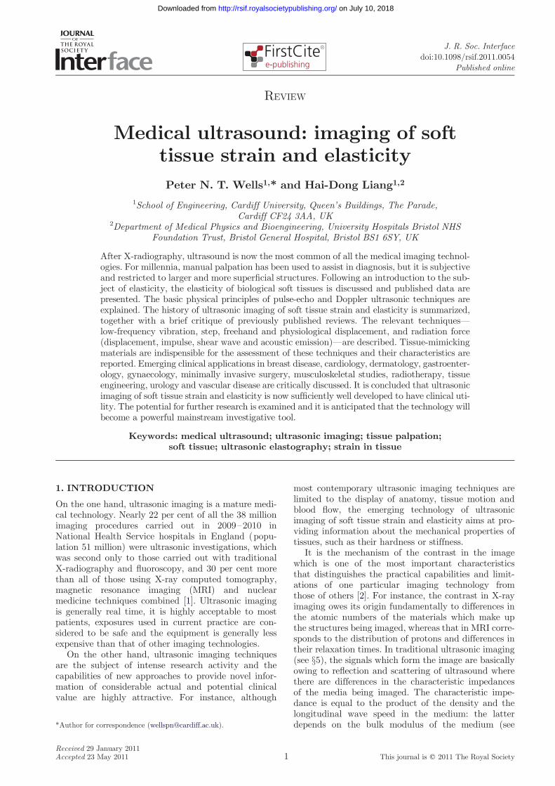
Figure 1 shows the typical relationship between stressand strain in soft tissues. With the stress increasing fromzero, the strain increases rapidly as free fluid is exuded,after which the ratio of stress over strain (Young’s mod-ulus) can be considered to be linear for small changes (i.e.for strains of less than a few per cent), but the elasticmodulus becomes progressively greater with increasingstrain. Consequently, the conditions under which anyparticular value was measured need to be specified.A further complication is that tissue may be viscoelastic(which may manifest itself as hysteresis on relaxation ofthe stress), poroelastic, anisotropic or contractile, or anycombination of these, quite apart from being normal ormodified by disease. Other factors which may be relevantare the age of the tissue, its temperature, and whether itis in vivo, in vitro or fixed.
The literature is bereft of data for the bulk modulusof soft tissues. Surprisingly, standard biomechanics text-books (such as [11]) are almost completely silent in thisrespect. Indeed, it seems that the best that can be done

stress
strain 0
0
Figure 1. Idealized stress–strain relationship for soft tissues.The inset (with stress on a magnified scale) shows that,when tissue is stressed, the strain initially increases rapidly(corresponding to the elimination of free fluid), after whichthe relationship is effectively linear over a small increase instress. With further increase in stress, the tissue becomesstrained decreasing rapidly (as it approaches the limit of itselasticity). The practical implication of this is that, in orderto obtain reproducible and useful values of Young’s modulus,the tissue needs to be slightly statically preloaded and themeasurement needs to be made over a small increment instress (i.e. in the linear region).
Table 1. Published values of Young’s modulus for varioustypes of tissues and other relevant materials. Where therange of conditions of measurement were specified by theauthors, the data are for low static preloading, low strainand low frequency. Ca, carcinoma.
tissue type E (kPa) reference
breast unspecified 29 [12]21–23 [13]
adipose 19 [14]1.9 [15]
ductal Ca 25 [14]in situ 12 [15]glandular 33 [14]fibrous 110 [14]
1.8 [15]invasive Ca 93 [14]
cervix unspecified 30–90 [16]
kidney unspecified 10 [17]6 [13]
liver unspecified 13 [18]10–17 [19]7–10 [13]1–3 [20]0.4–1.7 [21]
normal 10 [22]0.6–1.1 [23]
focal nodular 1.1–2.5 [23]hyperplasiachronic hepatitis 35 [18]cirrhosis 52 [18]
1.1–4.9 [23]VX2 Ca 0.3–0.9 [20]cholangiocarcinoma 3–12 [23]
muscle unspecified 14–16 [13]10–40 [20]7–57 [24]1.2–1.8 [21]
intercostal 100 [22]cardiac (systole) 100 [25]cardiac (diastole) 10 [25]along fibres 13 [26]across fibres 5.3 [26]VX2 Ca 2–8 [20]
prostate unspecified 0.8–4.0 [20]normal 62–69 [14]
17 [27]benign prostatic 36 [14]hypertrophycarcinoma 100 [14]
24 [27]
thrombus unspecified 8–38 [28]
uterus unspecified 30–90 [16]leiomyoma 60–220 [16]
rubber soft 990–3000 [29]agar 3% þ 70–100 [20]
gelatin 3%
4 Review. Medical ultrasonic elastography P. N. T. Wells and H.-D. Liang
on July 10, 2018http://rsif.royalsocietypublishing.org/Downloaded from
to gain insight into this is to use equation (3.4) tocalculate the probable range of K from published valuesof the speed of sound (cl) and density (r) for soft tissues.Even after 20 years since its publication, there is one book[10] which is arguably still the best source of reference.Omitting data for ‘soft tissues’ such as the lens of theeye, cartilage, skin and tendon, but including all othernormal and pathological soft tissues, values for cl rangefrom 1412 m s21 in fat to 1629 m s21 in muscle; the cor-responding values for r are 916 and 1060 kg m23,respectively. Substituting these values for cl and r inequation (3.4) gives an indication of the value of K;thus, K for soft tissues must range from about1800 MPa in fat to about 2800 MPa in muscle.
In the literature, it is usual for values of Young’s mod-ulus to be reported, rather than those of shear modulus.For soft tissues, however, Poisson’s ratio is usuallybetween 0.490 and 0.499. This is because tissue isalmost incompressible. Consequently, from equation (3.2),
E � 3G: ð4:1Þ
It is the value of G which is required for the calcu-lation of shear wave speed from equation (3.5) and so,in this review, equation (4.1) is used for conveniencewhenever conversion from values of E to those ofG—and vice versa—is necessary.
It is rather disappointing that the published data forYoung’s moduli of biological tissues, many of which arelisted in table 1, are often of only limited quantitativeuse. There are wide variations, particularly betweenthose reported by different authors—even for tissuesof the same types. Some of these differences may beowing to the sometimes indiscriminate treatment oflarge strains in Lagrangian (i.e. relative to the originallength) or Eulerian (i.e. relative to the strainedlength) representations (this is not problematic withsmall strains). Moreover, the data are sparse (table 1
J. R. Soc. Interface
is actually quite a comprehensive compendium). Never-theless, it can reasonably be concluded that typicalvalues of Young’s modulus are about 10 kPa for par-enchyma, 20 kPa for muscle and 50 kPa for connectivetissue. Table 1 also includes data for rubber (whichmight intuitively be thought to have elasticity similar

1828
96
22
106
2235
116
26
112
0
20
40
60
80
100
120
140
fat glandulartissue
fibroustissue
DCIS Ca
You
ng's
mod
ulus
(kP
a)
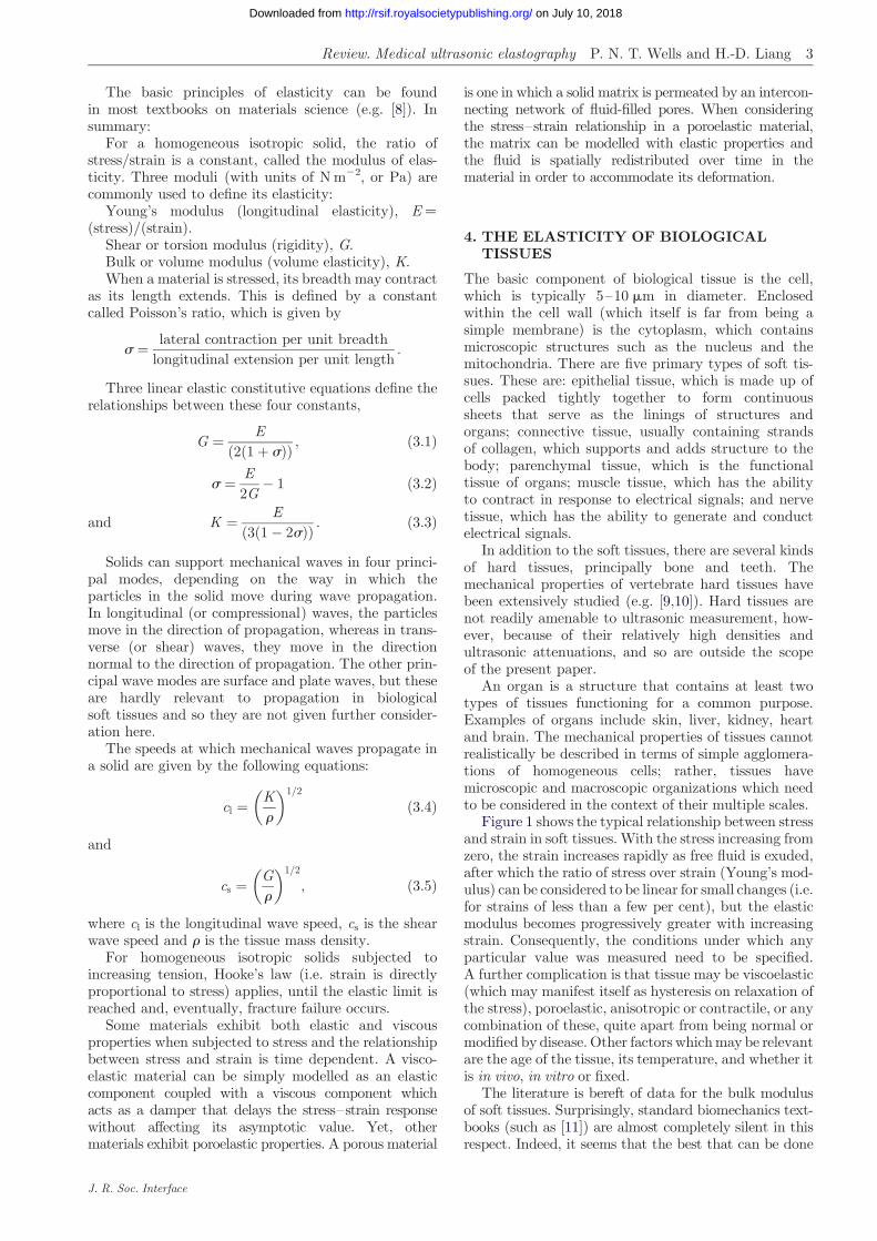
Figure 2. Young’s moduli of different types of breast tissues,measured with the same static preloading (5%) but at twoloading frequencies (0.1 Hz, black bars; 4 Hz, white bars).Although the measured value of Young’s modulus increasesslightly with the loading frequency, the effect is not parti-cularly marked. The practical implication of this is that,provided that the rate of change of the tissue displace-ment for the measurement is slow (i.e. quasi-static), staticconditions can be assumed to apply. Data from Krouskopet al. [14]. DCIS, ductal carcinoma in situ.
18 28
96
22
106
20 48
218
291
558
0
100
200
300
400
500
600
fat glandulartissue
fibroustissue
DCIS Ca
You
ng's
mod
ulus
(kP
a)
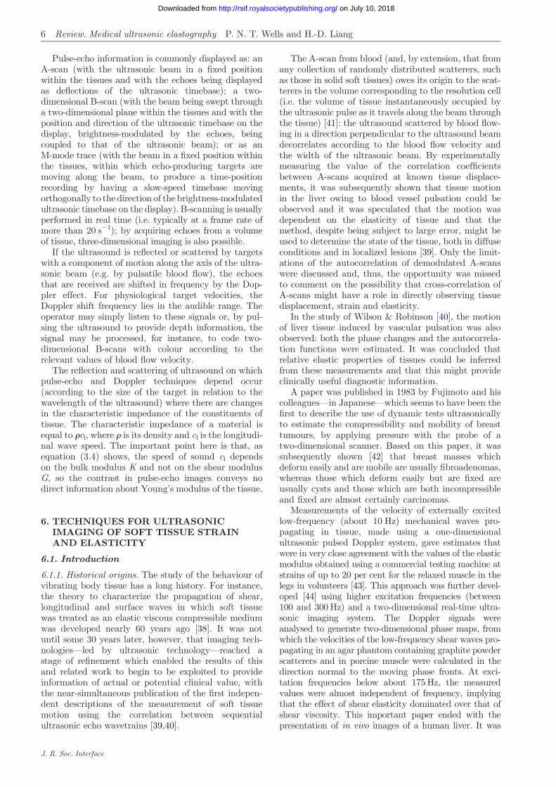
Figure 3. Young’s moduli of different types of breast tissues,measured at the same (quasi-static) loading frequency (0.1Hz) but at two levels of static preloading (5%, black bars;20%, white bars). The measured value of Young’s modulusincreases markedly with the level of static preloading. Thepractical implication of this is that, in order to obtain reprodu-cible measurements of Young’s modulus, the level of staticpreloading (provided that it is sufficient for the free fluid tobe eliminated) should be kept as small as practicable. Datafrom Krouskop et al. [14].
Review. Medical ultrasonic elastography P. N. T. Wells and H.-D. Liang 5
on July 10, 2018http://rsif.royalsocietypublishing.org/Downloaded from
to that of soft tissue, but clearly does not) and for awater-based material consisting of gelatin and agar(which is one of the family of tissue-mimickingmaterials: see §6.3).
Although the origins of many of these data are inade-quately specified, those in Krouskop et al. [14] are anotable exception. For various types of breast tissues,figure 2 summarizes the results of their mechanicalmeasurements at loading frequencies of 0.1 and 4 Hzwith 5 per cent static loading, and figure 3, summarizesthose for static loadings of 5 and 20 per cent with a0.1 Hz loading frequency. From figure 2, it is apparentthat Young’s modulus is not greatly dependent on theloading frequency, at least below 4 Hz, irrespective ofthe tissue type. For some types of tissues, however,figure 3 shows that Young’s modulus is highly dependenton the degree of static loading and that the ratios ofYoung’s moduli of different types of tissues (i.e. theirdynamic range) are greater at higher static loadings.
It has been stated [30] that the shear modulus (whichis approx. equal to one-third of Young’s modulus) ofsoft tissues varies over several orders of magnitude,whereas the variation in the bulk modulus is signifi-cantly less than one order of magnitude. Thisgeneralization has been perpetuated in the sub-sequently published literature. Within groups ofbroadly similar types of tissues, however, the shearmodulus data which they quoted are actually in nar-rower ranges, not much exceeding one order ofmagnitude. What is really important is that Young’smoduli of different types of tissues, including abnormaltissues, may be markedly different in structures inwhich the lack of significant difference in bulk modulimakes tissue characterization by traditional ultrasonicscanning problematic (see §5). In the breast, forexample, the bulk modulus hardly varies in differenttypes of tissues from about 2000 MPa, whereasYoung’s modulus typically ranges from about 20 kPain fatty tissues to about 100 kPa in carcinoma.
J. R. Soc. Interface
5. THE PHYSICAL PRINCIPLES OF PULSE-ECHO AND DOPPLER ULTRASOUND
The evolution of medical ultrasonics can be tracedthrough contemporary reviews (see [31–36]). For anup-to-date tutorial, see Halliwell [37]. A brief summaryof the physical principles is given below.
In order to be clinically useful, an ultrasonic imagingsystem typically needs to be able to resolve structures ofaround a millimetre in size at depths of up to around150 mm. Ultrasound travels at a speed of about1500 m s21 in soft tissues; this means that the frequencyneeds to be in the low megahertz range because thewavelength, which is one of the factors that determinethe spatial resolution and which is inversely pro-portional to the frequency, is, for example, 0.5 mm at3 MHz. A narrow beam of ultrasound is radiated byan aperture which is, say, at least 10 times the wave-length in size; a transducer, typically polarized leadzirconate titanate, produces a pulse of ultrasoundwhen excited by a brief electrical pulse. The tissue tobe imaged contains reflectors and scatterers that giverise to echoes which may be detected by the transducer,delayed in time according to their distances from thetransducer (i.e. by about 1.33 ms mm21).
The attenuation of ultrasound in soft tissues is about0.2–0.5 dB cm21 MHz21. Thus, attenuation increaseswith both distance and frequency, and this limits thepenetration (i.e. the depth from which the echoeshave amplitudes sufficient to be detected above thenoise) at any given frequency. In practice, this meansthat a frequency of about 3 MHz is optimal for abdomi-nal scanning, where a penetration depth of up to150 mm may be appropriate.
Thus, pulse-echo ultrasonic imaging is based on theprinciple that a directional beam of pulsed ultrasoundcan be generated and that echoes may be detectedfrom reflectors and scatterers in the beam, delayed intime according to their depths.
6 Review. Medical ultrasonic elastography P. N. T. Wells and H.-D. Liang
on July 10, 2018http://rsif.royalsocietypublishing.org/Downloaded from
Pulse-echo information is commonly displayed as: anA-scan (with the ultrasonic beam in a fixed positionwithin the tissues and with the echoes being displayedas deflections of the ultrasonic timebase); a two-dimensional B-scan (with the beam being swept througha two-dimensional plane within the tissues and with theposition and direction of the ultrasonic timebase on thedisplay, brightness-modulated by the echoes, beingcoupled to that of the ultrasonic beam); or as anM-mode trace (with the beam in a fixed position withinthe tissues, within which echo-producing targets aremoving along the beam, to produce a time-positionrecording by having a slow-speed timebase movingorthogonally to the direction of the brightness-modulatedultrasonic timebase on the display). B-scanning is usuallyperformed in real time (i.e. typically at a frame rate ofmore than 20 s21); by acquiring echoes from a volumeof tissue, three-dimensional imaging is also possible.
If the ultrasound is reflected or scattered by targetswith a component of motion along the axis of the ultra-sonic beam (e.g. by pulsatile blood flow), the echoesthat are received are shifted in frequency by the Dop-pler effect. For physiological target velocities, theDoppler shift frequency lies in the audible range. Theoperator may simply listen to these signals or, by pul-sing the ultrasound to provide depth information, thesignal may be processed, for instance, to code two-dimensional B-scans with colour according to therelevant values of blood flow velocity.
The reflection and scattering of ultrasound on whichpulse-echo and Doppler techniques depend occur(according to the size of the target in relation to thewavelength of the ultrasound) where there are changesin the characteristic impedance of the constituents oftissue. The characteristic impedance of a material isequal to rcl, where r is its density and cl is the longitudi-nal wave speed. The important point here is that, asequation (3.4) shows, the speed of sound cl dependson the bulk modulus K and not on the shear modulusG, so the contrast in pulse-echo images conveys nodirect information about Young’s modulus of the tissue.
6. TECHNIQUES FOR ULTRASONICIMAGING OF SOFT TISSUE STRAINAND ELASTICITY
6.1. Introduction
6.1.1. Historical origins. The study of the behaviour ofvibrating body tissue has a long history. For instance,the theory to characterize the propagation of shear,longitudinal and surface waves in which soft tissuewas treated as an elastic viscous compressible mediumwas developed nearly 60 years ago [38]. It was notuntil some 30 years later, however, that imaging tech-nologies—led by ultrasonic technology—reached astage of refinement which enabled the results of thisand related work to begin to be exploited to provideinformation of actual or potential clinical value, withthe near-simultaneous publication of the first indepen-dent descriptions of the measurement of soft tissuemotion using the correlation between sequentialultrasonic echo wavetrains [39,40].
J. R. Soc. Interface
The A-scan from blood (and, by extension, that fromany collection of randomly distributed scatterers, suchas those in solid soft tissues) owes its origin to the scat-terers in the volume corresponding to the resolution cell(i.e. the volume of tissue instantaneously occupied bythe ultrasonic pulse as it travels along the beam throughthe tissue) [41]: the ultrasound scattered by blood flow-ing in a direction perpendicular to the ultrasound beamdecorrelates according to the blood flow velocity andthe width of the ultrasonic beam. By experimentallymeasuring the value of the correlation coefficientsbetween A-scans acquired at known tissue displace-ments, it was subsequently shown that tissue motionin the liver owing to blood vessel pulsation could beobserved and it was speculated that the motion wasdependent on the elasticity of tissue and that themethod, despite being subject to large error, might beused to determine the state of the tissue, both in diffuseconditions and in localized lesions [39]. Only the limit-ations of the autocorrelation of demodulated A-scanswere discussed and, thus, the opportunity was missedto comment on the possibility that cross-correlation ofA-scans might have a role in directly observing tissuedisplacement, strain and elasticity.
In the study of Wilson & Robinson [40], the motionof liver tissue induced by vascular pulsation was alsoobserved: both the phase changes and the autocorrela-tion functions were estimated. It was concluded thatrelative elastic properties of tissues could be inferredfrom these measurements and that this might provideclinically useful diagnostic information.
A paper was published in 1983 by Fujimoto and hiscolleagues—in Japanese—which seems to have been thefirst to describe the use of dynamic tests ultrasonicallyto estimate the compressibility and mobility of breasttumours, by applying pressure with the probe of atwo-dimensional scanner. Based on this paper, it wassubsequently shown [42] that breast masses whichdeform easily and are mobile are usually fibroadenomas,whereas those which deform easily but are fixed areusually cysts and those which are both incompressibleand fixed are almost certainly carcinomas.
Measurements of the velocity of externally excitedlow-frequency (about 10 Hz) mechanical waves pro-pagating in tissue, made using a one-dimensionalultrasonic pulsed Doppler system, gave estimates thatwere in very close agreement with the values of the elasticmodulus obtained using a commercial testing machine atstrains of up to 20 per cent for the relaxed muscle in thelegs in volunteers [43]. This approach was further devel-oped [44] using higher excitation frequencies (between100 and 300 Hz) and a two-dimensional real-time ultra-sonic imaging system. The Doppler signals wereanalysed to generate two-dimensional phase maps, fromwhich the velocities of the low-frequency shear waves pro-pagating in an agar phantom containing graphite powderscatterers and in porcine muscle were calculated in thedirection normal to the moving phase fronts. At exci-tation frequencies below about 175 Hz, the measuredvalues were almost independent of frequency, implyingthat the effect of shear elasticity dominated over that ofshear viscosity. This important paper ended with thepresentation of in vivo images of a human liver. It was

displacementhigh-speedimaging
hydro-phone
inversion
strainstiffness modulus stiffnessmodulus
excitation
image
processing
detection
tissue
motion
radiation force
displace-ment
(6.2.5)
ARFIimaging (6.2.6)
shearwave(6.2.7)
vibro-acousto-graphy(6.2.8)
direct mechanical
low-frequencysurfacevibration(6.2.1)
quasistatic displacement
stepsurface(6.2.2)
freehandsurface(6.2.3)
physio-logical(6.2.4)
Figure 4. Overview of approaches to elastography. The methods of excitation are classified broadly as being by direct mechanicalaction or by radiation force: the section number in the paper corresponding to each technique is shown. The method of detectingand processing the effect of the excitation is also shown for each technique, as is the characteristic (stiffness, strain or (Young’s)elastic modulus), which is displayed in the corresponding image. ARFI, acoustic radiation force impulse.
2The term ‘elastogram’ was introduced in 1991 [74]. Originally, it wasreserved to describe quasi-static displacement strain images (see§6.2.2). Nowadays, however, the terms ‘elastogram’ and ‘elastography’tend to be used indiscriminately.
Review. Medical ultrasonic elastography P. N. T. Wells and H.-D. Liang 7
on July 10, 2018http://rsif.royalsocietypublishing.org/Downloaded from
concluded that ‘Although these results are preliminaryones, it may be said that the method proposed heregives a useful information [sic] about mechanical proper-ties of the tissues’. The dependence of velocity onelasticity was recognized (see equation (3.5)), but thepossibility of elasticity imaging was not specificallymentioned.
There are several methods that involve the use ofultrasound by which the elasticity of soft tissues canbe investigated, but which are outside the scope ofthis review. For instance, neither Doppler tissue ima-ging of the myocardium [45]—which is essentially avariant of ultrasonic Doppler colour flow imaging,with the rejection filter threshold selected to displaythe relatively strong echoes from the tissue ratherthan the weaker echoes from the blood and which pro-vides information about myocardial dyskinesia—northe measurement of the arterial blood flow pulse wavevelocity [46]—which increases with hardening of thevessel—depend on the direct measurement of eitherthe localized tissue strain or the localized shear wavespeed. Except where reference to such topics helps thecontext, they are not discussed in this review.
Thus, the foundations on which ultrasonic imagingof soft tissue strain and elasticity are built had beenfirmly laid by the end of the 1980s. The remainderof §6 describes the various approaches which havesubsequently been developed and, in many cases,introduced into clinical practice.
6.1.2. Overview of strain and elasticity imagingapproaches. As illustrated in figure 4, the first stage inelastography involves the excitation of the tissues. Exci-tation can be by direct mechanical means or byultrasonic radiation force. The excitation results in
J. R. Soc. Interface
(quasi)static or dynamic tissue displacement, thelatter being accompanied by the generation of shearwaves. Detection of the effects of excitation can be bythe ultrasonic Doppler effect, by ultrasonic pulse-echomethods or by acoustic emission. Finally, the resultantinformation can be displayed as images, either directlyof the spatial distributions of strains or shear wavesor of elastic moduli or tissue stiffnesses. Usually, theelastograms2 (whether of strain, elastic modulus orstiffness) can be fused with the corresponding ultrasonicB-scans, as this facilitates the identification of theanatomical structures to which they relate.
Tissue strain is a surrogate for tissue stiffness, wherestiffness can be considered to be the representative ofwhat might be felt with manual palpation: low tissuestrain corresponds to high tissue stiffness and viceversa. Thus, in practice, clinically useful informationcan often be provided by strain imaging. In the simplestof geometries, if both the stress and the strain areknown, the elastic modulus is given by their ratio. Inreal anatomical and pathological situations, however,the boundary conditions are such that the estimationof the shear modulus is far more complicated: theinverse problem that needs to be solved is to identifywhat distribution of elastic modulus would be consist-ent with the observed distributions of stress and strain.
Generally, in mechanical excitation methods (oftencalled low-frequency or quasi-static displacementmethods), the displacement is kept so small that theresultant tissue strain is never more than a few percent, and usually very much less. The ultrasonic

8 Review. Medical ultrasonic elastography P. N. T. Wells and H.-D. Liang
on July 10, 2018http://rsif.royalsocietypublishing.org/Downloaded from
measurement of the displacement depends on the deter-ministic nature of ultrasound backscattered by tissue.The wavelength of the ultrasound (for example, about500 mm in soft tissues at the typical frequency of3 MHz) is very much greater than the size of the smallscattering structures which are the origin of the speckle,which characterizes ultrasound backscattered by solidsoft tissues [48]. Consequently, the assumption that theechoes from a small volume of tissue can retain coherenceeven when their phase changes as the result of displace-ment may have at least limited validity, and this is thebasis of the ultrasonic measurements such as thosemade by cross-correlation of echo wavetrains acquiredbefore and after the application of stress. Moreover,because the magnitude of the strain is small, it is areasonable assumption that the echoes from any givensmall volume of tissue before and after being strainedcan be meaningfully cross-correlated.
If the compressor used for direct mechanical exci-tation is bigger than the homogeneous tissue specimenand free-slip conditions exist, the situation is quitestraightforward. It becomes more complicated whenthe compressor is smaller than the specimen, as, in prac-tice, it usually is: it may not then be realistic to assumethat the compression is uniaxial [49].
Radiation force excitation methods depend on thefact that, when an ultrasonic wave is absorbed orreflected, a force in the direction of propagation of thewave is the result. With complete absorption, therelationship is
F ¼Wcl; ð6:1Þ
where F is the force, W is the ultrasonic power and cl isthe longitudinal speed of propagation. For example,complete absorption of an ultrasonic wave travellingin water (or soft tissue) with a power of 1 W results ina force of about 0.7 mN, which is roughly equal to theforce of gravity acting on a mass of 70 mg. As illustratedin figure 4, there are several ways in which this phenom-enon can be applied in elastography.
As tissue volume can be considered to be incompres-sible, measurements in only one direction in a three-dimensional volume are sufficient to allow Young’smodulus to be determined. (Modest precompression oftissue is necessary to minimize the blood and freefluid volume, so that the tissue can then be assumedto be incompressible.) Another necessary assumptionis that the tissue is locally homogeneous; inaccurateresults are obtained near boundaries between differentkinds of tissues and this may limit the window size forcross-correlation. Internal and external tissue bound-aries can have marked effects on the distribution ofapplied stresses and this can make it problematic quan-titatively to analyse strain images [50]. An advantage ofradiation force excitation is that focused ultrasound canproduce highly localized motion and regions containingboundaries can usually be avoided.
Dynamic methods are limited by the ability to pro-pagate shear waves deep into tissue because of theirrelatively rapid attenuation. Static methods have theopposite problem: movement at any point in tissue
J. R. Soc. Interface
will affect all other points and this must be taken intoaccount in the estimation of Young’s modulus.
In elastography, the signal-to-noise ratio (SNRe) andthe contrast-to-noise ratio (CNRe) of the target to thebackground are commonly used to assess the accuracyof strain estimation methods. These are defined asfollows [51,52]:
SNRe ¼m
sð6:2Þ
and
CNRe ¼2ðmbackground � mtargetÞ
2
ðs2background þ s2
targetÞ; ð6:3Þ
where m is the mean measured strain and s is thestandard deviation in the measured strain in theregion of interest.
6.1.3. A brief summary of some relevant publishedreviews. Some reviews of elastography that have alreadybeen published are as follows (in chronological order):
1996
This landmark review [53] was published soonafter the opening of the era of ultrasonic imagingof soft tissue strain and elasticity. It is remarkablefor its prescience in the areas of elasticity data,source excitation, tissue models and boundaryconditions.1998
In this review [54], the principles of quasi-staticstep displacement ultrasonic elastography are dis-cussed in the context of medical image processing.Some early clinical results are presented.1999
This review [55] was written as an introductionfor mechanical engineers and others (as distinctfrom clinicians) to the then quite novel conceptof elastography. It is a comprehensive treatmentof the contemporary state of the art.2002
This review [56] aimed to expose, to a mainlyclinical readership, the creation and display of‘elastograms’ showing local strains, Young’smoduli and Poisson’s ratios in tissues subjectedto quasi-static displacements.2003
This review [57] discusses several methods of esti-mating tissue hardness using internal or externalmeans of applying stress and several associatedmethods of detecting the resulting strain, aimedat imaging mechanical characteristics oftissue. Both ultrasonic and magnetic resonancetechniques are considered.2003
This is a tutorial [58] intended primarily to intro-duce ultrasonic elastography to academic clinicalradiologists. The concept of quasi-static freehandscanning with software to produce strain imagesis discussed and some examples of using the tech-nique in the differential diagnosis of breast lesionsare included.2004
This review [59] briefly discusses the state of theart in ultrasonic elastography, in the context ofthe then newly available quasi-static near-real-time freehand scanners. The author foresaw thedevelopment of 10 clinically useful applications,of which perhaps four have now come to fruition.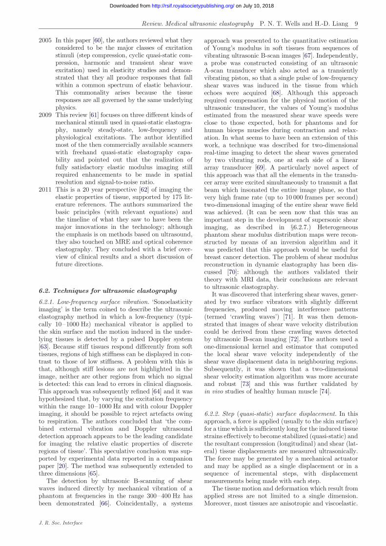
Review. Medical ultrasonic elastography P. N. T. Wells and H.-D. Liang 9
on July 10, 2018http://rsif.royalsocietypublishing.org/Downloaded from
2005
J. R. S
In this paper [60], the authors reviewed what theyconsidered to be the major classes of excitationstimuli (step compression, cyclic quasi-static com-pression, harmonic and transient shear waveexcitation) used in elasticity studies and demon-strated that they all produce responses that fallwithin a common spectrum of elastic behaviour.This commonality arises because the tissueresponses are all governed by the same underlyingphysics.
2009
This review [61] focuses on three different kinds ofmechanical stimuli used in quasi-static elastogra-phy, namely steady-state, low-frequency andphysiological excitations. The author identifiedmost of the then commercially available scannerswith freehand quasi-static elastography capa-bility and pointed out that the realization offully satisfactory elastic modulus imaging stillrequired enhancements to be made in spatialresolution and signal-to-noise ratio.2011
This is a 20 year perspective [62] of imaging theelastic properties of tissue, supported by 175 lit-erature references. The authors summarized thebasic principles (with relevant equations) andthe timeline of what they saw to have been themajor innovations in the technology; althoughthe emphasis is on methods based on ultrasound,they also touched on MRE and optical coherenceelastography. They concluded with a brief over-view of clinical results and a short discussion offuture directions.6.2. Techniques for ultrasonic elastography
6.2.1. Low-frequency surface vibration. ‘Sonoelasticityimaging’ is the term coined to describe the ultrasonicelastography method in which a low-frequency (typi-cally 10–1000 Hz) mechanical vibrator is applied tothe skin surface and the motion induced in the under-lying tissues is detected by a pulsed Doppler system[63]. Because stiff tissues respond differently from softtissues, regions of high stiffness can be displayed in con-trast to those of low stiffness. A problem with this isthat, although stiff lesions are not highlighted in theimage, neither are other regions from which no signalis detected: this can lead to errors in clinical diagnosis.This approach was subsequently refined [64] and it washypothesized that, by varying the excitation frequencywithin the range 10–1000 Hz and with colour Dopplerimaging, it should be possible to reject artefacts owingto respiration. The authors concluded that ‘the com-bined external vibration and Doppler ultrasounddetection approach appears to be the leading candidatefor imaging the relative elastic properties of discreteregions of tissue’. This speculative conclusion was sup-ported by experimental data reported in a companionpaper [20]. The method was subsequently extended tothree dimensions [65].
The detection by ultrasonic B-scanning of shearwaves induced directly by mechanical vibration of aphantom at frequencies in the range 300–400 Hz hasbeen demonstrated [66]. Coincidentally, a systems
oc. Interface
approach was presented to the quantitative estimationof Young’s modulus in soft tissues from sequences ofvibrating ultrasonic B-scan images [67]. Independently,a probe was constructed consisting of an ultrasonicA-scan transducer which also acted as a transientlyvibrating piston, so that a single pulse of low-frequencyshear waves was induced in the tissue from whichechoes were acquired [68]. Although this approachrequired compensation for the physical motion of theultrasonic transducer, the values of Young’s modulusestimated from the measured shear wave speeds wereclose to those expected, both for phantoms and forhuman biceps muscles during contraction and relax-ation. In what seems to have been an extension of thiswork, a technique was described for two-dimensionalreal-time imaging to detect the shear waves generatedby two vibrating rods, one at each side of a lineararray transducer [69]. A particularly novel aspect ofthis approach was that all the elements in the transdu-cer array were excited simultaneously to transmit a flatbeam which insonated the entire image plane, so thatvery high frame rate (up to 10 000 frames per second)two-dimensional imaging of the entire shear wave fieldwas achieved. (It can be seen now that this was animportant step in the development of supersonic shearimaging, as described in §6.2.7.) Heterogeneousphantom shear modulus distribution maps were recon-structed by means of an inversion algorithm and itwas predicted that this approach would be useful forbreast cancer detection. The problem of shear modulusreconstruction in dynamic elastography has been dis-cussed [70]: although the authors validated theirtheory with MRI data, their conclusions are relevantto ultrasonic elastography.
It was discovered that interfering shear waves, gener-ated by two surface vibrators with slightly differentfrequencies, produced moving interference patterns(termed ‘crawling waves’) [71]. It was then demon-strated that images of shear wave velocity distributioncould be derived from these crawling waves detectedby ultrasonic B-scan imaging [72]. The authors used aone-dimensional kernel and estimator that computedthe local shear wave velocity independently of theshear wave displacement data in neighbouring regions.Subsequently, it was shown that a two-dimensionalshear velocity estimation algorithm was more accurateand robust [73] and this was further validated byin vivo studies of healthy human muscle [74].
6.2.2. Step (quasi-static) surface displacement. In thisapproach, a force is applied (usually to the skin surface)for a time which is sufficiently long for the induced tissuestrains effectively to become stabilized (quasi-static) andthe resultant compression (longitudinal) and shear (lat-eral) tissue displacements are measured ultrasonically.The force may be generated by a mechanical actuatorand may be applied as a single displacement or in asequence of incremental steps, with displacementmeasurements being made with each step.
The tissue motion and deformation which result fromapplied stress are not limited to a single dimension.Moreover, most tissues are anisotropic and viscoelastic.

10 Review. Medical ultrasonic elastography P. N. T. Wells and H.-D. Liang
on July 10, 2018http://rsif.royalsocietypublishing.org/Downloaded from
For these reasons, one-dimensional strain measurementis likely only to provide a partial picture of the completesituation. Thus, the lateral strain should be estimated inorder to acquire two- or even three-dimensional data,although currently this is seldom the case.
The method seems first to have been comprehen-sively described in 1991 [47]. The aspiration is toproduce two-dimensional images of the distribution ofthe elastic modulus in tissue in vivo. Recognizing thatthe estimation of the elastic modulus from measure-ments of displacement requires the solution of theinverse problem, however, the technique is often usedsimply to produce real-time images corresponding tothe distribution of tissue strain.
Using cross-correlation for time-shift estimation,strain images were created from 40 to 60 A-scan linepairs obtained with 1–2 mm lateral translations of a2.25 MHz transducer between the pairs [47]. The com-pressor consisted of the front face of the transducerattached to an annulus to increase its overall size to44, 89 or 127 mm. Slight precompression was followedby step displacements of 0.5–1.0 mm for each linepair. Elastograms of various configurations of phantomsand of a bacon (i.e. cured pork belly) slab werepresented and it was rather modestly concluded that‘the method could become useful in a number ofapplications’.
Following their early work, Ophir and his colleagueshave published the results of their subsequent researchin numerous papers, the extent of which preemptstheir detailed review. (Their papers on elastography,published since 1991, of which there are more than110, are listed in http://www.elastography.com.) Forinstance, they contributed to the development ofstrain estimators, from studies of signal correlation[75], envelope decorrelation [76] and temporal stretch-ing [77–80], through zero-crossing tracking [81], tospectral cross-correlation and parametric spectral esti-mation [82,83]. Their papers on multi-resolutionimaging [84] and axial resolution [85] are also relevantto these studies. They analysed the stress distribu-tions owing to external compressors, with and withoutapodization [48,86,87]. There are many papers on con-trast, noise and resolution in ultrasonic elastography[88–96]. They tackled the problems of lateral resolutionand out-of-plane displacement [97–99] and demonstra-ted the feasibility of generating images of poroelasticity[100–102]. The paper by Doyley et al. [103] is represen-tative of their work on modulus elastography.
The publications of Ophir and his colleagues are themost numerous in the literature on the subject of sur-face step displacement techniques for ultrasonicelastography. Some principal publications of others onthis topic are outlined in the remainder of thissubsection.
Papers by other authors [104–108] are concerned withcross-correlation. Generally, this research involved exter-nal surface displacement, although there is one examplein which an angioplasty balloon containing an intravas-cular ultrasonic scanner was used to stress the wall of thecoronary artery [109], allowing plaques to be classified ashard, soft or homogeneous. Later, a compliant ballooncatheter with an integrated intravascular scanner was
J. R. Soc. Interface
used to demonstrate ex vivo images of thrombus andplaque [110]. It seems likely, however, that satisfactoryin vivo application would require motion compensation,which remains problematic.
Recently, the performances of the more importantstrain estimation strategies involving two-dimensionalwindows of ultrasonic radio-frequency and signal envel-ope data have been evaluated [111]. The authorscompared a single-resolution approach with a coarse-to-fine approach, both by finite-element modelling andexperimentally, and they quantified the accuracy ofmeasurements and the detectability of targets in axialand lateral dimensions, using the signal-to-noise and con-trast-to-noise ratios (equations (6.2) and (6.3)). Thecoarse-to-fine approach outperformed single-step displace-ment for strains larger than 1 per cent. On a coarse scale,the envelope data gave the better results.
Simple one-dimensional ultrasonic displacement esti-mates of the elastic moduli of muscle, liver andplasticized polyvinyl-chloride specimens have been com-pared with values obtained using an Instron model 1122universal testing machine [21]. With the Instron device,the stress–strain response was initially linear in allcases, for strains of up to 5 per cent; with these valuesas the reference, errors in ultrasonically determined dis-placement estimates of Young’s modulus were typicallyof the order of 25 per cent, although these errors weresubstantially reduced when the size of the compressorwas taken into account, and when multiple compressionlevels with small strain increments were applied. Sub-sequently, it was demonstrated that the optimal strainincrement for multi-compression in relatively homo-geneous tissue is about 0.35 per cent, but it wasconcluded that an increment of 0.15 per cent is moreappropriate for a large dynamic range [112].
With the simple static approach, it is the time orphase lag at the peak of the correlation functionwhich corresponds to the displacement. In practice,however, high displacement gradients and out-of-image-plane tissue motion combine to reduce the corre-lation coefficient. It was demonstrated that, for strainsgreater than 1 per cent, the correlation coefficientoften falls below 0.9, and the uncertainty in determin-ing the cross-correlation peak in the presence of noisyecho signals increases significantly [113]. This deterior-ating performance is at least partly owing to ‘peakhopping’: by applying a dynamic programming pro-cedure to find the most likely sequence of theconsequentially hidden states in the sequence ofmeasurements with strains of up to 6 per cent, signifi-cant improvement was demonstrated [114]. For largerstrains of up to 10 per cent, it was shown that awavelet-based peak search algorithm can provideacceptable results [115].
Conditioning the data by companding (signalcompression or expanding) allows relatively large dis-placements to be analysed while also minimizingadditive noise and losses from misregistration [116].Companding works by measuring the local tissue dis-placements from one- and two-dimensional correlationlags, typically at three spatial scales, and appropriatelycompressing or expanding the echo signals to enablethem to be cross-correlated. Satisfactory results can be

Review. Medical ultrasonic elastography P. N. T. Wells and H.-D. Liang 11
on July 10, 2018http://rsif.royalsocietypublishing.org/Downloaded from
obtained with strains as large as 5 per cent. Even betterresults can be obtained by iterative adaptive meshing[117] or optical flow processing [118], but at higher com-putational cost. In clinical practice, however, it is oftenappropriate to apply larger strains, which can enhancelesion detectability, and to accept that the imageswhich are obtained are essentially qualitative.
Except in idealized conditions that consequently areclinically unrepresentative, the accuracy of lateraldisplacement tracking is one of the factors that deter-mine the performance of displacement elastography.An obvious way to improve the accuracy of this esti-mate is to increase the sampling rate in the lateraldirection. An alternative approach is to first synthesizea lateral phase signal from the A-scan lines that areavailable and then to estimate the lateral displacementby zero-crossing detection [119]. More traditionally, alinear transducer array with a wide transmitted beamhas been used to acquire high frame-rate images (similarto the technique used for supersonic shear imaging: see§6.2.7), and this demonstrated results comparable tothose obtained using slower line-by-line image acqui-sition [120,121]. A review of the effects of variousparameters on lateral displacement estimation [122]concluded that the jitter error is minimized by smallA-scan line spacing, wide bandwidth and splineinterpolation.
Much of the published work on elastography neglectsthe fact that many tissues have poroelastic properties.Poroelasticity is due to the presence of mobile liquidwithin the tissue. In practice, this can often be ignoredif the tissue is subjected to precompression before thedisplacement owing to further loading is estimated. Inreality, however, complete characterization of a poro-elastic material with a linearly elastic solid phaserequires the measurement of Young’s modulus, Pois-son’s ratio and the permeability of the solid matrix tothe liquid which perfuses its pores [123]. Even thoughthe pores are too small to be imaged directly using cur-rently available techniques, both the volume fraction ofthe liquid and its resistance to flow through the poresare likely to be of diagnostic significance. Thus, in astudy of the time evolution of the strain within cylind-rical samples of tofu (bean curd) during sustainedcompression, parametric images were obtained whichrepresented Poisson’s ratio and the time-dependentelastic modulus [124].
It is fortunate that strain images are often suffi-ciently informative to be of clinical utility. Intuitively,however, images of distribution of Young’s moduluswould be probably both more reproducible and moreuseful for tissue characterization. In order to obtain aquantitative image of the elastic modulus with quasi-static displacement, the strain and displacement fieldsmust be measured and inverted based on an assumedmodel of the soft tissue mechanics. Thus, theoreticallypredicted and experimentally verified internal displace-ment and strain images have been obtained for a linearelastic model with complex boundary conditions [125].Subsequently, this approach was developed and usedto demonstrate the reduction of artefacts by a recon-struction procedure involving the solution of partialdifferential equations describing the mechanical
J. R. Soc. Interface
equilibrium of a deformed medium [126], and in thepresence of nonlinearity [127].
Using the multi-compression approach, elastic mod-ulus images have been produced by a coarse-to-finestrategy, initializing the process by the axial strain dis-tribution [128]. Using the confidence of displacementestimates as a weighting factor, the inverse problem ofelasticity reconstruction was solved and an accuracy of1 per cent was achieved in the estimate from phantomdata. The clinical feasibility of the method wasdemonstrated by in vivo breast imaging.
Although the approach was limited to studies ofphantoms with controlled axial movement of theprobe, encouraging results have been reported using atwo-dimensional capacitive micromachined ultrasonictransducer array [129]. It was concluded that three-dimensional tracking provided robust measurements ofdisplacement and it was predicted that volume dataacquisition with two-dimensional arrays will lead to asignificant advancement in the capabilities of elasticityimaging systems.
6.2.3. Freehand (quasi-static) surface displacement.The force exerted by the probe in contact with theskin varies as a natural consequence of freehand real-time ultrasonic B-scan imaging: this causes variationin tissue displacement which can be used for quasi-static elasticity imaging. This approach seems first tohave been described in 2001 [130]. The authors foundthat the strain sensitivity and contrast-to-noise ratioof freehand elastograms were comparable to thoseproduced by mechanically induced probe displacement,although the signal-to-noise ratio and the dynamicrange were somewhat worse. Using a similar freehandscanning technique [131], it was shown that high-quality elastograms could readily be obtained within vivo breast studies: real-time simultaneous displayof B-mode and strain images assisted in the processand the preliminary results suggested that the strainimage sequences for various breast pathologies wereunique.
Side-by-side display of real-time B-scan and strainimages facilitates the identification of anatomical fea-tures in relation to their elastic properties. In mostcommercially available scanners, however, the strainimage is overlaid on the B-scan as a ‘colour wash’,where colour corresponds to strain and brightness ispartly determined by the signal amplitude [132]. Itrequires a skilled operator to obtain good qualityimages with this simple approach. With the use of anormalization stage in the processing, however, goodpseudostrain images can be produced with a widerange of probe motions, rather than having to rely onsmooth manual compressions [133]. An alternativeapproach has been developed [134] called ‘assisted free-hand ultrasound’ (AFUSON). In this, the AFUSONdevice is held in stationary contact with the patient’sskin by the operator and the ultrasonic probe, thebody of which is housed within the device, is axiallymechanically displaced in a sequence of 0.25 mmincrements over a distance typically of 2–5 mm. Com-pared with completely freehand acquisition, this
12 Review. Medical ultrasonic elastography P. N. T. Wells and H.-D. Liang
on July 10, 2018http://rsif.royalsocietypublishing.org/Downloaded from
semi-automatic system reduces out-of-plane motiondecorrelation by 50 per cent and lateral motion by30 per cent, and increases within-scan repeatability by50 per cent.
Using a commercially available ultrasonic scannerequipped with software for freehand elastography anda tissue-mimicking phantom, the impact of the dynamicrange of the elasticity, the size of the region of interest,the frequency of probe movement, the rejection of elas-togram noise, the frame rate and the image persistence,and smoothing on imaging performance have beeninvestigated [135]. Perhaps unsurprisingly, it wasfound that the dynamic range of the elasticity had thegreatest effect on target visualization.
Using a linear ultrasonic transducer array with motor-driven sector scanning in the orthogonal direction,three-dimensional volumes of ultrasonic radio-frequencydata have been acquired before and after the applicationof a slight manual increment in the pressure of theprobe [136]. The total data acquisition time was ratherless than 2 s. Three-dimensional windows were used totrack the tissue displacement in all directions andthree-dimensional kernels for least-squares gradient esti-mates. It was shown that the sequential image volumeswere aligned sufficiently well for good signal correlationand that there was adequate axial strain variation toproduce satisfactory strain estimates.
6.2.4. Physiological displacement. The idea of makinguse of the arterial pressure pulse to gain insight intothe elasticity of the vessel wall is not a new one. Forinstance, a paper describing the measurement of thechanging diameter of the carotid artery in a normal vol-unteer by means of M-mode ultrasound was publishedin 1990 [137]. An example of the subsequent contro-versy which accompanied these kinds of techniquesconcerning their accuracies can be found in Stadleret al. [138].
It was then demonstrated that strain images of thewall of an arterial phantom could be estimated bycross-correlation of ultrasonic radio-frequency A-scansacquired by intravascular ultrasonic scanning (IVUS)during the passage of a pressure pulse simulating car-diac action [139]. In these experiments, the IVUScatheter was positioned at the centre of the lumen.When used in vivo, however, this is generally not thecase and it was only at end-diastole that elastogramswith sufficient reliability for plaque characterizationcould be obtained [140]. Motion compensation inIVUS elastography is a persistent problem [141].
The prospect of transcutaneous strain imaging basedon internal deformation of tissue in proximity to pulsat-ing blood vessels has been investigated [142]. Followingexperiments with phantoms containing flow channels,strain images were produced in tissue in the neighbour-hood of the brachial artery in vivo and it was concludedthat this approach has a potential role in real-time non-invasive measurement of relative pressure or vascularelasticity.
6.2.5. Radiation force displacement. The idea of usingfocused ultrasonic radiation force to displace tissue
J. R. Soc. Interface
and thus to acquire information about localized stiffnessseems first to have been published in 1990 [143]. Thetissue was displaced by the radiation force; when theultrasound was switched-off, the recoil of the tissuewas observed with an ultrasonic pulse-echo system.Empirically, it was found that the recoil rate was relatedto the stiffness of the tissue. This approach was laterrefined [144] and it was demonstrated that the time–displacement curves acquired from a phantom exhibiteda viscoelastic response which was consistent with theVoigt model. By studying the dynamics of singlelaser-induced bubbles subjected to pulsed ultrasonicradiation force, it was also shown that the maximumbubble displacement is inversely proportional toYoung’s modulus of the embedding medium [145].
With the growth of interest in the use of high-intensity focused ultrasonic surgery (HIFUS) [146,147],the possibility of monitoring minimally invasive thera-peutic tissue ablation by ultrasonic radiation forceimaging has recently begun to be investigated [148]. Inthis application, the technique is known as ‘harmonicmotion imaging for focused ultrasound’. By modulatingthe amplitude of the HIFUS beam at a frequency typi-cally of 15 Hz and simultaneously acquiring M-moderecordings (a process that is dependent on efficient filter-ing of the imaging signals), the displacement amplitudecan be seen to be reduced as the lesion is formed, becauseof its increasing hardness.
The fundamental concept of radiation force displace-ment underpins the techniques described in §§6.2.6 and6.2.7. These two approaches are sufficiently mature tomerit separate discussions.
6.2.6. Acoustic radiation force impulse imaging. It wasnot until the technique that is called acoustic radiationforce impulse (ARFI) imaging was described in 2002[149] that the feasibility of using short-duration acousticforces (pushing pulses) to cause localized displacementsdeep within tissue and to track these displacements byultrasonic cross-correlation, thus mapping viscoelasticproperties point by point but otherwise in a way similarto that used in surface displacement methods, beganwidely to be appreciated. With a modified diagnosticultrasonic scanner and a linear transducer array, afocused beam was used to apply pushing pulses to avolume of about 2 mm3 for up to 1 ms per pulse, withwhich, typically, the resultant displacement was about10 mm. Each tracking line was divided into a series ofshort search regions and the location of the peak in thecross-correlation function between a kernel in the firsttracking line and the corresponding position in thesecond tracking line was used to estimate the axial dis-placement. Using this system, the first two-dimensionalin vivo ARFI breast images were produced, co-registeredwith the corresponding B-scans.
In ARFI imaging, the lateral profile of the pushingbeam and, consequently, that of the radiation force isroughly Gaussian. The effect of the resultant distri-bution of the tissue displacement within the trackingbeam has been analysed and it was shown that, withcross-correlation, the estimate is typically equal to thesquare root of the peak displacement [150].

Review. Medical ultrasonic elastography P. N. T. Wells and H.-D. Liang 13
on July 10, 2018http://rsif.royalsocietypublishing.org/Downloaded from
Simplistically, ARFI images represent the spatial distri-bution of tissue stiffness. As the tissue recoils at the focalpoint following the application of a pushing pulse, shearwaves propagate away from this region [151]. By measur-ing the speed of these shear waves, the local value of theshear modulus can be estimated from equation (3.5);using this method in a phantom, ARFI imaging estimatesdiffered from direct measurements of the elastic modulusby not more than about 20 per cent. The image contrastfor spherical inclusions is greatest immediately after forcecessation: it increases as the size of the focal region isdecreased but frame rate and thermal considerationsimpose trade-offs with the hypothetical safety of the tech-nique [152]. Because the process of data acquisitiontypically requires 1–3 ms per tracking line pair, physio-logical motion can degrade the quality of in vivo images.This effect can be minimized by using tracking beamswith lower ultrasonic frequencies, and by adopting scan-ning strategies that take account of this motion [153].Model-based motion compensation, although not withoutlimitations, can also be helpful [154].
ARFI imaging has been compared with quasi-staticsurface displacement elastography [155]. ARFI imageswere found to be more homogeneous in both the back-ground and within inclusions, and they had bettercontrast, particularly for soft inclusions and beyondboundaries in the media at which slip could occur.
In considering the safety of ARFI imaging, the peaktemperature increase with a typical regime was estimatedto be about 0.148C for each pushing pulse [149]. Assumingthat an increase in temperature of up to 18C is acceptable[156] and considering the spatial distribution of thepushing beams during two-dimensional scanning, it wasconcluded that this regime did not pose an increased riskto the patient over that with traditional B-scan imaging.Further analysis [157] confirmed the safety of the methodfor the particular pulsing regime which was used and alsoshowed that, although the thermal expansion of thetissue is negligible, the change in the speed of sound maybe appreciable; this is also relevant to the related topic ofthermal strain imaging [158]. The thermal problem canbe ameliorated by tracking tissue displacements withparallel receiver beam-forming [159].
6.2.7. Radiation force induction of shear waves. It wasfirst reported in 1998 both that acoustic shear wavescould be induced remotely in tissue by the radiationforce of a focused ultrasonic beam and that these shearwaves could be detected, optically or by magnetic reson-ance, and displayed as an image, from which their speedand, hence, the elasticity of the tissue could be estimated[30]. It was concluded that the ultrasonic exposure necess-ary to induce detectable shear waves could be below thethreshold for bioeffects. It was also speculated that theshear waves might be able to be visualized with ultra-sound. Subsequently, it was confirmed that it was thuspracticable to measure the shear wave speed point bypoint by axial translation of the focused beam, fromwhich a two-dimensional image of Young’s moduluscould be displayed [160]. Later, a time-to-peak lateral dis-placement estimator was developed as an alternative tocorrelation-based algorithms for the measurement of
J. R. Soc. Interface
shear wave speed and it was demonstrated that thiscould be used to estimate Young’s modulus in the liversof normal volunteers [161].
A technique known as ‘spatially modulated ultra-sound radiation force’ (SMURF) imaging has beendeveloped [162]. Using a linear array transducer, asingle reference A-scan line is first acquired at somespecified position along the array. Two radiation forcepushing pulses are then transmitted in rapid succession:they are focused at the same depth but separated later-ally by an appropriate distance. A series of A-scan linesis then acquired, in the same position as the referenceA-scan line; correlation processing of these A-scansallows the time between the induced shear wave peaksto be estimated. The method (which is reminiscent ofthe crawling wave technique described in §6.2.1) hasbeen reported to be fast and accurate in the measure-ment shear modulus in a phantom and in ex vivoporcine liver [163].
The remote localized induction of shear waves intissue by a strongly focused beam of ultrasound, themeasurement of their speed by ultrasound and thefast two-dimensional imaging of Young’s modulus wasfirst demonstrated in 2004 [164]. With 4 MHz 100 mspulses applied to groups of elements in a linear arraytransducer with appropriate timing to form a focusedbeam sequentially at typically five discrete pointsalong the beam axis, shear waves are induced whichinterfere constructively to create a wavefront analogousto the Mach cone of an aircraft travelling at supersonicspeed. Thus, the source of the shear waves may bethought of as travelling along the beam axis at, relativeto that of the shear waves, supersonic speed. By time-sharing the operation of the linear array transducer,all the transducer elements are then excited simul-taneously by pulses with a duration appropriate forimaging and a set of about 50 two-dimensional imagesof the propagating shear wavefront is acquired at aframe rate of 5000 s21 by parallel processing of echoesreceived by appropriate groups of elements across thearray. Thus, the local values of the shear wave speed canbe estimated, from which a Young’s modulus map canbe constructed. Moreover, again by timesharing, B-scanimages can also be acquired and displayed parametricallywith the elasticity data. In practice, for breast scanning, allthis can be achieved within about 20 ms per frame, corre-sponding to a frame rate of 50 s21, which effectively is realtime. Because of the mechanism by which the extendedshear wavefront is created, the method is called ‘supersonicshear imaging’.
By modelling the propagation of radiation force-induced low-frequency (50–500 Hz) shear waves (such asthat used in supersonic shear imaging), the dependenceon the viscoelasticity of tissue was demonstrated [165].This led to the speculation that viscoelasticity mapsmight be more informative than maps of Young’s mod-ulus, considering that elasticity by itself has not provedto be completely adequate for tumour characterization.
6.2.8. Vibroacoustography. A variant of the use offocused ultrasonic radiation force to displace localizedregions with tissue has been described [166]. The foci oftwo ultrasonic beams are arranged to coincide at the

14 Review. Medical ultrasonic elastography P. N. T. Wells and H.-D. Liang
on July 10, 2018http://rsif.royalsocietypublishing.org/Downloaded from
beam cross-over point within the tissue. The two beamshave slightly different frequencies (typically differing by25 kHz) and so the tissue in the focal region experiencesa radiation force which fluctuates at the beat frequency.The tissues which are only in either one beam or theother also experience radiation forces, but these do notfluctuate and so only the tissue in the beam cross-overregion vibrates at the beat frequency. The amplitude ofthis tissue vibration depends on the local stiffness ofthe tissue, so that the associated acoustic emission atthe beat frequency, when detected by an external hydro-phone, can be used to create an image of spatial stiffnessdistribution when the focal point is scanned in a two-dimensional plane.
Three methods of stress field formation for vibro-acoustography have been compared [167]. The use of asingle amplitude-modulated focused transducer has themajor disadvantage that the oscillating radiation forceacts on the transducer itself and this generates a signalwhich tends to mask that owing to acoustic emissionfrom the tissue. With a confocal transducer, the centralelement is smaller than the concentric annulus and so ithas a greater focal depth of field. Physically, the optimalarrangement is for the beams produced by separate trans-ducers to cross over at the position of their coincident foci,but this may not be convenient for clinical application.The most promising compromise for clinical use is prob-ably to use a linear transducer array [168], but thepossibility that a disc-shaped sector array [169] might beused in some situations should not be discounted.
Vibroacoustography has been compared with otherdynamic radiation force methods of elastography[170]. The conclusion was that vibroacoustographycan detect displacements as small as a few nanometres,whereas displacements of at least a few micrometres arenecessary with other ultrasonic methods; this is becausethe hydrophone detector is highly sensitive. A conse-quence of this is that the ultrasonic intensity can below, giving confidence in the safety of the method.The spatial resolution of vibroacoustography is pro-portional to the width of the main lobe of the stressfield (typically 700 mm at 3 MHz). Another importantadvantage of vibroacoustography is that, unlike othercontemporary ultrasonic methods, it can detect smallhard inclusions, such as microcalcifications.
Although vibroacoustography has many desirableattributes for the investigation of the mechanical prop-erties of tissue, it suffers from the major disadvantagethat data have to be acquired separately from eachpoint within the imaged tissue. Even with the rapidbeam steering possible with a linear array transducer,the low frequency of the mechanical vibration (typically10–25 kHz) places a physical constraint on theminimum two- and three-dimensional scanning times.
6.3. Tissue-mimicking materials and phantomsfor evaluating the performance oftechniques for ultrasonic imaging of softtissue strain and elasticity
The convenient, reliable and reproducible evaluation ofthe performance of techniques for ultrasonic imaging ofsoft tissue strain and elasticity is dependent on the use
J. R. Soc. Interface
of consistent tissue-mimicking materials. This isbecause the characteristics of real tissues, whether invivo, in vitro or fixed, cannot be standardized in anymeaningful way.
For practical use, tissue-mimicking materials areusually fabricated into phantoms, which may be simpletest objects or designed to simulate relevant anatomicalstructures both in the overall shape and by incorporatingcomponents with differing characteristics. By the timethat the potential of ultrasonic elastography began to beof interest, phantoms for B-scan ultrasonography werealready in widespread use and commercially available. Inthat application, water-based gels [171] satisfy the require-ments for speed of sound (about 1540 m s21), attenuation(about 0.5 dB cm21 MHz21) and backscatter coefficient(between 1025 and 1022, between 2 and 7 MHz).
Tissue-mimicking phantoms for evaluating the per-formance of techniques for ultrasonic imaging of softtissue strain and elasticity need as closely as possibleto match the characteristics of real tissues with respectto speed of sound, density, attenuation, scattering,Young’s modulus, Poisson’s ratio and viscosity; andthese characteristics need to have the appropriate fre-quency and temperature dependencies. The materialsare also required to be non-toxic and stable over time.
Water-based gelatin gels containing n-propanol canprovide a wide range of stiffnesses approximating tothose of soft tissues [172]. The addition of formaldehydeor paraformaldehyde increases the melting point of thegel (paraformaldehyde generates cross-links morerapidly than formaldehyde) and also increases the stiff-ness in a predictable fashion. This slightly increases thespeed of sound, which is typically in the range 1550–1600 m s21. Graphite powder or glass beads can beadded to produce the desired attenuation and scatteringcharacteristics.
Although water-based gelatin gels are suitable forthe construction of solid phantoms, they tend to rup-ture easily with increasing stress. This makes themrather unsuitable for use as, for example, wall-lessblood vessel phantoms. It has been proposed that thisproblem could be avoided using polyvinyl alcohol cryo-gel (PVA-C), which acquires its properties by a freeze–thaw process [173]. This material can be modified tohave a speed of sound in the range 1500–1600 m s21
and an attenuation of 1–3 dB cm21 at 5 MHz. It is vir-tually incompressible and has been reported to haveelasticity close to that of a pig aorta. Detailed infor-mation about this material is available [174,175].Polyacrylamide gel is a similarly useful material [176].
The temporal stability of oil-in-gel dispersions, inwhich the base aqueous materials were either gelatin ora mixture of agar in gelatin, for use as elastographyphantoms has been reported [177]. Depending on theirformulation, these materials have been produced withYoung’s moduli of 5–130 kPa, speeds of sound of1492–1575 m s21 and attenuation coefficients of 0.14–0.47 dB cm21 MHz21. The stiffness of these phantomsremained unchanged within 1–2% over periods of atleast eight months.
Although the usual requirement is for phantoms thatmimic parenchymatous tissues and those with similarmechanical characteristics, there are some applications

Review. Medical ultrasonic elastography P. N. T. Wells and H.-D. Liang 15
on July 10, 2018http://rsif.royalsocietypublishing.org/Downloaded from
in which different properties are desirable. For example,using typical electrophoresis gel production protocols,the preparation of low-concentration acrylamide-basedphantoms mimicking the mechanical characteristics ofthe vitreous body of the eye has been described [178].With acrylamide concentrations of 1.6–1.7%, thespeed of sound was 1499–1510 m s21 and the attenu-ation coefficient was 0.07–0.14 dB cm21 MHz21; theelastic modulus was roughly proportional to the gel con-centration and could be adjusted to be similar to that ofthe vitreous body.
Recently, it has been reported that a materialcomposed of a mixture of styrene–ethylene/butylene–styrene copolymer in mineral oil, with the addition ofsilica or graphite powder scatterers, can be used satis-factorily to mimic the mechanical and acousticalproperties of soft tissues [179]. This material, whichhas proved to be stable over a period of at least16 months, can be formulated to have Young’s modulusin the range 2.2–150 kPa, speed of sound from 1420 to1464 m s21, and an attenuation coefficient from 0.4 to4.0 dB cm21 at 3.5 MHz.
Simplistically, it is assumed that the stress–strainrelationship in tissue is linear and this is realistic inthe case of small strains and considering the ratherqualitative nature of clinical elastography. It is appar-ent that a wide variety of materials exists for use asphantoms for ultrasonic elastography. There are severalmanufacturers of commercial phantoms and, for them,the choice of material must often be governed by proprie-tary considerations. Individual researchers tend naturallyto use the formulations which they themselves have devel-oped and with which they are familiar. For instance, abreast phantom (actually, of cylindrical shape) has beenconstructed for ultrasonic elastography, using dispersionsof microscopic droplets of safflower oil in solid aqueousgelatin [180]. Bound and unbound fibroadenomas, smallcancers, large cancers, glandular tissue and fat were simu-lated by changing the volume per cent of the oil, from10 per cent for simulated fibrodenomas to 70 per centfor simulated fat, to produce progressively harderinclusions of appropriate sizes.
Phantoms are invaluable in the development of elas-tography systems and for the assessment of theirperformance. None of the existing phantom materialsperfectly simulates the relevant ultrasonic and mechan-ical properties of soft tissues of any kind and the searchfor better tissue-mimicking materials is continuing.
7. EMERGING CLINICAL APPLICATIONS
7.1. Overview
The literature on established and emerging clinicalapplications of ultrasonic estimation and imaging ofsoft tissue strain and elasticity is by no means confinedto the biomedical ultrasonic journals. In §7.2, a fewexamples drawn from diverse publications are reviewed,not only to illustrate some of the uses of ultrasonic ima-ging of strain and elasticity in the context of severalclinical specialties, but also to demonstrate situationsin which the various techniques have been shown tobe applicable or to have promise for the future.
J. R. Soc. Interface
Apart from studies carried out using the specializedand advanced equipment which exists in some researchlaboratories, most clinical work is necessarily performedusing one or other of the scanners with elasticityimaging capability which are commercially available.In general, such machines are high-end real-timeB-scanners with additional software to derive elasto-grams, which depend on the changing probe pressureexperienced during freehand scanning.
Supplementary information about many clinicalapplications can be found in a review paper [181],which, although it was published 4 years ago, is still avery useful source of reference.
7.2. Applications in clinical specialties
7.2.1. Breast disease. Palpation of the breast is com-monplace whenever the presence of a lesion is apossibility. As a diagnostic test, however, it has notbeen shown to be effective in reducing breast cancermortality. Moreover, X-ray mammography and newertechniques such as MRI (even in younger women withdenser breasts, in whom X-ray mammography tendsto be unreliable), whether alone or in combination,miss some cancers and also have significant false-positive rates [182].
B-scan ultrasonography is in the same category,although its performance can be improved using micro-bubble contrast agents [183] and ultrasonic colourDoppler imaging can be helpful [184]. Where malig-nancy is suspected, biopsy is usually needed to obtaina definitive diagnosis. The deficiencies of all these tech-niques have stimulated the investigation of thepotential clinical advantages of ultrasonic elastographyof the breast in the context of both asymptomatic andsymptomatic lesions.
Using quasi-static compression of about 1 per cent, withside-by-side display of the ultrasonic two-dimensionalB-scans and the elastograms, it was possible to distinguishbetween malignant lesions and 73 per cent of fibroadeno-mas and 56 per cent of solid benign lesions [185]. Inanother study, a commercial ultrasonic scanner with free-hand compression for elastography was used to examinehistologically proven focal breast lesions [186]. WithB-scanning, the sensitivity was 96 per cent and the speci-ficity was 56 per cent; the corresponding figures withelastography were 81 and 89 per cent. When the qualitativeresults of elastography were replaced by calculations of theratios of the strains in normal and suspect areas of theimages, the sensitivity increased to 90 per cent, while thespecificity was unchanged at 89 per cent.
The presence of microcalcifications in breast lesions isa strong indicator of malignancy, but their visualizationis practically impossible using traditional ultrasonogra-phy. Vibroacoustography, however, has been shown[187] to be capable of imaging microcalcifications (ofabout 110 mm in diameter) in ex vivo human breasttissue specimens.
Using a commercially available scanner with free-hand ultrasonic elasticity imaging capability, earlierresults [181] were confirmed by comparing the apparentsizes of lesions as estimated from traditional B-scansand from elastograms [188]. It was found that, if the

16 Review. Medical ultrasonic elastography P. N. T. Wells and H.-D. Liang
on July 10, 2018http://rsif.royalsocietypublishing.org/Downloaded from
suspect B-scan area was greater than that of the elasto-gram, the lesion was likely to be benign, and vice versafor malignant lesions, with a specificity of 95 per cent.
Using the assisted freehand ultrasonic technique (see§6.2.3), the concept of ‘slip imaging’ for breast lesionassessment was introduced [189]: axial strain imagingwas supplemented by the measurement of lateral displa-cement or slip, to provide an estimate of lesion mobility.Although this resulted in a reduction in sensitivity, boththe diagnostic accuracy and the specificity increased.
Using a commercially available scanner with ARFIimaging capability, it was demonstrated that ultrasonicstrain imaging improved the classification of benign andmalignant breast masses, in comparison with ultrasonicB-scanning alone [190]. It was concluded that strainimaging can be helpful in deciding whether or not alesion should be biopsied.
As an example of the results of preliminary clinicalstudies, it has been demonstrated that supersonicshear imaging is a promising technique for quantitativemapping of breast tissue elasticity [191]. The techniquehas the advantage of being less operator dependentthan freehand compression, because the mechanicalexcitation is generated by the system itself.
The clinical applications of ultrasonic elastographyin the differential diagnosis of breast lesions have beenpragmatically reviewed [192].
7.2.2. Cardiology. Originally, echocardiography wasused to assess valvular function and, indeed, that isstill one of its principal clinical applications. In orderto obtain insight into cardiac performance, however,echocardiographic techniques were evolved to studythe regional behaviour of the myocardium. For this,the first important advance was the development oftissue Doppler imaging [45], but this is often difficultto interpret because of its angle dependency.
The thickening and thinning of the myocardiumduring the cardiac cycle can be estimated by ultrasonicpulse-echo imaging: the myocardium becomes thickerwhen the ventricles contract during systole and thinnerwhen they relax during diastole. This simple approachis not very satisfactory, however, because the heartrotates and is displaced as well as being deformedduring ventricular contraction and relaxation.
Regional ultrasonic myocardial strain and strain ratemeasurements have been in clinical use for more than10 years [193]. During this time, the technique has evolvedfrom being based on simple continuous one-dimensionalmeasurements of the thickness of the myocardium overthe cardiac cycle, through two-dimensional [194] tothree-dimensional [195] approaches, providing increas-ingly complete pictures of cardiac function. Thesedevelopments have reached an advanced stage of refine-ment with the application of two-dimensional speckletracking (see [196]) and they have been supplementedby the results of investigations of, for example, theimage frame rate necessary to track tissue motionsatisfactorily [197].
This discussion of ultrasonic myocardial strain andstrain rate measurements has been kept at a superficiallevel, because the topic is not strictly within the scope
J. R. Soc. Interface
of the present paper. While it is true that the change inthickness of the myocardium during the cardiac cycle isproperly described as the myocardial strain, and thatthe rate of change of strain is the myocardial strainrate, it must be clearly understood that these changesare accompanied by cyclical changes in the actual valueof Young’s modulus of the myocardium, as well as inthe myocardial blood volume. This is quite differentfrom the situation which prevails in tissues that are notcontractile, in which Young’s modulus is simply passivelydependent on the static and dynamic loading, and whichis the main thrust of the present paper.
Logically, a reasonable objective would be to con-tinuously measure the instantaneous regional value ofYoung’s modulus of the myocardium over the cardiaccycle. This is because the value of Young’s modulusdepends on the degree of myocardial contraction: itcan be expected to be characteristically cyclical in nor-mally functioning myocardium, but relatively constantin infarcted tissue. With contemporary techniques, itis not possible to track the rapidly moving myocardiumto allow the localized strain to be estimated by cross-correlation, let alone to model factors such as the ventri-cular pressure sufficiently realistically to allow theinstantaneous regional value of Young’s modulus ofthe myocardium over the cardiac cycle to be estimated.
It is apparent, however, that there is a reasonable pro-spect of measuring the instantaneous regional value ofYoung’s modulus of the myocardium over the cardiaccycle, by means of supersonic shear imaging. Thus, byapplying a linear transducer array directly to the exposedmyocardium in an experiment on sheep, the shear wavevelocity was measured in a small region at a rate of upto 20 s21 [25]. In this way, it was possible to estimatethe values of Young’s modulus, which varied cyclicallyfrom about 10 kPa in diastole to about 100 kPa in systole.The effect of local ischaemia following coronary arteryligature was also observed: a region of weakened contrac-tility was noted. The clinical implications of this work aretruly exciting.
7.2.3. Dermatology. High-resolution ultrasonicB-scanning of the skin is well established in clinicalpractice; using frequencies of 20–100 MHz, applicationsinclude wound healing assessment, tumour staging andresponse, boundary definition, eczema and ageingstudies [198].
In order to be clinically useful, ultrasonic skin elasto-graphy needs to have a resolution comparable to that ofultrasonic skin B-scanning (laterally, better thanaround 100 mm). In this context, it was demonstratedthat the greyscale level in 15–40 MHz B-scans increasesmarkedly with increasing strain, whereas the attenu-ation markedly decreases [199].
An instrument with a 50 MHz focused transducersurrounded by a small annulus vibrating at 300 Hzwhich generated shear waves in the skin with which itwas in contact has been described [200]. This allowedthe local elasticity to be estimated from the shearwave velocity, which was recovered from phase analysisof the ultrasound backscattered by the displacementfield. Measurements within the dermis are not
Review. Medical ultrasonic elastography P. N. T. Wells and H.-D. Liang 17
on July 10, 2018http://rsif.royalsocietypublishing.org/Downloaded from
influenced by the overlying layers and so the methodcan be expected, for instance, to provide insight intothe ageing processes of the skin.
As an alternative means of changing the pressure onthe skin in a step-wise fashion for high-frequency(20 MHz) ultrasonic elasticity imaging, a fluid-filledchamber with an opening in contact with the skin hasbeen used [201]. The chamber accommodated the trans-ducer and a linear scanning mechanism. The pressurein the chamber could be adjusted in steps from 0 to211 kPa (i.e. 0 to 2110 mbar): the negative pressurenot only served to decompress the tissue in a step-wisefashion, but also helped to keep the device in contactwith the skin. It was apparent that, in normal skin, resist-ance to decompression is primarily owing to the dermisand not to the subcutaneous fat. Differences were foundwhen imaging burns and a nevus. Moreover, the conse-quences of any tendency for the tissue to becomeengorged by blood and free fluids were not addressed.
7.2.4. Endocrinology. Ultrasonic elastography of thethyroid has been shown to be feasible using carotidartery pulsation (e.g. [202]). With this technique, theratio of ultrasonically estimated strain near the carotidartery to that of the suspect region of tissue was shownto be higher for papillary carcinoma than for nodulargoitre. It was speculated that this might be clinicallyuseful for guiding fine-needle aspiration biopsy inorder to reduce the possibility of false-negative resultsin patients with suspicion of malignancy.
7.2.5. Gastroenterology. Traditional imaging techniquesoften fail to detect hepatic metastases. This isparticularly so with B-scan ultrasonography. Althoughmicrobubble contrast agents can improve the reliabilityof B-scan ultrasonography [203,204], the cost is not tri-vial and their use involves logistical and regulatorydifficulties. This, combined with the fact that surgeonsoften discover unsuspected hard malignant lesions whenthey are able to palpate the exposed liver directly, hasencouraged the development of ultrasonic elastographyfor transabdominal liver scanning.
With a 40 Hz mechanical vibrator in contact with theskin close to a real-time ultrasonic scanner with a curvi-linear array transducer, the Doppler signals have beenused to estimate the two-dimensional shear wave velocitydistribution [18]. In the right lobe of the liver, the averagevelocities were 6.0 m s21 in healthy volunteers, 9.8 m s21
in those with chronic hepatitis and 11.9 m s21 in thosewith cirrhosis (the ribs prevented data acquisition fromthe left lobe). These are clinically useful distinctions.
The external application of quasi-static and vibrationexcitation for elasticity imaging is feasible only for struc-tures at rather limited distances from the abdominalwall. In appropriate circumstances, this problem can beavoided using endoscopic ultrasonic elastography, whichhas been shown to give promising results in cases oftumours situated close to the gastrointestinal tract[205]. The possibility that this approach might improvethe accuracy of guiding biopsy in the differential diagnosisof focal pancreatic masses, especially in the presence ofchronic pancreatitis, is particularly attractive.
J. R. Soc. Interface
The application of ARFI imaging at depths which areclinically realistic for abdominal imaging (including liver,gallbladder and hepatic vessels) in adults was firstreported in 2005 [206]. Encouraging results wereobtained. The technique allowed co-registration of thestrain images with B-scan and colour Doppler images.Motion due either to changing pressure on the hand-held transducer or to vascular pulsation, respiration ordigestion, however, was one of the problems, particularlybecause the line-by-line process of data acquisition wasnecessarily slow; to some extent, linear motion filterswere helpful. Also, it was concluded that the heatingeffect of the pushing pulses was within accepted safetylimits. Recently, it was demonstrated, using a commer-cially available scanner with ARFI imaging capability,that better diagnostic performance than that achievedwith B-scanning alone in the detection and assessmentof chronic liver disease was obtained when it wassupplemented by elastography [207].
At the current stage in the development of ultrasonicelastography, it is apparent that the supersonic shearimaging technique has important advantages overother approaches in the assessment of abdominal struc-tures. For instance, it was found, in intercostal studiesof the liver, that the technique was highly reproducibleand repeatable, and superior to ARFI imaging [22].A particular advantage of the technique was that, becauseof its very high frame rate (up to 50 frames per second), nosignificant motion artefacts were observed. Moreover, itwas possible to assess the mechanical behaviour of theliver over quite a large bandwidth (typically 50–400 Hz);in this way, shear wave dispersion could be demonstrated,opening up the prospect of shear wave spectroscopy.
Twenty years ago, it could be more dangerous for apatient with suspected appendicitis to have an ultrasonicscan than not to have one; this was because of the highfalse-negative rate of diagnostic ultrasonography at thattime [208]. However, with modern equipment, the resultsof B-scan ultrasonography are now much more reliable;in one study, the addition of freehand elastography wasreported to improve the sensitivity in the detection ofacute appendicitis from about 88 per cent to 100 per cent[209]. This result is particularly encouraging, because thestudy was done with a commercially available machine.
As an adjunct to freehand elastography, in an approachwhich significantly reduces the level of the skill required toproduce useful results, a 50 Hz mechanical vibrator hasbeen developed which, when applied to the abdominalwall over the liver, induces shear waves in the liver whichcan be detected by a 5 MHz pulse-echo transducermounted at the tip of the low-frequency vibrator [210].In this way, the propagation of the shear wave can beobserved over time, so that the shear wave speed canbe estimated directly. The technique has been used inthe study of cirrhosis in patients with hepatitis C [211],hepatitis B [212], biliary liver disease [213], alcoholic liverdisease [214], non-alcoholic fatty liver disease [215] andhepatic steatosis [216].
7.2.6. Gynaecology. Transvaginal B-scan ultrasonogra-phy is often the first-line imaging technique in theinvestigation of suspected gynaecological abnormalities.

18 Review. Medical ultrasonic elastography P. N. T. Wells and H.-D. Liang
on July 10, 2018http://rsif.royalsocietypublishing.org/Downloaded from
Uterine fibroids are a common cause of dysfunctionalbleeding. They are composed of the same smoothmuscle fibres as the uterine wall, but they are verymuch denser. As a result, they are often poorly visual-ized on traditional transvaginal ultrasonography, sothat their number and size can be wrongly estimated;this may cause difficulties during any subsequent sur-gery. Using a commercially available machine withsmall movements of the probe inducing tissue dis-placement, it was concluded that real-time ultrasonicelastography could provide detailed mapping andcharacterization of fibroids [217]. That is useful initself, but an important problem in cases of dysfunc-tional bleeding is the possibility that it may not bedue to fibroids, but to adenomyosis or other causes,for which the treatment is different. The frequency-dependent complex elastic moduli of human uterinetissue have already been characterized [16] and somepromising results have been obtained with ex vivouteri [218].
7.2.7. Minimally invasive surgery. Radio-frequencyablation is one of several approaches to the clinical man-agement of metastastic lesions in the liver. One of thechallenges with this technique is to ensure that all themalignant cells have been destroyed, and this cannotbe assessed reliably by traditional ultrasonography.A method has been described in which the ablationprobe itself was used as the tissue displacement devicefor ultrasonic elastography, thus minimizing the problemsof lateral slippage and non-axial motion experien-ced with other displacement approaches and imagingduring the respiratory cycle [219]. It was shown thatthe elastograms obtained in this way correlated wellwith histological examination. Using externally appliedlow-frequency vibration, good correspondence wasdemonstrated between three-dimensional elastographyand gross pathology in lesions induced by radio-frequencyablation in in vivo pig livers [220].
HIFU is becoming an established technique for track-less ablation of deep tissues [147]. It can be effective inthe treatment of tumours of the breast, kidney, liver,pancreas, prostate and uterus. One of the major pro-blems with HIFU, however, is that of predicting thepositions of the regions of ablation and monitoringtheir formation. This can be done quite well withMRI [221], but that technique is relatively expensiveand rather inconvenient. It was proposed that, by run-ning the HIFU transducer at a power below thatnecessary to form a lesion but sufficient to heat thetissue, it might be possible to use a diagnostic scannerto detect the resultant tissue strain by the associatedshift in the time-position of the backscattered ultra-sound and thus to determine the location at whichthe lesion would be formed. In an in vitro study, itwas concluded that this was likely to be feasible, pro-vided that means could be found to compensate forthe physiological motion which could be expected tooccur in vivo [222]. In another approach, which wasbased on earlier work [223], it was demonstrated that,even without breath-holding, elastograms of the prostatecould be obtained using a transrectal ultrasonic probe by
J. R. Soc. Interface
changing the volume of liquid in the coupling balloon,after HIFU had been used to treat the prostate withthe transrectal device [224]. In some patients, good cor-relation was found between MRI and ultrasonic strainimaging of the lesion. The method using externallyapplied vibration in pig livers also showed good corre-spondence between three-dimensional elastography andpathological findings for HIFU lesions [220].
In an in vivo experiment with mice, a harmonicmotion imaging system consisting of a HIFU transducerwith a coaxial phased-array imaging probe to detect thebackscattered ultrasound from the focal region whilethe HIFU beam was amplitude modulated at a fre-quency of 15 Hz was used [225]. This modulated theradiation force so that the tissue displacement and,hence, the tissue strain image could be obtained inreal time. In this way, the changes in tissue stiffnessaccompanying thermal lesioning could be monitored.
7.2.8. Musculoskeletal studies. Skeletal muscle tissue isanisotropic, with the fibres being aligned with the direc-tion of contraction. The results obtained using thesonoelastic imaging technique in the determination ofmuscle elasticity have been reported [24]. Using thedevice previously described [200] (see §7.2.2), it wasfound that the shear wave velocity perpendicular tothe fibres in contracted human biceps muscle was12 m s21, which was four times slower than that parallelto the fibres [226]. Subsequently, using supersonic shearimaging, the concept of muscle hardness in the quantit-ative assessments of neuromuscular pathology andrehabilitation began to be explored [227]. Because thistechnique gives rise to a wide frequency spectrum (about100–800 Hz) of shear waves, the viscoelasticity can beestimated with a single acquisition. The hope is that ultra-sonically estimated viscoelasticity might reduce the needfor biopsy in the diagnosis of neuromuscular diseaseswhich, although rare, have very significant impact onlife expectancy. Following the presentation of the firstresults of in vivo ultrasonic measurements of skeletalmuscle viscoelasticity [73], a complete set of quantitativein vivo parameters describing the mechanical propertiesof normal contracted and relaxed muscle has beenreported [228]. For instance, using a Voigt’s model to rep-resent the loading in biceps muscle, the shear modulusparallel to the fibres was estimated to increase fromabout 6 to 100 kPa when the loading increased from 0 to4 kg; the corresponding decrease transverse to the fibreswas from 1.6 to 1.1 kPa. For the viscosity, the correspond-ing changes were from 0.7 to 3.8 Pa s and from 0.9 to2.5 Pa s, respectively. Of course, the shear modulus ofmuscle is strongly dependent on loading and this againhighlights the caution with which the data presented intable 1 need to be applied.
The rare muscle diseases classified as idiopathic in-flammatory myopathies are characterized by slow butprogressive muscle weakness, fatigue, falling, pain andtenderness. Clinically, it is difficult to distinguish betweenpolymyositis, dermatomyositis and inclusion body myosi-tis [229]; the results of biochemical tests are oftenequivocal, biopsy is not a trivial procedure and MRI isunreliable as well as being expensive and not readily

Review. Medical ultrasonic elastography P. N. T. Wells and H.-D. Liang 19
on July 10, 2018http://rsif.royalsocietypublishing.org/Downloaded from
accessible. Using a commercial ultrasonic scanner withfreehand elastographic capability, however, it was con-cluded that the results were sufficiently promising tomerit further investigation [230].
The time-dependent local strain response of theaffected arms of patients with lymphoedema has beencompared with that of their contralateral arms [231].Although the results were not unequivocal, they indicatedthe possibility that poroelasticity imaging might prove tobe useful in the diagnosis and study of the condition.
Cleft lip and/or cleft palate occur in around one per700–1000 live births [232]. Cleft lip is usually surgicallycorrected in the neonatal period, by reconstruction ofthe upper lip and restoration of the circular muscle.The success of the procedure depends mainly on thecontinuity and functionality of the muscle and theamount and position of the residual scar tissue. It isdifficult to distinguish between the muscle and thescar tissue by B-scan ultrasonography. It has beendemonstrated, however, that ultrasonic elastography,using the natural voluntary movement of the lip tostrain the tissue, is capable of making this distinction,as the scar tissue is compressed when the muscleexpands [233].
The results of ultrasonic quasi-static strain measure-ments on Achilles tendons of rabbit showed that bettercorrelation with histology in both normal and inflamedtendons was obtained using strain measurement thanwith B-scan ultrasonography [234]. In healthy volunteersand using a commercial scanner with freehand elasto-graphic capability, excellent correlation was found withthe hardness of Achilles tendons; it was concluded thatthe role of the method in the assessment of tendinopathyshould be investigated [235]. It was observed thatthe reproducibility of elastograms obtained using thistechnique was better in the longitudinal than in thetransverse direction [236]. It was also reported thatthere could be different values of elasticity in the sameapparently normal tendon and it was suggested thatthis might be the result of presymptomatic damage.
Both the compressive and the tensile componentsof strain in bovine anterior cruciate ligaments and atthe ligament–bone insertions, using the techniqueof quasi-static ultrasonic elastography, have beendemonstrated [237].
7.2.9. Radiotherapy. In prostate brachytherapy, inwhich radioactive seeds are implanted in the gland inorder to irradiate the malignant tissue, the dosimetrydepends critically on having information about theprecise locations of the seeds. X-radiography, X-raycomputed tomography, MRI and B-scan ultrasonogra-phy all have deficiencies in this respect. One way inwhich this can be done is to vibrate the seeds with alow-frequency magnetic field and thus to detect themwith two-dimensional power Doppler imaging [238]. Ofcourse, this is not strictly an elastographic technique.
However, the feasibility of imaging brachytherapyseeds in a gel phantom by means of vibroacoustographyhas been demonstrated [239]. Remarkably good con-trast resolution was obtained when the excitationfrequency was selected to coincide with the resonant
J. R. Soc. Interface
frequency of the seeds. The potential of the method, apractical clinical procedure, deserves to be explored.
Gel dosimetry is an established technique for the ver-ification of complicated ionizing radiation fields, such asthose used in intensity-modulated radiotherapy. Forexample, ionizing radiation changes the magnetic prop-erties of certain polymer gels so that the dose to whichthey have been exposed can be deduced from magneticresonance images [240], but the cost and limited accessto MRI scanners restrict the utility of this approach.Exposure to ionizing radiation also modifies the elasticityof the material, however, and the possibility that ultra-sonic elastography could be used for gel dosimetry hasbegun to be explored [241]. Following the irradiation ofpart of a scatter-loaded gel block with a 21 Gy dose of6 MV photons, the shear moduli of irradiated and un-irra-diated regions were estimated by ultrasonically measuringthe displacement resulting from compression. The resultswere complicated by the boundary conditions butsuggested that, with further development, the approachcould become clinically useful.
7.2.10. Tissue engineering. Using a 50 MHz ultrasonicquasi-static elasticity microscope [242], elasticity micro-graphs of two model systems commonly used for tissueengineering have been obtained [243]. It was possible toidentify the upper cell layer of a tissue-engineeredsmooth muscle sample separately from its supportingmatrix, and collagen microspheres embedded in anotherwise homogeneous gel could be clearly depicted.The conclusion was that there are many potentialtissue engineering applications for ultrasonic elasticitymicrography.
7.2.11. Urology. A quasi-static compression rig for ultra-sonic elastography was used in in vitro experimentswith a goat kidney and a rabbit model of nephritisand the changes in the elasticity of the kidney withrenal damage and concomitant scarring could bedetected before this was revealed by renal functiontests [244]. Early loss of transplanted kidneys is usuallythe result of acute rejection, but long-term loss is oftenowing to chronic fibrosis. Thus, in patients with renaltransplants, a technique for assessing the degree offibrosis could provide a clinically useful indication ofoverall graft health. The results of freehand trans-abdominal ultrasonic elastography in the study of twokidney transplant patients with secondary end-stagerenal disease, one owing to diabetes and the other tolupus nephritis, revealed a threefold difference in renalcortical strain [245]. More data are needed but theapproach does seem to have clinical potential.
Current imaging techniques are unreliable bothin the screening of the disease and in the differentialdiagnosis of abnormalities of the prostate gland. Con-sequently, the ‘gold standard’ for the assessment ofthe prostate is needle biopsy, which is not only veryunpleasant for the patient and expensive for the serviceprovider, but also not without risks and errors. Inexperiments with prostatectomy specimens, it wasfound that three-dimensional ultrasonic elastographyperformed considerably better than B-scanning in the

20 Review. Medical ultrasonic elastography P. N. T. Wells and H.-D. Liang
on July 10, 2018http://rsif.royalsocietypublishing.org/Downloaded from
detection of prostate cancer [246]. Similarly, it was con-cluded that ultrasonically estimated tissue elasticity is apromising biomarker for prostate cancer [247]. Ofcourse, the results of neither of these studies are confir-mation that the method will be useful in clinicalpractice. Using ARFI imaging to examine freshlyexcised human prostate glands, it was possible to visu-alize the internal structures and it was demonstratedthat malignant lesions, benign hyperplasia, calcificationand atrophy all had characteristic appearances [248]. Atthe least, that technique holds out promise for the moreaccurate guidance of biopsy and the consequentialreduction in the false-negative rate of the procedure.The clinical applications, such as they are, of ultrasonicelastography in the differential diagnosis of prostatelesions have been pragmatically reviewed [192].
7.2.12. Vascular disease. In the UK, more than 44 percent of all deaths are owing to causes connected withthe heart and the cardiovascular system [249]. A verysubstantial proportion of these deaths are due to athero-sclerosis of the coronary and carotid arteries. Plaquesform in these arteries and, if they rupture, the resul-tant emboli can have catastrophic consequences. Thecharacterization of atherosclerotic plaques and the identifi-cation of those which are vulnerable to rupture is a hugechallenge for B-scan ultrasonography. For instance, somecorrelation was found between the texture of ultrasonicimages of excised carotid artery plaques and their histology[250]; however, that work served more to demonstrate thelimitations of histology as a ‘gold standard’ than to justifythe utility of the ultrasonic approach in the clinicalmanagement of individual patients.
Arteries pulsate in response to changing bloodpressure during the cardiac cycle. Following preliminaryencouraging results [140], data obtained with a radiallysteered-array real-time IVUS B-scanner with echo-tracking and motion compensation processing for thecreation of ultrasonic elastograms [251] demonstratedgood correlation between low strain values and regionsof calcification identified in atherosclerotic plaques incoronary arteries. A major difficulty with this approachis that of gross tissue motion and related artefacts,which tend to obscure the displacements that occurwithin the plaque itself. A technique aimed at motioncompensation based on block matching and opticalflow has been described [141] and this represents afurther step towards the practical clinical applicationof the method.
Although intravascular ultrasonic scanning mightseem to be the obvious way of attempting to producearterial wall strain images with blood pulse pressure dis-placement, it has the disadvantage of being invasiveand the disposable catheters are expensive. Encoura-ging results have been obtained with transcutaneousultrasonic scanning [252]; for instance, it was possibleto distinguish between the arteries of healthy youngindividuals and those of elderly patients with asympto-matic carotid stenoses. Promising results were alsoobtained using transcutaneous elastography of carotidartery plaques [253]. This may lead to clinically con-venient identification of plaque composition and
J. R. Soc. Interface
vulnerability, with relatively low cost and high patientacceptability, and seems to deserve to be explored.
The results of a pilot study in which ARFI imagingprovided measurements of the mechanical propertiesof carotid and politeal arteries have been presented[254]. It was concluded that the detection and discrimi-nation of hard and soft plaques might be helpful in theselection of treatment strategies and that the arterialstiffness in regions without atheroma might be a predic-tor of cardiovascular risk. A particularly encouragingreport [255] was that, in experimentally induced calcifi-cations in porcine femoral arteries, acoustic emissionsignals could be detected by means of vibroacoustogra-phy. This seems likely to prove to have been a clinicallyimportant development.
Using the supersonic shear imaging technique, it wasshown, in a healthy volunteer, that the shear modulusof the wall of the carotid artery is about 60 per centhigher in systole than it is in diastole [256]. This findingsupports the idea that the method might be valuable inthe clinical assessment of arterial stiffness.
Thermal strain imaging has been shown to becapable, in principle, of identifying lipid pools withinatherosclerotic plaques. The method depends on thedifferential changes in the speed of sound resultingfrom exposure to microwave pulses producing smallincreases in temperature (typically 1–28C), detectedduring intravascular scanning. A major problem withthis approach is that of bulk motion and tissue defor-mation; for this, compensation techniques have begunto be developed [257].
Venous thromboembolism is the most common causeof death among patients in hospital although, potentially,it is preventable [258]. The incidence of the condition is 1–3 per 1000 per year. Characteristically, a clot develops in adeep vein in the leg, after which parts of the clot detach, tobe transported by the circulation to cause the often fatalcomplication of pulmonary embolism. Ultrasonic Dopplercolour flow imaging with compression is nowadaysaccepted to be the best test when the presence of deepvein thrombosis is suspected. The operator obtains trans-verse images of the leg, showing arteries and veins, andapplies pressure to the probe: if the vein collapses beforethe artery, there is no clot, and vice versa if there is aclot. Clinically, however, there is then a further problem.If the clot is acute, the patient should be given intravenousheparin to disperse it, followed by oral anticoagulant; butif the clot is chronic, only oral anticoagulant should beprescribed. Using a rat model, good correlation has beendemonstrated between the age-related Young’s modulusof thrombus determined by ultrasonic elastography andthat subsequently directly measured ex vivo [28]. Thissuggests that ultrasonic thrombus elastography mightbe used to select the appropriate therapy for patientswith deep vein thrombosis.
8. CONCLUSIONS AND FUTUREPROSPECTS
Although ultrasonic elastography is a relatively newtechnology, it already has a place in the clinic. In par-ticular, there are two approaches—freehand surface

Review. Medical ultrasonic elastography P. N. T. Wells and H.-D. Liang 21
on July 10, 2018http://rsif.royalsocietypublishing.org/Downloaded from
displacement (§6.2.3) and supersonic shear imaging(§6.2.7)—which seem currently to be the most useful,at least partly because they are both implemented incommercially available machines. Moreover, the use offreehand vibration-assisted elastography (§7.2.7) hasachieved considerable popularity.
Looking into the future of elastography, Konofagou[59] anticipated that, during the decade to 2014, signifi-cant progress would be made in 10 areas. Already, shehas been at least partially vindicated in her predictionsconcerning: the imaging of normal tissues with distinct,symmetric or ordered structure, such as kidney andprostate; viscoelastic and poroelastic applications fortime-dependent mechanical property assessment; temp-erature monitoring and lesion detection in HIFUapplications; early detection of normal or pathologicalmyocardial or intravascular function; and localized radi-ation force techniques that can provide a direct motion-to-elastic-modulus relationship. This progress, however,leaves much still to be done, not only in these five areas,but also in the five other areas which she judged to beimportant: the characterization of tumour type basedon ‘signature’ images such as mobility and shear strain;fast algorithms and fast acquisition hardware with hand-held feedback for the most efficient clinical application ofelastography; full three-dimensional deformation imag-ing using two-dimensional arrays and four-dimensionalultrasonic data with the latest scanners; monitoring ofacupuncture needling effects to assess the mechanismbehind needle/tissue coupling; and applications at cellu-lar level through the use of higher frequency transducersor the acoustic microscope for small organ and tissueengineering applications.
Most recently, Parker et al. [62] concluded that thereare not only opportunities for the development of elasto-graphic techniques (such as through the enhancement ofthe capability of commercially available scanners andthe emergence of stand-alone devices, and advances inestimators that go beyond relative stiffness and Young’smodulus to provide insight intoviscosity, anisotropy, non-linearity, dispersion and their changes with disease), butalso through progress in developmental models of diseasesthat link genetic, cellular and gross pathological changeswith observations of biomechanical properties.
Indeed, there are troubling uncertainties and lacunaein the published data on the biomechanical properties oftissues in health and disease: a major effort is needed tocollect, classify and disseminate the information whichis available and to fill the gaps by making the requisitemeasurements. Other areas in which research needsto be pursued include the safety of all the differentapproaches to elastography, and the standardization ofsystem performance and analytical approaches.
In summary, soft tissue strain and elasticity imaging isalready a useful adjunct to real-time and Doppler ultra-sonic scanning techniques. Moreover, it is the subject ofintense contemporary research activity and can con-fidently be expected to become a powerful mainstreaminvestigative tool.
We are very grateful to the anonymous referees, whoseinsightful comments led to considerable improvements inour paper.
J. R. Soc. Interface
REFERENCES
1 Department of Health. 2010 Form KH10. See http://www.dh.gov.uk/en/Publicationsandstatistics/Statistics/Performancedataandstatistics/HospitalActivityStatistics/DH_077487.
2 Wells, P. N. T. 1982 Scientific basis of medical imaging.Edinburgh, UK: Churchill Livingstone.
3 Manduca, A., Oliphant, T. E., Dresner, M. A.,Mahowald, J. L., Kruse, S. A., Amromin, E., Femlee,J. P., Greenleaf, J. F. & Ehman, R. L. 2001 Magnetic res-onance elastography: non-invasive mapping of tissueelasticity. Med. Image Anal. 5, 237–254. (doi:10.1016/S1361-8415(00)00039-6)
4 Mariappan, Y. K., Glaser, K. J. & Ehman, R. L. 2010Magnetic resonance elastography: a review. Clin. Anat.23, 497–511. (doi:10.1002/ca.21006)
5 Ghaliounghi, P. 1963 Magic and medical science inancient Egypt. London, UK: Hodder and Stoughton.
6 Ho, P. Y. & Lisowski, F. P. 1996 A brief history ofChinese medicine, 2nd edn. Singapore: World ScientificPublishing.
7 Shorter, E. 1966 Primary care. In The Cambridge illus-trated history of medicine (ed. R. Porter), pp. 118–153.Cambridge, UK: Cambridge University Press.
8 Callister, W. D. 2000 Materials science and engineering:an introduction, 2nd edn. New York, NY: Wiley.
9 Currey, J. D. 1998 Mechanical properties of vertebratehard tissues. J. Eng. Med.: Proc. IMechE 212,399–412. (doi:10.1243/0954411981534178)
10 Duck, F. A. 1990 Physical properties of tissue. London,UK: Academic Press.
11 Fung, Y. C. 1993 Biomechanics, 2nd edn. New York, NY:Springer.
12 Han, L., Noble, J. A. & Burcher, J. A. 2003 A novelultrasound indentation system for measuring the biome-chanical properties of in vivo soft tissue. Ultrasound Med.Biol. 29, 813–823. (doi:10.1016/S0301-5629(02) 00776-7)
13 Egorov, V., Tsyuryupa, S., Kanilo, S., Kogit, M. &Sarvazyan, A. 2008 Soft tissue elastometer. Med.Eng. Phys. 30, 206–212. (doi:10.1016/j.medengphy.2007.02.007)
14 Krouskop, T. A., Wheeler, T. M., Kallel, K., Garra, B. S. &Hall, T. 1998 Elastic moduli of breast and prostate tissueunder compression. Ultrason. Imaging 20, 260–274.
15 Samani, A., Bishop, J., Luginbuhl, C. & Plewes, D. B.2003 Measuring the elastic modulus of ex vivo smalltissue samples. Phys. Med. Biol. 48, 2183–2198.(doi:10.1088/0031-9155/48/14/310)
16 Kiss, M., Hobson, M. A., Varghese, T., Harter, J.,Kliewer, M. A., Hartenbach, E. M. & Zagzebski, J. A.2006 Frequency-dependent complex modulus of theuterus. Phys. Med. Biol. 51, 3683–3695. (doi:10.1088/0031-9155/51/15/006)
17 Emelianov, S. Y., Lubinski, M. A., Skovoroda, A. R.,Erkamp, R. Q., Leavey, S. F., Wiggins, R. C. &O’Donnell, M. 1997 Reconstructive ultrasound elasticityimaging for renal pathology detection. In Proc. IEEEUltrason. Symp., Toronto, Canada, 5–8 October 1997,1123–1126. New York, NY: IEEE.
18 Sanada, M. et al. 2000 Clinical evaluation of sonoelasti-city measurement in liver using ultrasonic imaging offorced low-frequency vibration. Ultrasound Med. Biol.26, 1455–1460. (doi:10.1016/S0301-5629(00)00307-0)
19 Samur, E., Sedef, M., Basdogan, C., Avtan, L. &Duzgun, O. 2007 A robotic indenter for minimally inva-sive measurement and characterization of soft tissueresponse. Med. Imag. Anal. 11, 361–373. (doi:10.1016/j.media.2007.04.001)

22 Review. Medical ultrasonic elastography P. N. T. Wells and H.-D. Liang
on July 10, 2018http://rsif.royalsocietypublishing.org/Downloaded from
20 Parker, K. J., Huang, S. R., Musulin, R. A. & Lerner,R. M. 1990 Tissue response to mechanical vibrations for‘sonoelasticity imaging’. Ultrasound Med. Biol. 16,241–246. (doi:10.1016/0301-5629(90)90003-U)
21 Chen, E. J., Novakofski, J., Jenkins, K. & O’Brien, W. D.1996 Young’s modulus measurements of soft tissues withapplication to elasticity imaging. IEEE Trans. Ultrason.Ferroelect. Freq. Contr. 43, 191–194. (doi:10.1109/58.484478)
22 Muller, M., Gennisson, J. L., Deffieux, T., Tanter, M. &Fink, M. 2009 Quantitative viscoelasticity mapping ofhuman liver using supersonic shear imaging: preliminaryin vivo feasibility study. Ultrasound Med. Biol. 35, 219–229. (doi:10.1016/j.ultrasmedbio.2008.08.018)
23 Yeh, W. C., Li, P. C., Jeng, Y. M., Hsu, H. C., Kuo, P. L.,Li, M. L., Yang, P. M. & Lee, P. H. 2002 Elastic modulusmeasurements of human liver and correlation with path-ology. Ultrasound Med. Biol. 28, 467–474. (doi:10.1016/S0301-5629(02)00489-1)
24 Levinson, S. F., Shinagawa, M. & Sato, T. 1995 Sonoelasticdetermination of human skeletal muscle elasticity. J. Bio-mech. 28, 1145–1154. (doi:10.1016/0021-9290 (94)00173-2)
25 Couade, M., Pernot, M., Tanter, M., Messas, E., Bel, A., Ba,M., Hagege, A. A. & Fink, M. 2009 Quantitative imaging ofmyocardium elasticity using supersonic shear imaging. InProc. IEEE Int. Ultrason. Symp., Rome, Italy, 20–23September 2009, pp. 151–154. New York, NY: IEEE.
26 Urban, M. W. & Greenleaf, J. F. 2009 A Kramers-Kronig-based quality factor for shear wave propagation in softtissue. Phys. Med. Biol. 54, 5919–5933. (doi:10.1088/0031-9155/54/19/017)
27 Ahn, B. M., Kim, J., Ian, L., Rha, K. H. & Kim, H. J.2010 Mechanical property characterization using aminimally motorized indenter in an ex vivo indentationexperiment. Urology 76, 1007–1011. (doi:10.1016/j.urology.2010.02.025)
28 Xie, H. et al. 2005 Correspondence of ultrasound elas-ticity imaging to direct mechanical measurement ofaging in rats. Ultrasound Med. Biol. 31, 1351–1359.(doi:10.1016/j.ultrasmedbio.2005.06.005)
29 Crandall, S. H., Dahl, N. C. & Lardner, R. J. 1978 Anintroduction to the mechanics of solids, 2nd edn. p. 286.New York, NY: McGraw Hill.
30 Sarvazyan, A. P., Rudenko, O. V., Swanson, S. D.,Fowlkes, J. B. & Emelianov, S. Y. 1998 Shear wave elas-ticity imaging: a new ultrasonic technology in medicaldiagnosis. Ultrasound Med. Biol. 24, 1419–1435.(doi:10.1016/S0301-5629(98)00110-0)
31 Wells, P. N. T. 1969 Physical principles of ultrasonicdiagnosis. London, UK: Academic Press.
32 Wells, P. N. T. 1977 Biomedical ultrasonics. London,UK: Academic Press.
33 Wells, P. N. T. 1988 Ultrasound imaging. J. Biomed.Eng. 10, 548–554. (doi:10.1016/0141-5425(88)90114-8)
34 Wells, P. N. T. 1994 Ultrasonic colour flow imaging.Phys. Med. Biol. 39, 2113–2145. (doi:10.1088/0031-9155/39/12/001)
35 Wells, P. N. T. 1999 Ultrasonic imaging of the humanbody. Rep. Prog. Phys. 62, 671–722. (doi:10.1088/0034-4885/62/5/201)
36 Wells, P. N. T. 2006 Ultrasound imaging. Phys. Med.Biol. 51, R83–R98. (doi:10.1088/0031-9155/51/13/R06)
37 Halliwell, M. 2010 A tutorial on ultrasonic physics andimaging techniques. J. Eng. Med.: Proc. IMechE 224,127–142. (doi:10.1243/09544119JEIM656)
38 von Gierke, H. E., Oestreicher, H. L., Franke, E. K.,Parrack, H. O. & von Wittern, W. W. 1952 Physics ofvibrations in living tissues. J. Appl. Physiol. 4, 886–900.
J. R. Soc. Interface
39 Dickinson, R. J. & Hill, C. R. 1982 Measurement of softtissue motion using correlation between A-scans. Ultra-sound Med. Biol. 8, 263–271. (doi:10.1016/0301-5629(82)90032-1)
40 Wilson, L. S. & Robinson, D. E. 1982 Ultrasonic measure-ment of small displacements and deformations of tissue.Ultrason. Imaging 4, 71–82. (doi:10.1016/0161-7346(82) 90006-2)
41 Atkinson, P. & Berry, M. V. 1974 Random noise inultrasonic echoes diffracted by blood. J. Phys. A 7,1293–1302. (doi:10.1088/0305-4470/7/11/009)
42 Ueno, E., Tohno, E., Soeda, S., Asaoka, Y., Itoh, K.,Bamber, J. C., Blasczyk, M., Davey, J. & McKinna,J. A. 1988 Dynamic tests in real-time breast echography.Ultrasound Med. Biol. 14(Suppl. 1), 53–57. (doi:10.1016/0301-5629(88)90047-6)
43 Krouskop, T. A., Dougherty, D. R. & Vinson, F. S. 1987A pulsed Doppler ultrasonic system for making noninva-sive measurement of the mechanical properties of softtissue. J. Rehab. Res. Dev. 24, 1–8.
44 Yamakoshi, Y., Sato, J. & Sato, T. 1990 Ultrasonicimaging of internal vibration of tissue under forcedvibration. IEEE Trans. Ultrason. Ferroelect. Freq.Contr. 37, 45–53. (doi:10.1109/58.46969)
45 McDicken, W. N., Sutherland, G. R., Moran, C. M. &Gordon, L. N. 1992 Colour Doppler velocity imaging ofthe myocardium. Ultrasound Med. Biol. 18, 651–654.(doi:10.1016/0301-5629(92)90080-T)
46 Brands, P. J., Willigers, J. M., Ledoux, L. A. F., Rene-man, R. S. & Hoeks, A. P. G. 1998 A noninvasivemethod to estimate pulse wave velocity in arteries locallyby means of ultrasound. Ultrasound Med. Biol. 24,1325–1335. (doi:10.1016/S0301-5629(98)00126-4)
47 Ophir, J., Cespedes, H., Ponnekanti, H., Yazdi, Y. &Li, X. 1991 Elastography: a quantitative method for ima-ging the elasticity of biological tissues. Ultrason. Imaging13, 111–134. (doi:10.1016/0161-7346(91)90079-W)
48 Wells, P. N. T. & Halliwell, M. 1981 Speckle in ultrasonicimaging. Ultrasonics 19, 225–229. (doi:10.1016/0041-624X(81)90007-X)
49 Ponnekanti, H., Ophir, J. & Cespedes, I. 1992 Axialstress distributions between axial compressors in elasto-graphy: an analytical model. Ultrasound Med. Biol. 18,667–673. (doi:10.1016/0301-5629(92)90117-S)
50 Barbone, P. E. & Bamber, J. C. 2002 Quantitative elas-ticity imaging: what can and cannot be inferred fromstrain images. Phys. Med. Biol. 47, 2147–2164. (doi:10.1088/0031-9155/47/12/310)
51 Cespedes, E. I. & Ophir, J. 1993 Reduction of image noisein elastography. Ultrason. Imaging 15, 89–102. (doi:10.1006/uimg.1993.1008)
52 Bilgen, M. & Insana, M. F. 1997 Predicting target detect-ability in acoustic elastography. In Proc. IEEE Ultrason.Symp., Toronto, Canada, 5–8 October 1997, vol. 2,pp. 1427–1430. New York, NY: IEEE.
53 Gao, L., Parker, K. J., Lerner, R. M. & Levinson, S. F.1996 Imaging of the elastic properties of tissue—areview. Ultrasound Med. Biol. 22, 959–977. (doi:10.1016/S0301-5629(96)00120-2)
54 Chaturvedi, P., Insana, M. F. & Hall, T. J. 1998 Ultra-sonic and elasticity imaging to model disease-inducedchanges in soft-tissue structure. Med. Image Anal. 2,325–338. (doi:10.1016/S1361-8415(98)80014-5)
55 Ophir, J., Alam, S. K., Garra, B., Kallell, F., Konofagou,E., Krouskop, T. & Varghese, T. 1999 Elastography:ultrasonic estimation and imaging of the elastic proper-ties of tissues. J. Eng. Med.: Proc. IMechE 213,203–233. (doi:10.1243/0954411991534933)

Review. Medical ultrasonic elastography P. N. T. Wells and H.-D. Liang 23
on July 10, 2018http://rsif.royalsocietypublishing.org/Downloaded from
56 Ophir, J. et al. 2002 Elastography: imaging the elasticproperties of soft tissues with ultrasound. J. Med. Ultra-son. 29, 155–171. (doi:10.1007/BF02480847)
57 Greenleaf, J. F., Fatemi, M. & Insana, M. 2003 Selectedmethods for imaging elastic properties of biological tis-sues. Annu. Rev. Biomed. Eng. 5, 57–78. (doi:10.1146/annurev.bioeng.5.040202.121623)
58 Hall, T. J. 2003 Beyond the basics: elasticity imagingwith US. RadioGraphics 23, 1657–1671. (doi:10.1148/rg.236035163)
59 Konofagou, E. E. 2004 Quo vadis elasticity imaging? Ultra-sonics 42, 331–336. (doi:10.1016/j.ultras.2003.11.010)
60 Parker, K. J., Taylor, L. S., Gracewski, S. & Rubens,D. J. 2005 A unified view of imaging the elastic propertiesof tissue. J. Acoust. Soc. Am. 117, 2705–2712. (doi:10.1121/1.1880772)
61 Varghese, T. 2009 Quasi-static ultrasound elastography.Ultrasound Clin. 4, 323–338. (doi:10.1016/j.cult.2009.10.009)
62 Parker, K. J., Doyley, M. M. & Rubens, D. J. 2011Imaging the elastic properties of tissue: the 20 year per-spective. Phys. Med. Biol. 56, R1–R29. (doi:10.1088/0031-9155/56/1/R01)
63 Lerner, R. M. & Parker, K. J. 1988 Sonoelasticityimaging. In Acoustical imaging (ed. L. W. Kessler),pp. 317–327. New York, NY: Plenum Press.
64 Lerner, R. M., Huang, S. R. & Parker, K. J. 1990 ‘Sono-elasticity’ images derived from ultrasound signals inmechanically vibrated tissues. Ultrasound Med. Biol.16, 231–239. (doi:10.1016/0301-5629(90)90002-T)
65 Taylor, L. S., Porter, B. C., Rubens, D. J. & Parker, K. J.2000 Three-dimensional sonoelastography: principles andpractice. Phys. Med. Biol. 45, 1477–1494. (doi:10.1088/0031-9155/45/6/306)
66 Dutt, V., Kinnick, R. R., Muthupillai, R., Oliphant, T.E., Ehman, R. L. & Greenleaf, J. F. 2000 Acousticshear-wave imaging using echo ultrasound compared tomagnetic resonance elastography. Ultrasound Med. Biol.26, 397–403. (doi:10.1016/S0301-5629(99)00166-0)
67 Du, F., Levinson, S. F., Gracewski, S. M. & Parker, K. J.2000 Non-invasive quantitative reconstruction of tissueelasticity using an iterative forward approach. Phys. Med.Biol. 45, 1495–1509. (doi:10.1088/0031-9155/45/6/307)
68 Sandrin, L., Tanter, M., Gennisson, J. L., Catheline, S. &Fink, M. 2002 Shear elasticity probe for soft tissue with1-D transient elastography. IEEE Trans. Ultrason. Ferro-elect. Freq. Contr. 49, 436–446. (doi:10.1109/58.996561)
69 Sandrin, L., Tanter, M., Catheline, S. & Fink, M. 2002Shear modulus imaging with 2-D transient elastography.IEEE Trans. Ultrason. Ferroelect. Freq. Contr. 49, 426–435. (doi:10.1109/58.996560)
70 Park, E. & Maniatty, A. M. 2006 Shear modulus recon-struction in dynamic elastography: time harmonic case.Phys. Med. Biol. 51, 3697–3721. (doi:10.1088/0031-9155/51/15/007)
71 Wu, Z., Taylor, L. S., Rubens, D. J. & Parker, K. J. 2004Sonoelastographic imaging of interference patterns forestimation of the shear velocity of homogeneous bioma-terials. Phys. Med. Biol. 49, 911–922. (doi:10.1088/0031-9155/49/6/003)
72 Hoyt, K., Parker, K. J. & Rubens, D. J. 2007 Real-timeshear velocity imaging using sonoelastographic tech-niques. Ultrasound Med. Biol. 33, 1086–1097. (doi:10.1016/j.ultrasmedbio.2007.01.009)
73 Hoyt, K., Castenada, B. & Parker, K. J. 2008 Two-dimensional sonoelastographic shear velocity imaging.Ultrasound Med. Biol. 33, 276–288. (doi:10.1016/j.ultrasmedbio.2007.07.011)
J. R. Soc. Interface
74 Hoyt, K., Kneezel, T., Castaneda, B. & Parker, K. J. 2008Quantitative sonoelastography for the in vivo assessmentof skeletal muscle viscoelasticity. Phys. Med. Biol. 53,4063–4080. (doi:10.1088/0031-9155/53/15/004)
75 Varghese, T. & Ophir, J. 1996 Estimating tissue strainfrom signal decorrelation using the correlation coefficient.Ultrasound Med. Biol. 22, 1249–1254. (doi:10.1016/S0301-5629(96)00166-4)
76 Alam, S. K. & Ophir, J. 1997 On the use of envelope andRF signal decorrelation as tissue strain estimators. Ultra-sound Med. Biol. 23, 1427–1433. (doi:10.1016/S0301-5629(97)00186-5)
77 Varghese, T., Ophir, J. & Cespedes, I. 1996 Noisereduction in elastograms using temporal stretching withmulticompression averaging. Ultrasound Med. Biol. 22,1043–1052. (doi:10.1016/S0301-5629(96)00128-7)
78 Alam, S. K. & Ophir, J. 1997 Reduction of signal decorr-elation from mechanical compression of tissues bytemporal stretching: application to elastography. Ultra-sound Med. Biol. 23, 95–105. (doi:10.1016/S0301-5629(96)00164-0)
79 Varghese, T. & Ophir, J. 1997 Enhancement of echo-signal correlation in elastography using temporal stretch-ing. IEEE Trans. Ultrason. Ferroelect. Freq. Contr. 44,173–180. (doi:10.1109/58.585213)
80 Alam, S. K., Ophir, J. & Konofagou, E. E. 1998 An adap-tive strain estimator for elastography. IEEE Trans.Ultrason. Ferroelect. Freq. Contr. 45, 461–472. (doi:10.1109/58.660156)
81 Srinivasan, S. & Ophir, J. 2003 A zero-crossing strainestimator for elastography. Ultrasound Med. Biol. 29,227–238. (doi:10.1016/S0301-5629(02)00697-X)
82 Varghese, T., Konofagou, E. E., Ophir, J., Alam, S. K. &Bilgen, M. 2000 Direct strain estimation in elastographyusing spectral cross-correlation. Ultrasound Med. Biol.26, 1525–1537. (doi:10.1016/S0301-5629(00)00316-1)
83 Hoyt, K., Forsberg, F. & Ophir, J. 2005 Investigation ofparametric spectral estimation techniques for elasticityimaging. Ultrasound Med. Biol. 31, 1109–1121. (doi:10.1016/j.ultrasmedbio.2005.04.013)
84 Varghese, T., Bilgen, M. & Ophir, J. 1998 Multiresolutionimaging in elastography. IEEE Trans. Ultrason. Ferro-elect. Freq. Contr. 45, 65–75. (doi:10.1109/58.646912)
85 Righetti, R., Ophir, J. & Ktonas, P. 2002 Axial resolutionin elastography. Ultrasound Med. Biol. 28, 101–113.(doi:10.1016/S0301-5629(01)00495-1)
86 Ponnekanti, H., Ophir, J. & Cespedes, I. 1994 Ultrasonicimaging of the stress distribution in elastic media dueto an external compressor. Ultrasound Med. Biol. 20,27–33. (doi:10.1016/0301-5629(94)90014-0)
87 Konofagou, E., Dutta, P., Ophir, J. & Cespedes, I. 1996Reduction of stress nonuniformities by apodization ofcompressor displacement in elastography. UltrasoundMed. Biol. 22, 1229–1236. (doi:10.1016/S0301-5629(96)00147-0)
88 Ponnekanti, H., Ophir, J., Huang, Y. & Cespedes, I. 1995Fundamental mechanical limitations on the visualizationof elasticity contrast in elastography. UltrasoundMed. Biol. 21, 533–543. (doi:10.1016/0301-5629(94)00136-2)
89 Kallel, F., Bertrand, M. & Ophir, J. 1996 Fundamentallimitations on the contrast-transfer efficiency in elasto-graphy: an analytic study. Ultrasound Med. Biol. 22,463–470. (doi:10.1016/0301-5629(95)02079-9)
90 Kallel, F. & Ophir, J. 1998 Limits on the contrastof strain concentrations in elastography. UltrasoundMed. Biol. 24, 1215–1219. (doi:10.1016/S0301-5629(98)00106-9)

24 Review. Medical ultrasonic elastography P. N. T. Wells and H.-D. Liang
on July 10, 2018http://rsif.royalsocietypublishing.org/Downloaded from
91 Varghese, T. & Ophir, J. 1998 An analysis of elasto-graphic contrast-to-noise ratio. Ultrasound Med. Biol.24, 915–924. (doi:10.1016/S0301-5629(98)00047-7)
92 Srinivasan, S., Kallel, F. & Ophir, J. 2002 Estimating theelastographic signal-to-noise ratio using correlation coef-ficients. Ultrasound Med. Biol. 28, 359–368. (doi:10.1016/S0301-5629(01)00510-5)
93 Srinivasan, S., Kallel, F. & Ophir, J. 2002 The effects ofdigitization on the elastographic signal-to-noise ratio.Ultrasound Med. Biol. 28, 1521–1534. (doi:10.1016/S0301-5629(02)00652-X)
94 Srinivasan, S., Righetti, R. & Ophir, J. 2003 Trade-offsbetween the axial resolution and the signal-to-noiseratio in elastography. Ultrasound Med. Biol. 29,847–866. (doi:10.1016/S0301-5629(03)00037-1)
95 Srinivasan, S., Ophir, J. & Alam, S. K. 2004 Theoreticalderivation of SNR, CNR, and spatial resolution for a localadaptive strain estimator for elastography. UltrasoundMed. Biol. 30, 1185–1197. (doi:10.1016/j.ultrasmedbio.2004.07.013)
96 Srinivasan, S., Righetti, R. & Ophir, J. 2004 An experimen-tal characterization of elastographic spatial resolution:analysis of the trade-offs between spatial resolution andcontrast-to-noise ratio. Ultrasound Med. Biol. 30, 1269–1280. (doi:10.1016/j.ultrasmedbio.2004.07.003)
97 Konofagou, E. & Ophir, J. 1998 A new elastographicmethod for estimation and imaging of lateral displace-ments, lateral strains, corrected axial strains andPoisson’s ratios in tissues. Ultrasound Med. Biol. 24,1183–1199. (doi:10.1016/S0301-5629(98)00109-4)
98 Righetti, R., Srinivasan, S. & Ophir, J. 2003 Lateralresolution in elastography. Ultrasound Med. Biol. 29,695–704. (doi:10.1016/S0301-5629(03)00028-0)
99 Konofagou, E. E. & Ophir, J. 2000 Precision estimationand imaging of normal and shear components of the 3Dstrain tensor in elastography. Phys. Med. Biol. 45,1553–1563. (doi:10.1088/0031-9155/45/6/311)
100 Konofagou, E. E., Harrigan, T. P., Ophir, J. & Krouskop,T. A. 2001 Poroelastography: imaging the poro-elastic properties of tissues. Ultrasound Med. Biol. 30,215–228.
101 Righetti, R., Ophir, J., Srinivasan, S. & Krouskop, T. A.2004 The feasibility of using elastography for imagingthe Poisson’s ratio in porous media. UltrasoundMed. Biol. 30, 215–228. (doi:10.1016/j.ultrasmedbio.2003.10.022)
102 Righetti, R., Ophir, J. & Krouskop, T. A. 2005 A methodfor generating permeability elastograms and Poisson’sratio time-constant elastograms. Ultrasound Med. Biol.31, 803–816. (doi:10.1016/j.ultrasmedbio.2005.02.004)
103 Doyley, M. M., Srinivasan, S., Pendergrass, S. A., Wu, Z. &Ophir, J. 2005 Comparative evaluation of strain-based andmodel-based modulus elastography. Ultrasound Med. Biol.31, 787–802. (doi:10.1016/j.ultrasmedbio.2005.02.005)
104 O’Donnell, M., Skovoroda, A. R., Shapo, B. M. &Emelianov, S. Y. 1994 Internal displacement and strainimaging using ultrasonic speckle tracking. IEEE Trans.Ultrason. Ferroelect. Freq. Contr. 41, 314–325. (doi:10.1109/58.285465)
105 Lubinski, M. A., Emelianov, S. Y. & O’Donnell, M. 1999Speckle tracking methods for ultrasonic elasticity ima-ging using short time correlation. IEEE Trans.Ultrason. Ferroelect. Freq Contr. 46, 82–96. (doi:10.1109/58.741427)
106 Liu, J., Abbey, K. A. & Insana, M. F. 2004 Linearapproach to axial resolution in elasticity imaging. IEEETrans. Ultrason. Ferroelect. Freq. Contr. 51, 716–725.(doi:10.1109/TUFFC.2004.1308730)
J. R. Soc. Interface
107 Chen, H., Shi, H. & Varghese, T. 2007 Improvement of elas-tographic displacement estimation using a two-step cross-correlation method. Ultrasound Med. Biol. 33, 45–56.
108 Jiang, J. & Hall, T. J. 2009 A generalized speckle trackingalgorithm for ultrasonic strain imaging using dynamicprogramming. Ultrasound Med. Biol. 35, 1863–1879.(doi:10.1016/j.ultrasmedbio.2009.05.016)
109 Shapo, B. M., Crowe, J. R., Skovoroda, A. R., Eberle, M.J., Cohn, N. A. & O’Donnell, M. 1996 Displacement andstrain imaging of coronary arteries with intraluminalultrasound. IEEE Trans. Ultrason. Ferroelect. Freq.Contr. 43, 234–246. (doi:10.1109/58.485949)
110 Choi, C. D., Skovoroda, A. R., Emelianov, S. Y. &O’Donnell, M. 2002 An integrated compliant balloonultrasound catheter for intravascular strain imaging.IEEE Trans. Ultrason. Ferroelect. Freq. Contr. 49,1552–1560. (doi:10.1109/TUFFC.2002.1049737)
111 Lopata, R. G. P., Nillesen, M. M., Hansen, H. H. G.,Gerrits, I. H., Thijssen, J. & de Korte, C. L. 2009 Perform-ance evaluation of methods for two-dimensionaldisplacement and strain estimation using ultrasound radiofrequency data. Ultrasound Med. Biol. 35, 796–812.(doi:10.1016/j.ultrasmedbio.2008.11.002)
112 Du, H., Liu, J., Pellot-Barakat, C. & Insana, M. 2006Optimizing multicompression approaches to elasticityimaging. IEEE Trans. Ultrason. Ferroelect. Freq.Contr. 53, 90–99. (doi:10.1109/TUFFC.2006.1588394)
113 Emelianov, S. Y., Lubinski, M. A., Skovoroda, A. R.,Erkamp, R. Q., Leavey, S. F., Wiggins, R. C. &O’Donnell, M. 2000 Reconstructive elasticity imagingfor renal transplant diagnosis: kidney ex vivo results.Ultrason. Imaging 22, 197–205.
114 Petrank, Y., Huang, L. & O’Donnell, M. 2009 Reducedpeak-hopping artifacts in ultrasonic strain estimationusing the Viterbi algorithm. IEEE Trans. Ultrason.Ferroelect. Freq. Contr. 56, 1359–1367. (doi:10.1109/TUFFC.2009.1192)
115 Eskandari, H., Salcudean, S. E. & Rohling, R. 2007 Tissuestrain imaging using a wavelet transform-based peak searchalgorithm. IEEE Trans. Ultrason. Ferroelect. Freq. Contr.54, 1118–1130. (doi:10.1109/TUFFC.2007.366)
116 Chaturvedi, P., Insana, M. F. & Hall, T. J. 1998 Testingthe limitations of 2-D companding for strain imagingusing phantoms. IEEE Trans. Ultrason. Ferro-elect. Freq. Contr. 45, 1022–1031. (doi:10.1109/58.710585)
117 Zhu, Y., Chaturvedi, P. & Insana, M. F. 1999 Strain ima-ging with a deformable mesh. Ultrason. Imaging 20,127–146.
118 Pellot-Barakat, C., Mai, J. J., Kargel, C., Herment, A.,Trummer, B. & Insana, M. F. 2002 Accelerating ultra-sonic strain reconstructions by introducing mechanicalconstraints. Proc. SPIE 4684, 323–333. (doi:10.1117/12.467173)
119 Chen, X., Zohdy, M. J., Emelianov, S. Y. & O’Donnell, M.2004 Lateral speckle tracking using synthetic lateral phase.IEEE Trans. Ultrason. Ferroelect. Freq. Contr. 51, 540–550. (doi:10.1109/TUFFC.2004.1320827)
120 Park, S., Aglyamov, S. R. & Emelianov, S. Y. 2007Elasticity imaging using conventional and high-frame rateultrasound imaging: experimental study. IEEE Trans.Ultrason. Ferroelect. Freq. Contr. 54, 2246–2256.(doi:10.1109/TUFFC.2007.529)
121 Park, S., Aglyamov, S. R., Scott, W. S. & Emelianov,S. Y. 2007 Strain imaging using conventional and high-frame rate ultrasound imaging: numerical analysis.IEEE Trans. Ultrason. Ferroelect. Freq. Contr. 54,987–995. (doi:10.1109/TUFFC.2007.344)

Review. Medical ultrasonic elastography P. N. T. Wells and H.-D. Liang 25
on July 10, 2018http://rsif.royalsocietypublishing.org/Downloaded from
122 Luo, J. & Konofagou, E. E. 2009 Effects of variousparameters on lateral displacement estimation in ultrasoundelastography. Ultrasound Med. Biol. 35, 1352–1366.(doi:10.1016/j.ultrasmedbio.2009.03.001)
123 Berry, G. P., Bamber, J. C., Armstrong, C. G., Miller, N.R. & Barbone, P. E. 2006 Towards an acousticmodel-based poroelastic imaging method. I. Theoreticalfoundation. Ultrasound Med. Biol. 32, 547–567.(doi:10.1016/j.ultrasmedbio.2006.01.003)
124 Berry, G. P., Bamber, J. C., Miller, N. R., Barbone, P. E.,Bush, N. L. & Armstrong, C. G. 2006 Towards anacoustic model-based poroelastic imaging method. II.Experimental investigation. Ultrasound Med. Biol. 32,1869–1885. (doi:10.1016/j.ultrasmedbio.2006.07.013)
125 Skovoroda, A. R., Emelianov, S. Y., Lubinski, M. A.,Sarvazyan, A. P. & O’Donnell, M. 1994 Theoreticalanalysis and verification of ultrasound displacement andstrain imaging. IEEE Trans. Ultrason. Ferroelect. Freq.Contr. 41, 302–313. (doi:10.1109/58.285463)
126 Skovoroda, A. R., Emelianov, S. Y. & O’Donnell, M. 1995Tissue elasticity reconstruction based on ultrasonic displace-ment and strain images. IEEE Trans. Ultrason. Ferroelect.Freq. Contr. 42, 747–765. (doi:10.1109/58.393117)
127 Skovoroda, A. R., Lubinski, M. A., Emelianov, S. Y. &O’Donnell, M. 1999 Reconstructive elasticity imagingfor large deformations. IEEE Trans. Ultrason. Ferroelect.Freq. Contr. 46, 523–535. (doi:10.1109/58.764839)
128 Li, J., Cui, Y., Kadour, M. & Noble, J. A. 2008 Elasticityreconstruction from displacement and confidence measu-res of a multi-compressed ultrasound RF sequence. IEEETrans. Ultrason. Ferroelect. Freq. Contr 55, 319–326.(doi:10.1109/TUFFC.2008.651)
129 Fisher, T. G., Hall, T. J., Panda, S., Richards, M. S.,Barbone, P. E., Jiang, J., Resnick, J. & Barnes, S. 2010Volumetric elasticity imaging with a 2-D CMUT array.Ultrasound Med. Biol. 36, 978–990. (doi:10.1016/j.ultrasmedbio.2010.03.019)
130 Doyley, M. M., Bamber, J. C., Fuechsel, F. & Bush, N. L.2001 A freehand elastographic imaging approach forclinical breast imaging: system development and perform-ance evaluation. Ultrasound Med. Biol. 27, 1347–1357.(doi:10.1016/S0301-5629(01)00429-X)
131 Hall, T. J., Zhu, Y. & Spalding, C. 2003 In vivo real-timefreehand palpation imaging. Ultrasound Med. Biol. 29,427–435. (doi:10.1016/S0301-5629(02)00733-0)
132 Itoh, A., Ueno, E., Tohno, E., Kamma, H., Takahashi,H., Shiina, T., Yamakawa, M. & Matsumara, T. 2006Breast disease: clinical application of US elastographyfor diagnosis. Radiology 239, 341–350. (doi:10.1148/radiol.2391041676)
133 Lindop, J. E., Treece, G. M., Gee, A. H. & Prager, R. W.2008 An intelligent interface for freehand strain imaging.Ultrasound Med. Biol. 34, 1117–1128. (doi:10.1016/j.ultrasmedbio.2007.12.012)
134 Kadour, M. J. & Noble, J. A. 2009 Assisted-freehandultrasound elasticity imaging. IEEE Trans. Ultrason.Ferroelect. Freq. Contr. 56, 36–43. (doi:10.1109/TUFFC.2009.1003)
135 Havre, R. F., Elde, E., Gilja, O. D., Odegaard, S., Eide,G. E., Matre, K. & Nesje, L. B. 2008 Freehand real-time elastography: impact of scanning parameters onimage quality and in vitro intra- and interobserver vali-dations. Ultrasound Med. Biol. 33, 1638–1650. (doi:10.1016/j.ultrasmedbio.2008.03.009)
136 Treece, G. M., Lindop, J. E., Gee, A. H. & Prager, R. W.2008 Freehand ultrasound elastography with a 3-Dprobe. Ultrasound Med. Biol. 34, 463–474. (doi:10.1016/j.ultrasmedbio.2007.08.014)
J. R. Soc. Interface
137 Hoeks, A. P. G., Brands, P. J., Smeets, F. A. M. & Rene-man, R. S. 1990 Assessment of the distensibility ofsuperficial arteries. Ultrasound Med. Biol. 16, 121–128.(doi:10.1016/0301-5629(90)90139-4)
138 Stadler, R. W., Karl, W. C. & Lees, R. S. 1996 Newmethods for arterial diameter measurement from B-mode images. Ultrasound Med. Biol. 22, 25–34.(doi:10.1016/0301-5629(95)02017-9)
139 de Korte, C. L., Cespedes, E. I., van der Steen, A. F. W. &Lancee, C. T. 1997 Intravascular elasticity imaging usingultrasound: feasibility studies in phantoms. UltrasoundMed. Biol. 23, 735–746. (doi:10.1016/S0301-5629(97)00004-5)
140 de Korte, C. L., van der Steen, A. F. W., Cespedes, E. I.,Pasterkamp, G., Carlier, S. G., Mastik, F., Schoneveldt,A. H., Serruys, P. W. & Bom, N. 2000 Characterizationof plaque components and vulnerability with intravascu-lar ultrasound. Phys. Med. Biol. 45, 1465–1475. (doi:10.1088/0031-9155/45/6/305)
141 Danilouchkine, M. G., Mastik, F. & van der Steen,A. F. W. 2008 Improving IVUS palpography by incorpor-ation of motion compensation based on block matchingand optical flow. IEEE Trans. Ultrason. Ferroelect.Freq. Contr. 55, 2392–2404. (doi:10.1109/TUFFC.947)
142 Mai, J. J. & Insana, M. F. 2002 Strain imaging of internaldeformation. Ultrasound Med. Biol. 28, 1475–1484.(doi:10.1016/S0301-5629(02)00645-2)
143 Sugimoto, T., Ueha, S. & Itoh, K. 1990 Tissue hardnessmeasurement using radiation force of focused ultrasound.In Proc. IEEE Ultrason. Symp., Honolulu, HI, 4–7December 1990, pp. 1377–1380. New York, NY: IEEE.
144 Walker, W. F., Fernandez, F. J. & Negron, L. A. 2000 Amethod of imaging viscoelastic parameters with acousticradiation force. Phys. Med. Biol. 45, 1437–1447. (doi:10.1088/0031-9155/45/6/303)
145 Erpelding, T. N., Hollman, K. W. & O’Donnell, M. 2005Bubble-based acoustic radiation force elasticity imaging.IEEE Trans. Ultrason. Ferroelect. Freq. Contr. 52,971–979. (doi:10.1109/TUFFC.2005.1504019)
146 ter Haar, G. R. 1995 Ultrasound focal beam surgery.Ultrasound Med. Biol. 21, 1089–1100. (doi:10.1016/0301-5629(95)02010-1)
147 terHaar,G.&Coussios,C. 2007High intensity focusedultra-sound: physical principles and devices. Int. J. Hypertherm.23, 89–104. (doi:10.1080/0265673060118 6138)
148 Curiel, L., Huang, Y., Vykhodtseva, N. & Hynynen, K.2009 Focused ultrasound treatment of VX2 tumors con-trolled by local harmonic motion. Phys. Med. Biol. 54,3405–3419. (doi:10.1088/0031-9155/54/11/009)
149 Nightingdale, K. R., Soo, M. S., Nightingale, R. & Trahey,G. E. 2002 Acoustic radiation force impulse imaging: invivo demonstration and clinical feasibility. Ultrasound Med.Biol. 28, 227–235. (doi:10.1016/S0301-5629(01)00499-9)
150 McAleavey, S. A., Nightingale, K. R. & Trahey, G. E.2003 Estimates of echo correlation and measurementbias in acoustic radiation force impulse imaging. IEEETrans. Ultrason. Ferroelect. Freq. Contr 50, 631–641.(doi:10.1109/TUFFC.2003.1209550)
151 Palmeri, M. L., Sharma, A. C., Bouchard, R. R., Night-ingale, R. W. & Nightingale, K. R. 2005 A finite-element method model of soft tissue response to impul-sive acoustic radiation force. IEEE Trans. Ultrason.Ferroelect. Freq. Contr. 52, 1699–1712. (doi:10.1109/TUFFC.2005.1561624)
152 Nightingale, K. R., Palmeri, M. & Trahey, G. E. 2006Analysis of contrast in images generated with transientacoustic radiation. Ultrasound Med. Biol. 32, 61–72.(doi:10.1016/j.ultrasmedbio.2005.08.008)

26 Review. Medical ultrasonic elastography P. N. T. Wells and H.-D. Liang
on July 10, 2018http://rsif.royalsocietypublishing.org/Downloaded from
153 Fahey, B. J., Palmeri, M. & Trahey, G. E. 2007 The impactof physiological motion on tissue tracking during radiationforce imaging. Ultrasound Med. Biol. 33, 1149–1166.(doi:10.1016/j.ultrasmedbio.2007.01.007)
154 Fahey, B. J.,Hsu, S. J. & Trahey, G. E. 2008A novelmotioncompensation algorithm for acoustic radiation force elasto-graphy. IEEE Trans. Ultrason. Ferroelect. Freq. Contr.55, 1095–1111. (doi:10.1109/TUFFC.2008.762)
155 Melodelima, D., Bamber, J. C., Duck, F. A. & Shipley,J. A. 2007 Transient elastography using impulsive ultra-sound radiation force: a preliminary comparison withsurface palpation elastography. Ultrasound Med. Biol.33, 959–969. (doi:10.1016/j.ultrasmedbio.2006.12.004)
156 NCRP. 1992 Exposure criteria for medical diagnosticultrasound. I. Criteria based on thermal mechanisms.Report no. 113. National Council on Radiation Protec-tion and Measurements, Bethesda, MD.
157 Palmeri, M. L. & Nightingale, K. R. 2004 On the thermaleffects associated with radiation force imaging in softtissue. IEEE Trans. Ultrason. Ferroelect. Freq. Contr.51, 551–565. (doi:10.1109/TUFFC.2004.1320828)
158 Liang, H. D., Zhou, L. X., Wells, P. N. T. & Halliwell, M.2009 Temperature measurement by thermal strain ima-ging with diagnostic power ultrasound, with potentialfor thermal index determination. Ultrasound Med. Biol.35, 773–780. (doi:10.1016/j.ultrasmedbio.2008.10.017)
159 Dahl, J. J., Pinton, G. F., Palmeri, M. L., Agarwal, V.,Nightingale, K. R. & Trahey, G. E. 2007 A parallel track-ing method for acoustic radiation force impulse imaging.IEEE Trans. Ultrason. Ferroelect. Freq. Contr. 54, 301–312. (doi:10.1109/TUFFC.2007.244)
160 Nightingale, K. R., McAleavey, S. A. & Trahey, G. E. 2003Shear-wave generation using acoustic radiation force: invivo and ex vivo results. Ultrasound Med. Biol. 29, 1715–1723. (doi:10.1016/j.ultrasmedbio.2003.08.008)
161 Palmeri, M. L., Wang, M. H., Dahl, J. J., Frinkley, K. D. &Nightingale, K. R. 2008 Quantifying hepatic shear modulusin vivo using acoustic radiation force. Ultrasound Med. Biol.34, 546–558.
162 McAleavey, S. A., Menon, M. & Orszulak, J. 2007 Shear-modulus estimation by application of spatially-modu-lated impulsive acoustic radiation force. Ultrason.Imaging 29, 87–104.
163 McAleavey, S. A., Menon, M. & Elege, E. 2009 Shearmodulus imaging with spatially-modulated ultrasoundradiation force. Ultrason. Imaging 31, 217–234.
164 Bercoff, J., Tanter, M. & Fink, M. 2004 Supersonic shearimaging: a new technique for soft tissue elasticity map-ping. IEEE Trans. Ultrason. Ferroelect. Freq. Contr.51, 396–409. (doi:10.1109/TUFFC.2004.1295425)
165 Bercoff, J., Tanter, M., Muller, M. & Fink, M. 2004 Therole of viscosity in the impulse diffraction field of elasticwaves induced by acoustic radiation force. IEEE Trans.Ultrason. Ferroelect. Freq. Contr. 51, 1523–1536.(doi:10.1109/TUFFC.2004.1367494)
166 Fatemi, M. & Greenleaf, J. F. 1998 Ultrasound-stimu-lated vibro-acoustic spectrography. Science 280, 82–85.(doi:10.1126/science.280.5360.82)
167 Chen, S., Fatemi, M., Kinnick, R. & Greenleaf, J. F. 2004Comparison of stress field forming methods for vibro-acoustography. IEEE Trans. Ultrason. Ferroelect. Freq.Contr. 51, 313–321. (doi:10.1109/TUFFC.2004.1320787)
168 Silva, G. T., Greenleaf, J. F. & Fatemi, M. 2004 Lineararrays for vibro-acoustography: a numerical simulationstudy. Ultrason. Imaging 26, 1–17.
169 Silva, G. T., Chen, S., Frery, A. J., Greenleaf, J. F. &Fatemi, M. 2005 Stress field forming of sector array trans-ducers for vibro-acoustography. IEEE Trans. Ultrason.
J. R. Soc. Interface
Ferroelect. Freq. Contr. 52, 1943–1951. (doi:10.1109/TUFFC.2005.1561663)
170 Fatemi, M. & Greenleaf, J. F. 2000 Probing the dynamicsof tissue at low frequencies with the radiation force ofultrasound. Phys. Med. Biol. 45, 1449–1464. (doi:10.1088/0031-9155/45/6/304)
171 Madsen, E. L., Zagzebski, J. A. & Frank, G. R. 1982Oil-in-gelatin dispersions for use as ultrasonically tissue-mimicking materials. Ultrasound Med. Biol. 8,277–287. (doi:10.1016/0301-5629(82)90034-5)
172 Hall, T. J., Bilgen,M., Insana, M. F. & Krouskop,T. A. 1997Phantommaterials for elastography. IEEETrans.Ultrason.Ferroelect. Freq. Contr. 44, 1355–1365. (doi:10.1109/58.656639)
173 Brusseau, E., Fromageau, J., Finet, G., Delachatre, P. &Vray, D. 2001 Axial strain imaging of intravascular data:results on polyvinyl alcohol cryogel phantoms and caro-tid artery. Ultrasound Med. Biol. 27, 1631–1642.(doi:10.1016/S0301-5629(01)00451-3)
174 Fromageau, J., Brusseau, E., Vray, D., Gimenez, G. &Delachatre, P. 2003 Characterization of PVA cryogelfor intravascular ultrasound elasticity imaging. IEEETrans. Ultrason. Ferroelect. Freq. Contr. 50, 1318–1324. (doi:10.1109/TUFFC.2003.1244748)
175 Fromageau, J., Gennisson, J. L., Schmitt, C., Maurice, R.L., Mongrain, R. & Cloutier, G. 2007 Estimation of poly-vinyl alcohol cryogel mechanical properties with fourultrasound elastography methods and four comparisongold standard testings. IEEE Trans. Ultrason. Ferro-elect. Freq. Contr. 54, 498–509. (doi:10.1109/TUFFC.2007.273)
176 Klinikosz, T., Lewa, C. J. & Paczkowski, J. 2008 Propa-gation velocity and attenuation of a shear wave pulsemeasured by ultrasound detection in agarose and poly-acrylamide gels. Ultrasound Med. Biol. 34, 265–275.(doi:10.1016/j.ultrasmedbio.2007.07.005)
177 Madsen, E. L., Hobson, M. A., Shi, H., Varghese, T. &Frank, G. R. 2006 Stability of heterogeneous elastogra-phy phantoms made from oil dispersions in aqueousgels. Ultrasound Med. Biol. 32, 261–270. (doi:10.1016/j.ultrasmedbio.2005.10.009)
178 Negron, L. A., Viola, F., Toth, C. A. & Walker, W. F.2002 Development and characterisation of a vitreousmimicking material for radiation force imaging. IEEETrans. Ultrason. Ferroelect. Freq. Contr. 49, 1543–1551.(doi:10.1109/TUFFC.2002.1049736)
179 Oudry, J., Bastard, C., Miette, V., Willinger, R. &Sandrin, L. 2009 Copolymer-in-oil phantom materialsfor elastography. Ultrasound Med. Biol. 35, 1185–1197.(doi:10.1016/j.ultrasmedbio.2009.01.012)
180 Madsen, E. L., Hobson, M. A., Frank, G. R., Shi, H.,Jiang, J., Hall, T. J., Varghese, T., Doyley, M. M. &Weaver, J. B. 2006 Anthropomorphic breast phantomsfor testing elastography systems. Ultrasound Med. Biol.32, 857–874. (doi:10.1016/j.ultrasmedbio.2006.02.1428)
181 Garra, B. S. 2007 Imaging and estimation of tissue elas-ticity by ultrasound. Ultrasound Q. 23, 255–268.(doi:10.1097/ruq.0b013e31815b7ed6)
182 Elmore, J. G., Armstrong, K., Lehman, D. & Fletcher,S. W. 2005 Screening for breast cancer. J. Am. Med.Assoc. 293, 1245–1256. (doi:10.1001/jama.293.10.1245)
183 Forsberg, F., Piccoli, C. W., Merton, D. A., Palazzo, J. J. &Hall, A. L. 2007 Breast lesions: imaging with contrast-enhanced subharmonic US. Radiology 244, 718–726.(doi:10.1148/radiol.2443061588)
184 LeCarpentier, G. L. et al. 2008 Suspicious breast lesions:assessment of 3D Doppler US indexes. Radiology 249,463–469. (doi:10.1148/radiol.2492060888)

Review. Medical ultrasonic elastography P. N. T. Wells and H.-D. Liang 27
on July 10, 2018http://rsif.royalsocietypublishing.org/Downloaded from
185 Garra, B. S., Cespedes, E. I., Ophir, J., Spratt, S. R.,Zuurbier, R. A., Magnant, C. M. & Pennanen, M. F.1997 Elastography of breast lesions: initial clinicalresults. Radiology 202, 79–86.
186 Thomas, A., Degenhardt, F., Farrokh, A., Wojcinski, S.,Slowinski, T. & Fischer, T. 2010 Significant differen-tiation of focal breast lesions. Acad. Radiol. 17, 558–563. (doi:10.1016/j.acra.2009.12.006)
187 Fatemi, M., Wold, L. E., Alizad, A. & Greenleaf, J. F.2002 Vibro-acoustic tissue mammography. IEEE Trans.Med. Imag. 21, 1–8. (doi:10.1109/42.981229)
188 Barr, R. G. 2010 Real-time ultrasound elasticity of thebreast: initial clinical results. Ultrasound Q. 26, 61–66.(doi:10.1097/RUQ.0b013e3181dc7ce4)
189 Kadour, M. J., Adams, R., English, R., Parulekar, V.,Christopher, S. & Noble, J. A. 2010 Slip imaging: redu-cing ambiguity in breast lesion assessment. UltrasoundMed. Biol. 36, 2027–2035. (doi:10.1016/j.ultrasmedbio.2010.08.020)
190 Burnside, E. S., Hall, T. J., Sommer, A. M., Hesley,G. K., Sisney, G. A., Svensson, W. E., Fine, J. P.,Jiang, J. & Hangiandreou, N. J. 2007 Differentiatingbenign from malignant solid breast masses with USstrain imaging. Radiology 245, 401–410. (doi:10.1148/radiol.2452061805)
191 Tanter, M., Bercoff, J., Athanosiou, A., Deffieux, T., Gen-nison, J. L., Montaldo, G., Muller, M., Tardivon, A. &Fink, M. 2008 Quantitative assessment of breast viscoelas-ticity: initial clinical results using supersonic shear imaging.Ultrasound Med. Biol. 34, 1373–1386. (doi:10.1016/j.ultrasmedbio.2008.02.002)
192 Ginat, G. T., Destounis, S. V., Barr, R. G., Castaneda, B.,Strang, J. G. & Rubens, D. J. 2009 US elastography ofbreast and prostate lesions. RadioGraphics 29, 2007–2016. (doi:10.1148/rg.297095058)
193 D’hooge, J., Heimdal, A., Jamal, F., Kukulski, T.,Bijnens, B., Rademakers, F., Hatle, L., Suetens, P. &Sutherland, G. R. 2000 Regional strain and strain ratemeasurements by cardiac ultrasound: principles, imple-mentation and limitations. Eur. J. Echocard. 1,154–170. (doi:10.1053/euje.2000.0031)
194 Langeland, S., D’hooge, J., Claessens, T., Claus, P.,Verdonck, P., Suetens, P., Sutherland, G. R. & Bijnens,B. 2004 RF-based two-dimensional cardiac strain estima-tion: a validation study in a tissue-mimicking phantom.IEEE Trans. Ultrason. Ferrelectr. Freq. Contr. 51, 1537–1546. (doi:10.1109/TUFFC.2004.1367495)
195 Elen, A., Choi, H. F., Loeckx, D., Gao, H., Claus, P.,Suetens, P., Maes, F. & D’hooge, J. 2008 Three-dimen-sional cardiac strain estimation using spatio-temporalelastic registration of ultrasound images: a feasibilitystudy. IEEE Trans. Med. Imag. 27, 1580–1591.(doi:10.1109/TMI.2008.2004420)
196 Perk, G., Tunick, P. A. & Kronzon, I. 2007 Non-Dopplertwo-dimensional strain imaging by echocardiography—from technical considerations to clinical applications.J. Am. Soc. Echocardiol. 20, 234–243. (doi:10.1016/j.echo.2006.08.023)
197 Chen, H., Varghese, T., Rahko, P. S. & Zagzebski, J. A.2009 Ultrasound frame rate requirements for cardiac elas-tography: experimental and in vivo results. Ultrasonics49, 98–111. (doi:10.1016/j.ultras.2008.05.007)
198 Turnbull, D. H., Starkowski, B. G., Harasiewicz, K. A.,Semple, J. L., From, L., Gupta, A. K., Sauder, D. N. &Foster, F. S. 1995 A 40–100 MHz B-scan ultrasoundbackscatter microscope for skin imaging. UltrasoundMed. Biol. 21, 79–88. (doi:10.1016/0301-5629(94)00083-2)
J. R. Soc. Interface
199 Pan, L., Zan, L. & Foster, F. S. 1998 Ultrasonic andviscoelastic properties of skin under transverse mechan-ical stress in vitro. Ultrasound Med. Biol. 24, 995–1007. (doi:10.1016/S0301-5629(98)00071-4)
200 Gennisson, J. L., Baldeweck, T., Tanter, M., Catheline,S., Fink, M., Sandrin, L., Cornillon, C. & Querleux, B.2004 Assessment of elastic parameters of human skinusing dynamic elastography. IEEE Trans. Ultrason.Ferroelect. Freq. Contr. 51, 980–989. (doi:10.1109/TUFFC.2004.1324402)
201 Vogt, M. & Ermert, H. 2005 Development and evaluationof a high-frequency ultrasound-based system for in vivostrain imaging of the skin. IEEE Trans Ultrason. Ferro-elect. Freq. Contr. 52, 375–385. (doi:10.1109/TUFFC.2005.1417260)
202 Bae, U., Dighe, M., Dubinsky, T., Minoshima, S., Shamda-sani, T. & Kim, Y. 2007 Ultrasound thyroid elastographyusing carotid artery pulsation. J. Ultrasound Med. 26,797–805.
203 Blomley, M. J. K., Albrecht, T., Cosgrove, D. O., Patel,N., Jayaram, V., Butler-Barnes, J., Eckersley, R. J.,Bauer, A. & Schlief, R. 1999 Improved imaging of livermetastases with stimulated acoustic emission in the latephase of enhancement with the US contrast agent SHU 508A: early experience. Radiology 210, 409–416.
204 Stewart, V. R. & Sidhu, P. S. 2006 New directions inultrasound: microbubble contrast. Br. J. Radiol. 79,188–194. (doi:10.1259/bjr/17790547)
205 Saftoiu, A. & Vilman, P. 2006 Endoscopic ultrasoundelastography—a new imaging technique for the visualiza-tion of tissue elasticity distribution. J. Intestin. Liver Dis.15, 161–165.
206 Fahey, B. J., Nightingale, K. R., Nelson, R. C., Palmeri,M. L. & Trahey, G. E. 2005 Acoustic radiation forceimpulse imaging of the abdomen: demonstration of feasi-bility and utility. Ultrasound Med. Biol. 31, 1185–1198.(doi:10.1016/j.ultrasmedbio.2005.05.004)
207 Kim, J. I., Lee, J. Y., Kim, Y. J., Yoon, J. H., Kim, S. H.,Lee, J. M., Han, J. K. & Choi, B. I. 2010 Acoustic radi-ation force impulse elastography for chronic liverdisease: comparison with ultrasound-based scores ofexperienced radiologists, Child-Pugh scores and liverfunction tests. Ultrasound Med. Biol. 36, 1637–1643.(doi:10.1016/j.ultrasmedbio.2010.07.016)
208 Wells, P. N. T. 1988 Ultrasonography for diagnosingappendicitis. Br. Med. J. 297, 1129. (doi:10.1136/bmj.297.6656.1129-b)
209 Kapoor, A., Kapoor, A. & Mahajan, G. 2010 Real-timeelastography in acute appendicitis. J. Ultrasound Med.29, 871–877.
210 Sandrin, L. et al. 2003 Transient elastography: a newnoninvasive method for the assessment of hepatic fibro-sis. Ultrasound Med. Biol. 29, 1705–1715. (doi:10.1016/j.ultrasmedbio.2003.07.001)
211 Ziol, M. et al. 2005 Noninvasive assessment of liver fibro-sis by measurement of stiffness in patients with chronichepatitis C. Hepatology 41, 48–54. (doi:10.1002/hep.20506)
212 Marcellin, P., Ziol, M., Bedossa, P., Douvin, C.,Poupon, R., de Ledinghen, V. & Beaugrand, M. 2009Non-invasive assessment of liver fibrosis by stiffnessmeasurement in patients with chronic hepatitis B.Liver Int. 29, 242–247. (doi:10.1111/j.1478-3231.2008.01802.x)
213 Corpechot, C. et al. 2006 Assessment of biliary fibrosisby transient elastography in patients with PBC andPSC. Hepatology 43, 1118–1124. (doi:10.1002/hep.21151)

28 Review. Medical ultrasonic elastography P. N. T. Wells and H.-D. Liang
on July 10, 2018http://rsif.royalsocietypublishing.org/Downloaded from
214 Nahon, P. et al. 2008 Assessment of liver fibrosis usingtransient elastography in patients with alcoholic liver dis-ease. J. Hepatol. 49, 1062–1068. (doi:10.1016/j.jhep.2008.08.011)
215 Wong, V. W. et al. 2005 Diagnosis of fibrosis and cirrhosisusing liver stiffness measurement in non-alcoholic liverdisease. Hepatology 41, 48–54. (doi:10.1002/hep.20506)
216 Sasso, M., Beaugrand, M., de Ledinghen, V., Douvin, C.,Marcellin, P., Poupon, R., Sandrin, L. & Miette, V.2010 Controlled attenuation parameter (CAP): a novelVCTETM guided ultrasonic attenuation measurement forthe evaluation of hepatic steatosis: preliminary study andvalidation in a cohort of patients with chronic liver diseasefrom various causes. Ultrasound Med. Biol. 36, 1825–1835.(doi:10.1016/j.ultrasmedbio.2010.07.005)
217 Ami, O., Lamazou, F., Mabille, M., Levaillant, J. M.,Deffieux, X., Frydman, R. & Musset, D. 2009 Real-time transvaginal elastosonography of uterine fibroids.Ultrasound Obstet. Gynecol. 34, 486–488. (doi:10.1002/uog.7358)
218 Hobson, M. A. et al. 2007 In vitro uterine strain imaging.J. Ultrasound Med. 26, 899–908.
219 Varghese, T., Zagzebski, J. A. & Lee, F. T. 2002 Elasto-graphic imaging of thermal lesions in the liver in vivofollowing radiofrequency ablation: preliminary results.Ultrasound Med. Biol. 28, 1467–1473. (doi:10.1016/S0301-5629(02)00656-7)
220 Zhang, M., Casteneda, B., Hoyt, K., Christensen, J.,Saad, W., Bylund, K., Strang, J. G., Rubens, D. J. &Parker, K. J. 2008 Real-time sonoelastography of hepaticthermal lesions in a swine model. Med. Phys. 35, 4132–4141. (doi:10.1118/1.2968939)
221 Furusawa, H., Namba, K., Thomsen, F., Akiyama, A.,Bendet, C., Tanaka, C., Yasuda, Y. & Nakahara, H.2006 Magnetic resonance-guided focused ultrasound sur-gery of breast cancer: reliability and effectiveness. J. Am.Coll. Surg. 203, 54–63. (doi:10.1016/j.jamcollsurg.2006.04.002)
222 Miller, N. R., Bamber, J. C. & ter Haar, G. R. 2004Imaging of temperature-induced echo strain: preliminaryin vitro study to assess feasibility for guiding focusedultrasound surgery. Ultrasound Med. Biol. 30, 345–356.(doi:10.1016/j.ultrasmedbio.2003.11.016)
223 Souchon, R., Rouviere, O., Gelet, A., Detti, V., Sriniva-san, S., Ophir, J. & Chapelon, J. Y. 2003 Visualisationof HIFU lesions using elastography of human prostatein vivo: preliminary results. Ultrasound Med. Biol. 29,1007–1015. (doi:10.1016/S0301-5629(03)00065-6)
224 Curiel, L., Souchon, R.,Rouviere, O.,Gelet, A. & Chapelon,J. Y. 2005 Elastography for the follow-up of high-intensityfocused ultrasound prostate cancer treatment: initial com-parison with MRI. Ultrasound Med. Biol. 31, 1461–1468.(doi:10.1016/j.ultrasmedbio.2005.06.013)
225 Maleke, C. & Konofagou, E. E. 2010 In vivo feasibility ofreal-time monitoring of focused ultrasound surgery(FUS) using harmonic motion imaging (HMI). IEEETrans. Biomed. Eng. 57, 7–11. (doi:10.1109/TBME.2009.2027423)
226 Gennisson, J. L., Catheline, S., Chaffai, S. & Fink, M.2003 Transient elastography in anisotropic medium:application to the measurement of slow and fast shearwave speeds in muscles. J. Acoust. Soc. Am. 114, 536–541. (doi:10.1121/1.1579008)
227 Gennisson, J. L., Cornu, C., Catheline, S., Fink, M. &Portero, P. 2005 Muscle hardness assessment duringincremental isometric contraction using transient elasto-graphy. J. Biomech. 38, 1543–1550. (doi:10.1016/j.jbiomech.2004.07.013)
J. R. Soc. Interface
228 Gennisson, J. L., Deffieux, T., Mace, E., Montaldo, G.,Fink, M. & Tanter, M. 2010 Viscoelastic and anisotropicmechanical properties of in vivo muscle tissue assessed bysupersonic shear imaging. Ultrasound Med. Biol. 36,789–801. (doi:10.1016/j.ultrasmedbio.2010.02.013)
229 Mastaglia, F. L. & Phillips, B. A. 2002 Idiopathic in-flammatory myopathies: epidemiology, classification,and diagnostic criteria. Rheum. Dis. Clin. N. Am. 28,723–741. (doi:10.1016/S0889-857X(02)00021-2)
230 Botar-Jid, C., Damian, L., Dudea, S. M., Vasilescu, D.,Rednic, S. & Badea, R. 2010 The contribution of ultraso-nography and sonoelastography in assessment ofmyositis. Med. Ultrason. 12, 120–126.
231 Berry, G. P., Bamber, J. C., Mortimer, P. S., Bush, N. L.,Miller, N. R. & Barbone, P. E. 2008 The spatio-temporalstrain response of oedematous and nonoedematous tissueto sustained compression in vivo. Ultrasound Med. Biol.34, 617–629.
232 Moss, A. 2001 Controversies in cleft lip and palate man-agement. Ultrasound Obstet. Gynecol. 18, 420–421.(doi:10.1046/j.0960-7692.2001.00577.x)
233 de Korte, C. L., van Hees, N. J., Huyskens, R. W., Weijers,G., Katsaros, C. & Thijssen, J. M. 2005 Elastographyof healthy and reconstructed cleft lip. In Proc. IEEEUltrason. Symp., Rotterdam, The Netherlands, 18–21September 2005, pp. 40–43. New York, NY: IEEE.
234 Kuo, P. L., Li, P. C., Shun, C. T. & Lai, J. S. 1999 Strainmeasurements of rabbit Achilles tendons by ultrasound.Ultrasound Med. Biol. 25, 1241–1250. (doi:10.1016/S0301-5629(99)00078-2)
235 De Zordo, T., Fink, C., Feuchtner,G.M., Smekal, V.,Reindl,M. & Klauser, A. S. 2009 Real-time sonoelastographyfindings in healthy Achilles tendons. Am. J. Roentgenol.193, W134–W138. (doi:10.2214/AJR.08.1843)
236 Drakonaki, E. E., Allen, G. M. & Wilson, D. J. 2009 Real-time ultrasound elastography of the normal Achillestendon: reproducibility and pattern description. Clin.Radiol. 64, 1196–1202. (doi:10.1016/j.crad.2009.08.006)
237 Spalazzi, J. P., Gallina, J., Fung-Kee-Fung, S. D.,Konofagou, E. E. & Lu, H. H. 2006 Elastographic imagingof strain distribution in the anterior cruciate ligament andat the ligament-bone insertions. J. Orthopaed. Res. 24,2001–2010. (doi:10.1002/jor.20260)
238 McAleavey, S. A., Rubens, D. J. & Parker, K. J. 2003Doppler ultrasound imaging of magnetically vibratedbrachytherapy seeds. IEEE Trans. Biomed. Eng. 50,252–255. (doi:10.1109/TBME.2002.807644)
239 Mitri, F. G., Trompette, P. & Chapelon, J. Y. 2004Improving the use of vibro-acoustography for brachyther-apy metal seed imaging: a feasibility study. IEEE Trans.Med. Imag. 23, 1–6. (doi:10.1109/TMI.2003.819934)
240 Crescenti, R. A., Scheib, S. G., Schneider, U. & Gianolini, S.2007 Introducing gel dosimetry in a clinical environment:customization of polymer gel composition and magneticresonance imaging parameters used for 3-D dose verificationin radiosurgery and intensity modulated radiotherapy. Med.Phys. 34, 1286–1297. (doi:10.1118/1.2712042)
241 Crescenti, R. A., Bamber, J. C., Oberai, A. A., Barbone,P. A., Richter, J. P., Rivas, C., Bush, N. L. & Webb, S.2010 Quantitative ultrasonic elastography for gel dosim-etry. Ultrasound Med. Biol. 36, 268–275. (doi:10.1016/j.ultrasmedbio.2009.09.003)
242 Cohn, N. A., Emelianov, S. Y., Lubinski, M. A. &O’Donnell, M. 1997 An elasticity microscope. Part I.Methods. IEEE Trans. Ultrason. Ferroelect. Freq.Contr. 44, 1304–1319. (doi:10.1109/58.656634)
243 Cohn, N. A., Kim, B. S., Erkamp, R. Q., Mooney, D. J.,Emelianov, S. Y., Skovoroda, A. R. & O’Donnell, M. 2000

Review. Medical ultrasonic elastography P. N. T. Wells and H.-D. Liang 29
on July 10, 2018http://rsif.royalsocietypublishing.org/Downloaded from
High-resolution elasticity imaging for tissue engineering.IEEE Trans. Ultrason. Ferroelect. Freq. Contr. 47,956–966. (doi:10.1109/58.852079)
244 Emelianov, S. Y., Lubinski, M. A., Weitzel, W. F., Wig-gins, R. C., Skovoroda, A. R. & O’Donnell, M. A. 1995Elasticity imaging for early detection of renal pathology.Ultrasound Med. Biol. 21, 871–883. (doi:10.1016/0301-5629(94)00146-5)
245 Weitzel, W. F., Kim, K., Rubin, J. M., Wiggins, R. C., Xie,H., Chen, X., Emelianov, S. Y. & O’Donnell, M. 2004 Feasi-bility of applying ultrasound strain imaging to detect renaltransplant chronic allograft nephropathy. Kidney Int. 65,733–736. (doi:10.1111/j.1523-1755.2004.00435.x)
246 Taylor, L. S. et al. 2005 Prostate cancer: three-dimensionalsonoelastography for in vitro detection. Radiology 237,981–985. (doi:10.1148/radiol.2373041573)
247 Hoyt, K., Castaneda, B., Zhang, M., Nigewebar, P., di San-t’Agneso, P. A., Joseph, J. V., Strang, J., Rubens, D. J. &Parker, K. J. 2008 Tissue elasticity properties as bio-markers for prostate cancer. Cancer Biomark. 4, 213–225.
248 Zhai, L., Madden, J., Foo, W. C., Palmeri, M. L.,Mouraviev, V., Polaschik, T. J. & Nightingale, K. R.2010 Acoustic radiation force impulse imaging ofhuman prostates ex vivo. Ultrasound Med. Biol. 36,576–588. (doi:10.1016/j.ultrasmedbio.2009.12.006)
249 Shaw, M., Thomas, B., Davey Smith, G. & Dorling, D. 2008The grim reaper’s road map. Bristol, UK: Policy Press.
250 Rakebrandt, F., Crawford, D. C., Havard, D., Coleman,D. & Woodcock, J. P. 2000 Relationship betweenultrasound texture classification images and histology ofatherosclerotic plaque. Ultrasound Med. Biol. 26,1393–1402. (doi:10.1016/S0301-5629(00)00314-8)
251 Doyley, M. M., Mastik, F., de Korte, C. L., Carlier, S. G.,Cespedes, E. I., Serruys, P. W., Bom, N. & van der Steen,A. F. W. 2001 Advancing intravascular ultrasonic palpa-tion toward clinical applications. Ultrasound Med. Biol.27, 1471–1480. (doi:10.1016/S0301-5629(01)00457-4)
J. R. Soc. Interface
252 Schmitt, C., Soulez, G., Maurice, R. L., Giroux, M. F. &Cloutier, G. 2007 Noninvasive vascular elastography:toward a complementary characterization tool ofatherosclerosis in carotid arteries. Ultrasound Med.Biol. 33, 1841–1858. (doi:10.1016/j.ultrasmedbio.2007.05.020)
253 Ribbers, H., Lopata, R. G. P., Holewijn, S., Pasterkamp,G., Blankensteijn, J. D. & de Korte, C. L. 2007 Noninva-sive two-dimensional strain imaging of arteries:validation in phantoms and preliminary experience incarotid arteries in vivo. Ultrasound Med. Biol. 33, 530–540. (doi:10.1016/j.ultrasmedbio.2006.09.009)
254 Trahey, G. E., Palmeri, M. L., Bentley, R. C. & Nightin-gale, K. R. 2004 Acoustic radiation force impulse imagingof the mechanical properties of arteries: in vivo and invitro results. Ultrasound Med. Biol. 30, 1163–1171.(doi:10.1016/j.ultrasmedbio.2004.07.022)
255 Pislaru, C., Kantor, B., Kinnick, R. R., Anderson, J. L.,Aubry, M. C., Urban, M. W., Fatemi, M. & Greenleaf,J. F. 2008 In vivo vibroacoustography of large peripheralarteries. Invest. Radiol. 43, 243–252. (doi:10.1097/RLI.0b013e31816085fc)
256 Couade, M., Pernot, M., Prada, C., Messas, E., Emmer-ich, J., Bruneval, P., Criton, A., Fink, M. & Tanter, M.2010 Quantitative assessment of arterial wall biomecha-nical properties using shear wave imaging. UltrasoundMed. Biol. 36, 1662–1676. (doi:10.1016/j.ultrasmedbio.2010.07.004)
257 Shi, Y., de Ana, F. J., Chetcuti, S. J. & O’Donnell, M.2005 Motion artefact reduction for IVUS-based thermalstrain imaging. IEEE Trans Ultrason. Ferroelect. Freq.Contr. 52, 1312–1319. (doi:10.1109/TUFFC.2005.1509789)
258 Rashid, S. T., Thursz, M. R., Razvi, N. A., Voller, R.,Orchard, T., Rashid, S. T. & Shlebak, A. A. 2005 Venousthromboprophylaxis in UK medical inpatients. J. R. Soc.Med. 98, 507–512. (doi:10.1258/jrsm.98.11.507)