mechanisms of evolution by gene … · john postlethwait, member, biology kenneth prehoda, outside...
TRANSCRIPT

MECHANISMS OF EVOLUTION BY GENE DUPLICATION: THE ORIGINS OF
CORTICOSTEROID SIGNALING
by
SEAN MICHAEL CARROLL
A DISSERTATION
Presented to the Department of Biologyand the Graduate School of the University of Oregon
in partial fulfillment of the requirementsfor the degree of
Doctor of Philosophy
September 2009

11
University of Oregon Graduate School
Confirmation of Approval and Acceptance of Dissertation prepared by:
Sean Carroll
Title:
"Mechanisms of Evolution by Gene Duplication: The Origins of Corticosteroid Signaling"
This dissertation has been accepted and approved in partial fulfillment of the requirements forthe Doctor of Philosophy degree in the Department of Biology by:
Patrick Phillips, Chairperson, BiologyJoseph Thornton, Advisor, BiologyWilliam Cresko, Member, BiologyJohn Postlethwait, Member, BiologyKenneth Prehoda, Outside Member, Chemistry
and Richard Linton, Vice President for Research and Graduate Studies/Dean of the GraduateSchool for the University of Oregon.
September 5, 2009 .
Original approval signatures are on file with the Graduate School and the University of OregonLibraries.

111
An Abstract of the Dissertation of
Sean Michael Carroll for the degree of Doctor of Philosophy
in the Department ofBiology to be taken September 2009
Title: MECHANISMS OF EVOLUTION BY GENE DUPLICATION: THE ORIGINS
OF CORTICOSTEROID SIGNALING
Approved:Dr. Joseph W. Thornton, Advisor
Gene duplication underlies the evolution of many protein functions and is a
known stimulus for molecular innovation. Many models exist to explain the maintenance
of duplicate genes in the genome and the dynamics that drive the evolution of novel
protein functions; few if any of these models, however, incorporate knowledge of how
protein structures and functions actually evolve. A growing body of work on the
historical mechanisms of molecular evolution and the ways in which proteins evolve in
the lab has provided profound insights into the ways in which proteins respond to
mutation, selection, and drift. Evolutionary models of duplicate gene evolution could
greatly benefit from the knowledge gained from these mechanistic studies of protein
evolution.
My dissertation seeks to address this gap in knowledge by reconstructing the
process by which novel steroid signaling pathways evolved after gene duplication. I focus
specifically on a class of hormones called corticosteroids - critical regulators of the stress

IV
response, metabolism, and immunity - and the mineralocorticoid and glucocorticoid
receptors that mediate the steroid response. Both the enzymes that synthesize
corticosteroids and the hormone receptors are the result of ancient gene duplication
events, and I make use of methods in phylogenetics, molecular biology, and structural
biology to reconstruct the mechanisms and dynamics by which they evolved.
This dissertation comprises three separate but complementary studies that
illuminate the origins of corticosteroid signaling. In the first project, I show how lineage
specific steroid signaling arose in elasmobranchs as a novel hormone exploited the
structural promiscuity of preexistent receptors. Next, I describe how degenerative and
stabilizing mutations defined the divergence of the glucocorticoid receptor after gene
duplication. And finally, I use phylogenetic and functional analyses to reconstruct the
origins of corticosteroid synthesis with the duplication of enzymes in the steroid synthesis
pathway. Together, I provide a comprehensive reconstruction of the evolution of
corticosteroid signaling. This work also highlights specific evolutionary mechanisms
molecular exploitation, structural and functional promiscuity, degenerative mutations,
and stabilizing mutations - that could drive the evolution of novel protein functions after
gene duplication.
This dissertation includes both previously published and unpublished co-authored
materials.

CURRICULUM VITAE
NAME OF AUTHOR: Sean Michael Carroll
PLACE OF BIRTH: Havre de Grace, Maryland
DATE OF BIRTH: April 27, 1981
GRADUATE AND lThTDERGRADUATE SCHOOLS ATTENDED:
University of Oregon, Eugene, OregonMcDaniel College, Westminster, Maryland
DEGREES AWARDED:
Doctor of Philosophy in Biology, 2009, University of OregonBachelor of Arts in Biology, 2003, McDaniel College
AREAS OF SPECIAL INTEREST:
Molecular EvolutionStructural Biology
PROFESSIONAL EXPERIENCE:
Graduate Teaching Fellow, Department of Biology, University of Oregon, 20032004
Intern, Department of Biology, University of South Carolina, 2002
Intern, Osiris Therapeutics, Baltimore, Maryland, 2001
v
VI
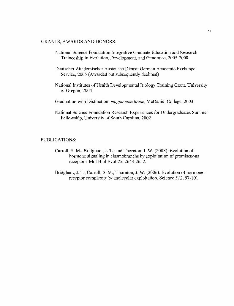
GRANTS, AWARDS AND HONORS:
National Science Foundation Integrative Graduate Education and ResearchTraineeship in Evolution, Development, and Genomics, 2005-2008
Deutscher Akademischer Austausch Dienst: German Academic ExchangeService, 2005 (Awarded but subsequently declined)
National Institutes of Health Developmental Biology Training Grant, Universityof Oregon, 2004
Graduation with Distinction, magna cum laude, McDaniel College, 2003
National Science Foundation Research Experiences for Undergraduates SummerFellowship, University of South Carolina, 2002
PUBLICATIONS:
Carroll, S. M., Bridgham, .T. T, and Thornton, J. W. (2008). Evolution ofhormone signaling in elasmobranchs by exploitation of promiscuousreceptors. Mol BioI Evol 25, 2643-2652.
Bridgham, J. T., Carroll, S. M., Thornton, J. W. (2006). Evolution of hormonereceptor complexity by molecular exploitation. Science 312, 97-101.

Chapter
TABLE OF CONTENTS
Page
Vll
I. INTRODUCTION 1Protein Evolution by Gene Duplication 1
Steroid Signaling as a Model System for Studies of Protein Evolution 18
Bridge to Chapter II 23
II. EVOLUTION OF HORMONE SIGNALING IN ELASMOBRANCHS BYEXPLOITATION OF PROMISCUOUS RECEPTORS 24Introduction............................................ 24
Methods 28
Results 31
Discussion 42
Bridge to Chapter III 48
III. DEGENERATIVE AND STABILIZING MUTATIONS IN THE ORIGINOF THE GLUCOCORTICOID RECEPTOR 49Introduction 49
Methods 52
Results 56
Discussion 70
Bridge to Chapter IV 74
IV. THE EVOLUTION OF CYP21 BY GENE DUPLICATION ANDCHROMOSOMAL RELOCATION 75Introduction 75
Methods 78
Results 81
Discussion 87
V. CONCLUSIONS :................................. 93
REFERENCES 96

Vlll
LIST OF FIGURES
Figure Page
2.1. The skate MR and GR are high- and low-sensitivity receptors, respectively,for 1a-B and other corticosteroids 32
2.2. GR and MR are widely coexpressed in skate 33
2.3. Sensitivity to 1a-B is taxonomically widespread 35
2.4. Sensitivity to 1a-B predates the evolution of 1a-B synthesis 39
2.5. The structural basis for receptor promiscuity 42
2.6. Receptor exploitation in the evolution of corticosteroid signaling 44
3.1. Schematic ofthe GR clade from the maximum likelihood phylogeny............ 57
3.2. AncGR1.1 and its descendents are low sensitivity receptors 59
3.3. Three historical substitutions reduce AncCR sensitivity................................. 62
3.4. The solved crystal structure of AncGR1.1 64
3.5. Lost hydrogen bonds in the region of position 116 66
3.6. C71S adds favorable bonds and buffers other mutations 67
4.1. The steroid synthesis pathway and major classes of hormone......................... 77
4.2. Maximum likelihood phylogeny of Cyp17 and Cyp21 proteins...................... 83
4.3. Conserved synteny in human regions flanking Cyp17 and Cyp21 85
LIST OF TABLES
Table Page
2.1. Sensitivity of extant and ancestral receptors to 1a-B 36
2.2. AncCR's sensitivity to 1a-B is robust to uncertainty 40
3.1. Sensitivity of extant and ancestral receptors to hormone................................. 60
3.2. Three historical GR substitutions reduce AncCR sensitivity 62
3.3. Statistical uncertainty in AncGR1.1 sites 69
4.1. Percent of sequence identity of amphioxus and lamprey Cyps........................ 81
IX

1
CHAPTER I
INTRODUCTION
PROTEIN EVOLUTION BY GENE DUPLICATION
Gene duplication is a widely accepted source of molecular innovation,
particularly for the evolution of novel protein functions. Many models exist to explain the
mechanisms that drive the maintenance and functional divergence of gene duplicates, but
few, if any, attempt to incorporate a growing body of work on structural and biophysical
mechanisms of protein evolution. Are there universal changes in protein structure that
underlie the functional diversification of proteins after gene duplication? The purpose of
this review is to synthesize our knowledge of evolution by gene duplication with
empirical analyses of protein structure to provide a more comprehensive understanding of
the mechanisms driving the evolution of novel protein functions.
The central goal of evolutionary biology is to provide a historical explanation for
the diversity and complexity of life. Just as Darwin's "principle of descent with
modification" (Darwin, 1964) explains the origin of species and their exquisitely
optimized forms, there are similar explanations for the patterns and processes of
evolution at the molecular level. Proteins occupy a special place in molecular evolution
as the workhorses of the cell; the interactions of proteins with nucleic acids, metabolites,

2and the environment drive virtually all cellular functions and underlie phenotypic
diversity and complexity. Therefore a central goal of molecular evolution is to
understand how proteins arise and diversify in function.
Properties ofProtein Structure and Function
Protein biology was in the midst of a "great biochemical revolution" (Dickerson,
2005) in the 1950's and '60's, as the molecular world was revealed in the light of X-ray
crystallography. No longer were the structures of DNA and proteins shrouded in mystery;
they could now be visualized in terms of discrete macromolecular structures. As a result,
a wealth of research emerged to explain the relationship between a protein's structure,
sequence, and function, and how they evolve. Early comparisons of proteins, particularly
the vertebrate hemoglobins, revealed many features of protein evolution that are relevant
today. Species differences in protein function were discovered to evolve from a common
structural scaffold; for example, the lineage-specific adaptations of bird, fish, and
crocodile hemoglobins are accomplished by one or a few mutations that result in only
minor changes to the protein fold (Perutz, 1983; Powers, 1972). Parts of some proteins
were discovered to be more "dispensable" than others for function (Anfinsen, 1959), in
what was a preface to the neutral theory of molecular evolution (Kimura, 1968). Changes
in some regions had little effect on function and were more tolerant to mutation, while
others were found to be essential and highly conserved between species. It was also
recognized that protein sequences and structures harbor an account of their evolutionary
past. Zuckerkandl and Pauling (Zuckerkandl and Pauling, 1965) deemed DNA and

3protein sequences "documents of evolutionary history," recording aspects of evolution
such as relationships to other sequences (phylogeny), the branches on which sequence
changes occurred, the rate of substitution, and even the sequences of long-extinct
ancestral molecules. Similarly, conserved macromolecular structures suggested common
descent of proteins from speciation and gene duplication events. This is best illustrated in
the classic papers by Perutz (Perutz et aI., 1960) and Kendrew (Kendrew et aI., 1960) on
the respective structures of human hemoglobin and whale myoglobin proteins. These and
other works within the hemoglobins (Ingram, 1961; Pauling and Zuckerkand1, 1963)
were the first to highlight the role of gene duplication as important for the evolution of
novel protein functions.
Ohno's seminal work solidified the importance of gene duplication in evolution,
and today there are three major ways - gene conservation, subfunctionalization, and
neofunctionalization - by which duplicate genes are preserved (Ohno, 1970; Piatigorsky
and Wistow, 1991; Hughes, 1994; Force et aI., 1999). This review focuses primarily on
the evolution of novel gene functions by neofunctionalization; the different fates of gene
duplicates and the population genetic models for their preservation are the subject of
numerous other reviews (Hahn, 2009; Lynch and Katju, 2004; Conant and Wolfe, 2008;
Roth et aI., 2007; Zhang, 2003). Ohno theorized that after gene duplication, one copy is
freed of its ancestral function and is allowed to explore previously "forbidden" mutations
in a period of relaxed or absent selection. Most often this mutation accumulation quickly
results in the degeneration and nonfunctionalization of the duplicate loci. In rare
instances, however, the mutational drift will stumble upon a novel gene function that
----_.__.. --------

4confers a fitness advantage and leads to the preservation of the duplicate loci. There are
several different models describing the mutational mechanisms of neofunctionalization,
and these are reviewed elsewhere (Hahn, 2009). Here, I discuss how knowledge of
protein evolution that has emerged since the time of Ohno and could enrich our
understanding of neofunctionalization as a mechanism for the preservation duplicate
genes and the subsequent evolution of novel protein functions. I avoid new terminology
or statements that further confuse an already heavily researched topic; my goal is to show
that common processes of protein evolution can increase the likelihood that duplicate
genes are preserved and evolve new functions.
Modern Concepts ofProtein Function and Evolution
Recent advances in our knowledge of protein functional evolution have come
from two major disciplines. The first includes work that seeks to empirically test
hypotheses on the historical mechanisms of protein evolution by employing the
manipulative experiments of molecular biology (Golding and Dean, 1998; Dean and
Thornton, 2007). Highlights of the so-called "functional synthesis" include the molecular
basis ofevolved pesticide resistance (Newcomb et aI., 1997), the adaptive landscape of
an ancient switch in coenzyme use (Miller et aI., 2006), and the diversification of opsins
to varying light conditions (Yokoyama et aI., 1999; Shi et aI., 2001; Yokoyama, 2002;
Yokoyama et aI., 2008). These case studies offer insights into historical processes but are
limited to the end-result of a single evolutionary "experiment" or outcome, and cannot
directly address things such as the repeatability or predictability of evolution (Gould,

51989; Lenormand et ai., 2009; Weinreich et ai., 2006; Blount et ai., 2008). However,
this approach is well complemented by protein engineers and experimental evolutionists
that can observe protein evolution in real time. Directed evolution, in particular, most
often seeks to evolve an enzyme to acquire some new desired function through repeated
rounds of mutation and selection (Arnold et ai., 2001; Bershtein and Tawfik, 2008a). The
researcher can monitor and control the strength and nature of selection, the pool of
variation to select upon, the properties of evolutionary intermediates, and the precise
mutational trajectories taken in one or any number of evolutionary experiments. These
experiments offer more precise measurements of protein evolution but lack the
complexity of natural systems. Collectively, mechanistic studies of historical evolution
and directed evolution experiments have illuminated such things as the evolutionary
potential ofpromiscuous protein functions, how proteins evolve in a period of relaxed
selection, and the fundamental importance of protein stability. Evolutionary models of
neofunctionalization could greatly benefit from this knowledge.
The Exploitation ofForiUt.'')us Protein Promiscuity
Shortly after 0000 ou~lined the fate of duplicate genes (0000, 1970), functional
promiscuity emerged as an im Jortant force in the evolution of novel protein functions.
Jensen (Jensen, 1976) championed the importance ofprotein promiscuity: "fortuitously
formed compounds that happened to be useful would have conferred a selective
advantage, thereby providing a basis for increased and more specific production of that
compound (by gene duplication and specialization by mutation)." In its broadest sense,

6promiscuity is merely a lack of specificity in protein functions. Proteins evolve under
purifying selection to maintain a native function, but in many instances this is
accompanied by one or more low-level and nonspecific functions that are of no selective
consequence. This phenomenon is most often attributed to enzymes, but promiscuity has
been observed in many different types of proteins across the tree of life (Copley, 2003;
Khersonsky et aI., 2006; O'Brien and Herschlag, 1999; D'Ari and Casadesus, 1998). A
recent genome-wide survey of E. coli found that promiscuity is quite common:
approximately 20% of gene knockouts are rescued by the promiscuous functions of
typically unrelated genes (Patrick et aI., 2007). These mutants were rescued by a large
sink oflatent and fortuitous protein functions (Patrick and Matsumura, 2008; Patrick et
aI., 2007; Jensen, 1976) that is potentially even larger when genes are exchanged between
E. coli strains or other bacteria (Hendrickson, 2009; Touchon et aI., 2009; Touchon et aI.,
2009).
Arguably, the exploitation ofpromiscuous activities blurs the lines between the
three outcomes of duplicate genes. Promiscuous activities can arise after gene duplication
and legitimately drive neofurl --;tionalization or, alternatively, they can be present before
duplication and selectively neutral. The exploitation of ancestral promiscuity is "new" in
the sense that these functions are newly exposed to selection, but the activities might
have arisen before or after gem' duplication. Both theory (Jensen, 1976; Bergthorsson et
aI., 2007) and empirical studies (Patrick and Matsumura, 2008; McLoughlin and Copley,
2008) suggest that these low-level promiscuous functions could be exposed to positive
selection if the protein (and its native function) are overexpressed. Duplicate proteins

7would amplify promiscuous activities and could lead to the "gene conservation" fate of
duplicates (Bergthorsson et aI., 2007; Ohno, 1970); subsequent mutations optimize the
functions of each paralog, to the detriment ofthe side-activity (Bergthorsson et aI., 2007;
Jensen, 1976). Selection upon promiscuous activities can also set the stage for
subfunctionalization after gene duplication. McLoughlin and Copley (McLoughlin and
Copley, 2008) devised an experimental system in which both the native and promiscuous
functions of an enzyme are suddenly necessary for survival. They effectively evolve a
situation of "gene sharing" (Jensen, 1976; Piatigorsky and Wistow, 1991; Hughes, 1994)
with noticable tradeoffs between the two functions. This multifunctional enzyme is a
model precursor for subfunctionalization by the partitioning and optmization of functions
between duplicates (Piatigorsky and Wistow, 1991; Hughes, 1994). In short, protein
promiscuity could expand the potential adaptive pathways to a new function and increase
the likelihood that duplicate genes are preserved.
Historical and real-time studies of protein evolution have illuminated ways in
which protein promiscuity increases evolvability. In some protein engineering
experiments, both the starting and end products of the selection process are specialists for
a particular function, and yet analyses of the mutational intermediates often find that
evolution proceeded through a generalist protein capable ofmultiple interactions
(Matsumura and Ellington, 2001; Fasan et aI., 2008). These results might suggest that
there is always a functional tradeoff between the native and evolved functions, but there
are many examples in which no such tradeoff exists (reviewed in Khersonsky et aI.,
2006). The dynamic nature of functional promiscuity has been captured in the lab by

8allowing proteins to neutrally diverge from their ancestral sequence. Experiments that
evolved an enzyme with random mutations plus purifying selection for the native
function found that the variants accrued a number of novel promiscuous activities in the
course of their divergence (Bloom et aI., 2007b; Amitai et aI., 2007). These and other
studies suggest that sequence divergence permits the exploration of an often-vast neutral
network of promiscuous activities (Amitai et aI., 2007; Bloom and Arnold, 2009;
Peisajovich and Tawfik, 2007). The exploitation of promiscuous activities by selection is
then highly contingent on evolutionary history (Patrick and Matsumura, 2008; Patrick et
aI., 2007) and the current sequence space occupied by the protein; latent promiscuous
functions could suddenly become adaptive after a shift in the environment or genome.
The exploitation of ancestral promiscuity for novel functions is elegantly
demonstrated in the origins of steroid signaling. In humans and other tetrapods, the
mineralocorticoid receptor (MR) is activated by aldosterone to regulate blood pressure
and salt balance, whereas the glucocorticoid receptor (GR) is activated by cortisol and
regulates various immune, metabolic, and stress responses. Phylogenetic analyses showed
that MR and GR descend from an ancient gene duplication, which begs the question: how
did the specific interactions ofMR with aldosterone and GR with cortisol arise? Using
ancestral state reconstruction, Bridgham et al. (Bridgham et aI., 2006) found that the
resurrected last common ancestor of MR and GR, named AncCR, was activated by a
variety of hormones, including those such as cortisol and aldosterone that had yet to
evolve. But why would a receptor be preadapted for hormones that had yet to appear?
The authors suggest that the AncCR's affinity is merely a byproduct of constraints

9imposed by structurally similar, more ancient endogenous hormones. The determination
of AncCR' s protein structure revealed a common binding mode for structurally
analogous hormones (Ortlund et aI., 2007), and suggested that empty "nooks" in the
hormone-binding pocket fortuitously fit hormones that evolved millions of years later
(Carroll et aI., 2008). The structural promiscuity that descended from AncCR facilitated
the recruitment of MR by aldosterone upon the later synthesis ofthe hormone in
tetrapods (Bridgham et aI., 2006). Similar instances of novel molecules recruiting
preexistent molecules for new functional roles have been found elsewhere in steroid
receptors (Thornton, 2001; Carroll et aI., 2008) and other in proteins (Cai et aI., 2007;
Krasowski et aI., 2005; Cardoso et aI., 2007).
Protein Functions in Response to Mutation
Neutral processes dominate most ofprotein evolution and playa major role in the
various outcomes ofgene duplication. The neutral theory predicts that most sequence
diversity is the result of random fixations ofmutation while purifYing selection maintains
protein function (Kimura, 1983). 0000 believed that after gene duplication, one copy is
"free to accumulate a series of forbidden mutations" in a period of mutational drift
(0000, 1970). The end-result of random mutations in a period of relaxed or absent
selection is most often nonfunctionalization, but these mutations can sometimes playa
creative role in the preservation ofduplicate genes. So how tolerant are proteins to
random mutational drift? Are there certain properties that make one protein more robust
to mutation than others? This could be particularly relevant to the period of "forbidden"

10mutations after gene duplication. If proteins crumble after only a few mutations then
the opportunity for a new or optimized function to arise is indeed extremely small.
Historical and engineered studies of protein evolution offer empirical tests of a protein's
response to periods of mutation accumulation with and without selection.
Typically, periods of "undirected" sequence evolution are unnecessary in directed
evolution. Starting with a protein and a native function (A), selection for activity (B)
usually proceeds by a simple uphill climb to the novel function (Tracewell and Arnold,
2009). However an alternative approach, and one that is particularly intriguing for studies
of historical evolution, is to select for new functions from libraries created by so called
"mutational drift" (Gupta and Tawfik, 2008); this term can be misleading because
sequences can be evolved with random mutations and no selection (drift), or random
mutations plus purifying selection for the native protein function (sequence divergence).
Typically, successive rounds of mutation and selection on the native function occur
before screening the variants for novel protein functions. Protein populations generated
using this technique have been found to display different properties than those under
constant selection, including greater protein stability and an increased opportunity for
new functions to arise (Bershtein et aI., 2008; Bloom et aI., 2007b; Bloom et aI., 2007a;
Bloom et aI., 2007).
A wealth of directed evolution experiments have used enzyme activity as a
surrogate for fitness to classify the distribution of the effects of random mutation. These
data (\reviewed in\ (Bloom and Arnold, 2009)) were generated from a broad sampling of
functionally diverse enzymes and give very similar results - most mutations are

11selectively neutral, a smaller proportion are deleterious, and a tiny fraction, typically
half to one-hundredth of a percent, are selectively advantageous for a novel protein
function. Structural analyses have shown, in accordance with the neutral theory (Kimura
and Ota, 1974) and earlier predictions (Anfinsen, 1959), that neutral mutations are not
equally distributed in the protein structure. Of920 "tolerated" mutations to a DNA repair
enzyme (AAG), mutations near the active site and DNA-binding surface are
underrepresented or absent while those on the surface and in protein loops are enriched
(Guo et aI., 2004). These results suggest that proteins are initially very tolerant to random
mutations, at least in some parts of the protein structure.
Repeated rounds of random mutagenesis shed light on the robustness of proteins
during prolonged mutation accumulation experiments. Bershtein et al. (Bershtein et aI.,
2006) subjected TEM-l j3-lactamase to a prolonged drift - upwards of 20 mutations per
gene - in the absence of purifying selection, and subsequently measured the proportion of
enzyme variants that continued to confer antibiotic resistance (fitness). When presented
with a high selective pressure (large dose of antibiotic), fitness decreased rapidly with
mutational load, so that only half of the variants confer resistance with just two
mutations. Given a lower dose of antibiotic, protein fitness is initially quite robust to
mutations; however, in later rounds of mutagenesis, the fitness decrease steepens and is
indicative of negative epistasis. The authors conclude that robustness and epistasis are
linked in proteins and determined by a common parameter - protein stability. In all, these
results show that the distribution of fitness effects of protein mutations can be dependent
on the presence or strength of selection, and likely the specific protein architecture. These

12factors could dominate the evolutionary dynamics of gene duplicates towards
nonfunctionalization versus other fates (Bershtein and Tawfik, 2008b).
Degenerative mutations accrued in weak or absent selection are known to playa
creative role in certain models of subfunctionalization (Force et al., 1999); a recent study
presents a case in which these mutations are the likely mechanism for
neofunctionalization. Steroid receptors most often function as hormone activated
transcription factors, possessing a largely modular domain architecture to coordinate
multiple subfunctions such as binding to DNA or to hormone (Beato, 1989). Amphioxus
possesses two steroid receptors that evolved with the duplication of an ancestral estrogen
activated receptor (Bridgham et al., 2008). One of these paralogs has retained an
ancestral-like function, whereas the other lost its subfunction to bind hormone. The
degenerative mutations in this copy created a novel receptor that competes for the same
DNA-binding elements as its paralog, effectively functioning as a transcriptional
repressor. These results demonstrate that degenerative evolution after gene duplication
may be a creative force in and of itself, significantly increasing the likelihood of
neofunctionalization.
The Importance ofProtein Stability
Long before knowledge of three-dimensional protein structures or even the nature
of the polypeptide chain, scientists recognized the importance of a properly folded
protein state. Outside of normal physiological conditions - such as changes in pH,
solvent, or temperature - proteins lose their native folded state and, with it, their function
(Nelson, 2003). Protein denaturation, however, is readily reversible when the unfolded
13chain is returned to its physiological norm. Anfinsen showed in his experiments that
renaturation is guided solely by the biophysical properties encoded in the protein's
sequence (Anfinsen, 1973), and a return to the native state is accomplished by finding the
most thermodynamically stable protein conformation. These early studies of protein
folding and stability inspired a wealth of research on the role that stability plays in
protein evolution, and how protein stability itself evolves.
Research has shown that protein stability is exquisitely adapted to an organism's
thermal environment. Enzymes from psychrophilic organisms are optimally active at cold
temperatures (Russell et aI., 1998; Davail et aI., 1994), while enzymes from thermophilic
organisms function at high temperatures (Singleton and Ame1unxen, 1973); these
adaptations have been recreated in the lab by selecting enzymes for increased thermal
tolerance (Counago et aI., 2006). The importance of thermal adaptation for protein
function suggests that stability is an important and evolutionary labile property of
proteins (Puigbo et aI., 2008). Remarkably, this tight relationship between protein activity
and thermostability has illuminated the thermal environment of reconstructed ancestral
proteins that existed some billions of years ago (Gaucher et aI., 2008; Gaucher et aI.,
2003).
So why aren't proteins selected to be overly stable? From a biophysical
perspective, there might be tradeoffs between the catalytic activity and thermostability of
thermal adapted enzymes (Arnold et aI., 2001). Enzymes adapted to high temperatures
are thought to have more stabilizing interactions to make their structures rigid, while
those adapted to cold should be more flexible to allow for conformational changes

14associated with catalytic activities. But an alternate theory is that, for most proteins,
marginal stability arises from passive evolutionary mechanisms. Taverna and Goldstein
(Taverna and Goldstein, 2002) suggest that proteins that meet the minimum stability
requirements will predominate simply because of the vastness of sequence space and the
rare chance that highly stable variants would arise by chance. Many random mutations
are inherently destabilizing, through a variety of mechanisms (Matthews, 1987;
Matthews, 1993). Tokuriki et al. (Tokuriki et aI., 2007) compute the distribution of
mutational effects on protein stability for 21 different proteins and find that, regardless of
protein architecture, most mutations are inherently destabilizing.
A protein's stability plays a major role in governing its tolerance to mutation.
Given the above assertions that proteins are only marginally stable and many mutations
are destabilizing, a protein would quickly unfold and lose activity in the absence of
selection. A protein's robustness to mutation is largely governed by a stability threshold,
below which the protein becomes unstable and loses activity (Bershtein et aI., 2006;
Bloom et aI., 2007). The protein's distance from this threshold, however, is dynamic and
evolvable. The stability threshold can be relaxed by decreasing selection for proper
function (Bershtein et aI., 2006), by gaining one or more stabilizing mutations that
increase the distance from the threshold (Bloom et aI., 2005; Bloom et aI., 2006), or even
by more global mechanisms such as increasing the expression of chaperone proteins
(Tokuriki and Tawfik, 2009). Mutations that increase stability are called "global
suppressors," because they allow a greater number of destabilizing mutations to
accumulate than would otherwise be allowed (Bershtein and Tawfik, 2008a; Mitraki et

15aI., 1991; ShortIe and Lin, 1985). Strikingly, proteins with global suppressor mutations
have an increased likelihood for novel functions to arise (Bershtein et aI., 2008; Bershtein
and Tawfik, 2008a; Bloom et aI., 2006), and destabilizing mutations that confer novel
functions are often balanced by changes to increase stability (Tokuriki et aI., 2008).
Increased protein stability can expand the range ofpromiscuous activities (Huang and
Palzkill, 1997; Petrosino et aI., 1998) by several proposed mechanisms (Tokuriki and
Tawfik, 2009; Wroe et aI., 2007). These results have profound implications for protein
evolution after gene duplication - factors such as relaxed purifYing selection or global
suppressor mutations that relieve the effects ofdeleterious mutations increase the
probability that beneficial mutations will arise.
Mechanistic Studies ofProtein Functional Evolution
Empirical studies of historical and engineered protein evolution have illuminated
several properties that can contribute to our understanding of how novel functions evolve
by gene duplication. First is the possibility that proteins are only as specific as they need
to be. Functional promiscuity is a common byproduct of native protein functions, present
in many different gene families across the tree of life. As long as promiscuous activities
do not interfere with normal cellular functions, they are allowed to persist and drift with
sequence divergence. Occasionally, though, this untapped and fortuitous promiscuity can
be exploited for new functional roles by a variety of mechanisms. Promiscuous activities
that arise and are selected upon before duplication (gene sharing) will lead to
subfunctionalization (0000, 1970; Hughes, 1994; Piatigorsky and Wistow, 1991); those

16that arise before or after duplication but are only exposed to selection after could lead
to the appearance of neofunctionalization (Ohno, 1970), or in actuality, preservation by
gene conservation (Bergthorsson et aI., 2007). The exploitation of fortuitous functional
promiscuity is not well explained by the canonical fates of duplicate genes.
Real-time studies of protein divergence allow us to monitor changes in native and
promiscuous protein functions, protein structure, and stability as they evolve with
mutation and varying degrees selection. These conditions simulate changes in functional
constraints brought about by gene duplication; those gene functions that are more robust
to mutation should have an increased likelihood of discovering creative mutations, and a
slightly decreased chance that mutations will be destructive. These degenerative
mutations, however, play creative roles in forms of subfunctionalization (Force et aI.,
1999) and as described above, neofunctionalization as well (Bridgham et aI., 2008). Thus
empirical studies of protein evolution have expanded our ideas of how proteins change in
response to mutation and varying degrees of selection.
Finally, protein stability has emerged as a global determinant of protein structure
and function and is in itself and evolvable character. Proteins are optimized by selection
to be only as stable as is necessary to properly fold and function. Marginal protein
stability could arise partly due to tradeoffs between flexibility and activity or because
many mutations are inherently destabilizing. Mutations that increase stability, however,
can have profound effects on subsequent protein evolution. These stabilizing and so
called "global suppressor" (ShortIe and Lin, 1985) mutations allow a greater number of
destabilizing and previously "forbidden" (Ohno, 1970) mutations to accrue than would

17normally be allowed. Suppressor mutations fortuitously expand the neutral networks
from which promiscuous or native functions arise, increase the chance of
neofunctionalization, and extend the time to nonfunctionalization.
Ohno's highly prescient synthesis forever placed gene duplication as one of the
major creative forces in evolution (Ohno, 1970). Since this time, the evolutionary
consequences of gene duplication have been found to include adaptation, speciation,
mutational robustness, evolvability, and complexity, amongst others (Postlethwait et aI.,
2004; Conant and Wolfe, 2006; Cooper et aI., 2007; Escriva et aI., 2006; Soltis and
Soltis, 1999; Teichmann and Babu, 2004; Wagner et aI., 2003; Hannay et aI., 2008;
Lynch and Conery, 2003; Hittinger and Carroll, 2007; Panopoulou and Poustka, 2005).
Gene duplication has arguably sparked the greatest interest in the evolution of novel gene
functions by neofunctionalization (Ohno, 1970; Hahn, 2009). Variations in the models of
neofunctionalization have sparked much debate, partly because of a lack of evidence on
how protein functions actually evolve; historical and engineered studies of protein
evolution can now illuminate these processes. This knowledge certainly will not
revolutionize the ideas founded by Ohno, but could help us to delineate the important
variables of neofunctionalization - when the novel function arises; whether mutations are
fixed by selection or drift; whether the mutations optimize, create, or destroy ancestral
functions; whether trajectories are repeatable and predictable or highly contingent; and
the effect of changing selective regimes on evolution. Studies of the specific mechanisms
by which proteins evolve could contribute to a greater understanding of how novel genes

18and gene networks evolve. My dissertation provides one such as example in the
evolution of corticosteroid signaling.
STEROID SIGNALING AS A MODEL SYSTEM FOR STUDIES OF PROTEIN
EVOLUTION
Steroid receptors and their honnones are an excellent system in which to study the
dynamics and mechanisms of protein evolution. Steroid receptors (SRs) are ligand
activated transcription factors and molecular mediators: honnone binding induces a
change in the receptor structure that causes cascade of interactions with chaperone
proteins, DNA response elements, and coregulatory proteins that ultimately results in the
regulation of gene expression in target tissues (Beato, 1989; Beato et aI., 1995). Steroid
honnones are produced by a series of enzymatic reactions from a cholesterol precursor
and are typically classified by their site of origin - sex steroids such as progestins,
androgens, and estrogens are synthesized predominantly in the gonads; corticosteroids
are made in the adrenal cortex. Steroid signaling regulates many critical facets of life
such as metabolism, immunity, reproduction, development, and the stress response
(Bentley, 1998). The ability of this system to regulate widely different biological
responses is due in large part to the specificity of a particular receptor for one or only a
few steroids ligands. It is of great interest then to understand how new receptors and
hormones evolve and are assembled into novel receptor-hormone interactions.

19SRs originated from a series of gene and whole-genome duplications followed
by the evolution of distinct hormone responses. Only recently were the evolutionary
relationships between these receptors delineated. Humans possess six SRs: the estrogen
receptors alpha (ERa) and beta (ER13), progesterone receptor (PR), androgen receptor
(AR), glucocorticoid receptor (GR), and mineralocorticoid receptor (MR). The diversity
of receptors evolved from a single estrogen receptor-like ancestor over 600 million years
ago (Thornton, 2001; Thornton et aI., 2003). Evolutionary offshoots in the process of SR
duplication and divergence are preserved across the tree of life: some invertebrates have a
single (estrogen) receptor (Thornton et aI., 2003; Keay et aI., 2006; Keay and Thornton,
2009), others diverged after the first gene duplication and have only two receptors
(Bridgham et aI., 2008; Paris et aI., 2008), basal vertebrates like lamprey have three
(Thornton, 2001), and jawed vertebrates typically have six (Bridgham et aI., 2006). Some
lineages have experienced additional rounds of duplication and possess additional SR
copies (Greenwood et aI., 2003; Bury et aI., 2003; Wu et aI., 2003). Much less is known
about the origins of steroid hormones, but work in this dissertation and elsewhere
suggests that the duplication and divergence of steroid-synthesizing enzymes can drive
the evolution of novel classes of steroids (Nelson, 1998; Nelson, 1999; Markov et aI.,
2008; Markov et aI., 2009).
The protein architecture of SRs makes them particularly amenable to molecular
analyses in the lab. SRs have a modular domain architecture, consisting of highly
conserved DNA-binding (DBD) and ligand-binding domains, and variable hinge, N
terminal, and C-terminal regions (Kumar and Thompson, 1999; Kumar and Thompson,
202005). The LBD alone is sufficient to bind hormone and mediate ligand-dependent
transcriptional activation, allowing the specific functions of the LBD to be cloned into
reporter constructs and assayed in vitro. Because of their biomedical importance, a wealth
of structural and functional data exists for human and other receptor LBDs, including a
large number of polymorphisms, disease states, and engineered proteins. X-ray
crystallography has revealed the structural basis of ligand-dependent transcriptional
activation in the receptor LBD. The activation function helix (AF-H) acts as a molecular
switch for transcriptional activation (Nagy and Schwabe, 2004). In the absence of ligand,
AF-H is extended or in an "off' position; the binding of a steroid agonist, however,
stabilizes the receptor structure and allows the AF-H to nestle against the receptor
surface. This repositioning of AF-H creates a novel protein-protein interaction surface
that allows coregulatory proteins to bind and recruit the transcriptional machinery to
upregulate the expression of target genes. Empirical knowledge of how SRs bind to and
are activated by hormone allows us to more easily dissect the molecular basis of
evolutionary changes in receptor-hormone interactions.
A combination of techniques from phylogenetics, molecular, and structural
biology permit highly mechanistic studies of SR evolution. This work is part of an
emerging "functional synthesis" that seeks to understand the processes of molecular
evolution at their most fundamental level (Dean and Thornton, 2007). Molecular
phylogenetics provides an evolutionary framework on which to describe the relative
timing of SR gene duplications, to explain the observed diversity of SR functions, and to
infer on which branches functional changes occurred (Thornton, 2001; Bridgham et aI.,

212006). The phylogeny also serves as a template for the reconstruction of ancestral
states. This technique is the closest thing that we have to molecular fossils or a time
machine: ancestral protein sequences are inferred by statistical models of the
evolutionary process, the ancestor is "resurrected" via gene synthesis, and this ancestor is
cloned and characterized with the same methods used for extant receptors (Thornton et
ai., 2003). Ancestral reconstruction allows us to empirically characterize the functions of
extinct genes, to track the branches on which amino acid substitutions occurred, and to
use manipulative experiments to determine the precise mechanism of functional change.
My dissertation employs this highly mechanistic approach to study the evolution of
corticosteroid signaling.
The Origins ofCorticosteroid Signaling
My work focuses specifically on corticosteroid hormones and their receptors the
MR and GR, to understand how new hormones and receptors evolve and are recruited
into novel molecular partnerships. Corticosteroids are typically classified as either
mineralocorticoids or glucocorticoids based on their respective abilities to activate MR or
GR. In humans, MR is activated predominantly by aldosterone for the regulation of salt
balance and blood pressure; GR is activated by cortsiol to control a variety of immune,
metabolic, and stress responses. There are, however, lineage-specific receptor-hormone
interactions that have evolved in several many species (Sturm et ai., 2005; Idler and
Truscott, 1966). So how can we explain the observed diversity and complexity of
corticosteroid signaling?

22GR and MR evolved with the duplication of a single ancestral receptor over
440 million years ago (Thornton, 2001; Bridgham et aI., 2006). Using methods in
ancestral state reconstruction and gene resurrection (Thornton et aI., 2003), our lab
previously demonstrated that the last common GR/MR ancestor functioned much like
extant MRs - highly sensitive to a variety of hormones - and that GR functions are
derived (Bridgham et aI., 2006; Ortlund et aI., 2007). After duplication, GRs evolved two
major changes in their response to ligand: 1) reduced sensitivity to all hormones, as is
found in the GR of an elasmobranchian cartilaginous fish (Carroll et aI., 2008), and on
top of this, 2) cortisol-specific activation, as is present in teleost and tetrapod receptors
(Bridgham et aI., 2006). The latter event is well studied: the historical mutations, their
effects on protein structure-function, and ancient mutational trajectories have all been
elucidated in the evolution of cortisol specificity (Bridgham et aI., 2006; Ortlund et aI.,
2007). Considerably less is known about earlier periods of corticosteroid receptor
evolution.
My dissertation seeks to reconstruct the evolutionary mechanisms and dynamics
that gave rise to corticosteroid signaling pathways. I use a combination of techniques
from phylogenetics, molecular biology, and structural biology to reveal the evolutionary
origins of hormones, their receptors, and their functional interactions. This dissertation
comprises three separate but complementary studies that elucidate the origins of
corticosteroid signaling. In the first project, I show how lineage-specific receptor
hormone interactions evolved by exploiting the structural promiscuity of receptors. This
work was originally published in the journal Molecular Biology and Evolution (vol. 25,

23iss. 12, pp. 2643-52, 2008). It was co-authored with Jamie T. Bridgham, who assisted
with materials and experimental design, and Joseph W. Thornton, who assisted with
experimental design and edited the manuscript. Next, I study how degenerative and
stabilizing mutations defined the functional divergence of GR after gene duplication. This
work is in preparation for publication and is co-authored with Eric A. Ortlund, who
assisted with experiments and experimental design, and Joseph W. Thornton, who
assisted with experimental design and edited the manuscript. Finally, I reconstruct the
molecular mechanisms of gene duplication that gave rise to the enzyme necessary for the
synthesis of corticosteroid hormones. This work is in preparation for publication and is
co-authored with Joseph W. Thornton, who assisted with experimental design and edited
the manuscript. Together, this dissertation describes how seemingly complex and
integrated facets of corticosteroid signaling can evolve by the simple and stepwise
process of gene duplication and divergence.
BRIDGE TO CHAPTER II
The interactions of corticosteroid hormones with their receptors represent a
simple, pairwise molecular interaction. These interactions are the basis for the molecular
networks that drive virtually all cellular processes and underlie phenotypic diversity and
complexity. To understand how molecular interactions evolve, I reconstructed the
evolution corticosteroid signaling in elasmobranchs, a subclass of cartilaginous fishes. I
show that novel receptor-hormone interactions can evolve by molecular exploitation, and
that they are facilitated by the structural promiscuity of receptors.

24
CHAPTER II
EVOLUTION OF HORMONE SIGNALING IN
ELASMOBRANCHS BY EXPLOITATION OF
PROMISCUOUS RECEPTORS
This work was originally published in the journal Molecular Biology and
Evolution (vol. 25, iss. 12, pp. 2643-52,2008). It was co-authored with Jamie T.
Bridgham, who assisted with materials and experimental design, and Joseph W.
Thornton, who assisted with experimental design and edited the manuscript.
INTRODUCTION
Virtually all cellular functions are driven by specific interactions among
biomolecules, such as enzymes and their substrates, transcription factors and their DNA
binding sites, and receptors and their ligands. Despite extensive "top-down" work on the
global structure of molecular interaction networks (Barabasi and Oltvai, 2004; Cork and
Purugganan, 2004; Teichmann and Babu, 2004; Wilkins, 2005), only limited knowledge
is available concerning the specific mechanisms and dynamics by which the molecular
interactions that constitute these networks evolve (Zhu et aI., 2005; Bridgham et aI.,

252006; Prud'homme et aI., 2006; Hittinger and Carroll, 2007; McGregor et aI., 2007;
Ortlund et aI., 2007).
Corticosteroid honnones and their receptors provide an elegant model of
molecular interactions, and the existence of a lineage-specific corticosteroid in the taxon
Elasmobranchii offers the opportunity to investigate how new honnone-receptor
interactions evolve. Corticosteroids are produced in the adrenal/interrenal gland through
an enzyme-mediated biosynthetic pathway and secreted into the blood. The classic
actions of corticosteroids are mediated by intracellular corticosteroid receptors (CRs),
members of the steroid honnone receptor family that also includes receptors for
androgens, progestins, and estrogens (Thornton, 2001). These proteins are ligand
activated transcription factors: upon binding to their honnone partners with high
specificity and affinity, the receptor changes confonnation, dimerizes, binds to specific
DNA response elements, and attracts coregulator proteins, resulting in increased
expression of nearby target genes (Beato et aI., 1995). Like other steroid receptors, CRs
have a modular domain structure, consisting of functionally autonomous conserved
domains for DNA-binding and for ligand-activated transcription, as well as a
nonconserved hinge and an N-terminal domain with additional ligand-independent
transcriptional regulatory functions (Kumar and Thompson, 2005). Most vertebrates
possess two paralogous CRs, a glucocorticoid receptor (GR) and mineralocorticoid
receptor (MR), which arose from a single ancestral CR in a large-scale gene duplication
some 470-440 million years ago (Thornton, 2001; Bridgham et aI., 2006). In bony
vertebrates, GR is activated by cortisol and regulates metabolism, immunity, and the

26stress response; MR controls electrolyte homeostasis and blood pressure and is
activated primarily by aldosterone in tetrapods and II-deoxycorticosterone in teleosts
(Bentley, 1998; Sturm et aI., 2005). In addition to their classic receptor-mediated effects,
some steroids also trigger rapid "non-classical" effects through other mechanisms
(Thomas et aI., 2005; Thomas et aI., 2004; Prossnitz et aI., 2007; Harvey et aI., 2008).
A better understanding of steroid hormone-receptor interactions in basal
vertebrates would help illuminate how OR, MR, and the physiological processes they
regulate have evolved (Baker et aI., 2007; Bury and Sturm, 2007). Some forty years ago
it was discovered that the plasma of elasmobranchs -sharks, skates, and rays - contains
very high levels of the corticosteroid la-hydroxycorticosterone (la-B), which is not
known to be produced in any other taxon (Idler and Truscott, 1966). la-B is synthesized
in the interrenal gland in copious amounts relative to other endogenous hormones (Idler
and Truscott, 1967; Truscott and Idler, 1972). It differs from other corticosteroids by the
addition of a hydroxyl group at the C I position of the steroid backbone. The enzyme
activity that drives this unusual la-hydroxylation reaction is present and functional in in
vitro preparations of elasmobranch steroidogenic tissues (Truscott and Idler, 1968), but
the underlying gene has not been identified. The molecular basis for la-B action in
elasmobranchs, particularly its potential interactions with MR and OR, remains
uncharacterized. It has been speculated that la-B functions as a dual ligand in both
glucocorticoid and mineralocorticoid signaling pathways (Oelsleichter and Musick, 1999;
Nunez and Trant, 1999; Nunez et aI., 2006; Manire et aI., 2007). la-B clearly has a
mineralocorticoid-like role in the regulation of salt and osmolyte balance (Hazon and

27Henderson, 1984; Armour et aI., 1993), but the evidence for its glucocorticoid-like
effects are more speculative. 1a-B levels have been shown to increase under stress and
reduced osmolarity (Hazon and Henderson, 1984; Armour et aI., 1993; Manire et aI.,
2007). Administration of glucocorticoids to elasmobranchs stimulates the regulation of
carbohydrate metabolism (Patent, 1970), cartilage growth (Gelsleichter and Musick,
1999), and immune responses (Walsh et aI., 2002). Like the glucocorticoids of other
vertebrates, synthesis of 1a-B is regulated by andrenocorticotropic hormone and
angiotensin II from the hypothalamic-pituitary adrenal axis (Honn and Chavin, 1978;
Hazon and Henderson, 1985; O'Toole et aI., 1990; Nunez and Vedeckis, 2002).
Although cortisol and other recognized glucocorticoids are absent or present at very low
concentrations in elasmobranchs, 1a-B circulates at extremely high levels in
elasmobranchs (Truscott and Idler, 1972; Kime, 1977; Armour et aI., 1993), just as
glucocorticoids do in most other vertebrates.
Here we characterize the functional interactions of 1a-B with the GR and MR of
an elasmobranch, the little skate (Leucoraja erinacea), and reconstruct how these
receptor-hormone interactions evolved. We combine molecular functional assays,
ancestral gene resurrection (Thornton, 2004), and analysis of protein structure to
determine the functions of GRand MR with respect to this hormone and to characterize
how the lineage-specific partnership of 1a-B with its receptors evolved.

28METHODS
Isolation and Reconstruction ofCorticosteroid Receptors.
Skate (Leucoraja erinacea) and hagfish (Myxine glutinosa) receptor ligand
binding domains (LBDs) were amplified using degenerate PCR and RACE from liver
cDNA (Bridgham et aI., 2006). Lamprey (Petromyzon marinus) CR cDNA was
amplified similarly from a cDNA library (Thornton, 2001). Teleost (Astatotilapia
burtoni) GR2a and MR were provided by R. Fernald, the human MR by R. Evans, and
the human GR by B. Darimont.
Reconstruction and synthesis of ancestral receptors was performed as described in
(Bridgham et aI., 2006; Ortlund et aI., 2007). Briefly, ancestral protein sequences were
inferred using maximum likelihood (ML) phylogenetic reconstruction and a large dataset
of steroid and related receptors. Nucleic acid sequences coding for the ligand binding
domain were synthesized de novo. Ambiguously reconstructed sites defined as having an
alternate amino acid state with a posterior probability greater than 0.20 or as having a
different maximum likelihood state when reconstructed on any tree in the 95% credible
set collected by Bayesian Markov Chain Monte Carlo analysis. Alternative states were
introduced singly into the maximum likelihood sequence using site-directed mutagenesis
(QuikChange II, Stratagene).

29Receptor Activation.
LBDs (with hinge and CTE) of extant receptors were amplified using high
fidelity PCR and cloned into pSG5-GAL4DBD (gift ofD. Furlow). Ancestral receptor
LBDs (with CTE) were cloned into pSG5-GAL4DBD with a human GR hinge region.
CHO-K1 cells were grown in 96-well plates in phenol red free a-MEM plus 10%
dextran-charcoal-stripped fetal bovine serum (Hyclone), then transfected with I ng of
receptor LBD, 100 ng ofpFRluc reporter, and 0.1 ng of normalization vector phRLtk
using Lipofectamine and Plus Reagents (Invitrogen). After 4 hours incubation, cells were
treated with fresh medium, allowed to recover overnight, then treated in triplicate with
hormone or vehicle control (ethanol) for 24 hours. Reporter expression was measured
assayed using Dual-Glo (Promega) and expressed as the ratio of firefly luciferase to
Renilla luciferase. Dose-response relationships were analyzed using Prism4 software
(GraphPad). Receptors were considered unresponsive ifthey displayed <2-fold activation
at >1 uM hormone. 1a-B was synthesized and provided by J. Rimoldi, University of
Mississippi.
Quantitative peR.
Expression of MR and GR was measured in various organs of1. erinacea
provided by J. St. George, Boston University. Total RNA was isolated using RNeasy
(Qiagen), and eDNA prepared using QuantiTect Reverse Transcription (Qiagen). Skeletal
and cardiac muscle samples were digested with proteinase K (Qiagen) to improve yield.
Primers were designed to amplify the ligand-dependent activation (AF-2) domain ofMR
30and GR LBDs. Housekeeping gene glyceraldehyde 3-phosphodehydrogenase
(GAPDH GenBank DQ382343) was isolated using degenerate PCR and used as an
internal reference for normalization. Q-PCR was performed on an ABI Prism 7900 HT
with Ix Quantitect SYBR Green PCR Master Mix (Qiagen), 0.3 flM each primer, and
skate cDNA. Cycling was as follows: 95°/15', [94°/15",54°/30",72°/30"] x 39 cycles,
followed by melting curve analysis from 65°-95°. Reactions were run in triplicate and
fluorescence detected during extension. Primer efficiencies (EMR = 1.96, EGR = 1.98, and
EGapdh = 1.97) were determined by standard curve analyses (Simon, 2003) of serially
diluted and linearized skate MR and GR in GAL4-DBD-pSG5 and skate Gapdh gene in
pCRl.l (Invitrogen).
In Silico Structural Analysis.
A model of la-B was constructed and energy minimized using ChemOffice Ultra
(CambridgeSoft) and imported into MacPyMOL (Delano Scientific LLC) along with the
crystal structure of AncCR/DOC (PDB 2Q3Y). la-B was aligned to DOC at the C IO
carbon with the steroid backbones oriented in the same plane.

31RESULTS
Higll- and low-sensitivity receptors/or la-B
To determine the intrinsic hormone sensitivity of skate GR and MR, we expressed
fusion constructs of receptor ligand-binding domains (LBDs) with the Gal4 DNA-binding
domain (DBD). We evaluated transcription of a UAS-driven luciferase reporter gene in
response to the predominant elasmobranch hormone la-B and several other steroid
hormones found at lesser concentrations in elasmobranchs, including 11
deoxycorticosterone (DOC), corticosterone (B), and II-dehydrocorticosterone (l1-DHC)
(Truscott and Idler, 1972). We also examined several mammalian corticosteroids not
present in elasmobranchs, including aldosterone (AIdo), c0l1isoi (F), and 11
deoxycortisol (DOF).
We found that both skate MR and GR LBDs are activated by la-B and other
corticosteroid hormones. MR is a high-sensitivity receptor, activating transcription in
response to low nanomolar concentrations of all hormones examined, including 1a-B.
GR, in contrast, is activated by the same hormones, but requires 2 to 4 orders of
magnitude higher concentrations to achieve half-maximal activation (Fig. 2.1). Based on
these results, endogenous concentrations of la-B and possibly other steroids in
elasmobranchs are expected to activate the MR, but only concentrations of la-B are
likely to be high enough to activate the GR (see Discussion). The difference in
quantitative sensitivity but not hormone specificity between GR and MR in

32elasmobranchs contrasts with the situation in humans and other bony vertebrates, in
which GR and MR have distinct hormone preferences.
IT <II (Il:\l)
Sk;llc:\IR <";k'llI.:(jR
(j()l) "~cj(l
() ()., '()'-." 73 .x-~. <) 73::'~.,' I '\ !\
() () 7 1I ()()
I .()..j. I\:-\{)~
21 l») 1() I c; 'i
FigUl·c 2.1. The skate MR and GR arc high- and low-sensitivity receptors, respectively,for 1a- and other cOlticosteroids. (A) Ligand-dependent transcriptional activity of
receptor ligand-binding domains was determined in the presence of increasingconcentrations of various hormones using a luciferase reporter gene assay. Foldactivation is the reporter activity of hormone-treated samples divided by vchicle-onlycontrol; points show the mean of three repJ icates plus SENt. four hormones found inelasmobranchs were tested: II-deoxycorticosterone (DOC), corticosterone (B),Ja-hydroxycOlticosterone (1 a·-D), and J I-dehydrocorticosterone (l1-DEL). (8) Skate
GR and MR hormone sensitivity. The concentration of hormone required for halfmaximal reporter activation (EC50) of skate MR and GR is shown in nanomolar (nM).NA, no activation, defined as Ee50> 1mM of hormone. A lelo, aldosterone; F, cortisol;DOF, II-deoxycortisol.

33MR al1d GR are Ubiquitously Coexpressed
To determine whether differences in gene expression could be important for
generating distinct tissue-specific gluco- or mineralocorticoid responses, we used
quantitative PCR to measure GR and MR transcripts across skate tissues. We found that
both MR and GR are ubiquitously expressed, with little variation in the relative quantities
of the two transcripts. GR and MR transcript levels, normalized to expression of the
housekeeping gene Gapdh, varied by less than an order of magnitude among tissues,
except for in skeletal muscle, where Gapdh levels were very high (Fig. 2.2A); a similar
pattern was observed when transcripts were not normalized to Gapdh (not shown). The
ratio of MR to GR expression, which is not affected by differences in Gapdh levels,
varied by a factor of less than two among tissues (Figs. 2.2B).
A B
10080604020
I----lI----l
f-l1-----1
I----i1---1
I----iH
I-lH
t---i
o
MuscleHeart
Epigonal OrganGutGill
Rectal GlandBrain
InterrenalOvaryLiver
Kidlley
4
_GRCJMR
3o
Muscle *Heart ...--~,
Epigonal Organ .fii-----t:'--,~~: ~~~~~~~==-= ....,'-_.
Reclal Gland ~~~=Brilin
InterrenalOvary iiiiiI!~_..Liver ~.
Kidney -t:=~~=-~r----.,r----__Percent Gilpdh Expression Percent MR Expression ot Total
Figure 2.2. MR and GR are widely coexpressed in skate. Abundance of MR, GR, andGapdh mRNAs were determined in various tissues of adult skate using Q-PCR. (A) Themean expression level and SEM was calculated for MR and GR from triplicate reactionsand nornmlized to Gapdh. Asterisk denotes a tissue-specific increase in Gapdhexpression that results in a decrease in normalized MR and GR expression. (B) RelativeMR and GR expression in each tissue. Gray bars (with SEM) show MR expression aspercent of total (MR plus GR) expression.

34la-B Activates CRsfrom other vertebrates
We next sought to determine how the lineage-specific partnership of 1a-B with
corticosteroid receptors was assembled during evolution. We began by determining
whether corticosteroid receptors from other species are also sensitive to la-B. MRs from
bony vertebrates - both tetrapods (Homo sapiens) and teleosts (Astatotilapia burtoni)
activated reporter gene transcription in the presence of sub-micromolar concentrations of
1a-B (Fig. 2.3, Table 2.1), despite the hormone's absence from lineages other than
elasmobranchs. The CR of an agnathan - the Atlantic hagfish (Myxine glutinosa), which
possesses a single unduplicated gene orthologous to both GR and MR - had similar 1a-B
sensitivity. Only the GRs of bony vertebrates - which are activated only by 17
hydroxylated corticosteroids like cortisol (Bridgham et aI., 2006) -- and the CR of the sea
lamprey (Petromyzon marinus) were insensitive to 1a-B.

35
Skale MR
Skale GR
Teleost MR
Human MR
Larnprey cr~
4
~l. ,.J
1. ... T-'
15
1 A1:LL~~':l,-~/IIo •
:il-./I
4
~l....JH,gfl,h CR n .r
:t o· •..J-11 ·10 -9 -8 -7 ·6 ·5
log[M]
Figure 2.3. OR and MR sensitivity to la-B is taxonomically widespread. Sensitivity ofcorticosteroid receptors from a variety of vertebrates to 1a-B was assessed using aluciferase reporter assay. Black and gray branches show receptors sensitive andinsensitive to 1a-B, respectively. Open boxes mark evolutionary loss of ancestralactivation by 1a-B in the most parsimonious scenario. Black boxes denote the evolutionof la-B synthesis in elasmobranchs.
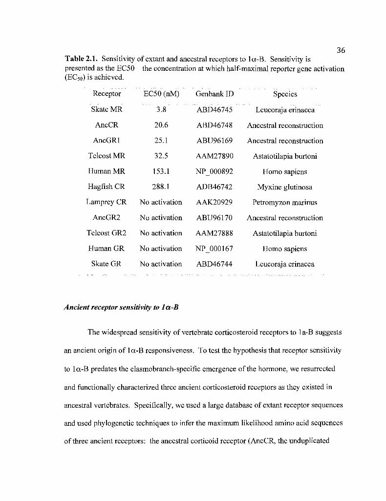
36Table 2.1. Sensitivity of extant and ancestral receptors to 1a-B. Sensitivity ispresented as the EC50 - the concentration at which half-maximal reporter gene activation(ECso) is achieved.
Receptor EC50 (nM) Genbank ID Species
Skate MR 3.8 ABD46745 Leucoraja erinacea
AncCR 20.6 ABD46748 Ancestral reconstruction
AncGR1 25.1 ABU96169 Ancestral reconstruction
Teleost MR 32.5 AAM27890 Astatotilapia burtoni
HumanMR 153.1 NP 000892 Homo sapiens
Hagfish CR 288.1 ADB46742 Myxine glutinosa
Lamprey CR No activation AAK20929 Petromyzon marinus
AncGR2 No activation ABU96170 Ancestral reconstruction
Teleost GR2 No activation AAM27888 Astatotilapia burtoni
HumanGR No activation NP 000167 Homo sapiens
Skate GR No activation ABD46744 Leucoraja erinacea
Ancient receptor sensitivity to 1a-B
The widespread sensitivity of vertebrate corticosteroid receptors to 1a-B suggests
an ancient origin of 1a-B responsiveness. To test the hypothesis that receptor sensitivity
to 1a-B predates the elasmobranch-specific emergence ofthe hormone, we resurrected
and functionally characterized three ancient corticosteroid receptors as they existed in
ancestral vertebrates. Specifically, we used a large database of extant receptor sequences
and used phylogenetic techniques to infer the maximum likelihood amino acid sequences
of three ancient receptors: the ancestral corticoid receptor (AncCR, the unduplicated

37ancestral gene from which extant MRs and GRs descend by gene duplication), GR in
the last common ancestor of all jawed vertebrates (AncGR1), and GR from the last
common ancestor of all bony vertebrates (AncGR2), after their split from cartilaginous
fishes (Fig. 4). We then synthesized DNAs coding for the ligand-binding domains of
these reconstructed proteins, expressed them, and characterized their functions using a
reporter transcription assay in cell culture (Bridgham et aI., 2006; Ortlund et aI., 2007).
As predicted, we found that the most ancient receptors -- AncCR and AncGRI -
are extremely sensitive to la-B, activating transcription with ECsos of ~20 nM.
AncGR2, in contrast, was unresponsive, as expected based on the lack of sensitivity to
la-B in its descendants, the GRs of tetrapods and teleosts (Fig. 2.4, Table 2.1). To
determine whether the la-B sensitivity of AncCR might be an artifact of uncertainty in
the inference of the ancestral sequence, we identified sites that were ambiguously
reconstructed (defined as having an alternative amino acid state with posterior probability
> 0.20). In all cases but five, the alternate state is found in other la-B-activated receptors
and is therefore not sufficient to abolish sensitivity to that hOl1none. Introducing each of
these five alternate states into the AncCR by site-directed mutagenesis had no effect on
ligand-activation (Table 2.2). Among sites that make contact with the ligand in the
AncCR crystal structure (Ortlund et aI., 2007), only one was ambiguously reconstructed;
introducing this alternate state into AncCR had no effect on sensitivity to la-B Crable
2.2). We conclude that AncCR's response to la-B is not an artifact of uncertainty in the
ancestral reconstruction.

To determine whether AncCR's sensitivity to la-B may be due to error in the
phylogeny on which the ancestral reconstruction is based, we inferred the maximum
likelihood sequence of AncCR on each of the 467 trees in the 95% credible set from a
large Bayesian analysis. At only one sequence site did the ancestral reconstructions
differ among trees. We introduced the alternate state at this site (A7V) into AncCR by
mutagenesis and found it had no effect on sensitivity to la-B (Table 2.2).
We conclude that AncCR and AncGRI were sensitive to la-B, and this ancient
sensitivity was retained in most ofthe lineages descending from those ancestors,
including the MR and GR of elasmobranchs. After the divergence of bony from
cartilaginous fishes, the GRs of bony vertebrates subsequently lost la-B sensitivity,
during the same period in which the receptor became cortisol-specific. This result
indicates that corticosteroid receptors were capable of being activated by la-B many
millions of years before synthesis of the hormone itself evolved in the elasmobranch
lineage.
38

39
4
3
2
Human CR
!eleost (jh~ ~]~~
15LL10 /:. ...~
c:oi:~lh/Iu. -11 -10 -9 -8 -7 -6 -5
Log [M]
MRs
Skate GR
Figure 2.4. Corticosteroid receptor sensitivity to la-B predates the evolution of la-Bsynthesis. Three ancestral receptors from early vertebrates (circled nodes) were"resurrected" by ancestral sequence reconstruction and gene synthesis. Graphs depictluciferase reporter activity in the presence of increasing la-B concentrations, relative tovehicle-only control. Dose-response curves for selected extant receptors are also shown.Black and gray circles denote ancestral receptors sensitive and insensitive, respectively tola-B. White rectangle shows loss of la-B activation; black rectangle, origin of la-Bsynthesis. AncCR represents the unduplicated ancestral gene from which GR and MRdescend; AncGRl is GR in the last common ancestor ofjawed vertebrates; AncGR2 isGR in the last common ancestor of bony vewrtebrates.

40Table 2.2. AncCR's sensitivity to la-B is robust to statistical and phylogeneticuncertainty. Alternate reconstructions of ancestral states were introduced into themaximum likelihood reconstruction of AncCR by site-directed mutagenesis, and theirsensitivity to la-B determined with a reporter gene assay.
Receptor EC50 (nMt
AncCR 20.6
AncCRA7V 23.3
AncCRA36G 3.0
AncCR S20T 10.5
AncCRK38R 12.3
AncCR S76A 4.5
AncCR Vl37A 9.8
AncCR V224A 8.1
Source of uncertaintyb
Phylogenetic
Stochastic - LBP
Stochastic - extant
Stochastic - extant
Stochastic - extant
Stochastic - extant
Stochastic - extant
Receptors insensitive tola-B with alternate stateC
PmaCR
Abu GR; Pma CR
HasGR
Hsa GR; Pma CR
PmaCR
Abu GR; Pma CR
a Sensitivity to 1a-B is reported as the concentration required for half-maximal activationof a luciferase reporter gene.
bphylogenetic uncertainty refers to maximum likelihood reconstructions that differamong trees in the 95% credible set. Stochastic uncertainty refers to non-optimalreconstructions with posterior probability> 0.2 on the maximum likelihood phylogeny.LBP, sites in the ligand binding pocket. Extant, alternate reconstruction is present in oneor more receptors insensitive to la-B.
CAbu, Astatotilapia burtoni; Hsa, Homo sapiens; Pma, Petromyzon marinus.
Structural Analysis of la-B Docked in the AncCR
To determine why ancient receptors were activated by la-B, we examined the
previously solved crystal structure of AncCR (Ortlund et aI., 2007). We hypothesized
that AncCR was structurally "preadapted" to bind la-B because of that hormone's
similarity to DOC, an ancient hormone that is the putative ancestral ligand for AncCR

41(Ortlund et aI., 2007). We generated a structural model of Ia-B docked into the
ligand-binding domain of the AncCR with DOC. Ia-B differs from DOC only by the
presence of hydroxy1groups at the C I and C II positions. The structure shows that
AncCR's ligand pocket contains ample room to accommodate the II-hydroxyl of Ia-B;
the receptor's previously identified ability to bind cortisol and aldosterone, which also
carry the II-hydroxyl, without conformational adjustment (Ortlund et aI., 2007). The
ligand pocket also contains unoccupied space in the alpha-plane above the C I carbon
(Fig. 2.5A); in the model of AncCR-with Ia-B, this space is occupied by and adequate to
accommodate the la-hydroxyl (Fig. 2.5B). A slight hydrophobic clash of this hydroxyl
with AncCR's Leu32 and Phe92, however, is the likely cause of the receptor's slightly
reduced sensitivity to Ia-B compared to other ligands that lack the la-hydroxyl. That
the receptor retains nanomolar sensitivity to Ia-B, however, indicates that minor
adjustments of the receptor backbone or side chain rotamers are sufficient to relieve the
clash.
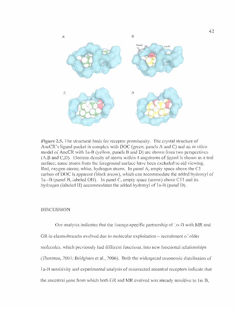
42B
c o
Figure 2.5. The structural basis [or receptor promiscuity. The crystal structure ofAncCR's ligand pocket in complex with DOC (green, panels A and C) and an i/1 si/icomodel of AncCR with 1a-8 (yellow, panels 8 and D) are shown from two perspectives(A,B and C,D). Electron density of atoms within 4 angstroms of ligand is shown as a tealsurface; some atoms from the foreground surface have been excluded to aid viewing.Red, oxygen atoms; white, hydrogen atoms. In panel A, empty space above the Clcarbon of DOC is apparent (black arrow), which can accommodate the added hydroxyl of1a -B (panel B, labeled OH). In panel C, empty space (arrow) abov C11 and itshydrogen (labeled H) accommodates the added hydroxyl of 1a-B (panel D).
DISCUSSION
Our analysis indicates that the lineage-specific partnership of 10.-8 with MR and
GR in elasmobranchs evolved due to molecular exploitation - recruitment of older
molecules, which previously had di fferent functions, into new functional relationships
(Thornton, 2001; Bridgham et aI., 2006). Both the widespread taxonomic distribution of
1a-B sensitivity and experimental analysis of resurrected ancestral receptors indicate that
the ancestral gene from which both GR and MR evolved was already sensitive to 1n-B,

- - -- --------
43millions of years before synthesis of the hormone itself evolved (Fig. 2.6). Sensitivity
to 1a-B has been retained to the present in numerous descendants of the AncCR,
including those in numerous species that lack 1a-B, such as mammals, in which
administration of exogenous 1a-B elicits a strong mineralocorticoid response (Idler et aI.,
1967). Our results indicate that the partnership of 1a-B with its receptors in
e1asmobranchs evolved when a newly synthesized hormone recruited pre-existing
receptors, which previously had different ligands, into new functional partnerships. This
evolutionary dynamic is similar to that previously observed for the interaction between
aldosterone and the MR of tetrapods (Bridgham et aI., 2006). Other steroid
hormone/receptor interactions evolved when intermediates in the biosynthesis of ligands
for more ancient receptors were recruited into partnerships with newly duplicated
receptors (Thornton, 2001; Thornton et aI., 2003). Several other apparent examples of
evolutionary recruitment of ancient molecules into new partnerships have recently been
described (Krasowski et aI., 2005; Cai et aI., 2007; Cardoso et aI., 2007), suggesting that
the evolution of specific molecular interactions by molecular exploitation may be a
dominant theme in the emergence of biological systems.

44ElasmobranchsTeleosts
.<:::::..~)~::: ..::::..~: :::.~~.;: ::.::: :: ~:):
. ......•. ....•....•./
.:""" . ....•.•.••. ••.•.. .,/ 1,,·B Synthesis
'. .."" .,/ Decreased GR Sensitivity............•......
.( .
. .'.'.
".....'.
Tetrapods., .':.:::: •• '> •• ~~~ ••••••••••••••••
Aldosterone Synthes;~"'" ............
...........
.........'.".
Increased GR Specificity··••Decreased GR Sensitivity···..
..•.•......•....
" ............
'.....'.".
".".
CR Activated by DOCPreadapted for 10-8 and Aldosterone
Figure 2.6. Receptor exploitation in the evolution of corticosteroid signaling. Thephylogeny traces the evolution of receptor genes and their ligand sensitivity (black andgray lines, sensitive and insensitive to la-B, respectively) within the divergence of majorvertebrate lineages (funnels). Closed circles at nodes on the phylogeny indicate ancestralreceptors that were resurrected and functionally characterized. Open boxes markfunctional shifts in the GR lineage. The origin of aldosterone and Ia-B synthesis isshown by black bars in the tetrapod and elasmobranch lineages, respectively.
Molecular exploitation is greatly facilitated by untapped promiscuity in ancient
proteins, which allows them to accommodate new partners that are minor variants of their
original binding partners. la-B is identical to the likely ancestral ligand DOC, except for
additional oxygen atoms at the C j and Cll positions. Our structural analysis of AncCR
indicates that the ancestral ligand pocket had ample room to accommodate these
modifications in la-B, when the hormone evolved later in the elasmobranchs. AncCR's
promiscuous response to steroids that had not yet evolved contrasts with its
unresponsiveness to the more ancient androgens, estrogens, or progestins, which differ
from the corticosteroids at other key locations on the steroid backbone (Bridgham et aI.,

452006). AncCR therefore appears to have been only as specific as it needed to be: it
excluded other endogenously produced hormones that would inappropriately activate the
receptor, but not other potential ligands whose synthesis had not yet evolved. When
steroids that fortuitously fit the pocket appeared later, this promiscuity set the stage for
the evolution of novel receptor-ligand partnerships with physiological or developmental
functions. Structural promiscuity - a ligand pocket that contains extra space or
flexibility, allowing to accommodate a range of ligands -- is a common protein feature
and is likely to have facilitated the evolution of new functions in both receptors and
enzymes (Brzozowski et aI., 1997; Bledsoe et aI., 2002; Pereira de Jesus-Tran et aI.,
2006; Nettles et aI., 2007; Suino-Powell et aI., 2008; James and Tawfik, 2001; Copley,
2003; Khersonsky et aI., 2006; Bloom et aI., 2007b).
Our results shed light on the functional roles of corticosteroids and their receptors
in elasmobranchs. The ligand binding domains of skate MR and GR differ little in
hormone specificity but radically in sensitivity: MR is activated by much lower
corticosteroid concentrations than are required to activate GR. Analysis of serum steroid
concentrations in a wide variety of elasmobranch species have indicated that 1a-B levels
are very high - up to several hundred nanomolar (Truscott and Idler, 1968; Kime, 1977)
- and that corticosterone, DOC, and II-DHC are present at orders of magnitude lower
concentrations (Idler and Truscott, 1967; Truscott and Idler, 1972; Kime, 1977; Hazon
and Henderson, 1984; Armour et aI., 1993; Rasmussen and Crow, 1993; Snelson et aI.,
1997). Our results indicate that skate MR has corticosteroid sensitivity similar to that of
tetrapod and teleost MRs (Bridgham et aI., 2006), whereas the skate GR's sensitivity is

46orders of magnitude lower. Although in vitro reporter assays using fusion proteins are
not precise predictors of in vivo dose-response relationships, physiologicalla-B levels, at
least under some conditions, are likely to be well beyond those necessary to activate skate
MR; it is possible that DOC and corticosterone may also serve as MR ligands. Skate GR,
in contrast, is likely to be activated only by elevated concentrations of la-B (and possibly
corticosterone) such as those that occur under conditions of stress (Manire et aI., 2007).
Our findings suggest the hypothesis that low corticosteroid levels regulate the MR to
control osmolarity, whereas stress-induced increases in the same hormones regulate the
GR to control the stress response. Like the elasmobranch GR, the GR of bony
vertebrates is a low-sensitivity receptor that activates the stress response only when
cortisol levels are very high (Bentley, 1998); unlike the elasmobranch GRs, however,
GRs in bony vertebrates evolved novel specificity as well, so that they are insensitive to
even high doses of mineralocorticoids (Bridgham et aI., 2006). Other factors, such as
post-translational modification and the availability of coregulators, may also contribute to
differences in GR and and MR function.
MR and GR are ubiquitously coexpressed, at roughly equal proportions, in a wide
variety of skate tissues. This is largely consistent with patterns of expression found in
teleosts (Bury et aI., 2003; Greenwood et aI., 2003; Sturm et aI., 2005) and tetrapods (de
Castro et aI., 1996; Le Menuet et aI., 2000). These data provide no evidence for
subfunctionalization of expression domains as important in the maintenance ofthe GR
and MR after duplication. We cannot rule out the possibility, however, that GR and MR

47may be differentially expressed on a finer scale, in specific cell types or groups of cells
or at specific stages.
The interaction of corticosteroid receptors, their ligands, and the DNA response
elements they regulate represent a relatively simple but physiologically essential
molecular network. Gene duplication provides raw material for the genesis and
expansion of such networks (Wagner, 1994; Teichmann and Babu, 2004), but duplication
alone is not enough to make a network more elaborate or generate a new one. To
increase complexity, functional interactions must diversify, either by the evolution of
new interactions or the partitioning of ancestral functions among new network nodes
generated by duplication. We have shown that the corticosteroid hormone/receptor
network was elaborated when receptor gene duplication and divergence, together with the
extension of a biosynthetic pathway, produced new receptors and hormones that were
slight structural and functional modifications oftheir ancestral forms. The receptors'
promiscuity set the stage for these new hormones to be integrated into the existing
receptor signaling network. Subsequently, divergence of the two sister receptors
leading to reduced sensitivity in the GR - resulted in markedly different quantitative
sensitivities to corticosteroid hormones and the possibility of distinct network responses
to varying levels of hormone. These observations indicate that the complex molecular
networks that drive physiological process can evolve by mechanisms as simple as
molecular exploitation and the partial degradation of function after gene duplication.
48BRIDGE TO CHAPTER III
These results lead to the question of how the degradation of GR functions
evolved. Because the skate GR is the only known receptor of its kind, reduced sensitivity
could have arisen within the cartilaginous fishes or in the divergence of GR after gene
duplication. The latter offers an intriguing hypothesis - that GR functions might have
evolved by high-frequency degenerative mutations after gene duplication. The GR
lineage could have begun the 'road to nonfunctionalization,' but was preserved when
degenerative mutations created a low sensitivity receptor that was recruited for novel
signaling roles. Chapter III seeks to test this hypothesis and reconstruct the changes in
GR sequence, structure, and function after duplication of AncCR. Using a combination of
ancestral gene resurrection, site-directed mutagenesis, and X-ray crystallography, I found
that the first GR ancestor was in fact a low sensitivity receptor. Reduced sensitivity
evolved by a number of mutations that destabilize the receptor structure, but are buffered
by an additional change that increases global receptor stability. These results suggest that
the neofunctionalization of GR was driven by high-probability degenerative mutations
that created a novel function through the partial degradation of ancestral structures.

49
CHAPTER III
DEGENERATIVE AND STABILIZING MUTATIONS IN
THE ORIGIN OF THE GLUCOCORTICOID RECEPTOR
This work is in preparation for publication and is co-authored with Eric A.
Ortlund, who assisted with experiments and experimental design, and Joseph W.
Thornton, who assisted with experimental design and edited the manuscript.
INTRODUCTION
Gene duplication provides the raw genetic material upon which new protein
functions can evolve. Ohno theorized that after gene duplication, one copy is freed from
purifYing selection and allowed to accumulate previously "forbidden" mutations that, in
very rare instances, lead to novel gene functions (Ohno, 1970). In most cases, however,
these mutations are degenerative, resulting in a loss of function, and the duplicate gene is
lost (Demuth and Hahn, 2009). Degenerative mutations can sometimes playa
constructive role in duplicate gene preservation if the gene contains multiple, independent
subfunctions. In the duplication-degeneration-complementation (DDC) model (Force et
aI., 1999), the various subfunctions of a single ancestral gene are partitioned between

50duplicate loci by complementary degenerative mutations; duplicates are preserved
because each is necessary to recapitulate the full complement of ancestral subfunctions.
This process, however, merely allots ancestral gene functions between daughter genes
and does not directly result in the evolution of novel functions. Recent work by Bridgham
et al. (Bridgham et aI., 2008) suggests that highly probable loss-of-function mutations
can in fact be the mechanism for neofunctionalization. In that case, degenerative
mutations in one duplicate copy knocked out a single modular protein subfunction and
created a novel competitive repressor of its paralog, suggesting that degenerative
evolution after gene duplication can be a creative force in and of itself, significantly
increasing the likelihood ofneofunctionalization by potentially loss-of-function
mutations. However, the prevalence of this process in the evolution of duplicate genes
and gene functions is not yet known.
We hypothesized that degenerative mutational processes may have played a
creative role in the preservation and divergence ofthe glucocorticoid receptor. The
glucocorticoid receptor (GR) functions as a ligand-activated transcription factor, relaying
the signal of a far-off secreted hormone to the local regulation of gene expression in
target tissues (Beato et aI., 1995). In humans, the GR is activated by cortisol to regulate
facets of immunity, metabolism, and the stress response (Bentley, 1998). GR and its
paralog the mineralocorticoid receptor (MR) arose by the duplication of a single ancestral
receptor over 440 million years ago (Thornton, 2001; Bridgham et aI., 2006). Using
methods in the phylogenetic reconstruction of ancestral proteins (Thornton et aI., 2003),
our group previously demonstrated that the last common GR/MR ancestor functioned

51much like extant MRs - highly sensitive to a variety of corticosteroid hormones - and
that GR functions are derived. After duplication, GRs evolved two major changes in their
response to ligand: 1) reduced sensitivity to all hormones, as is present in all known GRs,
and on top of this, 2) cortisol-specific activation, as is present in teleost and tetrapod
receptors (Bridgham et aI., 2006). The latter event is well studied: the historical
mutations, their effects on protein structure-function, and ancient mutational trajectories
have all been elucidated in the evolution of cortisol specificity (Bridgham et aI., 2006;
Ortlund et aI., 2007). The loss of GR sensitivity, however, has been largely unexplored
and could reflect the partial degeneration of ancestral receptor structures and functions.
The purpose of this study was to determine precisely when and how reduced GR
sensitivity evolved. Because all known GRs have reduced sensitivity, we hypothesized
that the very first GR also functioned as a low sensitivity receptor, and sought to test this
hypothesis using ancestral state reconstruction and gene resurrection (Thornton, 2004).
The protein sequence of the first GR ancestor - AncGRl.l - was reconstructed and
"resurrected," its sensitivity to hormone was assessed using a reporter gene assay, and its
protein structure was solved using X-ray crystallography. We show that after gene
duplication, the GR lineage evolved by the partial degradation of a highly sensitive
ancestor to become a novel low sensitivity receptor for corticosteroid signaling.

52METHODS
Receptor Isolation.
The little skate (Leucoraja erinacea) GR ligand-binding domain (LBD) was
isolated previously using degenerate PCR and RACE with liver cDNA (Bridgham et aI.,
2006). The skate GR protein sequence was used in a tblastn search of the elephant shark
genome (http://esharkgenome.imcb.a-star.edu.sg/) to identify the GR LBD; gene-specific
primers were designed to amplify the elephant shark GR LBD coding sequence. All other
Chondrichthyan GR LBDs were isolated by hemi-degenerate PCR from cDNA using a
degenerate primer in the GR DNA-binding domain (DBD) in combination with a gene
specific primer for a ~ 25 bp sequence conserved in the 3' -UTR of the elephant shark and
skate (5'-TCATATGCACTACATATGGTTTACAGA-3'). High-fidelity PCR was used
to amplify GR LBDs from five cartilaginous fish species: elephant shark (Callorhincus
milii), Atlantic sharpnose shark (Rhizoprionodon terraenovae), brownbanded
bambooshark (Chiloscyllium punctatum), small-spotted catshark (Scyliorhinus canicula),
and Atlantic stingray (Dasyatis sabina). Template cDNA for PCR was graciously
provided by B. Venkatesh (c. milii and C. punctatum) and S. Nunez (R. terraenovae, S.
canicula, and D. sabina).
Phylogenetic Analysis.
The conserved DNA- and ligand-binding domains of 100 steroid receptor protein
sequences were aligned using Clustal X (Larkin et aI., 2007). Maximum likelihood

53phylogenetics was performed using PhyML_aLRT (Anisimova and Gascuel, 2006)
assuming the Jones model of evolution (Jones et aI., 1992) and a four-category discrete
gamma distribution of among-site rate variation, with the shape parameter estimated from
the data; this model was previously shown to be highly supported, with 100% posterior
probability, when this and other models are compared in a Bayesian analysis (Bridgham
et aI., 2006). Support at nodes was calculated as the approximate likelihood ratio (LR) of
the maximum likelihood tree versus the next best tree that does not contain that node
(Anisimova and Gascuel, 2006).
Ancestral Sequence Reconstruction and Gene Resurrection.
The maximum likelihood tree topology differed from previously published SR
phylogenies (Bridgham et aI., 2006) with respect to the placement ofjawless fish
receptors; to account for this discrepancy in trees, ancestral receptor sequences were
reconstructed over both the experimental and published (Bridgham et aI., 2006) trees
weighted by their probability. Ancestral states were infened using the Ancestral
Reconstruction Tool [Victor Hanson-Smith, unpublished] given the above alignment
matrix and the JTT model. For any ancestor relevant to our study, no site in the inferred
sequence possessed amino acid states that differed between trees. A nucleic acid
sequence coding for the LBD of the last common ancestor of all GRs (AncGRl.l) and
optimized for expression in mammalian cells was synthesized de novo (Genscript,
Piscataway, NJ) and characterized as described below.

S4
Tests ofEvolutionary Rates.
Rates of protein evolution were analyzed in HyPhy (Pond et aI., 2005). A
likelihood ratio test (LRT) was used to compare the relative branch lengths from the last
common GR/MR ancestor (AncCR) to the last common ancestors of all GRs (AncGRI.I)
or MRs (AncMR1). The likelihood was calculated given the alignment, maximum
likelihood tree, and the JTT model. The alternate and null hypotheses of the LRT
optimized branch lengths given the full (unconstrained) likelihood model or one in which
the MR and GR branches post-gene duplication were constrained to have the same
length. The likelihood ratio of alternate and null models was determined and a p-value
calculated using a chi-squared distribution with one degree of freedom.
Receptor Characterization.
LBDs were cloned as fusion proteins into a pSG5-GaI4DBD expression vector
(gift ofD. Furlow) and cotransfected using Lipofectamine and Plus Reagents (Invitrogen,
Carlsbad, CA) with a UAS-driven luciferase reporter gene (pFRluc) into mammalian cell
culture (CHO-Kl) and grown in phenol red-free u-MEM plus 10% dextran-charcoal
stripped fetal bovine serum (Hyclone, Logan, UT). Cells were incubated with transfection
reagents for four hours, after which they were treated with fresh medium; after recovery,
cells were treated in triplicate with hormone or vehicle control, and incubated overnight.
Reporter expression was measured using Dual-Glo (Promega, Madison, WI) and dose-

55response relationships analyzed using Prism4 (GraphPad, La Jolla, CA). Site-directed
mutagenesis was carried out using QuickChange II (Stratagene, La Jolla, CA) and clones
verified by DNA sequencing.
Protein Growth, Purification, and Crystallography.
AncGRl.l was subcloned into apMCSG7-MBP-His expression vector,
transformed into BL2l (DE3) pLysS cells, and grown to an OD600 of 0.8-1.0. Cultures
were induced with 0.1 uM IPTG plus 50 uM of the steroid II-deoxycorticosterone
(DOC) and grown overnight at 16° C. Purification of AncGR1.l was performed using
nickel affinity chromatography and a sizing column. Pure AncGRl.l was concentrated to
3.7 mg/mL and dialyzed into a crystallization buffer consisting of20 mM Tris, pH 6.5,
150 mM NaCl, 5% glycerol, 50 uM CHAPS, and 50 uM hormone (11
deoxycorticosterone).
Multiple sparse matrix screens were set with AncGRl.l protein using a Phoenix
crystallization robot (Art Robbins Instruments, SUlmyvale, CA); hits formed at 22° C
from the Salt Rx screen (Hampton Research, Aliso Viejo, CA). Crystals were optimized
at 22° C in hanging drop diffusion plates with: 2.5-2.8 M sodium acetate trihydrate, pH
7.0,0.1 M BIS-TRIS propane, pH 7.0, and a small peptide designed from the TIF2 Box3
steroid receptor coactivator protein. Crystals were soaked in a cryoprotectant solution
containing 20% glycerol and flash-frozen in liquid nitrogen. Data was collected to 1.95 A
resolution at the South East Regional Collaborative Access Team (SER-CAT) at the

56Advanced Photon Source (Argonne National Laboratory). Initial phasing of the
AncGR1.l plus DOC structure was determined using molecular replacement of the
AncCR with DOC (2Q3V); refinement of the structure was carried out using COOT
version 0.5 (Emsley and Cowtan, 2004) and CCP4i (http://www.ccp4.ac.uk/pjb.html).
The root mean square deviation (rmsd), a measure of the overall similarity between
protein backbones, was calculated using CaspR (Claude et aI., 2004).
RESULTS
GR Pltylogenetics, Evolutionary Rate Variation, and ReconstructedAncestral States
To increase taxon sampling for ancestral reconstruction we isolated additional
GRs sampled from throughout the cartilaginous fishes. The GR phylogeny and nodes at
which ancestral receptor sequences were reconstructed are shown in Fig. 3.1. This
phylogeny was used to test for differences in evolutionary rates after duplication and to
reconstruct ancestral receptor states.
To determine whether novel GR functions evolved by increased rates of protein
evolution after gene duplication, we sought to compare the relative branch lengths of the
GR and MR lineages stemming from the AncCR. The classic model of
neofunctionalization (0000, 1970) predicts that the duplicate lineage acquiring a novel
function should evolve quickly with the rapid fixation of beneficial mutations, while the
ancestral functions are maintained by purifying selection in the paralogous copy and this
lineage continues to evolve slowly. For phylogenies such as ours with thorough taxon

57sampling, we can test for differences in the rate of duplicate gene evolution on '
branches that cover the same amount oftime. We utilized a likelihood-based test to
assess the relative branch lengths of the GR and MR lineages from the duplication of
AncCR to the divergence of cartilaginous fishes, so that any differences in branch length
would reflect unequal rates of substitution after gene duplication. The AncCR:AncGR1.1
branch is noticeably longer (0.123) than the corresponding AncCR:AncMRl branch
(0.041) (Fig. 3.1), but we found only a marginally non-significant difference between the
rates of divergence after gene duplication (p = 0.06).
r1~CM~R~1"-========~=======J194 MlI1eralocorticoid receptors (MRs)01)·1"1
IAncGRUI
0123
527 X 1()'"
AncGR2C~ e.95 X 10'rh
Elephant shark
9,67 X 10d Teleost GRs
Tetrapod Gr-{s
2,14 X 10"
1,20
Small-spotted catshark
6,98
Atlantic sharpnose shark291 x10'"
Brownbanded bambooshark
Atlantic stingray
oro,ro
<0S'oCtil
iii::rGJ:;0til
0.2 63,5
Little skate
Figure 3.1. Schematic of the GR clade from the maximum likelihood phylogeny. Thelikelihood ratio and the names of relevant ancestral sequences are shown at major nodes;branch lengths following the duplication of AncCR are also displayed. AncCR, the lastcommon ancestor of all MRs and GRs; AncGR1.1, the GR ancestor of cartilaginous andbony fishes; AncGR2, the GR ancestor of ray- and lobe-finned fishes; AncMR1, the MRancestor of cartilaginous and bony fishes. Black, high sensitivity receptors; gray, lowsensitivity receptors; black dash, the evolution of reduced sensitivity. TheAncCR:AncMRl and AncCR:AncGR1.1 branch lengths were analyzed using alikelihood-based test of relative rates.
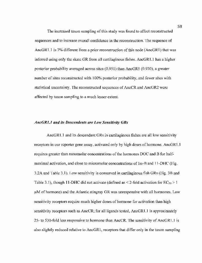
58The increased taxon sampling of this study was found to affect reconstructed
sequences and to increase overall confidence in the reconstruction. The sequence of
AncGR1.1 is 7% different from a prior reconstruction of this node (AncGR1) that was
inferred using only the skate GR from all cartilaginous fishes. AncGR1.l has a higher
posterior probability averaged across sites (0.951) than AncGRI (0.930), a greater
number of sites reconstructed with 100% posterior probability, and fewer sites with
statistical unceliainty. The reconstructed sequences of AncCR and AncGR2 were
affected by taxon sampling to a much lesser extent.
AncGRl.l and its Descendents are Low Sensitivity GRs
AncGR1.l and its descendant GRs in cartilaginous fishes are all low sensitivity
receptors in our reporter gene assay, activated only by high doses of hormone. AncGR1.1
requires greater than nanomolar concentrations of the hormones DOC and B for half
maximal activation, and close to micromolar concentrations of la-B and II-DHC (Fig.
3.2A and Table 3.1). Low sensitivity is conserved in cartilaginous fish GRs (Fig. 3B and
Table 3.1), though II-DHC did not activate (defined as < 2-fold activation for ECso > 1
!AM of hormone) and the Atlantic stingray GR was unresponsive with all hormones. Low
sensitivity receptors require much higher doses of hormone for activation than high
sensitivity receptors such as AncCR; for all ligands tested, AncGR1.l is approximately
25- to 530-fold less responsive to hormone than AncCR. The sensitivity of AncGR1.l is
also slightly reduced relative to AncGR1, receptors that differ only in the taxon sampling

59used for ancestral reconstruction. The low sensitivity of AncGRl.l strongly suggests
that a reduced response to hormone evolved soon after the duplication ofAncCR.
A
'ISAncGR1.1
.2 DOCro 10-, • !l()
lltHli;<,<
'D,\ \1 1 UC i
~
ll-+-O-...............""'I"'".........,......""""........""'I"'".............."""'"-! 2 11 iO ·9 -L ·7 ·6 ·S
Log [tvl]
[]
·5
.(; 'I
I'I I2' ·7
I !,~ 0 • I •u
UJ !' •en..Q 1<;
!- ~ ~I
Figure 3.2. AncGR 1.1 and its dcscendent GRs in cartilaginous fishes are low sensitivityreceptors. A) Ligand-dependent transcriptional activation of the AncGR 1.1 was measuredin the presence of increasing concentrations of hormone using a luciferase reporter gencassay. Points are the me8n of triplic8te rcactions with the standard error of the me8n. Fourhormones were tested: II-deoxycorticosterone (J )OC), corticosterone (13), 11dehydrocorticosterone (11-DI-I C), and 1(X-hydroxycorticosterone (1 a.-B). B) High andlow sensitivity receptors. Dosc«response curves were calculated for a number of receptorhormone pairs and plotted as the log effective concentration for half maximal activation(log EC50) in molar with standard error. Larger values, Jow sensitivity; smaller values,high sensitivity. Hormones are the same as in (A); l1-DIIe did not activate cartilaginousfish GRs in our assay (defined as < 2··fold activation for EC';(J > I ~lM of hormone) and isnot shown. Receptors are grouped by reconstructed ancestors or extant cartilaginous fishGRs. AncGR 1 is a prior reconstruction of AncGR 1.1; cartilaginolls fish species are listedTable 3.1.

60Table 3.1. Sensitivity of extant and ancestral receptors to hormone. Dose-responsecurves were calculated for receptors in the presence of hormone; the data is plotted as thelog ECso plus standard error.
DOC B Il-DHC la-B
Receptor log ECso SE log ECso SE log ECso SE log ECso SE
AncCR -10.870 0.34 -10.410 0.29 -7.315 0.17 -7.992 0.16
AncGRl -9.182 0.20 -8.944 0.15 -7.304 0.12 -7.735 0.19
AncGRI.1 -8.144 0.20 -8.151 0.16 -5.924 0.14 -6.232 0.09
CmilGR -7.761 0.25 -8.196 0.28 NA -6.438 0.16
Rter GR -7.623 0.12 -7.912 0.16 NA -6.433 0.12
Cpun GR -6.143 0.16 -7.320 0.21 NA -6.552 0.18
Scan GR -6.702 0.24 -7.322 0.17 NA -5.767 0.16
Leri GR -6.515 0.21 -7.548 0.28 NA -5.700 0.13
Dsab GR NA NA NA NA
a AncGR1, prior reconstruction of AncGR1.l; Cmil, elephant shark (Callorhincus milii);Rter, Atlantic sharpnose shark (Rhizoprionodon terraenovae); Cpun, brownbandedbambooshark (Chiloscyllium punctatum); Scan, small-spotted catshark (Scyliorhinuscanicula); Leri, little skate (Leucoraja erinacea); Dsab, Atlantic stingray (Dasyatissabina).
b NA, no activation, defined as < 2-fold activation for ECso > 1 f!M of hormone
The molecular basis ofdecreased hormone sensitivity
To dissect the mechanism of reduced hormone sensitivity we examined the
historical substitutions that occurred on the branch from AncCR to AncGR1.l. Out of 36
changes on this branch, we chose a set of candidate substitutions that were hypothesized
to cause the reduced hormone sensitivity of AncGR1.1. Seventeen of these substitutions
were already tested in AncGRI and did not reduce receptor sensitivity, leaving 19
remaining candidates. Of these, only 7 cause a radical change in amino acid properties

61and are therefore predicted to strongly affect receptor function. To test whether any of
these 7 candidate substitutions recapitulate the loss of GR sensitivity, we introduced each
historical change into the maximum likelihood (ML) AncCR background using site
directed mutagenesis and tested the effect on hormone sensitivity in the reporter gene
assay with increasing concentrations of DOC.
Of seven candidate substitutions, two markedly reduced AncCR's sensitivity to
hormone. The V43A and Rl16H single mutants reduced the EC50 of DOC by over two
orders of magnitude to near AncGR1.1-like values (Fig. 3.3 and Table 3.2). Other
mutations caused slight reductions in AncCR sensitivity (Table 3.2), but the V43A and
Rl16H substitutions are clearly those with the greatest effect. When combined, V43A
and Rl16H produce a nearly unresponsive receptor that required over 105 times the dose
of hormone required to activate the ML AncCR, a value orders of magnitude higher than
required to activate AncGR1.1 or any of the extant GRs. Both V43A and R116H
occurred on the branch to AncGR1.1, suggesting that additional substitutions buffer the
loss of sensitivity conferred by these mutations.

62
-(i
~ -7
U -8,-..,'-'0
-9I/~
Uw.J -10:lfJ,..'-'
- I ]
-I ~
Figure 3.3. Three historical substitutions are sufficient to reduce AncCR sensitivity.Shown is the log ECso of molar II-deoxycorticosterone (DOC) with a high sensitivityreceptor (AncCR), a low sensitivity receptor (AncGR1.1), and single or combinationAncCR mutants. The V43A and Rl16H substitutions were identified from candidatesites; C71 S was identified from the structure of AncGR1.1.
Table 3.2. Two historical GR substitutions markedly reduce AncCR sensitivity.Candidate substitutions were introduced into AncCR by site-directed mutagenesis andtheir effects on receptor sensitivity measured as the change in log ECso of molar 11deoxycorticosterone (DOC) from the maximum likelihood (ML) reconstruction ofAncCR.
Receptor log ECso SE
AncCRML -10.870 0.34
LIM -10.250 0.30
V43A -8.443 0.15
K83Q -10.330 0.41
A107S -10.350 0.24
Ql13K -9.773 0.43
Change inSensitivity
(Fold)
- 4.2
- 267.3
- 3.5
- 3.3
- 12.5
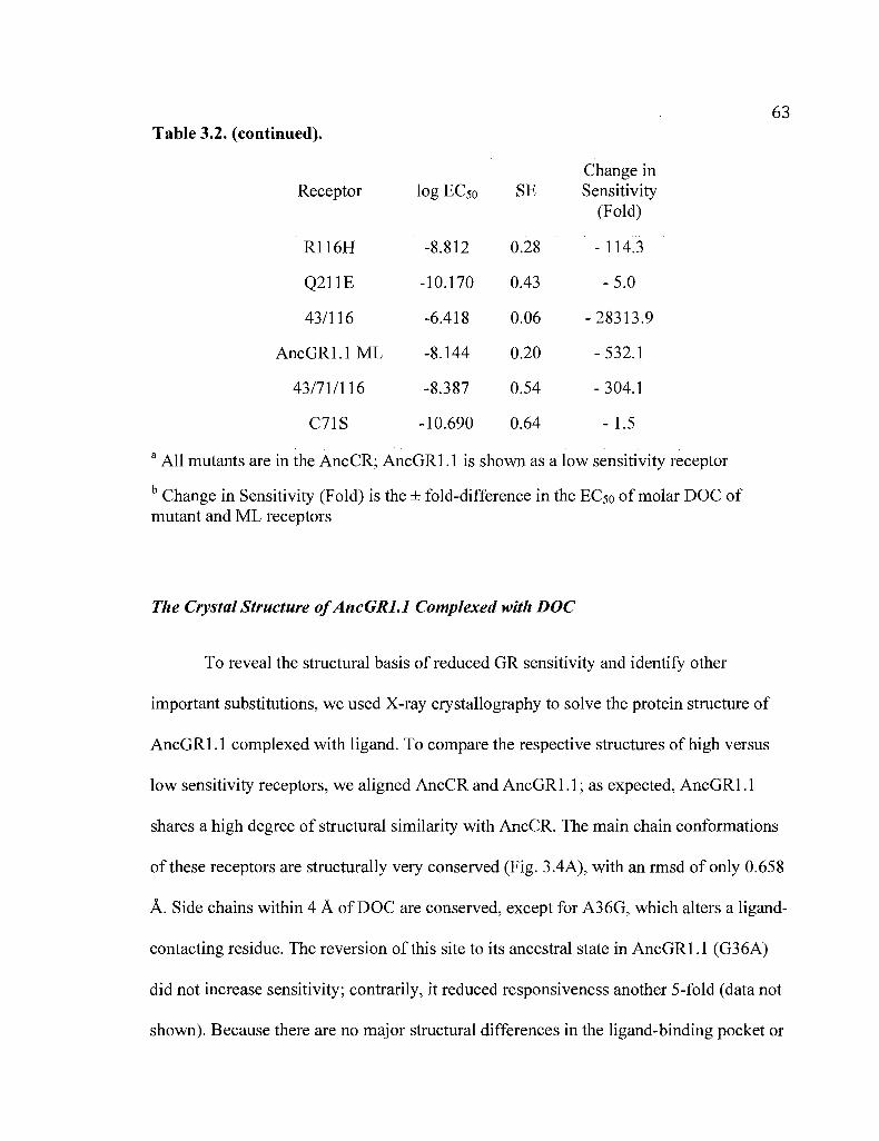
63Table 3.2. (continued).
Change inReceptor log ECso SE Sensitivity
(Fold)
Rl16H -8.812 0.28 - 114.3
Q211E -10.170 0.43 - 5.0
43/116 -6.418 0.06 - 28313.9
AncGR1.1 ML -8.144 0.20 - 532.1
43/71/116 -8.387 0.54 - 304.1
C71S -10.690 0.64 - 1.5
a All mutants are in the AncCR; AncGR1.1 is shown as a low sensitivity receptor
b Change in Sensitivity (Fold) is the ± fold-difference in the ECso of molar DOC ofmutant and ML receptors
The Crystal Structure 0/AncGRJ.l Complexed with DOC
To reveal the structural basis of reduced GR sensitivity and identifY other
important substitutions, we used X-ray crystallography to solve the protein structure of
AncGR1.1 complexed with ligand. To compare the respective structures of high versus
low sensitivity receptors, we aligned AncCR and AncGR1.1; as expected, AncGR1.1
shares a high degree of structural similarity with AncCR. The main chain conformations
ofthese receptors are structurally very conserved (Fig. 3.4A), with an rmsd of only 0.658
A. Side chains within 4 A of DOC are conserved, except for A36G, which alters a ligand-
contacting residue. The reversion of this site to its ancestral state in AncGR1.1 (G36A)
did not increase sensitivity; contrarily, it reduced responsiveness another 5-fold (data not
shown). Because there are no major structural differences in the ligand-binding pocket or

64the protein backbone, we focused on evolutionary changes in protein structure induced
by the site 43 and 116 substitutions to reveal the mechanism of decreased sensitivity.
/\
c
,.
A
Figure 3.4. The solved crystal structure of AncGRl.l in complex with 11deoxycorticosterone (DOC) compared to the previously solved structure of AncCR withDOC (2Q3Y). A) AncCR and AncGR 1.1 are highly structurally conserved despite 36substitutions between the two receptors. Gray, AncCR; yellow, AncGR1.I; spheres,sequence differences; magenta sphere, position 43; cyan sphere, position 116; green,DOC; AF-I-I, activation function helix. B) and C) show the structural environment aroundposition 43. The substitution of V43 in AncCR (B) for A43 in AncGRl.l (C) is thoughtto weaken the hydrophobic interactions of the surrounding helices.
NJlltflliollS tl1(1l Redllce Sellsitivity Call.')e a Loss (~rFavomble llllemcliolls
Structural analysis of the V43A substitution suggested a loss of hydrophobic
interactions from AncCR to AncGRI.I. V43 in AncCR (Fig. 3.4B) helps to stabilize
helix 3 (H3) against I-IS and the ligand. The V43A substitution results in a smaller side
chain and weaker hydrophobic interactions in the AncGR 1.1 structure (Fig. 3.4C). The

65loss of contacts with V43A is likely to destabilize the packing ofH3 against the
receptor and affect ligand contacting residues and/or the hydrophobic cleft. The
hydrophobic cleft is an important coactivator docking site on the receptor surface formed
at the interface ofH3, H5, and the activation function helix (H12 or AF-H) (Feng et aI.,
1998); these coactivators bridge the receptor-ligand complex to the transcriptional
machinery and are necessary for receptor function. Destabilizing mutations around the
cleft would likely decrease coactivator binding and weaken ligand-dependent
transcriptional activation.
Position 116 is antipodal to 43 on the receptor structure and a hub in an extensive
and stabilizing hydrogen bond network. In AncCR, Rl16 is part of a large network ofh
bonds running from the C-terminalloop ofH5 through the inner-facing side chains ofH7
and into H6 (Fig. 3.5A). This network is greatly reduced in AncGRl.l due to the Rl16H
substitution and possibly another substitution at the neighboring site 113 (Fig. 3.5B).
Hl16 has a much-reduced ability to form hydrogen bonds, and could be repelled by the
like positively charged side chain ofKl13; the latter site forms a novel salt bridge to
E100, further rearranging the ancestral h-bond network. Indeed, Ql13K was identified in
the original group of seven candidate substitutions from AncCR to AncGRl.1 and had
the third greatest effect on sensitivity after the changes at sites 43 and 116 (Table 3.2).

66/\ B
1<113
Figure 3.5. AncGR 1.1 lost hydrogen bonds in the region of position 116. A) R 116 inAncCR is part of an extensive and stabilizing hydrogen bond network. B) This h-bondnetwork is greatly reduced in AncGRl.l with an R 116H substitution and a neighboringQ] 13K substitution that rearranges the ancestral pattern of h-bonds.
The loss of hydrogen bonds due to substitutions at sites 116 and 113, as well as
the loss of hydrophobic interactions at position 43, suggests that the novel function of
AncGRI.l, its reduced sensitivity, evolved because of the partial degeneration of
ancestral structures and functions. Our results indicated that other substitutions build
favorable interactions in the AncGR] .1 to buffer mutations at sites 43 and 116; we
returned to the AncGRl.1 structure to identify these sites.
e7lS Add,,, Core Molecular B01lding amI Suppresses Desensitizing Nfl/lations
By analyzing tbe positions of AncCl :AncGRl.l changes on the protein structure,
C71 S emerged as a candidate historical substitution for new favorable interactions in
AncGR 1.1. C71 S creates in AncGR 1.1 a new water-mediated hydrogen bond network

67with L139 and TI43 that stabilizes the receptor core (Fig. 3.6); C71 in AncCR cannot
form this stl"Ucture. We introduced S71 into AncCR and found that this mutation
(AncCR_C71 S) has absolutely no effect on receptor sensitivity (Fig. 3.3 and Table 3.2).
In combination with the destabilizing mutations (AncCR_V43A, C71 S, R 116H), C71 S
recapitulates the evolution of AncGR 1. J sensitivity by buffering the effects of mutations
at sites 43 and J 16.
l139143
.,- ..
571
Figul'e 3.6. A C7I S substitution from AncCR to AncGR 1.1 adds favorable hydrogenbonds and buffers the effects of other mutations. S7I creates a novel water-mediatedhydrogen bond network with L 139 and T143, potentially stabilizing the core oftheAncGRl.1 structure.
Statisticalullcertaillfy ill J'ecollstl'llctetl AllcGRJ. J sites
To determine whether reduced sensitivity is all artifact of chance errors in the
reconstruction, we assessed plausible alternate reconstructions of AncGRl.1 sites. We
examined those sites that are most likely to change AncGR 1.1 function: a) those shown

68in this study to be important for receptor sensitivity, and b) those positions with
plausible alternate states (PP > 0.20) that are present in one or more high sensitivity
receptors and therefore might increase receptor sensitivity. Alternate reconstructions were
introduced into AncGR1.1 using mutagenesis and receptor sensitivity was measured as
the response to DOC.
None of the alternate reconstructions in the AncGR1.1 sequence undermine the
conclusion that the ancestral GR was a low-sensitivity receptor. All substitutions have
virtually no effect on receptor sensitivity (Table 3.3), except for H116Q. This alternate
reconstruction produces a modest (7-fold) increase in the response of AncGR1.l to DOC
but is still approximately 80-fold less responsive to hormone than AncCR. Further, the
structural basis for reduced sensitivity does not change with the alternate reconstruction:
both H 116 and Q 116 would result in a loss of hydrogen bonds from the highly sensitive
AncCR state (RI16). These results indicate that the reduced sensitivity of AncGR1.l is
robust to uncertainty in the sequence reconstruction, as is the biophysical mechanism that
caused reduced sensitivity

69Table 3.3. Statistical uncertainty in the reconstruction of functionally importantAncGRl.l sites. Plausible alternate reconstructions of AncGRl.l were introduced intothe maximum likelihood (ML) sequence and assayed for their effects on the EC50 ofmolar II-deoxycorticosterone (DOC). Sites are those identified from the reducedsensitivity of AncCR or from plausible alternate AncGR1.1 states (defined as > 0.20 PPwith the state present in high sensitivity receptors).
Change inReceptor PP (Alt) log EC50 SE Sensitivity
(Fold)
AncGR1.1 ML - -8.144 0.20
T25S 0.27 -8.245 0.32 + 1.3
A43V* 0.45 -8.041 0.17 - 1.3
EII0D 0.30 -8.196 0.25 + 1.1
K113Q* 0.28 -7.993 0.21 - 1.4
H116Q* 0.28 -8.972 0.22 + 6.7
T214S 0.24 -8.159 0.18 + 1.0
T214A 0.22 -8.137 0.28 - 1.0
• Statistical uncertainty in sites important for sensitivity.
a All mutants are in the AncCR; AncGRl.l is shown as a low sensitivity receptor
b Change in Sensitivity (Fold) is the ± fold-difference in the EC50ofmolar DOC ofmutant and ML receptors
C PP (Alt) is the posterior probability of plausible alternate states
DISCUSSION
The Evolution ofGR Functions by Gene Duplication
Our work suggests that GR arose by the partial degradation of structures and
functions present in a highly sensitive ancestor. After duplication of the ancestral

70corticoid receptor, mutations in the GR lineage resulted in the loss of hydrophobic
interactions, hydrogen bonds, and ligand-binding residues. The degradation of these
structures created in the GR ancestor a reduced sensitivity to hormone, where up to two
orders of magnitude more ligand is needed for transcriptional activation. Several single
mutations are sufficient to recapitulate the reduction of hormone sensitivity after gene
duplication, but their combined effects are too much and result in a largely nonfunctional
receptor. Receptor function is maintained by an additional substitution that increases core
receptor stability and buffers the degenerative effects of other mutations. Additional
substitutions then optimized the functions of the GR ancestor as a low sensitivity
corticosteroid receptor.
The common ancestor of all jawed vertebrates, in which AncGR1.1 existed,
possessed MR and GR that functioned similarly to the respective high and low sensitivity
receptors of cartilaginous fishes. In these animals, MR is activated by baseline
corticosteroid levels, while GR is activated only by the highest physiological doses of
these same hormones (Carroll et aI., 2008). Our results demonstrate that the last common
ancestor of all GRs was a low sensitivity receptor, and that reduced sensitivity is
conserved in the GRs of cartilaginous fishes. The other descendents of AncGR1.1 - the
GRs ofteleosts and tetrapods - acquired increased specificity to cortisol (Bridgham et aI.,
2006). Other innovations in corticosteroid-receptor signaling have evolved in multiple
different lineages (Bury and Sturm, 2007; Prunet et aI., 2006; Keightley et aI., 1998).

71The Structural Basis ofDecreased Receptor Sensitivity
After duplication of AncCR, a combination of potentially stabilizing and
destabilizing mutations generated the reduced hormone sensitivity of AncGR1.1.
Thermodynamic stability is a global determinant of protein function and is in itself an
evolvable character. Periods of random mutation accumulation can quickly degrade
protein structures and, past a certain stability threshold, cause the protein to unfold and
lose activity (Bershtein et ai., 2006; Bloom et ai., 2005; Bloom and Arnold, 2009; Bloom
et ai., 2007). Conversely, stabilizing mutations are thought to buffer the protein beyond
the stability threshold and allow a greater number of otherwise deleterious mutations to
accrue (ShortIe and Lin, 1985; Mitraki et ai., 1991; Huang and Palzkill, 1997; Bershtein
and Tawfik, 2008a). These so~called "global suppressor" mutations (ShortIe and Lin,
1985) might facilitate the preservation of duplicate genes. Periods of relaxed selection
and random mutation accumulation are an important part of evolution by
neofunctionalization but most often result in nonfunctionalization and gene loss (0000,
1970). Suppressor mutations would allow a greater number of mutations to accrue than
would otherwise be tolerable, effectively increasing the likelihood of
neofunctionalization and decreasing the chances of nonfunctionalization. Here, multiple
destabilizing mutations were buffered by the added stability of C71 S to generate a novel
low sensitivity receptor that was integrated into corticosteroid signaling pathways. Given
the complexity of GR functions and its modular protein domain architecture, it is
unknown whether the GR locus was preserved after duplication by mutations conferring
decreased sensitivity, or mutations to other protein domains.

72Site 71 was first identified as a determinant of protein stability in the
recombinant expression of human GR (Bledsoe et aI., 2002); in fact, all crystal structures
of corticosteroid receptors with agonist bound have had to engineer a serine at site 71 to
express soluble protein (Kauppi et aI., 2003; Bledsoe et al., 2005; Li et al., 2005; Ortlund
et al., 2007). We found that this exact experiment was performed over 440 MYA, where
a C71 S substitution increased the stability of AncGR1.1. Given its neutrality in AncCR,
the C71 S substitution may have been a chance permissive mutation for decreased
sensitivity (Ortlund et al., 2007) or arisen later in GR evolution as a compensatory
mutation for the loss of stability caused by other degenerative mutations (Depristo et al.,
2005).
Accuracy ofAncestral State Reconstructions
This study displays the power of ancestral sequence reconstruction for
mechanistic studies of molecular evolution, but our analyses demonstrate that measures
should be taken to assess the accuracy of inferred ancestral states. We found that the
choice or depth of taxon sampling can affect the identity and statistical support of
reconstructed sequences. Reconstructions at the same node - AncGR1 and AncGR1.1
have slightly different sequences and functions due only to the number of cartilaginous
fish GRs used in the analysis. Opportunities to increase taxon sampling and break up long
phylogenetic branches should increase the accuracy of maximum likelihood
reconstructions, as has been found in reconstructions by parsimony (Salisbury and Kim,
2001). Equally important is an assessment of the quality of the inferred sequence. Three

73of four sites important to the decreased sensitivity of GRs are ambiguously
reconstructed in AncGR1.1; fortunately, alternate states at these sites have little effect on
receptor sensitivity. Nonetheless, alternate reconstructions could potentially alter the
function of reconstructed ancestral states in other model systems and skew the
evolutionary inferences drawn from these results. Finally, we show that evolutionary
changes to protein structure can allow substitutions that are predicted by the likelihood
model to be low probability mutations.
New Functions can arise by tlte Partial Degeneration 0/Ancestral Functions
Our study highlights a creative role for partialloss-of-function mutations in the
evolution of gene functions. This is similar to the process described by Bridgham et aI,
(2008), where after gene duplication, complete loss-of-function mutations in one modular
protein domain of one duplicate can create a competitive repressor of the fully functional
paralog. The degradation of GR, however, presents a slightly different mechanism in the
partial loss of activity within a modular function - the receptor ligand-binding domain
that created a novel differential response to hormone levels, Steroid signaling relies on
very precise molecular cues, and even slight changes in receptor sensitivity could have
noticeable effects on the biological response (Simons, 2006). After duplication of
AncCR, the MR ligand-binding domain retained ancestral sensitivity, whereas the
evolution ofreduced sensitivity created a novel regulator of steroid signaling in the GR
lineage, These examples demonstrate how mutations that impair native protein functions
can drive the evolution of novel functional roles.

74
BRIDGE TO CHAPTER IV
Despite the wealth of knowledge on steroid receptor evolution, very little is
known about the origins of the hormones themselves. It is thought that corticosteroids
arose by the duplication of enzymes involved in the synthesis of more ancient hormones
such as sex steroids, but a rigorous test of this hypothesis has yet to be done. In Chapter
IV, I reconstruct both the timing and the mechanism of the gene duplication that gave rise
to the Cyp21 enzyme that drives corticosteroid synthesis. Using genomic analyses,
molecular phylogenetics, and analyses of conserved synteny, I show that the gene
duplication that gave rise to Cyp21 occurred prior to the divergence of vertebrates, and by
a complex set of mutational events.
75
CHAPTER IV
THE EVOLUTION OF CYP21 BY GENE DUPLICATION
AND CHROMOSOMAL RELOCATION
This work is in preparation for publication and is co-authored with Joseph W.
Thornton, who assisted with experimental design and edited the manuscript.
INTRODUCTION
Corticosteroids are critical regulators of vertebrate immunity, glucose
metabolism, and the stress response (Bentley, 1998). Corticosteroids are produced in the
adrenal gland, released into the bloodstream, and received by the mineralocorticoid and
glucocorticoid receptors, ligand-activated transcription factors that are expressed in target
cells (Beato, 1989). The chemical synthesis of corticosteroids is driven by the 21
hydroxylase activity of the CYP21 enzyme, and mutations to this gene cause human
disease in the form of congenital adrenal hyperplasia (White and Speiser, 2000). Despite
their fundamental importance, very little is known about the evolutionary origins of
corticosteroid and the CYP21 enzyme.

76A reasonable hypothesis is that corticosteroids evolved by elaborating upon
preexistent steroidogenic pathways. The synthesis of corticosteroids and other steroid
hormones - including sex steroids such as progestins, androgens, and estrogens - is
accomplished by successive enzymatic modifications to a cholesterol precursor.
Progestins are the first products of steroid synthesis, after which the pathway bifurcates to
produce either corticosteroids or, alternatively, androgens and finally estrogens (Fig. 4.1).
The synthesis of sex steroids is quite ancient (Thornton, 2001; Thornton et aI., 2003;
Keay and Thornton, 2009), and corticosteroids are thought to be a recent evolutionary
offshoot of this pathway. Both CYP21 and the enzyme CYP17 share certain progestin
hormones (progesterone and 17-hydroxyprogesterone) as substrates for the synthesis of
corticosteroids or sex steroids, respectively. It has been proposed that Cyp21 arose by the
duplication of a Cyp17-like ancestor that functioned in the synthesis of sex steroids, but a
rigorous test of this hypothesis has yet to be done.

77
rtleo tel I
CYP21 ]
)i I I-Dcoxycorl sol170H·Proge- INane
GYP17 I!Andr ton diane
Progestins §]Progesterone ., -11.DcoxYCOnlcosterone
CYP171 ~
17 H·Progn nol rip
Prcgnollolon'
CYP17 ~
GYP17I!DII.A
Androgens EstrogensFigure 4.1. - The steroid synthesis pathway and major classes of hormone. Arrowsindicate the conversion of one steroid to anothcr by enzymes (boxed). Those reactionspertaining to CYP21 and CYP 17 are shown in bold.
Studies of steroid biology in basal vertebrates and their closcst outgroups offcr
insights into Cyp21 evo!ution. Genomic and molecular analyses havc shown that the
steroidogenic pathway is largely conscrved in amphioxus, a close chordate outgroup to
vertebrates, minus C'yp2/ and other enzymes in cOlticosteroid synthesis (Castro el aI.,
2005; Mizuta et aI., 200~: Mizuta and Kubokawa, 2007). Amphioxus possesses a single
C);p/7-like gene (MizLlta and Kubokawa, 2007) but its true homology to Cyp17 has been
questioned (Markov ct aI., 2009) and its enzymatic functions have yet to be characterized.
In another phylogenetically informative taxa, our lab has shown that lampreys - the most
basal vertebrate ingroup - possess a single steroid receptor that is co-orthologous to the
mineralocorticoid (MR) and glucocorticoid (GR) receptors of higher vertebrates and
specific for corticosteroids (Thornton, 2001; Bridgham et aI., 2006). The in Fiva and in

78vitro conversion of labeled steroid precursors to corticosteroids has been observed in
these same animals (Weisbart and Youson, 1975; Weisbart and Youson, 1977; Weisbart
et aI., 1978). These data suggest that lamprey diverged after the Cyp17/Cyp21 duplication
and should possess copies of both genes, whereas basal craniates such as amphioxus
diverged before the duplication and should have a single, co-orthologous copy of
Cyp17/Cyp21.
To elucidate the timing of the Cyp17/Cyp21 duplication, we sought to determine
what Cyps are present in the amphioxus and sea lamprey and to clearly discem their
phylogenetic relationships. In addition, we sought to determine the molecular mechanism
of gene duplication using analyses of syntenic conservation between the human CYP17
and CYP21 genomic loci. Our results show that a Cyp17/Cyp21 ancestor duplicated just
prior to the evolution of vertebrates by a complex set of mutational events.
METHODS
Isolation and Analysis ofCyp Sequences
The sequence of the published amphioxus (B. belcheri) Cyp17-like gene (Mizuta
and Kubokawa, 2007) was used in a blastn search of the B. floridae genome
(http://genome.jgi-psf.org/BraflllBrafll.home.html) (Putnam et aI., 2008). A predicted
cDNA encoding Cyp17 (BRAFLDRAFT_121141) was downloaded along with over 50
kb of the genomic scaffold (BeV2_122) on which BRAFLDRAFT_121141 was
predicted. Primers were designed to the predicted start and stop codons using Geneious

79vA.7A (Biomatters LTD, Auckland, NZ). Amphioxus Cyp17 was amplified from
ovary eDNA using high-fidelity PCR and cloned into the pCR2.! sequencing vector
(Invitrogen, Carlsbad, CA).
Lamprey Cyps were identified in the 5.9x preliminary assembly of the P. marinus
genome (http://pre.ensembl.org/index.html) using a tblastn search of related Cyp17 and
Cyp21 sequences. Sequences with the reciprocal best hits against the Genbank nr
database were downloaded as the entire genome contig from the genome assembly.
Contigs with hits to Cyp17 were PMAR3:Contig38214:1 :11273:1 and
PMAR3 :Contig9569: 1:15185: 1; those with hits to Cyp21 were
PMAR3:Contig36955:1:9414:1 and PMAR3:Contig5425:1:27435:1. The assembly of
contigs and identification of putative start and stop codons were computed in Geneious.
Lamprey Cyp17 and Cyp21 were PCR amplified from ovary eDNA and cloned into the
pCR2.1 sequencing vector.
Cyp exon/intron boundaries were determined by aligning PCR-amplified eDNA
sequences with the corresponding genomic contig or scaffold. The identification of
transposable elements and low complexity DNA sequences was determined manually by
blastx searchers of non-coding DNA, and by RepeatMasker
(http://www.repeatmasker.org). Protein alignments and the presentation of sequence
motifs was performed in Geneious.

80Molecular Phylogenetics
The lamprey and amphioxus sequences were added to a large dataset of related
Cyps with Cypla and Cyplb genes as the outgroup. Sequences were aligned using
MUSCLE (Edgar, 2004). ProtTest2.1 (Abascal et aI., 2005) was used to select the best
evolutionary model for phylogenetics according to the Akaike Information Criterion
framework (Posada and Buckley, 2004). Maximum likelihood phylogenetics was
performed in PhyML_aLRT (Guindon and Gascuel, 2003; Anisimova and Gascuel, 2006)
using the selected JTT model (Jones et aI., 1992) assuming an estimated proportion of
invariant sites plus a four-category gamma distribution with alpha estimated from the
data. Statistical confidence at nodes was calculated as the "i statistic or I-p, where p is
the estimated probability that the likelihood ratio support of a given node would occur by
chance if that node is actually false (Anisimova and Gascuel, 2006).
Analyses ofChromosomal Location and Conserved Gene Order
Chromosome synteny - conservation of gene content and order - was identified
using the Synteny Database (Catchen et aI., 2009). Searches were conducted using the
human, chicken, and amphioxus genomes with a window size of 100 genes. Briefly, this
method uses reciprocal best hits from blast to identify paralogous and orthologous genes
and their positions, and subsequently uses a sliding window to discover conserved gene
order in nearby chromosomal regions. The Synteny Database was also used to examine

81the reciprocal best hits of predicted genes on the amphioxus scaffold containing Cyp17
(BeV2_122) with their putative orthologs in humans.
RESULTS
Identification ofAmphioxus and Lamprey Cyps
A single gene similar to Cyp17 was identified in amphioxus, and two genes
similar to Cyp17 and Cyp21 were found in lamprey. We compared the predicted protein
sequences of these Cyps to those of human and eel and found that there is considerable
sequence divergence between enzymes (Table 4.1). All sequences have very low amino
acid similarity, but the amphioxus and lamprey Cyp17 genes are most similar to the eel
Cyp17, and lamprey Cyp21 is most similar to eel Cyp21. All Cyp sequences show greater
similarity in previously documented conserved regions, including the Ono-sequence (Ono
et aI., 1988), Ozols' tridecapeptide sequence (Ozols et aI., 1981), and the heme binding
site (data not shown).
Table 4.1. Percent of sequence identity between amphioxus, lamprey, zebrafish, andhuman Cyps. The percent of identical states between alignable sites are shown.
Amphioxus Lamprey Lamprey
Cyp17 Cyp17 Cyp21
Amphioxus Cyp17 - 37 32
Lamprey Cyp17 37 - 37
Eel Cyp17 39 52 36
Human Cyp17 34 42 32

82Table 4.1. (continued).
Amphioxus Lamprey Lamprey
Cyp17 Cyp17 Cyp2l
Lamprey Cyp2l 32 37
Eel Cyp2l 38 34 40
HumanCyp2l 31 33 34
Maximum Likelihood Phylogenetics ofCyp Protein Sequences
The maximum likelihood phylogeny of the amphioxus, lamprey, and related Cyps
sequences places the timing of an ancient Cyp17/Cyp21 gene duplication. Lamprey
Cyp17 and Cyp21 are orthologous to their respective clades, and the single amphioxus
Cyp17-like gene is co-orthologous to these genes. These relationships are strongly
supported in the analysis: the placements oflamprey Cyp17 and Cyp21, the Cyp17/Cyp21
duplication, and the co-orthologous gene of amphioxus are all supported by a X2 statistic
of 1.00. Despite high support at most nodes, there are several instances where the
topology does not match the accepted species phylogeny, reflecting the rapid evolution of
Cyp sequences in certain lineages (ie. mammalian Cypl7). Nonetheless, these results
strongly suggest that: a) amphioxus has a single gene co-orthologous to Cyp17 and
Cyp21, b) lamprey has orthologs of both enzymes, and c) the duplication that gave rise to
the vertebrates Cyp17 and Cyp21 lineages occurred between the divergence of amphioxus
and lamprey.

83
Zcllrafish
r_-([===~Z('hrali.'\h
'-------1 1':°\1"",""Uolphin
OX7 I)O!!lbt
Mouse
..,...,.,.-I[C=-l~~i~:'ICh:lCk'--- Zcur;lli:.h
(j'<"0N
..,...----l'l QllnilCllic\{rl\
L...---TllrllrFro~DyhovsJ<y',<; Fro~
'-------Ilogfisli
')\1 II 0 r:-Ol' Shrl'p
11111111:1111 Chillq)
I H:dlOOIlManllust'j
'-----------Llll1ll'rc) (),,17
'-----------1 1I1111)l'l'~ ()p21'------------[C~n. helcheri Cypl7
'",phi""" (B.n"r;,!.,,·, (\,,17
0.4
Figure 4.2. Maximum likelihood phy logeny of Cyp 17 and Cyp2I proteins. Lamprey andamphioxus Cyps are marked in bold red. The Cyp 17/Cyp2I gene duplication is markedwith a box. Statistical accuracy at nodes was calculated as the l statistic or 1-1', where pis the estimated probability that the likelihood ratio support of a giv n node would occurby chance if that node is actually false (Anisimova and Gascuel, 2006).
Tile Molecular Nlecllallism ofthe Cyp17/Cyp21 Duplicflti011
To determine whether CypJ 7 and C)lp2} were generated by whole-genome
duplication, we performed an automated search (or syntenic conservation between the
human chromosomes on which eyp17 and CYP2) reside using the Synteny Database
(Catchen et aI., 2009). Analyses of synteny - homologous genes conserved in duplicated

84chromosomal regions - are useful for identifying large-scale duplication events such
as whole-genome duplication. If generated by this mechanism, we expected to find
conserved paralogous synteny between the genomic neighborhoods of the human CYP17
and CYP21 genes. Instead, we found a different pattern synteny that suggests Cyp21
arose by a more complex set of mutational mechanisms. In the synteny analysis, human
CYP17 lies on chromosome 10 (position 10q24.3) and form a four-fold paralogous
cluster with regions of chromosomes 2,5, and 4 (Fig. 4.3A). The presence of four-fold
paralogous clusters suggests that these regions were generated by two rounds of large
scale duplications from a single ancestral chromosome. CYP21 lies on chromosome 6
(position 6p21.3) and forms a different tetra-paralogon with regions of chromosomes 1,
9, and 19 (Fig. 4.3B). Together, these non-overlapping four-fold paralogous clusters
suggest that one or both of Cyp17 and Cyp21 relocated in the course of their evolution,
moving into regions that were generated by two rounds of whole-genome duplication.
Whether the Cyp17/Cyp21 duplication was the result of duplicative transposition (and
transfer) or whole-genome duplication followed by transposition is unknown.

85
A
2 \
5 ----------\~r___--\
' .. '."
Hsap Cyp17
-4
10 - .1-\-- _
B
19
9
6
Figure 4.3. Conserved synteny in human chromosomal regions flanking Cyp17 andCyp21. Shown arc the paralogons in which human Cyp 17 (A) and Cyp21 (B) reside,though syntenic clusters were only identificd by neighboring genes. I-Iumanchromosomes are shown as darker and thicker horizontal lines; connecting gray linesshow reciprocal best hits of paralogous human genes. The approximate positions ofCyp17 and Cyp21 are indicated. Red lines identify the relationships of four-foldparalogous genes: NFKB2 in (A) and RGL2 in (B).
86
To determine which paralog relocated after the Cyp17/Cyp21 duplication, we
sought to trace each tetra-paralogous chromosomal cluster in humans back to a single co
orthologous region in amphioxus using the Synteny Database (Catchen et aI., 2009) and
previously published synteny analyses. The CYP21 cluster includes many loci in the
major histocompatibility complex (MHC) and is one of the most well-studied examples
of four-fold synteny, homologous to a single chromosomal segment in amphioxus (Castro
et aI., 2004; Abi-Rached et aI., 2002). The amphioxus Cyp17/Cyp21 co-ortholog is not
present in this region, indicating that Cyp21 relocated after genome duplication. The
history of the Cyp17 locus is less clear, as we were unable to trace the human CYP17
cluster back to a single homologous region in amphioxus. The genomic neighborhood of
the amphioxus co-ortholog was shown previously to have only weak synteny with human
chromosomes 10,8,3, and X (Putnam et aI., 2008). Collectively, these results do not
allow us to discern the location of the ancestral Cyp17/Cyp21 locus, and it is unclear
exactly how many times Cyps have relocated in the course of their evolution.
DISCUSSION
Corticosteroid Signaling Evolvedfrom Preexistent Estrogen Signaling Pathways
Our work sheds light on the timing and mechanism of the gene duplication that
gave rise to Cyp21 and corticosteroid synthesis. We identified orthologs of Cyp I7 and
Cyp21 in the sea lamprey and, in contrast to other analyses (Mizuta and Kubokawa,

872007; Markov et aI., 2009), a single gene that is co-orthologous to Cyp17 and Cyp21
in an amphioxus (B. floridae). This suggests that corticosteroids arose in vertebrates by
the duplication of enzymes that previously functioned in the synthesis of sex steroids.
Given these results, we can reconstruct the timing of when corticosteroids and
their receptors evolved. Amphioxus possesses only the receptors and hormones for
estrogen signaling (Mizuta et aI., 2008; Mizuta and Kubokawa, 2007; Bridgham et aI.,
2008); some time between the divergence of amphioxus and lamprey, primitive
vertebrates evolved corticosteroid-sensitive receptors (Thornton, 2001) and the Cyp21
enzyme necessary for corticosteroid synthesis. The order by which corticosteroids and
their receptors evolved - either receptor-first or hormone-first - is not known; prior work,
however, suggests that both are plausible scenarios for the evolution of novel receptor
hormone interactions ( Thornton, 2001; Bridgham et aI., 2006; Carroll et aI., 2008).
There is some debate as to the antiquity of estrogen synthesis and the ancestral
pathway from which the synthesis of corticosteroids arose. Work from our lab and others
strongly suggests that estrogens and their receptors are quite ancient (Thornton, 2001;
Keay and Thornton, 2009; Garcia-Alonso et aI., 2006; D'Aniello et aI., 1996; Di Cosmo
et aI., 2001; Di Cosmo et aI., 2002), while others have suggested that these hormones
evolved independently in vertebrate and invertebrate lineages (Markov et aI., 2008;
Markov et aI., 2009). This latter hypothesis, however, is questionable due to inferences
made from weakly supported Cyp phylogenies. Due to the high rates of sequence
divergence between Cyp homologs, the validity of Cyp phylogenetic analyses should be
carefully assessed. This study found very strong support for clades within the vertebrate

88Cyp17 and Cyp21 lineages that are incongruent with the established species
phylogeny; similar false relationships are likely to form with greater phylogenetic
distances. Because of these inherent difficulties, we believe that a combination of
analyses - including molecular phylogenetics, genomic analyses, and molecular assays
are necessary for robust inferences of Cyp and steroid honnone evolution.
The Mechanism ofthe Cyp17/Cyp21 Duplication
There are four common molecular mechanisms of gene duplication, including:
whole-genome duplication (polyploidization), duplicative transposition (non-allelic
homologous recombination and non-homologous end joining), unequal crossing over
(tandem duplication), and retrotransposition (Hahn, 2009; Hastings et aI., 2009; Paques
and Haber, 1999). The least likely mechanism for the Cyp17/Cyp21 duplication is
retroposition, as this model would require that the duplicate Cyp locus regained all of its
intronic sequences after duplication. Cyp17 and Cyp21 may have evolved by tandem
duplication, but this requires two separate mutational events (tandem followed by
duplicative transposition) and there is no additional evidence supporting tandem
duplication.
Several lines of evidence suggested that the duplication creating Cyp17 and
Cyp21 was part of a large-scale and possibly whole-genome duplication event. First, the
timing of the duplication - between amphioxus and lamprey - is consistent with
hypotheses of polyploidization in vertebrate genome evolution. Almost forty years ago,

89Ohno proposed that several rounds of whole-genome duplication occurred in some
vertebrate ancestor(s) (Ohno, 1970); today, the "2R" hypothesis (Hughes, 1999) is
generally accepted as one round of whole-genome duplication between amphioxus and
lamprey, and another round after lamprey but before the split ofjawed vertebrates
(Holland et aI., 1994). Second is that steroid receptors have expanded by en bloc
duplications that are associated with the 2R. Amphioxus has two steroid receptor genes,
lamprey has three (one copy was lost after duplication), and jawed vertebrates such as
humans have six (Thornton, 2001; Bridgham et aI., 2008; Bridgham et aI., 2006); genome
mapping analyses show that at least four of the human steroid receptors are syntenic with
one another and evolved by the twice-duplication of a common ancestral chromosome
(Thornton, 2001). Our synteny analyses clearly demonstrate that the chromosomes on
which CYP17 and CYP21 reside are the products of two rounds of whole-genome
duplication; the fact that these clusters are non-overlapping, however, means that one or
more Cyps have translocated in the course of their evolution and we cannot discern
whether it was a whole-genome duplication that specifically gave rise to Cyp17 and
Cyp21. Nevertheless, the ancestral Cyp17/Cyp21 locus was undoubtedly duplicated in
this whole-genome duplication event, but the products of this duplication may have been
lost in the course of evolution and the event that gave rise to Cyp21 may have been
driven by other molecular mechanisms.
Duplicative transposition is an alternative mechanism for the Cyp17/Cyp21
duplication. We found many instances of transposon insertions (data not shown) in the
genomic sequences of amphioxus and lamprey Cyps, which are known to facilitate

90recombination and duplicative transposition (Gray, 2000). The amphioxus Cyp17
locus actually has an inverted, partial tandem duplicate that most likely evolved by non
allelic homologous recombination of flanking DNA transposon sequences. These
transposable elements can induce chromosomal duplications and rearrangements through
many different mechanisms and are enriched at the boundaries of segmental duplications
in humans (Bailey et aI., 2003) and flies (Fiston-Lavier et aI., 2007). Transposon
mediated recombination could have duplicated the Cyp17/Cyp21 ancestor and moved
these genes to their current chromosomal locations in one mutational event. It is also
possible that Cyp21 arose by whole-genome duplication followed by duplicative
transposition and the loss of the ancestral Cyp21 locus. Because evolutionary divergence
prevents us from "anchoring" the ancestral Cyp locus, the exact mechanism by which
Cyp21 evolved is unknown. Improved annotations of the amphioxus, lamprey, and other
phylogenetically informative genomes would improve synteny analyses and help to
delineate the mechanism of duplication.
Interestingly, the process of recombination associated with duplicative
transposition persists at the human CYP21 locus. Over 95% ofCYP21 disease state
alleles are the result of recombination between the functionallocus and a nearby
pseudogene (CYP21P) that shares 98% sequence identity with its functional paralog
(White et aI., 1986; Higashi et aI., 1986). Recombination causes deletions and gene
conversions in CYP21, whereby nonfunctional mutations from the pseudogenic copy are
recombined into the functional locus. The CYP21 locus is particularly prone to these
events because of its position in the MHC class III cluster, a region in which high rates of

91recombination are maintained by balancing selection to generate allelic diversity in
immune effector molecules (Klitz et aI., 1986; Takahata et aI., 1992). Thus, similar
mutational processes of recombination might underlie the origins of Cyp21 and the
generation of disease state alleles.
In addition to facilitating gene duplication, transposable elements could have
played a role in the origins of 21-hydroxylase activities in Cyp21. Both amphioxus and
lamprey Cyps have acquired independent retroelement insertions in their intronic
sequences, as well as variable exonlintron boundaries between genes (data not shown).
Transposon insertions are normally deleterious to gene function, but they can sometimes
serve as the substrate for novel coding sequence in a process called "exon recruitment"
(Nekrutenko and Li, 2001; Kazazian, 2004). Retroposons can also contain their own
splice sites and result in novel altematively spliced transcripts (Makalowski et aI., 1994),
or copy and paste exons to new genomic locations (Moran et aI., 1999). Mutational
processes associated with the insertion of transposable elements might have equally ifnot
more important than sequence substitutions to the divergence of Cyp21 from a Cyp17
like ancestor. Functional analysis and characterization of sites at exonlintron boundaries
would help to explore this hypothesis.
The Novelty ofCorticosteroid Signaling
Corticosteroid signaling is a critical regulator of vertebrate physiology and a
vertebrate-specific innovation. The players necessary for corticosteroid synthesis evolved
92prior to the divergence of lamprey, as did the corticosteroid-sensitive receptors
(Thornton, 2001), arising from preexistent components of estrogen signaling. Even the
adrenal gland - the organ in which corticosteroids are produced - shares a common
developmental origin with the gonads (Hatano et aI., 1996), and adrenal-like cells persist
in testicular tissue (Val et aI., 2006). In the evolution of Cyp21, seemingly "novel"
enzymatic functions arose by redirecting the hydroxylase activity of Cyp17 to a new atom
on the same steroid substrates (progesterone and 17-hydroxyprogesterone). The distance
between the C17 and C21 carbons on which the respective activities of Cyp17 and Cyp21
are carried out is a mere 2.6 A. This shows how new protein functions, and in fact entire
pathways of endocrine signaling, can arise by subtle changes in protein structure and
function.

93
CHAPTER V
CONCLUSIONS
Gene and whole-genome duplications provide the raw material for molecular
innovation. My dissertation describes the mechanisms and dynamics by which gene
duplication and divergence gave rise to corticosteroid signaling in vertebrates.
Corticosteroid hormones and their receptors are an ideal system in which to study how
molecular diversity and complexity evolve. A combination of techniques from
evolutionary biology and traditional molecular analyses permit highly mechanistic
studies of the ways in which novel hormones, receptors, and their interactions arise. My
work shows that seemingly complex facets of corticosteroid evolution can evolve by
simple processes that elaborate upon preexistent molecular functions.
Much like the exploitation of promiscuous enzyme activities, the MR and GR of
the skate evolved lineage-specific functions from the promiscuous activities of their
ancestor. The skate MR and GR were preadapted for signaling with la-B due to the
structural promiscuity of AncCR. The sensitivity of AncCR to la-B is a byproduct of
activation towards structurally similar, more ancient ligands. The AncCR is not
necessarily a nonspecific or a generalist receptor; it is only as specific as it needs to be to
activate with corticosteroids but does not distinguish much among them. After

94duplication of AncCR and the synthesis of 1a-B in elasmobranchs, MR and GR were
recruited for lineage-specific signaling functions. Thus, 1a-B signaling evolved as the
fortuitous byproduct of structures adapted for preexistent functions.
Similarly, "novel" functions arose in the GR lineage by the partial degradation of
ancestral structures and functions. I used ancestral sequence reconstruction and gene
resurrection to show that the first GR ancestor functioned as a low sensitivity receptor,
much like the little skate GR. After gene duplication, the GR lineage accrued mutations
that reduced the stability and sensitivity of the receptor through the loss of hydrophobic
interactions, hydrogen bonds, and direct ligand contacts. This loss, however, was
buffered by an accompanying substitution to increase core receptor stability. Reduced
sensitivity in GR created an added level of complexity in corticosteroid signaling - a low
sensitivity receptor with a response distinct from that of MR or ancestral receptors. Other
gain-of-function mutations likely occurred in other GR domains or non-coding regions,
but its "novel" low sensitivity response to hormone arose by degenerative mutations after
gene duplication.
Finally, I show that corticosteroid hormones evolved by gene duplication and
elaborations upon existing steroid synthesis pathways. The duplication giving rise to the
Cyp17 and Cyp21 lineages occurred just prior to the divergence of lamprey, making
corticosteroids a vertebrate innovation. The timing of this duplication coincides with a
round of whole-genome duplication proposed by the 2R hypothesis (Holland et ai.,
1994), but analyses of gene synteny suggest that an array of mutational mechanisms led
to the duplication and translocation of the Cyp21 locus. Additional genomic and

95molecular analyses of Cyps in basal vertebrates will help to elucidate the origins of
corticosteroid synthesis.
The origin of corticosteroids and their receptors presents a model system by
. which novel protein functions arise by gene duplication. This system arose largely by the
exploitation of preexistent forms: structural and functional promiscuity of the AncCR, the
degenerative evolution of ancestral forms and functions in GR, and the elaboration of
preexistent steroid synthesis pathways for the production of corticosteroids. The
evolutionary processes underlying the origins of novel hormones, receptors, and
interactions are not unlike those observed in other historical studies of evolution, or in
protein engineering experiments in the lab. These are mechanisms by which protein
functions evolve.

96
REFERENCES
Abascal, F., Zardoya, R., and Posada, D. (2005). ProtTest: selection of best-fit models ofprotein evolution. Bioinfonnatics 21, 2104-2105.
Abi-Rached, L., Gilles, A., Shiina, T., Pontarotti, P., and Inoko, H. (2002). Evidence ofen bloc duplication in vertebrate genomes. Nat Genet 31,100-105.
Amitai, G., Gupta, R., and Tawfik, D. S. (2007). Latent evolutionary potentials under theneutral mutational drift of an enzyme. HFSP J. 1, 67-78.
Anfinsen, C. (1973). Principles that govern the folding of protein chains. Science 181,223-230.
Anfinsen, C. B. (1959). The Molecular Basis of Evolution (New York: Wiley).
Anisimova, M., and Gascuel, O. (2006). Approximate likelihood-ratio test for branches:A fast, accurate, and powerful alternative. Syst BioI 55, 539-552.
Annour, K. J., O'Toole, L. B., and Hazon, N. (1993). The effect of dietary proteinrestriction on the secretory dynamics of 1 alpha-hydroxycorticosterone and ureain the dogfish, Scyliorhinus canicula: a possible role for 1 alphahydroxycorticosterone in sodium retention. J Endocrinol138, 275-282.
Arnold, F. H., Wintrode, P. L., Miyazaki, K., and Gershenson, A. (2001). How enzymesadapt: lessons from directed evolution. Trends Biochem Sci 26, 100-106.

97
Bailey, J. A., Liu, G., and Eichler, E. E. (2003). An Alu transposition model for the originand expansion of human segmental duplications. Am J Hum Genet 73, 823-834.
Baker, M. E., Chandsawangbhuwana, C., and Ollikainen, N. (2007). Structural analysisof the evolution of steroid specificity in the mineralocorticoid and glucocorticoidreceptors. BMC Evol BioI 7, 24.
Barabasi, A. L., and Oltvai, Z. N. (2004). Network biology: understanding the cell'sfunctional organization. Nat Rev Genet 5, 101-113.
Beato, M. (1989). Gene regulation by steroid hormones. Cell 56, 335-344.
Beato, M., Herrlich, P., and Schutz, G. (1995). Steroid hormone receptors: many actors insearch ofaplot. Cell 83, 851-857.
Bentley, P. J. (1998). Comparative Vertebrate Endocrinology (New York: CambridgeUniversity Press).
Bergthorsson, u., Andersson, D. I., and Roth, J. R. (2007). Ohno's dilemma: evolution ofnew genes under continuous selection. P Natl Acad Sci Usa 104, 17004-17009.
Bershtein, S., Segal, M., Bekerman, R., Tokuriki, N., and Tawfik, D. S. (2006).Robustness-epistasis link shapes the fitness landscape of a randomly driftingprotein. Nature 444, 929-932.
Bershtein, S., Goldin, K., and Tawfik, D. S. (2008). Intense neutral drifts yield robust andevolvable consensus proteins. Journal of Molecular Biology 379, 1029-1044.
Bershtein, S., and Tawfik, D. S. (2008a). Advances in laboratory evolution of enzymes.Current opinion in chemical biology 12, 151-158.

98
Bershtein, S., and Tawfik, D. S. (2008b). OOOo's model revisited: measuring thefrequency of potentially adaptive mutations under various mutational drifts. MolBioI Evol 25, 2311-2318.
Bledsoe, R. K, Madauss, K. P., Holt, J. A., Apolito, C. J., Lambert, M. H., Pearce, K. H.,Stanley, T. B., Stewart, E. L., Trump, R P., Willson, T. M., and Williams, S. P.(2005). A ligand-mediated hydrogen bond network required for the activation ofthe mineralocorticoid receptor. J BioI Chem 280,31283-31293.
Bledsoe, R K, Montana, V. G., Stanley, T. B., Delves, C. J., Apolito, C. 1., McKee, D.D., Consler, T. G., Parks, D. J., Stewart, E. L., Willson, T. M., Lambert, M. H.,Moore,1. T., Pearce, K H., and Xu, H. E. (2002). Crystal structure of theglucocorticoid receptor ligand binding domain reveals a novel mode of receptordimerization and coactivator recognition. Cell]]0, 93-105.
Bloom,1. D., and Arnold, F. (2009). In the Light of Evolution III: Two Centuries ofDarwin Sackler Colloquium: In the light of directed evolution: Pathways ofadaptive protein evolution. Proc Natl Acad Sci USA 106, 9995-10000 .
Bloom, 1. D., Arnold, F. H., and Wilke, C. (2007). Breaking proteins with mutations:threads and thresholds in evolution. Mo(Syst BioI 3, 1-2.
Bloom, J. D., Labthavikul, S. T., Otey, C. R, and Arnold, F. H. (2006). Protein stabilitypromotes evolvability. Proc Natl Acad Sci USA 103, 5869-5874.
Bloom, J. D., Lu, Z., Chen, D., Raval, A., Venturelli, O. S., and Arnold, F. H. (2007a).Evolution favors protein mutational robustness in sufficiently large populations.BMC BioI 5, 29.
Bloom, J. D., Romero, P. A., Lu, Z., and Arnold, F. H. (2007b). Neutral genetic drift canalter promiscuous protein functions, potentially aiding functional evolution. BioIDirect 2, 17.

99
Bloom, J. D., Silberg, J. J., Wilke, C., Drummond, D. A., Adami, C., and Arnold, F. H.(2005). Thermodynamic prediction of protein neutrality. P Natl Acad Sci Usa102,606-611.
Bloom, J. D., Raval, A., and Wilke, C. O. (2007). Thermodynamics of neutral proteinevolution. Genetics 175, 255.
Blount, Z. D., Borland, C. Z., and Lenski, R. E. (2008). Historical contingency and theevolution of a key innovation in an experimental population of Escherichia coli. PNatl Acad Sci Usa 105,7899-7906.
Bridgham, J. T., Carroll, S. M., and Thornton, J. W. (2006). Evolution of hormonereceptor complexity by molecular exploitation. Science 312, 97-101.
Bridgham, J. T., Brown, J. E., RodrIguez-MarI, A, Catchen, J. M., and Thornton, J. W.(2008). Evolution of a new function by degenerative mutation in cephalochordatesteroid receptors. PLoS Genetics 4, e 1000191.
Brzozowski, A M., Pike, A. C., Dauter, Z., Hubbard, R. E., Bonn, T., Engstrom, 0.,Ohman, L., Greene, G. L., Gustafsson, J. A, and Carlquist, M. (1997). Molecularbasis of agonism and antagonism in the oestrogen receptor. Nature 389, 753-758.
Bury, N. R., and Sturm, A (2007). Evolution of the corticosteroid receptor signallingpathway in fish. Gen Comp Endocrinol153, 47-56.
Bury, N. R., Sturm, A, Le Rouzic, P., Lethimonier, C., Ducouret, B., Guiguen, Y.,Robinson-Rechavi, M., Laudet, V., Rafestin-Oblin, M. E., and Prunet, P. (2003).Evidence for two distinct functional glucocorticoid receptors in teleost fish. J MolEndocrinol 31, 141-156.

100Cai, S. Y., Xiong, L., Wray, C. G., Ballatori, N., and Boyer, J. L. (2007). The
famesoid X receptor FXRalphaINR1H4 acquired ligand specificity for bile saltslate in vertebrate evolution. Am J Physio1 Regul Integr Comp Physiol 293,R1400-9.
Cardoso, 1. C., Vieira, F. A., Gomes, A. S., and Power, D. M. (2007). PACAP, VIP andtheir receptors in the metazoa: insights about the origin and evolution of theligand-receptor pair. Peptides 28, 1902-1919.
Carroll, S. M., Bridgham, J. T., and Thornton, J. W. (2008). Evolution of hormonesignaling in elasmobranchs by exploitation of promiscuous receptors. Mol BioIEvol 25, 2643-2652.
Castro, L. F., Furlong, R. F., and Holland, P. W. (2004). An antecedent of the MHClinked genomic region in amphioxus. Immunogenetics 55, 782-784.
Castro, L. F., Santos, M. M., and Reis-Henriques, M. A. (2005). The genomicenvironment around the Aromatase gene: evolutionary insights. BMC Evol BioI5,43.
Catchen,1. M., Conery, 1. S., and Postlethwait, 1. H. (2009). Automated identification ofconserved synteny after whole-genome duplication. Genome Res 19, 1497-1505.
Claude, 1. B., Suhre, K., Notredame, c., Claverie, J. M., and Abergel, C. (2004). CaspR:a web server for automated molecular replacement using homology modelling.Nucleic Acids Res 32, W606-9.
Conant, G., and Wolfe, K. (2008). Turning a hobby into ajob: How duplicated genes findnew functions. Nat Rev Genet 9, 938-950.
Conant, G. c., and Wolfe, K. H. (2006). Functional partitioning of yeast co-expressionnetworks after genome duplication. PLoS Bioi 4, e109.

101Cooper, G. M., Nickerson, D. A., and Eichler, E. E. (2007). Mutational and selective
effects on copy-number variants in the human genome. Nat Genet 39, S22-9.
Copley, S. D. (2003). Enzymes with extra talents: moonlighting functions and catalyticpromiscuity. CUIT Opin Chern BioI 7, 265-272.
Cork, J. M., and Purugganan, M. D. (2004). The evolution of molecular genetic pathwaysand networks. Bioessays 26,479-484.
Counago, R., Chen, S., and Shamoo, Y. (2006). In vivo molecular evolution revealsbiophysical origins of organismaI fitness. Mol Cell 22, 441-449.
D'Aniello, A., Di Cosmo, A., Di Cristo, C., Assisi, L., Botte, V., and Di Fiore, M. M.(1996). Occurrence of sex steroid hormones and their binding proteins in Octopusvulgaris lam. Biochem Biophys Res Commun 227, 782-788.
D'Ari, R., and Casadesus, J. (1998). Underground metabolism. Bioessays 20, 181-186.
Darwin, C. (1964). On the Origin of Species (Cambridge: Harvard University Press).
Davail, S., Feller, G., Narinx, E., and Gerday, C. (1994). Cold adaptation of proteins.Purification, characterization, and sequence of the heat-labile subtilisin from theantarctic psychrophile Bacillus TA41. Journal of Biological Chemistry 269,17448-17453.
de Castro, M., Elliot, S., Kino, T., Bamberger, c., Karl, M., Webster, E., and Chrousos,G. P. (1996). The non-ligand binding beta-isoform of the human glucocorticoidreceptor (hGR beta): tissue levels, mechanism of action, and potential physiologicrole. Mol Med 2,597-607.
Dean, A. M., and Thornton, 1. W. (2007). Mechanistic approaches to the study ofevolution: the functional synthesis. Nat Rev Genet 8, 675-688.

102
Demuth,1. P., and Hahn, M. W. (2009). The life and death of gene families. Bioessays31,29-39.
Depristo, M., Weinreich, D., and Hartl, D. L. (2005). Missense meanderings in sequencespace: a biophysical view of protein evolution. Nat Rev Genet 6, 678-687.
Di Cosmo, A., Di Cristo, C., and Paolucci, M. (2001). Sex steroid hormone fluctuationsand morphological changes of the reproductive system of the female of Octopusvulgaris throughout the annual cycle. J Exp Zool289, 33-47.
Di Cosmo, A., Di Cristo, C., and Paolucci, M. (2002). A estradiol-17beta receptor in thereproductive system of the female of Octopus vulgaris: characterization andimmunolocalization. Mol Reprod Dev 61,367-375.
Dickerson, R. E. (2005). Present At the Flood: How Structural Molecular Biology CameAbout (Sunderland, Mass: Sinauer Associates).
Edgar, R. C. (2004). MUSCLE: multiple sequence alignment with high accuracy andhigh throughput. Nucleic Acids Res 32, 1792-1797.
Emsley, P., and Cowtan, K. (2004). Coot: model-building tools for molecular graphics.Acta Crystallogr D BioI Crystallogr 60, 2126-2132.
Escriva, H., Bertrand, S., Germain, P., Robinson-Rechavi, M., Umbhauer, M., Cartry, J.,Duffraisse, M., Holland, L., Gronemeyer, H., and Laudet, V. (2006).Neofunctionalization in vertebrates: the example ofretinoic acid receptors. PLoSGenet 2, el02.
Fasan, R., Meharenna, Y. T., Snow, C. D., Poulos, T. L., and Arnold, F. H. (2008).Evolutionary history of a specialized p450 propane monooxygenase. Journal ofMolecular Biology 383, 1069-1080.

103
Feng, W., Ribeiro, R. C., Wagner, R. L., Nguyen, H., Apriletti, J. W., Fletterick, R. J.,Baxter, J. D., Kushner, P. J., and West, B. L. (1998). Hormone-dependentcoactivator binding to a hydrophobic cleft on nuclear receptors. Science 280,1747-1749.
Fiston-Lavier, A. S., Anxolabehere, D., and Quesneville, H. (2007). A model ofsegmental duplication formation in Drosophila melanogaster. Genome Res 17,1458-1470.
Force, A., Lynch, M., Pickett, F. B., Amores, A., Van, Y. L., and Postlethwait, J. (1999).Preservation of duplicate genes by complementary, degenerative mutations.Genetics 151,1531-1545.
Garcia-Alonso, J., Hoeger, D., and Rebscher, N. (2006). Regulation of vitellogenesis inNereis virens (Annelida: Polychaeta): effect of estradiol-17beta on eleocytes.Comp Biochem Physiol A Mol Integr Physiol143, 55-61.
Gaucher, E. A., Govindarajan, S., and Ganesh, O. K. (2008). Palaeotemperature trend forPrecambrian life inferred from resurrected proteins. Nature 451, 704-707.
Gaucher, E. A., Thomson, 1. M., Burgan, M. F., and Benner, S. A. (2003). Inferring thepalaeoenvironment of ancient bacteria on the basis of resurrected proteins. Nature425,285-288.
Gelsleichter, J., and Musick, 1. A. (1999). Effects of insulin-like growth factor-I,corticosterone, and 3,3', 5-tri-iodo-L-thyronine on glycosaminoglycan synthesis invertebral cartilage of the cleamose skate, Raja eglanteria. J Exp Zoo1284, 549556.
Golding, G. B., and Dean, A. M. (1998). The structural basis of molecular adaptation.Mol BioI Evo115, 355-369.

104Gould, S. J. (1989). Wonderful life: The burgess shale and the nature of life (New
York: Norton)
Gray, Y. H. (2000). It takes two transposons to tango: transposable-element-mediatedchromosomal rearrangements. Trends Genet 16, 461-468.
Greenwood, A. K., Butler, P. C., White, R. B., DeMarco, D., Pearce, D., and Fernald, R.D. (2003). Multiple corticosteroid receptors in a teleost fish: distinct sequences,expression patterns, and transcriptional activities. Endocrinology 144,4226-4236.
Guindon, S., and Gascuel, O. (2003). A simple, fast, and accurate algorithm to estimatelarge phylogenies by maximum likelihood. Syst BioI 52, 696-704.
Guo, H. H., Choe, J., and Loeb, L. A. (2004). Protein tolerance to random amino acidchange. Proc Natl Acad Sci D SAlOl, 9205-9210.
Gupta, R., and Tawfik, D. S. (2008). Directed enzyme evolution via small and effectiveneutral drift libraries. Nat Meth 5, 939-942.
Hahn, M. W. (2009). Distinguishing Among Evolutionary Models for the Maintenance ofGene Duplicates. J Hered 100, 605-617.
Hannay, K., Marcotte, E. M., and Vogel, C. (2008). Buffering by gene duplicates: ananalysis of molecular correlates and evolutionary conservation. BMC Genomics9,609.
Harvey, B. J., Alzamora, R., Stubbs, A. K., Irnaten, M., McEneaney, V., and Thomas, W.(2008). Rapid responses to aldosterone in the kidney and colon. J SteroidBiochem Mol Bioi 108, 310-317.
Hastings, P. 1., Lupski, J. R., Rosenberg, S. M., and Ira, G. (2009). Mechanisms ofchange in gene copy number. Nat Rev Genet 10, 551-564.

105
Hatano, 0., Takakusu, A., Nomura, M., and Morohashi, K. (1996). Identical origin ofadrenal cortex and gonad revealed by expression profiles of Ad4BP/SF-I. GenesCells 1, 663-671.
Hazon, N., and Henderson, I. W. (1984). Secretory dynamics of 1 alphahydroxycorticosterone in the elasmobranch fish, Scyliorhinus canicula. JEndocrinol103,205-211.
Hazon, N., and Henderson, I. W. (1985). Factors affecting the secretory dynamics of 1alpha-hydroxycorticosterone in the dogfish, Scyliorhinus canicula. Gen CompEndocrinol 59, 50-55.
Hendrickson, H. (2009). Order and disorder during Escherichia coli divergence. PLoSGenet 5, elO00335.
Higashi, Y, Yoshioka, H., Yamane, M., Gotoh, 0., and Fujii-Kuriyama, Y (1986).Complete nucleotide sequence of two steroid 21-hydroxylase genes tandemlyarranged in human chromosome: a pseudogene and a genuine gene. Proc NatlAcad Sci USA 83, 2841-2845.
Hittinger, C. T., and Carroll, S. B. (2007). Gene duplication and the adaptive evolution ofa classic genetic switch. Nature 449, 677-681.
Holland, P. W., Garcia-Fernandez, J., Williams, N. A., and Sidow, A. (1994). Geneduplications and the origins of vertebrate development. Dev Suppl 125-133.
Honn, K. V., and Chavin, W. (1978). In vitro tropic action of ACTH upon adrenocorticalcyclic nucleotide (cAMP, cGMP) production and corticosterone output in blue(Prionace glauca Linnaeus) and in mako (Isurus oxyrinchus Rafinesque) sharks.Gen Comp Endocrinol36, 161-169.

106Huang, W., and Palzkill, T. (1997). A natural polymorphism in -lactamase is a global
suppressor. Proceedings of the National Academy of Sciences 94(16),8801-8806.
Hughes, A. L. (1994). The evolution of functionally novel proteins after gene duplication.Proc BioI Sci 256, 119-124.
Hughes, A. L. (1999). Phylogenies of developmentally important proteins do not supportthe hypothesis of two rounds of genome duplication early in vertebrate history. JMol Evol 48, 565-576.
Idler, D. R., Freeman, H. C., and Truscott, B. (1967). Biological activity and proteinbinding of 1 alpha-hydroxycorticosterone: an interrenal steroid in elasmobranchfish. Gen Comp Endocrinol9, 207-213.
Idler, D. R, and Truscott, B. (1966). I-alpha-hydroxycorticosterone from cartilaginousfish: a new adrenal steroid in blood. Journal of the Fisheries Research Board ofCanada 23,615-619.
Idler, D. R, and Truscott, B. (1967). I-alpha-hydroxycorticosterone: synthesis in vitroand properties of an interrenal steroid in the blood of cartilaginous fish (genusRaja). Steroids 9, 457-477.
Ingram, V. M. (1961). Gene evolution and the haemoglobins. Nature 189, 704-708.
James, L. C., and Tawfik, D. S. (2001). Catalytic and binding poly-reactivities shared bytwo unrelated proteins: The potential role of promiscuity in enzyme evolution.Protein Sci 10,2600-2607.
Jensen, R A. (1976). Enzyme recruitment in evolution of new function. Annu RevMicrobioI30,409-425.

107Jones, D. T., Taylor, W. R., and Thornton, J. M. (1992). The rapid generation of
mutation data matrices from protein sequences. Comput Appl Biosci 8, 275-282.
Kauppi, B., Jakob, C., Famegardh, M., Yang, J., Ahola, H., Alarcon, M., Calles, K.,Engstrom, 0., Harlan, J., Muchmore, S., Ramqvist, A. K., Thorell, S., Ohman, L.,Greer, J., Gustafsson, J. A., Carlstedt-Duke, J., and Carlquist, M. (2003). Thethree-dimensional structures of antagonistic and agonistic forms of theglucocorticoid receptor ligand-binding domain: RU-486 induces atransconformation that leads to active antagonism. J BioI Chern 278, 2274822754.
Kazazian, H. H. 1. (2004). Mobile elements: drivers of genome evolution. Science 303,1626-1632.
Keay, 1., Bridgham, J. T., and Thornton, J. W. (2006). The Octopus vulgaris estrogenreceptor is a constitutive transcriptional activator: evolutionary and functionalimplications. Endocrinology 147, 3861-3869.
Keay, J., and Thornton, J. W. (2009). Hormone-activated estrogen receptors in annelidinvertebrates: implications for evolution and endocrine disruption. Endocrinology150,1731-1738.
Keightley, M. C., Curtis, A. J., Chu, S., and Fuller, P. 1. (1998). Structural determinantsof cortisol resistance in the guinea pig glucocorticoid receptor. Endocrinology139,2479-2485.
Kendrew, J. C., Dickerson, R. E., Strandberg, B. E., Hart, R. G., Davies, D. R., Phillips,D. C., and Shore, V. C. (1960). Structure of myoglobin: A three-dimensionalFourier synthesis at 2 A resolution. Nature 185,422-427.
Khersonsky, 0., Roodveldt, C., and Tawfik, D. S. (2006). Enzyme promiscuity:evolutionary and mechanistic aspects. CUIT Opin Chern BioI 10, 498-508.

108Kime, D. E. (1977). Measurement of 1alpha-hydroxycorticosterone and other
corticosteroids in e1asmobranch plasma by radioimmunoassay. Gen CompEndocrinol33, 344-351.
Kimura, M. (1983). The neutral theory of molecular evolution (Cambridge, UK:Cambridge University Press).
Kimura, M. (1968). Evolutionary rate at the molecular level. Nature 217, 624-626.
Kimura, M., and Ota, T. (1974). On some principles governing molecular evolution. ProcNatl Acad Sci USA 71, 2848-2852.
Klitz, W., Thomson, G., and Baur, M. P. (1986). Contrasting evolutionary historiesamong tightly linked HLA loci. Am J Hum Genet 39,340-349.
Krasowski, M. D., Yasuda, K., Hagey, L. R., and Schuetz, E. G. (2005). Evolution of thepregnane x receptor: adaptation to cross-species differences in biliary bile salts.Mol Endocrinol19, 1720-1739.
Kumar, R., and Thompson, E. B. (1999). The structure of the nuclear hormone receptors.Steroids 64, 310-319.
Kumar, R., and Thompson, E. B. (2005). Gene regulation by the glucocorticoid receptor:structure:function relationship. J Steroid Biochem Mol BioI 94, 383-394.
Larkin, M. A., Blackshie1ds, G., Brown, N. P., Chenna, R., McGettigan, P. A.,McWilliam, H., Valentin, F., Wallace, 1. M., Wilm, A., Lopez, R., Thompson, J.D., Gibson, T. 1., and Higgins, D. G. (2007). Clustal Wand Clustal X version 2.0.Bioinformatics 23, 2947-2948.

109Le Menuet, D., Viengchareun, S., Penfornis, P., Walker, F., Zennaro, M. C., and
Lombes, M. (2000). Targeted oncogenesis reveals a distinct tissue-specificutilization of alternative promoters of the human mineralocorticoid receptor genein transgenic mice. J BioI Chern 275, 7878-7886.
Lenormand, T., Roze, D., and Rousset, F. (2009). Stochasticity in evolution. Trends EcolEvo1 (Arnst) 24, 157-165.
Li, Y, Suino, K., Daugherty, J., and Xu, H. E. (2005). Structural and biochemicalmechanisms for the specificity of hormone binding and coactivator assembly bymineralocorticoid receptor. Mol Cell 19, 367-380.
Lynch, M., and Conery, J. S. (2003). The origins of genome complexity. Science 302,1401-1404.
Lynch, M., and Katju, V. (2004). The altered evolutionary trajectories of gene duplicates.Trends Genet 20,544-549.
Makalowski, W., Mitchell, G. A., and Labuda, D. (1994). Alu sequences in the codingregions ofrnRl\TA: a source of protein variability. Trends Genet 10,188-193.
Manire, C. A., Rasmussen, L. E. L., Maruska, K. P., and Tricas, T. C. (2007). Sex,seasonal, and stress-related variations in elasmobranch corticosteroneconcentrations. Comp Biochem Phys A 148,926-935.
Markov, G. V., Paris, M., Bertrand, S., and Laudet, V. (2008). The evolution of theligand/receptor couple: a long road from comparative endocrinology tocomparative genomics. Mol Cell Endocrinol 293, 5-16.
Markov, G. V., Tavares, R., Dauphin-Villemant, C., Demeneix, B. A., Baker, M. E., andLaudet, V. (2009). Independent elaboration of steroid hormone signalingpathways in metazoans. Proc Nad Acad Sci USA 106, 11 913-11918.

110Matsumura, 1., and Ellington, A. D. (2001). In vitro evolution of beta-glucuronidase
into a beta-galactosidase proceeds through non-specific intermediates. J Mol BioI305,331-339.
Matthews, B. W. (1987). Genetic and structural analysis of the protein stability problem.Biochemistry 26, 6885-6888.
Matthews, B. W. (1993). Structural and genetic analysis of protein stability. Annu RevBiochem 62, 139-160.
McGregor, A. P., Orgogozo, V., Delon, 1., Zanet, J., Srinivasan, D. G., Payre, F., andStem, D. L. (2007). Morphological evolution through multiple cis-regulatorymutations at a single gene. Nature 448, 587-590.
McLoughlin, S. Y., and Copley, S. D. (2008). A compromise required by gene sharingenables survival: Implications for evolution of new enzyme activities. P NatlAcad Sci Usa 105,13497-13502.
Miller, S. P., Lunzer, M., and Dean, A. M. (2006). Direct demonstration of an adaptiveconstraint. Science 314,458-461.
Mitraki, A., Fane, B., Haase-Pettingell, C., Sturtevant, J., and King, 1. (1991). Globalsuppression of protein folding defects and inclusion body formation. Science 253,54-58.
Mizuta, T., Asahina, K., Suzuki, M., and Kubokawa, K. (2008). In vitro conversion ofsex steroids and expression of sex steroidogenic enzyme genes in amphioxusovary. J Exp Zool Part A Ecol Genet Physiol 309, 83-93.
Mizuta, T., and Kubokawa, K. (2007). Presence of sex steroids and cytochrome P450(CYP) genes in amphioxus. Endocrinology 148, 3554-3565.

111Moran, J. V., DeBerardinis, R. 1., and Kazazian, H. H. J. (1999). Exon shuffling by
L1 retrotransposition. Science 283, 1530-1534.
Nagy, L., and Schwabe, J. W. (2004). Mechanism of the nuclear receptor molecularswitch. Trends Biochem Sci 29, 317-324.
Nekrutenko, A., and Li, W. H. (2001). Transposable elements are found in a largenumber of human protein-coding genes. Trends Genet 17, 619-621.
Nelson, D. R. (1998). Metazoan cytochrome P450 evolution. Comp Biochem Physiol CPharmacol Toxicol Endocrinol121, 15-22.
Nelson, D. R. (1999). Cytochrome P450 and the individuality of species. Arch BiochemBiophys 369, 1-10.
Nelson, P. (2003). Biological physics: Energy, information, life (New York: W.H.Freeman).
Nettles, K. W., Bruning, 1. B., Gil, G., O'Neill, E. E., Nowak, 1., Guo, Y., Kim, Y.,DeSombre, E. R., Dilis, R., Hanson, R. N., Joachimiak, A., and Greene, G. L.(2007). Structural plasticity in the oestrogen receptor ligand-binding domain.EMBO Rep 8, 563-568.
Newcomb, R. D., Campbell, P. M., Ollis, D. L., Cheah, E., Russell, R. J., and Oakeshott,1. G. (1997). A single amino acid substitution converts a carboxylesterase to anorganophosphorus hydrolase and confers insecticide resistance on a blowfly. ProcNatl Acad Sci USA 94, 7464-7468.
Nunez, B. S., Evans, A. N., Simpson, M. A., Wong, W. P., and Ip, Y. K. (2006).Characterization of cDNAs encoding cholesterol side chain cleavage and 3betahydroxysteroid dehydrogenase in the freshwater stingray Potamotrygon motoro.Comp Biochem Physiol B Biochem Mol Bioi 145, 306-317.

112Nunez, B. S., and Vedeckis, W. V. (2002). Characterization of promoter IB in the
human glucocorticoid receptor gene. Mol Cell Endocrinol189, 191-199.
Nunez, S., and Trant, J. M. (1999). Regulation of interrenal gland steroidogenesis in theAtlantic stingray (Dasyatis sabina). J Exp Zool 284, 517-525.
O'Brien, P. J., and Herschlag, D. (1999). Catalytic promiscuity and the evolution of newenzymatic activities. Chemistry & Biology 6, R91-R105.
O'Toole, L. B., Armour, K. J., Decourt, C., Hazon, N., Lahlou, B., and Henderson, 1. W.(1990). Secretory patterns of 1 alpha-hydroxycorticosterone in the isolatedperifused interrenal gland of the dogfish, Scyliorhinus canicula. J Mol Endocrinol5,55-60.
Ohno, S. (1970). Evolution By Gene Duplication (Berlin, New York: Springer-Verlag).
Ono, H., Iwasaki, M., Sakamoto, N., and Mizuno, S. (1988). cDNA cloning and sequenceanalysis of a chicken gene expressed during the gonadal development andhomologous to mammalian cytochrome P-450c17. Gene 66, 77-85.
Ortlund, E. A., Bridgham, 1. T., Redinbo, M. R., and Thornton, J. W. (2007). Crystalstructure of an ancient protein: Evolution by conformational epistasis. Science317, 1544-1548.
Ozols, J., Heinemann, F. S., and Johnson, E. F. (1981). Amino acid sequence of ananalogous peptide from two forms of cytochrome P-450. J BioI Chern 256,11405-11408.
Panopoulou, G., and Poustka, A. J. (2005). Timing and mechanism of ancient vertebrategenome duplications -- the adventure of a hypothesis. Trends Genet 21, 559-567.

113Paques, F., and Haber, J. E. (1999). Multiple pathways of recombination induced by
double-strand breaks in Saccharomyces cerevisiae. Microbiol Mol BioI Rev 63,349-404.
Paris, M., Pettersson, K., Schubert, M., Bertrand, S., Pongratz, 1., Escriva, H., andLaudet, V. (2008). An amphioxus orthologue of the estrogen receptor that doesnot bind estradiol: insights into estrogen receptor evolution. BMC Evol BioI 8,219.
Patent, G. J. (1970). Comparison of some hormonal effects on carbohydrate metabolismin an elasmobranch (Squalus acanthias) and a holocephalan (Hydrolagus colliei).Gen Comp Endocrinol14, 215-242.
Patrick, W. M., and Matsumura, 1. (2008). A study in molecular contingency: glutaminephosphoribosylpyrophosphate amidotransferase is a promiscuous and evolvablephosphoribosylanthranilate isomerase. Journal of Molecular Biology 377, 323336.
Patrick, W. M., Quandt, E. M., Swartzlander, D. B., and Matsumura, 1. (2007). Multicopysuppression underpins metabolic evolvability. Mol BioI Evo124, 2716-2722.
Pauling, L., and Zuckerkandl, E. (1963). Chemical paleogenetics: molecular "restorationstudies" of extinct forms of life. Acta Chern Scand 17, S9-S 16.
Peisajovich, S. G., and Tawfik, D. S. (2007). Protein engineers turned evolutionists. NatMeth 4,991-994.
Pereira de Jesus-Tran, K., Cote, P. L., Cantin, L., Blanchet, J., Labrie, F., and Breton, R.(2006). Comparison of crystal structures of human androgen receptor ligandbinding domain complexed with various agonists reveals molecular determinantsresponsible for binding affinity. Protein Sci 15, 987-999.

114Perutz, M. F., ROSSMANN, M. G., CULLIS, A. F., MUIRHEAD, H., WILL, G.,
and NORTH, A. C. (1960). Structure of haemoglobin: a three-dimensionalFourier synthesis at 5.5-A. resolution, obtained by X-ray analysis. Nature 185,416-422.
Perutz, M. F. (1983). Species adaptation in a protein molecule. Mol BioI Evol 1, 1-28.
Petrosino, J., Cantu, C. 3rd, and Palzkill, T. (1998). beta-Lactamases: protein evolution inreal time. Trends Microbiol 6, 323-327.
Piatigorsky, 1., and Wistow, G. (1991). The recruitment of crystallins: new functionsprecede gene duplication. Science 252, 1078-1079.
Pond, S. L., Frost, S. D., and Muse, S. V. (2005). HyPhy: hypothesis testing usingphylogenies. Bioinformatics 21,676-679.
Posada, D., and Buckley, T. R. (2004). Model selection and model averaging inphylogenetics: advantages of akaike information criterion and bayesianapproaches over likelihood ratio tests. Syst BioI 53, 793-808.
Postlethwait, J., Amores, A., Cresko, W., Singer, A., and Yan, Y. L. (2004). Subfunctionpartitioning, the teleost radiation and the annotation of the human genome. TrendsGenet 20, 481-490.
Powers, D. A. (1972). Hemoglobin adaptation for fast and slow water habitats insympatric catostomid fishes. Science 177, 360-362.
Prossnitz, E. R., Arterburn, 1. B., and Sklar, L. A. (2007). GPR30: A G protein-coupledreceptor for estrogen. Mol Cell EndocrinoI265-266, 138-142.

115Prud'homme, B., Gompel, N., Rokas, A., Kassner, V. A., Williams, T. M., Yeh, S.
D., True, J. R., and Carroll, S. B. (2006). Repeated morphological evolutionthrough cis-regulatory changes in a pleiotropic gene. Nature 440, 1050-1053.
Prunet, P., Sturm, A., and MilIa, S. (2006). Multiple corticosteroid receptors in fish: fromold ideas to new concepts. Gen Comp Endocrinol147, 17-23.
Puigbo, P., Pasamontes, A., and Garcia-Vallve, S. (2008). Gaining and losing thethermophilic adaptation in prokaryotes. Trends Genet 24, 10-14.
Putnam, N. H., Butts, T., Ferrier, D. E., Furlong, R. F., Hellsten, D., Kawashima, T.,Robinson-Rechavi, M., Shoguchi, E., Terry, A., Yu, 1. K., Benito-Gutierrez, E. L.,Dubchak, 1., Garcia-Fernandez, J., Gibson-Brown, J. J., Grigoriev, 1. V., Horton,A. C., de Jong, P. J., Jurka, .1., Kapitonov, V. V., Kohara, Y., Kuroki, Y.,Lindquist, E., Lucas, S., Osoegawa, K., Pennacchio, L. A., Salamov, A. A., Satou,Y., Sauka-Spengler, T., Schmutz, J., Shin-I, T., Toyoda, A., Bronner-Fraser, M.,Fujiyama, A., Holland, L. Z., Holland, P. W., Satoh, N., and Rokhsar, D. S.(2008). The amphioxus genome and the evolution of the chordate karyotype.Nature 453, 1064-1071.
Rasmussen, L. E. L., and Crow, G. L. (1993). Serum Corticosterone Concentrations inImmature Captive Whitetip Reef Sharks, Triaenodon Obesus. Journal ofExperimental Zoology 267,283-287.
Roth, C., Rastogi, S., Arvestad, L., Dittmar, K., Light, S., Ekman, D., and Liberles, D. A.(2007). Evolution after gene duplication: models, mechanisms, sequences,systems, and organisms. J Exp Zool B Mol Dev Evo1308, 58-73.
Russell, R. J., Gerike, D., Danson, M. 1., Hough, D. W., and Taylor, G. L. (1998).Structural adaptations of the cold-active citrate synthase from an Antarcticbacterium. Structure 6, 351-361.
Salisbury, B. A., and Kim, 1. (2001). Ancestral state estimation and taxon samplingdensity. Syst BioI 50, 557-564.

116
Shi, Y., Radlwimmer, F. B., and Yokoyama, S. (2001). Molecular genetics and theevolution of ultraviolet vision in vertebrates. Proc Natl Acad Sci USA 98,11731-11736.
ShortIe, D., and Lin, B. (1985). Genetic analysis of staphylococcal nuclease:identification of three intragenic" global" suppressors of nuclease-minusmutations. Genetics 110, 539-555.
Simon, P. (2003). Q-Gene: processing quantitative real-time RT-PCR data.Bioinformatics 19,1439-1440.
Simons, S. S. J. (2006). How much is enough? Modulation of dose-response curve forsteroid receptor-regulated gene expression by changing concentrations oftranscription factor. Curr Top Med Chern 6, 271-285.
Singleton, R., and Amelunxen, R. E. (1973). Proteins from thermophilic microorganisms.Microbiology and Molecular Biology Reviews 37, 320-342.
Snelson, F. F., Jr., Rasmussen, L. E., Johnson, M. R., and Hess, D. L. (1997). Serumconcentrations of steroid hormones during reproduction in the Atlantic stingray,Dasyatis sabina. Gen Comp Endocrinol108, 67-79.
Soltis, D. E., and Soltis, P. S. (1999). Polyploidy: recurrent fonnation and genomeevolution. Trends Ecol Evo114, 348-352.
Sturm, A., Bury, N., Dengreville, L., Fagart, J., Flouriot, G., Rafestin-Oblin, M. E., andPrunet, P. (2005). II-deoxycorticosterone is a potent agonist of the rainbow trout(Oncorhynchus mykiss) mineralocorticoid receptor. Endocrinology 146, 47-55.
Suino-Powell, K., Xu, Y., Zhang, C., Tao, Y. G., Tolbert, W. D., Simons, S. S., Jr., andXu, H. E. (2008). Doubling the size of the glucocorticoid receptor ligand bindingpocket by deacylcortivazol. Mol Cell BioI 28, 1915-1923.

117
Takahata, N., Satta, Y., and Klein, 1. (1992). Polymorphism and balancing selection atmajor histocompatibility complex loci. Genetics 130, 925-938.
Taverna, D. M., and Goldstein, R. A. (2002). Why are proteins marginally stable?Proteins 46, 105-109.
Teichmann, S. A., and Babu, M. M. (2004). Gene regulatory network growth byduplication. Nat Genet 36,492-496.
Thomas, P., Pang, Y., Filardo, E. J., and Dong, J. (2005). Identity of an estrogenmembrane receptor coupled to a G protein in human breast cancer cells.Endocrinology 146, 624-632.
Thomas, P., Pang, Y., Zhu, Y., Detweiler, C., and Doughty, K. (2004). Multiple rapidprogestin actions and progestin membrane receptor subtypes in fish. Steroids 69,567-573.
Thornton, J. W. (2001). Evolution of vertebrate steroid receptors from an ancestralestrogen receptor by ligand exploitation and serial genome expansions. Proc Nat!Acad Sci USA 98,5671-5676.
Thornton, J. W. (2004). Resurrecting ancient genes: experimental analysis of extinctmolecules. Nat Rev Genet 5,366-375.
Thornton, J. W., Need, E., and Crews, D. (2003). Resurrecting the ancestral steroidreceptor: ancient origin of estrogen signaling. Science 301, 1714-1717.
Tokuriki, N., and Tawfik, D. S. (2009). Chaperonin overexpression promotes geneticvariation and enzyme evolution. Nature 459, 668-673.

118Tokuriki, N., Stricher, F., Schymkowitz, 1., Serrano, L., and Tawfik, D. S. (2007).
The stability effects of protein mutations appear to be universally distributed.Journal of Molecular Biology 369, 1318-1332.
Tokuriki, N., Stricher, F., Serrano, L., and Tawfik, D. S. (2008). How protein stabilityand new functions trade off. PLoS Comput BioI 4, e1000002.
Tokuriki, N., and Tawfik, D. S. (2009). Protein dynamism and evolvability. Science 324,203-207.
Touchon, M., Hoede, C., Tenaillon, 0., Barbe, V., Baeriswyl, S., Bidet, P., Bingen, E.,Bonacorsi, S., Bouchier, C., Bouvet, 0., Calteau, A., Chiapello, H., Clermont, 0.,Cruveiller, S., Danchin, A., Diard, M., Dossat, C., Karoui, M., Frapy, E., Garry,L., Ghigo, 1., Gilles, A., Johnson, 1., Le Bouguenec, C., Lescat, M., Mangenot, S.,Martinez-Jehanne, V., Matic, 1., Nassif, X., Oztas, S., Petit, M., Pichon, C., Rouy,Z., Ruf, C., Schneider, D., Tourret, 1., Vacherie, B., Vallenet, D., Medigue, c.,Rocha, E. P., and Denamur, E. (2009). Organised genome dynamics in theEscherichia coli species results in highly diverse adaptive paths. PLoS Genet 5,e1000344.
Tracewell, C. A., and Arnold, F. H. (2009). Directed enzyme evolution: climbing fitnesspeaks one amino acid at a time. Current opinion in chemical biology 13, 3-9.
Truscott, B., and Idler, D. R. (1968). The widespread occurrence of a corticosteroid 1alpha-hydroxylase in the interrenals of elasmobranchii. J Endocrinol 40, 515-526.
Truscott, B., and Idler, D. R. (1972). Corticosteroids in plasma of elasmobranchs. CompBiochem Physiol A 42, 41-50.
Val, P., Jeays-Ward, K., and Swain, A. (2006). Identification of a novel population ofadrenal-like cells in the mammalian testis. Dev BioI 299, 250-256.

119Wagner, A. (1994). Evolution of gene networks by gene duplications: a mathematical
model and its implications on genome organization. Proc Natl Acad Sci USA 91,4387-4391.
Wagner, G. P., Amemiya, c., and Ruddle, F. (2003). Hox cluster duplications and theopportunity for evolutionary novelties. Proc Nat! Acad Sci USA 100, 1460314606.
Walsh, C. J., Wyffels, J. T., Bodine, A R, and Luer, C. A (2002). Dexamethasoneinduced apoptosis in immune cells from peripheral circulation andlymphomyeloid tissues ofjuvenile clearnose skates, Raja eglanteria. Dev CompImmunoI26,623-633.
Weinreich, D. M., Delaney, N. F., DePristo, M. A, and Hartl, D. L. (2006). Darwinianevolution can follow only very few mutational paths to fitter proteins. Science312,111-114.
Weisbart, M., and Youson, J. H. (1975). Steroid formation in the larval and parasiticadult sea lamprey, Petromyzon marinus L. Gen Comp Endocrinol 27, 517-526.
Weisbart, M., and Youson, J. H. (1977). In vivo formation of steroids from [1,2,6,7-3H]progesterone by the sea lamprey, Petromyzon marinus L. J Steroid Biochem 8,1249-1252.
Weisbart, M., Youson, 1. H., and Wiebe, J. P. (1978). Biochemical, histochemical, andultrastructural analyses of presumed steroid-producing tissues in the sexuallymature sea lamprey, Petromyzon marinus L. Gen Comp Endocrinol34, 26-37.
White, P. C., and Speiser, P. W. (2000). Congenital adrenal hyperplasia due to 21hydroxylase deficiency. Endocr Rev 21,245-291.
White, P. C., New, M. 1., and Dupont, R (1986). Structure of human steroid 21hydroxylase genes. Proc Nat! Acad Sci USA 83, 5111-5115.

120
Wilkins, A. S. (2005). Recasting developmental evolution in terms of genetic pathwayand network evolution... and the implications for comparative biology. Brain ResBull 66, 495-509.
Wroe, R., Chan, H. S., and Bomberg-Bauer, E. (2007). A structural model of latentevolutionary potentials underlying neutral networks in proteins. HFSP J. 1, 79-87.
Wu, K. H., Tobias, M. L., Thornton, J. W., and Kelley, D. B. (2003). Estrogen receptorsin Xenopus: duplicate genes, splice variants, and tissue-specific expression. GenComp Endocrinol133, 38-49.
Yokoyama, S., Tada, T., Zhang, H., and Britt, L. (2008). Elucidation of phenotypicadaptations: Molecular analyses of dim-light vision proteins in vertebrates. ProcNatl Acad Sci USA 105, 13480-13485.
Yokoyama, S., Zhang, H., Radlwimmer, F. B., and Blow, N. S. (1999). Adaptiveevolution of color vision of the Comoran coelacanth (Latimeria chalumnae). ProcNatl Acad Sci USA 96, 6279-6284.
Yokoyama, S. (2002). Molecular evolution of color vision in vertebrates. Gene 300, 6978.
Zhang, J. Z. (2003). Evolution by gene duplication: an update. Trends in Ecology &Evolution 18, 292-298.
Zhu, G., Golding, G. B., and Dean, A. M. (2005). The selective cause of an ancientadaptation. Science 307, 1279-1282.
Zuckerkandl, E., and Pauling, L. (1965). Molecules as documents of evolutionary history.J Theor BioI 8, 357-366.