mechanical response properties of ventroposterior medial thalamic neurons in the alert monkey
TRANSCRIPT

Exp Brain Res (1987) 67:603-614 E ,x _'mental Bran Research �9 Springer-Verlag 1987
Mechanical response properties of ventroposterior medial thalamic neurons in the alert monkey
M.C. Bushnell and G.H. Duncan
Facult6 de M6decine Dentaire et Centre de Recherche en Sciences Neurologiques, Universit6 de Montr6al, Montr6al, Qu6bec, Canada
Summary. The properties of trigeminal cutaneous thalamic neurons were explored in alert cynomolgous monkeys to determine receptive field and response characteristics. Two monkeys received juice reward for sitting quietly while an investigator probed the monkey's face with mechanical stimuli. Extracellular single unit recordings were made from the ventropos- terior medial thalamic nucleus (VPM), and mechani- cal response properties were evaluated for each cell having an extraoral cutaneous receptive field. Of 89 cells examined, 90% responded best to innocuous tactile stimulation, and were classified as low threshold. The other 10%, classified as wide dynamic range, showed a graded response to increasingly intense stimuli, with a maximum discharge to noxious pinch. Of the low threshold neurons, most exhibited excitatory responses, with about half being rapidly adapting and the others slowly adapting. The spon- taneous activity of 11% of the low threshold neurons was inhibited by stimulation of the neuron's receptive field. There was no systematic difference in receptive field size for the various types of neurons, but the receptive fields of wide dynamic range cells were smaller than those previously observed in tri- geminothalamic neurons of the medullary dorsal horn. The wide dynamic range and inhibitory low threshold neurons were located primarily in the caudal third of VPM, while the excitatory low threshold neurons were located throughout. In sum- mary, response characteristics of VPM neurons show more diversity in the alert monkey than has been reported in paralyzed and/or anesthetized animals.
Key words: Somatosensory thalamus - Trigeminal - Monkey
Offprint requests to: M.C. Bushnell, Facult6 de M6decine Dentaire, Ddpartement de Stomatologie, Universit6 de Montrdal, Case Postale 6128, Succursale A, Montrdal, Qudbec, Canada H3C 3J7
Introduction
Previous studies of the ventrobasal thalamus have not yielded a consensus regarding the neurophy- siological properties of the ventroposterior medial nucleus (VPM proper), the region of the ventrobasal complex receiving trigeminal projections (Mountcas- tie and Henneman 1952). Although most inves- tigators have described an overwhelming majority of low threshold mechanoreceptive neurons in VPM (Darian-Smith 1964, 1966; Loe et al. 1977; Pollin and Albe-Fessard 1979; Dykes et al. 1981; Golovchinsky et al. 1981; Jones and Friedman 1982; Kaas et al. 1984; Yokota et al. 1985), some have more recently observed significant populations of wide dynamic range (WDR) and nociceptive-specific neurons in cat (Yokota and Matsumoto 1983a, 1983b; Yokota et al. 1985) and monkey (Casey and Morrow 1983). In addition others have demonstrated responses to tooth pulp stimulation (Shigenaga et al. 1973; Woda et al. 1975; Albe-Fessard et al. 1977), which is thought to activate predominantly nociceptive fibers (Sessle 1979).
Among the reports describing low threshold neurons in VPM, some investigators have observed mainly those with rapidly adapting (RA) properties, that is, neurons that produce a burst of activity to the onset and offset of a sustained mechanical stimulus (Darian-Smith 1964, 1966; Loe et al. 1977). Other investigators, however, have described a substantial population of neurons with slowly adapting (SA) properties, i.e. a tonic response to sustained mechan- ical stimulation (Dykes et al. 1981; Golovchinsky et al. 1981; Kaas et al. 1984). Still others have encoun- tered only SA neurons in VPM (Jones and Friedman 1982).
One factor contributing to the lack of consensus may be that most descriptions of VPM neurons are incorporated within larger surveys of the ventrobasal

604
complex (Loe et al. 1977; Pollin and Albe-Fessard 1979; Dykes et al. 1981; Golovchinsky et al. 1981; Casey and Morrow 1983; Kaas et al. 1984); as a result VPM frequently has not been completely explored. In addition, all but two studies (Pollin and Albe- Fessard 1979; Casey and Morrow 1983) have been conducted in either anesthetized or paralyzed ani- mals, procedures which can modify the responsive- ness of thalamic neurons (Casey 1966; Guilbaud et al. 1981).
The present study explores the unitary discharge properties of VPM neurons in alert monkeys. We have concentrated the investigation on those single units responsive to cutaneous stimulation of the face in order to determine the size and location of receptive fields, the response characteristics to innocuous and brief noxious mechanical stimulation, and the topographical distribution of submodalities within VPM. A preliminary report of some of these data has been presented (Duncan and Bushnell 1985).
15 16 ,
o . - 3 4 k. J " / . - . t
f( \
- ~ / / t \ ",',a'::a~,~,~ - ~ S ~ ~ ' '~,
Fig. 1. Reconstruction of lateral radiographs of the skull during two electrode penetrations (15 and 16). The x-ray tube was attached to the monkey chair so that the distance and angle remained constant for each radiograph. Four stereotaxicaUy placed screws (1-4), the auditory meatus (5), and the inferior margin of the orbit (6) were used as reference points
Methods
Two adult male cynomolgous monkeys (M. fascicularis) weighing between five and six kilograms were used in this investigation. The monkeys were first trained to sit quietly in a primate chair for approximately two hours per day. They were rewarded with fruit juice and raisins for remaining quiet while the investigator touched the monkey's face in a manner that would allow the determination of mechanical response properties and receptive fields of neurons.
After training, each monkey underwent aseptic surgery under general pentobarbital anesthesia. A 16 mm-diameter stainless steel chamber, with an attached device for stabilizing the head, was implanted stereotaxically over the sensorimotor cortex so that electrodes passing through the center reached the ventrobasal complex of the thalamus (A8.0, L7.0, Shantha et al. 1968). A corresponding craniotomy was performed, and the chamber was secured to the skull with titanium screws and neurosurgical acrylic. Several of the screws were placed stereotaxically, to be used as markers on radiographs. The dura was left intact. The monkeys were allowed to recover from the surgery for one week before recording sessions were begun.
During the experimental sessions, extracellular single-unit activity was recorded using glass-coated tungsten microelectroces plated with gold and platinum (Merrill and Ainsworth 1972). The experimenter tested each unit for responses to mechanical stimula- tion of the monkey's face and body as the electrode was advanced. Neurons were classified as cutaneous if they responded to stimuia- tion of the skin surface. If a neuron responded only to manipula- tion of a joint or muscle it was classified as deep, and was excluded from further analysis. A detailed evaluation of receptive field and mechanical stimulus-response characteristics was undertaken for each cell receiving extraoral cutaneous trigeminal input. The cell's responsiveness to light tactile stimulation was tested by manual application of gentle pressure or brushing to the receptive field. When presenting such stimuli, the experimenter's hand was rested against the primate chair, to avoid movement or vibration. In order to test for responsiveness to noxious stimulation, a short- duration manual pinch was applied within a neuron's receptive
field. Ethical considerations concerning the presentation of these stimuli were in keeping with those established in previous studies of awake, minimally trained monkeys (Casey and Morrow 1983). The examination of intraoral receptive fields was necessarily kept to a minimum, and responses to thermal stimuli were not studied. Although some neurons with receptive fields on the body were examined in detail, the present report concerns only those cells with trigeminal input.
Cutaneous neurons were classified as rapidly adapting low threshold (RA), slowly adapting low threshold (SA), wide dynamic range (WDR), or inhibitory low threshold (INH). Neurons were categorized as low threshold when they responded maximally to a light touch of the skin or hair. Rapidly adapting neurons were those that responded only transiently to steady skin indentation or hair deflection, while SA neurons were those that produced a sustained response. Wide dynamic range neurons showed a graded response to increasingly intense stimuli into the noxious range. Inhibitory neurons showed an inhibition of spon- taneous activity when the receptive field was touched.
Since many penetrations were made over a period of weeks, not every penetration was marked. Instead, standardized lateral and frontal radiographs were normally taken at the conclusion of each penetration. The stereotaxic location of the electrode tip was confirmed by its relationship to the stereotaxically placed screws and bony landmarks (Aggleton 1985), and was compared to the locations of the other electrode penetrations (Fig. 1). After the VPM area had been systematically explored, the monkey was deeply anesthetized with pentobarbitol and perfused with normal saline followed by 10% formalin in 1% NaC1. The thalamus was cut in 20-1xm frozen sections at the angle of the electrode penetrations and was stained with cresyl violet. The positions of electrode tracks were plotted on camera lucida drawings of the thalamus, and the locations of neurons were plotted relative to the tracks, using microdrive readings, stereotaxic coordinates and radiographic localizations of the microelectrode for each penetra- tion.

605
A - LOW THRESHOLD A 8.5-9.0
N=14 ~ ~.J A 7.5"8.0 ~ ~ _ , ~ ~ . ~ N = 16L ~ I A7.0
B - WIDE DYNAMIC RANGE
N=0 N-2 (',~ D ~ CC N = ~
MD
C - INHIBITORY
N=0 N=4 /
, N=5
Fig. 2A-C. Locations of recording sites within VPM, plotted on composite camera lucida drawings of VPM approximately at anterior 8.5-9.0 (left), 7.5-8.0 (middle), and 7.0 (right) (using readings of Shantha et al. 1968). A shows RA cells (open circles) and SA cells (filled circles). B shows WDR cells, and C depicts INH neurons. Neurons whose locations could not be determined with reasonable accuracy have been omitted. However, all reconstruction is somewhat appoximate, as the neurons were recorded during many penetrations made throughout a period of several weeks per thalamus
Results
Eighty-nine cells having receptive fields on the face were studied in three thalami of two monkeys. Thirty-six of these were classified as RA, 36 as SA, eight as WDR and nine as INH. Five of the 89 cells
had exclusively ipsilateral receptive fields and four others had bilateral receptive fields.
In our sample, the various cell types were not distributed evenly throughout the nucleus. The RA and SA cells were found in the anterior, middle, and posterior parts of the nucleus (Fig. 2A). In contrast,

606
A t,, I0 ~I I 111'I i ~} ; ,
, , . . .... .,,' " , ....
lib W A / ' ~ ' ~ ~ ._.x X
S L O W L Y ADAPTING N --- 20
B
/
,~,v' , i / / ' / / h / / /.//2
�9 ,'," ~ ,//h/~ ~ .... ' "
........ / I N ":::::::::: ~ ~ -
--:'----~ ~i~ ..~------~ ~ ~,
',,,If
RAPIDLY ADAPTING N = 2 0
, \~,\t I I l l I'l lit ({,/I,//////I/Z"
@ ~ 7.4 :
WIDE D Y N A M I C RANGE N = 7
i, } ~JII/~L, ,,r k\
I N H I B I T O R Y N = 6
Fig. 3A-I). Receptive field boundaries for SA (A), RA (B), WDR (C), and INH (D) cells. For all cell types, the receptive fields were usually confined to one trigeminal division, but some perioral receptive fields included parts of both the maxillary and mandibular divisions. A few cells had bilateral receptive fields (see B, C), and some had exclusively ipsilateral receptive fields (see B, D). Only receptive fields that had been clearly delineated are drawn here, so that not all cells of each type are included
the WDR neurons were never found in the anterior portion of VPM, and tended to be concentrated in the posterior third (Fig. 2B). Similarly, the INH neurons were found only in the middle and posterior parts of VPM (Fig. 2C). However, because of the small sample size, statistical comparisons cannot be made.
Despite the fact that SA and RA cells were found in similar distributions throughout VPM, there was a tendency for these cells to be clustered by cell type. When cells were isolated in close proximity to one
another in the same electrode penetration, there was a strong tendency to show the same response proper- ties, i.e., rapid or slow adaptation. Statistical analysis revealed that, in a given electrode penetration, both RA and SA cells followed cells of the same category more frequently than cells of another category (X- square, p < 0.01).
Receptive field sizes and locations were similar among the various cell types, as shown in Fig. 3. Usually, receptive fields were confined to one divi- sion of the trigeminal distribution, except for some

A
....... �9
UNIT E1 8.3 SA LT CONTRALATERAL RF
B
t/It ~ L
%
UNIT E1 8.5 RA LT IPSILATERAL RF
U3 W
.J
O_
50-
30
10
~ ~ 60-
40
20
0 ~ ~ 0 ~ t touch release ~oo m8 touch release 800 ~s
TIME Fig. 4A, B. Responses of an SA neuron (A) and an R A neuron (B) during a gentle sustained touch of the cell's receptive field. Each cell's receptive field is shown as the stippled area on the monkey's face. Three traces of neuronal activity are shown for each cell. The histograms at the bottom sum the activity of a single neuron discriminated from the above activity. Each histogram is plotted as impulses/s in 200 ms bins. The arrows indicating "touch" and "release" show the time at which the experimenter was signalled to touch or release the monkey's face. In this and subsequent graphs, since the stimuli were presented manually, the delay between this signal and the actual touch was variable. Consequently, the increase in neuronal activity did not always begin at the same time. This variability did not reflect a highly variable latency in the onset of neuronal response, but a variability in stimulus presentation
cells with perioral receptive fields that included parts of the maxilla and mandible. Nevertheless, these perioral receptive fields were no larger than receptive fields confined to one division, and were found among all cell classes (Fig. 3). Occasionally, recep- tive fields near the mouth crossed the midline, as shown in Fig. 3B, C, for RA and WDR cells, respectively. Five cells had small, ipsilateral recep- tive fields,- Four of these are shown in Fig. 3 (parts B and D). Although the bilateral and ipsilateral recep- tive fields were not found for all cell types, the small number of cells encountered with such receptive fields precludes an analysis of possible differences.
Figure 4 shows typical responses of SA and RA neurons to a gentle sustained touch of the receptive field. Each graph shows the neuronal activity during three consecutive trials in which the receptive field was gently touched, pressure maintained, and then released. The histograms at the bottom demonstrate
that for the SA cell (Fig. 4A) there was sustained neuronal activity during the touch, while for the RA cell (Fig. 4B) there was a brisk "on" response, a period of adaptation and finally, an "off" response. All SA and RA cells responded maximally to innocu- ous touch of the skin. Most of these cells also responded when the skin was pinched, but the response to pinch was never greater than that to a gentle touch.
Activity of cells classified as WDR is shown in Figs. 5 and 6. All WDR cells responded to both innocuous touch and noxious pinch, but the response to pinch was always greater than that to light touch. Some WDR cells responded only feebly to gently touch, as exemplified by the cell in Fig. 5. Figure 5A shows that the cell's activity increased when the receptive field was touched. When the intensity of the stimulus was increased (pinch), the response frequency likewise increased. Figure 5B illustrates

608
,\\\\,,,\\t t r it IIIIt(llW(lll,ii/lll~
,llli(/"((l'{~
UNIT E16.7
WDR CONTRALATERAL RF
A
B
C
i i It
/ PINCH TOUCH
, , " . r T T ] ' t ~ N ~ [ ~ ~ , ~ r , y ~ ',~ . . . . . . . . . Tr
TOUCH
'.~l[~r']",~lrW1]'rq'~T['rf 'I l,r,~l~,[,lt~lTl,,,.~,,..r ~
PINCH
TOUCH PINCH
100]
0 I [
j I i I_
1
400 ms
I I
TOUCH PINCH
Fig. 5A-C. Responses of a WDR neuron during a touch followed by a pinch (A), and during separate trials of touch and pinch (B). The histo- grams in C sum discriminated activity taken from the data shown in B. The lines under each record and the histograms show the duration of each stimulus. Data in the histo- grams are presented in 200 ms bins
the neuronal activity during two trials in which the stimulus was only gentle pressure to the receptive field and two trials in which the stimulus was a more forceful pinch. The gentle and noxious stimuli were presented on alternate trials. The histograms in Fig. 5C sum this activity and show that the response to touch was quite weak while the response to pinch was robust. Figure 6 shows a W D R cell that had more spontaneous activity and a more robust response to innocuous touch than that demonstrated in Fig. 5. Figure 6A, B show two trials each, in which the receptive field was alternately stimulated by sustained gentle Pressure (touch) (A) and by a pinch (B). As with the cell in Fig. 5, the response fre- quency was greater when the receptive field was
stimulated by a noxious stimulus than by an innocu- ous one.
Figure 7 illustrates the final category of cell encountered in VPM, i.e., a cell whose discharge was inhibited by touching the neuron 's receptive field. As with many VPM cells of all categories, this cell showed a high rate of spontaneous activity. When the receptive field was touched with gentle pressure, the neuronal activity decreased and remained inhibited until the stimulus was removed. For this cell, there was a rebound excitation after the stimulus was withdrawn, which only gradually decreased to the prestimulation level over the next five to ten seconds. This post-stimulus excitation was not characteristic of the other eight I N H cells encountered.

609
. , 4 , , \V '< o c,'t' ,l't tl'tt IL ,,, l id At r l @z,/i]z/~
C \
UNIT E1 8.4
W D R
C O N T R A L A T E R A L RF
A
1 0 0
8O 09
CO LU 60 09
D 4 0 el_
2O
0 ~ + touch release
B
1 O0
80
60
40
20
0
l i i " ~ t ' l " Jk+ l kN i t l l i f d~ f l l i i t h /~ /~ /~ i l , ,.,i'~ i[L[ Jl.i. lm'e"lP'llr I+~1!1' lllNi'l+llti ! . . . . . g llr]iIrF
m/-
# f
T IME
pinch release 8 0 0 m s
Fig. 6A, B. Responses of a W D R neuron during two trials in which the receptive field was stimulated by a gentle pressure (A) and two trials in which the stimulus was a noxious pinch (B). The histograms are sums, in 200 ms bins, of discriminated data taken from the above records
Discussion
Most cells observed in the VPM of alert monkeys that showed cutaneous mechanical sensibility could be classified as low threshold neurons, in that they responded best to hair deflection or nonnoxious touch of the skin. Of these, about half had rapidly adapting and half had slowly adapting properties. However, in addition to low threshold ceils excited by touch, there was a population (8/89) showing wide dynamic range properties, that responded best to noxious mechanical stimulation. Finally, about 10% of cells were inhibited by touch of the skin. These response characteristics show more diversity than has been previously reported in studies of paralyzed or anesthetized animals.
Properties of low threshold neurons in VPM
The majority of studies of neurons in the ventrobasal complex of cats or monkeys have found that VPM and the ventroposterior lateral nucleus (VPL) con-
tain mainly RA neurons (Perl and Whitlock 1961; Darian-Smith 1964, 1966; Baker 1971; Miller 1973; Loe et al. 1977; Iwamura and Inubushi 1974). However, a few studies have reported substantial populations of SA neurons in VPM and VPL (Dykes et al. 1981; Golovchinsky et al. 1981; Jones and Friedman 1982; Kaas et al. 1984). In one of these studies (Jones and Friedman 1982), the investigators had the impression that all units in VPM were SA, but they did not do a quantitative assessment to verify this. All but one of these investigations (Baker 1971) used anesthetized and/or paralyzed animals. In our study, using awake, behaving monkeys, we found that about half of low threshold neurons exhibited SA properties. This high incidence of SA neurons in awake animals suggests that anesthesia may change the dynamic response characteristics of low threshold neurons in thalamus, at it has been shown to do in somatosensory cortex (Duncan et al. 1982). Changes in response characteristics of WDR neurons in VPL reported after chloralose administration (Guilbaud et al. 1981) add further weight to this hypothesis.
The distribution of RA and SA cells throughout

610
UNIT J22.4
INHIBITORY LT
CONTRALATERAL RF
O9
O9 uJ 30 O9 J
n
5 0 n = 1 0
10
0
,i ~ l r~ ~ ,I '~' :
t o u c h r e l e a s e 8oo ms TIME
Fig. 7. Responses of an INH neuron during stimulation of the receptive field by gentle sustained pressure. The records show four representa- tive trials of neuronal activity. The histogram sums the discriminated data from these and six other trials in which the same stimulus was pre- sented. Data are averaged in 200 ms bins
VPM was not uniform in this study; the cells tended to be clustered, so that during an electrode penetra- tion, a few RA cells would be isolated sequentially, followed by a few SA cells, or visa versa. Dykes et al. (1981) found a similar clustering of RA and SA neurons in VPL and VPM of anesthetized squirrel monkeys, and Lenz et al. (1986) observed this type of clustering in the somatosensory thalamus of humans. Kenshalo et al. (1980) made the casual observation that nociceptive neurons in VPL of anesthetized monkeys were also located in clusters. Although a few of our WDR neurons were found in close proximity, our sample was too Small to evaluate possible clustering of these cells. Previous elec- trophysiological and anatomical studies have demon- strated a rod-like somatotopic and modality repre-
sentation in VPM and VPL that leads to focal projections on the somatosensory cortex (Jones and Friedman 1982; Jones et al. 1986a). Further, clusters of RA and SA neurons were found in area 3B of the somatosensory cortex of monkeys (Sur et al. 1984). The clustering of RA and SA neurons in the present study is consistent with the idea of a microsegrega- tion of neuron types in VB thalamus that in turn project to different parts of somatosensory cortex (Jones et al. 1986a).
Nociceptive responses in VPM
In our study, about 10% of cells encountered showed graded responses to increasingly intense mechanical

611
stimuli, so that the cells responded to touch, but responded more to pressure and pinch (see Figs. 5 and 6). There is extensive evidence that WDR and NS neurons in the spinal and medullary dorsal horns project to ventrobasal thalamus (VPL and VPM) in monkey (Willis et al. 1974; Price et al. 1976; Applebaum et al. 1979; Giesler et al. 1981; Bushnell et al. 1984), suggesting that nociceptive neurons should exist in VPL and VPM. Nevertheless, several comprehensive surveys of VB failed to find such neurons in anesthetized and/or paralyzed monkeys (Mountcastle and Henneman 1952; Poggio and Mountcastle 1963; Loe et al. 1977). Additionally, recordings of neurons in the VB thalamus of unanes- thetized humans have not revealed nociceptive cells (Jasper and Bertrand 1966; Ohye et al. 1972; Donaldson 1973; Lenz et al. 1986). However, small populations of nociceptive neurons now have been found in the ventrobasal thalamus of the monkey, including a few in VPM (Perl and Whitlock 1961; Pollin and Albe-Fessard 1979; Kenshalo et al. 1980; Casey and Morrow 1983). A large population of nociceptive VPL neurons was observed in one study of anesthetized monkeys (Chung et al. 1986). The large majority of neurons in these studies were WDR, with a small percentage of nociceptive specific (NS) neurons found in some studies (Perl and Whitlock 1961; Kenshalo et al. 1980; Chung et al. 1986). In the studies utilizing unanesthetized mon- keys, as in the present report, all nociceptive neurons were WDR. This complete absence of NS neurons may reflect a sampling error caused by small sample sizes and difficulties in presenting noxious stimuli to awake animals. However, these data, combined with those of anesthetized monkeys, suggest that there is at least a predominance of WDR over NS neurons in VPL and VPM of monkeys.
The preponderance of WDR nociceptive neurons in VPM of monkey contrasts with findings in VPM of anesthetized cat. Yokota and colleagues (Yokota and Matsumoto 1983a, 1983b; Yokota et al. 1985) found that most nociceptive neurons in cat VPM were NS, with only about a fourth of VPM nociceptive neurons being WDR. These findings suggest a possible species difference in the response properties of nociceptive thalamic neurons.
Several physiological studies in the cat have demonstrated WDR and NS neurons in a "shell" arrangement around VPL and VPM, and not in these nuclei proper (Honda et al. 1983; Kniffki and Mizumura 1983; Yokota and Matsumoto 1983a, 1983b; Yokota et al. 1985). Anatomical studies of spinothalamic tract terminations support this segre- gation (Boivie 1971; Berkeley 1980). However, in monkey there is much more overlap between dorsal
O9 ,_I ._I LLI
r r a
I-. Z W C.) rr" W
,oo- 7
80
I MDH (n=30)
VPM (n=7)
0 1 DIV 2 DIV
RF S I Z E
3 DIV
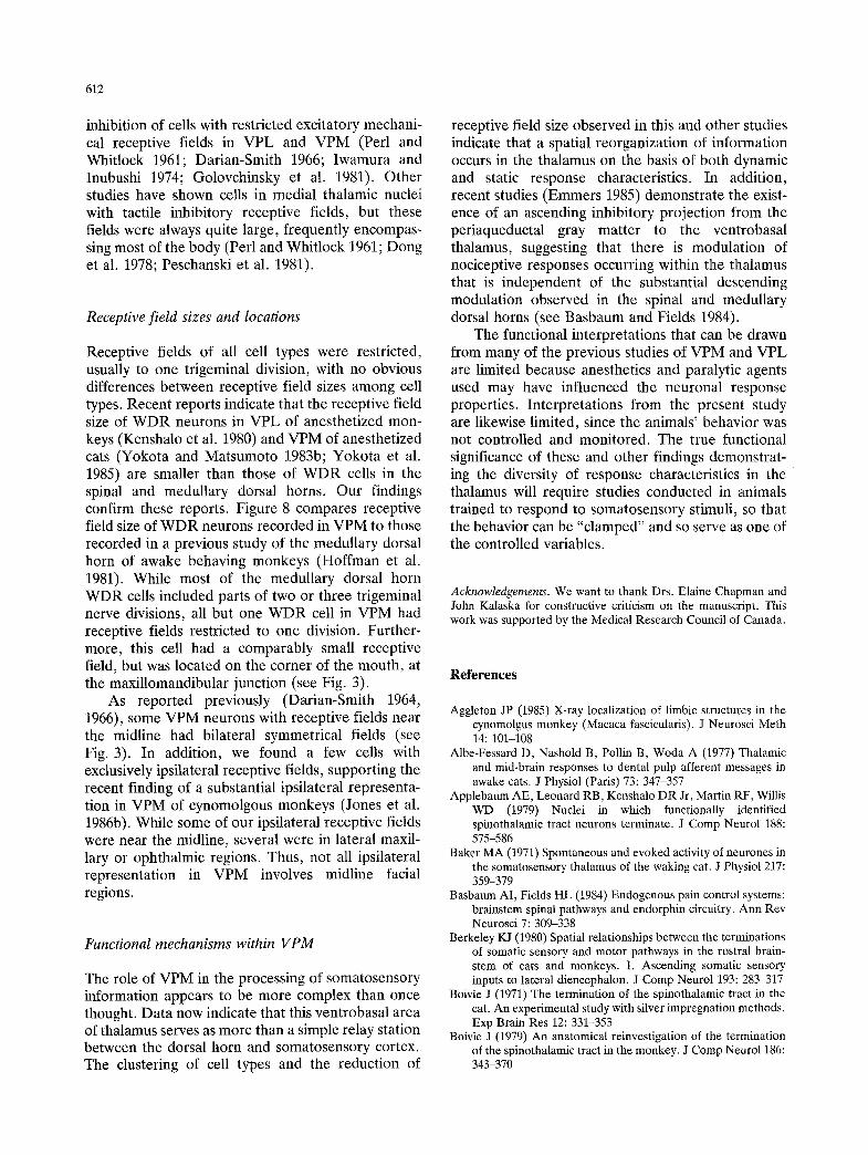
Fig. 8. Histogram showing the number of divisions of the trigeminal distribution involved in the receptive fields of WDR neurons in the medullary dorsal horn and VPM thalamus
column and spinothalamic tract input into VB (Mehler et al. 1960; Bowsher 1961; Bovie 1979; Berkeley 1980). Correspondingly, WDR and NS neurons are found intermixed with low threshold neurons (Kenshalo et al. 1980; Casey and Morrow 1983). Our study confirms this mixing of WDR neurons with low treshold cells of VPM, instead of the "shell" arrangement of WDR cells in VPM reported for cat. However, consistent with reports in cat (Yokota and Matsumoto 1983a, 1983b; Yokota et al. 1985), the nociceptive neurons in the present study were primarily confined to the posterior third of VPM (see Fig. 2).
Inhibitory responses of VPM neurons
About 10% of VPM neurons encountered in the present study exhibited inhibitory responses to mechanical skin stimulation. Such inhibition has only infrequently been reported. Perl and Whitlock (1961) described a few units in cat and monkey recorded from VB that had restricted, exclusively inhibitory receptive fields, similar to the inhibitory receptive fields of our study. This type of cell contrasts with other thalamic cells showing inhibitory properties. Several investigators have reported surround-type
612
inhibition of cells with restricted excitatory mechani- cal receptive fields in VPL and VPM (Perl and Whitlock 1961; Darian-Smith 1966; Iwamura and Inubushi 1974; Golovchinsky et al. 1981). Other studies have shown cells in medial thalamic nuclei with tactile inhibitory receptive fields, but these fields were always quite large, frequently encompas- sing most of the body (Perl and Whitlock 1961; Dong et al. 1978; Peschanski et al. 1981).
Receptive field sizes and locations
Receptive fields of all cell types were restricted, usually to one trigeminal division, with no obvious differences between receptive field sizes among cell types. Recent reports indicate that the receptive field size of W DR neurons in VPL of anesthetized mon- keys (Kenshalo et al. 1980) and VPM of anesthetized cats (Yokota and Matsumoto 1983b; Yokota et al. 1985) are smaller than those of WDR cells in the spinal and medullary dorsal horns. Our findings confirm these reports. Figure 8 compares receptive field size of W D R neurons recorded in VPM to those recorded in a previous study of the medullary dorsal horn of awake behaving monkeys (Hoffman et al. 1981). While most of the medullary dorsal horn WDR cells included parts of two or three trigeminal nerve divisions, all but one W D R cell in VPM had receptive fields restricted to one division. Further- more, this cell had a comparably small receptive field, but was located on the corner of the mouth, at the maxillomandibular junction (see Fig. 3).
As reported previously (Darian-Smith 1964, 1966), some VPM neurons with receptive fields near the midline had bilateral symmetrical fields (see Fig. 3). In addition, we found a few cells with exclusively ipsilateral receptive fields, supporting the recent finding of a substantial ipsilateral representa- tion in VPM of cynomolgous monkeys (Jones et al. 1986b). While some of our ipsilateral receptive fields were near the midline, several were in lateral maxil- lary or ophthalmic regions. Thus, not all ipsilateral representation in VPM involves midline facial regions.
Functional mechanisms within VPM
The role of VPM in the processing of somatosensory information appears to be more complex than once thought. Data now indicate that this ventrobasal area of thalamus serves as more than a simple relay station between the dorsal horn and somatosensory cortex. The clustering of cell types and the reduction of
receptive field size observed in this and other studies indicate that a spatial reorganization of information occurs in the thalamus on the basis of both dynamic and static response characteristics. In addition, recent studies (Emmers 1985) demonstrate the exist- ence of an ascending inhibitory projection from the periaqueductal gray matter to the ventrobasal thalamus, suggesting that there is modulation of nociceptive responses occurring within the thalamus that is independent of the substantial descending modulation observed in the spinal and medullary dorsal horns (see Basbaum and Fields 1984).
The functional interpretations that can be drawn from many of the previous studies of VPM and VPL are limited because anesthetics and paralytic agents used may have influenced the neuronal response properties. Interpretations from the present study are likewise limited, since the animals' behavior was not controlled and monitored. The true functional significance of these and other findings demonstrat- ing the diversity of response characteristics in the thalamus will require studies conducted in animals trained to respond to somatosensory stimuli, so that the behavior can be "clamped" and so serve as one of the controlled variables.
Acknowledgements. We want to thank Drs. Elaine Chapman and John Kalaska for constructive criticism on the manuscript. This work was supported by the Medical Research Council of Canada.
References
Aggleton JP (1985) X-ray localization of limbic structures in the cynomolgus monkey (Macaca fascicularis). J Neurosci Meth 14:101-108
Albe-Fessard D, Nashold B, Pollin B, Woda A (1977) Thalamic and mid-brain responses to dental pulp afferent messages in awake cats. J Physiol (Paris) 73:347-357
Applebaum AE, Leonard RB, Kenshalo DR Jr, Martin RF, Willis WD (1979) Nuclei in which functionally identified spinothalamic tract neurons terminate. J Comp Nenrol 188: 575-586
Baker MA (1971) Spontaneous and evoked activity of neurones in the somatosensory thalamus of the waking cat. J Physiol 217: 359-379
Basbaum AI, Fields HL (1984) Endogenous pain control systems: brainstem spinal pathways and endorphin circuitry. Ann Rev Neurosci 7:309-338
Berkeley KJ (1980) Spatial relationships between the terminations of somatic sensory and motor pathways in the rostral brain- stem of cats and monkeys. I. Ascending somatic sensory inputs to lateral diencephalon. J Comp Neurol 193:283-317
Boivie J (1971) The termination of the spinothalamic tract in the cat. An experimental study with silver impregnation methods. Exp Brain Res 12:331-353
Boivie J (1979) An anatomical reinvestigation of the termination of the spinothalamic tract in the monkey. J Comp Neuro1186: 343-370

613
Bowsher D (1961) The termination of secondary somatosensory neurons within the thalamus of Macaca mulatta: an experi- mental degeneration study. J Comp Neurol 117:213-227
Bushnell MC, Duncan GH, Dubner R, He LF (1984) Activity of trigeminothalamic neurons in medullary dorsal horn of awake monkeys trained in a thermal discrimination task. J Neurophysiol 52:170-187
Casey KL (1966) Unit analysis of nociceptive mechanisms in the thalamus of the awake squirrel monkey. J Neurophysiol 29: 727-750
Casey KL, Morrow TJ (1983) Ventral posterior thalamic neurons differentially responsive to noxious stimulation of the awake monkey. Science 221:675-677
Chung JM, Lee KH, Surmeier DJ, Sorkin LS, Kim J, Willis WD (1986) Response characteristics of neurons in the ventral posterior lateral nucleus of the monkey thalamus. J Neurophysiol 56:370-390
Darian-Smith I (1964) Cortical projections of thalamic neurones excited by mechanical stimulation of the face of the cat. J Physiol 171:339-360
Darian-Smith I (1966) Neural mechanisms of facial sensation. Intl Rev Neurobiol 9:301-395
Donaldson IML (1973) The properties of some human thalamic units. Brain 96:419-440
Dong WK, Ryu H, Wagman IH (1978) Nociceptive responses of neurons in medial thalamus and their relationship to spinothalamic pathways. J Neurophysiol 41:1592-1613
Duncan GH, Dreyer DA, McKenna TM, Whitsel BL (1982) Dose- and time-dependent effects of ketamine on SI neurons with cutaneous receptive fields. J Neurophysiol 47:677-699
Duncan GH, Bushnell MC (1985) Mechanical response properties of trigeminal thalamic neurons in the alert monkey. Soc Neurosci Abstr 11:22
Dykes RW, Sur M, Merzenich MM, Kaas JH, Nelson RJ (1981) Regional segregation of neurons responding to quickly adapt- ing, slowly adapting, deep and pacinian receptors within thalamic ventroposterior lateral and ventroposterior inferior nuclei in the squirrel monkey (Saimiri sciureus). Neuroscience 6:1687-1692
Emmers R (1985) Stimulation of the periaqueductal gray subdues sensitized pain in morphine- and meperidine-dependent rats. Exp Neurol 88:405-417
Giesler GJ Jr, Yezierski RP, Gerhart KD, Willis WD (1981) Spinothalamic tract neurons that project to medial and/or lateral thalamic nuclei: evidence for a physiologically novel population of spinal cord neurons. J Neurophysiol 46: 1285-1308
Golovchinsky V, Kruger L, Saporta SA, Stein BE, Young DW (1981) Properties of velocity-mechanosensitive neurons of the cat ventrobasal thalamic nucleus with special reference to the concept of convergence. Brain Res 209:355-374
Guilband G, Peschanski M, Gautron M (1981) Functional changes in ventrobasal thalamic neurones responsive to noxious and non-noxious cutaneous stimuli after chloralose treatment: new evidence for the presence of pre-existing "silent connec- tions" in the adult nervous system? Pain 11:9-19
Hoffman DS, Dubner R, Hayes RL, Medlin TP (1981) Neuronal activity in medullary dorsal horn of awake monkeys trained in a thermal discrimination task. I. Responses of innocuous and noxious thermal stimuli. J Neurophysiol 46:409-427
Honda CN, Meuse S, Perl ER (1983) Neurons in ventrobasal region of cat thalamus selectively responsive to noxious mechanical stimulation. J Neurophysiol 49:662-673
Iwamura Y, Inubushi S (1974) Regional diversity in excitatory and inhibitory receptive-field organization of cat thalamic ven- trobasal neurons. J Neurophysiol 37:910-919
Jasper H, Bertrand G (1966) Thalamic units involved in somatic
sensation and voluntary and involuntary movements in man. In: D Purpura, M Yahr (eds) The thalamus. Columbia University Press, New York, pp 365-390
Jones EG, Friedman DP (1982) Projection pattern of functional components of thalamic ventrobasal complex on monkey somatosensory cortex. J Neurophysiol 48:521-544
Jones EG, Hendry SHC, Brandon C (1986a) Cytochrome oxidase staining reveals functional organization of monkey somatosensory thalamus. Exp Brain Res 62:438-442
Jones EG, Schwark HD, Callahan PA (1986b) Extent of the ipsilateral representation in the ventral posterior medial nucleus of the monkey thalamus. Exp Brain Res 63:310-320
Kaas JH, Nelson RJ, Sur M, Dykes RW, Merzenich MM (1984) The somatotopic organization of the ventroposterior thalamus of the squirrel monkey, Saimiri sciureus. J Comp Neurol 226: 111-140
Kenshalo DR Jr, Giesler GJ, Leonard RB, Willis WD (1980) Responses of neurons in primate ventral posterior lateral nucleus to noxious stimuli. J Neurophysiol 43:1594-1614
Kniffki K-D, Mizumura K (1983) Responses of neurons in VPL and VPL-VL of the cat to algesic stimulation of muscle and tendon. J Neurophysiol 49:649-661
Lenz FA, Dostrovsky JO, Tasker RR (1986) Functional properties of human somatosensory thalamus. Soc Neurosci Abstr 12: 329
Loe PR, Whitsel BL, Dreyer DA, Metz CB (1977) Body representation in ventrobasal thalamus of macaque: a single- unit analysis. J Neurophysiol 40:1339-1355
Mehler WR, Feferman ME, Nauta WJH (1960) Ascending axon degeneration following anterolateral cordotomy. An experi- mental study in the monkey. Brain 83:718-751
Merrill EG, Ainsworth A (1972) Glass-coated platinum-plated tungsten microelectrodes. Med Biol Eng 10:662-672
Millar J (1973) The topography and receptive fields of ventropos- terolateral thalamic neurons excited by afferents projecting through the dorsolateral funiculus of the spinal cord. Exp Neurol 41:303-313
Mountcastle VB, Henneman E (1952) The representation of tactile sensibility in the thalamus of the monkey. J Comp Neurol 97:409-440
Ohye C, Fukamachi A, Narabayashi H (1972) Spontaneous and evoked activity of sensory neurons and their organization in the human thalamus. J Neurol 203:219-234
Perl ER, Whitlock DG (1961) Somatic stimuli exciting spinothalamic projections to thalamic neurons in cat and monkey. Exp Neurol 3:256-296
Peschanski M, Guilbaud G, Gautron M (1981) Posterior intralami- nat region in rat: neuronal responses to noxious and nonnoxi- ous cutaneous stimuli. Exp Neurol 72:226-238
Poggio GF, Mountcastle VB (1963) The functional properties of ventrobasal thalamic neurons studied in unanesthetized mon- keys. J Neurophysiol 26:775-806
Pollin B, Albe-Fessard D (1979) Organization of somatic thalamus in monkeys with and without section of dorsal spinal tracts. Brain Res 173:431-449
Price DD, Dubner R, Hu JW (1976) Trigeminothalamic neurons in nucleus caudalis responsive to tactile, thermal, and nociceptive stimulation of monkey's face. J Neurophysiol 39: 936-953
Sessle BJ (1979) Is the tooth pulp a 'pure' source of noxious input? In: Bonica JJ, Liebeskind JC, Albe-Fessard DG (eds) Ad- vances in pain research and therapy, Vol 3. Raven Press, New York, pp 245-260
Shanta TR, Manocha SL, Bourne GH (1968) A stereotaxic atlas of the Java monkey (Macaca irus). Williams and Wilkins, Baltimore
Shigenaga Y, Matano S, Okada K, Sakai A (1973) The effects of

614
tooth pulp stimulation in the thalamus and hypothalamus of the rat. Brain Res 63:402407
Sur M, Wall JT, Kaas JH (1984) Modular distribution of neurons with slowly adapting and rapidly adapting responses in area 3b of somatosensory cortex in monkeys. J Neurophysiol 51: 724-744
Willis WD, Trevino DL, Coulter JD, Maunz RA (1974) Responses of primate spinothalamic tract neurons to natural stimulation of hindlimb. J Neurophysiol 37:358-372
Woda A, Azerad J, Guilbaud G, Besson JM (1975) Etude microphysiologique des projections thalamiques de la pulpe dentalre chez le chat. Brain Res 89:193-213
Yokota T, Matsumoto N (1983) Somatotopic distribution of trigeminal nociceptive specific neurons within the caudal somatosensory thalamus of cat. Neurosci Lett 39:125-130
Yokota T, Matsumoto N (1983) Location and functional organiza- tion of trigeminal wide dynamic range neurons within the nucleus ventralis posteromedialis of the cat. Neurosci Lett 39: 231-236
Yokota T, Koyama N, Matsumoto N (1985) Somatotopic distribu- tion of trigeminal nociceptive neurons in ventrobasal complex of cat thalamus. J Neurophysiol 53:1403-1416
Received November 20, 1986 / Accepted February 27, 1987