measurement of anti-thyroglobulin igg in serum by novel and sensitive immune complex transfer enzyme...
TRANSCRIPT
Chn B~ochem, Vol 22, pp 277-284, 1989 0009-9120/89 $3 00 + 00 Printed m Canada All rights reserved. Copyright © 1989 The Canadian Socmty of Chmcal Chemists
Measurement of Anti-Thyroglobulin IgG in Serum by Novel and Sensitive Immune Complex Transfer
Enzyme Immunoassay
TAKEYUKI KOHNO, 1 TOMOHIRO MITSUKAWA, 2 SHIGERU MATSUKURA, 2 YOSHIZO TSUNETOSHI, 3 and EIJI ISHIKAWA 1
1Department of Biochemistry, 2Third Department of Internal Medicine, and 3Department of Public Health, Medical College of Miyazaki, Kiyotake, Miyazaki 889-16, Japan
Anti-thyroglobuhn IgG in human serum was measured by a novel and senstt~ve immune complex transfer enzyme immunoassay. Anti- thyroglobulin IgG in human serum was reacted with dinitrophenyl thyroglobuhn, and the complex formed between human anti-thyro- globuhn IgG and dinitrophenyl thyroglobuhn was trapped onto an affinity-purified rabbit (anti-dinitrophenyl bovine serum albumin) IgG-coated polystyrene ball. The polystyrene ball was washed to elimmate most nonspeciftc IgG in test serum, and the complex was eluted from the polystyrene ball, to which nonspecific IgG had been adsorbed, with dinitrophenyI-L-tysine and transferred to a clean pol- ystyrene ball coated with rabbit anti-thyroglobuhn IgG. The amount of human anti-thyroglobulin IgG in the complex on the rabbit antl- thyroglobulin IgG-coated polystyrene ball was estimated usmg rabbit (antt-human IgG ~-chain) Fab'-horseradish peroxidase conjugate. The present enzyme immunoassay was 1,000 to 3,0004old more sensitive than the conventional enzyme tmmunoassay, m which a human thyroglobulin-coated polystyrene ball was incubated with serum contaming human anti-thyroglobuhn IgG and, after washmg, with rabbit (anti-human IgG q,-chain) Fab'-horseradish peroxidase con- jugate. By the present enzyme immunoassay, antt-thyroglobulin IgG was demonstrated in about 10% of healthy subjects and in all pattents wtth Graves' disease and chronic thyroiditis. The principle of the present method may make tt possible to sensitively measure other autoantibodies including anti-thyroid peroxidase and antl-thyrotropm receptor antibodies to aid diagnosis of thyroid diseases.
KEY WORDS: autoantibodies; anti-thyroglobulin IgG; novel enzyme immunoassay; thyroglobulin; peroxidase; Graves' disease; chromc thyroiditis.
immunological binding of nonspecific immunoglobulins in test serum to the solid phase, which cannot be easily eliminated (2,3).
This paper describes the measurement of anti-thy- roglobulin IgG in human serum by a novel and sensi- tive enzyme immunoassay (immune complex transfer enzyme immunoassay), in which nonspecific IgG in test serum was efficiently eliminated and the sensitivity was improved 1,000 to 3,000-fold as compared with that of the conventional enzyme immunoassay. By this en- zyme immunoassay, anti-thyroglobulin IgG was dem- onstrated in about 10% of healthy subjects and in all patients with Graves' disease and chronic thyroiditis.
Materials and methods
BUFFERS
The regularly used buffers were 10 mmol/L sodium phosphate buffer, pH 7.0, containing 0.1 mol/L NaC1 and 1.0 g/L bovine serum albumin (fraction V, Armour Pharmaceutical Co., Kankakee, Illinois) (buffer A) and 10 mmol/L sodium phosphate buffer, pH 7.0 containing 0.1 mol/L NaCI (buffer B).
Introduction
In the conventional enzyme immunoassay for anti- bodies in serum, antigen-coated solid phase is incu- bated with serum to trap specific immunoglobulins and subsequently reacted with anti-immunoglobulin anti- body-enzyme conjugate to estimate the amount of the specific immunoglobulins on the solid phase (1). The sensitivity of this assay is seriously limited by non-
Correspondence: Professor Eiji Ishikawa, M.D., Ph.D., De- partment of Biochemistry, Medical College of Miyazaki, Ki- yotake, Miyazaki 889-16, Japan.
Manuscript received June 20, 1988; revised September 2, 1988; accepted September 15, 1988.
ENZYMES
Horseradish peroxidase (EC 1.11.1.7) (grade I, lyoph- ilized RZ = 3.0) and pepsin from porcine gastric mucosa (EC 3.4.23.1) were obtained from Boehringer Mann- helm GmbH, Mannheim, FRG.
ANTISERA
Rabbit anti-human thyroglobulin serum was a gen- erous gift from the Chemo-Sero-Therapeutic Research Institute, Kumamoto, Japan. Rabbit (anti-human IgG ~/-chain) IgG was obtained from Medical and Biological Laboratories Co., Ltd., Nagoya, Japan. Rabbit (anti- dinitrophenyl bovine serum albumin) serum was ob- tained from Miles Laboratories, Inc., Elkhart, Indiana.
CLINICAL BIOCHEMISTRY, VOLUME 22, AUGUST 1989 277
KOHNO, MITSUKAWA, MATSUKURA, TSUNETOSHI, AND ISHIKAWA
IGG, F(AB') 2 AND FAB'
IgG was prepared from serum by fractionation with Na2SO4 followed by passage through a column of DEAE- cellulose (4). F(ab')2 was prepared by digestion of IgG with pepsin, and Fab' was prepared by reduction of F(ab')2 with 2-mercaptoethylamine (4). The amounts of IgG and its fragments were calculated from the ab- sorbance at 280 nm (4).
DINITROPHENYL BOVINE SERUM ALBUMIN
1. Mercaptosuccinylated bovine serum albumin
Bovine serum albumin (20 mg, fraction V, Armour Pharmaceutical Co.) in 2 mL of 0.1 mol/L sodium phos- phate buffer, pH 7.5, was incubated with 0.2 mL of 0.11 mol/L S-acetylmercaptosuccinic anhydride (Na- karai Chemicals, Ltd., Kyoto, Japan) in N,N-dime- thylformamide at 30°C for 30 min. The reaction mixture was incubated with 0.5 mL of 1.0 mol/L Tris-HC1 buffer, pH 7.0, 0.2 mL of 0.1 mol/L EDTA, pH 7.0 and 0.3 mL of I mol/L hydroxylamine, pH 7.0, at 30°C for 15 min and subjected to gel filtration on a column (1 x 30 cm) of Sephadex G-25 (Pharmacia Fine Chemicals AB, Uppsala, Sweden) using 0.1 mol/L sodium phosphate buffer, pH 6.0, containing 5 mmol/L EDTA. The amount of bovine serum albumin was calculated from the ab- sorbance at 280 nm (5). The average number of thiol groups introduced per bovine serum albumin molecule was 8.2 (4).
2. Maleimide-dinitrophenyl-L-lysine
An aliquot (5.0 mL) of 5.5 mmol/L dinitrophenyl-L- lysine-HC1 (Tokyo Kasei Kogyo, Co., Ltd., Tokyo, Ja- pan) in 0.1 mol/L sodium phosphate buffer, pH 7.0, containing 5 mmol/L EDTA was incubated with 0.5 mL of 5.5 mmol/L N-succinimidyl-6-maleimidohexanoate (Dojindo Laboratories, Kumamoto, Japan) in N,N-di- methylformamide at 30°C for 30 min.
Fine Chemicals AB, Uppsala, Sweden) as follows. CNBr- activated Sepharose 4B (1 g) was swollen in 200 mL of 1 mmol/L HC1 and washed on a filter (MF-Millipore Type PH, Miltipore Co., Bedford, Massachusetts) with 200 mL of 0.1 mol/L sodium borate buffer, pH 8.0, con- taining 0.5 mol/L NaC1 (coupling buffer). Each protein (10 mg) in 10 mL of the coupling buffer was incubated with the CNBr-activated Sepharose 4B at 4°C for 20 h with continuous shaking. After incubation, the protein- coupled Sepharose 4B was washed with 200 mL of the same buffer as described above, and incubated with 10 mL of 0.2 mol/L glycine-NaOH buffer, pH 8.0, at room temperature for 2 h with continuous shaking. Finally, the protein-coupled Sepharose 4B was washed as de- scribed above successively with 200 mL each of the coupling buffer, 0.1 mol/L sodium acetate buffer, pH 4.0, containing 0.5 mol/L NaC1 and the coupling buffer and stored in buffer A containing 1.0 g/L NAN3.
HUMAN THYROGLOBULIN
Partially purified human thyroglobulin was gener- ously supplied by the Chemo-Sero-Therapeutic Re- search Institute, Kumamoto, Japan. The partially pu- rifled thyroglobulin, which had been prepared from thyroid glands by fractionation with ammonium sulfate (7), was further purified by chromatography on a col- umn of DEAE-cellulose (8). Subsequently, the purified thyroglobulin (3.0 mg) in 1.5 mL of 0.1 mol/L sodium phosphate buffer, pH 7.0, containing 1 g/L NaN3 was passed through a column (0.9 x 5.5 cm) of rabbit (anti- human IgG ~-chain) IgG-Sepharose 4B using the same buffer at a flow rate of 1 mL/h and subjected to gel filtration on a column (1.5 x 45 cm) of Ultrogel AcA 22 (LKB, Stockholm, Sweden) using the same buffer. Homogeneity of the purified thyroglobulin was con- firmed by polyacrylamide gel electrophoresis in the presence of sodium dodecyl sulfate and urea (8). The amount of thyroglobulin was calculated from the ab- sorbance at 280 nm by taking the extinction coefficient as 1.0 g- ' - liter • cm ~ (8).
3. Dinitrophenyl bovine serum albumin DINITROPHENYL THRYOGLOBULIN
The mercaptosuccinylated bovine serum albumin (15 mg) in 3 mL of 0.1 mol/L sodium phosphate buffer, pH 6.0, containing 5 mmol/L EDTA was incubated with 4.5 mL of the maleimide-dinitrophenyl-L-lysine solu- tion at 30°C for 30 min. The reaction mixture was sub- jected to gel filtration on a column (1.5 x 45 cm) of Sephadex G-25 using 0.1 mol/L sodium phosphate buffer, pH 7.5. The average number of dinitrophenyl groups introduced per bovine serum albumin molecule was 5.5, which was calculated from the absorbance at 360 nm by taking the molar extinction coefficient as 17,400 tool -1 - liter • cm -1 (6).
PROTEIN-SEPHAROSE 4B
(Anti-human IgG ,/-chain) IgG, dinitrophenyl bovine serum albumin and purified human thyroglobulin were coupled to CNBr-activated Sepharose 4B (Pharmacia
1. Mercaptosuccinylated human thyroglobulin
The purified human thyroglobulin (0.85 rag) in 0.3 mL of 0.1 mol/L sodium phosphate buffer, pH 7.5 was incubated with 0.03 mL of 55 mmol/L S-acetylmercap- tosuccinic anhydride (Nakarai Chemicals, Ltd., Kyoto, Japan) in N,N-dimethylformamide at 30°C for 30 rain. The reaction mixture was subjected to gel filtration on a column (1 x 30 cm) of Sephadex G-25 using 0.1 tool/ L sodium phosphate buffer, pH 7.0 containing 5 mmol/ L EDTA. Four mL of the fractions containing 0.64 mg of S-acetylmercaptosuccinylated human thyroglobulin was incubated with 0.4 ml of i mol/L hydroxylamine, pH 7.0, at 30°C for 15 rain and subjected to gel filtration as described above using 0.1 mol/L sodium phosphate buffer, pH 6.0, containing 5 mmol/L EDTA. The av- erage number of thiol groups introduced per human thyroglobulin molecule was 20 (4).
278 CLINICAL BIOCHEMISTRY, VOLUME 22, AUGUST 1989
ENZYME IMMUNOASSAY OF ANTI-THYROGLOBULIN IGG
2. Maleimide-dinitrophenyl-L-lysine
The maleimide-dinitrophenyl-L-lysine solution was prepared as described above.
3. Dinttrophenyl thyroglobulin
The mercaptosuccinylated human thyroglobulin (0.5 rag) in 1.0 mL of 0.1 mol/L sodium phosphate buffer, pH 6.0, containing 5 mmol/L EDTA was incubated with the maleimide-dinitrophenyl-L-lysine solution (0.03 mL) at 30°C for 30 min, and subsequently with 0.1 mol/L N-ethylmaleimide (5 ~L) in the same buffer at 30°C for 15 min. The reaction mixture was subjected to gel filtration on a column (1 × 30 cm) of Sephadex G-25 using 0.1 mol/L sodium phosphate buffer, pH 7.0, containing 1 g/L NAN3. The average number of dini- trophenyl groups introduced per human thyroglobulin molecule was 16 (6).
AFFINITY-PURIFICATION OF ANTIBODIES
(Anti-dinitrophenyl bovine serum albumin) IgG (27 rag) in 4 mL of 0.1 mol/L sodium phosphate buffer, pH 7.0, containing 1 g/L NaN3 was applied to a column 10.55 × 1.7 cm) ofdinitrophenyl bovine serum albumin- Sepharose 4B at a flow rate of i mL/h using the same buffer. The specific IgG was eluted with 3.2 mmol/L HC1, pH 2.5, at a flow rate of 60 mL/h, and the eluate was immediately neutralized by addition of 1 mol/L sodium phosphate buffer, pH 7.0. The amount of the affinity-purified IgG was 2.3 mg.
Human anti-thyroglobulin IgG (1 mg) from serum of patients with Graves' disease in 0.35 mL of 0.1 mol/L sodium phosphate buffer, pH 7.0, containing 1 g/L NaN3 was applied to a column (1 × 2 mm) of human thyroglobulin-Sepharose 4B at a flow rate of 0.5 mL/h using the same buffer. The specific IgG was eluted with 3.2 mmol/L HC1, pH 2.5, as described above. The amount of the affinity-purified IgG (0.7 ~g) was measured by enzyme immunoassay (9).
PROTEIN-COATED POLYSTYRENE BALLS
Polystyrene balls (3.2 mm in diameter, Precision Plastic Ball Co., Chicago, Illinois) were coated by phys- lcal adsorption with affinity-purified rabbit (anti-di- nitrophenyl bovine serum albumin) IgG (0.1 g/L), rab- bit anti-human thyroglobulin IgG (0.1 g/L) and the pu- rifled human thyroglobulin (0.1 g/L) (10).
(ANTI-HUMAN IGG w-CHAIN) FAB'-PEROXIDASE CONJUGATES
Rabbit (anti-human IgG ~/-chain) Fab' was conju- gated to horseradish peroxidase using N-succinimidyl- 6-maleimidohexanoate (12). The amount of the conju- gate was calculated from peroxidase activity (4).
PRESENT ENZYME IMMUNOASSAY FOR ANTI- THYROGLOBULIN IGG IN SERUM
An aliquot (20 ~L) of human serum containing anti- thyroglobulin IgG was incubated with 80 ~L of buffer A containing 50 fmol of dinitrophenyl thyroglobulin, 3.75 g/L nonspecific rabbit IgG and I g/L NAN3, 50 ~L of 10 mmol/L sodium phosphate buffer, pH 7.0, con- taining 1.0 mol/L NaC1, 1 g/L bovine serum albumin and 1 g/L NaN3 and an affinity-purified rabbit (anti- dinitrophenyl bovine serum albumin) IgG-coated poly- styrene ball, which had been treated with nonspecific rabbit IgG (3), at 20°C for 20 h. After incubation, the polystyrene ball was washed twice by addition and as- piration of 2 mL of buffer B and incubated with I mmol/ L dinitrophenyl-L-lysine and 2 g/L nonspecific rabbit IgG in 0.15 mL of buffer A containing 1 g/L NaN3 at 20°C for 3 h to elute the complex of anti-thyroglobulin IgG and dinitrophenyl thyroglobulin. After removal of the polystyrene ball, the eluate was incubated with a rabbit anti-thyroglobulin IgG-coated polystyrene ball, which had been treated with nonspecific rabbit IgG (3), at 20°C for 3 h. After incubation, the rabbit anti-thy- roglobulin IgG-coated polystyrene ball was washed as described above, and incubated with rabbit (anti-hu- man IgG ~/-chain) Fab'-peroxidase conjugate (50 ng) in 0.15 mL of buffer A at 20°C for 3 h. Finally, the poly- styrene ball was washed and bound peroxidase activity was assayed for 10 min as described above.
To test the presence of anti-thyroglobulin IgG more specifically, an aliquot (20 txL) of serum was preincu- bated with 30 txL of buffer A containing 15 pmol of the purified human thyroglobulin (or bovine thyroglobulin, Sigma Chemical Co., St. Louis, Missouri) and 1 g/L NaN3 at 20°C for 3 h. The reaction mixture was mixed with 50 ~L of buffer A containing 50 fmol of dinitro- phenyl thyroglobulin, 6.0 g/L nonspecific rabbit IgG and I g/L NAN3, and 50 txL of 10 mmol/L sodium phos- phate buffer, pH 7.0, containing 1.0 mol/L NaC1, 1 g/L bovine serum albumin and 1 g/L NaN3 and was processed as described above.
CONVENTIONAL ENZYME IMMUNOASSAY FOR ANTI- THYROGLOBULIN IGG IN SERUM
ASSAY OF PEROXIDASE ACTIVITY
The activity ofperoxidase was assayed by fluorimetry at 30°C using 3-(4-hydroxyphenyl)propionic acid as substrate (11). The fluorescence intensity was mea- sured relative to 1 mg/L quinine in 50 mmol/L H2SO4 using 320 nm for excitation and 405 nm for emission in a Shimadzu spectrofluorophotometer (RF-510, Shi- madzu Seisakusho Ltd., Kyoto, Japan).
Human serum containing anti-thyroglobulin IgG was diluted 1 × 10⁵-fold with buffer A containing 1 g/L NAN3, and an aliquot (0.15 mL) of the diluted serum was incubated with a human thyroglobulin-coated polystyrene ball at 37°C for 3 h. After incubation, the polystyrene ball was washed as described above and incubated with rabbit (anti-human IgG ~-chain) Fab'- peroxidase conjugate (50 ng) in 0.15 mL of buffer A at 37°C for 3 h. Finally, the polystyrene ball was washed
CLINICAL BIOCHEMISTRY, VOLUME 22, AUGUST 1989 279
KOHNO, MITSUKAWA, MATSUKURA, TSUNETOSHI, AND ISHIKAWA
as described above and bound peroxidase activity was assayed for 10 min as described above. The assay back- ground in the absence of anti-thyroglobulin IgG was assessed by preincubation with purified human thy- roglobulin as described above.
EXPRESSION OF THE DETECTION LIMIT OF
ANTI-THYROGLOBULIN IGG
The detection limit of anti-thyroglobulin IgG in serum was expressed as the maximal dilution of serum con- taining anti-thyroglobulin IgG with pooled serum from heal thy subjects or the minimal amount of the affinity- purified anti-thyroglobulin IgG which gave a bound peroxidase activity significantly in excess of tha t ob- served with serum from heal thy subjects (the back- ground). A significant difference from the background was confirmed by the t-test (n = 5, P < 0.001).
OTHER METHODS
Measurement of anti- thyroglobulin antibodies by hemagglut inat ion was performed using a commercial kit (Seroclit-TG, Sanko Junyaku Co., Ltd., Tokyo, Ja- pan). Serum levels of human thyroid-st imulat ing hor- mone (hTSH) were measured by sandwich enzyme im- munoassay as described previously (13). Serum levels of T4 and T3 were measured by competitive radioim- munoassay using commercial kits (Tetrabead-125 and T-3.Riabead, respectively, Dainabot Co., Ltd., Tokyo, Japan).
R e s u l t s
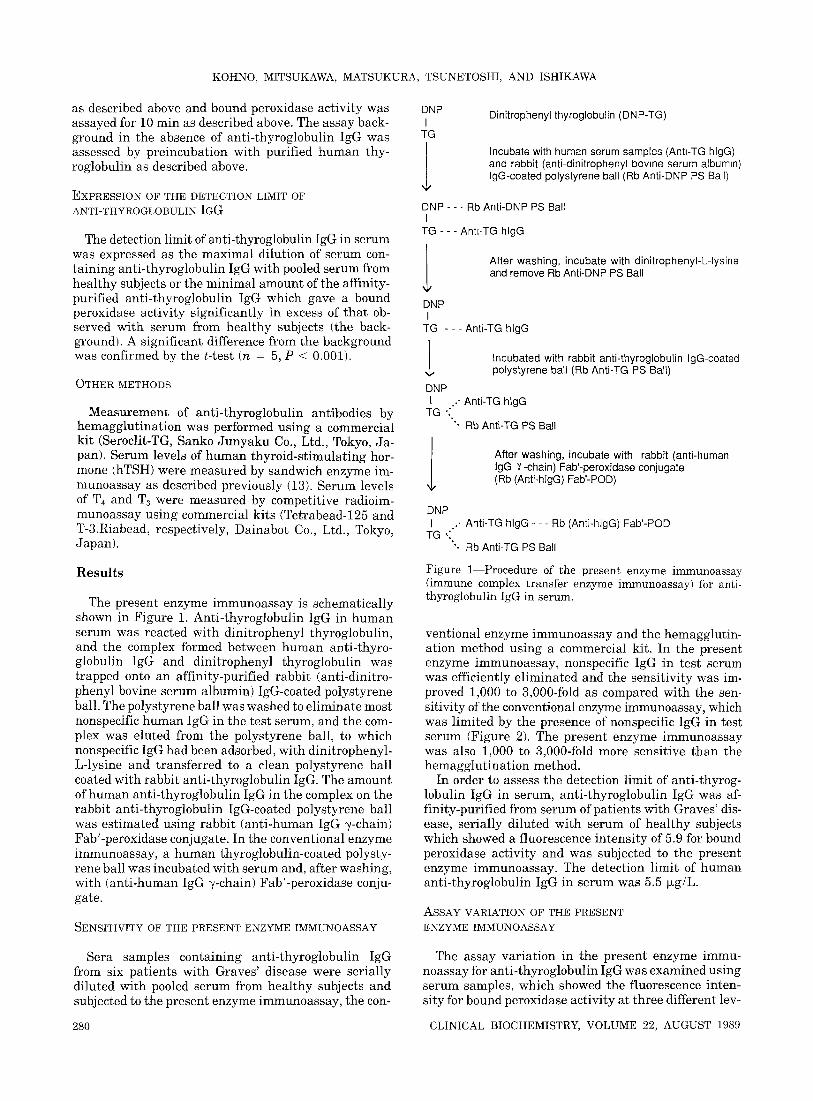
The present enzyme immunoassay is schematically shown in Figure 1. Anti-thyroglobulin IgG in human serum was reacted with dinitrophenyl thyroglobulin, and the complex formed between human anti-thyro- globulin IgG and dinitrophenyl thyroglobulin was trapped onto an affinity-purified rabbit (anti-dinitro- phenyl bovine serum albumin) IgG-coated polystyrene ball. The polystyrene ball was washed to eliminate most nonspecific human IgG in the test serum, and the com- plex was eluted from the polystyrene ball, to which nonspecific IgG had been adsorbed, with dinitrophenyl- L-lysine and transferred to a clean polystyrene ball coated with rabbit anti-thyroglobulin IgG. The amount of human anti-thyroglobulin IgG in the complex on the rabbit anti-thyroglobulin IgG-coated polystyrene ball was est imated using rabbit (ant i-human IgG ~-chain) Fab'-peroxidase conjugate. In the conventional enzyme immunoassay, a human thyroglobulin-coated polysty- rene ball was incubated with serum and, after washing, with (anti-human IgG ~/-chain) Fab'-peroxidase conju- gate.
SENSITIVITY OF THE PRESENT ENZYME IMMUNOASSAY
DNP I
TG
l DNP - - - Rb Anti-DNP PS Ball
I TG - - - Anh-TG hlgG
l DNP I
TG - - - Anti-TG hlgG
Dinitrophenyl thyroglobulin (DNP-TG)
Incubate with human serum samples (Anb-TG hlgG) and rabbit (anti-dinitrophenyl bovine serum albumin) lgG-coated polystyrene ball (Rb Anti-DNP PS Ball)
After washing, incubate with dinitrophenyl-L-lysine and remove Rb Anti-DNP PS Ball
Incubated with rabbit anti-thyroglobulin IgG-coated polystyrene ball (Rb Anti-TG PS Ball)
DNP I .. Anti-TG hlgG
TG ":~ *. Rb Anti-TG PS Ball
After washing, incubate with rabbit (anti-human IgG 7-chain) Fab'-peroxidase conjugate (Rb (Anti-hlgG) Fab'-POD)
DNP I ., Anti-TG hlgG - - - Rb (Anti-hlgG) Fab'-POD
TG ,:" ". Rb Anti-TG PS Ball
Figure 1--Procedure of the present enzyme immunoassay (immune complex transfer enzyme immunoassay) for anti- thyroglobulin IgG in serum.
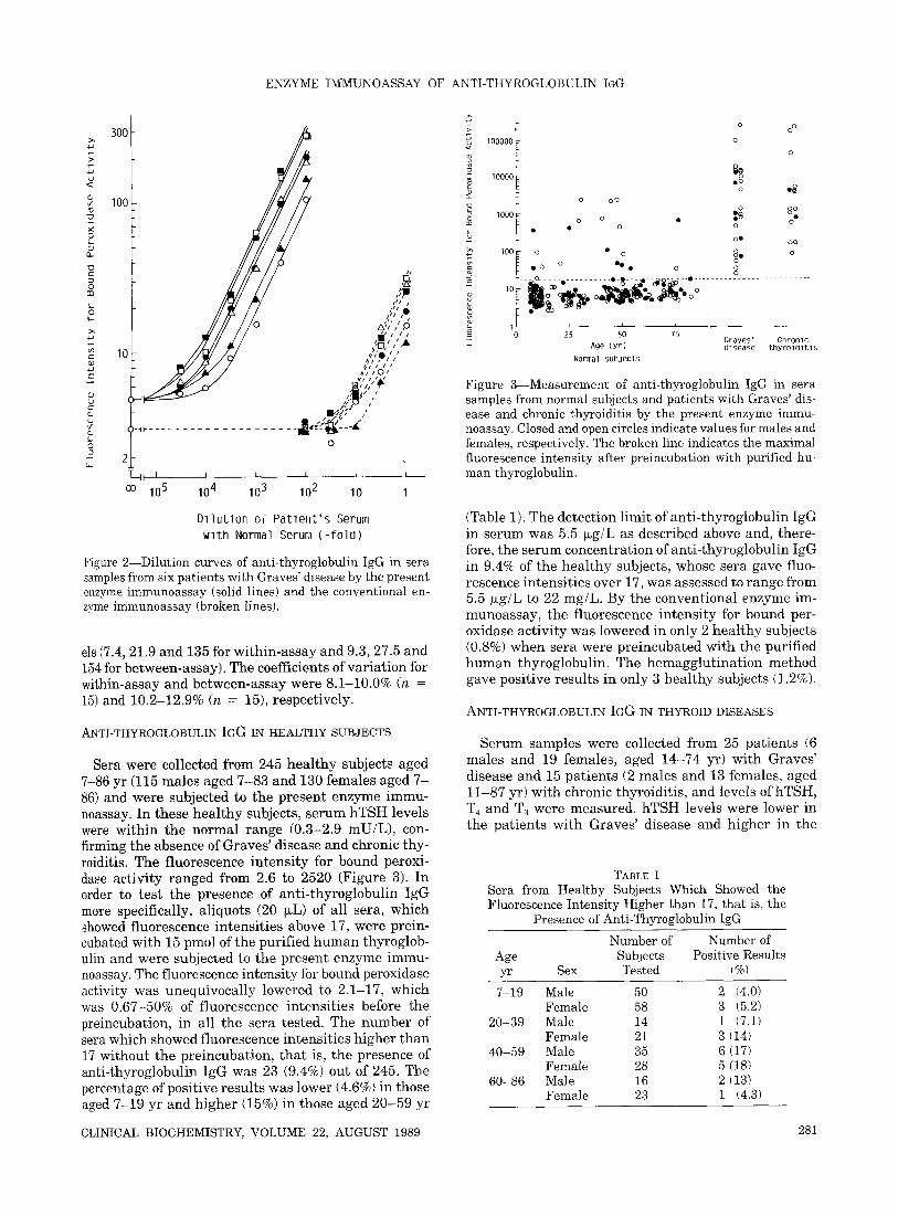
ventional enzyme immunoassay and the hemagglutin- ation method using a commercial kit. In the present enzyme immunoassay, nonspecific IgG in test serum was efficiently el iminated and the sensitivity was ira- proved 1,000 to 3,000-fold as compared with the sen- sitivity of the conventional enzyme immunoassay, which was limited by the presence of nonspecific IgG in test serum (Figure 2). The present enzyme immunoassay was also 1,000 to 3,000-fold more sensitive than the hemagglut inat ion method.
In order to assess the detection limit of anti-thyrog- lobulin IgG in serum, anti-thyroglobulin IgG was af- finity-purified from serum of patients with Graves' dis- ease, serially diluted with serum of heal thy subjects which showed a fluorescence intensi ty of 5.9 for bound peroxidase activity and was subjected to the present enzyme immunoassay. The detection limit of human anti-thyroglobulin IgG in serum was 5.5 ~g/L.
ASSAY VARIATION OF THE PRESENT ENZYME IMMUNOASSAY
Sera samples containing anti-thyroglobulin IgG from six patients with Graves' disease were serially diluted with pooled serum from heal thy subjects and subjected to the present enzyme immunoassay, the con-
The assay variat ion in the present enzyme immu- noassay for ~ anti- thyroglobulin IgG was examined using serum samples, which showed the fluorescence inten- sity for bound peroxidase activity at three different lev-
280 CLINICAL BIOCHEMISTRY, VOLUME 22, AUGUST 1989
ENZYME IMMUNOASSAY OF ANTI-THYROGLOBULIN IGG
300
>
i0o
Q_
i ; I I L 0 J ~ • z I •
A, , , ,0
, t , [ ~ l / / A 10 ,,';'o ' '
4p r t l
4 / i O z = i / l l l ~ t
( q ]" , '
W ( )_~p . . . . . . . . . . . . . . . . . . . .
o 0
=- 2- T_._~ I I I I I i I
oo 105 104 103 102 10 I
Dl lu t i on of Pa t ien t ' s Serum wi th Normal Serum ( - f o l d )
Figure 2--Dilution curves of anti-thyroglobulin IgG in sera samples from six patients with Graves' disease by the present enzyme immunoassay (solid lines) and the conventional en- zyme immunoassay (broken lines).
els (7.4, 21.9 and 135 for wi thin-assay and 9.3, 27.5 and 154 for between-assay). The coefficients of var ia t ion for within-assay and between-assay were 8.1-10.0% (n = 15) and 10.2-12.9% (n = 15), respectively.
ANTI-THYROGLOBULIN IGG IN HEALTHY SUBJECTS
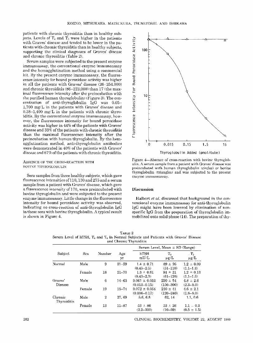
Sera were collected from 245 hea l thy subjects aged 7-86 yr (115 males aged 7-83 and 130 females aged 7 - 86) and were subjected to the present enzyme immu- noassay. In these hea l thy subjects, se rum hTSH levels were within the normal range (0.3-2.9 mU/L), con- firming the absence of Graves ' disease and chronic thy- roiditis. The fluorescence in tens i ty for bound peroxi- dase activity ranged from 2.6 to 2520 (Figure 3). In order to test the presence of ant i- thyroglobulin IgG more specifically, aliquots (20 ~L) of all sera, which showed fluorescence intensit ies above 17, were prein- cubated with 15 pmol of the purified h u m a n thyroglob- ulin and were subjected to the present enzyme immu- noassay. The fluorescence intensity for bound peroxidase activity was unequivocally lowered to 2.1-17, which was 0.67-50% of fluorescence intensit ies before the preincubation, in all the sera tested. The number of sera which showed fluorescence intensit ies higher than 17 without the preincubation, tha t is, the presence of anti-thyroglobulin IgG was 23 (9.4%) out of 245. The percentage of positive resul ts was lower (4.6%) in those aged 7-19 yr and higher (15%) in those aged 20-59 yr
100000
I0000
L
I000
1oo
o ° o o
o
o o o o ~
o o o ° o ~ o
o • o ~ o • o • • o o
oO o o
o • o 8 o o o
oO o ° ° • o o o
- - - -o . . . . . . . . . . o - ~ - - : : . - - # ~ - - o . . . . . . . . . . . . . . . . . . . . . . . .
i L I H
25 50 75 G r a v e s ' C h r o n l c
Age ( y r ) 01 sease t h y r o l d l t l s
Norma l S u b j e c t s
Figure 3--Measurement of anti-thyroglobulin IgG in sera samples from normal subjects and patients with Graves' dis- ease and chronic thyroiditis by the present enzyme immu- noassay. Closed and open circles indicate values for males and females, respectively. The broken line indicates the maximal fluorescence intensity after preincubation with purified hu- man thyroglobulin.
(Table 1). The detection l imit of anti- thyroglobulin IgG in serum was 5.5 ~g/L as described above and, there- fore, the se rum concentration of anti- thyroglobulin IgG in 9.4% of the hea l thy subjects, whose sera gave fluo- rescence intensit ies over 17, was assessed to range from 5.5 ~xg/L to 22 mg/L. By the conventional enzyme im- munoassay, the fluorescence intensi ty for bound per- oxidase activity was lowered in only 2 heal thy subjects (0.8%) when sera were preincubated with the purified h u m a n thyroglobulin. The hemagglu t ina t ion method gave positive resul ts in only 3 heal thy subjects (1.2%).
ANTI-THYROGLOBULIN IGG IN THYROID DISEASES
Serum samples were collected from 25 pat ients (6 males and 19 females, aged 14-74 yr) with Graves ' disease and 15 pat ients (2 males and 13 females, aged 11-87 yr) wi th chronic thyroiditis, and levels of hTSH, T4 and T3 were measured, hTSH levels were lower in the pat ients with Graves ' disease and higher in the
TABLE 1 Sera from Healthy Subjects Which Showed the Fluorescence Intensity Higher than 17, that is, the
Presence of Anti-Thyroglobulin IgG
Number of Number of Age Subjects Positive Results yr Sex Tested (%)
7-19 Male 50 2 (4.0) Female 58 3 (5.2)
20-39 Male 14 1 (7.1) Female 21 3 (14)
40-59 Male 35 6 (17) Female 28 5 (18)
60-86 Male 16 2 [13) Female 23 I (4.3)
CLINICAL BIOCHEMISTRY, VOLUME 22, AUGUST 1989 281
KOHNO, MITSUKAWA, MATSUKURA, TSUNETOSHI. AND ISHIKAWA
patients with chronic thyroidit is than in hea l thy sub- jects. Levels of T4 and T3 were higher in the pat ients with Graves ' disease and tended to be lower in the pa- t ients with chronic thyroidit is than in hea l thy subjects, support ing the clinical diagnoses of Graves ' disease and chronic thyroidit is (Table 2).
Serum samples were subjected to the present enzyme immunoassay, the conventional enzyme immunoassay and the hemaggtu t ina t ion method using a commercial kit. By the present enzyme immunoassay, the fluores- cence intensity for bound peroxidase activity was higher in all the pat ients with Graves ' disease (26-254,000) and chronic thyroidit is (86-223,000) than 17 (the max- imal fluorescence intensity after the preincubation with the purified h u m a n thyroglobulin) (Figure 3). The con- centrat ion of ant i- thyroglobulin IgG was 0.05- 1,700 mg/L in the pat ients with Graves ' disease and 0.38-1,400 mg/L in the pat ients with chronic thyro- iditis. By the conventional enzyme immunoassay , how- ever, the fluorescence intensi ty for bound peroxidase activity was higher in 44% of the pat ients with Graves ' disease and 33% of the pat ients with chronic thyroidit is than the max imal fluorescence intensi ty after the preincubation with h u m a n thyroglobulin. By the hem- agglut inat ion method, ant i- thyroglobulin antibodies were demonstra ted in 40% of the pat ients with Graves ' disease and 67% of the pat ients with chronic thyroiditis.
ABSENCE OF THE CROSS-REACTION WITH BOVINE THYROGLOBULIN
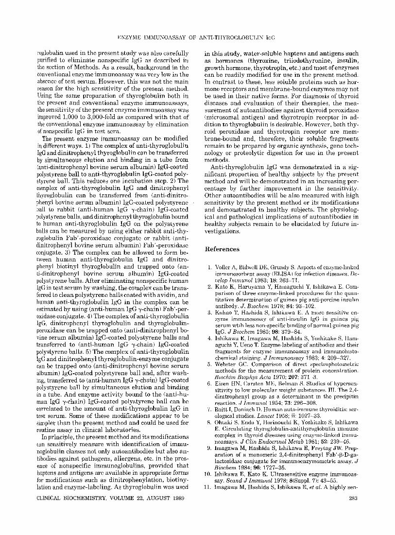
Sera samples from three hea l thy subjects, which gave fluorescence intensit ies of 110,130 and 275 and a serum sample from a pat ient with Graves ' disease, which gave a fluorescence intensi ty of 170, were preincubated with bovine thyroglobulin and were subjected to the present enzyme immunoassay. Litt le change in the fluorescence intensi ty for bound peroxidase activity was observed, indicating no cross-reaction of ant i- thyroglobulin IgG in these sera with bovine thyroglobulin. A typical result is shown in Figure 4.
>,,
{.9
0
" O
© cfD
0
>,,,
L9 e -
0
i ,
lOG
10
A L~
1 I II I I I I
0 0.015 0.15 1.5 15
Thyroglobul in Added (pmol/ tube)
Figure 4--Absence of cross-reaction with bovine thyroglob- ulin. A serum sample from a patient with Graves' disease was preincubated with human thyroglobulin [circles) or bovine thyroglobulin (triangles) and was subjected to the present enzyme lmmunoassay.
D i s c u s s i o n
Halber t et al. discussed tha t background in the con- ventional enzyme immunoassay for anti- thyroglobulin IgG migh t have been lowered by el iminat ion of non- specific IgG from the prepara t ion of thyroglobulin im- mobilized onto solid phase (14). The prepara t ion of thy-
TABLE 2 Serum Level of hTSH, T4 and T3 in Normal Subjects and Patients with Graves' Disease
and Chronic Thyroiditis
Serum Level, Mean -+ SD (Range)
Subject Sex Number Age hTSH T 4 , T3 yr mU/L ~g/L ~g/L
Normal Male 9 21-59 1.4 +- 0.71 89 -+ 26 1.2 -+ 0.09 (0.45-2.5) (51-110) (1.1-1.3)
Female 18 21-70 1.3 -+ 0.61 91 -+ 21 1.2 -+ 0.13 (0.43-2.3) (61-120) (1.1-1.5)
Graves' Male 6 14-63 0.061 -+ 0.052 220 -+ 54 4.8 -+ 2.6 Disease (0.013-0.15) (150-300) [2.3-8.0)
Female 19 15-74 0.072 -+ 0.054 210 -+ 41 4.6 -- 2.1 (0.006-0.17) (120-240) (1.8-8.0)
Chronic Male 2 27, 69 5.6, 6.8 62, 14 1.1, 0.6 Thyroiditis
Female 13 11-87 53 -+ 86 53 -+ 26 1.1 -+ 0.3 (2.2-300) (10-99) (0.5 -+ 1.5)
282 CLINICAL BIOCHEMISTRY, VOLUME 22, AUGUST 1989
ENZYME IMMUNOASSAY OF ANTI-THYROGLOBULIN IGG
roglobulin used in the present study was also carefully purified to eliminate nonspecific IgG as described in the section of Methods. As a result, background in the conventional enzyme immunoassay was very low in the absence of test serum. However~ this was not the main reason for the high sensitivity of the present method. Using the same preparat ion of thyroglobulin both in the present and conventional enzyme immunoassays, the sensitivity of the present enzyme immunoassay was improved 1,000 to 3,000-fold as compared with tha t of the conventional enzyme immunoassay by elimination of nonspecific IgG in test sera.
The present enzyme immunoassay can be modified in different ways. 1) The complex of anti- thyroglobulin IgG and dinitrophenyl thyroglobulin can be t ransferred by simultaneous elution and binding in a tube from (anti-dinitrophenyl bovine serum albumin) IgG-coated polystyrene ball to anti- thyroglobulin IgG-coated poly- styrene ball. This reduces one incubation step. 2) The complex of anti- thyroglobulin IgG and dinitrophenyl thyroglobulin can be t ransferred from (anti-dinitro- phenyl bovine serum albumin) IgG-coated polystyrene ball to rabbit (ant i-human IgG ~-chain) IgG-coated polystyrene balls, and dinitrophenyl thyroglobulin bound to human anti-thyroglobulin IgG on the polystyrene balls can be measured by using ei ther rabbit anti-thy- roglobulin Fab'-peroxidase conjugate or rabbit ~anti- dinitrophenyl bovine serum albumin) Fab'-peroxidase conjugate. 3) The complex can be allowed to form be- tween human anti- thyroglobulin IgG and dinitro- phenyl biotinyl thyroglobulin and trapped onto (an- ti-dinitrophenyl bovine serum albumin) IgG-coated polystyrene balls. After el iminating nonspecific human IgG in test serum by washing, the complex can be trans- ferred to clean polystyrene balls coated with avidin, and human anti-thyroglobulin IgG in the complex can be estimated by using (ant i-human IgG ~/-chain) Fab'-per- oxidase conjugate. 4) The complex of anti- thyroglobulin IgG, dinitrophenyl thyroglobulin and thyroglobulin- peroxidase can be trapped onto (anti-dinitrophenyl bo- vine serum albumin) IgG-coated polystyrene balls and transferred to (ant i -human IgG ~-chain) IgG-coated polystyrene balls. 5) The complex of anti- thyroglobulin IgG and dinitrophenyl thyroglobulin-enzyme conjugate can be trapped onto (anti-dinitrophenyl bovine serum albumin) IgG-coated polystyrene ball and, after wash- ing, transferred to (anti-human IgG ~-chain) IgG-coated polystyrene ball by simultaneous elution and binding in a tube. And enzyme activity bound to the (anti-hu- man IgG ~-chain) IgG-coated polystyrene ball can be correlated to the amount of anti- thyroglobulin IgG in test serum. Some of these modifications appear to be simpler than the present method and could be used for routine assay in clinical laboratories.
In principle, the present method and its modifications can sensitively measure with identification of immu- noglobulin classes not only autoantibodies but also an- tibodies against pathogens, allergens, etc. in the pres- ence of nonspecific immunoglobulins, provided that haptens and antigens are available in appropriate forms for modifications such as dinitrophenylation, biotiny- lation and enzyme-labeling. As thyroglobulin was used
CLINICAL BIOCHEMISTRY, VOLUME 22, AUGUST 1989
in this study, water-soluble haptens and antigens such as hormones (thyroxine, tri iodothyronine, insulin, growth hormone, thyrotropin, etc.) and most of enzymes can be readily modified for use in the present method. In contrast to these, less soluble proteins such as hor- mone receptors and membrane-bound enzymes may not be used in their native forms. For diagnosis of thyroid diseases and evaluation of their therapies, the mea- surement of autoantibodies against thyroid peroxidase (microsomal antigen) and thyrotropin receptor in ad- dition to thyroglobulin is desirable. However, both thy- roid peroxidase and thyrotropin receptor are mem- brane-bound and, therefore, their soluble fragments remain to be prepared by organic synthesis, gene tech- nology or proteolytic digestion for use in the present methods.
Anti-thyroglobulin IgG was demonstrated in a sig- nificant proportion of heal thy subjects by the present method and will be demonstrated in an increasing per- centage by fur ther improvement in the sensitivity. Other autoantibodies will be also measured with high sensitivity by the present method or its modifications and demonstrated in heal thy subjects. The physiolog- ical and pathological implications of autoantibodies in heal thy subjects remain to be elucidated by future in- vestigations.
R e f e r e n c e s
1. Voller A, Bidwell DE, Grundy S. Aspects of enzyme-hnked immunosorbent assay (ELISA) for infection diseases. De- velop Immunol 1983; 18: 263-71.
2. Kato K, Haruyama Y, Hamaguchi Y, Ishikawa E. Com- parison of three enzyme-linked procedures for the quan- titative determination of guinea pig anti-porcine insuhn antibody. J. Bmchem 1978; 84: 93-102.
3. Kohno T, Hashida S, Ishlkawa E. A more sensitive en- zyme immunoassay of anti-insulin IgG in guinea pig serum wt~h less non-specific binding of normal guinea pig IgG. J Bzochem 1985; 98: 379-84.
4. Ishikawa E, Imagawa M, Hashida S, Yoshitake S, Ham- aguchi Y, Ueno T. Enzyme-labeling of antibodies and their fragments for enzyme immunoassay and immunoh~sto- chemical staining. J Immunoassay 1983; 4: 209-327.
5. Webster GC. Comparison of direct spectrophotometric methods for the measurement of protein concentration. Bwchim Bzophys Acta 1970; 207: 371-3.
6. Eisen HN, Carsten ME, Belman S. Studies of hypersen- sitivity to low molecular weight substances. III. The 2,4- dimtrophenyl group as a determinant m the precipitin reaction. J Immunol 1954; 73: 296-308.
7. Roitt I, Doniach D. Human auto-immune thyroiditis: ser- ological studies. Lancet 1958; ii: 1027-33.
8. Ohtaki S, Endo Y, Horinouchi K, Yoshitake S, Ishikawa E. Circulating thyroglobulin-antithyroglobulin immune complex in thyroid diseases using enzyme-linked immu- noassays. J Clin Endocr~nol Metab 1981; 52: 239-46.
9. |magawa M, Hashida S, Ishikawa E, Freytag JW. Prep- aration of a monomeric 2,4-dinitrophenyl Fab'-~-D-ga- lactosidase conjugate for immunoenzymometric assay. J Biochem 1984; 96: 1727-35.
10. Ishikawa E, Kato K. Ultrasensitive enzyme immunoas- say. Scand J Immunol 1978; 8(Suppl. 7): 43-55.
11. Imagawa M, Hashida S, Ishikawa E, et al. A highly sen-
283
KOHNO, MITSUKAWA, MATSUKURA, TSUNETOSHI, AND ISHIKAWA
sltive sandwich enzyme immunoassay for insuhn in hu- man serum developed using capybara anti- insuhn Fab '- horseradish peroxidase conjugate. Anal Lett 1983; 16(B19): 1509-23.
12. Hashida S, Imagawa M, Inoue S, Ruan K-h, Ishikawa E. More useful malelmide compounds for the conjugation of Fab' to horseradish peroxidase through thiol groups in the hinge. J Appl Biochem 1984; 6: 56-63.
13. Inoue S, Hashida S, Ishikawa E, et al. Highly sensitive
sandwich enzyme immunoassay for human thyroid-stim- ulating hormone (hTSH) in serum using monoclonal anti- hTSH ~-subunit IgGl-coated polystyrene balls and poly- clonal ant i -human chorionic gonadotropin Fab'-horse- radish peroxidase conjugate. Anal Lett 1986; 19~7&8): 845-61.
14. Halbert SP, Bastomsky CH, Anken M. A rapid standard- ized enzyme immunoassay for autoantibodies to thyro- globulin. Clin Chim Acta 1983: 127: 69-76.
284 CLINICAL BIOCHEMISTRY, VOLUME 22, AUGUST 1989