maureen n. hood, phd, rn, rt (mr), fsmrt - nasci.org mr safety... · ventilators clothing...
TRANSCRIPT
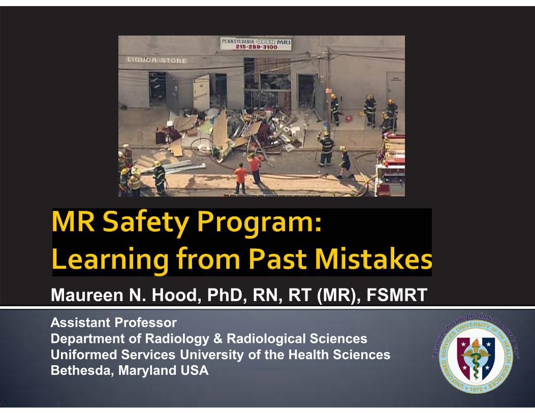
Maureen N. Hood, PhD, RN, RT (MR), FSMRT
Assistant Professor Department of Radiology & Radiological Sciences Uniformed Services University of the Health Sciences Bethesda, Maryland USA
� The opinions or assertions contained herein are the private views of the authors and are not to be construed as official or reflecting the views of the Uniformed Services University of the Health Sciences or the Department of Defense of the United States of America.
� I have an in-kind research relationship with GE Healthcare.

� Physical layout � Staff � Training � Policies
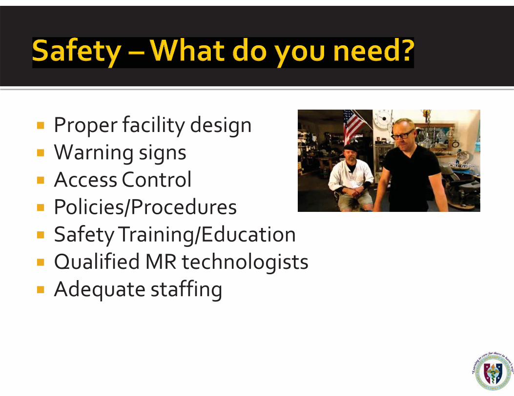
� Proper facility design � Warning signs � Access Control � Policies/Procedures � Safety Training/Education � Qualified MR technologists � Adequate staffing

� All you need is a walk-through ferromagnetic detector and a tech.
� Wrong!!! � Need multiple layers of protection � Nothing can replace a good MR Technologist � Must use screening forms AND interviews � Use tools such as hand held magnets,
ferromagnetic detectors and hand held wands to check for incidental items as needed.
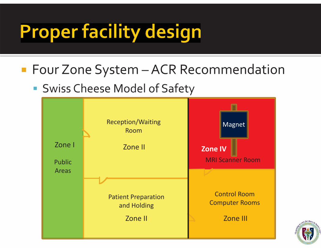
� Four Zone System – ACR Recommendation � Swiss Cheese Model of Safety
Zone I Zone II Zone IV
Zone III Zone II
Reception/Waiting Room
Patient Preparation and Holding
Control Room Computer Rooms
MRI Scanner Room
Magnet
Public Areas
� Zone I � Freely accessible to the general public. � Outside the actual MRI environment.
� Zone II
� Interface between the public area and the controlled MRI environment.
� In general – this is the reception area for MRI. Patients and non-MRI personnel check-in here.

� Zone III - Controlled access area. � ONLY MR Personnel have free access to Zone III.
� Restricted at all times.
� No access by any non-MR persons unless escorted by MRI personnel.
� Zone IV - The MR scan room. � Only screened persons may enter.
� Pts, visitors, staff � Only screened and approved equipment. � NO Exceptions - The scanner is ALWAYS ON!
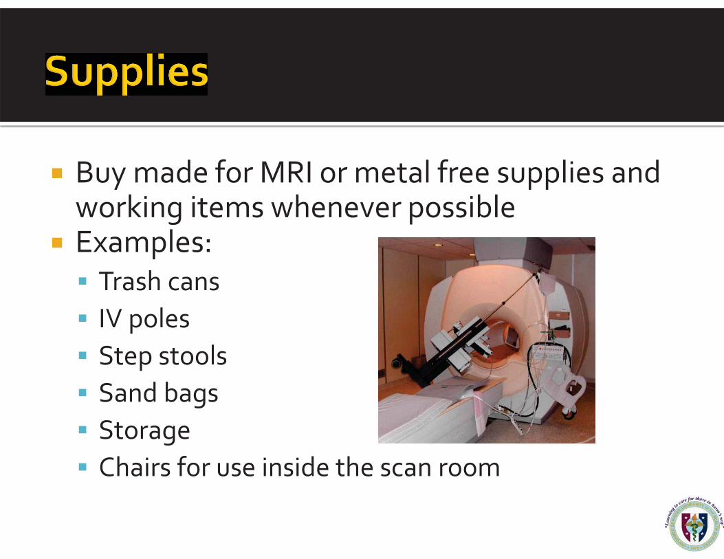
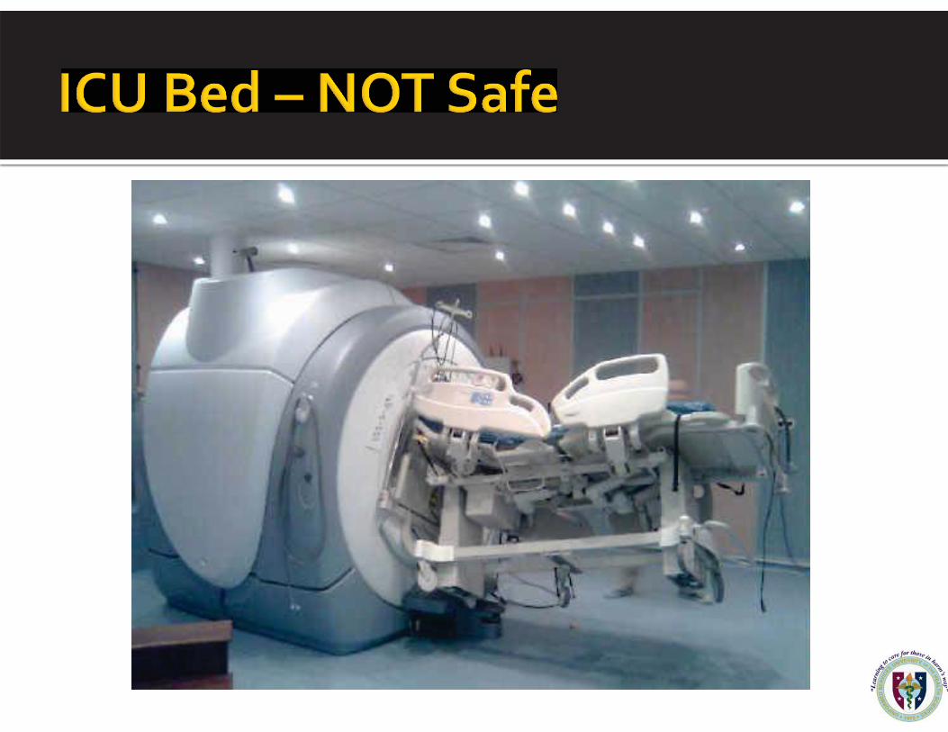
� Buy made for MRI or metal free supplies and working items whenever possible
� Examples: � Trash cans � IV poles � Step stools � Sand bags � Storage � Chairs for use inside the scan room

• MR Safe: an item that poses no known hazards in all MRI environments.
• MR Conditional: an item that has been demonstrated to pose no known hazards in a specified MRI environment with specified conditions of use. Additional conditions, including specific configurations of the item, may be required.
• MR Unsafe: an item that is known to pose hazards in all MRI
environments. MR Unsafe items include magnetic items such as a pair of ferromagnetic scissors.
American Society of Testing and Materials

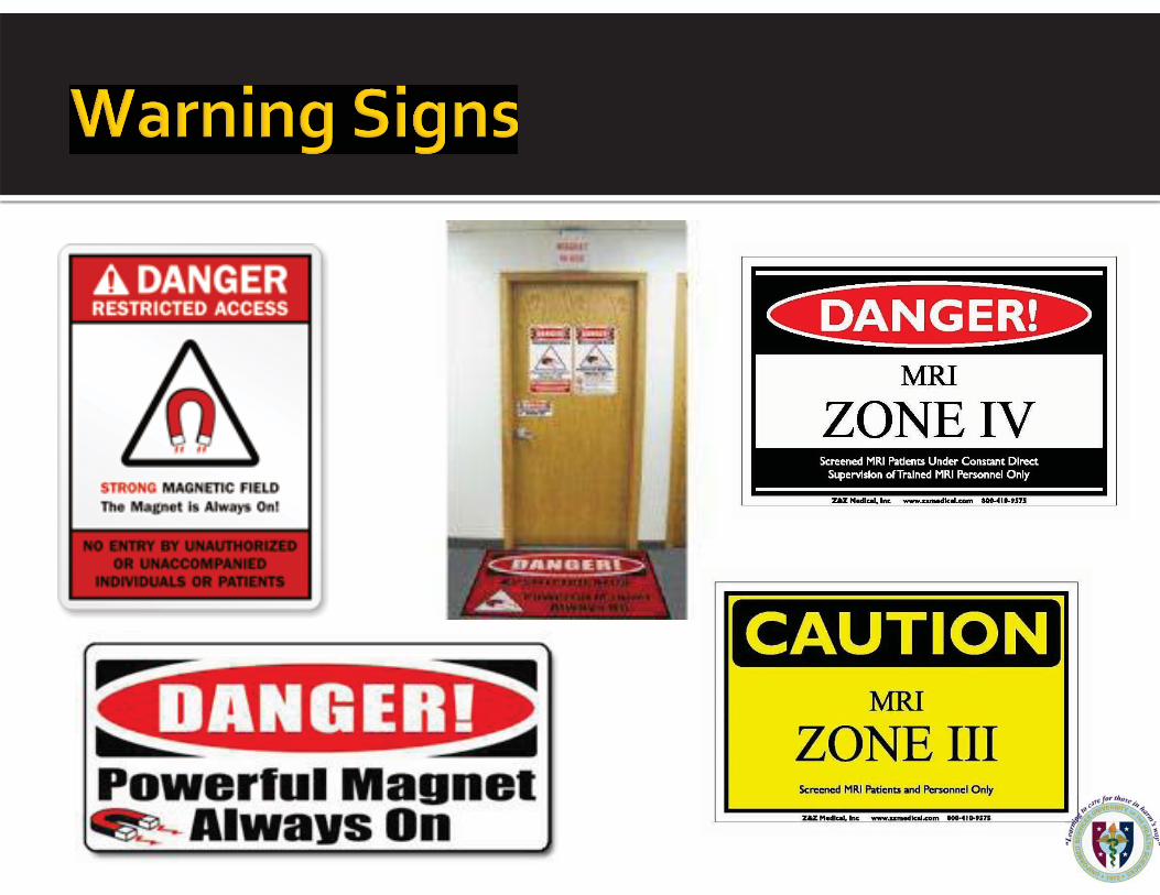
� Warning signs work great.
� Do not rely solely on signs � Many people don’t read signs � People think the signs don’t pertain to them � People think the scanner is off when it is quiet
� So wrong, yet so common � Never let your guard down
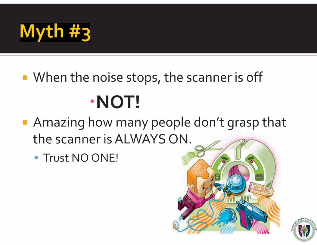
� When the noise stops, the scanner is off
�NOT! � Amazing how many people don’t grasp that
the scanner is ALWAYS ON. � Trust NO ONE!

� MR technologists must have advanced knowledge of MRI safety � MR Registry � Continued education and training in MR safety
� Never work solo � Dangerous on many levels � Minimum:
▪ 2 techs, or ▪ Tech + aide

� Radiologists working in MR must have advanced training in MR safety
� Radiologists ultimately make the decisions on who gets scanned � Risk / Benefit analysis � Radiologist need to support their technologists

� Blanket policies are best � Very dangerous – there’s always an exception � Example: All stents are safe
▪ Not true – best to know what the stent is to make a logical decision
▪ Stacked? Is there a potential heating issue? ▪ 1.5 T, 3T? 7T?
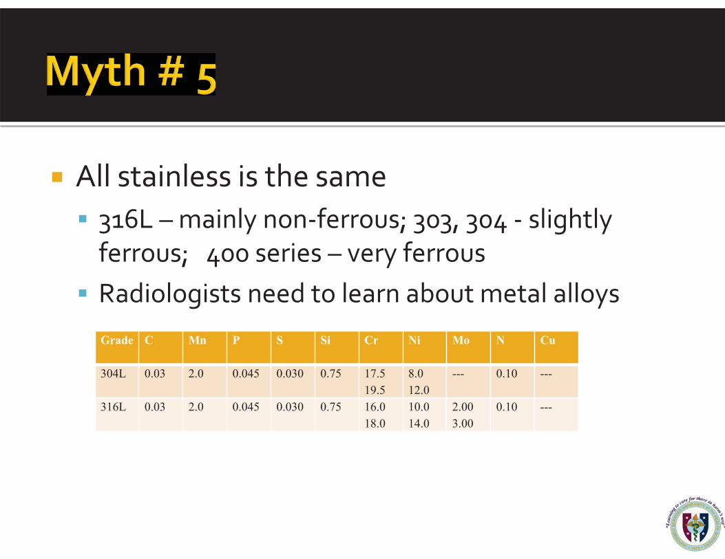
� All stainless is the same � 316L – mainly non-ferrous; 303, 304 - slightly
ferrous; 400 series – very ferrous � Radiologists need to learn about metal alloys
Grade C Mn
P
S
Si
Cr
Ni
Mo
N
Cu
304L
0.03
2.0
0.045
0.030
0.75
17.5 19.5
8.0 12.0
---
0.10
---
316L
0.03
2.0
0.045
0.030
0.75
16.0 18.0
10.0 14.0
2.00 3.00
0.10
---

� All MR staff given regular MR safety training � Radiologists & MR Techs
� Advanced Training � Administrative Staff
� Basics, plus roles in an emergency � Anesthesiology, sedation teams, etc
� Basic + safety pertaining to their working environment
� Other regular users
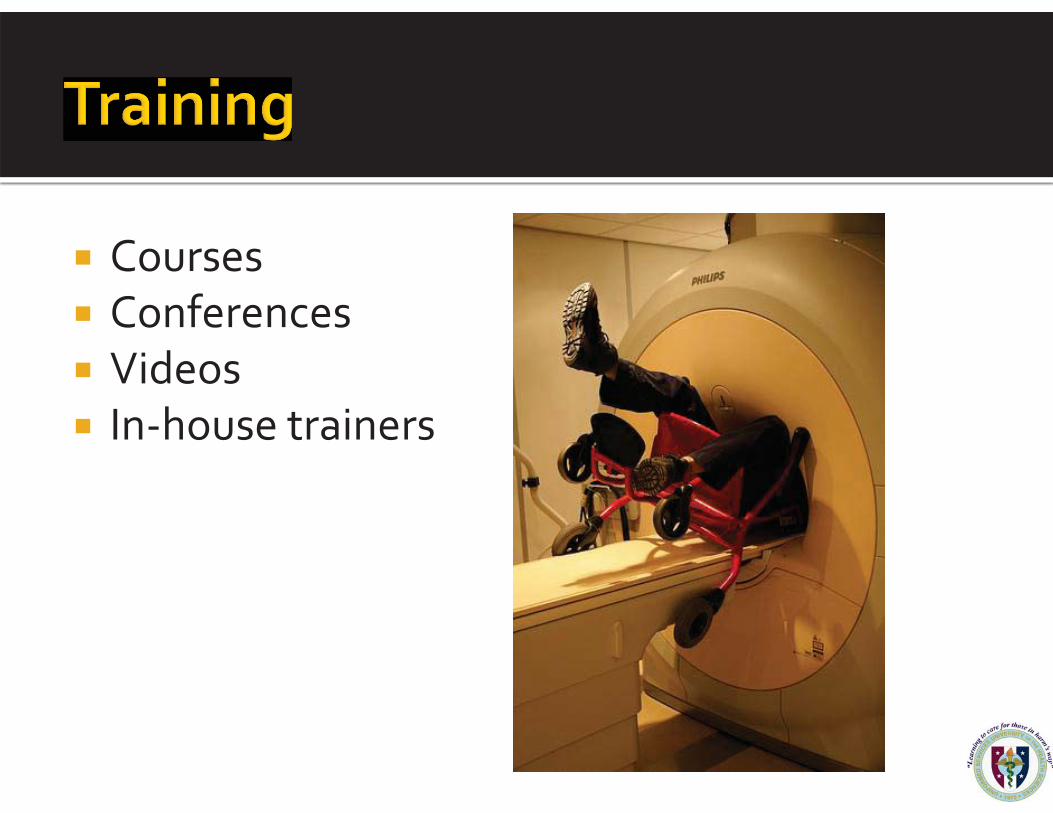
� Courses � Conferences � Videos � In-house trainers
� All scanners are the same � Field strengths, surface coils, gradient coils, etc.
vary � Hot spots cannot be predicted in advance – too
many variables � Bare skin touching can cause burns, especially at
higher field strengths

� SOP must include safety policies � Implants � IV infusion pumps � Ventilators � Clothing � Family/Visitors � 100% supervision of non-MR people in Zones
III & IV.

� Safety Training � annually
� Screening � form and interview
� Increased safety for Tesla systems above 1.5T
� Medication/Monitoring � Follow institution policies
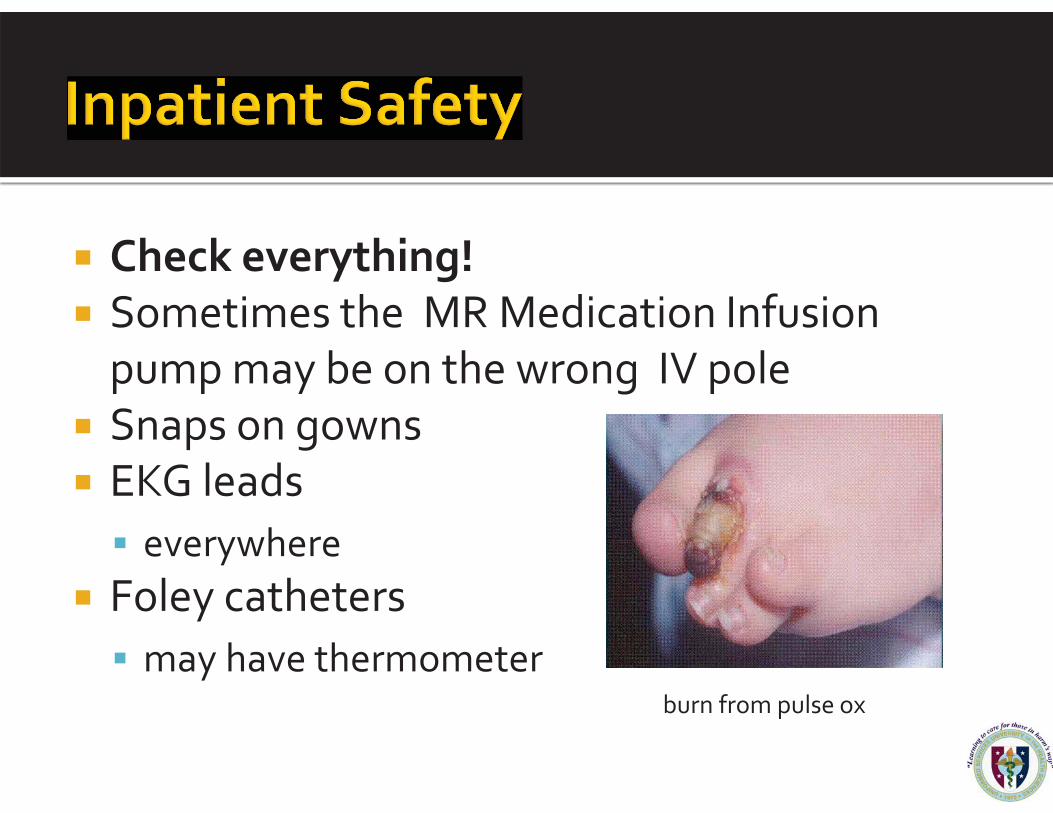
� Check everything! � Sometimes the MR Medication Infusion
pump may be on the wrong IV pole � Snaps on gowns � EKG leads
� everywhere � Foley catheters
� may have thermometer
burn from pulse ox
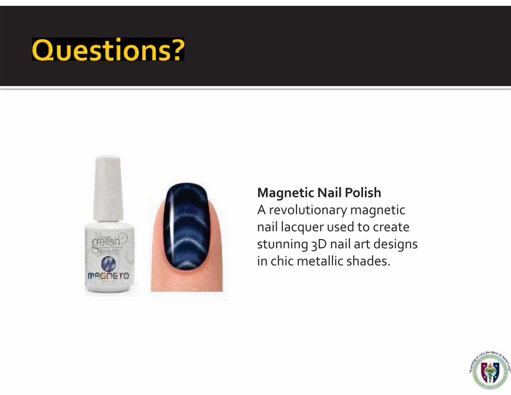
Magnetic Nail Polish A revolutionary magnetic nail lacquer used to create stunning 3D nail art designs in chic metallic shades.
� American Society for Testing and Materials (ASTM) International. (2005). Designation: F2503-08, Standard Practice for Marking Medical Devices and Other Items for Safety in the Magnetic Resonance Environment. ASTM International, West Conshohocken, PA. Retrieved November 20, 2011 from http://www.astm.org/Standards/F2503.htm.
� Becerra, R. J., Harsh, M. J., Boskamp, E. B., Guclu, C. C., Duby,T., Havens, T. J., Hink, R. S., Noonan, J. P., Peterson, W. T., Piepenberg, S. J., Stormont, R. S., & Vavrek, R. M. (2006). Instrumentation: Magnets, gradients, and coils. In Edelaman, R. R., Hesselink, J. R., Zlatkin, M.B., Crues, J. V. III (Eds.), Clinical Magnetic Resonance Imaging (pp 105-136). Philadelphia: Saunders, Elsevier.
� Bushberg, J.T., Seibert, J. A., Leidholdt E. M., & Boone, J. M. (1994). Magnetic resonance imaging. In Bushberg, J.T., Seibert, J. A., Leidholdt E. M., & Boone, J. M. (Ed.), The Essential Physics of Medical Imaging (pp 291-366). Baltimore, Maryland: Williams & Wilkins.
� Edelman, R. R., Dunkle, E. E., Li, W., Kissinger, K. V., & Thangaraj, K. (2006). Practical considerations and image optimization. In Edelaman, R. R., Hesselink, J. R., Zlatkin, M.B., Crues, J. V. III (Eds.), Clinical Magnetic Resonance Imaging (pp 58-104). Philadelphia: Saunders, Elsevier.
� Garcia-Bournissen, F., Shrim, A., & Koren, G. (2006 ). Safety of gadolinium during pregnancy. Canadian Family Physician, 52:309-10. � Gilk, T., & Latino, R. J. (2011). MRI safety 10 years later: What we learned from the accident that killed Michael. Patient Safety & Quality
Healthcare. Retrieved November 20 2011 from http://www.reliability.com/mri/. � Haik, J., Daniel, S., Tessone, A., Orenstein, A., & Winkler, E. (2009). MRI induced fourth-degree burn in an extremity, leading to amputation.
Burns, 35(2), 294-296. Hallett, M., & Cohen, L.G. (1989). Magnetism. A new method for stimulation of nerve and brain. Journal of the American Medical Association, 262(4), 538-541.
� Hornak, J. P. (2011). The Basics of NMR. Retrieved November 20, 2011 from http://www.cis.rit.edu/htbooks/nmr/chap-3/chap-3.htm. � Jauchem JR. Effects of drugs on thermal responses to microwaves. General Pharmacology, 1985;16(4):307-10. � Jerrolds, J. & Keene, S. (2009). MRI Safety at 3T versus 1.5T. The Internet Journal of World Health and Societal Politics. 6,1. � Kanal, E., Barkovich, A.J., Bell, C., Borgstede, J.P., Bradley, W.G. Jr., Froelich, J.W., Gilk, T, Gimbel, J.R., Gosbee, J., Kuhni-Kaminski, E.,
Lester, J.W. Jr., Nyenhuis, J., Parag, Y.,Schaefer, D.J., Sebek-Scoumis, E.A., Weinreb, J., Zaremba, L.A., Wilcox, P., Lucey, L., Sass, N.; ACR Blue Ribbon Panel on MR Safety. ACR guidance document for safe MR practices: (2007). American Journal of Roentgenology, 188(6):1447-74. Kanal, E., Shellock, F. G.(1990). Burns associated with clinical MR examinations. Radiology 1990;175:595.
� Lange, S., & Nguyen, Q. N. (2006) Cables and electrodes can burn patients during MRI. Nursing, 36, (11), 18. � Levine, G.N., Gomes, A.S., Arai, A.E., Bluemke, D.A., Flamm, S.D., Kanal, E., Manning, W.J., Martin, E.T, Smith, J.M, Wilke, N.,
Shellock, F.S.; American Heart Association Committee on Diagnostic and Interventional Cardiac Catheterization; American Heart Association Council on Clinical Cardiology; American Heart Association Council on Cardiovascular Radiology and Intervention. (2007). Safety of magnetic resonance imaging in patients with cardiovascular devices: an American Heart Association scientific statement from the Committee on Diagnostic and Interventional Cardiac Catheterization, Council on Clinical Cardiology, and the Council on Cardiovascular Radiology and Intervention: endorsed by the American College of Cardiology Foundation, the North American Society for Cardiac Imaging, and the Society for Cardiovascular Magnetic Resonance. Circulation. 11;116(24), 2878-91.Levitt, M., Benjamin, V., & Kricheff, I. I. (1990). Potential misinterpretation of cervical spondylosis with cord compression caused by metallic artifacts in magnetic resonance imaging of the postoperative spine. Neurosurgery. 27(1):126-9. McRobbie, D. W., Moore, E. A., Graves, M J., & Prince, M. R. (2007). Welcome to the MR unit. In: McRobbie, D. W., Moore, E. A., Graves, M J., & Prince, M. R. (Eds.) MRI: From Picture to Proton, 2nd ed (pp 11-29). New York: Cambridge Press.
� Miller. G. (1987). Exposure guidelines for magnetic fields. American Industrial Hygiene Association Journal, 48, 957-968. � Mugler, J. P. III. (2006). Basic Principles. In Edelaman, R. R., Hesselink, J. R., Zlatkin, M.B., Crues, J. V. III (Eds.), Clinical Magnetic
Resonance Imaging (pp 23-57). Philadelphia: Saunders, Elsevier. � Nazarian, S., Hansford, R., Roguin, A., Goldsher, D., Zviman, M.M., Lardo, A.C., Caffo, B.S., Frick, K.D., Krau,t M.A., Kamel, I.R.,
Calkins, H., Berger, R.D., Bluemke, D.A., Halperin, H.R. (2011). A prospective evaluation of a protocol for magnetic resonance imaging of patients with implanted cardiac devices. Annals of Internal Medicine. 4;155(7), 415-24.Nordbeck, P., Weiss, I., Ehses, P., Ritter ,O., Warmuth, M., Fidler, F., Herold, V., Jakob, P.M., Ladd, M.E., Quick, H.H., & Bauer, W.R. (2009). Measuring RF-induced currents inside implants: Impact of device configuration on MRI safety of cardiac pacemaker leads. MagneticResonance in Medicine. 61(3), 570-8. Schaefer, D.J. (1993). Bioeffects of MRI and Patient Safety. In: Bronskill, M.J, Sprawls, P., (Ed.). The Physics of MRI: 1992 AAPM Summer School Proceedings (pp 607-646). Woodbury, NY: American Association of Physics in Medicine.

� Nielsen-Bohlman, L., Panzer, A. M., Hamlin, B., & Kindig, D. A., Eds. Committee on Health Literacy Board on Neuroscience and Behavioral Health. (2004). Health Literacy A Prescription to End Confusion. Retrieved 10 February 2012 from http://hospitals.unm.edu/health_literacy/pdfs/HealthLiteracyExecutiveSummary.pdf
� Shellock, F. G. (2011). Reference Manual for Magnetic Resonance Safety, Implants, & Devices: 2011 edition. Los Angeles, CA: Biomedical Research Publishing Group.
� Shellock F. G., & Kanal E. (1996). Burns associated with the use of monitoring equipment during MR procedures. Journal of Magnetic Resonance Imaging, 6, 271–272.
� Shellock, F. G., & Shellock, V. J. (1998). Cardiovascular catheters and accessories: ex vivo testing of ferromagnetism, heating, and artifacts associated with MRI. Journal of Magnetic Resonance Imaging, 8, 1338–1342.
� Shellock, F.G., Woods, T.O., & Crues, J.V. (2009). MR Labeling Information for Implants and Devices: Explanation of Terminology. Radiology, 253: 26-30.
� The Joint Commission. (2010). Medical Staff (CAMH/Hospitals): Permission to Administer Moderate Sedation. Retrieved 4 December 2011 from http://www.jointcommission.org/standards_information/jcfaqdetails.aspx?StandardsFAQId=230&StandardsFAQChapterId=74
� U.S. Department of Health and Human Services, Food and Drug Administration, Center for Devices and Radiological Health. (1998). Guidance for Industry: Guidance for the Submission of Premarket Notifications for Magnetic Resonance Diagnostic Devices. Retrieved November 30 2011 from http://www.fda.gov/MedicalDevices/DeviceRegulationandGuidance/GuidanceDocuments/ucm073817.htm.
� U.S. Department of Health and Human Services, Food and Drug Administration, Center for Devices and Radiological Health. (2008). Guidance for Industry and FDA Staff: Establishing Safety and Compatibility of Passive Implants in the Magnetic Resonance (MR) Environment. Retrieved November 30, 2011 from http://www.fda.gov/cdrh/osel/guidance/1685.pdf.
� U.S. Department of Health and Human Services, Food and Drug Administration, Center for Devices and Radiological Health. (2009). Alerts and Notices (Medical Devices). Retrieved November 30, 2011 from http://www.fda.gov/MedicalDevices/Safety/AlertsandNotices/TipsandArticlesonDeviceSafety/ucm064761.htm.
� U.S. Department of Health and Human Services, Food and Drug Administration, Center for Devices and Radiological Health. (2011). Revo MRI SureScan Pacing System - P090013. Retrieved November 30, 2011 from http://www.fda.gov/MedicalDevices/ProductsandMedicalProcedures/DeviceApprovalsandClearances/Recently-ApprovedDevices/ucm244469.htm.