management of multiple impacted teeth: a case … reviews the diagnosis and management of impacted...
TRANSCRIPT

Management of multiple missing teeth … Ajith SD et al Journal of International Oral Health 2014; 6(3):93-98
93
Case ReportReceived: 2nd December 2013 Accepted: 13th March 2014 Conflict of Interest: None
Source of Support: Nil
Management of Multiple Impacted Teeth: A Case Report and ReviewSreedevi D Ajith1, Smitha Shetty2, Huma Hussain3, Tejavathy Nagaraj4, M Srinath5
Contributors:1Professor & Head, Department of Orthodontics and Dentofacial Orthopedics, Sri Rajiv Gandhi College of Dental Sciences and Hospital, Bangalore, Karnataka, India; 2Senior Lecturer, Department of Orthodontics and Dentofacial Orthopedics, Sri Rajiv Gandhi College of Dental Sciences and Hospital, Bangalore, Karnataka, India; 3Post Graduate Student, Department of Orthodontics and Dentofacial Orthopedics, Sri Rajiv Gandhi College of Dental Sciences and Hospital, Bangalore, Karnataka, India; 4Professor & Head, Department of Oral Medicine and Radiology, Sri Rajiv Gandhi College of Dental Sciences and Hospital, Bangalore, Karnataka, India; 5Professor, Department of Orthodontics and Dentofacial Orthopedics, Narayana Dental College, Nellore, Andhra Pradesh, India.Correspondence:Dr. Sreedevi D Ajith. Department of Orthodontics and Dentofacial Orthopedics, Sri Rajiv Gandhi College of Dental Sciences and Hospital, Bangalore, Karnataka, India. Phone: +91-9449649496. Email: [email protected] to cite the article:Ajith SD, Shetty S, Hussain H, Nagaraj T, Srinath M. Management of multiple impacted teeth: A case report and review. J Int Oral Health 2014;6(3):93-8.Abstract:Interdisciplinary care for the management of impacted teeth provides a holistic method of treating patients. Careful planning is necessary to reach the desired treatment goals. This article attempts to highlight the importance of diagnosis and adequate treatment planning for successful eruption of impacted teeth. The concept of forced eruption to improve the bone morphology of the impacted teeth has been used to treat a case of multiple impacted teeth. This paper reviews the diagnosis and management of impacted teeth. A case report of multiple impacted maxillary anterior teeth of a 13-year-old female patient has been presented.
Key Words: Bone morphology, forced eruption, multiple impacted teeth
IntroductionImpacted teeth are those that will not erupt completely. This is based on clinical and radiographic assessment. Abnormal eruption paths within the dentoalveolar process may result in impactions and serious clinical ramifications.1-3 Orthodontic intervention to bring these impacted teeth into the line of occlusion is important for long-term function and stability.
IncidenceThe classic distribution in order of frequency of impaction of permanent teeth can be summarized as follows: Lower third molars, upper third molars, upper canines, upper and lower
premolars, upper incisors, lower canines, lower incisors, upper and lower first molars and upper and lower second molars.2,3
The prevalence of maxillary permanent canine impaction is 1-2% in the general population.4,5 This is most likely due to an extended development period, and the long, tortuous path of eruption before the canine emerges into full occlusion.6-8 Failure of eruption of the mandibular canine is an unusual event. The incidence of maxillary canine impaction is in the range of 0.8-2.8% and the prevalence is 0.9-2.2%. Studies have shown increased female predilection of impaction than males.9-11
EtiologyPrimary reasons for impacted teeth are genetics,12 endocrine deficiency, irradiation, palatal clefts, developmental abnormalities, dento maxillary disharmony, late or missing root development, growth disharmony between pre-maxilla and maxilla, and transverse growth deficiency of the anterior maxilla.13
The secondary reasons are loss of guidance of the lateral incisor (microdontia or absence),14-16 trauma, premature extraction causing space problems by crowding of the anterior segment, root malformation, pericoronary pathology, ectopic germ position, thick fibrous tissue, 17 mesiodistal dimension of the nasal fossae, unerupted canine at the borderline of a palatal cleft. 18
InvestigationsA through diagnosis is essential to detect and prevent impactions. This includes a detailed familial history, clinical inspection, and palpation between the ages of 9-10 years and also a thorough radiographic examination. Radiographic evaluation will include intra oral periapical films, tube‑shift technique or Clark’s same lingual opposite buccal (SLOB) rule, occlusal films, frontal and lateral cephalograms, panoramic films, computed tomography or cone beam computed tomography.
TreatmentIn cases where deciduous teeth are present along with impacted teeth it should be noted that the long-term prognosis for retaining the deciduous teeth is poor, regardless of the length of the root and crown height. Because of the poor prognosis, the deciduous teeth will need to be extracted at a later date. Various treatment options for correcting impacted teeth will include the following:1. Autotransplantation2. Extraction and movement of the adjacent teeth in its
position
94
Management of multiple missing teeth … Ajith SD et al Journal of International Oral Health 2014; 6(3):93-98Management of multiple missing teeth … Ajith SD et al Journal of International Oral Health 2014; 6(3):93-98
3. Extraction of the impacted canine and use of segmental osteotomy to move the posterior section forward
4. Replacement of the impacted teeth if they cannot be saved5. Surgical exposure of the impacted teeth and orthodontic
traction to bring the tooth into the line of occlusion. The final option is obviously the most desirable approach.
Ingber19 showed that in the concept of forced eruption the teeth could be erupted for the purpose of lengthening the clinical crown, altering the gingival margins, and leveling the osseous defects. This concept of forced eruption and its association with bone migration has been taken advantage of to treat a patient with multiple impacted teeth.20,21
The following is a description of a case report of multiple impacted teeth, which were treated by improving the 3D morphology of bone in that area and then guiding the eruption of the impacted teeth.
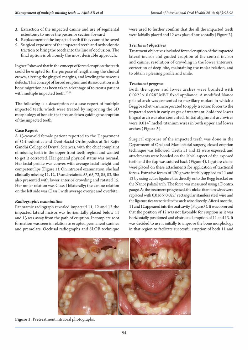
Case ReportA 13-year-old female patient reported to the Department of Orthodontics and Dentofacial Orthopedics at Sri Rajiv Gandhi College of Dental Sciences, with the chief complaint of missing teeth in the upper front teeth region and wanted to get it corrected. Her general physical status was normal. Her facial profile was convex with average facial height and competent lips (Figure 1). On intraoral examination, she had clinically missing 11, 12, 13 and retained 53, 65, 72, 85, 83. She also presented with lower anterior crowding and rotated 15. Her molar relation was Class I bilaterally; the canine relation on the left side was Class I with average overjet and overbite.
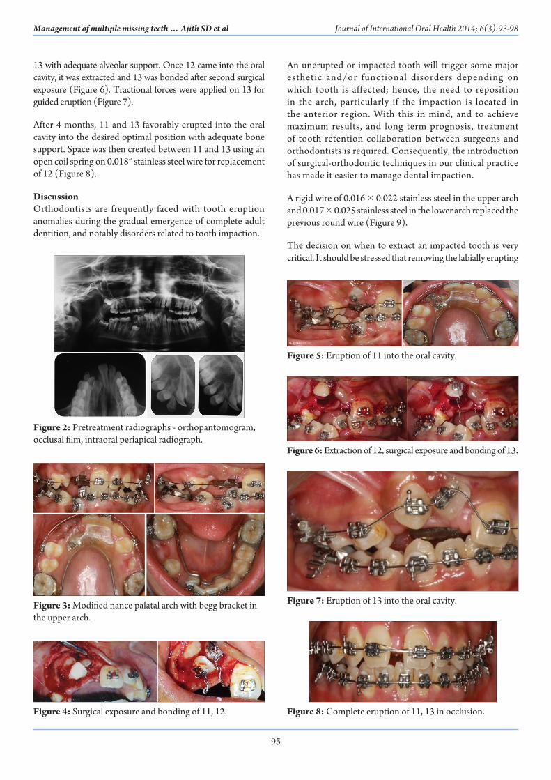
Radiographic examinationPanoramic radiograph revealed impacted 11, 12 and 13 the impacted lateral incisor was horizontally placed below 11 and 13 was away from the path of eruption. Incomplete root formation was seen in relation to erupted permanent canines and premolars. Occlusal radiographs and SLOB technique
were used to further confirm that the all the impacted teeth were labially placed and 12 was placed horizontally (Figure 2).
Treatment objectivesTreatment objectives included forced eruption of the impacted lateral incisor and guided eruption of the central incisor and canine, resolution of crowding in the lower anteriors, correction of deep bite, maintaining the molar relation, and to obtain a pleasing profile and smile.
Treatment progressBoth the upper and lower arches were bonded with 0.022″ × 0.028″ MBT fixed appliance. A modified Nance palatal arch was cemented to maxillary molars in which a Begg bracket was incorporated to apply traction forces to the impacted teeth in early stages of treatment. Soldered lower lingual arch was also cemented. Initial alignment archwires were 0.014” nickel titanium wires in both upper and lower arches (Figure 3).
Surgical exposure of the impacted teeth was done in the Department of Oral and Maxillofacial surgery, closed eruption technique was followed. Teeth 11 and 12 were exposed, and attachments were bonded on the labial aspect of the exposed teeth and the flap was sutured back (Figure 4). Ligature chains were placed on these attachments for application of tractional forces. Extrusive forces of 120 g were initially applied to 11 and 12 by using active ligature ties directly onto the Begg bracket on the Nance palatal arch. The force was measured using a Dontrix gauge. As the treatment progressed, the nickel titanium wires were replaced with 0.016 × 0.022” rectangular stainless steel wire and the ligature ties were tied to the arch wire directly. After 4 months, 11 and 12 appeared into the oral cavity (Figure 5). It was observed that the position of 12 was not favorable for eruption as it was horizontally positioned and obstructed eruption of 11 and 13. It was decided to use it initially to improve the bone morphology in that region to facilitate successful eruption of both 11 and
Figure 1: Pretreatment intraoral photographs.
95
Management of multiple missing teeth … Ajith SD et al Journal of International Oral Health 2014; 6(3):93-98Management of multiple missing teeth … Ajith SD et al Journal of International Oral Health 2014; 6(3):93-98
13 with adequate alveolar support. Once 12 came into the oral cavity, it was extracted and 13 was bonded after second surgical exposure (Figure 6). Tractional forces were applied on 13 for guided eruption (Figure 7).
After 4 months, 11 and 13 favorably erupted into the oral cavity into the desired optimal position with adequate bone support. Space was then created between 11 and 13 using an open coil spring on 0.018” stainless steel wire for replacement of 12 (Figure 8).
DiscussionOrthodontists are frequently faced with tooth eruption anomalies during the gradual emergence of complete adult dentition, and notably disorders related to tooth impaction.
An unerupted or impacted tooth will trigger some major esthetic and/or functional disorders depending on which tooth is affected; hence, the need to reposition in the arch, particularly if the impaction is located in the anterior region. With this in mind, and to achieve maximum results, and long term prognosis, treatment of tooth retention collaboration between surgeons and orthodontists is required. Consequently, the introduction of surgical-orthodontic techniques in our clinical practice has made it easier to manage dental impaction.
A rigid wire of 0.016 × 0.022 stainless steel in the upper arch and 0.017 × 0.025 stainless steel in the lower arch replaced the previous round wire (Figure 9).
The decision on when to extract an impacted tooth is very critical. It should be stressed that removing the labially erupting
Figure 2: Pretreatment radiographs - orthopantomogram, occlusal film, intraoral periapical radiograph.
Figure 4: Surgical exposure and bonding of 11, 12.
Figure 3: Modified nance palatal arch with begg bracket in the upper arch.
Figure 5: Eruption of 11 into the oral cavity.
Figure 6: Extraction of 12, surgical exposure and bonding of 13.
Figure 7: Eruption of 13 into the oral cavity.
Figure 8: Complete eruption of 11, 13 in occlusion.

96
Management of multiple missing teeth … Ajith SD et al Journal of International Oral Health 2014; 6(3):93-98Management of multiple missing teeth … Ajith SD et al Journal of International Oral Health 2014; 6(3):93-98
and out of arch canine is not indicated, despite its poor esthetic appearance. Improvement in the short term esthetics can be achieved with such an extraction; however it may complicate and compromise the orthodontic treatment results, including the ability to provide the patient with a functional occlusion. The extraction of the impacted tooth, especially the canine, may be considered only in the following situations: (1) If it is ankylosed and cannot be transplanted, (2) if it is undergoing external or internal root resorption, (3) dilacerations of the root, (4) if the impaction is severe or unfavorably placed for example if the canine is stuck between central incisor and lateral incisor, (5) if the occlusion is acceptable, with the first premolar in the position of the canine and with an otherwise functional occlusion without crowding, (6) presence cystic formation, infection and other pathologies, (7) If the patient does not desire orthodontic treatment.3-5
Various modalities for the management of impacted canines exist. Given below are some of them.
Interceptive treatmentAn impacted maxillary anterior tooth in a child is problematic because of its esthetic unacceptability. If the problem can be treated earlier, neither orthodontists nor parents want to wait for complete eruption of the permanent dentition before starting comprehensive orthodontic treatment. Nevertheless, both parents and patient should be informed of the possibility of failure before extensive measures are undertaken to save a severely impacted tooth.2
Best long-term results are gained by preventing upper canine impaction. This entails selectively extracting the deciduous teeth by ages 8 or 9. Williams used this interceptive approach when treating in Class I uncrowded cases with canine impaction. In almost 91% of cases, removing the deciduous canine if done before 11 years of age will allow the canine to erupt into its correct postion15 64% success rate is shown when the canine is impacted is mesial to the midline of the lateral incisor. 13,14
Corrective treatmentEarly diagnosis and interception of the probable impaction is the best approach in tis management. Orthodontic treatment followed by surgical exposure of the canine to bring it into its correct position in the arch should be given importance. Following are the procedures for the management of impacted canines:1. Open orthodontic eruption2. Closed orthodontic eruption3. Extraction of impacted tooth or teeth.
Some of the surgical techniques used to manage impacted maxillary canines are gingivectomy,10 apically positioned flap,10 closed eruption,11 closed flap,22 open eruption,5 open window eruption,22 and tunnel traction.23
Many techniques have been used to move impacted teeth into occlusion orthodontically. Some of the techniques are cantilever system24 which requires less frequent activation, temporary anchorage devices25 which provide absolute anchorage for tooth movement, double-archwire mechanics26 has the advantage of minimizing root resorption, easy-way-coil system,27auxiliary arm from transpalatal arch.28 Auxiliary spring29 and K-9 spring30 to name a few.
Orthodontists suggest that adequate space be created in the dental arch to allow for proper alignment of impacted teethe and later expose the tooth surgically so that a mechanical traction force can be applied to help erupt the tooth. It has been shown that many methods can efficiently create space for impacted teeth, but the easiest would be to use closed-coil springs and eyelets, provided noting is obstructing the path of the impacted tooth.
We should first determine whether the impacted tooth could be successfully aligned in its proper position on the basis of its position and orientation and the amount of root formation. It is important to plan when and how the impacted tooth will be moved to its final position, as well as the positions of adjacent teeth and the intermaxillary relationships.
Ingber has shown that to improve the clinical crown, alter gingival margins and to level the osseous defect, a tooth could be erupted orthodontically. In order to improve the 3D quality of the bone, forced eruption has been quite extensively used in the recent times. A non-surgical technique for an increasing amount of bone available for development of the site for implant and pontic placement is orthodontic extrusion. This is also called forced eruption.31 In this patient, we saw that the position of the lateral incisor was not favorable; however we started the forced eruption of the lateral incisor to avoid the bone loss which could have been caused due to the early extraction of the lateral incisor.1,2,6
Figure 9: Archwires: Upper arch 0.016 × 0.022″ stainless steel lower arch-0.017 × 0.025″ stainless steel, open coil spring between 11 and 13.

97
Management of multiple missing teeth … Ajith SD et al Journal of International Oral Health 2014; 6(3):93-98Management of multiple missing teeth … Ajith SD et al Journal of International Oral Health 2014; 6(3):93-98
A close eruption technique was followed in this case to allow the tooth to erupt the attached gingiva with good attached gingiva and periodontal attachment and less chances of vertical relapse.
Designing and applying an ideal force system relative to the center of resistance of the tooth can be challenging. With impactions and in this case multiple impactions, applying the desired force system is complicated still further. 1,8 In this case, we have used active ligature ties initially with Nance palatal arch as anchorage, so as to apply light eruptive forces to avoid excessive forces which can cause root resorption, but the disadvantage is that it has a high decay rate. After the permanent central incisor and canine sufficiently erupted in the mouth, these teeth were included in the aligning archwires.
Movement of an impacted central incisor could be impossible because of ankylosis and external root resorption. Furthermore, even successfully treated patients can have irregular root formation or an unaesthetic gingival margin after alignment. However, these complications did not occur in this patient.
ConclusionDental impaction confronts the practitioner with a serious challenge. Timely treatment for dental impaction is important. Precise diagnoses, an accurate treatment plan coupled with forced eruption using light forces are important to achieve the desired long term results. This case reports describes treatment of multiple impacted teeth, where a technique of guided eruption was used to improve the bone morphology around the impacted teeth for better prognosis.
References1. Conley RS, Boyd SB, Legan HL, Jernigan CC, Starling C,
Potts C. Treatment of a patient with multiple impacted teeth. Angle Orthod 2007;77:735-41.
2. Pinho T, Neves M, Alves C. Impacted maxillary central incisor: Surgical exposure and orthodontic treatment. Am J Orthod Dentofacial Orthop 2011;140:256-65.
3. Jacoby H. The etiology of maxillary canine impactions. Am J Orthod 1983;84:125-32.
4. Bedoya MM, Park JH. A review of the diagnosis and management of impacted maxillary canines. J Am Dent Assoc 2009;140:1485-93.
5. Bishara SE. Impacted maxillary canines: A review. Am J Orthod Dentofacial Orthop 1992;101:159-71.
6. Becker A (Editor). The Orthodontic Treatment of Impacted Teeth, 2nd ed. Wiley Blackwell; 2007. p. 1-228.
7. Mitchell L (Editor). An Introduction to Orthodontics, 3rd ed. Oxford University Press; 2007. p. 147-56.
8. Bansal N, Valiathan A, Bansal K, Parkar F. Management of multiple impacted teeth. Contemp Clin Dent 2012;3:129-33.
9. Manne R, Gandikota C, Juvvadi SR, Rama HR, Anche S. Impacted canines: Etiology, diagnosis, and orthodontic
management. J Pharm Bioallied Sci 2012;4:S234-8.10. Pankaj A, Akshay D. Management of impacted teeth in
orthodontic practice. J Clin Diagn Res 2011;5:894-8.11. Kokich VG. Surgical and orthodontic management of
impacted maxillary canines. Am J Orthod Dentofacial Orthop 2004;126(3):278-83.
12. Vichi M, Franchi L. Eruption anomalies of the maxillary permanent cuspids in children with cleft lip and/or palate. J Clin Pediatr Dent 1996;20:149-53.
13. McConnell TL, Hoffman DL, Forbes DP, Janzen EK, Weintraub NH. Maxillary canine impaction in patients with transverse maxillary deficiency. ASDC J Dent Child 1996;63:190-5.
14. Sasakura H, Yoshida T, Murayama S, Hanada K, Nakajima T. Root resorption of upper permanent incisor caused by impacted canine. An analysis of 23 cases. Int J Oral Surg 1984;13:299-306.
15. Ericson S, Kurol J. Radiographic examination of ectopically erupting maxillary canines. Am J Orthod Dentofacial Orthop 1987;91:483-92.
16. Peck S, Peck L, Kataja M. Site‑specificity of tooth agenesis in subjects with maxillary canine malpositions. Angle Orthod 1996;66(6):473-6.
17. Goho C. Delayed eruption due to overlying fibrous connective tissue. ASDC J Dent Child 1987;54 (5):359-60.
18. Benoit R, Leduc JP, Genon P. Orthodontic and periodontal considerations for labially placed canines in cleft lip and palate. J Parodontol 1989;8(2):139-54.
19. Ingber JS. Forced eruption. I. A method of treating isolated one and two wall infrabony osseous defects-rationale and case report. J Periodontol 1974;45(4):199-206.
20. Ingber JS. Forced eruption: Part II. A method of treating nonrestorable teeth – Periodontal and restorative considerations. J Periodontol 1976;47(4):203-16.
21. Salama H, Salama M. The role of orthodontic extrusive remodeling in the enhancement of soft and hard tissue profiles prior to implant placement: A systematic approach to the management of extraction site defects. Int J Periodontics Restorative Dent 1993;13:312-33.
22. Jacoby H. The ‘ballista spring” system for impacted teeth. Am J Orthod 1979;75:143-51.
23. Crescini A, Clauser C, Giorgetti R, Cortellini P, Pini Prato GP. Tunnel traction of infraosseous impacted maxillary canines. A three-year periodontal follow-up. Am J Orthod Dentofacial Orthop 1994;105:61-72.
24. Fischer TJ, Ziegler F, Lundberg C. Cantilever mechanics for treatment of impacted canines. J Clin Orthod 2000;34(11):647-50.
25. Park HS, Kwon OW, Sung JH. Micro-implant anchorage for forced eruption of impacted canines. J Clin Orthod 2004;38(5):297-302.
26. Kim SH, Choo H, Hwang YS, Chung KR. Double-archwire mechanics using temporary anchorage devices to relocate ectopically impacted maxillary canines. World J Orthod 2008;9(3):255-66.
27. Schubert M. A new technique for forced eruption of

98
Management of multiple missing teeth … Ajith SD et al Journal of International Oral Health 2014; 6(3):93-98Management of multiple missing teeth … Ajith SD et al Journal of International Oral Health 2014; 6(3):93-98
impacted teeth. J Clin Orthod 2008;42(2):175-9.28. Tausche E, Harzer W. Treatment of a patient with Class II
malocclusion, impacted maxillary canine with a dilacerated root, and peg-shaped lateral incisors. Am J Orthod Dentofacial Orthop 2008;133(5):762-70.
29. Kornhauser S, Abed Y, Harari D, Becker A. The resolution of palatally impacted canines using palatal-occlusal force
from a buccal auxiliary. Am J Orthod Dentofacial Orthop 1996;110(5):528-34.
30. Kalra V. The K-9 spring for alignment of impacted canines. J Clin Orthod 2000;34(10):606-10.
31. Mantzikos T, Shamus I. Forced eruption and implant site development: Soft tissue response. Am J Orthod Dentofacial Orthop 1997;112:596-606.