management of histologic abnormalities of the cervix · management of histologic abnormalities of...
TRANSCRIPT
Management of Histologic Abnormalities of the CervixMARK SPITZER, M.D., New York Methodist Hospital, Brooklyn, New YorkBARBARA S. APGAR, M.D., M.S., University of Michigan Medical School, Ann Arbor, MichiganGREGORY L. BROTZMAN, M.D., Medical College of Wisconsin, Milwaukee, Wisconsin
At approximately the same time that results from the National Cancer Institute’s atypical squa-mous cells of undetermined sig-
nificance (ASC-US) low-grade squamous intraepithelial lesion triage study (ALTS)1 were published, the American Society for Col-poscopy and Cervical Pathology (ASCCP)2-4 sponsored a consensus conference to develop comprehensive, evidence-based guidelines for women with cytologic and histologic abnormalities of the cervix. The ASCCP developed new options for management of cervical intraepithelial neoplasia (CIN) and ranked them according to the strength of the recommendation and quality of the evidence (Table 12).3 The terminology used in the guidelines is detailed in Table 2.2
CIN 1: OverviewThere is a high level of intraobserver and interobserver variability in the histologic diagnosis of CIN 1.5,6 In ALTS, an expert
pathology review committee downgraded 41 percent of CIN 1 diagnoses to normal and upgraded 13 percent of CIN 1 diagnoses to CIN 2-3.5 Studies7,8 of women with histo-logic CIN 1 have found that 23 to 55 percent of patients undergoing loop electrosurgical excision procedures (LEEP) actually have CIN 2-3.
A literature review,9 meta-analysis,10 and two-year follow-up data from ALTS11 found that 10 to 15 percent of CIN 1 lesions progress to CIN 2-3, and that 0.3 percent progress to cancer. It was impossible to ascertain whether CIN 2-3 was present at the beginning of the observation period and discovered later, or whether CIN 1 lesions had progressed. It is difficult to develop management protocols that treat only those women with CIN 1 who have or will develop CIN 2-3 because it is not known which CIN 1 lesions will regress or progress. The consensus guidelines attempt to strike a balance between overtreatment of a nonprogressive human papillomavirus
The American Society for Colposcopy and Cervical Pathology sponsored a consensus confer-ence in 2001 to develop evidence-based guidelines for women with histologic abnormalities of the cervix. The options for management of cervical intraepithelial neoplasia 1, 2, and 3 are ranked according to the strength of the recommendation and the quality of the evidence. Fol-low-up with repeat cytology at six and 12 months or DNA testing for high-risk types of human papillomavirus at 12 months is the preferred management approach for women with cervical intraepithelial neoplasia 1 and satisfactory initial colposcopy. If results from repeat cytology are reported as atypical squamous cells of undetermined significance or greater, or if DNA human papillomavirus testing is positive for oncogenic types of the virus, repeat colposcopy is preferred. When the initial colposcopy is unsatisfactory, a diagnostic excisional procedure is preferred. Fol-low-up without treatment is acceptable only in women who are pregnant and adolescents with cervical intraepithelial neoplasia 1 who had unsatisfactory colposcopy. Biopsy-confirmed cervi-cal intraepithelial neoplasia 2 and 3 requires treatment except during pregnancy and in compli-ant adolescents with cervical intraepithelial neoplasia 2 and negative endocervical curettage. When colposcopy is satisfactory, treatment includes ablative or excisional procedures. A diag-nostic excisional procedure is recommended in women with biopsy-confirmed cervical intraepi-thelial neoplasia 2 or 3 and unsatisfactory colposcopy. (Am Fam Physician 2006;73:105-12. Copyright © 2006 American Academy of Family Physicians.)
Downloaded from the American Family Physician Web site at www.aafp.org/afp. Copyright© 2006 American Academy of Family Physicians. For the private, noncommercial use of one individual user of the Web site. All other rights reserved. Contact [email protected] for copyright questions and/or permission requests.
106 American Family Physician www.aafp.org/afp Volume 73, Number 1 ◆ January 1, 2006
Cervical Abnormalities
(HPV) infection (i.e., CIN 1) and failure to identify and treat lesions with true malig-nancy potential (i.e., CIN 2-3).
The degree of certainty that the most advanced lesion has been recognized and sampled is an important consideration. When the transformation zone is visu-alized completely (i.e., satisfactory col-poscopy) and endocervical curettage is negative, physicians can be reasonably cer-tain that the histology represents the most serious lesion. However, if the colposcopy is unsatisfactory or the endocervical curet-tage is positive, unrecognized CIN 2-3 or cancer may be present, and further diag-nostic testing is indicated.
Most experts advocate observation with-out treatment when colposcopy is sat-isfactory12 because most cases of CIN 1 spontaneously regress and because most cases of invasive cancer occur in women who are lost to follow-up.13 The risk of a woman with histologic CIN 1 subsequently developing CIN 2-3 is 9 to 16 percent,11,13,14 similar to the risk of finding CIN 2-3 in women with ASC-US.1,11,15,16 This statistic suggests that women with CIN 1 can be followed safely with protocols similar to those for women with ASC-US.2,3
In ALTS, repeat cytology at six and 12 months cumulatively detected 85 per-cent of CIN 3 lesions in women with ASC-US, whereas HPV DNA testing detected 95 percent of CIN 3 lesions over
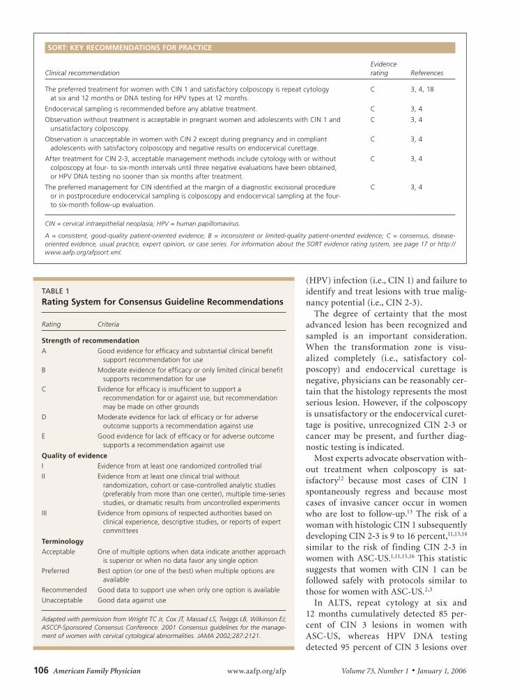
SORT: Key ReCOmmeNdATIONS fOR PRACTICe
Clinical recommendation
Evidence rating
References
The preferred treatment for women with CIN 1 and satisfactory colposcopy is repeat cytology at six and 12 months or DNA testing for HPV types at 12 months.
C 3, 4, 18
Endocervical sampling is recommended before any ablative treatment. C 3, 4
Observation without treatment is acceptable in pregnant women and adolescents with CIN 1 and unsatisfactory colposcopy.
C 3, 4
Observation is unacceptable in women with CIN 2 except during pregnancy and in compliant adolescents with satisfactory colposcopy and negative results on endocervical curettage.
C 3, 4
After treatment for CIN 2-3, acceptable management methods include cytology with or without colposcopy at four- to six-month intervals until three negative evaluations have been obtained, or HPV DNA testing no sooner than six months after treatment.
C 3, 4
The preferred management for CIN identified at the margin of a diagnostic excisional procedure or in postprocedure endocervical sampling is colposcopy and endocervical sampling at the four- to six-month follow-up evaluation.
C
3, 4
CIN = cervical intraepithelial neoplasia; HPV = human papillomavirus.
A = consistent, good-quality patient-oriented evidence; B = inconsistent or limited-quality patient-oriented evidence; C = consensus, disease- oriented evidence, usual practice, expert opinion, or case series. For information about the SORT evidence rating system, see page 17 or http://www.aafp.org/afpsort.xml.
table 1
Rating System for Consensus Guideline Recommendations
Rating Criteria
Strength of recommendation
A Good evidence for efficacy and substantial clinical benefit support recommendation for use
B Moderate evidence for efficacy or only limited clinical benefit supports recommendation for use
C Evidence for efficacy is insufficient to support a recommendation for or against use, but recommendation may be made on other grounds
D Moderate evidence for lack of efficacy or for adverse outcome supports a recommendation against use
E Good evidence for lack of efficacy or for adverse outcome supports a recommendation against use
Quality of evidence
I Evidence from at least one randomized controlled trial
II Evidence from at least one clinical trial without randomization, cohort or case-controlled analytic studies (preferably from more than one center), multiple time-series studies, or dramatic results from uncontrolled experiments
III Evidence from opinions of respected authorities based on clinical experience, descriptive studies, or reports of expert committees
Terminology
Acceptable One of multiple options when data indicate another approach is superior or when no data favor any single option
Preferred Best option (or one of the best) when multiple options are available
Recommended Good data to support use when only one option is available
Unacceptable Good data against use
Adapted with permission from Wright TC Jr, Cox JT, Massad LS, Twiggs LB, Wilkinson EJ; ASCCP-Sponsored Consensus Conference. 2001 Consensus guidelines for the manage-ment of women with cervical cytological abnormalities. JAMA 2002;287:2121.
January 1, 2006 ◆ Volume 73, Number 1 www.aafp.org/afp American Family Physician 107
Cervical Abnormalities
a two-year follow-up period.17 When combining HPV testing’s high sensitivity for CIN 3 with evidence that only persistent HPV progresses to CIN 3, the consen-sus conference concluded that HPV DNA testing at 12 months provides an acceptable alternative to repeat cytology at six and 12 months in untreated women with CIN 1.18 The addition of colposcopy would help ensure that CIN 2-3 is not missed, but it would increase costs and necessitate access to colposcopic services. No studies have demonstrated the superiority of follow-up colposcopy compared with cytology alone. Additionally, no data suggest that extending the period of observa-tion for CIN 1 beyond 24 months is unsafe in compliant populations, although rates of spontaneous regression or progression may increase.19
The safety of a conservative follow-up protocol in women with CIN 1 is less well-established when col-poscopy is unsatisfactory or endocervical curettage is positive. Although one study7 showed that the rate of detection of CIN 2-3 in the conization specimens of these women was only about 10 percent and was lower than the rate in women with satisfactory colposcopy, the consensus panel determined that a diagnostic excisional procedure is more appropriate than observation because of the consequences of missing an occult invasive cancer and because the literature is limited.3
Ablative procedures (e.g., cryotherapy, fulguration, laser ablation, cold coagulation) and excisional modali-ties (e.g., LEEP, laser conization, cold knife conization) are effective for treating women with CIN 1 and satisfac-tory colposcopy (Figure 120).21 Excisional methods often are recommended over ablation for treatment of recur-rent or persistent CIN because these lesions often are
located in the endocervical canal. Additionally, visual-ization of the squamocolumnar junction in a previously treated patient cannot guarantee that the transforma-tion zone has been fully visualized.
Studies22,23 comparing various ablative and excisional modalities in women with satisfactory colposcopy have shown similar success rates in the treatment of CIN. Although LEEP conizations are associated with less blood loss, shorter operative times, and a higher rate of satisfactory posttreatment colposcopy compared with cold knife conization, the pathologic margins are more likely to be involved and may be more difficult to inter-pret.24 Ultimately, the decision about which therapeutic option is best must be individualized.
managing Biopsy-Confirmed CIN 1Repeat cytology at six and 12 months or DNA testing for high-risk HPV types at 12 months is the preferred man-agement approach for women with CIN 1 and satisfactory colposcopy (AII recommendation, Figure 24).3,4,18 If results from repeat cytology are reported as atypical squamous cells (ASC) or greater, or if DNA test results are positive for high-risk HPV types, referral for repeat colposcopy is preferred (AII recommendation).3,4 After two consecu-tive negative cytologic smears or one negative HPV DNA
table 2
Terminology Used in the 2001 Consensus Guidelines
diagnostic excisional procedure: obtaining a histologic sample of the transformation zone and endocervical canal using LEEP or laser, cold-knife, or LEEP conization.
endocervical assessment: evaluation of the endocervical canal for neoplasia using colposcopy or endocervical sampling.
endocervical sampling: obtaining a histologic specimen by endocervical curettage or cytobrush or obtaining a cytologic sample with a cytobrush.
Satisfactory colposcopy: the margins of the lesion(s) and the entire squamocolumnar junction are visible.
LEEP = loop electrosurgical excision procedure.
Information from reference 2.
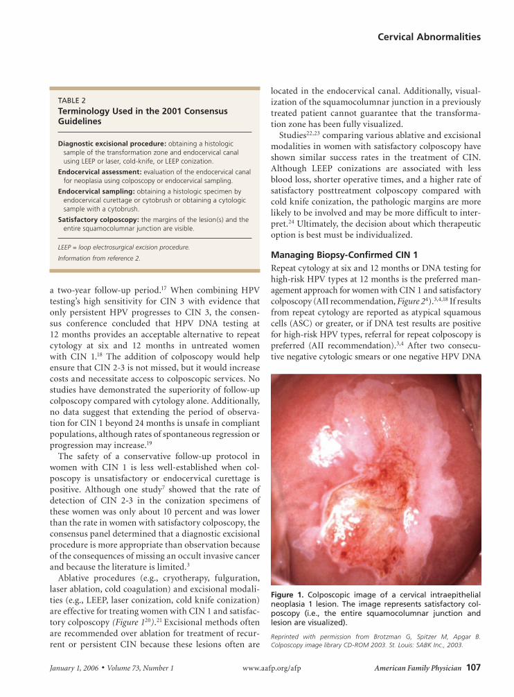
figure 1. Colposcopic image of a cervical intraepithelial neoplasia 1 lesion. the image represents satisfactory col-poscopy (i.e., the entire squamocolumnar junction and lesion are visualized).
Reprinted with permission from Brotzman G, Spitzer M, Apgar B. Colposcopy image library CD-ROM 2003. St. Louis: SABK Inc., 2003.
108 American Family Physician www.aafp.org/afp Volume 73, Number 1 ◆ January 1, 2006
test, it is preferred that patients continue annual cytologic screening (BII recommendation) and recommended that they continue to be screened at annual intervals (BIII recommendation).3,4 Adding colposcopy to the follow-up protocol is acceptable (AII recommendation).3,4
Treatment of women with biopsy-confirmed CIN 1 and satisfactory colposcopy is acceptable (AI recom-mendation) and may include individualized ablative or excisional modalities (AI recommendation).3,4 Endo-cervical sampling is recommended before ablation of CIN (AII recommendation).3,4 Excisional modalities are preferred in women who have undergone previous abla-tive therapy and have recurrent biopsy-confirmed CIN 1 (BII recommendation).3,4
Diagnostic excisional procedures are preferred when colposcopy is unsatisfactory in women with biopsy-confirmed CIN 1 (AII recommendation, Figures 320 and 44).3,4 Follow-up without treatment is acceptable in pregnant and immunosuppressed women and adoles-cents with biopsy-confirmed CIN 1 and an unsatisfac-tory colposcopy (CIII recommendation).3,4 Application of podophyllin to the cervix or vagina, ablative treat-ment in women with unsatisfactory colposcopy, and
hysterectomy as the primary treatment for biopsy- confirmed CIN 1 are unacceptable treatment options (EII recommendation).3,4
CIN 2-3: OverviewApproximately 43 percent of untreated CIN 2 and 32 percent of CIN 3 will regress spontaneously; 35 per-cent of CIN 2 and 56 percent of CIN 3 will persist; and 22 percent of CIN 2 and 14 percent of CIN 3 will prog-ress to carcinoma-in-situ or invasive cancer.25 Therefore, except in special circumstances, women with biopsy-confirmed CIN 2-3 should be treated. Based on natural history studies, the recommendations for women with CIN 2 and CIN 3 are combined.3
Effective treatment of biopsy-confirmed CIN 2-3 requires the removal of the entire transformation zone rather than just the removal of the lesion.3 When colpos-copy is satisfactory, any ablative or excisional modality will treat CIN effectively (Figures 520 and 64). However, because excisional modalities allow for the pathologic identification of unanticipated microinvasive or occult invasive cancer, some physicians prefer these methods to treat biopsy-confirmed CIN 2-3.26
management of Biopsy-Confirmed CIN 1 with Satisfactory Colposcopy
figure 2.
The rightsholder did not grant rights to reproduce this item in electronic media. For the missing item, see the original print version of this publication.
This algorithm is available at: http://www.asccp.org/consensusguidelines.
January 1, 2006 ◆ Volume 73, Number 1 www.aafp.org/afp American Family Physician 109
Cervical Abnormalities
Because a small number of women with biopsy- confirmed CIN 2-3 and unsatisfactory colposcopy have occult invasive cancer,27 excisional procedures should be performed (Figure 7 20). Cold knife and LEEP conizations effectively diagnose and treat these women. The patho-logic margin of specimens from cold knife conization is less frequently involved and is easier to interpret than the margin of LEEP conizations, although the complica-tion rate of cold knife conization is greater.24,27
Positive conization margins or positive endocervical curettage performed at the time of a diagnostic exci-sional procedure is predictive of recurrent or persistent CIN,28,29 which occurs in up to 7 percent of women with negative endocervical margins and 30 percent of women with positive endocervical margins.30 Therefore, it is recommended that women with positive margins be counseled about the relative risks of observation versus further treatment and that their management be individualized. Hysterectomy is appropriate in selected patients.3
SPeCIAl CIRCUmSTANCeS
There are certain circumstances in which observation is preferred because the risk of progression of CIN 2-3 is very small and the risk of treatment is relatively high. The risk of progression of CIN 2-3 to invasive cancer during pregnancy is minimal,31 and the rate of postpartum spon-taneous regression is high.32 Excisional procedures during pregnancy are associated with significant complications, including bleeding and preterm labor.33,34 Therefore, treatment during pregnancy should be limited to women in whom invasive cancer cannot be ruled out.33 Invasive cancer is rare in adolescents, and the rate of spontaneous regression is high enough in those with biopsy-proven CIN 235 that some experts on the consensus panel favored observation without treatment for appropriately coun-seled, compliant adolescents with biopsy-confirmed CIN 2.3 Although the failure rate after treatment in women infected with human immunodeficiency virus 1
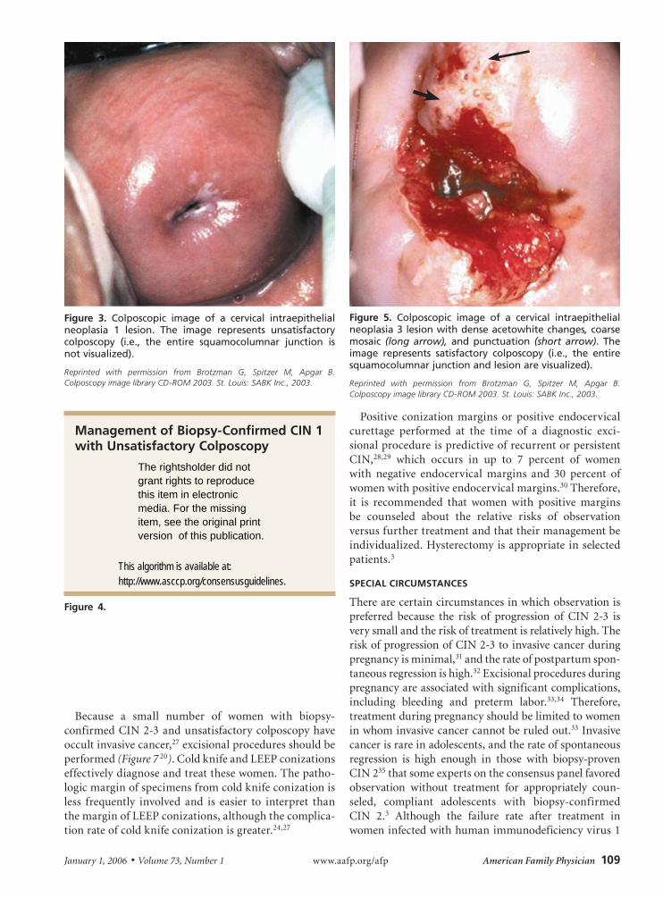
figure 3. Colposcopic image of a cervical intraepithelial neoplasia 1 lesion. the image represents unsatisfactory colposcopy (i.e., the entire squamocolumnar junction is not visualized).
Reprinted with permission from Brotzman G, Spitzer M, Apgar B. Colposcopy image library CD-ROM 2003. St. Louis: SABK Inc., 2003.
management of Biopsy-Confirmed CIN 1 with Unsatisfactory Colposcopy
figure 4.
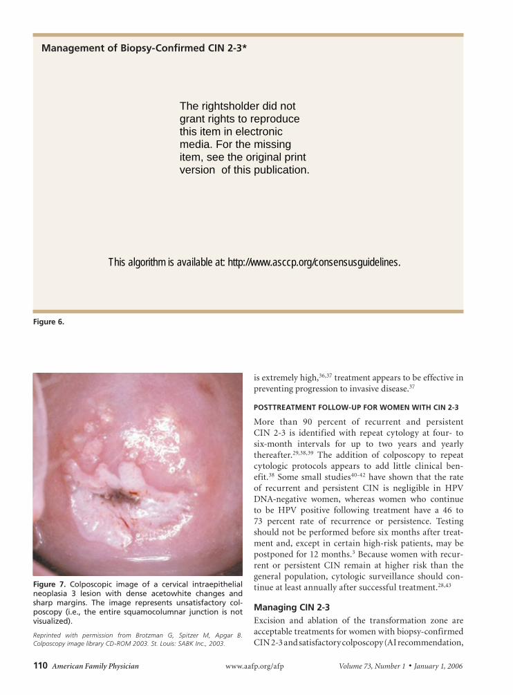
figure 5. Colposcopic image of a cervical intraepithelial neoplasia 3 lesion with dense acetowhite changes, coarse mosaic (long arrow), and punctuation (short arrow). the image represents satisfactory colposcopy (i.e., the entire squamocolumnar junction and lesion are visualized).
Reprinted with permission from Brotzman G, Spitzer M, Apgar B. Colposcopy image library CD-ROM 2003. St. Louis: SABK Inc., 2003.
The rightsholder did not grant rights to reproduce this item in electronic media. For the missing item, see the original print version of this publication.
This algorithm is available at: http://www.asccp.org/consensusguidelines.
110 American Family Physician www.aafp.org/afp Volume 73, Number 1 ◆ January 1, 2006
is extremely high,36,37 treatment appears to be effective in preventing progression to invasive disease.37
POSTTReATmeNT fOllOw-UP fOR wOmeN wITh CIN 2-3
More than 90 percent of recurrent and persistent CIN 2-3 is identified with repeat cytology at four- to six-month intervals for up to two years and yearly thereafter.29,38,39 The addition of colposcopy to repeat cytologic protocols appears to add little clinical ben-efit.38 Some small studies40-42 have shown that the rate of recurrent and persistent CIN is negligible in HPV DNA-negative women, whereas women who continue to be HPV positive following treatment have a 46 to 73 percent rate of recurrence or persistence. Testing should not be performed before six months after treat-ment and, except in certain high-risk patients, may be postponed for 12 months.3 Because women with recur-rent or persistent CIN remain at higher risk than the general population, cytologic surveillance should con-tinue at least annually after successful treatment.28,43
managing CIN 2-3Excision and ablation of the transformation zone are acceptable treatments for women with biopsy-confirmed CIN 2-3 and satisfactory colposcopy (AI recommendation,
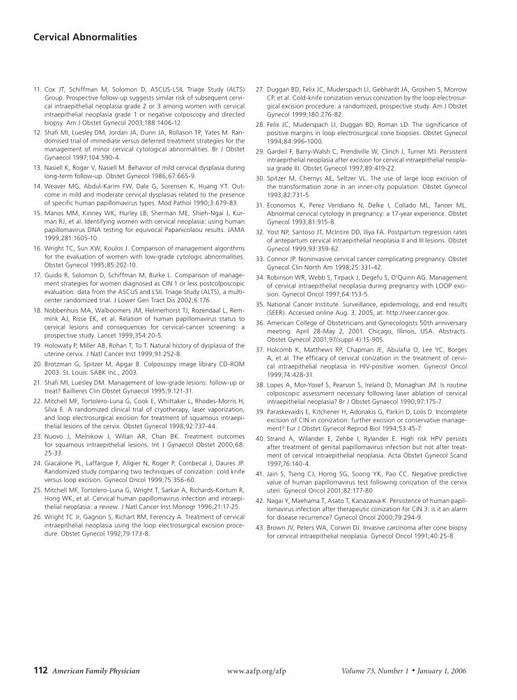
figure 7. Colposcopic image of a cervical intraepithelial neoplasia 3 lesion with dense acetowhite changes and sharp margins. the image represents unsatisfactory col-poscopy (i.e., the entire squamocolumnar junction is not visualized).
Reprinted with permission from Brotzman G, Spitzer M, Apgar B. Colposcopy image library CD-ROM 2003. St. Louis: SABK Inc., 2003.
management of Biopsy-Confirmed CIN 2-3*
figure 6.
The rightsholder did not grant rights to reproduce this item in electronic media. For the missing item, see the original print version of this publication.
This algorithm is available at: http://www.asccp.org/consensusguidelines.
January 1, 2006 ◆ Volume 73, Number 1 www.aafp.org/afp American Family Physician 111
Cervical Abnormalities
Figure 64).3,4 Excisional modalities are preferred in women with recurrent CIN 2-3 (AII recommendation).3,4 A diag-nostic excisional procedure is recommended in women with biopsy-confirmed CIN 2-3 and unsatisfactory col-poscopy (AII recommendation).3,4 Observation of CIN 2-3 without treatment is unacceptable except in special circumstances (EII recommendation).3,4 Hysterectomy is unacceptable as a primary therapy for women with CIN 2-3 (EII recommendation).3,4
follow-Up After Treatment for Biopsy-Confirmed CIN 2-3Acceptable follow-up protocols after treatment of CIN 2-3 include cytology or a combination of cytology and colposcopy at four- to six-month intervals until three negative evaluations have been performed (AII recom-mendation, Figure 64).3,4 Annual cytologic follow-up is recommended thereafter (AII recommendation).3,4 A cytologic result of ASC is the recommended threshold for referral to colposcopy during follow-up (AII recommen-dation).3,4 Surveillance with HPV DNA testing performed no sooner than six months after treatment also is accept-able (BII recommendation).3,4 A positive test for high-risk HPV types is the recommended threshold for referral to colposcopy (BIII recommendation).3,4 If HPV testing is negative, annual cytologic screening is recommended (BIII recommendation).3,4 Repeat conization or hysterec-tomy based on a single positive HPV test that is not cor-roborated by other findings (e.g., cytology, colposcopy, histology) is unacceptable (DIII recommendation).3,4
If CIN is identified at the margin of a diagnostic excisional procedure or on a postprocedure endocervi-cal curettage, it is preferred that endocervical sampling be added to one of the previous follow-up protocols (BII recommendation).3,4 When CIN 2-3 is identified at the endocervical margin or in the endocervical sam-pling obtained after the diagnostic excisional procedure, a repeat diagnostic excisional procedure is acceptable (AII recommendation).3,4 Hysterectomy is acceptable when repeat diagnostic excision is not feasible (BII rec-ommendation)3,4 or for women with recurrent or persis-tent CIN 2-3 (BII recommendation).3,4
follow-Up After Treatment for Biopsy-Confirmed CIN 2-3: Special Circumstances In compliant adolescents with histologic CIN 2, satisfac-tory colposcopy, and negative endocervical curettage, observation with colposcopy and cytology at four- to six-month intervals for one year is acceptable (BII rec-ommendation).3 Ablation or excision is required for adolescents with CIN 3 (BIII recommendation).3,4
The AuthorsMARK SPITZER, M.D., is professor of clinical obstetrics and gynecology at Weill Medical College of Cornell University, New York, and chairman of the Department of Obstetrics and Gynecology at New York Methodist Hospital, Brooklyn. He received his medical degree from Albert Einstein College of Medicine of Yeshiva University, Bronx, New York, and com-pleted a residency in obstetrics and gynecology at affiliated hospitals.
BARBARA S. APGAR, M.D., M.S., is clinical professor of family medicine at the University of Michigan Medical School, Ann Arbor. Dr. Apgar received her medical degree from Texas Tech University Health Sciences Center School of Medicine, Lubbock, where she also completed a family practice residency. She received a master’s degree from the University of Michigan and completed a faculty development fellowship at Michigan State University College of Human Medicine, East Lansing. Dr. Apgar is an associate editor of American Family Physician.
GREGORY L. BROTZMAN, M.D., is professor of family and community medicine at the Medical College of Wisconsin in Milwaukee. Dr. Brotzman received his medical degree from Southern Illinois University School of Medicine, Springfield, where he completed a family practice residency.
Address correspondence to Mark Spitzer, M.D., Dept. of Obstetrics and Gynecology, New York Methodist Hospital, 506 Sixth St., Brooklyn, NY 11215 (e-mail: [email protected]). Reprints are not available from the authors.
Author disclosure: Dr. Brotzman is on the speakers bureau for Digene Corp. and Merck & Co., Inc. Dr. Spitzer is on the speakers bureau for Quest Diagnostics and Merck & Co., Inc.
RefeReNCeS
1. Solomon D, Schiffman M, Tarone R; ALTS Study group. Comparison of three management strategies for patients with atypical squamous cells of undetermined significance: baseline results from a randomized trial. J Natl Cancer Inst 2001;93:293-9.
2. Wright TC Jr, Cox JT, Massad LS, Twiggs LB, Wilkinson EJ; ASCCP-Sponsored Consensus Conference. 2001 Consensus guidelines for the management of women with cervical cytological abnormalities. JAMA 2002;287:2120-9.
3. Wright TC Jr, Cox JT, Massad LS, Carlson J, Twiggs LB, Wilkinson EJ; American Society for Colposcopy and Cervical Pathology. 2001 Consensus guidelines for the management of women with cervical intraepithelial neoplasia. Am J Obstet Gynecol 2003;189:295-304.
4. Wright TC Jr, Cox JT, Massad LS, Carlson J, Twiggs LB, Wilkinson EJ. 2001 Consensus guidelines for the management of women with cervi-cal intraepithelial neoplasia. J Low Genit Tract Dis 2003;7:154-67.
5. Stoler MH, Schiffman M. Interobserver reproducibility of cervical cyto-logic and histologic interpretations: realistic estimates from the ASCUS-LSIL Triage Study. JAMA 2001;285:1500-5.
6. Hopman EH, Voorhorst FJ, Kenemans P, Meyer CJ, Helmerhorst TJ. Observer agreement on interpreting colposcopic images of CIN. Gyne-col Oncol 1995;58:206-9.
7. Spitzer M, Chernys AE, Shifrin A, Ryskin M. Indications for cone biopsy: pathologic correlation. Am J Obstet Gynecol 1998;178:74-9.
8. Massad LS, Halperin CJ, Bitterman P. Correlation between col-poscopically directed biopsy and cervical loop excision. Gynecol Oncol 1996;60:400-3.
9. Ostor AG. Natural history of cervical intraepithelial neoplasia: a critical review. Int J Gynecol Pathol 1993;12:186-92.
10. Melnikow J, Nuovo J, Willan AR, Chan BK, Howell LP. Natural history of cervical squamous intraepithelial lesions: a meta-analysis. Obstet Gynecol 1998;92:727-35.
112 American Family Physician www.aafp.org/afp Volume 73, Number 1 ◆ January 1, 2006
Cervical Abnormalities
11. Cox JT, Schiffman M, Solomon D, ASCUS-LSIL Triage Study (ALTS) Group. Prospective follow-up suggests similar risk of subsequent cervi-cal intraepithelial neoplasia grade 2 or 3 among women with cervical intraepithelial neoplasia grade 1 or negative colposcopy and directed biopsy. Am J Obstet Gynecol 2003;188:1406-12.
12. Shafi MI, Luesley DM, Jordan JA, Dunn JA, Rollason TP, Yates M. Ran-domised trial of immediate versus deferred treatment strategies for the management of minor cervical cytological abnormalities. Br J Obstet Gynaecol 1997;104:590-4.
13. Nasiell K, Roger V, Nasiell M. Behavior of mild cervical dysplasia during long-term follow-up. Obstet Gynecol 1986;67:665-9.
14. Weaver MG, Abdul-Karim FW, Dale G, Sorensen K, Huang YT. Out-come in mild and moderate cervical dysplasias related to the presence of specific human papillomavirus types. Mod Pathol 1990;3:679-83.
15. Manos MM, Kinney WK, Hurley LB, Sherman ME, Shieh-Ngai J, Kur-man RJ, et al. Identifying women with cervical neoplasia: using human papillomavirus DNA testing for equivocal Papanicolaou results. JAMA 1999;281:1605-10.
16. Wright TC, Sun XW, Koulos J. Comparison of management algorithms for the evaluation of women with low-grade cytologic abnormalities. Obstet Gynecol 1995;85:202-10.
17. Guido R, Solomon D, Schiffman M, Burke L. Comparison of manage-ment strategies for women diagnosed as CIN 1 or less postcolposcopic evaluation: data from the ASCUS and LSIL Triage Study (ALTS), a multi-center randomized trial. J Lower Gen Tract Dis 2002;6:176.
18. Nobbenhuis MA, Walboomers JM, Helmerhorst TJ, Rozendaal L, Rem-mink AJ, Risse EK, et al. Relation of human papillomavirus status to cervical lesions and consequences for cervical-cancer screening: a prospective study. Lancet 1999;354:20-5.
19. Holowaty P, Miller AB, Rohan T, To T. Natural history of dysplasia of the uterine cervix. J Natl Cancer Inst 1999;91:252-8.
20. Brotzman G, Spitzer M, Apgar B. Colposcopy image library CD-ROM 2003. St. Louis: SABK Inc., 2003.
21. Shafi MI, Luesley DM. Management of low-grade lesions: follow-up or treat? Baillieres Clin Obstet Gynaecol 1995;9:121-31.
22. Mitchell MF, Tortolero-Luna G, Cook E, Whittaker L, Rhodes-Morris H, Silva E. A randomized clinical trial of cryotherapy, laser vaporization, and loop electrosurgical excision for treatment of squamous intraepi-thelial lesions of the cervix. Obstet Gynecol 1998;92:737-44.
23. Nuovo J, Melnikow J, Willan AR, Chan BK. Treatment outcomes for squamous intraepithelial lesions. Int J Gynaecol Obstet 2000;68: 25-33.
24. Giacalone PL, Laffargue F, Aligier N, Roger P, Combecal J, Daures JP. Randomized study comparing two techniques of conization: cold knife versus loop excision. Gynecol Oncol 1999;75:356-60.
25. Mitchell MF, Tortolero-Luna G, Wright T, Sarkar A, Richards-Kortum R, Hong WK, et al. Cervical human papillomavirus infection and intraepi-thelial neoplasia: a review. J Natl Cancer Inst Monogr 1996;21:17-25.
26. Wright TC Jr, Gagnon S, Richart RM, Ferenczy A. Treatment of cervical intraepithelial neoplasia using the loop electrosurgical excision proce-dure. Obstet Gynecol 1992;79:173-8.
27. Duggan BD, Felix JC, Muderspach LI, Gebhardt JA, Groshen S, Morrow CP, et al. Cold-knife conization versus conization by the loop electrosur-gical excision procedure: a randomized, prospective study. Am J Obstet Gynecol 1999;180:276-82.
28. Felix JC, Muderspach LI, Duggan BD, Roman LD. The significance of positive margins in loop electrosurgical cone biopsies. Obstet Gynecol 1994;84:996-1000.
29. Gardeil F, Barry-Walsh C, Prendiville W, Clinch J, Turner MJ. Persistent intraepithelial neoplasia after excision for cervical intraepithelial neopla-sia grade III. Obstet Gynecol 1997;89:419-22.
30. Spitzer M, Chernys AE, Seltzer VL. The use of large loop excision of the transformation zone in an inner-city population. Obstet Gynecol 1993;82:731-5.
31. Economos K, Perez Veridiano N, Delke I, Collado ML, Tancer ML. Abnormal cervical cytology in pregnancy: a 17-year experience. Obstet Gynecol 1993;81:915-8.
32. Yost NP, Santoso JT, McIntire DD, Iliya FA. Postpartum regression rates of antepartum cervical intraepithelial neoplasia II and III lesions. Obstet Gynecol 1999;93:359-62.
33. Connor JP. Noninvasive cervical cancer complicating pregnancy. Obstet Gynecol Clin North Am 1998;25:331-42.
34. Robinson WR, Webb S, Tirpack J, Degefu S, O’Quinn AG. Management of cervical intraepithelial neoplasia during pregnancy with LOOP exci-sion. Gynecol Oncol 1997;64:153-5.
35. National Cancer Institute. Surveillance, epidemiology, and end results (SEER). Accessed online Aug. 3, 2005, at: http://seer.cancer.gov.
36. American College of Obstetricians and Gynecologists 50th anniversary meeting. April 28-May 2, 2001. Chicago, Illinois, USA. Abstracts. Obstet Gynecol 2001;97(suppl 4):1S-90S.
37. Holcomb K, Matthews RP, Chapman JE, Abulafia O, Lee YC, Borges A, et al. The efficacy of cervical conization in the treatment of cervi-cal intraepithelial neoplasia in HIV-positive women. Gynecol Oncol 1999;74:428-31.
38. Lopes A, Mor-Yosef S, Pearson S, Ireland D, Monaghan JM. Is routine colposcopic assessment necessary following laser ablation of cervical intraepithelial neoplasia? Br J Obstet Gynaecol 1990;97:175-7.
39. Paraskevaidis E, Kitchener H, Adonakis G, Parkin D, Lolis D. Incomplete excision of CIN in conization: further excision or conservative manage-ment? Eur J Obstet Gynecol Reprod Biol 1994;53:45-7.
40. Strand A, Wilander E, Zehbe I, Rylander E. High risk HPV persists after treatment of genital papillomavirus infection but not after treat-ment of cervical intraepithelial neoplasia. Acta Obstet Gynecol Scand 1997;76:140-4.
41. Jain S, Tseng CJ, Horng SG, Soong YK, Pao CC. Negative predictive value of human papillomavirus test following conization of the cervix uteri. Gynecol Oncol 2001;82:177-80.
42. Nagai Y, Maehama T, Asato T, Kanazawa K. Persistence of human papil-lomavirus infection after therapeutic conization for CIN 3: is it an alarm for disease recurrence? Gynecol Oncol 2000;79:294-9.
43. Brown JV, Peters WA, Corwin DJ. Invasive carcinoma after cone biopsy for cervical intraepithelial neoplasia. Gynecol Oncol 1991;40:25-8.