management of difficult airway · slcoa national guidelines / difficult airway 9 2 management of...
TRANSCRIPT
SLCOA National Guidelines
Management ofDifficult Airway
SLCOA National Guidelines
List of ContributorsDr. Deepthi AttygalleDr. Hemantha RajapakseDr. Kalum RanatungeDr. Nalini RodrigoDr. Nilmini Wijesuriya
Contents
List of Contributors 08
Introduction 09
Objectives 09
Definitions 10
Assessment and preparation for trachealintubation 11
Difficult airway drills 17
Adult non-obstetric difficult airway management drill 17
Obstetric difficult airway management drill 26
Management of acute upper airway obstruction 32
Management of desaturation under anaesthesia 37
Extubation & Follow up 41
References 44
9SLCOA National Guidelines / Difficult Airway
2 Management of difficult airway
2.1 IntroductionThe incidence of difficult tracheal intubation following inductionof general anaesthesia is rare and has been estimated at 3-18%. Itis the most critical emergency that an anaesthetist can be facedwith and may lead to hypoxaemic anaesthetic death, brain damageor serious soft tissue damage. When the junior anaesthetist isconfronted with an unanticipated difficult tracheal intubationespecially out of hours he should concentrate on maintenance ofoxygenation and prevention of airway trauma until senior helparrives.These guidelines will address the following aspects:
These guidelines are constructed with regard to the culture, skilland equipment available in Sri Lanka. Therefore some equipmentthat are mentioned in standard text books or guidelines formulatedin other countries may not be included. That does not preclude theanaesthetist to use them if and when available and if adequatelyexperienced in using them. Wall charts have been produced to usein conjunction with these guidelines for quick reference.
2.2 ObjectivesThese guidelines are formulated to help the non-specialistanaesthetic medical officers to manage different clinical scenariosassociated with unanticipated difficult tracheal intubation in adults.Although these scenarios may be multifactorial, a structuredsequence of actions would detect most of the causes. Childrenwith unpredicted difficult airways should be managed by aconsultant at an early stage. Management of predicted difficultairway is not within the scope of the non-specialist anaesthetistand he should always discuss such cases with the consultantanaesthetist. When specialist opinion is not available the patientshould be transferred to the nearest hospital where a consultant isavailable, preferably after discussion and stabilization.
10 SLCOA National Guidelines / Difficult Airway
2.3 Definitions
2.3.1 Difficult airway: the clinical situation in which aconventionally trained anesthetist experiences difficulty with facemask ventilation, laryngoscopy, tracheal intubation or fails tointubate the trachea.A difficult airway may be caused by several factors:
• patient factors• clinical setting• skills of the practitioner
2.3.2 Difficult face mask ventilation: Inability of an unassistedanaesthetist to maintain oxygen saturation (SpO2) more than 90%with positive pressure mask ventilation using 100% oxygen in apatient whose oxygen saturation (SpO2) was more than 90% beforeinduction and/or it is not possible to prevent or reverse signs ofinadequate ventilation with positive pressure mask ventilation.
• This may occur due to one or more of the followingproblems: inadequate mask seal, excessive gas leak, or excessiveresistance to the ingress or egress of gas.
• Signs of inadequate face mask ventilation: absent orinadequate chest movement, absent or inadequate breath sounds,auscultatory signs of severe obstruction, cyanosis, gastric air entryor dilatation, decreasing or inadequate oxygen saturation (SpO2),absent or inadequate exhaled carbon dioxide, and haemodynamicchanges associated with hypoxaemia or hypercarbia (e.g.,hypertension, tachycardia, arrhythmia).
• 2.3.3 Difficult laryngoscopy: Inability to visualize anyportion of the vocal cords after multiple attempts at conventionallaryngoscopy. (Cormack and Lehane laryngoscopy grade 3 or 4)
11SLCOA National Guidelines / Difficult Airway
• 2.3.4 Difficult tracheal intubation: Tracheal intubationwhich requires more than 3 attempts, or more than 10 minuteswith a conventional laryngoscope.
• 2.3.5 Failed intubation: Placement of the endotrachealtube fails after multiple intubation attempts.
2. 4 Assessment and preparation for trachealintubation following routine induction ofgeneral anaesthesia in a non-obstetric patient
2.4.1 Pre-operative Assessment of the airwayAll patients should undergo an airway evaluation pre-surgeryand this should be recorded on the anaesthesia record. Butthis assessment is imperfect in predicting problems always,so an airway strategy should be drawn up for each patient tocover the entire period of anaesthetic care, particular at thestart and end of anaesthesia.
History• Intubation problems during previous
anaesthetics• Neonates, elderly, pregnant women• Facial/maxillary trauma, cervical spine injury,
previous surgery• Snoring and sleep apnoea• Infection or inflammation, orofacial or neck
oedema• Rheumatoid disease or surgery of the neck or
degenerative spinal diseases• Tumours, radiation-related scarring, burns
12 SLCOA National Guidelines / Difficult Airway
Physical examination: Any factor which interfereswith the line of vision
• Buck teeth, missing or loose teeth, especially upperleft incisors)
• Inability to extend the head in the erect position• Short neck• Receding mandible (anterior larynx)• Inability to protrude the lower jaw
Special tests• Mallampati grade, (also checks the degree of mouth
opening, size and mobility of the tongue and otherstructures in the mouth; Grade e” 3)
• Thyromental distance (< 6.5 cm or < 3 fingerbreaths)
• Sterno-mental distance < 12.5 cm• Delikan’s sign
See Reference pp. 4-4 for more details (with permission)
If preoperative assessment indicates the possibility of a difficultairway, aspiration prophylaxis should be given and theconsultant anaesthetist should be consulted. Junior medicalofficers should not handle a predicted difficult airway in theabsence of a consultant anaesthetist.
2.4.2 Preparation for difficult airwaymanagementAnaesthetists should be ready to deal with difficulties inintubation at any time. The correct equipment, drugs and trainedpersonnel must be immediately available. This will include:
13SLCOA National Guidelines / Difficult Airway
2.4.2 (a) Personnel• A trained anaesthetist (conventionally trained for 6 monthsand approved by a consultant anaesthetist to be satisfactory).Intubation with a bougie should be practiced simulating aLehane & Cormack Grade 3.• A trained assistant: as there are no trained operatingdepartment assistants in government hospitals, a nurse or asuitable minor employee should be trained for this purposewhen the anaesthetist is on his own.2.4.2 (b) Recommended equipment for routine airwaymanagementThe following list of equipment is recommended for routineintubation.
• Adjustable operating table or trolley• Wee’s detector (Oesophageal detector device)• ECG, pulse oximetry, NIBP, capnography,
stethoscope• Oxygen source and tubing• Anaesthetic breathing system / Ambu bag• Reliable suction equipment• Facemasks• Oropharyngeal/ Nasopharyngeal airways: three sizes• Laryngeal Mask Airways• Two working laryngoscope handles with a selection
of blades• A variety of endotracheal tubes in a range of sizes,
and tape or ties for securing the ETT• Tracheal tube introducer (“gum-elastic” bougie)• Magill’s forceps• Malleable stylet• A cricothyroidotomy kit
14 SLCOA National Guidelines / Difficult Airway
2.4.2 (c) Drugs• Appropriate induction/sedative/ paralytic agents• Essential emergency drugs
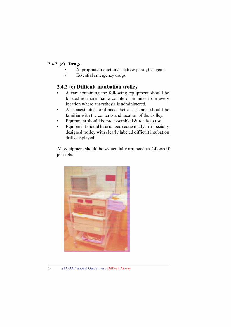
2.4.2 (c) Difficult intubation trolley• A cart containing the following equipment should be
located no more than a couple of minutes from everylocation where anaesthesia is administered.
• All anaesthetists and anaesthetic assistants should befamiliar with the contents and location of the trolley.
• Equipment should be pre assembled & ready to use.• Equipment should be arranged sequentially in a specially
designed trolley with clearly labeled difficult intubationdrills displayed
All equipment should be sequentially arranged as follows ifpossible:
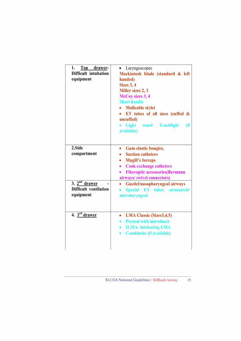
15SLCOA National Guidelines / Difficult Airway
1. Top drawer- Difficult intubation equipment
• Laryngoscopes Mackintosh blade (standard & left handed) Sizes 3, 4 Miller sizes 2, 3 McCoy sizes 3, 4 Short handle • Malleable stylet • ET tubes of all sizes (cuffed & uncuffed) • Light wand Trachlight (if available)
2.Side compartment
• Gum elastic bougies, • Suction catheters • Magill’s forceps • Cook exchange catheters • Fiberoptic accessories(Bermann airways/ swivel connectors)
3. 2nd drawer - Difficult ventilation equipment
• Guedel/nasopharyngeal airways • Special ET tubes -armoured/ microlaryngeal
4. 3rd drawer • LMA Classic (Sizes3,4,5) • Proseal with introducer • ILMA- intubating LMA • Combitube (if available)
16 SLCOA National Guidelines / Difficult Airway
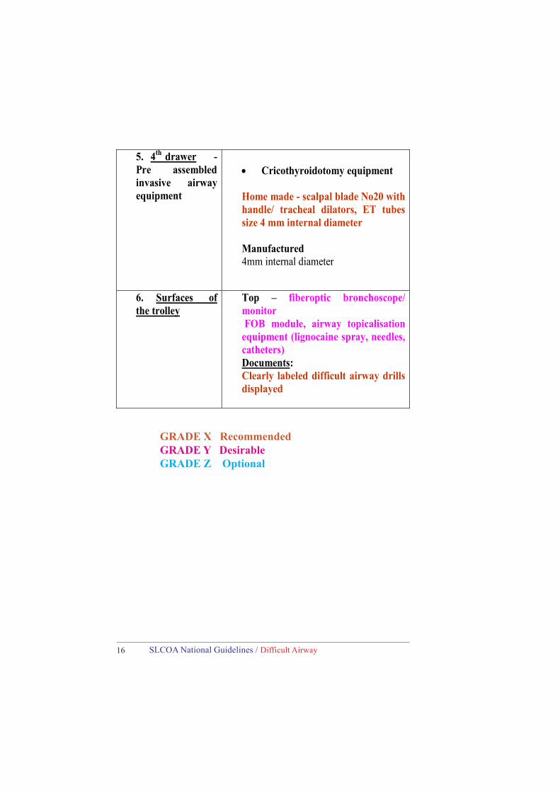
5. 4th drawer -Pre assembled invasive airway equipment
• Cricothyroidotomy equipment Home made - scalpal blade No20 with handle/ tracheal dilators, ET tubes size 4 mm internal diameter Manufactured 4mm internal diameter
6. Surfaces of the trolley
Top – fiberoptic bronchoscope/ monitor FOB module, airway topicalisation equipment (lignocaine spray, needles, catheters) Documents: Clearly labeled difficult airway drills displayed
GRADE X RecommendedGRADE Y DesirableGRADE Z Optional

17SLCOA National Guidelines / Difficult Airway
2.5 Difficult airway drills
2.5.1 Adult non-obstetric difficult airwaymanagement drill
2.5.1.1Unanticipated difficult laryngoscopy/intubation afterroutine induction of anaesthesia
Activate PLAN A
PLAN A: Correct possible causes GRADE X• If there is a difficulty in inserting laryngoscope, considerand correct possible causes:
Poor head position - adjust pillowsBreast in the way - retract breast and/or use shorthandle or polio bladeUse BURP (Backward, Upward, Rightward Pressure)Relaxation sub optimal - wait
Difficult intubation:Cormack and Lehane Grade 3 or 4 after optimal positioning,adequate muscle relaxation and best attempt.• Send for skilled help (nurse to phone)• Use external laryngeal manipulation or BURP manoeuvre(Backward, Upward and Rightward Pressure on the thyroidcartilage)• If grade 4 view:– maintain oxygenation with mask ventilation with or withoutan oral airway– send a nurse to call consultant– awaken patient
18 SLCOA National Guidelines / Difficult Airway
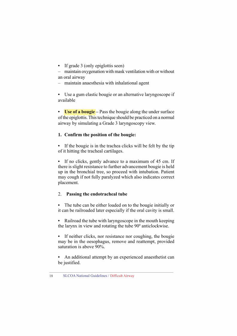
• If grade 3 (only epiglottis seen)– maintain oxygenation with mask ventilation with or withoutan oral airway– maintain anaesthesia with inhalational agent
• Use a gum elastic bougie or an alternative laryngoscope ifavailable
• Use of a bougie – Pass the bougie along the under surfaceof the epiglottis. This technique should be practiced on a normalairway by simulating a Grade 3 laryngoscopy view.
1. Confirm the position of the bougie:
• If the bougie is in the trachea clicks will be felt by the tipof it hitting the tracheal cartilages.
• If no clicks, gently advance to a maximum of 45 cm. Ifthere is slight resistance to further advancement bougie is heldup in the bronchial tree, so proceed with intubation. Patientmay cough if not fully paralyzed which also indicates correctplacement.
2. Passing the endotracheal tube
• The tube can be either loaded on to the bougie initially orit can be railroaded later especially if the oral cavity is small.
• Railroad the tube with laryngoscope in the mouth keepingthe larynx in view and rotating the tube 900 anticlockwise.
• If neither clicks, nor resistance nor coughing, the bougiemay be in the oesophagus, remove and reattempt, providedsaturation is above 90%.
• An additional attempt by an experienced anaesthetist canbe justified.
19SLCOA National Guidelines / Difficult Airway
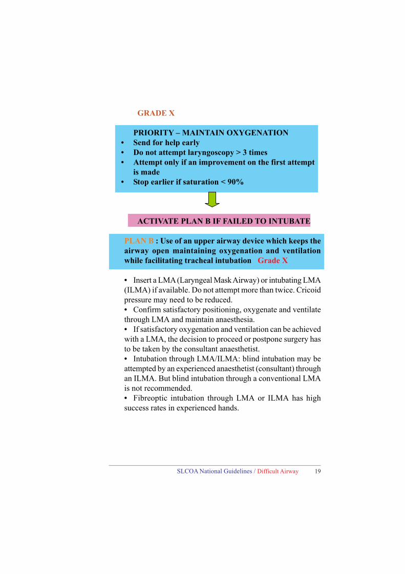
GRADE X
PRIORITY – MAINTAIN OXYGENATION• Send for help early• Do not attempt laryngoscopy > 3 times• Attempt only if an improvement on the first attempt
is made• Stop earlier if saturation < 90%
ACTIVATE PLAN B IF FAILED TO INTUBATE
PLAN B : Use of an upper airway device which keeps theairway open maintaining oxygenation and ventilationwhile facilitating tracheal intubation Grade X
• Insert a LMA (Laryngeal Mask Airway) or intubating LMA(ILMA) if available. Do not attempt more than twice. Cricoidpressure may need to be reduced.• Confirm satisfactory positioning, oxygenate and ventilatethrough LMA and maintain anaesthesia.• If satisfactory oxygenation and ventilation can be achievedwith a LMA, the decision to proceed or postpone surgery hasto be taken by the consultant anaesthetist.• Intubation through LMA/ILMA: blind intubation may beattempted by an experienced anaesthetist (consultant) throughan ILMA. But blind intubation through a conventional LMAis not recommended.• Fibreoptic intubation through LMA or ILMA has highsuccess rates in experienced hands.
20 SLCOA National Guidelines / Difficult Airway
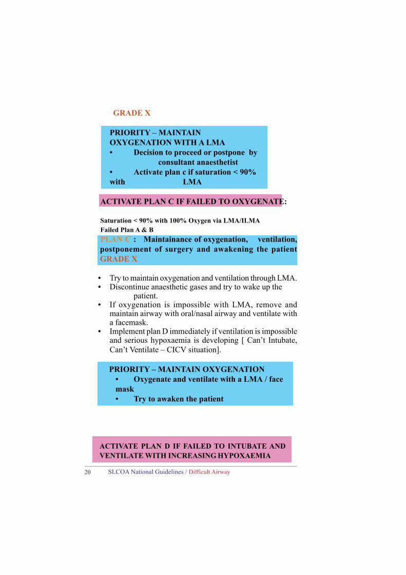
GRADE X
PRIORITY – MAINTAINOXYGENATION WITH A LMA• Decision to proceed or postpone by
consultant anaesthetist• Activate plan c if saturation < 90%with LMA
ACTIVATE PLAN C IF FAILED TO OXYGENATE:
Saturation < 90% with 100% Oxygen via LMA/ILMAFailed Plan A & BPLAN C : Maintainance of oxygenation, ventilation,postponement of surgery and awakening the patientGRADE X
• Try to maintain oxygenation and ventilation through LMA.• Discontinue anaesthetic gases and try to wake up the
patient.• If oxygenation is impossible with LMA, remove and
maintain airway with oral/nasal airway and ventilate witha facemask.
• Implement plan D immediately if ventilation is impossibleand serious hypoxaemia is developing [ Can’t Intubate,Can’t Ventilate – CICV situation].
PRIORITY – MAINTAIN OXYGENATION• Oxygenate and ventilate with a LMA / facemask• Try to awaken the patient
ACTIVATE PLAN D IF FAILED TO INTUBATE ANDVENTILATE WITH INCREASING HYPOXAEMIA
21SLCOA National Guidelines / Difficult Airway
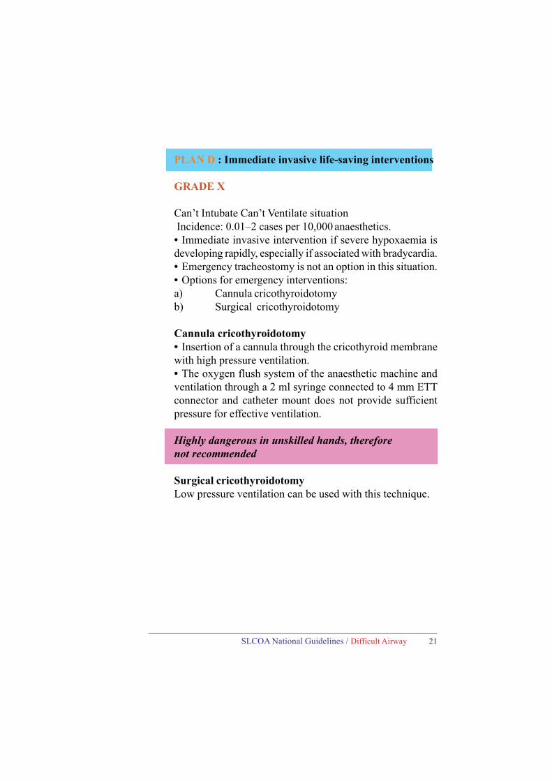
PLAN D : Immediate invasive life-saving interventions
GRADE X
Can’t Intubate Can’t Ventilate situation Incidence: 0.01–2 cases per 10,000 anaesthetics.• Immediate invasive intervention if severe hypoxaemia isdeveloping rapidly, especially if associated with bradycardia.• Emergency tracheostomy is not an option in this situation.• Options for emergency interventions:a) Cannula cricothyroidotomyb) Surgical cricothyroidotomy
Cannula cricothyroidotomy• Insertion of a cannula through the cricothyroid membranewith high pressure ventilation.• The oxygen flush system of the anaesthetic machine andventilation through a 2 ml syringe connected to 4 mm ETTconnector and catheter mount does not provide sufficientpressure for effective ventilation.
Highly dangerous in unskilled hands, thereforenot recommended
Surgical cricothyroidotomyLow pressure ventilation can be used with this technique.

22 SLCOA National Guidelines / Difficult Airway
CALL THE SURGEON FOR HELP
Technique• Identify cricothyroid membrane after optimalpositioning by extending the head.• Ask the surgeon to make a stab incision throughskin and membranes (short and rounded scalpel No 20 orminitrach scalpel if available).• Perform blind dissection to enlarge the incision withscalpel handle, forceps or dilator.• Insert a 4 mm endotracheal tube and inflate the cuffavoiding endobronchial intubation.• Check for correct placement and ventilate with abreathing system/Ambu bag.• For difficult cases eg: obese patients, use a bougiethrough the incision or a tracheostomy retractor to pullthe cricoid cartilage downwards.
• Every anaesthetist should be able to performinvasive rescue airway interventions
• Invasive airway access is a temporarymeasure to restore oxygenation
• Convert to a formal tracheostomy within 24hours
2.5.1.2Unanticipated difficult intubation during rapid sequenceinduction (RSI) in a non-obstetric patient• These patients are at risk of vomiting or regurgitation
and subsequent pulmonary aspiration of gastric contents.Laryngoscopy and insertion of a bougie or LMA mayprove difficult due to cricoid pressure.
• All patients undergoing RSI should be preoxygenated andcricoid pressure applied as consciousness is lost. Therecommended force is 30 N, which should be reduced iflaryngoscopy is difficult or airway obstruction occurs.
23SLCOA National Guidelines / Difficult Airway
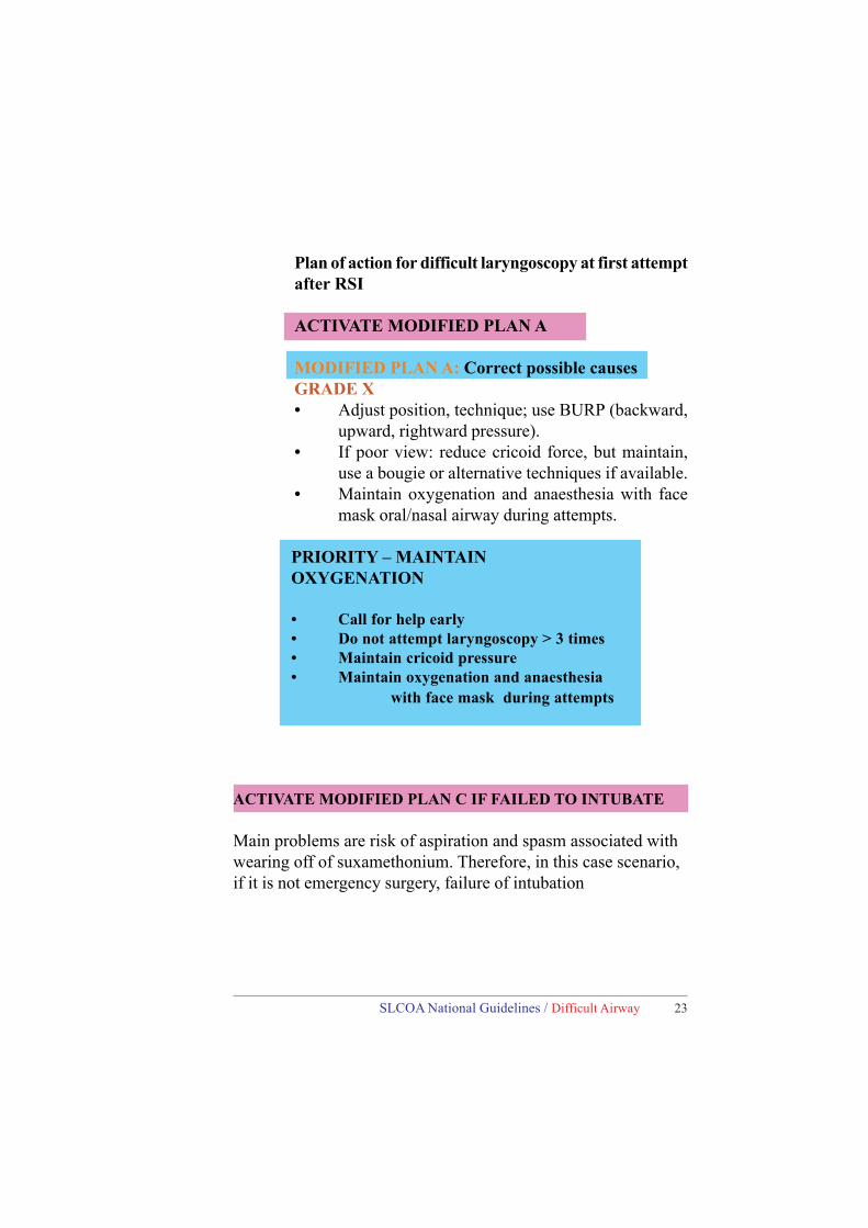
Plan of action for difficult laryngoscopy at first attemptafter RSI
ACTIVATE MODIFIED PLAN A
MODIFIED PLAN A: Correct possible causesGRADE X• Adjust position, technique; use BURP (backward,
upward, rightward pressure).• If poor view: reduce cricoid force, but maintain,
use a bougie or alternative techniques if available.• Maintain oxygenation and anaesthesia with face
mask oral/nasal airway during attempts.
PRIORITY – MAINTAINOXYGENATION
• Call for help early• Do not attempt laryngoscopy > 3 times• Maintain cricoid pressure• Maintain oxygenation and anaesthesia
with face mask during attempts
ACTIVATE MODIFIED PLAN C IF FAILED TO INTUBATE
Main problems are risk of aspiration and spasm associated withwearing off of suxamethonium. Therefore, in this case scenario,if it is not emergency surgery, failure of intubation
24 SLCOA National Guidelines / Difficult Airway
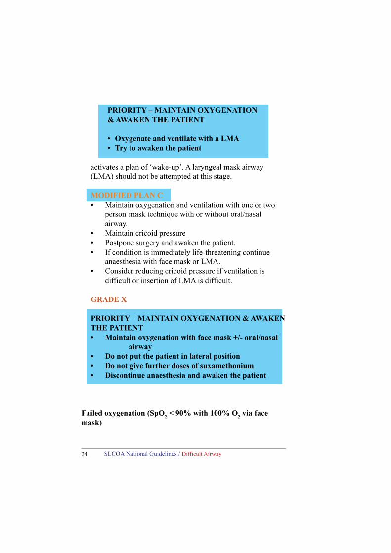
PRIORITY – MAINTAIN OXYGENATION& AWAKEN THE PATIENT
• Oxygenate and ventilate with a LMA• Try to awaken the patient
activates a plan of ‘wake-up’. A laryngeal mask airway(LMA) should not be attempted at this stage.
MODIFIED PLAN C• Maintain oxygenation and ventilation with one or two
person mask technique with or without oral/nasalairway.
• Maintain cricoid pressure• Postpone surgery and awaken the patient.• If condition is immediately life-threatening continue
anaesthesia with face mask or LMA.• Consider reducing cricoid pressure if ventilation is
difficult or insertion of LMA is difficult.
GRADE X
PRIORITY – MAINTAIN OXYGENATION & AWAKENTHE PATIENT• Maintain oxygenation with face mask +/- oral/nasal
airway• Do not put the patient in lateral position• Do not give further doses of suxamethonium• Discontinue anaesthesia and awaken the patient
Failed oxygenation (SpO2 < 90% with 100% O2 via facemask)
25SLCOA National Guidelines / Difficult Airway
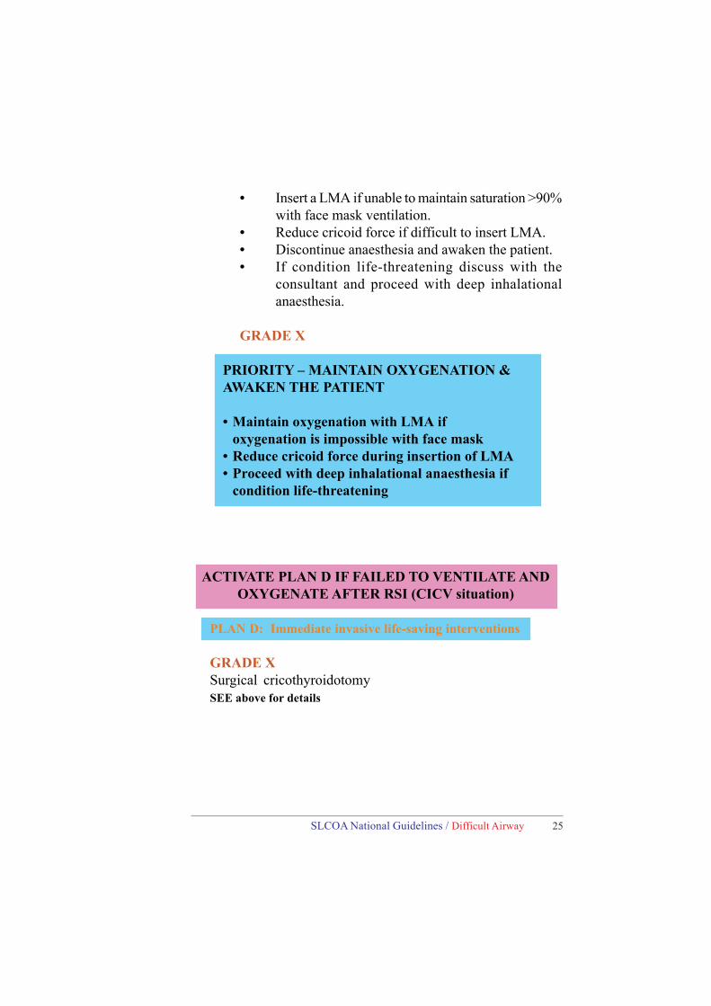
• Insert a LMA if unable to maintain saturation >90%with face mask ventilation.
• Reduce cricoid force if difficult to insert LMA.• Discontinue anaesthesia and awaken the patient.• If condition life-threatening discuss with the
consultant and proceed with deep inhalationalanaesthesia.
GRADE X
PRIORITY – MAINTAIN OXYGENATION &AWAKEN THE PATIENT
• Maintain oxygenation with LMA ifoxygenation is impossible with face mask
• Reduce cricoid force during insertion of LMA• Proceed with deep inhalational anaesthesia if
condition life-threatening
ACTIVATE PLAN D IF FAILED TO VENTILATE ANDOXYGENATE AFTER RSI (CICV situation)
PLAN D: Immediate invasive life-saving interventions
GRADE XSurgical cricothyroidotomySEE above for details

26 SLCOA National Guidelines / Difficult Airway
2.5.2 Obstetric difficult airway management drill
INTRODUCTION• Although frequency of general anaesthesia in obstetric isdeclining, it still remains the single most frequent anaesthetic causefor maternal morbidity and mortality. Hypoxic brain damage anddeath due to failure of maintaining adequate oxygenation at thetime of induction and intubation are the most dreadedcomplications. Anatomical and physiological changes in pregnancyand emergency intervention in inadequately assessed patients makethe situation more challenging. The incidence of failed intubationis almost 10 times higher in obstetric population than in the generalsurgical population. Therefore regional anaesthesia should beconsidered first in every case. This guideline is intended for use inadult obstetric patient who are undergoing general anaesthesia.This guideline is not intended for use in non-obstetric adult patients.
OBJECTIVES
• To avoid general anaesthetic related maternal morbidityand mortality by providing definitive guidelines so thatrecommended techniques will be used at every stage of generalanaesthesia.• To assess and evaluate pregnant mothers early with regardto anticipated difficult airway and formulate plans for intubation.
COMPETENCY/TRAINING
All qualified anaesthetists who are involved in obstetric anaesthesiamust initially complete a six month in house training in non-obstetric rapid sequence induction and difficult airway managementand should obtain competency before embarking on generalanaesthesia in obstetrics. Training and post-training period shouldinclude at least 10 general anaesthetics in obstetric patients undersupervision before working unsupervised.
27SLCOA National Guidelines / Difficult Airway
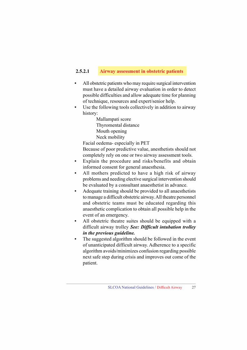
2.5.2.1 Airway assessment in obstetric patients
• All obstetric patients who may require surgical interventionmust have a detailed airway evaluation in order to detectpossible difficulties and allow adequate time for planningof technique, resources and expert/senior help.
• Use the following tools collectively in addition to airwayhistory:
Mallampati scoreThyromental distanceMouth openingNeck mobility
Facial oedema- especially in PETBecause of poor predictive value, anesthetists should notcompletely rely on one or two airway assessment tools.
• Explain the procedure and risks/benefits and obtaininformed consent for general anaesthesia.
• All mothers predicted to have a high risk of airwayproblems and needing elective surgical intervention shouldbe evaluated by a consultant anaesthetist in advance.
• Adequate training should be provided to all anaesthetiststo manage a difficult obstetric airway. All theatre personneland obstetric teams must be educated regarding thisanaesthetic complication to obtain all possible help in theevent of an emergency.
• All obstetric theatre suites should be equipped with adifficult airway trolley See: Difficult intubation trolleyin the previous guideline.
• The suggested algorithm should be followed in the eventof unanticipated difficult airway. Adherence to a specificalgorithm avoids/minimizes confusion regarding possiblenext safe step during crisis and improves out come of thepatient.
28 SLCOA National Guidelines / Difficult Airway
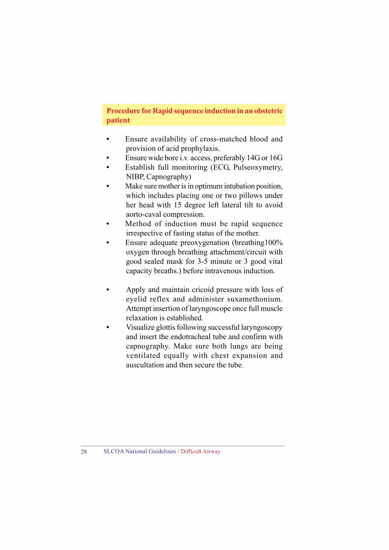
Procedure for Rapid sequence induction in an obstetricpatient
• Ensure availability of cross-matched blood andprovision of acid prophylaxis.
• Ensure wide bore i.v. access, preferably 14G or 16G• Establish full monitoring (ECG, Pulseoxymetry,
NIBP, Capnography)• Make sure mother is in optimum intubation position,
which includes placing one or two pillows underher head with 15 degree left lateral tilt to avoidaorto-caval compression.
• Method of induction must be rapid sequenceirrespective of fasting status of the mother.
• Ensure adequate preoxygenation (breathing100%oxygen through breathing attachment/circuit withgood sealed mask for 3-5 minute or 3 good vitalcapacity breaths.) before intravenous induction.
• Apply and maintain cricoid pressure with loss ofeyelid reflex and administer suxamethonium.Attempt insertion of laryngoscope once full musclerelaxation is established.
• Visualize glottis following successful laryngoscopyand insert the endotracheal tube and confirm withcapnography. Make sure both lungs are beingventilated equally with chest expansion andauscultation and then secure the tube.
29SLCOA National Guidelines / Difficult Airway
2.5.2.1 Unanticipated difficult laryngoscopy/intubation after rapid sequence induction in anobstetric patient
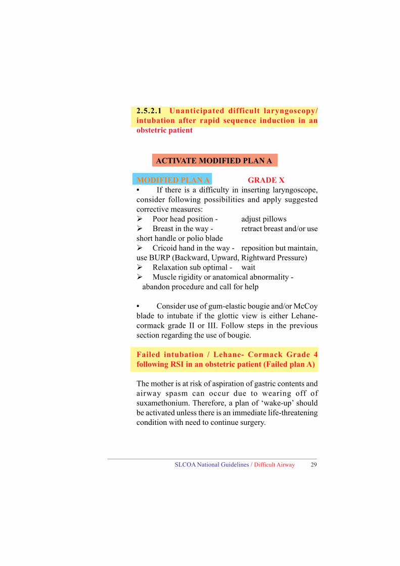
ACTIVATE MODIFIED PLAN A
MODIFIED PLAN A GRADE X• If there is a difficulty in inserting laryngoscope,consider following possibilities and apply suggestedcorrective measures:
Poor head position - adjust pillowsBreast in the way - retract breast and/or use
short handle or polio bladeCricoid hand in the way - reposition but maintain,
use BURP (Backward, Upward, Rightward Pressure)Relaxation sub optimal - waitMuscle rigidity or anatomical abnormality -
abandon procedure and call for help
• Consider use of gum-elastic bougie and/or McCoyblade to intubate if the glottic view is either Lehane-cormack grade II or III. Follow steps in the previoussection regarding the use of bougie.
Failed intubation / Lehane- Cormack Grade 4following RSI in an obstetric patient (Failed plan A)
The mother is at risk of aspiration of gastric contents andairway spasm can occur due to wearing off ofsuxamethonium. Therefore, a plan of ‘wake-up’ shouldbe activated unless there is an immediate life-threateningcondition with need to continue surgery.
30 SLCOA National Guidelines / Difficult Airway
ACTIVATE MODIFIED PLAN C (FAILEDINTUBATION DRILL)
MODIFIED PLAN C
• Do not panic.• Call for senior help. Send somebody other than the
involved anaesthetist.• Maintain oxygenation with 100% oxygen and cricoid
pressure with one or two person mask technique withor without oral /nasal airway. Provision of adequateoxygenation must be the priority at this stage.
• Attempt gentle positive pressure ventilation whilemaintaining cricoid pressure to provide/maintainoxygenation and to protect airway.
• Do not repeatedly attempt to insert laryngoscope or tovisualize glottis.
• Do not give second dose of suxamethonium.• Do not turn the mother to a side as this makes
maintenance of airway and oxygenation more difficult.But maintain the left lateral tilt.
• If adequate positive pressure ventilation is possible, andoxygenation is adequate (SpO2 >93%) determinewhether there is urgency/need to proceed with theplanned operation.
• If surgery is not life-saving, awaken the patient:
Discontinue inhalation agent, maintain airway with100% oxygen until patient is awake.
Turn to lateral position once awake.Decision to proceed with surgery at this stage usingspinal/epidural or CSE should be made afterdiscussing with the consultant anaesthetist.
GRADE XIf regional anaesthesia is contra-indicated considerlocal infiltration anaesthesia by the surgeon.
31SLCOA National Guidelines / Difficult Airway
The degree of anaesthesia and analgesia may be suboptimal and therefore mother should be fully informedabout the technique. GRADE X
If regional techniques are not indicated, awake fiber-opticintubation followed by general anaesthesia may beconsidered when facilities and expertise is available.GRADE Y
• If surgery is life-saving, continue anaesthesia withSPONTANEOUSLY BREATHING TECHNIQUE:
Deepen anaesthesia with inhalational agent in 100%oxygen while allowing the mother to breathespontaneously.Maintain airway with existing method. Maintainsaturation > 93%.Provide adequate analgesia /hypnosis / amnesia once thebaby is born. Consider fentanyl, alfentanil, midazolam,propofol.Start an infusion of syntocinon to overcome uterinerelaxation caused by high dose of inhalational agents.
FAILED OXYGENATION (SpO2 < 90%) VIA FACEMASK.
Maintain tight seal with face mask and apply oxygenwith closed breathing system valve.Ease cricoid pressure to allow adequate ventilation. Ifthe ventilation is possible without cricoid pressure,allow the patient to wake up if surgery is not life-saving.If operation is life-saving consider spontaneousbreathing technique as above.Insert a laryngeal mask airway (LMA) if ventilation isdifficult / impossible even without cricoid pressure. If ventilation is possible with the laryngeal maskairway, consider waking up or continuing deepanaesthesia depending on the urgency as above.
32 SLCOA National Guidelines / Difficult Airway
ACTIVATE PLAN D IF VENTILATION ISIMPOSSIBLE AND SATURATION IS < 90%
PLAN D: Immediate invasive life-saving interventionsto rescue the motherGRADE Xa) Cannula cricothyroidotomyb) Surgical cricothyroidotomy
SEE Page 20 for details
MATERNAL CARDIAC ARREST
Perform Caesarean section and deliver the baby as soonas possible.CPR will be otherwise unsuccessful as the gravid uterusimpedes the cardiac output.
• In each successfully managed difficult airwaysituation, recovery should be very cautious as prematureextubation could lead to serious complications again.
2.5.3 Management of acute upper airwayobstruction
2.5.3.1 Introduction
Obstruction of the airway occurs quite commonly in associationwith general anaesthesia. The anaesthetist is often called to stabilizeand intubate patients who present with acute airway obstruction inthe ETU/ICU or ward set up.Airway management is a fundamental anaesthetic responsibilityand skill. Airway obstruction demands a rapid and organisedapproach to its diagnosis and management and undue delay usuallyresults in desaturation and a potential threat to life.
33SLCOA National Guidelines / Difficult Airway
An uncomplicated pre-learned sequence of airway rescueinstructions is an essential part of every anaesthetist’s clinicalpractice requirements.
2.5.3.2 DefinitionIt is a potentially life-threatening event caused by a blockage ofthe upper airway, which can be in the trachea, larynx or oropharynx.
2.5.3.3 Common causes1. Upper airway• Oropharyngeal : tumours, haematoma, infection (epiglottitis,
croup), foreign bodies, sleep apnoea, obesity, facial oedema,burns
• Lesions in and around the larynx : malignancy, infections,laryngospasm, laryngeal oedema, inhalational injury
2. Mid-tracheal obstruction• Retrosternal goitre• Tracheal stenosis
3. Lower tracheal and bronchial obstruction• Tumours
2.5.3.4 Presentation
2.5.3.4 a) Partial airway obstruction-• Stridor- Inspiratory –obstruction at or above upper trachea Expiratory - obstruction of the lower trachea or bronchi.• Increased work of breathing –suprasternal, intercostal and
subcostal retraction along with an increased use of accessorymuscles of respiration. Paradoxical ‘see-saw’ movement ofabdomen and chest.
34 SLCOA National Guidelines / Difficult Airway
• Choking – sudden onset associated with coughing andaphonia indicates foreign body inhalation.
• Wheezing• Agitation, panic, changes in consciousness or
unconsciousness• Desaturation, hypercapnia
2.5.3.4 b) Complete airway obstruction-• Cyanosis• Inability to speak or breathe• Severe desaturation associated with bradycardia, cardio-respiratory arrest
2.5.3.4 c) Upper Airway Obstruction under anaesthesia-Breathing spontaneously:• Poor movement of the reservoir bag with or without above signsOn IPPV :• Increased airway pressure with decreased chest movement• Noisy respiration• Wheezing
2.5.3.5 Management2.5.3.5 a) Life threatening upper airway obstruction in theETU/ICU/Ward- Send for senior help; ask a nurse to phone.- Do not disturb the patient or delay for investigations, i.v. cannulation
etc.- Do not change the patient’s preferred position.- Administer 100% humidified O2 via facemask, attach pulse oximeter.- Nebulized adrenaline 1: 1000 5mg (400 mcg/kg up to a maximum of
5mg for children)- Take the patient to the theatre. Induce anaesthesia with halothane or
sevoflurane in oxygen and intubate with a small ETT. Have the ENTsurgeon ready to intervene if necessary (tracheostomy or rigidventilating broncoscopy)
- Do not give i.v. induction agents, sedatives or muscle relaxants before intubation as these may lead to catastrophic CICV (Can’t IntubateCan’t Ventilate) situation.
35SLCOA National Guidelines / Difficult Airway
2.5.3.5 b) Options for immediately life threateningevents and senior help not available
Severe cases: Perform cricothyroidotomy or tracheotomyunder local anaesthesia if intubation is not possible.Less severe cases: Perform intubation or tracheostomyunder deep inhalational anaesthesia with face mask or LMAif not improving with nebulized adrenaline.Children and uncooperative patients: Administer deepinhalational anaesthesia with preservation of spontaneousbreathing. Apply a moderate degree of CPAP which iseffective at keeping the airway open. Local anaesthetic sprayto the larynx may be helpful once the patient is deep andstable and give atropine to prevent bradycardia in children.
2.5.3.5 c) Acute upper airway obstruction under anaesthesia
Laryngospasm : Acute glottic closure by the vocal cordsThis is a common cause for acute airway obstruction in the peri-operative period.It is a potentially life-threatening condition which, if poorlymanaged can cause severe hypoxaemia, pulmonary aspiration andpost-obstructive negative pressure pulmonary oedema.
CausesSurgical stimulation or airway instrumentation orremoval under light anaesthesiaRegurgitation, vomiting or aspirationBlood or secretions in the pharynxIrritant volatile agent and exacerbation of a soiledairwayChildren are more prone to laryngospasm and becomecyanosed more rapidly than adults. Hypoxia associatedwith severe bradycardia is a preterminal event in them.
36 SLCOA National Guidelines / Difficult Airway
PresentationInspiratory stridor/airway obstruction/absentinspiratory sounds if complete obstruction.Increased respiratory efforts, tracheal tugParadoxical chest/abdomen (“see-saw) movementsDesaturation, bradycardia, central cyanosis
Immediate managementCall for helpStop stimulationRemove oral airway/LMA if cause for laryngospasmClear the airwayOpen the airway (head tilt, chin lift and jaw thrust)Give 100% oxygen (with inhalational agent ifappropriate)Deepen anaesthesia if appropriate (propofol 1-2 mg/kg iv bolus) or allow to wake upInsert oral /nasal airway if depth of anaesthesiaadequateApply CPAP (close the expiratory valve and maintaina tight seal by holding the mask with two hands) andgently ventilate while looking for a causeIf all above measures fail give suxamethonium 0.25 -0.5 mg/kg IV or 2-4 mg/kg IM if access is notavailable. Use early in complete airway obstructionas IPPV exacerbates the condition by inflating thestomach and forces the arytenoids and false cordsagainst the true vocal cords. Give atropine 10mcg/kgfor children or if associated with bradycardia.Intubate and ventilate if necessary
Subsequent managementMonitor in the recovery until stable, or in a HDU
or ICU depending on the conditionExclude pulmonary aspiration, negative pressure
pulmonary oedemaExplain what happened and reassure the patient as
awareness is possible.
37SLCOA National Guidelines / Difficult Airway
2.5.4 Management of Desaturation under anaesthesia
2.5.4.1 IntroductionDesaturation under anaesthesia (SpO2 < 93%) is apotentially life-threatening event that can result from manycauses. Inability to recognize the cause and manage itpromptly may lead to hypoxic brain injury or death.Correct use of a structured drill has been shown to identify99% of possible causes for desaturation within 40-60seconds.Persistent desaturation should be managed with handventilation using 100% oxygen, return to a supine positionand completion of a checklist of possible causes (COVER-ABCD). Blood gases, chest X-ray and bronchoscopy maybe required if no apparent cause can be found.
2.5.4.2 Causes for desaturation under anaesthesia:
I Equipment problems
a) Anaesthetic machine – related :Hypoxic gas mixtureAnaesthetic machine errorNo gas flow during preoxygenationDisconnection, leaks,Breathing system : damage to inner tube in Bainsystem, obstruction of tubings, catheter mount,connectors, filterMonitor error
b) Endotracheal tube –related:Leaks, kinking, obstructionMisplacement- oesophageal, endobronchial,accidental extubationFailed intubation / failed ventilation
38 SLCOA National Guidelines / Difficult Airway
II Patient problems
a) Airway - obstruction if unintubated- secretions,blood or debris, gastric contents, foreign body,laryngospasm
b) Breathing – hypoventilation, bronchospasm, lobarcollapse, pneumo- or haemothorax, gaseousdistension of the abdomen (esp. in infants)
c) Circulation – cardiac arrest, haemodynamicinstability, congestive cardiac failure with pulmonaryoedema, obstruction to circulation from excessiveintrathoracic pressure(inadvertent PEEP), pulmonaryembolism, cardiac tamponade, reversal of a shunt
d) Other – worsening of pre-existing conditions,obesity, sepsis
e) Drugs – anaphylaxis, malignant hyperpyrexia
2.5.4.3 Desaturation drill• Confirm diagnosis: check pulse oximeter waveform• Exclude cardiac arrest• Call for help• Administer 100% oxygen and hand ventilate• Complete “COVER ABCD” algorithm if intubated or
“AB COVER CD” if breathing spontaneously toidentify the cause for desaturation.
“COVER ABCD”
C1 CirculationCheck rate, rhythm and volume of the central pulse.Check ETCO2 which indicates cardiac output.Initiate CPR if no central pulse.
39SLCOA National Guidelines / Difficult Airway
C2 ColourGive 100% oxygen; ensure that only oxygen flow
meter is operating. Look for central cyanosis. Checkpulse oxymeter.
O1 OxygenCheck flow meter settings to ensure that mixture is
not hypoxic.
O 2 Oxygen analyzerCheck that the oxygen analyser shows a rising oxygen
concentration distal to the common gas outlet.
V1 VentilationVentilate the lungs by hand to assess breathing circuit
integrity, check the patency of the catheter mount,connector and filter*, airway patency, chestcompliance and air entry by “feel” and carefulobservation and auscultation.
Also inspect capnography trace.
V2 VaporiserNote settings and levels of agents. Check all vaporiserfiller ports, seatings and connections for liquid or gasleaks during pressurisation of the system.Consider the possibility of the wrong agent being inthe vaporiser.
E1 Endotracheal tubeSystematically check the endotracheal tube (if in use).Ensure that it is patent with no leaks or kinks orobstructions.Check capnograph for tracheal placement and oximeterfor possible endobronchial position.If necessary, adjust, deflate cuff, pass a catheter, orremove and replace.
40 SLCOA National Guidelines / Difficult Airway
E2 Elimination
Eliminate the anaesthetic machine and ventilate with self-inflating (e.g. Ambu) bag with 100% oxygen (from alternativesource if necessary). Retain gas monitor sampling port if available (but be aware of
possible problems).
R1 Review monitors
Review all monitors in use (preferably oxygen analyser,capnograph, oximeter, blood pressure, electrocardiograph (ECG),temperature and neuromuscular junction monitor). For proper use,the algorithm requires all monitors to have been correctly sited,checked and calibrated.
R2 Review equipment
Review all other equipment in contact with or relevant to thepatient (e.g. diathermy, humidifiers, heating blankets, endoscopes,probes, prostheses, retractors and other appliances).
A Airway
Check patency of the unintubated airway. Consider laryngospasm or presence of foreign body, blood,
gastric contents, nasopharyngeal or bronchial secretions. Mucus plugs or bronchial secretions can causes marked
desaturation especially in young children.
B Breathing
Assess pattern, adequacy and distribution of ventilation.
Consider, examine and auscultate for bronchospasm, pulmonary oedema, lobar collapse, pneumo- or haemothorax and impaired ventilation due to retractors.
41SLCOA National Guidelines / Difficult Airway
C Circulation
Repeat evaluation of peripheral perfusion, pulse, bloodpressure, ECG and filling pressures (where possible) andhypovolaemia or any possible obstruction to venous return suchas embolism, raised intrathoracic pressure (e.g. inadvertent PEEP),sepsis, myocardial depression and poor cardiac output or directinterference to (e.g. stimulation by central line) or tamponade ofthe heart.
Note any trends on records.
D Drugs
Review intended (and consider possible unintended) drug orsubstance administration.
Consider whether the problem may be due to unexpectedeffect (anaphylaxis, malignant hyperpyrexia), a failure ofadministration or wrong dose, route or manner of administrationof an intended or “wrong drug”.
Review all possible routes of drug administration.
2.6 Extubation & Follow upWhen the airway has proved difficult to manage a difficult airwayfollow-up should be initiated in the postoperative period.
2.6.1 Strategy for Extubation of the Difficult Airway
The strategy should depend on the surgery, the condition of thepatient, and the skills and preferences of the anaesthetist.
Take the following into consideration before extubation:
GRADE X
42 SLCOA National Guidelines / Difficult Airway
1. Risks and benefits of awake extubation versus deepextubation with the use of a nasopharyngeal airway or LMA.
2. Extubate with the expiratory valve tightly closed to producea cough.
3. Presence of any general clinical factors that may impairventilation after extubation.
4. Short-term use of a device that can serve as a guide forreintubation if necessary. Eg : Cook airway exchanger – it isusually inserted through the lumen of the tracheal tube andinto the trachea before the tracheal tube is removed. It isrigid to facilitate intubation and hollow to facilitateventilation and administer oxygen.
5. Give prophylactic dexamethasone if there is a risk oflaryngeal oedema and monitor closely in the post-operativeperiod.
2.6.2 Follow-up Care GRADE X
Document the presence and nature of the airway difficultyand management strategy in the anaesthetic record.
Explain to the patient or responsible person the difficulty,and the importance of informing the next anaesthetist of theproblem.
Evaluate and follow up complications of difficult airwaymanagement: sore throat, difficulty in swallowing, pain orswelling of the face, bleeding, tracheal and esophagealperforation, chest pain, pneumothorax, aspiration, and dentaldamage. ENT referral if necessary. Express regret formorbidity.
43SLCOA National Guidelines / Difficult Airway
2.6.3 COMPLETE A DIAGNOSIS CARD ANDHAND OVER TO THE PATIENT- this is a useful way ofmaking certain that you have documented events properly.
Reasons/comments
Difficult mask
ventilation?
Yes /
No
Difficult direct
laryngoscopy?
Yes /
No
Difficult
tracheal intubation?
YES /
NO
Laryngoscopy grade 1 / 2 / 3 / 4
Equipment used:
Other information:
Is awake intubation necessary in the future?
Complications :
Name of anaesthetist:
Grade: Date:
If you require further information please contact the
Anaesthetic Department.
44 SLCOA National Guidelines / Difficult Airway
References
1. Management of difficult and failed intubation in obstetrics-BJA/CPDE, vol1, No4, 2004
2. Airway management in obstetrics. - Indian J Anesth 2005;49(4): 325-338
3. Difficult airway society guidelines for management ofunanticipated difficult intubation –Anaesthsia2004,53,p675-694
4. Hand Book of Anaesthesia 3rd Edition 2004
5. American Society of Anesthesiologists Task Force onManagement of the Difficult Airway. Practice guidelinesfor management of the difficult airway: an updated reportby the American Society of Anesthesiologists Task Forceon Management of the Difficult Airway.Anesthesiology 2003 May;98(5):1269-77.
6. Prediction and Management of Difficult TrachealIntubation. Update in Anaesthesia WFSA Issue 9 (1998)Article 9: Page 2 of 4