management of a complex case using conventional complete denture...
TRANSCRIPT

Citation: Păuna M, Babiuc I and Farcaşiu AT. Management of a Complex Case using Conventional Complete Denture Restorations. Austin J Dent. 2016; 3(3): 1038.
Austin J Dent - Volume 3 Issue 3 - 2016ISSN : 2381-9189 | www.austinpublishinggroup.com Păuna et al. © All rights are reserved
Austin Journal of DentistryOpen Access
Abstract
Clinical experience has demonstrated that in complete edentulous patients, more than in other areas of prosthetics, particularities of the case play an important role. The prosthodontic treatment must provide restorations that are bio-functional and bio-prophylactic for the surrounding tissues. In this aim, an edentulous patient must be submitted to a methodical clinical examination in order to establish the degree of difficulty of the prosthetic treatment. Additional investigation as cephalometric radiographs can be useful. In our daily practice, we are rarely confronted with a normal morphology of the denture bearing oral structures. Managing abused tissues in a patient with morphologic abnormalities due to faulty prostheses sometimes proves difficult. Preventing the deterioration of oral status must be a condition in providing a chance for the success of the following rehabilitations, mainly in the situation when the complete edentulousness appears in a young or middle age patient.
This article is a clinical report on the prosthodontic management of a severe mandibular atrophy with a particular conformation of the alveolar crest correlated with the necessity of a maxillary immediate complete denture in a patient with dramatic perturbation of esthetics.
Keywords: Mandibular particular conformation; Maxillary-mandibular relationship; Conventional complete dentures
Intraoral clinical examination revealed a severe atrophy of the mandibular bone and abnormal anatomic situation. A massive protuberance was found in the buccal side of the first molar region on the right side of the mandible. It measured 12 mm in length, 8 mm in
IntroductionThe success of conventional complete denture therapy can be
evaluated based on some prognostic indicators. The fabrication of technically correct dentures, a favorable conformation of the mandibular crest and the accuracy of jaw relations are positive indicators for success, whereas patient neuroticism and a poorly formed mandibular ridge are negative indicators [1]. Following these criteria, severe atrophy of the mandible poses a great prosthodontic challenge [2-6] especially in conjunction with other particular morphological aspects [7,8].
Case ReportA 53 years old female patient was referred to the Removable
Prosthodontics. Department, Faculty of Dentistry, ”Carol Davila” University of Medicine and Pharmacy from Bucharest. Due to family problems, she neglected her oral health for a long period of time. Masticatory function, esthetics and speech were severely affected (Figure 1,2).
The patient presented two incorrect dentures which favored the development of periodontal disease and functional problems. The upper denture was inappropriately designed, with poor fit and reduced tissue support. As a consequence, the remaining maxillary anterior teeth were tilted on an almost horizontal position [9,10]. The mandibular complete denture, fabricated 10 years before, immediately after all the remaining teeth were extracted, had a particularly undexteded and ill-fitting base. Both dentures displayed very poor esthetics and incorrect occlusal relations (Figure 3).
Case Report
Management of a Complex Case using Conventional Complete Denture RestorationsPăuna M*, Babiuc I and Farcaşiu ATDepartment of Removable Prosthodontics, Carol Davila University of Medicine and Pharmacy, Romania
*Corresponding author: Mihaela Pauna, Department of Removable Prosthodontics, Faculty of Dentistry, Carol Davila University of Medicine and Pharmacy, Bucharest, Romania
Received: May 07, 2016; Accepted: July 11, 2016; Published: July 13, 2016
Figure 1: Facial aspect at first visit.
Figure 2: Appearance of maxillary incisors.

Austin J Dent 3(3): id1038 (2016) - Page - 02
Păuna M Austin Publishing Group
Submit your Manuscript | www.austinpublishinggroup.com
width and 5 mm in height. The area was covered by clinically normal soft tissue without sensitivity upon palpation and did not present any history of pain while the denture was used.
The patient recalled that 10 years before, when the teeth were extracted from this area, iodoform gauze was applied to enhance the healing. The radiological investigation reveals the formation of an abnormal bony architecture, with visible iodine crystals (Figure 4). Bone formation after contact with iodoform was a rare thing but it was mentioned by Thomson & Miles in 1904 [11].
The panoramic X-ray revealed a severely compromised status of the remaining maxillary teeth. The advanced loss of periodontal support presented a predominantly horizontal pattern, in conjunction with angular defects, which left most of the roots unsupported. As a result, the buccal migration of the incisors took place and all the remaining teeth displayed an increased mobility. The roots supporting the fixed restoration in the first quadrant presented an extensive loss of periodontal support as a result of endo-perio lesions.
The mandibular alveolar crest presented an increased resorbtion in the areas related to the opposing teeth, when the vertical position of the mental foramen and mandibular canal were taken as reference. In the alveolar area corresponding to teeth 46 and 47 a bone condensation and a protuberance is visible (Figure 5).
The cephalometric evaluation with the old restorations in the mouth reveal an almost hypodivergent skeletal pattern (FMA=21), with an increased facial index (FI=0.8), which suggests an anterior rotation of the mandible. Other measurements also support the diagnosis of reduced vertical dimension and anterior rotation of the mandible: GoGn – SN (30 as compared to a normal value of 32), NsaNsp-M (17, normally 28), NsaXi-XiPM (36, normally 43), Nsa-Me. Angular measurements show a normal position of the maxilla and mandible in relation to the cranial base.
Figure 3: Lower and upper old dentures.
Figure 4: Radiological aspect of the mandibular abnormal protuberance.
Figure 5: Initial OPG investigation.
Figure 6: Lateral cephalometric radiograph.
Figure 7: Patient’s facial profile.
Figure 8: Mandible master cast.
Figure 9: Section of the right mandibular edentulous ridge in the middle of the protuberance.
Austin J Dent 3(3): id1038 (2016) - Page - 03
Păuna M Austin Publishing Group
Submit your Manuscript | www.austinpublishinggroup.com
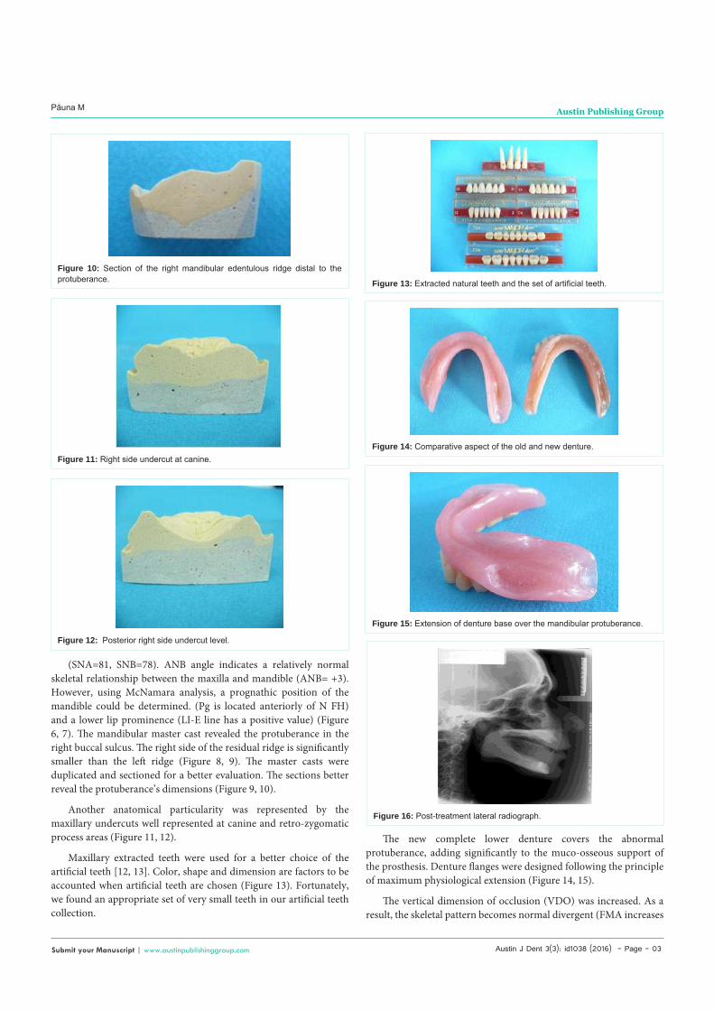
(SNA=81, SNB=78). ANB angle indicates a relatively normal skeletal relationship between the maxilla and mandible (ANB= +3). However, using McNamara analysis, a prognathic position of the mandible could be determined. (Pg is located anteriorly of N FH) and a lower lip prominence (LI-E line has a positive value) (Figure 6, 7). The mandibular master cast revealed the protuberance in the right buccal sulcus. The right side of the residual ridge is significantly smaller than the left ridge (Figure 8, 9). The master casts were duplicated and sectioned for a better evaluation. The sections better reveal the protuberance’s dimensions (Figure 9, 10).
Another anatomical particularity was represented by the maxillary undercuts well represented at canine and retro-zygomatic process areas (Figure 11, 12).
Maxillary extracted teeth were used for a better choice of the artificial teeth [12, 13]. Color, shape and dimension are factors to be accounted when artificial teeth are chosen (Figure 13). Fortunately, we found an appropriate set of very small teeth in our artificial teeth collection.
Figure 10: Section of the right mandibular edentulous ridge distal to the protuberance.
Figure 11: Right side undercut at canine.
Figure 12: Posterior right side undercut level.
The new complete lower denture covers the abnormal protuberance, adding significantly to the muco-osseous support of the prosthesis. Denture flanges were designed following the principle of maximum physiological extension (Figure 14, 15).
The vertical dimension of occlusion (VDO) was increased. As a result, the skeletal pattern becomes normal divergent (FMA increases
Figure 13: Extracted natural teeth and the set of artificial teeth.
Figure 14: Comparative aspect of the old and new denture.
Figure 15: Extension of denture base over the mandibular protuberance.
Figure 16: Post-treatment lateral radiograph.
Austin J Dent 3(3): id1038 (2016) - Page - 04
Păuna M Austin Publishing Group
Submit your Manuscript | www.austinpublishinggroup.com
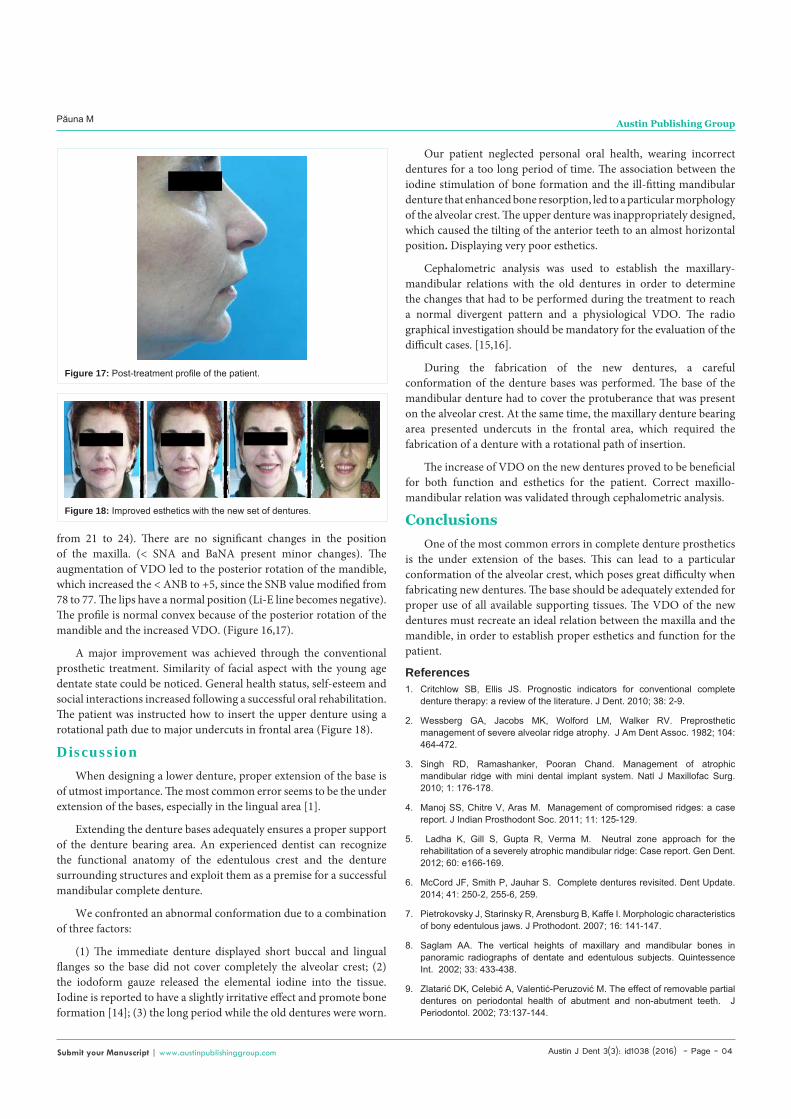
from 21 to 24). There are no significant changes in the position of the maxilla. (< SNA and BaNA present minor changes). The augmentation of VDO led to the posterior rotation of the mandible, which increased the < ANB to +5, since the SNB value modified from 78 to 77. The lips have a normal position (Li-E line becomes negative). The profile is normal convex because of the posterior rotation of the mandible and the increased VDO. (Figure 16,17).
A major improvement was achieved through the conventional prosthetic treatment. Similarity of facial aspect with the young age dentate state could be noticed. General health status, self-esteem and social interactions increased following a successful oral rehabilitation. The patient was instructed how to insert the upper denture using a rotational path due to major undercuts in frontal area (Figure 18).
DiscussionWhen designing a lower denture, proper extension of the base is
of utmost importance. The most common error seems to be the under extension of the bases, especially in the lingual area [1].
Extending the denture bases adequately ensures a proper support of the denture bearing area. An experienced dentist can recognize the functional anatomy of the edentulous crest and the denture surrounding structures and exploit them as a premise for a successful mandibular complete denture.
We confronted an abnormal conformation due to a combination of three factors:
(1) The immediate denture displayed short buccal and lingual flanges so the base did not cover completely the alveolar crest; (2) the iodoform gauze released the elemental iodine into the tissue. Iodine is reported to have a slightly irritative effect and promote bone formation [14]; (3) the long period while the old dentures were worn.
Figure 17: Post-treatment profile of the patient.
Our patient neglected personal oral health, wearing incorrect dentures for a too long period of time. The association between the iodine stimulation of bone formation and the ill-fitting mandibular denture that enhanced bone resorption, led to a particular morphology of the alveolar crest. The upper denture was inappropriately designed, which caused the tilting of the anterior teeth to an almost horizontal position. Displaying very poor esthetics.
Cephalometric analysis was used to establish the maxillary-mandibular relations with the old dentures in order to determine the changes that had to be performed during the treatment to reach a normal divergent pattern and a physiological VDO. The radio graphical investigation should be mandatory for the evaluation of the difficult cases. [15,16].
During the fabrication of the new dentures, a careful conformation of the denture bases was performed. The base of the mandibular denture had to cover the protuberance that was present on the alveolar crest. At the same time, the maxillary denture bearing area presented undercuts in the frontal area, which required the fabrication of a denture with a rotational path of insertion.
The increase of VDO on the new dentures proved to be beneficial for both function and esthetics for the patient. Correct maxillo-mandibular relation was validated through cephalometric analysis.
ConclusionsOne of the most common errors in complete denture prosthetics
is the under extension of the bases. This can lead to a particular conformation of the alveolar crest, which poses great difficulty when fabricating new dentures. The base should be adequately extended for proper use of all available supporting tissues. The VDO of the new dentures must recreate an ideal relation between the maxilla and the mandible, in order to establish proper esthetics and function for the patient.
References1. Critchlow SB, Ellis JS. Prognostic indicators for conventional complete
denture therapy: a review of the literature. J Dent. 2010; 38: 2-9.
2. Wessberg GA, Jacobs MK, Wolford LM, Walker RV. Preprosthetic management of severe alveolar ridge atrophy. J Am Dent Assoc. 1982; 104: 464-472.
3. Singh RD, Ramashanker, Pooran Chand. Management of atrophic mandibular ridge with mini dental implant system. Natl J Maxillofac Surg. 2010; 1: 176-178.
4. Manoj SS, Chitre V, Aras M. Management of compromised ridges: a case report. J Indian Prosthodont Soc. 2011; 11: 125-129.
5. Ladha K, Gill S, Gupta R, Verma M. Neutral zone approach for the rehabilitation of a severely atrophic mandibular ridge: Case report. Gen Dent. 2012; 60: e166-169.
6. McCord JF, Smith P, Jauhar S. Complete dentures revisited. Dent Update. 2014; 41: 250-2, 255-6, 259.
7. Pietrokovsky J, Starinsky R, Arensburg B, Kaffe I. Morphologic characteristics of bony edentulous jaws. J Prothodont. 2007; 16: 141-147.
8. Saglam AA. The vertical heights of maxillary and mandibular bones in panoramic radiographs of dentate and edentulous subjects. Quintessence Int. 2002; 33: 433-438.
9. Zlatarić DK, Celebić A, Valentić-Peruzović M. The effect of removable partial dentures on periodontal health of abutment and non-abutment teeth. J Periodontol. 2002; 73:137-144.
Figure 18: Improved esthetics with the new set of dentures.

Austin J Dent 3(3): id1038 (2016) - Page - 05
Păuna M Austin Publishing Group
Submit your Manuscript | www.austinpublishinggroup.com
10. Bergman B, Hugoson A, Olsson CO. Caries, periodontal and prosthetic findings in patients with removable partial dentures: A ten-year longitudinal study. J Prosthe Dent. 1982; 48: 506-514.
11. Thomson A, Miles A. A Manual of Surgery. Kaplan Publishing. 2009; 528.
12. Seck A, Guèye M, Dieng L, Mbodj EB, Ndiaye C, Seck MT, et al. Correlations between colorimetric parameters of teeth, eyes and skin. Perspectives in the choice of tooth shade for complete denture. Odontostomatol Trop. 2013; 36: 17-25.
13. Sellen PN, Jagger DC, Harrison A. The selection of anterior teeth appropriate for the age and sex of the individual. How variable are dental staffs in their choice? J Oral Rehabil. 2002; 29: 853-857.
14. Strajnić L, Misković B. Computerized cephalometric evaluation of changes following treatment with complete dentures. Med Pregl. 2012; 65: 163-167.
15. Palac A, Bitanga P, Capkun V, Kovacic I. Association of cephalometric changes after 5 years of complete dentures wearing and oral health-related quality-of-life. Acta Odontol Scand. 2013; 71: 449-456.
16. Singh, V, Das S, Sharma N. Iodoform: A boon in disguise. Open Journal of Stomatology. 2012; 2: 322-325.
Citation: Păuna M, Babiuc I and Farcaşiu AT. Management of a Complex Case using Conventional Complete Denture Restorations. Austin J Dent. 2016; 3(3): 1038.
Austin J Dent - Volume 3 Issue 3 - 2016ISSN : 2381-9189 | www.austinpublishinggroup.com Păuna et al. © All rights are reserved