malpositionsincisives orthodontie mandibulairesdel ... - sop.asso.fr · 19 revue...
TRANSCRIPT
soumis pour publication le 10 avril 2011accepté pour publication le 22 juin 2011 Revue d’Odonto-Stomatologie/Février 2012
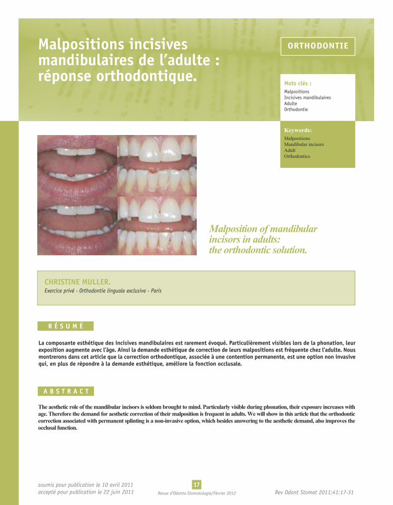
La composante esthétique des incisives mandibulaires est rarement évoqué. Particulièrement visibles lors de la phonation, leurexposition augmente avec l’âge. Ainsi la demande esthétique de correction de leurs malpositions est fréquente chez l’adulte. Nousmontrerons dans cet article que la correction orthodontique, associée à une contention permanente, est une option non invasivequi, en plus de répondre à la demande esthétique, améliore la fonction occlusale.
Malpositions incisivesmandibulaires de l’adulte :réponse orthodontique.
ORTHODONTIE
Malposition of mandibularincisors in adults:the orthodontic solution.
Rev Odont Stomat 2011;41:17-31
R É S U M É
The aesthetic role of themandibular incisors is seldombrought tomind. Particularly visible during phonation, their exposure increases withage. Therefore the demand for aesthetic correction of theirmalposition is frequent in adults.Wewill show in this article that the orthodonticcorrection associated with permanent splinting is a non-invasive option, which besides answering to the aesthetic demand, also improves theocclusal function.
A B S T R A C T
17
CHRISTINE MULLER.Exercice privé - Orthodontie linguale exclusive - Paris
Mots clés :MalpositionsIncisives mandibulairesAdulteOrthodontie
Keywords:MalpositionsMandibular incisorsAdultOrthodontics
18Revue d’Odonto-Stomatologie/Février 2012
ORTHODONTIE
Introduction
La visibilité des incisives maxillaires lors du sourire estun critère esthétique majeur. C’est pourquoi en termed’esthétique, la préoccupation principale des chercheurss’est focalisée sur les incisives maxillaires en analysant,par exemple, les modifications de l’attractivité des souriresen fonction de différents paramètres : proportions (Wolfartet coll., 2005), axe longitudinal (Zlowodski et coll.,2008) etc....
Si cet aspect est prépondérant chez l’adulte jeune, unautre paramètre crucial de l’apparence esthétique estsouvent oublié chez l’adulte et le senior : il s’agit de lacomposante esthétique des incisives mandibulaires. Viget Brundo (1977) constataient déjà que le rôle des incisivesmandibulaires était plus important que celui que ne luiaccordait la littérature. Ils ont montré qu’avec l’âge,l’exposition des incisives maxillaires diminue graduel-lement et que ce phénomène est accompagné d’uneaugmentation de la visibilité des incisives mandibulaires.Cade (1979) notait ainsi que le rôle des incisivesmandibulaires dans l’esthétique des patients de 60 anset plus est le même que celui des incisives maxillaireschez les jeunes de moins de 30 ans.
C’est particulièrement le cas lors de la phonation, fonctionsociale très importante, pendant laquelle les incisivesmandibulaires sont largement exposées. Une simpleobservation minutieuse d’un enregistrement vidéo suf-fit pour s’en convaincre (Sackstein 2007, 2008).
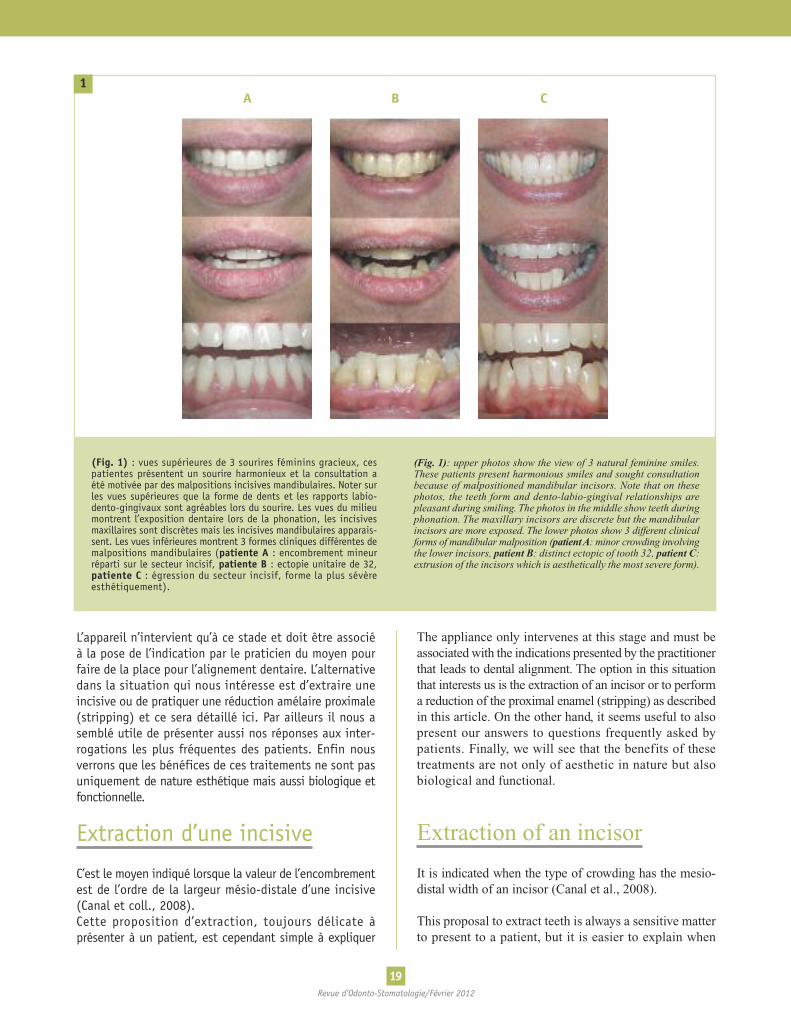
L’ensemble de ces constatations explique pourquoi chezle senior, qui présente souvent des malpositions desincisives mandibulaires liées au vieillissement, l’alignementdentaire devient le 1er souhait esthétique de cettetranche d’âge (Wulfman et coll., 2009) mais aussi pourquoiil ne faut pas traiter à la légère la demande concernantles désordres de ce secteur (fig. 1).
Le but de cet article clinique est de décrire une réponse non-prothétique à cette demande, une réponse orthodontiquetout à fait dans la logique d’une dentisterie moderne lamoins invasive possible (Tirlet et Attal, 2009). L’ap-pareillage utilisé pour traiter tous les cas présentés danscet article est l’appareillage Incognito (3M). Rappelonsque le dispositif n’est qu’un outil avec ses limites. N’ou-blions pas l’essentiel, son emploi est sous-tendu par unexamen clinique, la pose d’un diagnostic et l’établisse-ment d’objectifs.
Introduction
The visibility of the maxillary incisors while smiling is amajor aesthetic criterion. That is why in an aestheticstandpoint, the principal concern of researchers is focalizedon the maxillary incisors by analyzing for example thechanges in the attractiveness of smiles according to thedifferent parameters, proportions (Wolfart et al., 2005),longitudinal axis (Zlowodski et al., 2008), etc.
If this aspect is predominant in young adults, anothercritical parameter of aesthetic appearance is often forgot-ten in adults and the elderly: it is the aesthetic role of themandibular incisors. Vig and Brundo (1977) alreadynoticed that the role of the mandibular incisors was moreimportant than what is mentioned in the literature. Theyshowed that with age, the exposure of themaxillary incisorsgradually decreases and that this phenomenon is ac-companied by the increase in the visibility of the mandibularincisors. Cade (1979) also noted that the aesthetic role ofthe mandibular incisors in patients aged 60 years or moreis the same as that of the maxillary incisors of the youngof less than 30 years.
Particularly, in the case of phonation being a very importantsocial function during which the mandibular incisors aremostly exposed. A tedious observation of a simple videorecording is enough to be convinced (Sackstein 2007,2008).
All these observations explain why in the elderly, whooften showmalposition of the mandibular incisors, relatedto aging, the dental alignment becomes their initialaesthetic desire (Wulfman et al., 2009). That is the reasonwhy consultations regarding the disorders of this area inthis age group should not be taken lightly (fig. 1).
The aim of this clinical article is to illustrate the non-prosthetic response to this demand, an orthodontic solutionthat is completely in the logic of modern dentistry and asminimally invasive as possible (Tirlet and Attal, 2009).The appliance used to treat the cases presented in thisarticle is the Incognito (3M) appliance. It calls to mindthat the procedure is only a tool with its limits. Don’t forgetthat what is essential is that its use is preceded by a clinicalexamination, diagnosis and setting up of objectives.
19Revue d’Odonto-Stomatologie/Février 2012
A B C
L’appareil n’intervient qu’à ce stade et doit être associéà la pose de l’indication par le praticien du moyen pourfaire de la place pour l’alignement dentaire. L’alternativedans la situation qui nous intéresse est d’extraire uneincisive ou de pratiquer une réduction amélaire proximale(stripping) et ce sera détaillé ici. Par ailleurs il nous asemblé utile de présenter aussi nos réponses aux inter-rogations les plus fréquentes des patients. Enfin nousverrons que les bénéfices de ces traitements ne sont pasuniquement de nature esthétique mais aussi biologique etfonctionnelle.
Extraction d’une incisive
C’est le moyen indiqué lorsque la valeur de l’encombrementest de l’ordre de la largeur mésio-distale d’une incisive(Canal et coll., 2008).Cette proposition d’extraction, toujours délicate àprésenter à un patient, est cependant simple à expliquer
The appliance only intervenes at this stage and must beassociated with the indications presented by the practitionerthat leads to dental alignment. The option in this situationthat interests us is the extraction of an incisor or to performa reduction of the proximal enamel (stripping) as describedin this article. On the other hand, it seems useful to alsopresent our answers to questions frequently asked bypatients. Finally, we will see that the benefits of thesetreatments are not only of aesthetic in nature but alsobiological and functional.
Extraction of an incisor
It is indicated when the type of crowding has the mesio-distal width of an incisor (Canal et al., 2008).
This proposal to extract teeth is always a sensitive matterto present to a patient, but it is easier to explain when
1
(Fig. 1) : vues supérieures de 3 sourires féminins gracieux, cespatientes présentent un sourire harmonieux et la consultation aété motivée par des malpositions incisives mandibulaires. Noter surles vues supérieures que la forme de dents et les rapports labio-dento-gingivaux sont agréables lors du sourire. Les vues du milieumontrent l’exposition dentaire lors de la phonation, les incisivesmaxillaires sont discrètes mais les incisives mandibulaires apparais-sent. Les vues inférieures montrent 3 formes cliniques différentes demalpositions mandibulaires (patiente A : encombrement mineurréparti sur le secteur incisif, patiente B : ectopie unitaire de 32,patiente C : égression du secteur incisif, forme la plus sévèreesthétiquement).
(Fig. 1): upper photos show the view of 3 natural feminine smiles.These patients present harmonious smiles and sought consultationbecause of malpositioned mandibular incisors. Note that on thesephotos, the teeth form and dento-labio-gingival relationships arepleasant during smiling. The photos in the middle show teeth duringphonation. The maxillary incisors are discrete but the mandibularincisors are more exposed. The lower photos show 3 different clinicalforms of mandibular malposition (patientA: minor crowding involvingthe lower incisors, patient B: distinct ectopic of tooth 32, patient C:extrusion of the incisors which is aesthetically the most severe form).
20Revue d’Odonto-Stomatologie/Février 2012
ORTHODONTIE
quand l’encombrement est sévère, car les patients ontpresque toujours conscience de «l’excès dentaire» qu’ilsobservent souvent s’aggraver depuis des années. Il estintéressant de constater que régulièrement, davantageque la « mutilation », ce qui génère le plus de stress, estl’idée de l’asymétrie du résultat final et le fait d’avoir àassumer un édentement antérieur après l’extraction.Les questions les plus fréquentes sont :
1) « Est-ce-que je vais être de travers avec 3 dentsdevant ? »Rassurer le patient est très simple en montrant des castraités et en expliquant que cette anomalie de nombrepasse complètement inaperçue à condition que les axesdes 3 incisives mandibulaires soient bien verticaux aprèsla fermeture de l’espace d’extraction.
Le cas clinique suivant (fig. 2 et 3) illustre les bénéficesobtenus grâce au « sacrifice » d’une incisive. La patienteâgée de 40 ans consulte au sujet de malpositionsmandibulaires. Elle est médecin et se plaint de réflexionsde petits patients. Sa demande ne concerne pas l’améliorationde son sourire ni de son occlusion (classe 1 bilatérale) maisla normalisation du secteur incisif mandibulaire. De plus,elle a noté le caractère évolutif de l’encombrement etcherche aussi une solution qui réponde au besoin destabilisation de sa situation à moyen et long terme.Deux projets ont été proposés à la patiente. Un traitementmandibulaire répondant à sa demande et un traitementbimaxillaire avec l’objectif d’amélioration de son sourire(correction de la courbe incisive inversée). La patientea choisi le projet bimaxillaire. Le temps orthodontique(appareil lingual bimaxillaire + extraction de 31) a duré15 mois à l’issue duquel des contentions permanentes decanines à canines ont été collées.L’occlusion a été respectée.
2) La 2e interrogation porte sur l’édentement antérieur.« Est-ce que je vais avoir un trou devant ? ».La plupart du temps, le manque de place est majeur(puisque l’encombrement justifie le « sacrifice » d’une unitédentaire), l’extraction va générer finalement un espace de1 ou 2 mm seulement qui sera rapidement comblé (C’est lecas sur la figure 2 où la vue occlusale de l’arcade mandibulairemontre un diastème de 1,5 mm immédiatement aprèsl’extraction).Mais parfois la dent à extraire n’est pas celle en malpositionla plus sévère. Dans ces cas, l’espace engendré provoqueun véritable édentement antérieur et non plus un diastèmeet il est capital d’assurer la compensation esthétique del’édentement, le jour de l’extraction. La figure 4 montre4 possibilités de masquer un site édenté après extraction(fig. 4).
there is severe crowding because the patient is alwaysaware of the « excess in teeth » that they often observeworsening through the years. It is interesting to note thatconstantly what creates more stress, even more than« mutilation », is the idea of the asymmetry in the finaloutcome and the fact of accepting the loss of an anteriortooth after extraction. The questions often asked are:
1) “Will I be deformed with 3 front teeth?”
It is simple to reassure the patient by showing cases thathave been treated and by explaining that this peculiarityin number is completely unnoticeable as long as the axisof the 3 mandibular incisors are correctly vertical afterthe closure of the extraction space.
The following clinical case (fig. 2 and 3) illustrates theadvantages obtained as a result of “sacrificing” an incisor.The 40-year-old female patient consulted formalpositioningof the mandibular teeth. She is a medical doctor andcomplains of remarks from her patients. Her request doesnot concern the improvement of her smile or her occlusion(bilateral Class I) but the alignment of the mandibularincisors. Moreover, she observed the evolutive characterof the crowding and also seeks a solution to her situation,which answers her need formedium and long-term stability.Two proposals were presented to the patient: 1) a mandibulartreatment responding to her demand and 2) a bi-maxil-lary treatment with the objective of improving her smile(correction of the incisal reverse curve). The patient chosethe bi-maxillary proposition. The orthodontic treatmentperiod (extraction of tooth 31 then bi-maxillary lingualappliance) lasted for 15 months after which a permanentcanine-to-canine splinting was placed.The occlusion was respected.
2) The 2nd question concerns the edentulous anteriorregion. “Will I have a hole in front?”.Most of the time, the lack of space is important since thecrowding justifies « sacrificing » a tooth. The extractionwill finally result to a space of only 1 or 2 mm that willeasily be filled. This is the case in figure 2 where themandibular occlusal view of the arch shows a diastema of1.5 mm immediately after extraction.
But sometimes the tooth to be extracted is not the oneseverely malpositioned. In this case, the space producedgive rise to a real edentulous anterior space and not adiastema.Thus during the day of extraction, it is essential toensure aesthetics to compensate for the tooth loss (fig. 4).
21Revue d’Odonto-Stomatologie/Février 2012
2
4
1
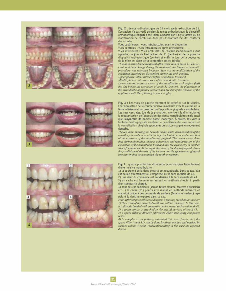
Fig. 2 : temps orthodontique de 15 mois après extraction de 31.L’occlusion n’a pas varié pendant le temps orthodontique, le dispositiforthodontique lingual a été bien supporté car il n’y a jamais eu demodification de l’occlusion donc pas d’inconfort lors des contactsdes arcades.Vues supérieures : vues intrabuccales avant orthodontie.Vues centrales : vues intrabuccales après orthodontie.Vues inférieures : Vues occlusales de l’arcade mandibulaire avant(gauche) le jour de l’extraction de 31 (centre) et de la pose dudispositif orthodontique (centre) et enfin le jour de la dépose etde la mise en place de la contention collée (droite).15-month orthodontic treatment after extraction of tooth 31. The oc-clusion did not change during the treatment; the lingual orthodonticprocedure was tolerated because there was no modification of theocclusion therefore no discomfort during the arch contact.Upper photos: intra-oral view before orthodontic treatment.Middle photos: intra-oral view after orthodontic treatment.Lower photos: occlusal views of the mandibular arch before (left)the day before the extraction of tooth 31 (center), the placement ofthe orthodontic appliance (center) and the day of the removal of theappliance with the splinting in place (right).
Fig. 3 : Les vues de gauche montrent le bénéfice sur le sourire,l’harmonisation de la courbe incisive maxillaire avec la courbe de lalèvre inférieure et la correction de l’exposition gingivale mandibulaire.Les vues centrales, lors de la phonation, montrent la diminution etla régularisation de l’exposition des dents mandibulaires mais aussique l’asymétrie de nombre passe inaperçue. À droite, les vues àl’échelle dento-gingivale montrent le parallélisme des axes incisifs etla normalisation gingivale spontanée qui a accompagné le mouvementdentaire.The left views showing the benefits on the smile, harmonization of themaxillary incisal curve with the inferior labial curve and correctionof the exposure of the mandibular gingival. The center views showthat during phonation, there is a decrease and regularization of theexposition of the mandibular teeth and that the asymmetry in numberwas left unnoticed. At the right, the view of the dento-gingival showsthe parallelism of the axis of the incisors and the spontaneous gingivalrestoration that accompanied the tooth movement.
Fig. 4 : quatre possibilités différentes pour masquer l’édentementd’une incisive mandibulaire :1) la couronne de la dent extraite est récupérable. Dans ce cas, elleest collée directement au composite sur la face mésiale de 42.2) une dent du commerce est solidarisée à la face mésiale de 41.3) un cache est façonné au fauteuil en méthode directe à partird’un composite chargé.4) dans des cas complexes (senior, teinte saturée, facettes d’abrasionsetc...) le cache (31) pourra être réalisé en méthode indirecte etmaquillé grâce à des colorants de surface (Ivoclar-Vivadent) rap-pelant la dentine exposée dans ce cas.Four different possibilities to disguise a missing mandibular incisor:1) The crown of the extracted tooth can still be retrieved. In this case,it is directly bonded with composite on the mesial surface of tooth 42.2) a tooth pontic is attached to the mesial surface of tooth 41.3) a space filler is directly fabricated chair-side using compositeresin.4) in complex cases (elderly, saturated tint, wear facets, etc.) thespace filler (tooth 31) can be done by direct method and masked bysurface colors (Ivoclar-Vivadent)recalling in this case the exposeddentin.
3
2
3 4
22Revue d’Odonto-Stomatologie/Février 2012
ORTHODONTIE
5 6
Réduction amélaire proximale(stripping)
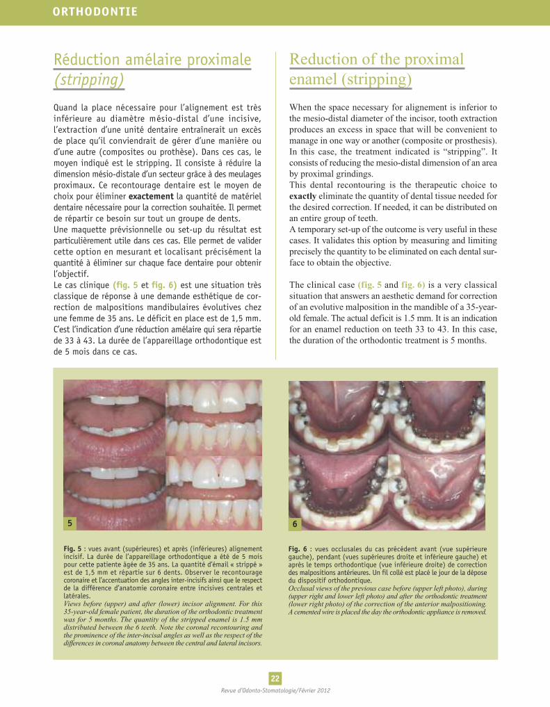
Quand la place nécessaire pour l’alignement est trèsinférieure au diamètre mésio-distal d’une incisive,l’extraction d’une unité dentaire entraînerait un excèsde place qu’il conviendrait de gérer d’une manière oud’une autre (composites ou prothèse). Dans ces cas, lemoyen indiqué est le stripping. Il consiste à réduire ladimension mésio-distale d’un secteur grâce à des meulagesproximaux. Ce recontourage dentaire est le moyen dechoix pour éliminer exactement la quantité de matérieldentaire nécessaire pour la correction souhaitée. Il permetde répartir ce besoin sur tout un groupe de dents.Une maquette prévisionnelle ou set-up du résultat estparticulièrement utile dans ces cas. Elle permet de validercette option en mesurant et localisant précisément laquantité à éliminer sur chaque face dentaire pour obtenirl’objectif.Le cas clinique (fig. 5 et fig. 6) est une situation trèsclassique de réponse à une demande esthétique de cor-rection de malpositions mandibulaires évolutives chezune femme de 35 ans. Le déficit en place est de 1,5 mm.C’est l’indication d’une réduction amélaire qui sera répartiede 33 à 43. La durée de l’appareillage orthodontique estde 5 mois dans ce cas.
Reduction of the proximalenamel (stripping)
When the space necessary for alignement is inferior tothe mesio-distal diameter of the incisor, tooth extractionproduces an excess in space that will be convenient tomanage in one way or another (composite or prosthesis).In this case, the treatment indicated is “stripping”. Itconsists of reducing the mesio-distal dimension of an areaby proximal grindings.This dental recontouring is the therapeutic choice toexactly eliminate the quantity of dental tissue needed forthe desired correction. If needed, it can be distributed onan entire group of teeth.A temporary set-up of the outcome is very useful in thesecases. It validates this option by measuring and limitingprecisely the quantity to be eliminated on each dental sur-face to obtain the objective.
The clinical case (fig. 5 and fig. 6) is a very classicalsituation that answers an aesthetic demand for correctionof an evolutive malposition in the mandible of a 35-year-old female. The actual deficit is 1.5 mm. It is an indicationfor an enamel reduction on teeth 33 to 43. In this case,the duration of the orthodontic treatment is 5 months.
Fig. 5 : vues avant (supérieures) et après (inférieures) alignementincisif. La durée de l’appareillage orthodontique a été de 5 moispour cette patiente âgée de 35 ans. La quantité d’émail « strippé »est de 1,5 mm et répartie sur 6 dents. Observer le recontouragecoronaire et l’accentuation des angles inter-incisifs ainsi que le respectde la différence d’anatomie coronaire entre incisives centrales etlatérales.Views before (upper) and after (lower) incisor alignment. For this35-year-old female patient, the duration of the orthodontic treatmentwas for 5 months. The quantity of the stripped enamel is 1.5 mmdistributed between the 6 teeth. Note the coronal recontouring andthe prominence of the inter-incisal angles as well as the respect of thedifferences in coronal anatomy between the central and lateral incisors.
Fig. 6 : vues occlusales du cas précédent avant (vue supérieuregauche), pendant (vues supérieures droite et inférieure gauche) etaprès le temps orthodontique (vue inférieure droite) de correctiondes malpositions antérieures. Un fil collé est placé le jour de la déposedu dispositif orthodontique.Occlusal views of the previous case before (upper left photo), during(upper right and lower left photo) and after the orthodontic treatment(lower right photo) of the correction of the anterior malpositioning.A cementedwire is placed the day the orthodontic appliance is removed.
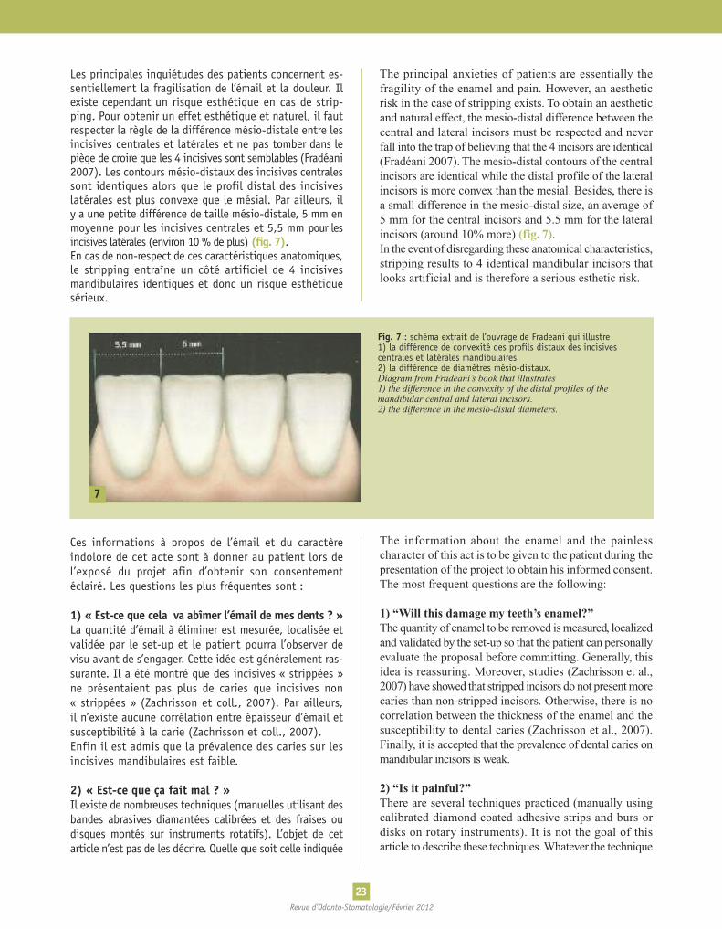
Les principales inquiétudes des patients concernent es-sentiellement la fragilisation de l’émail et la douleur. Ilexiste cependant un risque esthétique en cas de strip-ping. Pour obtenir un effet esthétique et naturel, il fautrespecter la règle de la différence mésio-distale entre lesincisives centrales et latérales et ne pas tomber dans lepiège de croire que les 4 incisives sont semblables (Fradéani2007). Les contours mésio-distaux des incisives centralessont identiques alors que le profil distal des incisiveslatérales est plus convexe que le mésial. Par ailleurs, ily a une petite différence de taille mésio-distale, 5 mm enmoyenne pour les incisives centrales et 5,5 mm pour lesincisives latérales (environ 10 % de plus) (fig. 7).En cas de non-respect de ces caractéristiques anatomiques,le stripping entraîne un côté artificiel de 4 incisivesmandibulaires identiques et donc un risque esthétiquesérieux.
Ces informations à propos de l’émail et du caractèreindolore de cet acte sont à donner au patient lors del’exposé du projet afin d’obtenir son consentementéclairé. Les questions les plus fréquentes sont :
1) « Est-ce que cela va abîmer l’émail de mes dents ? »La quantité d’émail à éliminer est mesurée, localisée etvalidée par le set-up et le patient pourra l’observer devisu avant de s’engager. Cette idée est généralement ras-surante. Il a été montré que des incisives « strippées »ne présentaient pas plus de caries que incisives non« strippées » (Zachrisson et coll., 2007). Par ailleurs,il n’existe aucune corrélation entre épaisseur d’émail etsusceptibilité à la carie (Zachrisson et coll., 2007).Enfin il est admis que la prévalence des caries sur lesincisives mandibulaires est faible.
2) « Est-ce que ça fait mal ? »Il existe de nombreuses techniques (manuelles utilisant desbandes abrasives diamantées calibrées et des fraises oudisques montés sur instruments rotatifs). L’objet de cetarticle n’est pas de les décrire. Quelle que soit celle indiquée
The principal anxieties of patients are essentially thefragility of the enamel and pain. However, an aestheticrisk in the case of stripping exists. To obtain an aestheticand natural effect, the mesio-distal difference between thecentral and lateral incisors must be respected and neverfall into the trap of believing that the 4 incisors are identical(Fradéani 2007). The mesio-distal contours of the centralincisors are identical while the distal profile of the lateralincisors is more convex than the mesial. Besides, there isa small difference in the mesio-distal size, an average of5 mm for the central incisors and 5.5 mm for the lateralincisors (around 10% more) (fig. 7).In the event of disregarding these anatomical characteristics,stripping results to 4 identical mandibular incisors thatlooks artificial and is therefore a serious esthetic risk.
The information about the enamel and the painlesscharacter of this act is to be given to the patient during thepresentation of the project to obtain his informed consent.The most frequent questions are the following:
1) “Will this damage my teeth’s enamel?”The quantity of enamel to be removed ismeasured, localizedand validated by the set-up so that the patient can personallyevaluate the proposal before committing. Generally, thisidea is reassuring. Moreover, studies (Zachrisson et al.,2007) have showed that stripped incisors do not presentmorecaries than non-stripped incisors. Otherwise, there is nocorrelation between the thickness of the enamel and thesusceptibility to dental caries (Zachrisson et al., 2007).Finally, it is accepted that the prevalence of dental caries onmandibular incisors is weak.
2) “Is it painful?”There are several techniques practiced (manually usingcalibrated diamond coated adhesive strips and burs ordisks on rotary instruments). It is not the goal of thisarticle to describe these techniques.Whatever the technique
23Revue d’Odonto-Stomatologie/Février 2012
7
Fig. 7 : schéma extrait de l’ouvrage de Fradeani qui illustre1) la différence de convexité des profils distaux des incisivescentrales et latérales mandibulaires2) la différence de diamètres mésio-distaux.Diagram from Fradeani’s book that illustrates1) the difference in the convexity of the distal profiles of themandibular central and lateral incisors.2) the difference in the mesio-distal diameters.
indicated by the practitioner, it is clear that it must be per-fectly mastered (quantitatively and qualitatively) andpainless as it is in peripheral enamel zone.
The following case (fig. 8 and 9) presents a 40-year-oldfemale who wishes to correct the malpositioning thatoccurred over time, she talks about “lifting the teeth”.It illustrates this therapeutic option in a severe case ofcrowding where the set-up is a key element to validatethis option.
par le praticien, il est évident qu’elle doit être parfaitementmaîtrisée (quantitativement et qualitativement) et indolorepuisque dans la zone périphérique amélaire.
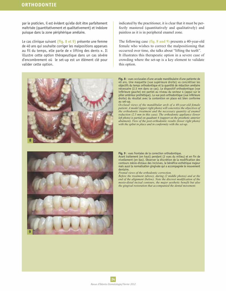
Le cas clinique suivant (fig. 8 et 9) présente une femmede 40 ans qui souhaite corriger les malpositions apparuesau fil du temps, elle parle de « lifting des dents ». Ilillustre cette option thérapeutique dans un cas sévèred’encombrement où le set-up est un élément clé pourvalider cette option.
24Revue d’Odonto-Stomatologie/Février 2012
ORTHODONTIE
8
9
Fig. 8 : vues occlusales d’une arcade mandibulaire d’une patiente de40 ans. Une maquette (vue supérieure droite) va concrétiser lesobjectifs du temps orthodontique et la quantité de réduction amélairenécessaire (2,5 mm dans ce cas). Le dispositif orthodontique (vueinférieure gauche) est partiel au niveau du secteur 4 (appui sur lepilier antérieur prothétique). La vue post-orthodontique (vue inférieuredroite) du résultat avec la contention en place est bien conformeau set-up.Occlusal views of the mandibular arch of a 40-year-old femalepatient. A model (upper right photo) will concretize the objectives ofthe orthodontic treatment and the necessary quantity of enamelreduction (2.5 mm in this case). The orthodontic appliance (lowerleft photo) is partial on quadrant 4 (support on the prosthetic anteriorabutment). View of the post-orthodontic results (lower right photo)with the splint in place and in conformity with the set-up.
Fig. 9 : vues frontales de la correction orthodontique.Avant traitement (en haut) pendant (2 vues du milieu) et en fin denivellement (en bas). Observer la discrétion de la modification descontours mésio-distaux des incisives, le bénéfice esthétique majeurmais aussi la normalisation gingivale qui a accompagnée le mouvementdentaire.Frontal views of the orthodontic correction.Before the treatment (above), during (2 middle photos) and at theend of the alignment (below). Note the discreet modification of themesio-distal incisal contours, the major aesthetic benefit but alsothe gingival restoration that accompanied the dental movement.
Benefits of these treatmentsAfter the method, we will see in this part that the benefitsof this correction are numerous and we will review theiraesthetic, biological, functional and mechanic benefits.
1)Aesthetic benefitThe facial aesthetic of an individual should not be basedsolely on the smile. Thus, one can have a harmonioussmile and a real aesthetic problem during phonation. Ingeneral, patients (except for professional hairdressers...),rarely look at themselves in the mirror while speaking.On the other hand, with the development of handheldvideo cameras, an esthetic problem during phonationstarts to be a regular reason for consultation, “I saw myselfon a film...”.
This esthetic concern is all the more important as one canconceal unsightly teeth when one smiles, it is on the otherhand much more difficult to control and to hide whilespeaking. The clinical case in figure 10 shows the dif-ference between the severity of a minor malposition andthe severe aesthetic defect that it causes during phonation.
Bénéfices de ces traitementsAprès la méthode, nous allons voir dans cette partie queles bénéfices de cette correction sont multiples et nouspasserons en revue les bénéfices esthétique, biologique,fonctionnel et mécanique.
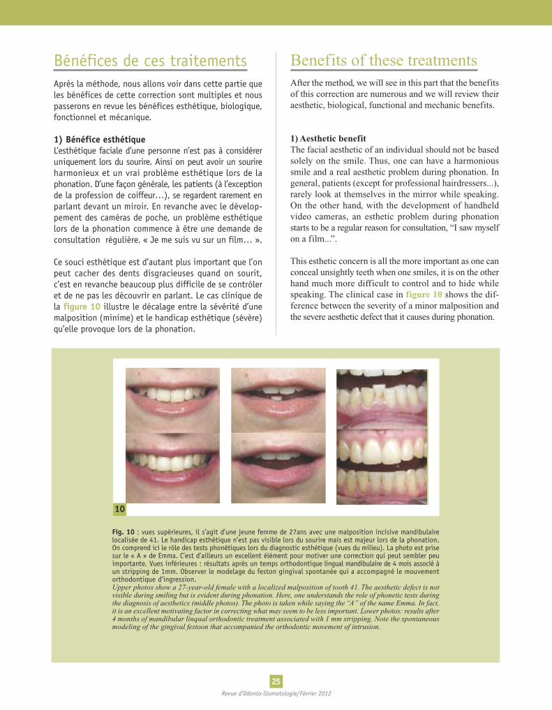
1) Bénéfice esthétiqueL’esthétique faciale d’une personne n’est pas à considéreruniquement lors du sourire. Ainsi on peut avoir un sourireharmonieux et un vrai problème esthétique lors de laphonation. D’une façon générale, les patients (à l’exceptionde la profession de coiffeur…), se regardent rarement enparlant devant un miroir. En revanche avec le dévelop-pement des caméras de poche, un problème esthétiquelors de la phonation commence à être une demande deconsultation régulière. « Je me suis vu sur un film… ».
Ce souci esthétique est d’autant plus important que l’onpeut cacher des dents disgracieuses quand on sourit,c’est en revanche beaucoup plus difficile de se contrôleret de ne pas les découvrir en parlant. Le cas clinique dela figure 10 illustre le décalage entre la sévérité d’unemalposition (minime) et le handicap esthétique (sévère)qu’elle provoque lors de la phonation.
25Revue d’Odonto-Stomatologie/Février 2012
10
Fig. 10 : vues supérieures, il s’agit d’une jeune femme de 27ans avec une malposition incisive mandibulairelocalisée de 41. Le handicap esthétique n’est pas visible lors du sourire mais est majeur lors de la phonation.On comprend ici le rôle des tests phonétiques lors du diagnostic esthétique (vues du milieu). La photo est prisesur le « A » de Emma. C’est d’ailleurs un excellent élément pour motiver une correction qui peut sembler peuimportante. Vues inférieures : résultats après un temps orthodontique lingual mandibulaire de 4 mois associé àun stripping de 1mm. Observer le modelage du feston gingival spontanée qui a accompagné le mouvementorthodontique d’ingression.Upper photos show a 27-year-old female with a localized malposition of tooth 41. The aesthetic defect is notvisible during smiling but is evident during phonation. Here, one understands the role of phonetic tests duringthe diagnosis of aesthetics (middle photos). The photo is taken while saying the “A” of the name Emma. In fact,it is an excellent motivating factor in correcting what may seem to be less important. Lower photos: results after4 months of mandibular linqual orthodontic treatment associated with 1 mm stripping. Note the spontaneousmodeling of the gingival festoon that accompanied the orthodontic movement of intrusion.
26Revue d’Odonto-Stomatologie/Février 2012
ORTHODONTIE
2) Bénéfices biologiquesTout d’abord notons que le caractère évolutif desmalpositions incisives mandibulaires est souvent perçupar les patients et parfois même plus que par les praticiensqui n’ont pas toujours à leur disposition, dans leurdossier-patient, des éléments objectifs pour l’appréciercomme des photos au fil des années.
Seule l’observation de documents à 2 ou 3 ans d’intervallepermet de connaître le caractère évolutif des malpositionspour chacun. Les patients, étant très conscients de cephénomène, sont demandeurs d’une correction et ac-ceptent facilement la notion de contention. Le premierbénéfice biologique sera la maîtrise du phénomène « devieillissement » grâce à un dispositif de contentionpermanent collé à la fin du temps orthodontique (Mulleret coll., 2009). Le dispositif de contention aura une doublemission : maintenir le résultat le temps de la réorgani-sation de l’os et des fibres desmodontales autour de lanouvelle position (consensus sur 2 années actuellement)et d’autre part, passé ce délai, il préviendra de nouveaules effets possibles de la dérive mésiale physiologiquequi se poursuit toute la vie donc à différencier d’unphénomène de récidive.
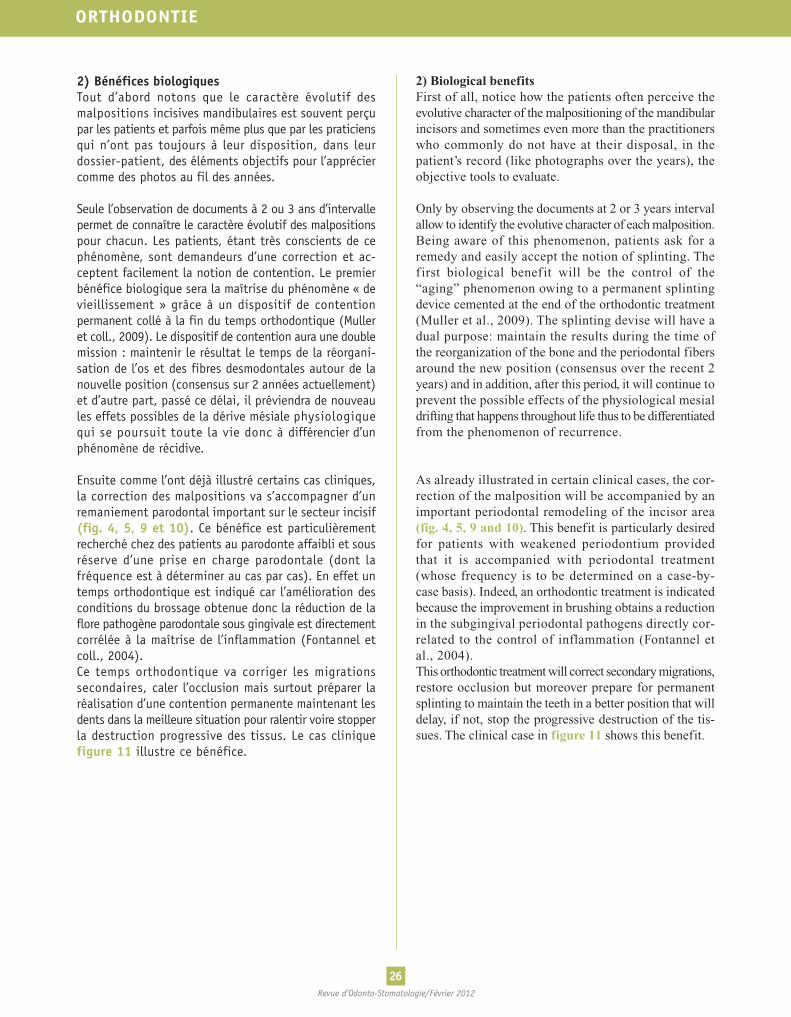
Ensuite comme l’ont déjà illustré certains cas cliniques,la correction des malpositions va s’accompagner d’unremaniement parodontal important sur le secteur incisif(fig. 4, 5, 9 et 10). Ce bénéfice est particulièrementrecherché chez des patients au parodonte affaibli et sousréserve d’une prise en charge parodontale (dont lafréquence est à déterminer au cas par cas). En effet untemps orthodontique est indiqué car l’amélioration desconditions du brossage obtenue donc la réduction de laflore pathogène parodontale sous gingivale est directementcorrélée à la maîtrise de l’inflammation (Fontannel etcoll., 2004).Ce temps orthodontique va corriger les migrationssecondaires, caler l’occlusion mais surtout préparer laréalisation d’une contention permanente maintenant lesdents dans la meilleure situation pour ralentir voire stopperla destruction progressive des tissus. Le cas cliniquefigure 11 illustre ce bénéfice.
2) Biological benefitsFirst of all, notice how the patients often perceive theevolutive character of the malpositioning of the mandibularincisors and sometimes even more than the practitionerswho commonly do not have at their disposal, in thepatient’s record (like photographs over the years), theobjective tools to evaluate.
Only by observing the documents at 2 or 3 years intervalallow to identify the evolutive character of eachmalposition.Being aware of this phenomenon, patients ask for aremedy and easily accept the notion of splinting. Thefirst biological benefit will be the control of the“aging” phenomenon owing to a permanent splintingdevice cemented at the end of the orthodontic treatment(Muller et al., 2009). The splinting devise will have adual purpose: maintain the results during the time ofthe reorganization of the bone and the periodontal fibersaround the new position (consensus over the recent 2years) and in addition, after this period, it will continue toprevent the possible effects of the physiological mesialdrifting that happens throughout life thus to be differentiatedfrom the phenomenon of recurrence.
As already illustrated in certain clinical cases, the cor-rection of the malposition will be accompanied by animportant periodontal remodeling of the incisor area(fig. 4, 5, 9 and 10). This benefit is particularly desiredfor patients with weakened periodontium providedthat it is accompanied with periodontal treatment(whose frequency is to be determined on a case-by-case basis). Indeed, an orthodontic treatment is indicatedbecause the improvement in brushing obtains a reductionin the subgingival periodontal pathogens directly cor-related to the control of inflammation (Fontannel etal., 2004).This orthodontic treatmentwill correct secondarymigrations,restore occlusion but moreover prepare for permanentsplinting to maintain the teeth in a better position that willdelay, if not, stop the progressive destruction of the tis-sues. The clinical case in figure 11 shows this benefit.
11
Fig. 11 : vues supérieures, il s’agit d’une patiente âgée de 50 ans. Elle a été suivie pour une parodontite agres-sive (prise en charge parodontale Dr. L. Jaoui) Elle présente des migrations secondaires qui prennent la formede diastèmes à l’arcade maxillaire et d’égression à la mandibule. Observer l’exposition radiculaire des incisivesmandibulaires lors de la phonation (vues du milieu).Vues inférieures, après le temps orthodontique bimaxillaire de nivellement qui va faciliter la prise en chargedu problème bactérien. Observer l’amplitude du nivellement incisif et la diminution de l’exposition radiculairemandibulaire lors de la phonation mais aussi du sourire.Upper photos show a 50-year-old female patient. She was treated for aggressive periodontitis (periodontaltreatment by Dr. L. Jaoui). She has secondary migrations that take the form of a maxillary arch diastema andextrusion of the mandibular teeth. Note root exposure of the mandibular incisors during phonation (middlephoto). Lower photos, after the bi-maxillary orthodontic alignment that will help in treating the bacterialproblem. Note the extent of the incisor alignment and the decrease in the exposure of the mandibular rootsduring phonation and smiling.
27Revue d’Odonto-Stomatologie/Février 2012
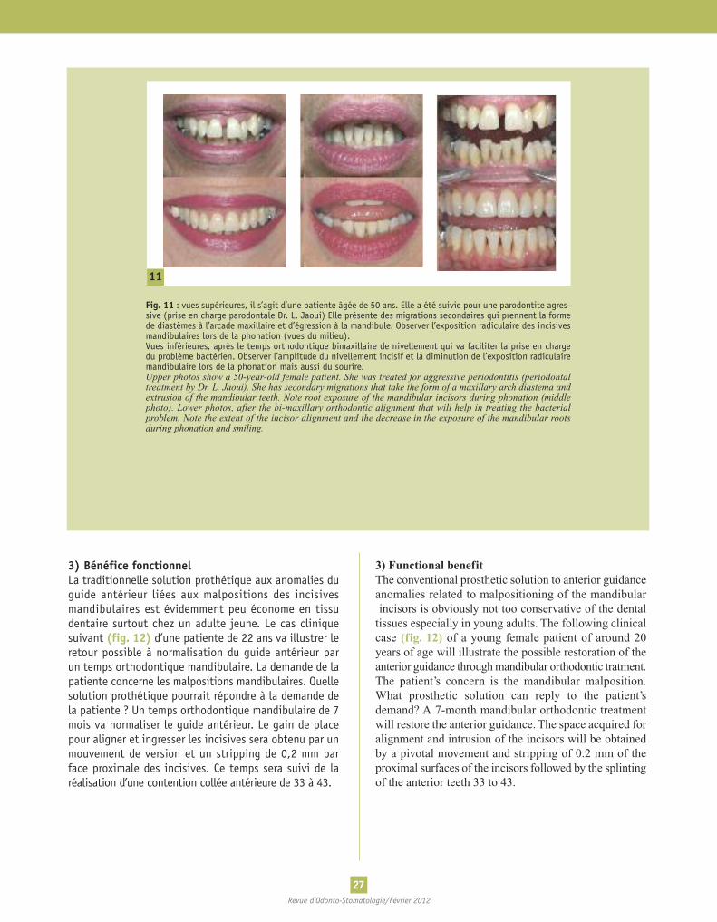
3) Bénéfice fonctionnelLa traditionnelle solution prothétique aux anomalies duguide antérieur liées aux malpositions des incisivesmandibulaires est évidemment peu économe en tissudentaire surtout chez un adulte jeune. Le cas cliniquesuivant (fig. 12) d’une patiente de 22 ans va illustrer leretour possible à normalisation du guide antérieur parun temps orthodontique mandibulaire. La demande de lapatiente concerne les malpositions mandibulaires. Quellesolution prothétique pourrait répondre à la demande dela patiente ? Un temps orthodontique mandibulaire de 7mois va normaliser le guide antérieur. Le gain de placepour aligner et ingresser les incisives sera obtenu par unmouvement de version et un stripping de 0,2 mm parface proximale des incisives. Ce temps sera suivi de laréalisation d’une contention collée antérieure de 33 à 43.
3) Functional benefitThe conventional prosthetic solution to anterior guidanceanomalies related to malpositioning of the mandibularincisors is obviously not too conservative of the dentaltissues especially in young adults. The following clinicalcase (fig. 12) of a young female patient of around 20years of age will illustrate the possible restoration of theanterior guidance throughmandibular orthodontic tratment.The patient’s concern is the mandibular malposition.What prosthetic solution can reply to the patient’sdemand? A 7-month mandibular orthodontic treatmentwill restore the anterior guidance. The space acquired foralignment and intrusion of the incisors will be obtainedby a pivotal movement and stripping of 0.2 mm of theproximal surfaces of the incisors followed by the splintingof the anterior teeth 33 to 43.
28Revue d’Odonto-Stomatologie/Février 2012
ORTHODONTIE
12
Fig. 12 : femme âgée de 22 ans. Sur les vues supérieures avant traitement, elle présente une égression si sévère dusecteur incisif mandibulaire que la vue du sourire de face est perturbée (exposition gingivale mandibulaire lorsdu sourire). Vues inférieures, après un temps d’orthodontie mandibulaire uniquement (C’est le choix de lapatiente de ne pas corriger les malpositions supérieures) le secteur antérieur est nivelé. Observer le remaniementgingival qui a accompagné le mouvement dentaire. Une contention collée permanente est réalisée afin demaintenir la correction.A young 22-year-old female. On the upper photos before treatment, she presents a very severe extrusion of themandibular incisors that the vision of the smile is unbalanced (exposure of the mandibular gingiva while smiling).Lower photos show views of the aligned anterior teeth after a mandibular orthodontic treatment. It is the choiceof the patient not to correct the maxillary malposition. Note the gingival remodeling that accompanied the toothmovement. A permanent cemented splint is fixed to maintain the correction.
13
Fig. 13 : patiente âgée de 60 ans, c’est le 3e jeu de couronnes 11, 21 qui est en place lors de la consultationorthodontique (vues supérieures). L’hypothèse de forces iatrogènes au cours de la fonction est émise etl’indication d’un nivellement orthodontique mandibulaire (stripping 2 mm) qui va supprimer les forces iatrogèness’exerçant sur les prothèses maxillaires est posée. Les vues inférieures montrent la normalisation obtenue en uneannée.A 60-year-old female patient, it’s the 3rd set of crowns on teeth 11 and 21 in place during the orthodontic consultation(upper photos). The hypothesis of iatrogenic forces during function is considered and the indication for a mandibularorthodontic alignment (2 mm stripping) that will suppress the iatrogenic forces on the maxillary prosthesis isproposed. The lower photos show the restoration obtained after a year.
29Revue d’Odonto-Stomatologie/Février 2012
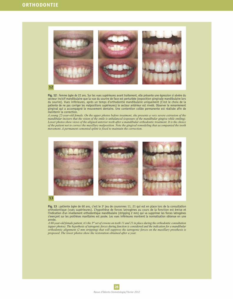
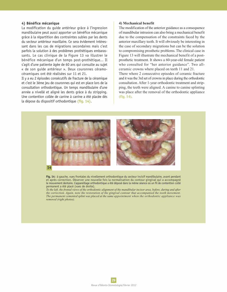
4) Bénéfice mécaniqueLa modification du guide antérieur grâce à l’ingressionmandibulaire peut aussi apporter un bénéfice mécaniquegrâce à la répartition des contraintes subies par les dentsdu secteur antérieur maxillaire. Ce sera évidement intéres-sant dans les cas de migrations secondaires mais c’estparfois la solution à des problèmes prothétiques embaras-sants. Le cas clinique de la Figure 13 va illustrer lebénéfice mécanique d’un temps post-prothétique... Ils’agit d’une patiente âgée de 60 ans qui consulte au sujet« de son guide antérieur ». Deux couronnes céramo-céramiques ont été réalisées sur 11 et 21.Il y a eu 2 épisodes consécutifs de fracture de la céramiqueet c’est le 3ème jeu de couronnes qui est en place lors de laconsultation orthodontique. Un temps mandibulaire d’uneannée a nivellé et aligné les dents grâce à du stripping.Une contention collée de canine à canine a été placée dèsla dépose du dispositif orthodontique (fig. 14).
4) Mechanical benefitThemodification of the anterior guidance as a consequenceofmandibular intrusion can also bring amechanical benefitdue to the compensation of the constraints faced by theanterior maxillary teeth. It will obviously be interesting inthe case of secondary migrations but can be the solutionto compromising prosthetic problems. The clinical case inFigure 13 will illustrate the mechanical benefit of a post-prosthetic treatment. It shows a 60-year-old female patientwho consulted for “her anterior guidance”. Two all-ceramic crowns where placed on teeth 11 and 21.There where 2 consecutive episodes of ceramic fractureand itwas the 3rd set of crowns in place during the orthodonticconsultation.After 1-year orthodontic treatment and strip-ping, the teeth were aligned. A canine to canine splintingwas place after the removal of the orthodontic appliance(fig. 14).
14
Fig. 14 : à gauche, vues frontales du nivellement orthodontique du secteur incisif mandibulaire, avant pendantet après correction. Observer une nouvelle fois la normalisation du contour gingival qui a accompagnéle mouvement dentaire. L’appareillage orthodontique a été déposé dans la même séance où un fil de contention collépermanent a été placé (vues de droite).To the left, the frontal views of the orthodontic alignment of the mandibular incisor area, before, during and afterthe correction. Again, note the restoration of the gingival contour that accompanied the tooth movement.The permanent cemented splint was placed at the same appointment when the orthodontic appliance wasremoved (right photos).
30Revue d’Odonto-Stomatologie/Février 2012
ORTHODONTIE
Quel que soit le projet global, le temps orthodontique de correction des malpositions mandibulaires ne selimite pas à une « petite amélioration esthétique ». Cet article a montré l’association ; 1) d’un objectif esthétiqued’harmonisation du secteur antérieur (alignement : correction des malpositions, rotations et version et, nivel-lement : correction des égressions ou alignement vertical des bords libres) ; 2) d’un objectif biologique :la correction de la malposition mais aussi l’arrêt de son évolution avec la conception et la mise en place d’undispositif de stabilisation indissociable de la correction ; 3) d’un objectif fonctionnel avec l’amélioration duguide antérieur et 4) d’un objectif mécanique grâce à l’amélioration de la répartition des contraintes lors desfonctions.
Ce type de projet illustre complètement le principe de soins modernes et plus globaux mais aussi lesmoins invasifs possible. L’objet de cet article était d’illustrer que l’orthodontie de l’adulte a aujourd’hui uneplace de choix dans les propositions thérapeutiques à offrir à nos patients.
Whatever the overall project, the orthodontic treatment to correct mandibular malpositions is notlimited to a “small aesthetic improvement”. This article shows the association of; 1) an aesthetic objective ofharmonization of the anterior sector (alignment: correction of the malpositions, rotations, pivot and aligning:correction of extrusions or vertical alignment of free borders); 2) a biological objective: it is the correction of themalposition but also the interruption of its evolution with the objective and the placement of a stabilizing deviceto maintain the correction; 3) a functional objective with the improvement of the anterior guidance and 4) amechanical objective as a result of the improvement in the reparation of the functional constraints.
This type of project completely illustrates the principles of a modern treatment that is more comprehensiveand as possible, less invasive. The objective of this article was to illustrate that today adult orthodontics has itsplace in the choice of therapeutic propositions to offer to our patients.
Conclusion
Demande de tirés-à-part :Christine Muller - 222, Bd Raspail - 75014 PARIS
Traduction : Marie-Grace Poblete-Michel

CANAL P., SALVADORI A.Orthodontie de l’adulte. Ed: Masson Paris, 2008. Cat 3
CADE R.E.The role of the mandibular anterior teeth in complete dentureesthetics. J Prosthet Dent 1979;42(4):368-370. Cat 3
FRADEANI M.Analyse esthétique. Ed: Quintess Intern 2007. Cat 3
FONTANNEL F., BRION M., DANAN M.Parodontites sévères et orthodontie. Ed: CDP Paris, 2004. Cat 3
MULLER C.H., HITMI L., ROUSSARIE F., ATTAL J-P.Méthode simple et rapide de contention indirecte.Orthodont France. 2009;80:233-238. Cat 4
SACKSTEIN M.Display of mandibular and maxillary anterior teeth duringsmiling and speech: age and sex correlations.Int J Prosthod 2008;21:149-151. Cat 2
SACKSTEIN M.A digital video photographic technique for esthetic evaluationof anteror mandibular teeth. J prosth Dent 2007;97:246-247.Cat 2
TIRLET G., ATTAL J-P.Le gradient thérapeutique: un concept médical pour lestraitements esthétiques.Inform dent 2009;(41/42):2561-2568. Cat 4
VIG R.G., BRUNDO G.C.The kinetics of anterior tooth display.J Prosth Dent 1978;39(5):502-504. Cat 4
WOLFART S., THORMANN H., FREITAG S., KERN M.Assessment of dental appearance following changes inincisor proportions. Europ J Oral Sci 2005;113(2):159-165. Cat 4
WULFMAN C., TEZENAS DU MONTCEL S., JONAS P.,FATTOUH J., RIGNON-BRET CH.Aesthetic demand of french senior: a large-scale study.Gerodontology 2010;27(4):266-271. Cat 1
ZACHRISSON B.U., NYØYGAARD L., MOBARAK K.Dental health assessed more than 10 years after interproximalenamel reduction of mandibular anterior teeth.Amer J Orthod Dentofac Orthop 2007;131(2):162-169. Cat 1
ZLOWODZKI A-S., TIRLET G., ATTAL J-P.Perception comparée de l’esthétique du sourire. Influence del’axe longitudinal des incisives maxillaires.Inform Dent 2008;42:2531-2538. Cat 3
1Revue d’Odonto-Stomatologie/Février 2012
Bibliographie
31