malignant vs benign neoplasms recognition and treatment · pdf filemalignant vs benign...
TRANSCRIPT

Tracey C. Vlahovic, DPM FFPM RCPS (Glasg) Associate Professor, J Stanley and Pearl Landau Faculty Fellow Temple University School of Podiatric Medicine, Philadelphia, PA
Malignant vs Benign Neoplasms Recognition and Treatment
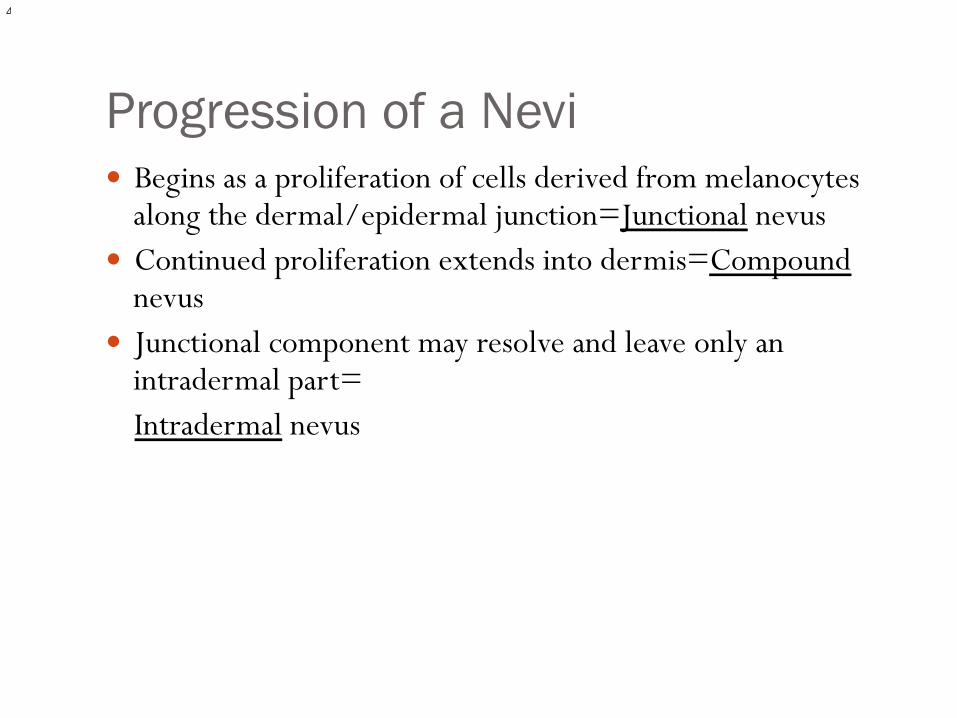
Progression of a Nevi � Begins as a proliferation of cells derived from melanocytes
along the dermal/epidermal junction=Junctional nevus � Continued proliferation extends into dermis=Compound
nevus � Junctional component may resolve and leave only an
intradermal part= Intradermal nevus
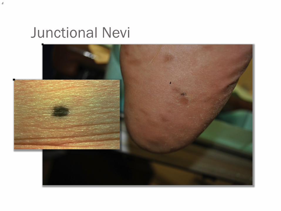
Junctional Nevi
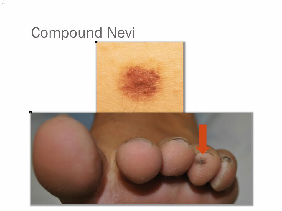
Compound Nevi

Intradermal Nevi
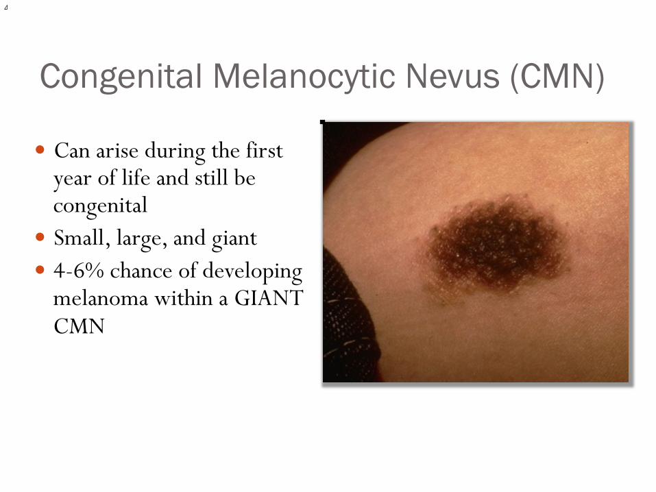
Congenital Melanocytic Nevus (CMN)
� Can arise during the first year of life and still be congenital
� Small, large, and giant � 4-6% chance of developing
melanoma within a GIANT CMN
When good moles go bad…Dysplastic and Atypical Nevi � Dysplastic is a discouraged and controversial term for an
atypical looking nevi � So use ATYPICAL nevi to describe:
� Marked variegation in color: “fried egg” � Loss of normal symmetry � Larger than ordinary >6mm � Is different histologically from melanoma, but full thickness
excision with 0.2 cm margin of normal skin
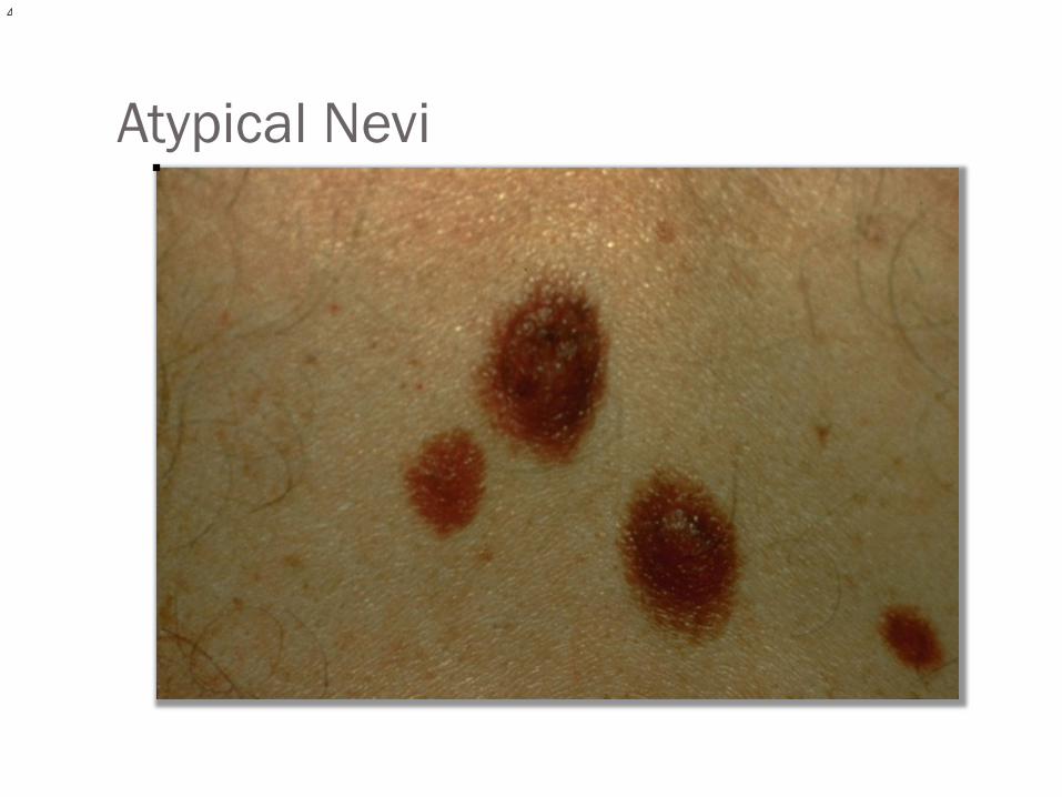
Atypical Nevi
Dermatofibroma � Small, firm, flat or papule on lower extremities can range
from 2mm-3cm � Skin colored or brown � “Dimple sign” when lateral pressure applied � Etiology not known—post shave or arthropod bite? � Excise it…or leave it alone
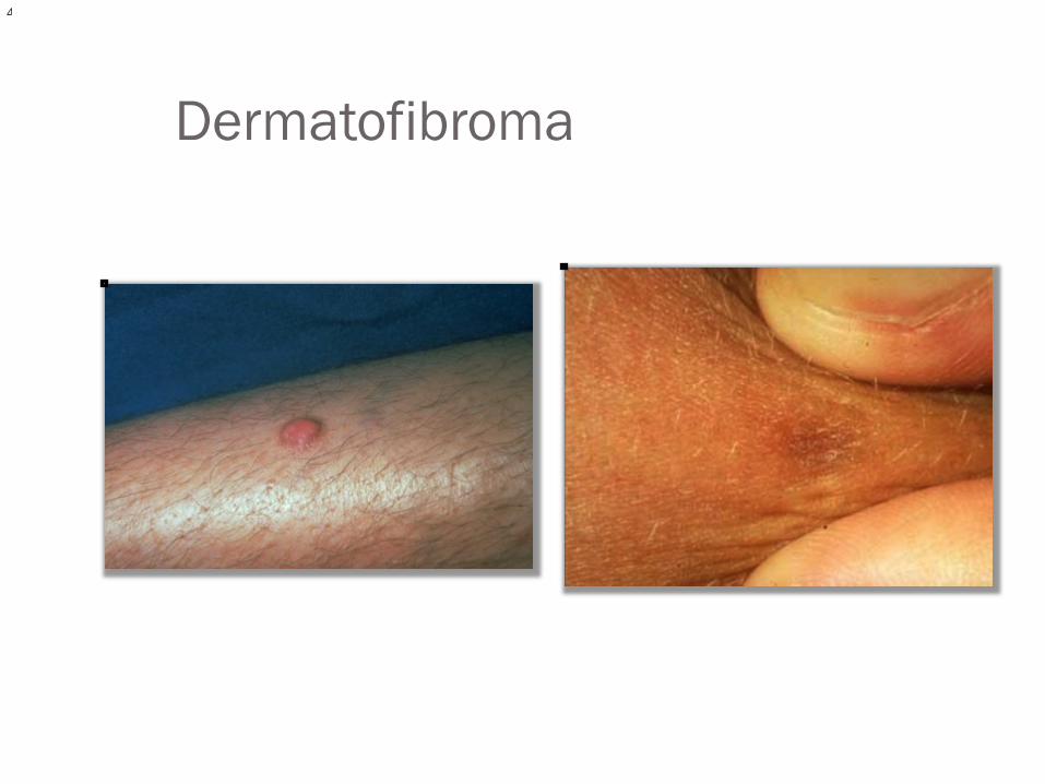
Dermatofibroma
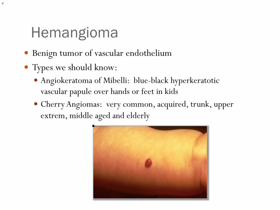
Hemangioma � Benign tumor of vascular endothelium � Types we should know:
� Angiokeratoma of Mibelli: blue-black hyperkeratotic vascular papule over hands or feet in kids
� Cherry Angiomas: very common, acquired, trunk, upper extrem, middle aged and elderly
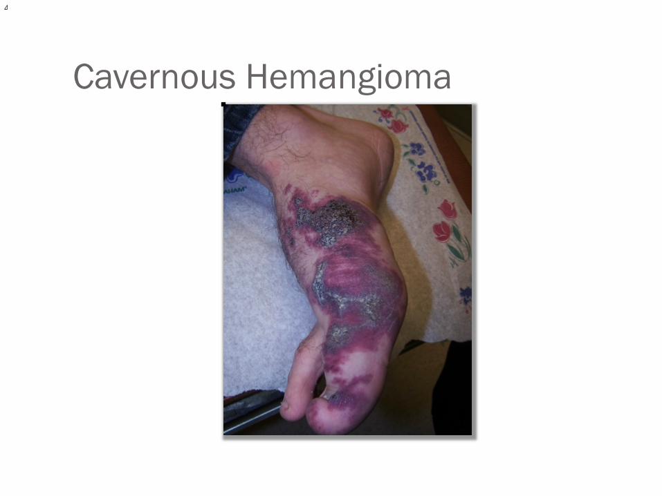
Cavernous Hemangioma
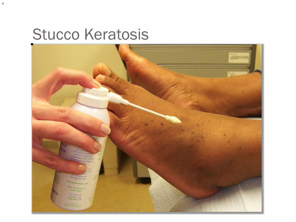
Stucco Keratosis

Malignant Lesions � Pre-malignant: Actinic Keratosis, Bowen’s � Kaposi’s � Basal Cell Carcinoma � Squamous Cell Carcinoma � Melanoma

Premalignant: Actinic Keratoses and Bowen’s Disease
� Actinic keratoses are sun induced pre cancerous lesions of skin. Elderly pts with light skin color and sun exposure; 2-5% become SCC
� Bowen’s Disease or SCC in situ: on ANY skin surface; persistent erythematous slightly indurated plaques with scale

Actinic Keratoses
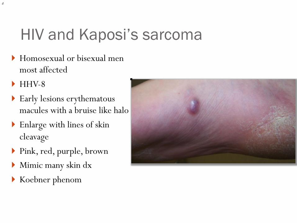
HIV and Kaposi’s sarcoma } Homosexual or bisexual men
most affected } HHV-8 } Early lesions erythematous
macules with a bruise like halo } Enlarge with lines of skin
cleavage } Pink, red, purple, brown } Mimic many skin dx } Koebner phenom
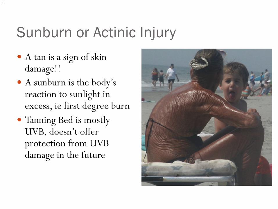
Sunburn or Actinic Injury � A tan is a sign of skin
damage!! � A sunburn is the body’s
reaction to sunlight in excess, ie first degree burn
� Tanning Bed is mostly UVB, doesn’t offer protection from UVB damage in the future
Is the Lesion Malignant? � Has it changed recently? � Does it have an onset over age 40? � Is it a lesion they have had since childhood that has
recently changed? � Does it itch? � Is it the site of a scar or ulcer?
� Does the ulcer contain hyper-proliferative tissue, non-healing over 6 months? Has it changed?
� Is it an area that does not heal? � Meet criteria for general risk factors (next slide)
Risk Factors for all skin cancers � Exposure to carcinogenic agents
� UV (sunlight, tanning), PUVA, radiation, arsenic, HPV, cigarette smoking
� Genetic syndromes � XP, albinism, Basal Cell Nevus syndrome � Personal or Family hx of melanoma
� Predisposing clinical scenario � Non-healing wound
� Immunosuppression � Organ transplant, AIDS
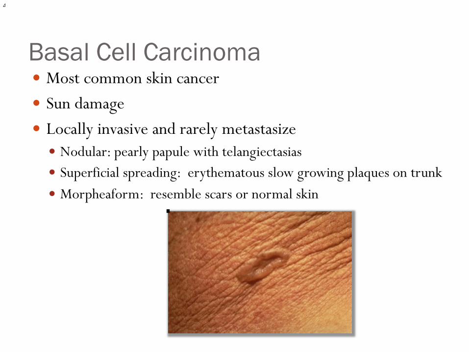
Basal Cell Carcinoma � Most common skin cancer � Sun damage � Locally invasive and rarely metastasize
� Nodular: pearly papule with telangiectasias � Superficial spreading: erythematous slow growing plaques on trunk � Morpheaform: resemble scars or normal skin

Basal Cell Treatment � Excision � Cryotherapy � Electrodessication and
currettage � Moh’s surgery � Topical (Aldara)
Squamous Cell Carcinoma � May resemble BCC, warts, but usually look like an ill-defined
red lesion with a rough surface � More scale on surface—even produce a cutaneous horn � Verrucous carcinoma are a variant that can occur on feet � More aggressive than BCC and likely to metastasize
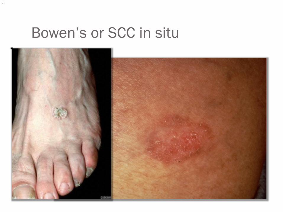
Bowen’s or SCC in situ
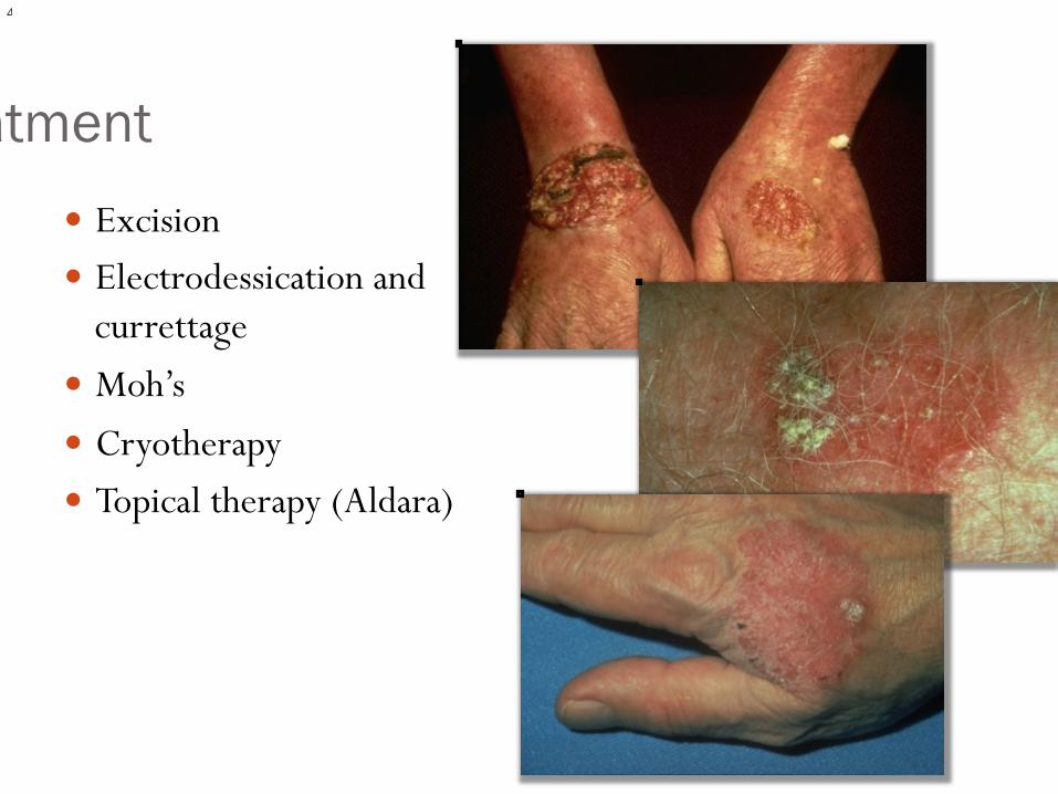
SCC treatment � Excision � Electrodessication and
currettage � Moh’s � Cryotherapy � Topical therapy (Aldara)
Malignant Melanoma � Most common malignancy in women 25-29 yrs; 1 in 100 for
all people � UVA and mutation in tumor suppressor genes � Fair complexions, who have atypical nevi, family member
with melanoma or self � Can occur anywhere, but trunk in men, legs in women
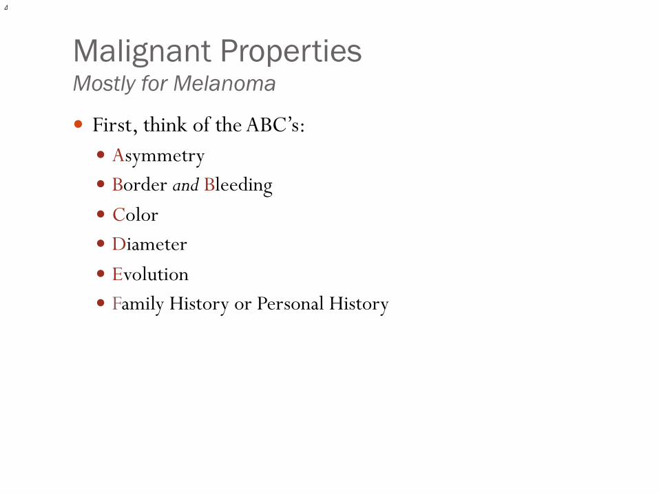
Malignant Properties Mostly for Melanoma
� First, think of the ABC’s: � Asymmetry � Border and Bleeding � Color � Diameter � Evolution � Family History or Personal History
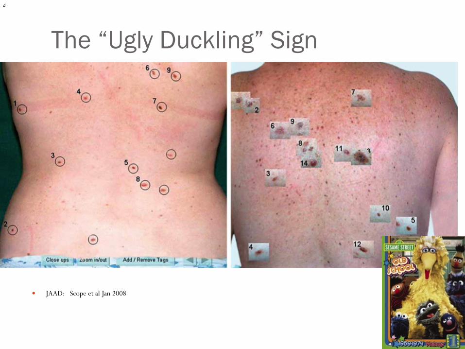
The “Ugly Duckling” Sign
� JAAD: Scope et al Jan 2008
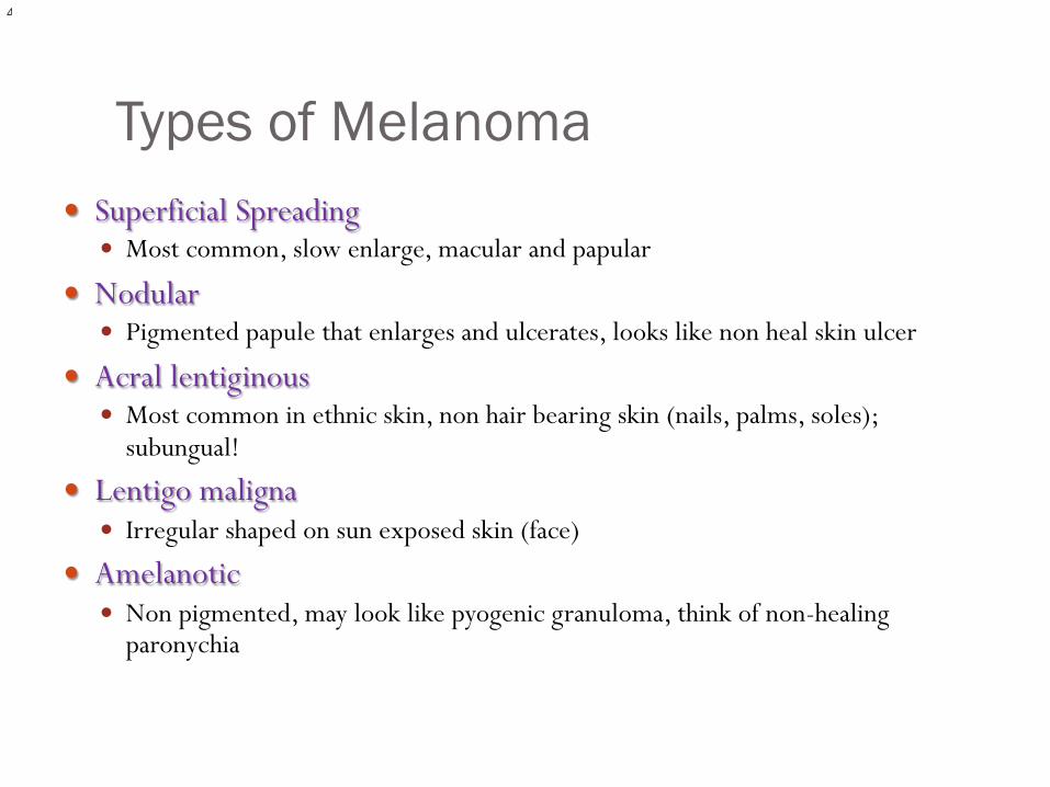
Types of Melanoma � Superficial Spreading
� Most common, slow enlarge, macular and papular
� Nodular � Pigmented papule that enlarges and ulcerates, looks like non heal skin ulcer
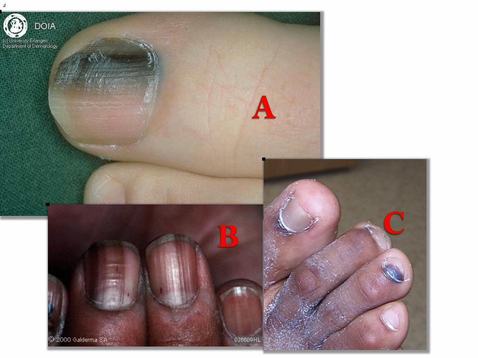
� Acral lentiginous � Most common in ethnic skin, non hair bearing skin (nails, palms, soles);
subungual!
� Lentigo maligna � Irregular shaped on sun exposed skin (face)
� Amelanotic � Non pigmented, may look like pyogenic granuloma, think of non-healing
paronychia
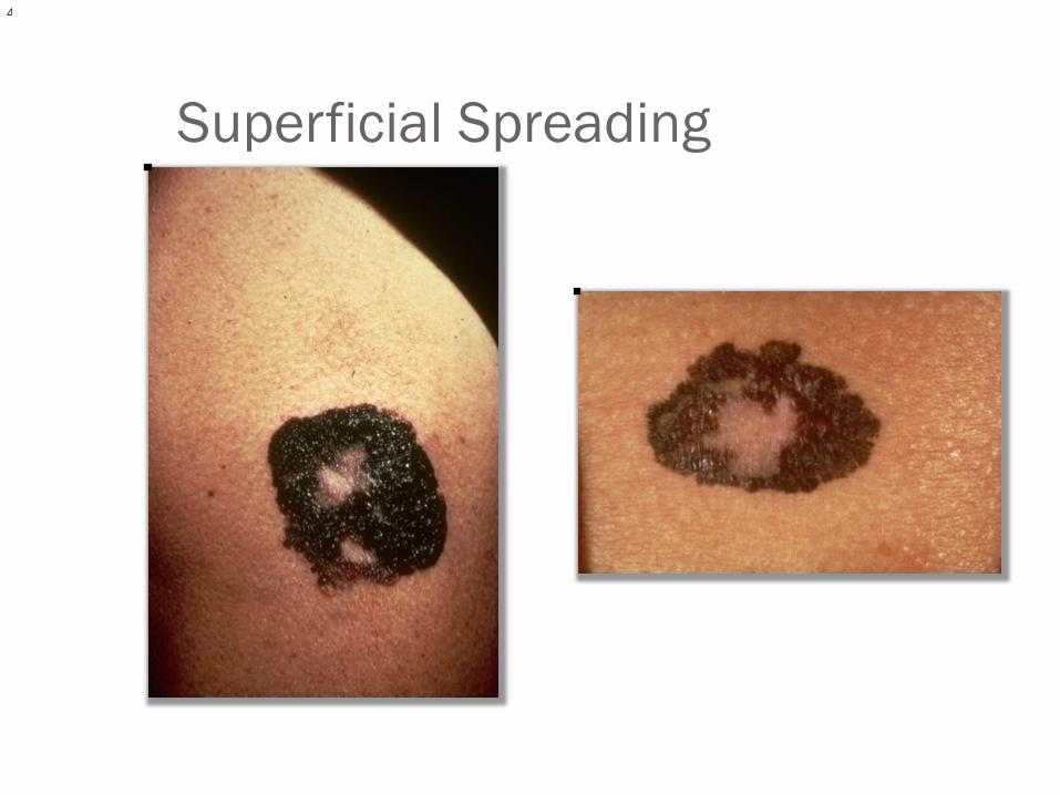
Superficial Spreading
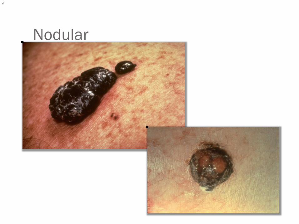
Nodular
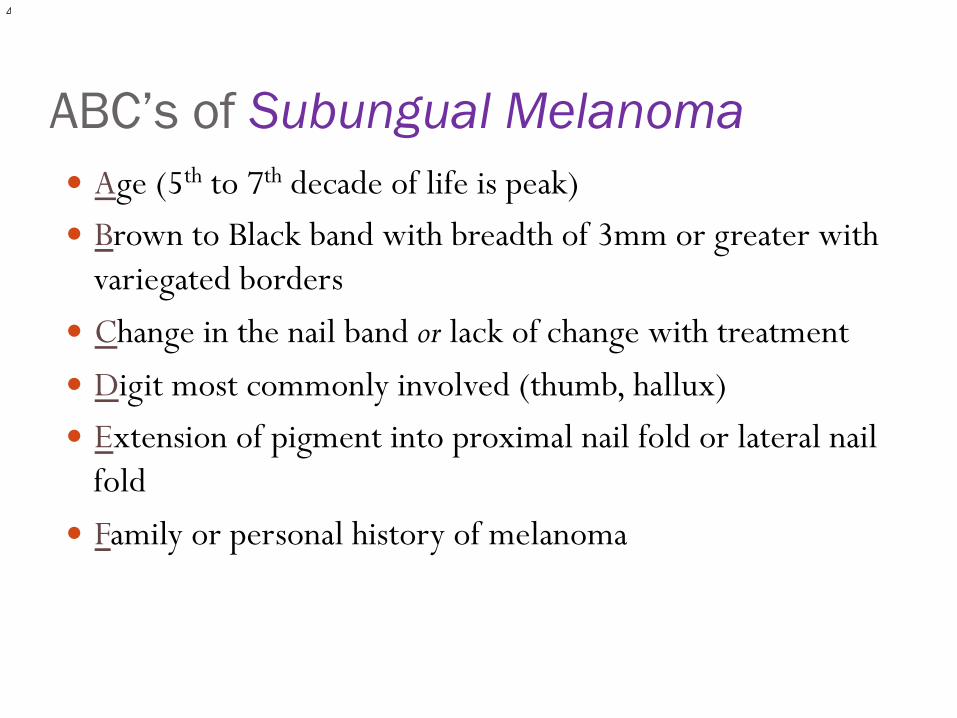
ABC’s of Subungual Melanoma � Age (5th to 7th decade of life is peak) � Brown to Black band with breadth of 3mm or greater with
variegated borders � Change in the nail band or lack of change with treatment � Digit most commonly involved (thumb, hallux) � Extension of pigment into proximal nail fold or lateral nail
fold � Family or personal history of melanoma
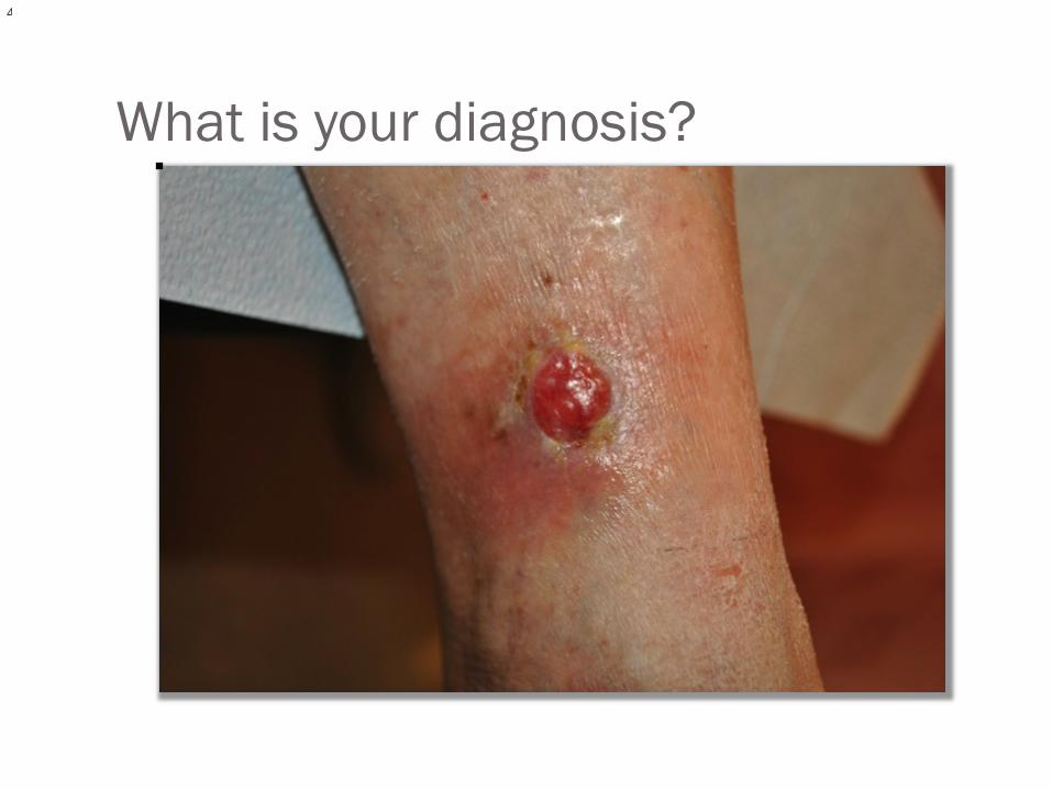
What is your diagnosis?
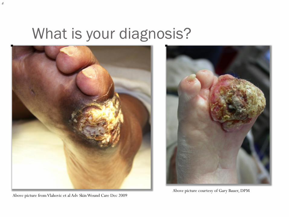
What is your diagnosis?
Above picture courtesy of Gary Bauer, DPM Above picture from Vlahovic et al Adv Skin Wound Care Dec 2009
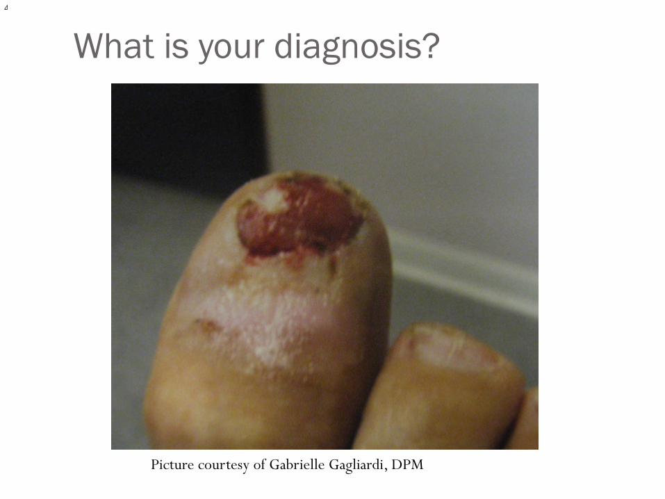
What is your diagnosis?
Picture courtesy of Gabrielle Gagliardi, DPM
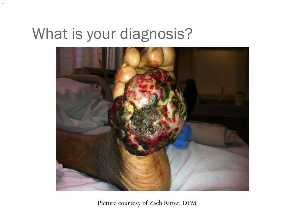
What is your diagnosis?
Picture courtesy of Zach Ritter, DPM

What is your diagnosis?
Dermatoscope picture
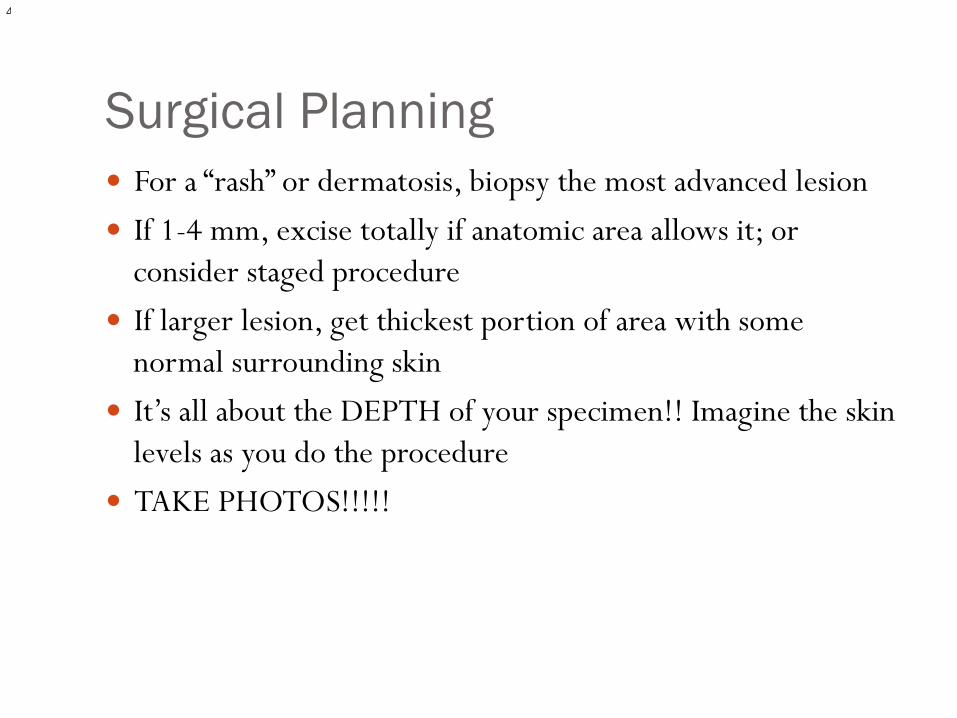
Surgical Planning � For a “rash” or dermatosis, biopsy the most advanced lesion � If 1-4 mm, excise totally if anatomic area allows it; or
consider staged procedure � If larger lesion, get thickest portion of area with some
normal surrounding skin � It’s all about the DEPTH of your specimen!! Imagine the skin
levels as you do the procedure � TAKE PHOTOS!!!!!
Surgical Planning � Relaxed skin tension lines � An untouched primary lesion dependent on DDx---papules
better than macules � Edge of lesion vs Center � Stay away from old blisters, but if no choice, get surrounding
“good” skin � Shave and Curettage are not appropriate for skin dermatitis
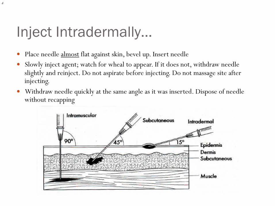
Inject Intradermally… � Place needle almost flat against skin, bevel up. Insert needle � Slowly inject agent; watch for wheal to appear. If it does not, withdraw needle
slightly and reinject. Do not aspirate before injecting. Do not massage site after injecting.
� Withdraw needle quickly at the same angle as it was inserted. Dispose of needle without recapping
Shave Biopsy � Indications: Nevi (Moles) flat and papular � Benign, EXOPHYTIC growths
� Plantar lesions are ENDOPHYTIC (ie warts)
� Not helpful or appropriate for skin rashes � Unless you have experience in doing a deeper shave
(saucerization), it will not give you the depth or level of invasion that an incisional/excisional will give � I personally would not do on a suspected melanoma on the
lower extremity
Technique � Intradermal technique of local anesthesia � Blade parallel with skin; can use #15 blade or autoclaved
disposable shaving blade � Topical hemostatic agent (Aluminum chloride) and cauterize � Pigmentation remains 25%
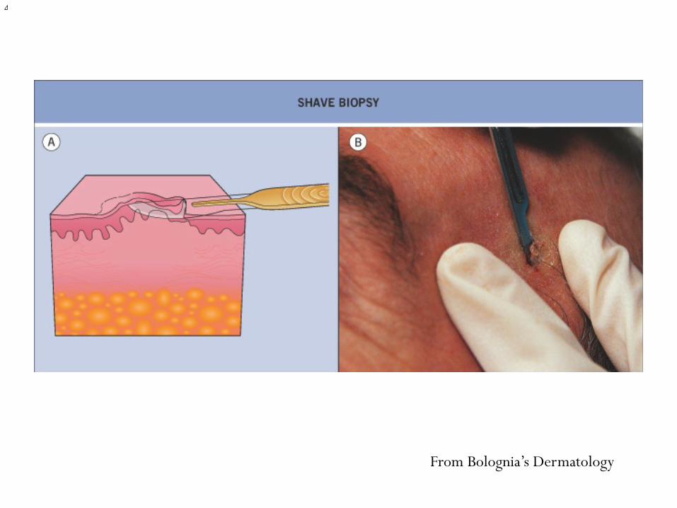
From Bolognia’s Dermatology
From Bolognia’s Dermatology
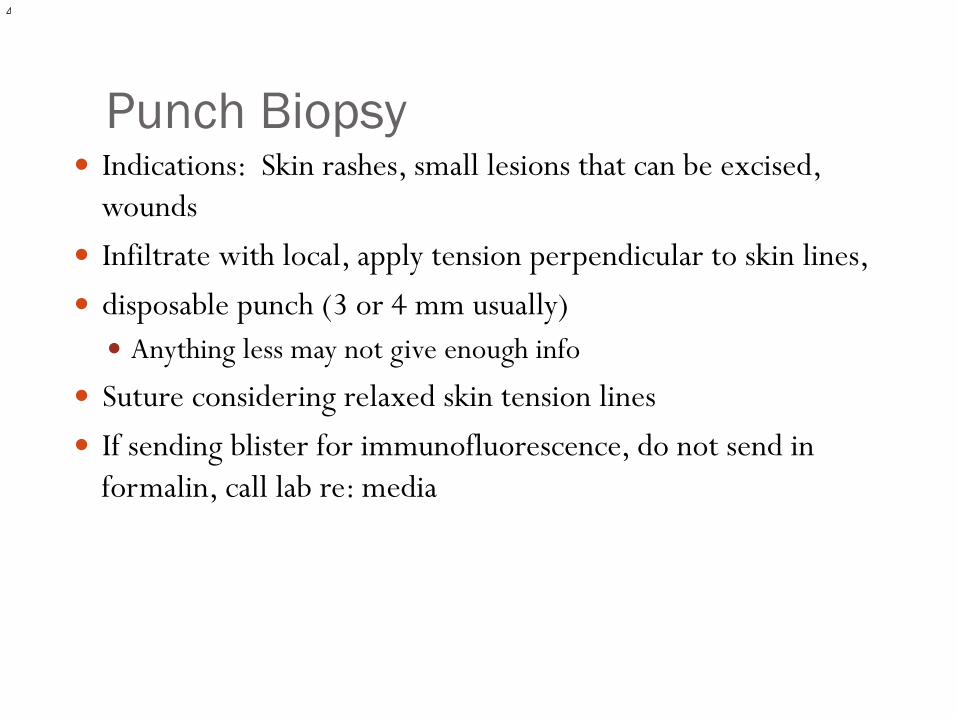
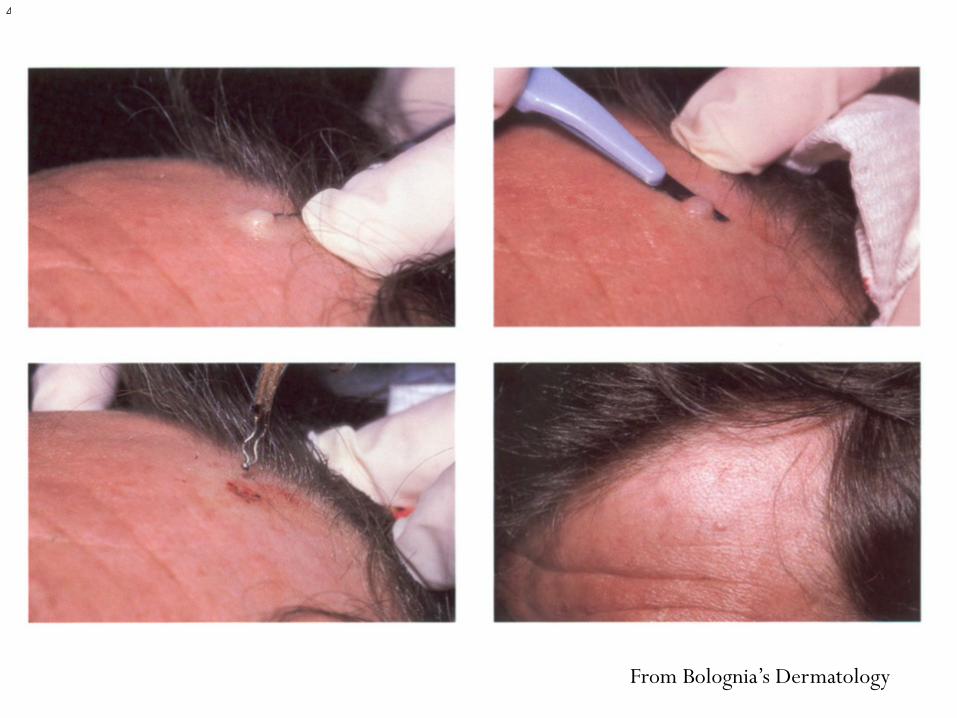
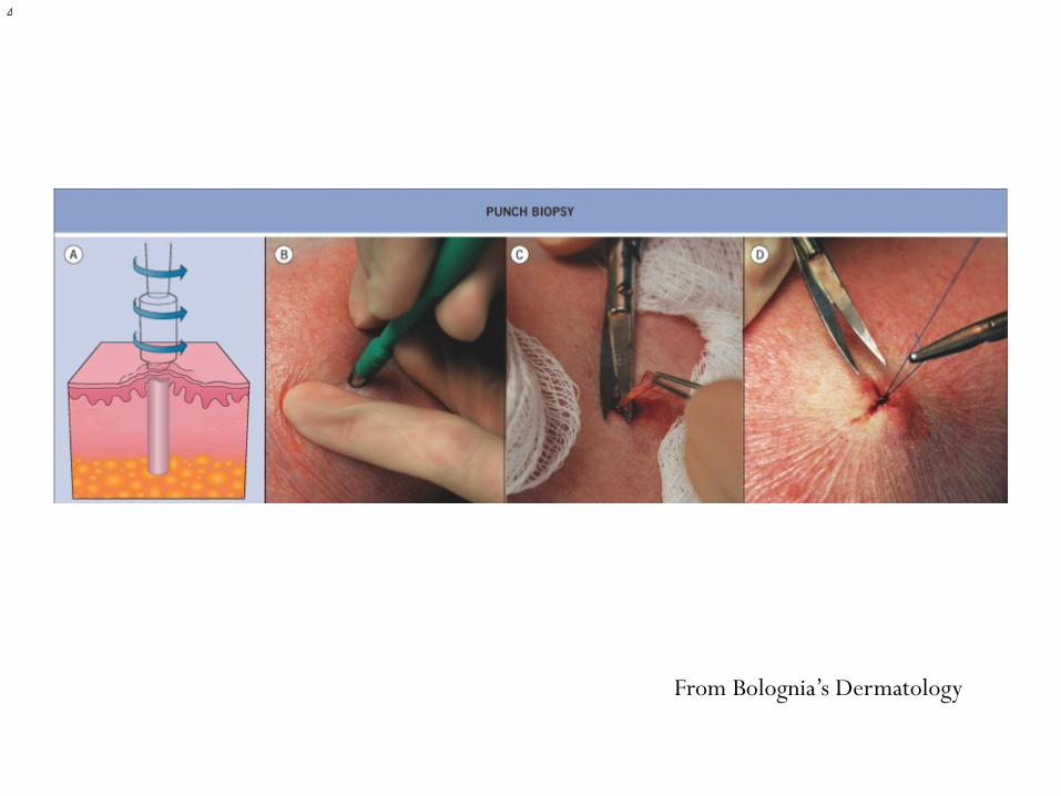
Punch Biopsy � Indications: Skin rashes, small lesions that can be excised,
wounds � Infiltrate with local, apply tension perpendicular to skin lines, � disposable punch (3 or 4 mm usually)
� Anything less may not give enough info
� Suture considering relaxed skin tension lines � If sending blister for immunofluorescence, do not send in
formalin, call lab re: media
From Bolognia’s Dermatology
From Bolognia’s Dermatology
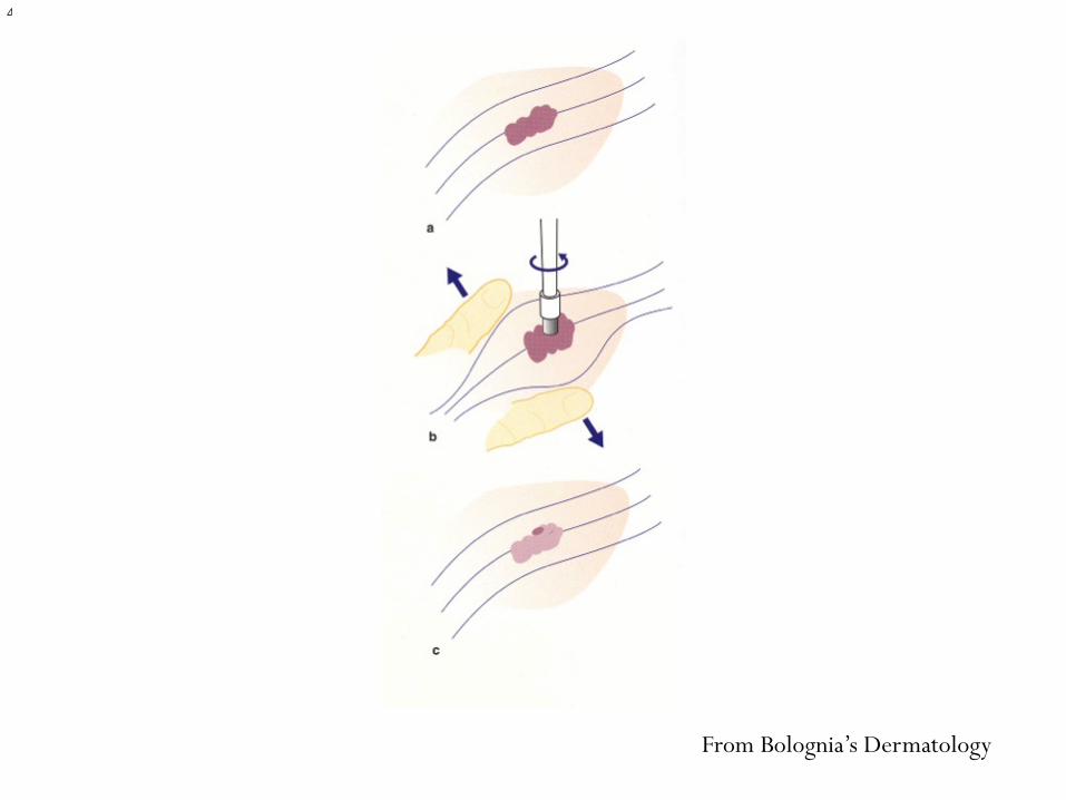
What’s wrong with this picture??
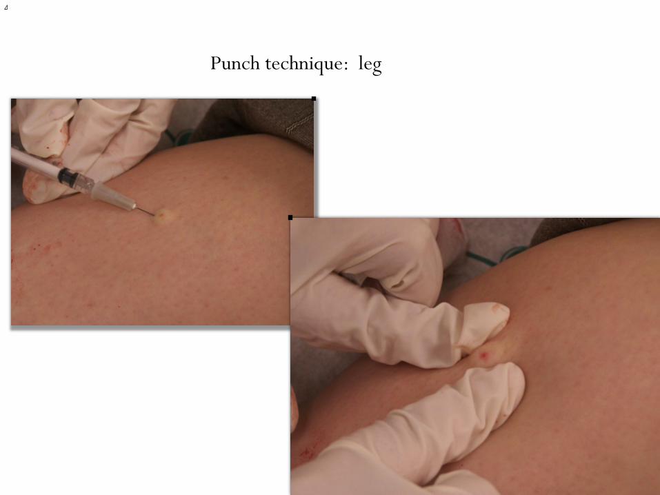
Punch technique: leg
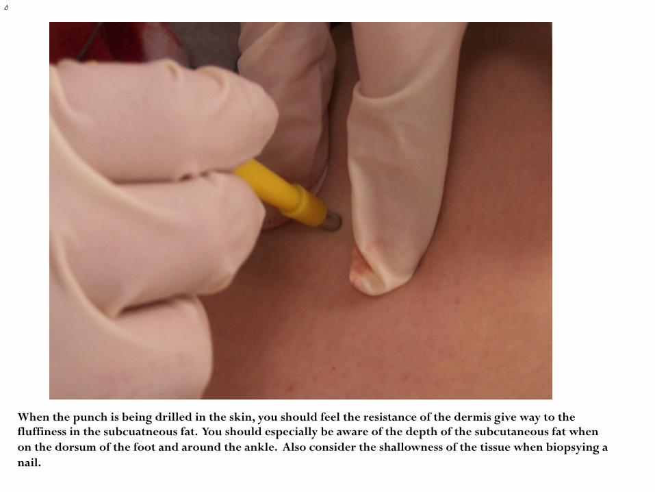
When the punch is being drilled in the skin, you should feel the resistance of the dermis give way to the fluffiness in the subcuatneous fat. You should especially be aware of the depth of the subcutaneous fat when on the dorsum of the foot and around the ankle. Also consider the shallowness of the tissue when biopsying a nail.
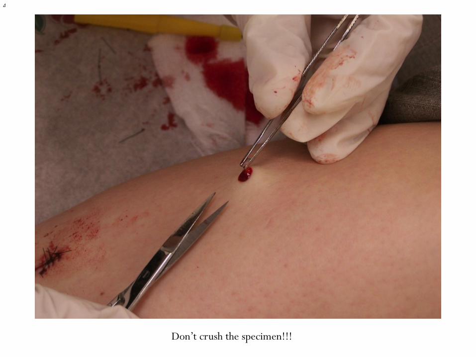
Don’t crush the specimen!!!

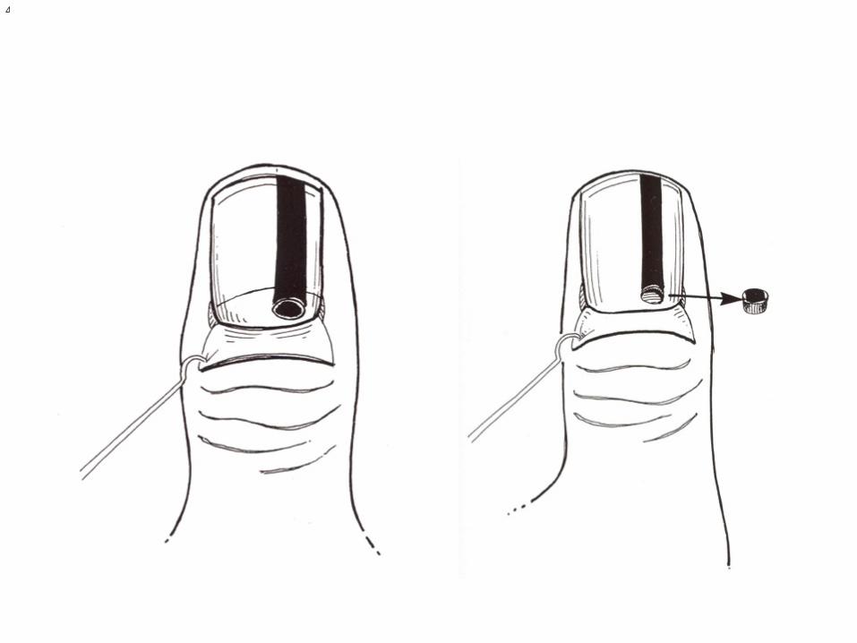
Why biopsy a nail? � Subungual melanomas and nevi can appear as a longitudinal
pigmented streak in a nail. � Known as longitudinal melanonychia until a pathologic
diagnosis formed � In caucasians, suspect melanoma until proven otherwise � In AA, use ABC’s of melanoma to determine if you should
biopsy
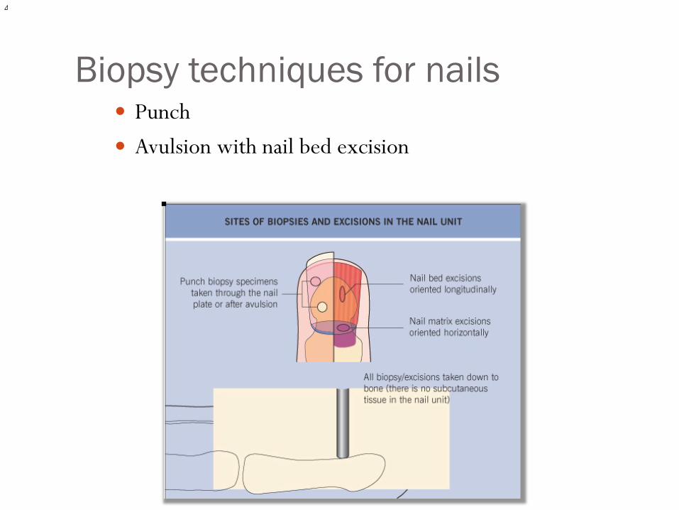
Biopsy techniques for nails � Punch � Avulsion with nail bed excision
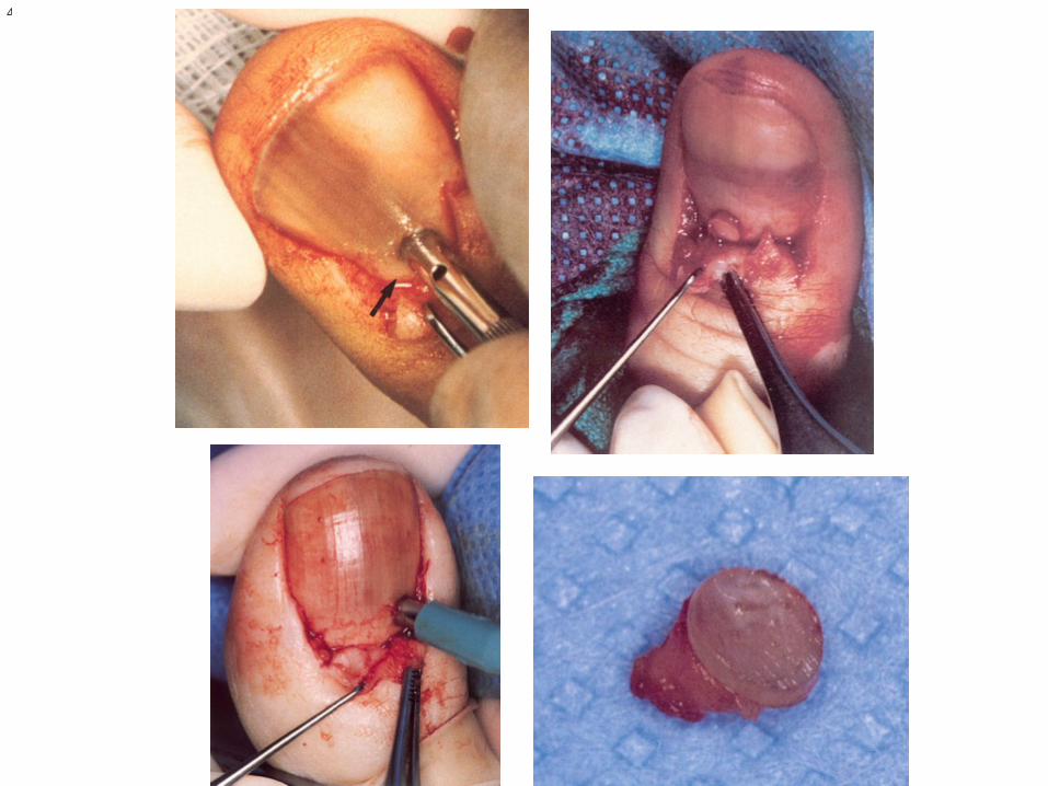
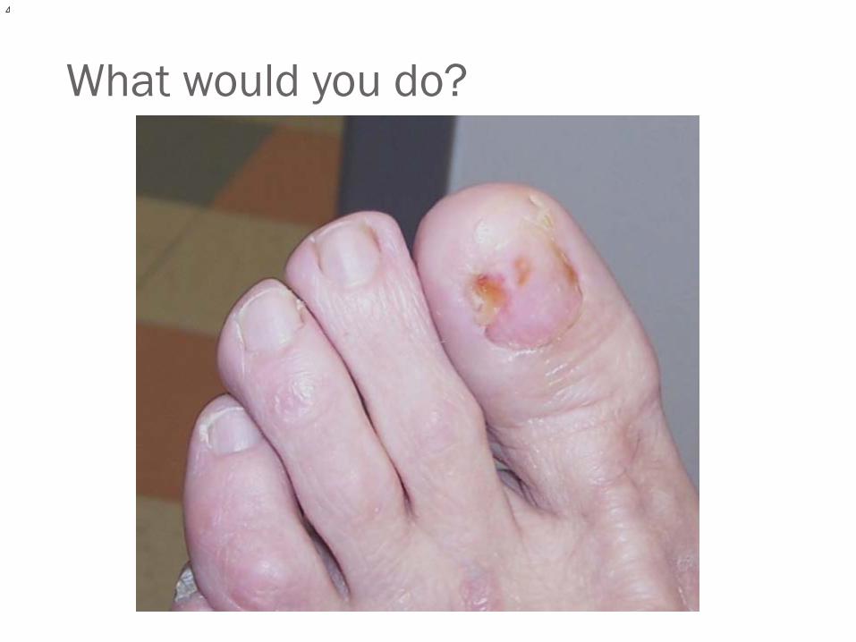
What would you do?

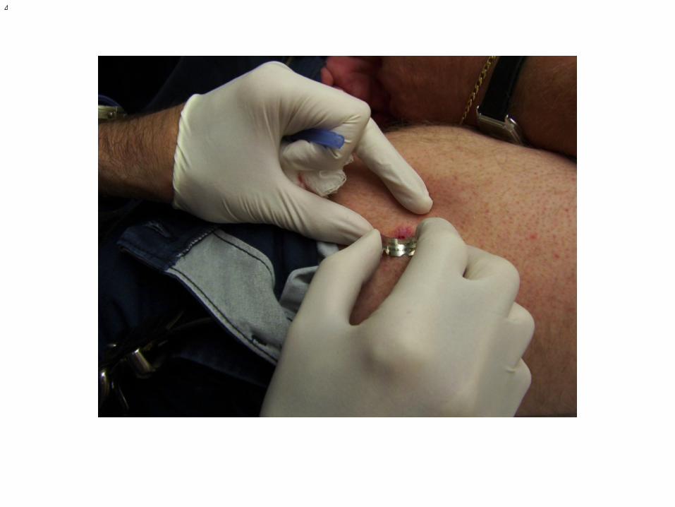
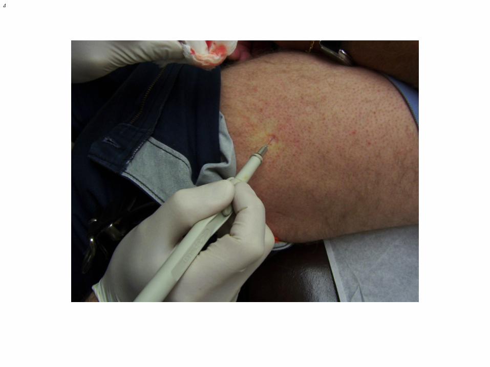
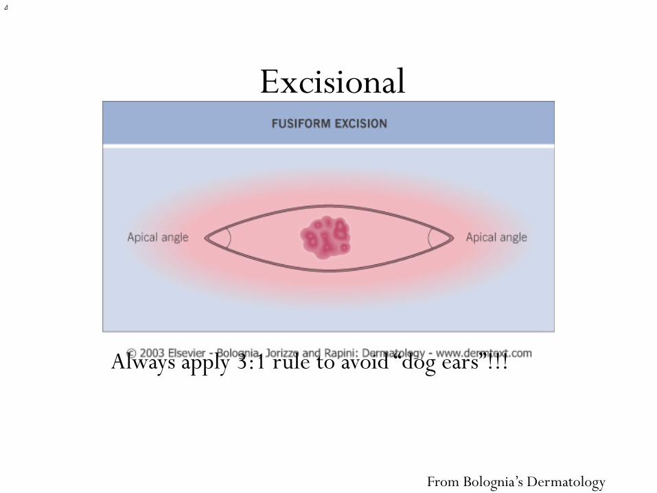
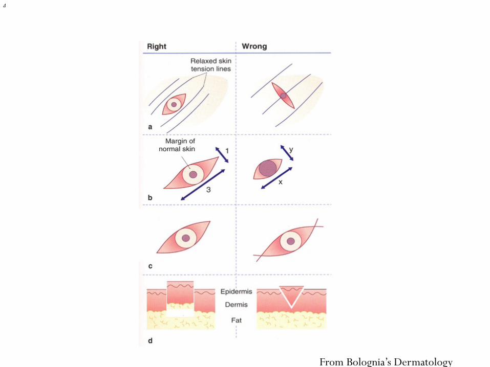
Excisional � Basic elliptical has length 3x as long as the width; can use 2 mm
margin for first excision � Great for moles that are suspicious; goes down to fat; complete
excision � Consider relaxed skin tension lines for best scar outcome � If you suspect a melanoma arising from a nevus, this is the best
technique to use
Excisional
Always apply 3:1 rule to avoid “dog ears”!!!
From Bolognia’s Dermatology
From Bolognia’s Dermatology
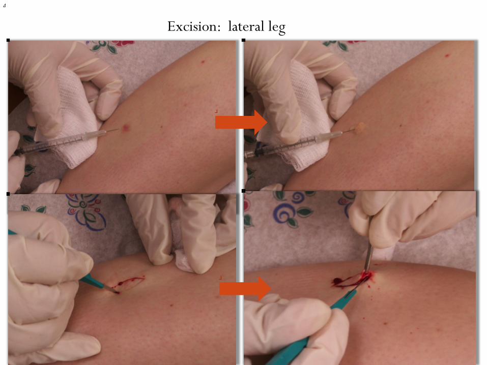
Excision: lateral leg
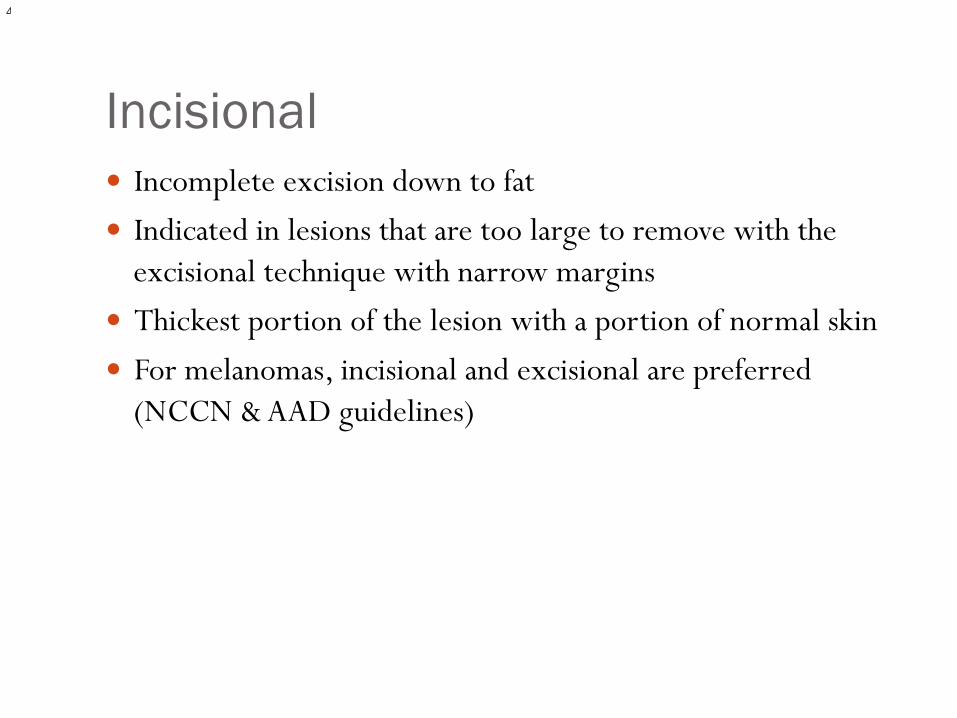
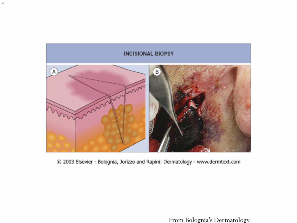
Incisional � Incomplete excision down to fat � Indicated in lesions that are too large to remove with the
excisional technique with narrow margins � Thickest portion of the lesion with a portion of normal skin � For melanomas, incisional and excisional are preferred
(NCCN & AAD guidelines)

Why include a margin of normal skin? � Excisional vs Punch vs Shave by Stell et al 2007
� Punches have a high positive lateral margin rate � Unless you do a “punch excision”
� Shaves have a high positive deep margin rate � Shows inadequate sampling � Saucerization technique gives deeper specimen, but remember the
thickness of the skin plantarly! � Really consider not doing this for a suspected plantar melanoma; although
it has been shown useful for “thin” melanomas � Most common biopsy sent to paths
� NCCN and AAD prefer Excisional first (or incisional if too large) for suspected melanoma for best chance at proper staging
From Bolognia’s Dermatology
Assisting the Dermatopathologist… � On the requisition sheet, provide the following info:
� Age, sex, type of biopsy, site (laterality, too) � Duration of lesion biopsied and of disease, Size, Description of
lesion (color, distribution pattern) � Your clinical diagnosis and differential diagnosis � Anything else that you feel would aid in diagnosis (photos, drug
history, etc)

What next? � Tell patient about site care and pain control � Return for suture removal and review of path diagnosis in
10-14 days
Figuring out the Path report � Communication is crucial between you and the
dermatopathologist � If you have questions, don’t hesitate to call
� In situ=superficial lesion � May say wider excision recommended � May ask for another specimen to do immunofluorescence
staining (for melanoma and vesicles) � Depth/margins/mitotic rate
Clark’s, Breslow’s, and 5 year survival rate � Breslow’s Depth is the most important prognostic indicator
per AJCC guidelines � Clark’s has little value beyond 1mm thickness � Clark Level I= tumor cells in epidermis only (melanoma in situ)
� 100% cured with adequate excision � Clark Level II=tumor cells into dermis (but do not fill papillary
dermis) � 95-99% 5 year survival with Breslow depth <0.75 mm
Clark’s, Breslow’s, and Survival � Clark Level III=tumor cells into and fill papillary dermis
� 5 yr survive 90% with depth of 0.76-1.49mm � Clark Level IV=tumor cells into reticular dermis
� 5 yr survive 75% with depth of 1.50-4.00 mm � Clark Level V=tumor cells through dermis and into
subcutaneous fat � 5 yr survive<50% with depth >4.00mm

New AJCC guidelines � Ann Surg Oncol 2010 � Tumor
� thickness, ulcerated or not ulcerated?, mitotic rate
� Node � Nodal micrometastases staining
� Metastasis � Site or sites of distant metastases
Melanomaprognosis.org
Once the biopsy results return: Treatment � General guidelines for surgical margins for total excision
In situ: 5mm border of normal skin Breslow’s depth of <1mm wide excision with 1cm normal
margins 1-2mm depth should have 1-2 cm margins 2-4mm should have 2 cm margins >4mm should have 2 cm+ margins
http://www.nccn.org/professionals/physician_gls/PDF/melanoma.pdf
Need for further surgery � Now you need to do a sterile procedure for a total excision!
� Can still be an office based procedure
� Keep in mind a diagnosis of melanoma will follow that patient indefinitely—especially in cases of life insurance
Working with other docs… � Need a graft? Get plastics or use a skin substitute � Staged closure? Dermaclose � Need a consult for full body check with dermatology � Moh’s surgery � Oncology consult and eval when needed
Moh’s Surgery � Treatment of choice for difficult skin tumors; has 98% 5
year cure rate � Dermatological surgeon or Plastic surgeon who is Moh’s
fellowship trained � Serial excision & histologic exam; then repeat process until
margins clear and close wound � Can be used for digital and pedal malignant lesions to avoid
amputation…as long as nodes are clear � For Basal Cell, Squamous Cell, Verrucous carcinoma (low
grade squamous)
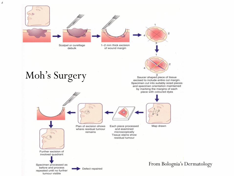
Moh’s Surgery
From Bolognia’s Dermatology

Post Moh’s surgery

References 1. “Proper Biopsy” in Histologic Diagnosis of Inflammatory Skin Diseases: An Algorithmic Method Based on Pattern Analysis THIRD EDITION by
A. Bernard Ackerman, Almut Böer, Bruce Bennin, and Geoffrey J. Gottlieb
2. Olbricht S. “Biopsy techniques and basic excisions.” In Bolognia JL, Rapini RP, et al. (eds) Dermatology (1st Edition) Mosby, London, pp 2269-2286, 2003.
3. Sina B, Kao G, Deng A, Gaspari A. Skin biopsy for inflammatory and common neoplastic skin diseases: optimum time, best location, and preferred techniques. A critical review. J Cutan Pathol (epub ahead of print) 2009 Jan 27; 1-6.
4. Mehregan D, Dooley V. How to get the most out of your skin biopsies.” Int J Dermatol 2007; 46(7): 727-33.
5. Jankovic A, Binic I, Ljubenovic M. Basal cell carcinoma is not granulation tissue in the venous leg ulcer. Int J Low Extrem Wounds 2008 7(3):182-4.
6. Spear M. Pyoderma gangrenosum: an overview. Plastic Surg Nurs 2008 28(3):154-7.
7. Dawber R, De Berker D, Baran R. “Science of the nail apparatus.” In Baran R, Dawber R (eds) Diseases of the Nails and their Management (2nd edition); 1994. pp. 1-34.
8. Dawber RP, Baran R, Berker D. Science of the nail apparatus. In: Baran R, Dawber RP, eds. Diseases of the Nails and their Management (2nd edition). Blackwell Scientific Publications, Oxford, England; pp 1-34, 1994.
9. Rich P. “Nail Surgery” In Bolognia JL, Rapini RP, et al. (eds) Dermatology (1st Edition) Mosby, London, pp 2321-2230, 2003.
10. Sladden MJ et al “Surgical Excision margins for primary cutaneous melanoma” The Cochrane Library 2009, issue 4
11. Gershenwald JE et al “TNM staging system for cutaneous melanoma and beyond” Ann Surg Oncol 2010 17: 1475
12. Sober AJ et al “Method of Biopsy and Incidence of Positive Margins in Primary Melanoma” Ann Surg Oncol 2007 14(2): 274
13. Stell VH et al “Method of Biopsy and Incidence of Positive Margins in Primary Melanoma” Ann Surg Oncol 2007 14(2): 893