magnetic properties of ultrasmall iron-oxide nanoparticles
TRANSCRIPT
Journal of Alloys and Compounds 595 (2014) 153–157
Contents lists available at ScienceDirect
Journal of Alloys and Compounds
journal homepage: www.elsevier .com/locate / ja lcom
Magnetic properties of ultrasmall iron-oxide nanoparticles
http://dx.doi.org/10.1016/j.jallcom.2014.01.1120925-8388/� 2014 Elsevier B.V. All rights reserved.
⇑ Corresponding author. Tel.: +381 11 3408694; fax: +381 11 3408607.E-mail address: [email protected] (D. Milivojevic).
Dušan Milivojevic a,⇑, Branka Babic-Stojic a, Vukoman Jokanovic a, Zvonko Jaglicic b,Darko Makovec c, Nataša Jovic a
a Vinca Institute of Nuclear Sciences, University of Belgrade, P.O. Box 522, 11001 Belgrade, Serbiab Institute of Mathematics, Physics and Mechanics, Jadranska 19, 1000 Ljubljana, Sloveniac Department for Materials Synthesis, Jozef Stefan Institute, Jamova 39, 1000 Ljubljana, Slovenia
a r t i c l e i n f o a b s t r a c t
Article history:Received 22 November 2013Received in revised form 15 January 2014Accepted 16 January 2014Available online 24 January 2014
Keywords:Nanostructured materialsMagnetic fluidsMagnetic measurementsUltrasmall superparamagnetic iron oxideUSPIO
The work presents structural and magnetic properties of ultrasmall magnetic nanoparticles consisting ofinorganic iron oxide core and organic ester shell, dispersed in an organic fluid, synthesized via polyolroute. The structure analysis shows that nanoparticles are crystalline, less than 3 nm in size, mutuallyclearly separated. The magnetic properties are in accordance with the size of the nanoparticles and donot indicate interparticle interactions. The particles show pure superparamagnetic behavior with verylow blocking temperature. ZFCFC bifurcation and ac susceptibility peaks are at temperatures TB < 12 K.The properties of fluid were compared with dried powder sample. Drying of fluid brings about interac-tions between the magnetic nanoparticles that considerably affect spin dynamics of the particles. Thesurface of nanoparticles has a significant influence on their behavior. The Mössbauer parameters indicateexistence of c-Fe2O3 core and non-stoichiometric surface layer. Magnetic field dependent magnetizationanalysis suggests smaller apparent size of the particles d0 = 0.56 nm. High magnetic anisotropy due tosurface layer anisotropy was measured to be of the order 106 erg/cm3 that is two orders of magnitudehigher than that in bulk material.
� 2014 Elsevier B.V. All rights reserved.
1. Introduction
Magnetic nanoparticles (MNPs) and their dispersions in variousmedia have been for a long time of scientific and technologicalinterest. Magnetic dipole and van der Waals interactions have ten-dency to agglomerate the particles. In order to prevent agglomera-tion the magnetic particles should be small enough (usually about10 nm in size) and coated with a shell of an appropriate material.This coating can be a surfactant made of long chained molecules,or ionic if it is an electric shell [1–3]. The particles are usually madeof maghemite c-Fe2O3, magnetite Fe3O4, and other ferrites of thetype MFe2O4, where M = Mn, Co, Ni, Cu. The carrier liquid is usuallyan organic solvent or water.
Besides wide range of technological applications [1], in recentyears a lot of research work has been devoted to various biomedi-cal applications of dispersions of magnetic nanoparticles. Biocom-patible nanoparticles dispersed in aqueous or physiologicalmedium [4] can be used in Magnetic Resonance Imaging (MRI) ascontrast agents [3,5,6], as drug carriers [3,7] and in magnetichyperthermia treatment [3,5,7,8].
Iron oxides are known as T2 MRI contrast agents but ultrasmallnanoparticles can act as both T1 and T2 contrast agents [9–11].Ultrasmall iron oxide/ferrite nanoparticles having longer circula-tion half-life in the body can avoid fast uptake by the macrophagesand provide better contrast [9,12,13]. Commonly used iron oxidenanoparticle contrast agents with a hydrodynamic size of over50 nm remain primarily intravascular which severely underminestheir targeting specificity.
The characterization of ultrasmall iron oxide nanoparticles(USPIO) is rather difficult task. The ultrasmall particles have highsurface to volume ratio. They may be considered to be a less-orderedsystem which is neither completely crystalline nor completelyamorphous [13]. This property is reflected in discordant data ob-tained using different experimental methodologies. Other problemis that some experimental methodologies require drying of samples.
In the present work we have synthesized USPIO in organiccarrier fluid and studied magnetic properties. We compared it tothe dried powder form. Microstructure, morphology, static anddynamic magnetic properties of the prepared fluid were studiedusing X-ray diffraction (XRD), transmission electron microscopy(TEM), atomic force microscopy (AFM), dynamic light scattering(DLS), dc magnetization, ac susceptibility measurements andMössbauer spectroscopy.
154 D. Milivojevic et al. / Journal of Alloys and Compounds 595 (2014) 153–157
2. Experimental details
2.1. Synthesis
The synthesis was carried out using tris(acetylacetonate)Fe(III) (FeIII(acac)3),diethylene glycol (DEG) and propionic acid. The Fe precursor compound, 34 g ofFeIII(acac)3, was dissolved in 200 ml of DEG. The solution was autoclaved at120 �C for 2 h, than temperature was increased to 165 �C to obtain esters of dieth-ylene glycol acetylacetonate. The reaction was promoted by iron ions which cata-lyzed this reaction. After this procedure the mixture was rapidly cooled to 100 �Cby flow of water through the wall of autoclave. Finally, 37.63 g of propionic acidwas added with a small quantity (0.9 ml) of sulphuric acid as catalyst and reactionwas done at 145 �C for 2 h to obtain well dispersed iron oxide nanoparticles insideester shell. The obtained colloid solution was brown and very stable. Besides thefluid sample, a powder sample was also prepared by drying the fluid at 200 �C for10 h.
The idea of this route is to enable networking around iron oxide nanoparticles, tolower toxicity of DEG by making esters and prolong circulation time by coveringUSPIO with esters, (diethylenglycolactetylacetonate and diethylenglycolpropionate).
2.2. Experimental technique
X-ray characterization of the samples was carried out on a Philips PW 1050powder diffractometer using Cu Ka radiation. High-resolution transmission elec-tron microscopy (HRTEM) and energy dispersive X-ray spectroscopy (EDXS) werecarried out using transmission electron microscope JEOL 2010 equipped with an en-ergy-dispersive X-ray spectrometer (EDXS) microanalysis system (LINK ISIS EDS300) operated at 200 kV. The samples were prepared by drying a drop of the dilutedsuspension on a copper-grid-supported transparent carbon foil. The size distribu-tion of the nanoparticles was measured by dynamic light scattering (DLS) usingZetasizer Nano ZS (Malvern, UK) equipped with green laser (532 nm). Intensity ofscattered light was detected at the angle of 173�. All measurements were conductedat room temperature. Ten measurements were performed for each sample. All dataprocessing was done by the Zetasizer software 6.20 (Malvern instruments). Mag-netic measurements were carried out on a SQUID magnetometer (MPMS XL-5,Quantum Design) equipped with an ac option. Zero-field-cooled (ZFC) and field-cooled (FC) measurements of magnetization were performed at applied magneticfields from 50 to 1000 Oe in the temperature range from 2 to 300 K. Magnetic fielddependence of magnetization was measured at applied magnetic fields up to50 kOe. The ac magnetization measurements were made under an ac exciting fieldwith amplitude of 6.5 Oe using different frequencies in the range 1–1500 Hz. 57FeMössbauer spectrum was recorded at room temperature using a conventionaltransmission Mössbauer spectrometer operating in constant acceleration mode.The source was 57Co in Rh matrix. Experimental data were fitted to Lorentzianabsorption lines by using a least-square based method. The spectrometer was cal-ibrated using a-Fe foil at room temperature. All the isomer shifts are given relativeto the center of the a-Fe spectrum.
3. Results and discussion
3.1. Structure analysis
The X-ray diffraction lines of the samples are very broad and noinformation about structure can be obtained. Estimation of averagecrystallite size from the width of the X-ray diffraction lines usingScherrer’s formula gives sizes lower than 1.6 nm, which is the sizeof only two crystal lattice parameters or the material is amorphous.
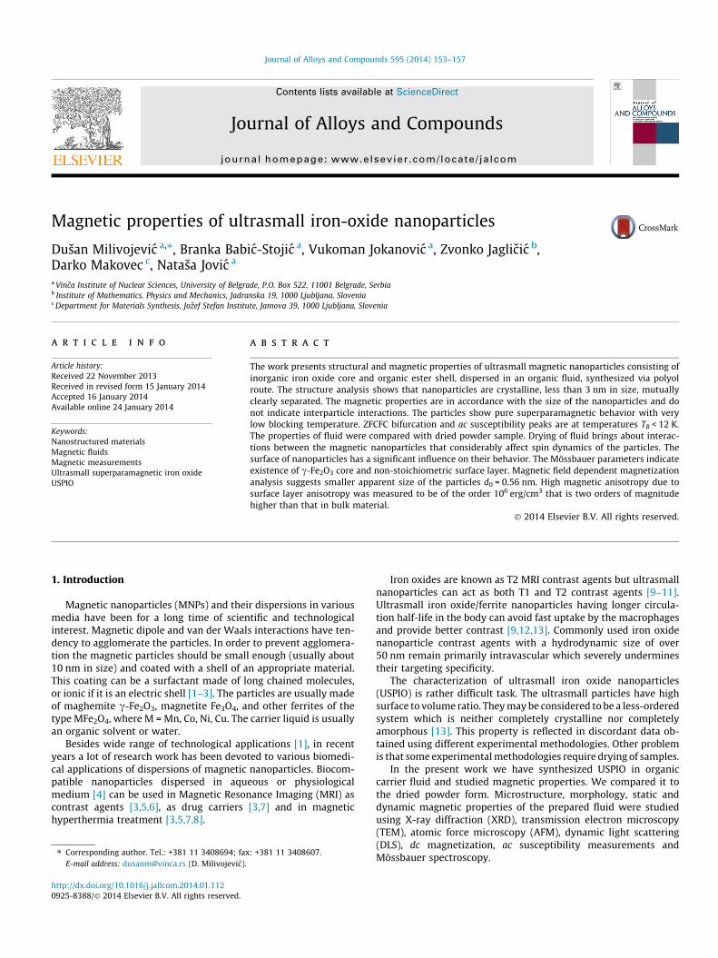
The morphology, particle size and chemical composition werecharacterized by HRTEM and EDXS. TEM analysis (Fig. 1) showsthat after drying the suspension on the TEM support, the sampledeposited in the form of small deposits containing small nanopar-ticles of uniform, but various sizes. The nanoparticles of one sizeare always deposited together. The majority of the nanoparticlesare below 3 nm, but also larger nanoparticles around 5 nm in sizewere observed. HRTEM shows that the nanoparticles are crystal-line. EDXS shows presence of Fe and O.
The AFM micrographs reveal the well separated nanostructuresof around 50 nm in size (Fig. 1(d)).
The size distribution of the nanoparticles in the organic fluid wasstudied using dynamic light scattering (DLS). Number of particles asa function of hydrodynamic particle diameter can be well describedby a log-normal distribution f ðdÞ ¼ ð1=
ffiffiffiffiffiffiffi2pp
rdÞ � exp½�ln2ðd=d0Þ=2r2� yielding the distribution width r = 0.22 and median of the
distribution d0 = 48.82 nm, which is related to the average hydrody-namic diameter dav = d0exp(r2/2) = 50.01 nm.
The particle diameters obtained by DLS and AFM techniques arevery close to each other but are an order of magnitude larger thanthe values obtained by XRD and TEM. This is because DLS and AFMmeasure the size of possibly agglomerated particles together withthe organic shell.
It is apparent in Fig. 1(a) and (b) that the particles are mutuallyclearly separated but clustered to form larger groups. Since TEMprocedure includes drying of fluid, clusterization could be due tofluid evaporation that turns the sample into a similar to the pow-der sample.
Discordant structural data for USPIO nanoparticles obtainedwith different structural methods have been already discussed inthe paper by Di Marco et al. [13].
3.2. Mössbauer spectra analysis
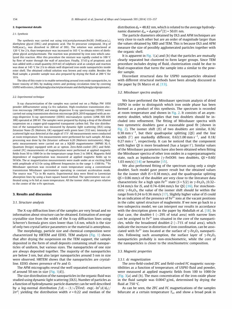
We have performed the Mössbauer spectrum analysis of driedUSPIO in order to distinguish which iron oxide phase has beenformed as a product of this synthesis. The spectrum is recordedat room temperature and shown in Fig. 2. It consists of an asym-metric doublet, which implies that two doublets should be in-cluded into refinement. The fitting of Mössbauer spectra withtwo symmetric doublets gave a reasonable good fit (shown inFig. 2). The isomer shift (IS) of two doublets are similar, 0.36/0.38 mm s�1, but their quadrupolar splitting (QS) and the linewidth (U) are markedly different, 0.63/1.00 mm s�1, and 0.18/0.27 mm s�1, respectively. It can be noticed that the componentwith higher QS is more broadened (has a larger U). Similar valuesof the Mössbauer parameters have also been obtained when fittingthe Mössbauer spectra of other iron oxides in a superparamagneticstate, such as lepidocrocite (c-FeOOH: two doublets, QS = 0.60/1.03 mm/s) [14] or hematite [15].
We also performed fitting of the spectrum using only a singledoublet. Such model gave poorer result but the obtained valuesfor the isomer shift IS = 0.38 mm/s, and the quadrupolar splittingQS = 0.86 mm/s of the doublet are very close to the literature datacharacteristic for a high spin Fe3+ state (S = 5/2) in c-Fe2O3 (0.32–0.34 mm/s for IS, and 0.74–0.84 mm/s for QS) [16]. For stoichiom-etric c-Fe2O3, the value of the isomer shift should lie within therange from 0.24 to 0.36 mm/s [17]. Slightly higher value of IS couldbe an indication of the presence of Fe2+ ions at the vacant positionsin the cubic spinel structure of maghemite. If we now go back to atwo subspectra model, we can interpret our results in accordancewith the description given in the paper by Abdullah et al. [18]. Inthat case, the doublet-1 (�29% of total area) with narrow linescan be assigned to Fe3+ ions situated in the core of the nanoparti-cles, while the broadened doublet-2 (�71% of total area), whichindicate the increase in distortion of iron coordination, can be asso-ciated with Fe3+ ions located at the surface of c-Fe2O3 nanoparti-cles. Following such assumption, the surface layer of c-Fe2O3
nanoparticles probably is non-stoichiometric, while the core ofthe nanoparticles is closer to the stoichiometric composition.
3.3. Magnetic properties
3.3.1. dc magnetizationThe zero-field-cooled ZFC and field-cooled FC magnetic suscep-
tibilities as a function of temperature, of USPIO fluid and powder,were measured at applied magnetic fields from 100 to 1000 Oe(Fig. 3(a) and (b). The mass concentration of the iron-oxide phasein the fluid sample was 0.0047 g/mL, determined by drying thefluid at 750 �C.
As can be seen, the ZFC and FC magnetizations of the samplesbifurcate at a certain temperature Tirr and show a broad peak in
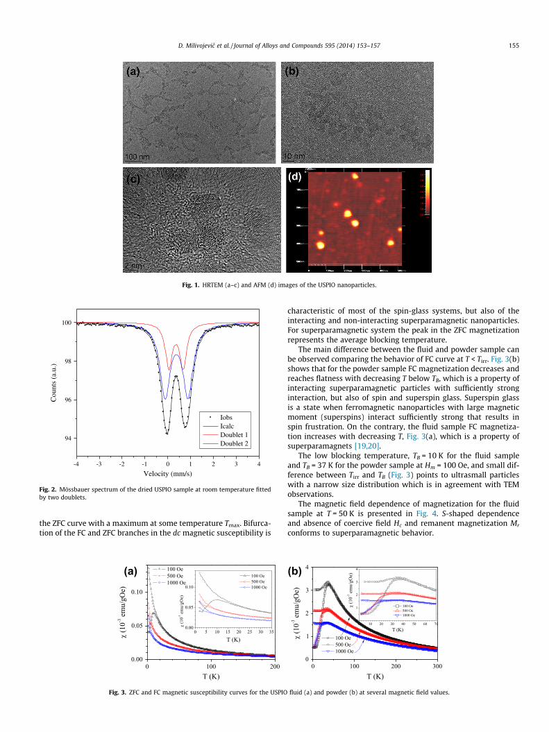
Fig. 1. HRTEM (a–c) and AFM (d) images of the USPIO nanoparticles.
-4 -3 -2 -1 0 1 2 3 4
94
96
98
100
Cou
nts
(a.u
.)
Velocity (mm/s)
IobsIcalcDoublet 1Doublet 2
Fig. 2. Mössbauer spectrum of the dried USPIO sample at room temperature fittedby two doublets.
D. Milivojevic et al. / Journal of Alloys and Compounds 595 (2014) 153–157 155
the ZFC curve with a maximum at some temperature Tmax. Bifurca-tion of the FC and ZFC branches in the dc magnetic susceptibility is
(a)
Fig. 3. ZFC and FC magnetic susceptibility curves for the USPIO
characteristic of most of the spin-glass systems, but also of theinteracting and non-interacting superparamagnetic nanoparticles.For superparamagnetic system the peak in the ZFC magnetizationrepresents the average blocking temperature.
The main difference between the fluid and powder sample canbe observed comparing the behavior of FC curve at T < Tirr. Fig. 3(b)shows that for the powder sample FC magnetization decreases andreaches flatness with decreasing T below TB, which is a property ofinteracting superparamagnetic particles with sufficiently stronginteraction, but also of spin and superspin glass. Superspin glassis a state when ferromagnetic nanoparticles with large magneticmoment (superspins) interact sufficiently strong that results inspin frustration. On the contrary, the fluid sample FC magnetiza-tion increases with decreasing T, Fig. 3(a), which is a property ofsuperparamagnets [19,20].
The low blocking temperature, TB = 10 K for the fluid sampleand TB = 37 K for the powder sample at Hm = 100 Oe, and small dif-ference between Tirr and TB (Fig. 3) points to ultrasmall particleswith a narrow size distribution which is in agreement with TEMobservations.
The magnetic field dependence of magnetization for the fluidsample at T = 50 K is presented in Fig. 4. S-shaped dependenceand absence of coercive field Hc and remanent magnetization Mr
conforms to superparamagnetic behavior.
(b)
fluid (a) and powder (b) at several magnetic field values.
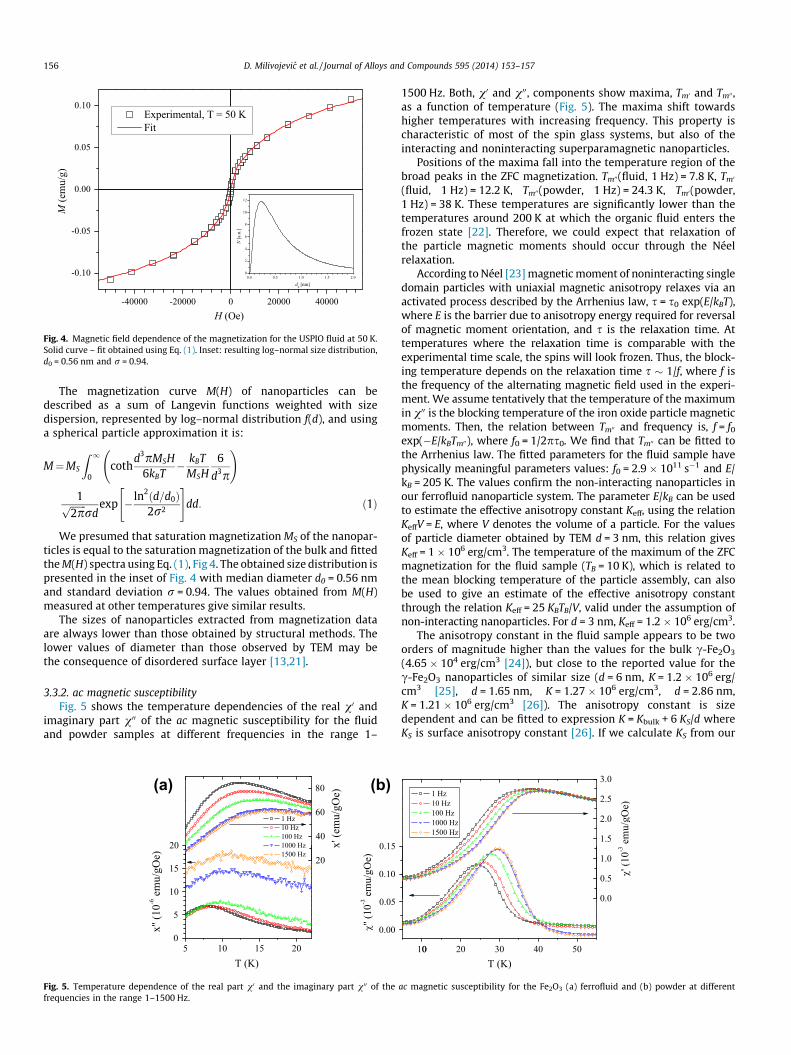
Fig. 4. Magnetic field dependence of the magnetization for the USPIO fluid at 50 K.Solid curve – fit obtained using Eq. (1). Inset: resulting log–normal size distribution,d0 = 0.56 nm and r = 0.94.
156 D. Milivojevic et al. / Journal of Alloys and Compounds 595 (2014) 153–157
The magnetization curve M(H) of nanoparticles can bedescribed as a sum of Langevin functions weighted with sizedispersion, represented by log–normal distribution f(d), and usinga spherical particle approximation it is:
M¼MS
Z 1
0coth
d3pMSH6kBT
� kBTMSH
6
d3p
!
1ffiffiffiffiffiffiffi2pp
rdexp � ln2ðd=d0Þ
2r2
" #dd: ð1Þ
We presumed that saturation magnetization MS of the nanopar-ticles is equal to the saturation magnetization of the bulk and fittedthe M(H) spectra using Eq. (1), Fig 4. The obtained size distribution ispresented in the inset of Fig. 4 with median diameter d0 = 0.56 nmand standard deviation r = 0.94. The values obtained from M(H)measured at other temperatures give similar results.
The sizes of nanoparticles extracted from magnetization dataare always lower than those obtained by structural methods. Thelower values of diameter than those observed by TEM may bethe consequence of disordered surface layer [13,21].
3.3.2. ac magnetic susceptibilityFig. 5 shows the temperature dependencies of the real v0 and
imaginary part v00 of the ac magnetic susceptibility for the fluidand powder samples at different frequencies in the range 1–
(a) (b)
Fig. 5. Temperature dependence of the real part v0 and the imaginary part v00 of thefrequencies in the range 1–1500 Hz.
1500 Hz. Both, v0 and v00, components show maxima, Tm0 and Tm00,as a function of temperature (Fig. 5). The maxima shift towardshigher temperatures with increasing frequency. This property ischaracteristic of most of the spin glass systems, but also of theinteracting and noninteracting superparamagnetic nanoparticles.
Positions of the maxima fall into the temperature region of thebroad peaks in the ZFC magnetization. Tm00(fluid, 1 Hz) = 7.8 K, Tm0
(fluid, 1 Hz) = 12.2 K, Tm00(powder, 1 Hz) = 24.3 K, Tm0(powder,1 Hz) = 38 K. These temperatures are significantly lower than thetemperatures around 200 K at which the organic fluid enters thefrozen state [22]. Therefore, we could expect that relaxation ofthe particle magnetic moments should occur through the Néelrelaxation.
According to Néel [23] magnetic moment of noninteracting singledomain particles with uniaxial magnetic anisotropy relaxes via anactivated process described by the Arrhenius law, s = s0 exp(E/kBT),where E is the barrier due to anisotropy energy required for reversalof magnetic moment orientation, and s is the relaxation time. Attemperatures where the relaxation time is comparable with theexperimental time scale, the spins will look frozen. Thus, the block-ing temperature depends on the relaxation time s � 1/f, where f isthe frequency of the alternating magnetic field used in the experi-ment. We assume tentatively that the temperature of the maximumin v00 is the blocking temperature of the iron oxide particle magneticmoments. Then, the relation between Tm00 and frequency is, f = f0
exp(�E/kBTm00), where f0 = 1/2ps0. We find that Tm00 can be fitted tothe Arrhenius law. The fitted parameters for the fluid sample havephysically meaningful parameters values: f0 = 2.9 � 1011 s�1 and E/kB = 205 K. The values confirm the non-interacting nanoparticles inour ferrofluid nanoparticle system. The parameter E/kB can be usedto estimate the effective anisotropy constant Keff, using the relationKeffV = E, where V denotes the volume of a particle. For the valuesof particle diameter obtained by TEM d = 3 nm, this relation givesKeff = 1 � 106 erg/cm3. The temperature of the maximum of the ZFCmagnetization for the fluid sample (TB = 10 K), which is related tothe mean blocking temperature of the particle assembly, can alsobe used to give an estimate of the effective anisotropy constantthrough the relation Keff = 25 KBTB/V, valid under the assumption ofnon-interacting nanoparticles. For d = 3 nm, Keff = 1.2 � 106 erg/cm3.
The anisotropy constant in the fluid sample appears to be twoorders of magnitude higher than the values for the bulk c-Fe2O3
(4.65 � 104 erg/cm3 [24]), but close to the reported value for thec-Fe2O3 nanoparticles of similar size (d = 6 nm, K = 1.2 � 106 erg/cm3 [25], d = 1.65 nm, K = 1.27 � 106 erg/cm3, d = 2.86 nm,K = 1.21 � 106 erg/cm3 [26]). The anisotropy constant is sizedependent and can be fitted to expression K = Kbulk + 6 KS/d whereKS is surface anisotropy constant [26]. If we calculate KS from our
0
ac magnetic susceptibility for the Fe2O3 (a) ferrofluid and (b) powder at different
D. Milivojevic et al. / Journal of Alloys and Compounds 595 (2014) 153–157 157
values of Keff we obtain KS = 0.048 erg/cm2 (from the Arrheniuslaw) and KS = 0.065 erg/cm2 (from TB) which is close to the reportedvalues for maghemite, KS = 0.04 erg/cm2 [26].
The powder sample fitted with the Arrhenius law gives unphys-ical value for f0 = 1.3 � 1017 s�1. The unphysical value is due tointerparticle interactions in the powder sample. The experimentaldependence ln f (T) is close to linear for the measured range of fvalues. The apparent linear dependence cannot be satisfactorilyfitted with 3 parameters functions, that include interparticleinteractions, like Vogel–Fulcher law s = s0 exp(E/kB(T � T0)) orpower law s = s0 [Tf/(Tf – Tc)]zm [27].
Disordered nanoparticle system with a random orientation ofeasy axes, size distribution and conflicting character of dipoleinteractions may have properties characteristic to spin glass state.To reveal what regime do the interparticle interactions belong to,we have considered the empirical parameter c = DTf/[Tf Dlog10 f][27] which delineates the frequency sensitivity of Tf between thevarious systems. The value of c decreases with increasing interac-tions. The parameter c is of the order of 0.01 in strongly interactingnanoparticle systems showing collective freezing of the particlemoments and of the order of 0.1 in the superparamagnetic systemswith noninteracting and weakly interacting nanoparticles [27–30].
We found cF = 0.104 for the fluid sample, and cP = 0.053 for thepowder sample. It can be seen that the parameter c obtained forthe fluid sample corresponds to noninteracting superparamagneticnanoparticles whose spin dynamics can be described in the frame-work of the Néel model, while the powder sample displays someproperties of collective dynamics.
4. Conclusion
Ultrasmall magnetic nanoparticles consisting of inorganic ironoxide core and organic ester shell, dispersed in an organic fluid,were synthesized via polyol route. Applied synthesis method ap-pears to produce very low size iron-oxide nanoparticles, less than3 nm in diameter, mutually clearly separated. Magnetic propertiesshow anticipated properties in accordance with the size of thenanoparticles. The nanoparticles in fluid are noninteracting andsuperparamagnetic with very low blocking temperature. Dryingof fluids leads to a development of interparticle interactions.
The study reveals the high influence of surface layer on theproperties of the nanoparticles: (i) high magnetic anisotropy ofthe order of �106 erg/cm3 originating from the surface anisotropy,(ii) smaller apparent magnetic size of the nanoparticles,d0 = 0.56 nm, as observed in magnetization analysis, (iii) core–shellstructure of the iron oxide nanoparticles as observed by Mössbauerspectroscopy. The fitted Mössbauer parameters indicate the exis-
tence of stoichiometric Fe3+ ions in the core of the c-Fe2O3
nanoparticles, and distortion of iron coordination that can beattributed to non-stoichiometric c-Fe2O3 at the surface.
Acknowledgment
Financial support for this study was granted by the Ministry ofEducation, Science and Technological Development of the Republicof Serbia, Project No. 172026.
References
[1] S. Odenbach, J. Phys.: Condens. Matter 15 (2003) S1497.[2] S. Odenbach, J. Phys.: Condens. Matter 16 (2004) R1135.[3] C. Scherer, A.M. Figueiredo Neto, Braz. J. Phys. 35 (2005) 718.[4] S. Chandra, K.C. Barick, D. Bahadur, Adv. Drug Deliver. Rev. 63 (2011) 1267.[5] C.C. Berry, J. Phys. D: Appl. Phys. 42 (2009) 224003.[6] H. Tan, J.M. Xue, B. Shuter, X. Li, J. Wang, Adv. Funct. Mater. 20 (2010) 722.[7] Rahisuddin, P.K. Sharma, M. Salim, G. Garg, Int. J. Pharm. Sci. Rev. Res. 5 (2010)
115.[8] S. Laurent, S. Dutz, U.O. Hafeli, M. Mahmoudi, Adv. Colloid Interface 166 (2011)
8.[9] F. Hu, Y.S. Zhao, Nanoscale 4 (2012) 6235.
[10] J. Wan, W. Cai, X. Meng, E. Liu, Chem. Commun. (2007) 5004.[11] Y. Gossuin, P. Gillis, A. Hocq, Q.L. Vuong, A. Roch, WIREs Nanomed.
Nanobiotechnol. 1 (2009) 299.[12] F. Hu, H.M. Joshi, V.P. Dravid, T.J. Meade, Nanoscale 2 (2010) 1884.[13] M. Di Marco, C. Sadun, M. Port, I. Guilbert, P. Couovreur, C. Dubernet, Int. J.
Nanomed. 2 (4) (2007) 609.[14] A. Jagminas, K. Mazeika, E. Juška, J. Reklaitis, D. Baltrunas, Appl. Surf. Sci. 256
(2010) 3993.[15] L. Machala, R. Zboril, A. Gedanken, J. Phys. Chem. B 111 (2007) 4003.[16] A.H. Lu, J.J. Nitz, M. Comotti, C. Weidenthaler, K. Schlichte, C.W. Lehmann, O.
Terasaki, F. Schuth, J. Am. Chem. Soc. 132 (2010) 14152.[17] V. Tzitzios, G. Basina, A. Bakandritsos, C.G. Hadjipanayis, H. Mao, D. Niarchos,
G.C. Hadjipanayis, J. Tucek, R. Zboril, J. Mater. Chem. 20 (2010) 5418.[18] F. Bou-Abdullah, E. Carney, N.D. Chasteen, P. Arosio, A.J. Viescas, G.C.
Papaefthymiou, Biophys. Chem. 130 (2007) 114.[19] M. Suzuki, S.I. Fulem, I.S. Suzuki, L. Wang, C.J. Zhong, Phys. Rev. B 79 (2009)
024418.[20] D. Parker, I. Lisiecki, M.P. Pileni, J. Phys. Chem. Lett. 1 (2010) 1139.[21] M.P. Morales, S Veintemillas-Verdaguer, M.I. Montero, C.J. Serna, A. Roig, Ll.
Casas, B. Martinez, F. Sandiumenge, Chem. Mater. 11 (1999) 3058.[22] B. Babic-Stojic, V. Jokanovic, D. Milivojevic, Z. Jaglicic, D. Makovec, N. Jovic, M.
Marinovic-Cincovic, J. Appl. Phys. 113 (2013) 234311.[23] L. Néel, Rev. Mod. Phys. 25 (1953) 293.[24] H. Takei, S. Chiba, J. Phys. Soc. Jpn. 21 (1966) 1255.[25] J.M.D. Coey, D. Khalafalla, Phys. Status Solidi A 11 (1972) 229.[26] M.J. Martinez Perez, R. de Miguel, C. Carbonera, M. Martinez Julvez, A. Lostao,
C. Piquer, C. Gomez Moreno, J. Bartolome, F. Luis, Nanotechnology 21 (2010)465707.
[27] J.A. Mydosh, Spin Glasses: An Experimental Introduction, Taylor and Francis,London, 1993.
[28] J.L. Dormann, L. Bessais, D. Fiorani, J. Phys. C: Solid State Phys. 21 (1988) 2015.[29] J.L. Dormann, D. Fiorani, R. Cherkaoui, E. Tronc, F. Lucari, F. DOrazio, L. Spinu,
M. Noguès, H. Kachkachi, J.P. Jolivet, J. Magn. Magn. Mater. 203 (1999) 23.[30] R. Mahendiran, Y. Breard, M. Hervieu, B. Raveau, P. Schiffer, Phys. Rev. B 68
(2003) 104402.