magnetic properties of nanostructured ferrimagnetic zinc ferrite
TRANSCRIPT

Magnetic properties of nanostructured ferrimagnetic zinc ferrite
This article has been downloaded from IOPscience. Please scroll down to see the full text article.
2000 J. Phys.: Condens. Matter 12 7795
(http://iopscience.iop.org/0953-8984/12/35/314)
Download details:
IP Address: 171.67.34.205
The article was downloaded on 13/10/2012 at 05:38
Please note that terms and conditions apply.
View the table of contents for this issue, or go to the journal homepage for more
Home Search Collections Journals About Contact us My IOPscience
J. Phys.: Condens. Matter 12 (2000) 7795–7805. Printed in the UK PII: S0953-8984(00)13657-4
Magnetic properties of nanostructured ferrimagnetic zincferrite
C N Chinnasamy†, A Narayanasamy†, N Ponpandian†, K Chattopadhyay‡,H Guerault§ and J-M Greneche§‖† Materials Science Centre, Department of Nuclear Physics, University of Madras, GuindyCampus, Madras 600 025, India‡ Department of Metallurgy, Indian Institute of Science, Bangalore 560 012, India§ Laboratoire de Physique de l’Etat Condense, UPRESA CNRS 6087, Universite du Maine,Faculte des Sciences, 72085 Le Mans Cedex 9, France
E-mail: [email protected]
Received 3 May 2000, in final form 17 July 2000
Abstract. Nanostructured ZnFe2O4 ferrites with different grain sizes were prepared by highenergy ball milling for various milling times. Both the average grain size and the root meansquare strain were estimated from the x-ray diffraction line broadening. The lattice parameterinitially decreases slightly with milling and it increases with further milling. The magnetization isfound to increase as the grain size decreases and its large value is attributed to the cation inversionassociated with grain size reduction. The 57Fe Mossbauer spectra were recorded at 300 K and 77 Kfor the samples with grain sizes of 22 and 11 nm. There is no evidence for the presence of theFe2+ charge state. At 77 K the Mossbauer spectra consist of a magnetically ordered componentalong with a doublet due to the superparamagnetic behaviour of small crystalline grains with thesuperparamagnetic component decreasing with grain size reduction. At 4.2 K the sample with11 nm grain size displays a magnetically blocked state as revealed by the Mossbauer spectrum.The Mossbauer spectrum of this sample recorded at 10 K in an external magnetic field of 6 Tapplied parallel to the direction of gamma rays clearly shows ferrimagnetic ordering of the sample.Also, the sample exhibits spin canting with a large canting angle, maybe due to a spin-glass-likesurface layer or grain boundary anisotropies in the material.
1. Introduction
Small particles of magnetic materials exhibit interesting magnetic properties [1, 2]. Recentlyfine particles of spinel ferrites synthesized by sol–gel or mechanochemical methods wereshown to have magnetic properties markedly different from that of their bulk counterparts. Ofall the spinel systems, ZnFe2O4 is found to be the most interesting one to study the effect ofgrain size on the magnetic properties. ZnFe2O4 is a normal spinel in the crystalline bulk formwith Zn2+ ions only on the A-sites and Fe3+ ions only on the B-sites and is characterized asan ordered antiferromagnet below 10 K. Any change in the cation distribution in this ferritedue to grain size reduction, therefore, can easily be studied. In view of this a considerableamount of research has been done to investigate the effect of grain size on the cation distributionand magnetic properties of nanocrystalline ZnFe2O4 [3–10]. All these studies agree in oneaspect that ZnFe2O4 is magnetically ordered with high ordering temperature and magneticmoment. But they differ in predicting the type of magnetic ordering. Some of them conclude
‖ Author to whom all correspondence should be addressed.
0953-8984/00/357795+11$30.00 © 2000 IOP Publishing Ltd 7795
7796 C N Chinnasamy et al
that nanocrystalline ZnFe2O4 is ferromagnetically ordered due to clustering of Fe3+ ions andonly one report published very recently predicts that the magnetic ordering is of ferrimagnetictype in the case where zinc ferrite ultrafine powders were prepared by the supercritical sol–gel method [11]. Goya et al [12] have synthesized nanocrystalline ZnFe2O4 by using themechanochemical method. The main conclusions from their study are (i) the presence ofcompeting ferro- and antiferromagnetic exchange interactions arising from Fe clustering,(ii) the breaking of superexchange bonds due to oxygen vacancies produced while milling and(iii) the decrease of hyperfine magnetic fields with milling process due to oxygen vacancies.Also, magnetization does not saturate even under a magnetic field of 6.5 T. The Mossbauerisomer shifts show evidence for the presence of the Fe2+ charge state formed due to the oxygenvacancies. Jiang et al [13] have produced nanocrystalline ZnFe2O4 by ball milling the bulkferrite prepared by using the conventional ceramic route. From the increase in the relativeintensities of the second and fifth lines of the Mossbauer sextet in a small external magneticfield of 0.7 T applied perpendicular to the gamma ray direction, they conclude that the magneticordering could be either ferromagnetic or ferrimagnetic.
In order to confirm the type of magnetic ordering and to study the effect of grain boundaryand surface spins on the magnetic properties of nanostructured ZnFe2O4, we have carriedout Mossbauer and magnetization studies on different nanostructured powders of ZnFe2O4.They were prepared by high energy mechanical milling from the crystalline bulk ZnFe2O4
obtained by the conventional ceramic route. It is important to remember that, in the synthesisof nanostructured particles using mechanical milling, nanocrystalline grains are welded toeach other through grain boundaries. Our results confirm without any ambiguity that milledZnFe2O4 powders exhibit ferrimagnetic ordering due to change in the cation distribution whenthe grain size is reduced to the nanometre scale. The system also shows spin canting due to aspin-glass-like surface layer or grain boundary anisotropies in the material.
2. Experiment
Zinc ferrite was synthesized in polycrystalline form by the conventional ceramic method froma mixture of α-Fe2O3 (Cerac, 99.95%) and ZnO (Cerac, 99.5%) taken in the atomic ratio of 1:1respectively. The formation of single-phase spinel was confirmed by x-ray powder diffractionusing Fe Kα (λ = 1.9373 Å) radiation and a long time pattern using Cu Kα (λ = 1.540 56 Å)radiation to obtain a structural and a microstructural insight into the nanostructured powdersmilled for 20 h. The milling of the as-prepared ZnFe2O4 was carried out in air in a planetaryball mill (Fritsch Pulverisette, P7) with tungsten carbide balls and vials to reduce the grain size.After selected milling times, a small amount of powder was removed from the vial for analysis.The chemical analysis of these samples confirmed that they have very low levels of impurityelements (a small amount of WC estimated at about 5 wt% resulting from the balls and vials).
Magnetic measurements were carried out at the University of Madras, India using avibrating sample magnetometer (EG&G PARC, model 4500) with a maximum available fieldof 7 kOe. 57Fe Mossbauer spectrometry was carried out at Le Mans, France at differenttemperatures with and without external magnetic field using a constant acceleration Mossbauerspectrometer with a source of 25 mCi 57Co diffused into an Rh matrix. Natural iron foil wasused as a standard for velocity calibration.
3. Results and discussion
3.1. X-ray studiesThe x-ray diffractograms of the as-prepared (0 hour) and the milled samples for various millingtimes are shown in figure 1. The milled zinc ferrite samples retain their spinel structure
Magnetic properties of nanostructured zinc ferrite 7797
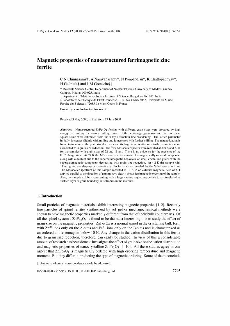
Figure 1. X-ray diffractograms (Fe Kα) of ZnFe2O4 milled for various times.
0 5 10 15 200
20
40
60
80
100
Ave
rage
gra
in s
ize
(nm
)
Milling Time (h)
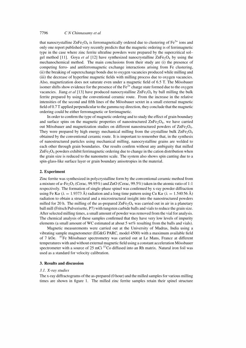
Figure 2. Average grain size of ZnFe2O4 powders versus milling time.
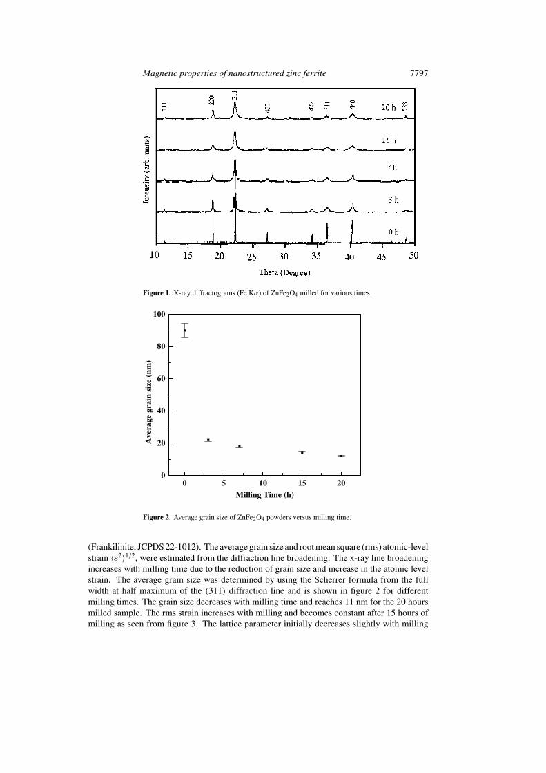
(Frankilinite, JCPDS 22-1012). The average grain size and root mean square (rms) atomic-levelstrain 〈ε2〉1/2, were estimated from the diffraction line broadening. The x-ray line broadeningincreases with milling time due to the reduction of grain size and increase in the atomic levelstrain. The average grain size was determined by using the Scherrer formula from the fullwidth at half maximum of the (311) diffraction line and is shown in figure 2 for differentmilling times. The grain size decreases with milling time and reaches 11 nm for the 20 hoursmilled sample. The rms strain increases with milling and becomes constant after 15 hours ofmilling as seen from figure 3. The lattice parameter initially decreases slightly with milling
7798 C N Chinnasamy et al
0 5 10 15 200.0
0.1
0.2
0.3
<ε2 >
1/2
Milling Time (h)
Figure 3. The root mean square strain versus milling time.
0 5 10 15 20
0.844
0.846
0.848
0.850
(14 nm)
(17 nm)(22 nm)
(90 nm)
(11 nm)
a (n
m)
Milling time (h)
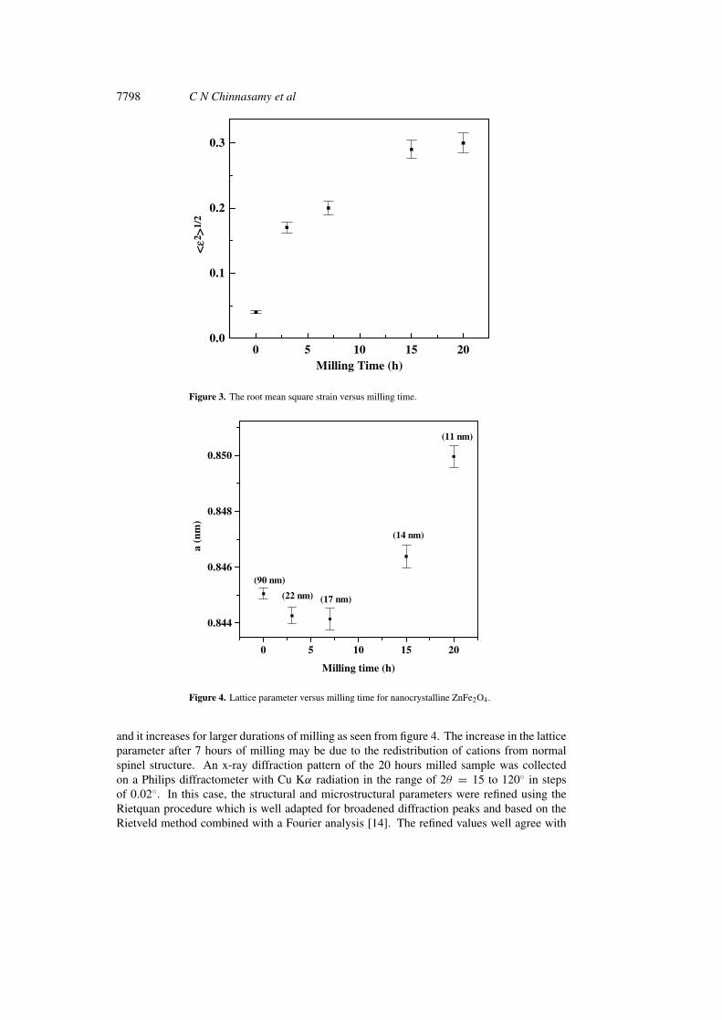
Figure 4. Lattice parameter versus milling time for nanocrystalline ZnFe2O4.
and it increases for larger durations of milling as seen from figure 4. The increase in the latticeparameter after 7 hours of milling may be due to the redistribution of cations from normalspinel structure. An x-ray diffraction pattern of the 20 hours milled sample was collectedon a Philips diffractometer with Cu Kα radiation in the range of 2θ = 15 to 120◦ in stepsof 0.02◦. In this case, the structural and microstructural parameters were refined using theRietquan procedure which is well adapted for broadened diffraction peaks and based on theRietveld method combined with a Fourier analysis [14]. The refined values well agree with
Magnetic
propertiesofnanostructured
zincferrite
7799
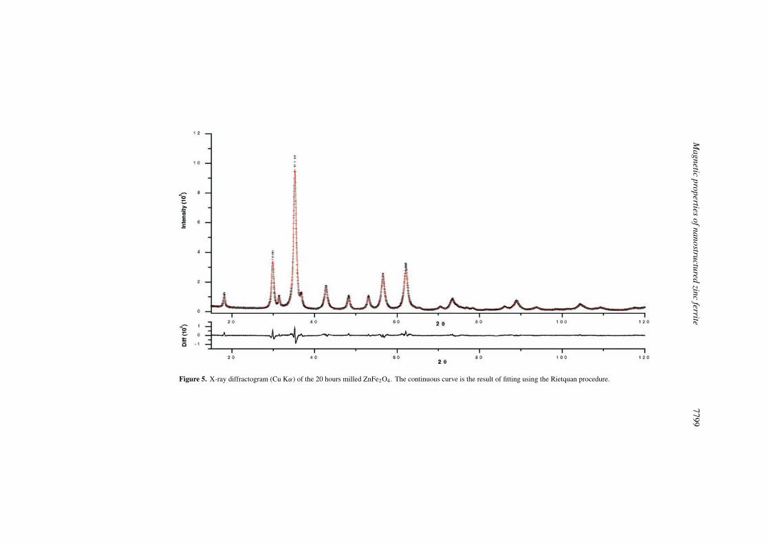
Figure 5. X-ray diffractogram (Cu Kα) of the 20 hours milled ZnFe2O4. The continuous curve is the result of fitting using the Rietquan procedure.
7800 C N Chinnasamy et al
-8000 -4000 0 4000 8000
-10
-5
0
5
10
15
* 11 nm - 900oC annealed (5 h)
*90 nm
22 nm
17 nm
14 nm
11 nm
Ma
gn
etis
ati
on
(em
u/g
m)
Applied Field (Oe)
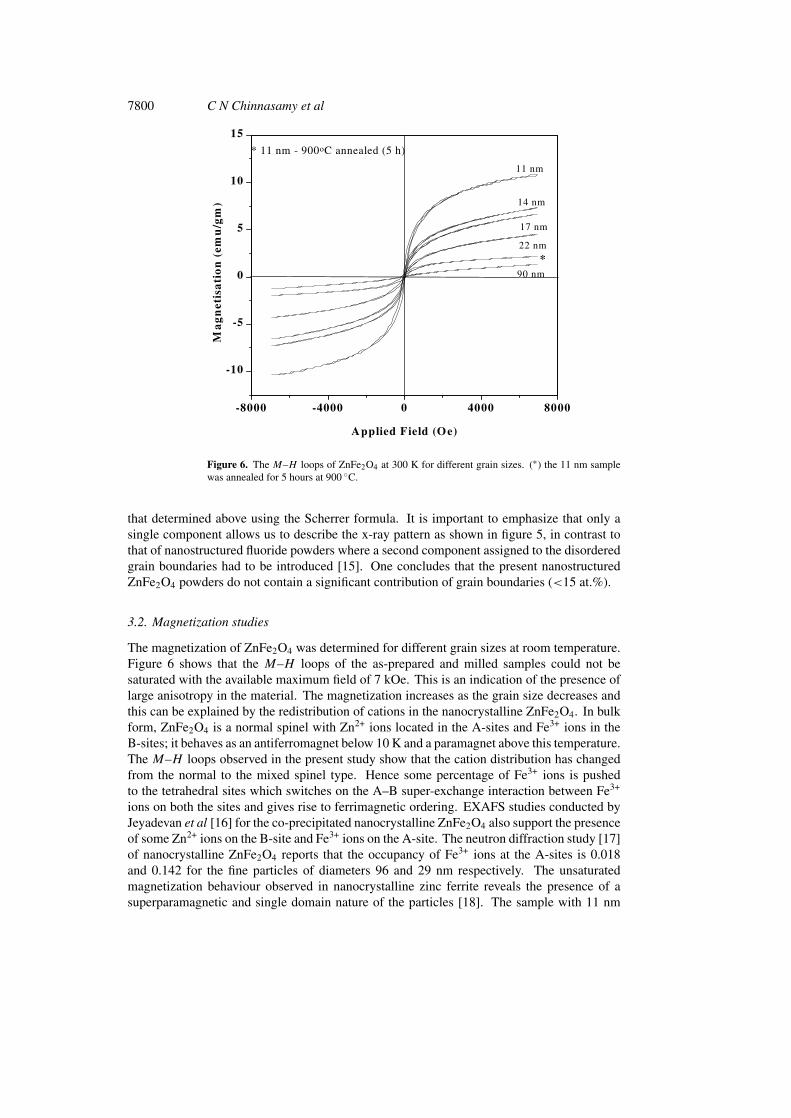
Figure 6. The M–H loops of ZnFe2O4 at 300 K for different grain sizes. (∗) the 11 nm samplewas annealed for 5 hours at 900 ◦C.
that determined above using the Scherrer formula. It is important to emphasize that only asingle component allows us to describe the x-ray pattern as shown in figure 5, in contrast tothat of nanostructured fluoride powders where a second component assigned to the disorderedgrain boundaries had to be introduced [15]. One concludes that the present nanostructuredZnFe2O4 powders do not contain a significant contribution of grain boundaries (<15 at.%).
3.2. Magnetization studies
The magnetization of ZnFe2O4 was determined for different grain sizes at room temperature.Figure 6 shows that the M–H loops of the as-prepared and milled samples could not besaturated with the available maximum field of 7 kOe. This is an indication of the presence oflarge anisotropy in the material. The magnetization increases as the grain size decreases andthis can be explained by the redistribution of cations in the nanocrystalline ZnFe2O4. In bulkform, ZnFe2O4 is a normal spinel with Zn2+ ions located in the A-sites and Fe3+ ions in theB-sites; it behaves as an antiferromagnet below 10 K and a paramagnet above this temperature.The M–H loops observed in the present study show that the cation distribution has changedfrom the normal to the mixed spinel type. Hence some percentage of Fe3+ ions is pushedto the tetrahedral sites which switches on the A–B super-exchange interaction between Fe3+
ions on both the sites and gives rise to ferrimagnetic ordering. EXAFS studies conducted byJeyadevan et al [16] for the co-precipitated nanocrystalline ZnFe2O4 also support the presenceof some Zn2+ ions on the B-site and Fe3+ ions on the A-site. The neutron diffraction study [17]of nanocrystalline ZnFe2O4 reports that the occupancy of Fe3+ ions at the A-sites is 0.018and 0.142 for the fine particles of diameters 96 and 29 nm respectively. The unsaturatedmagnetization behaviour observed in nanocrystalline zinc ferrite reveals the presence of asuperparamagnetic and single domain nature of the particles [18]. The sample with 11 nm
Magnetic properties of nanostructured zinc ferrite 7801
0,92
0,94
0,96
0,98
1,00-4 -2 0 2 4
V (mm/s)
20h 300K
Tra
nsm
issi
on-8 -4 0 4 8
0,97
0,98
0,99
1,00
V (mm/s)
20h 77K
-4 -2 0 2 4
0,90
0,92
0,94
0,96
0,98
V (mm/s)
7h 300K
-8 -4 0 4 80,90
0,92
0,94
0,96
0,98
7h 77K
V (mm/s)
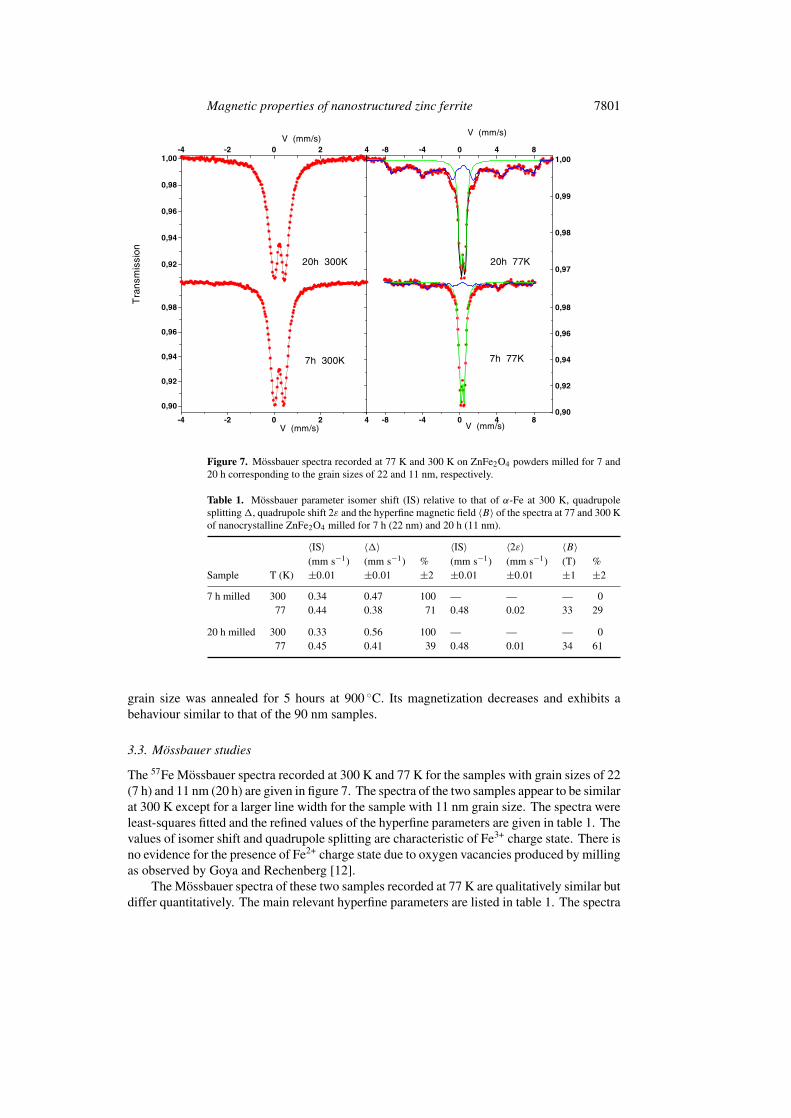
Figure 7. Mossbauer spectra recorded at 77 K and 300 K on ZnFe2O4 powders milled for 7 and20 h corresponding to the grain sizes of 22 and 11 nm, respectively.
Table 1. Mossbauer parameter isomer shift (IS) relative to that of α-Fe at 300 K, quadrupolesplitting �, quadrupole shift 2ε and the hyperfine magnetic field 〈B〉 of the spectra at 77 and 300 Kof nanocrystalline ZnFe2O4 milled for 7 h (22 nm) and 20 h (11 nm).
〈IS〉 〈�〉 〈IS〉 〈2ε〉 〈B〉(mm s−1) (mm s−1) % (mm s−1) (mm s−1) (T) %
Sample T (K) ±0.01 ±0.01 ±2 ±0.01 ±0.01 ±1 ±2
7 h milled 300 0.34 0.47 100 — — — 077 0.44 0.38 71 0.48 0.02 33 29
20 h milled 300 0.33 0.56 100 — — — 077 0.45 0.41 39 0.48 0.01 34 61
grain size was annealed for 5 hours at 900 ◦C. Its magnetization decreases and exhibits abehaviour similar to that of the 90 nm samples.
3.3. Mossbauer studies
The 57Fe Mossbauer spectra recorded at 300 K and 77 K for the samples with grain sizes of 22(7 h) and 11 nm (20 h) are given in figure 7. The spectra of the two samples appear to be similarat 300 K except for a larger line width for the sample with 11 nm grain size. The spectra wereleast-squares fitted and the refined values of the hyperfine parameters are given in table 1. Thevalues of isomer shift and quadrupole splitting are characteristic of Fe3+ charge state. There isno evidence for the presence of Fe2+ charge state due to oxygen vacancies produced by millingas observed by Goya and Rechenberg [12].
The Mossbauer spectra of these two samples recorded at 77 K are qualitatively similar butdiffer quantitatively. The main relevant hyperfine parameters are listed in table 1. The spectra
7802 C N Chinnasamy et al
consist of a magnetically ordered component and a quadrupole doublet and the latter may bedue to the superparamagnetic behaviour of small particles. For the samples with smaller grainsize, the relative intensity of the magnetically ordered component is greater (61%) than thatof the other sample (29%) with a larger grain size. This is a clear evidence for the increase ofthe cation inversion parameter with decrease of grain size.
Prolonged ball milling induces ferrimagnetic moment at 300 K as is evident from figure 6but magnetically ordered Mossbauer spectra are obtained only from 77 K. The observeddifference between the two measurements is due to the magnetic relaxations in the smallgrains, namely superparamagnetism and collective magnetic excitations. The magnetic fieldapplied in the magnetization measurements greatly suppresses the relaxation leading to themeasurement of non-zero magnetic moment whereas a broad quadrupolar doublet is obtainedin the Mossbauer spectrum due to superparamagnetic behaviour of the sample. At lowtemperatures the relaxation effects are slowed down and a magnetically ordered Mossbauerspectrum is observed. Similar results have been obtained by Hamdeh et al [4] also.
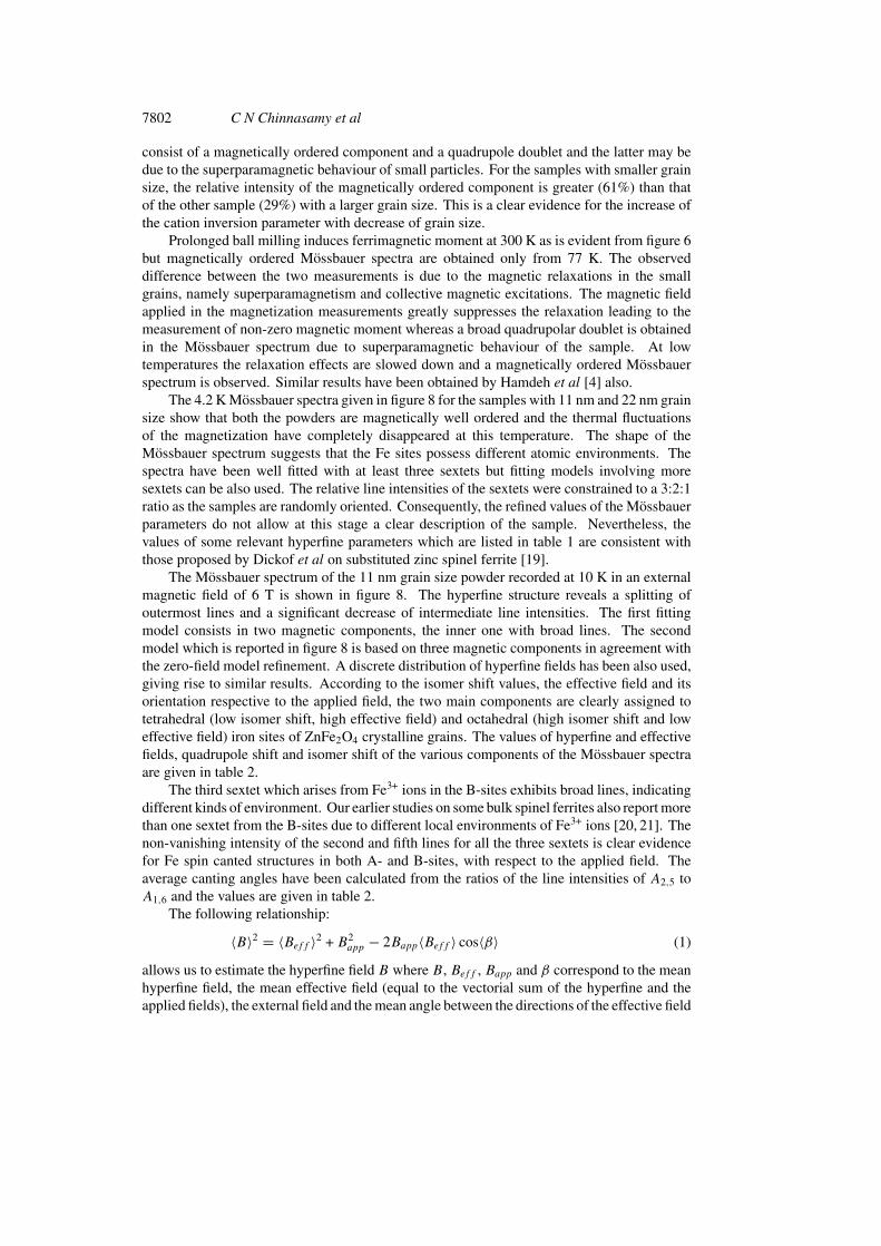
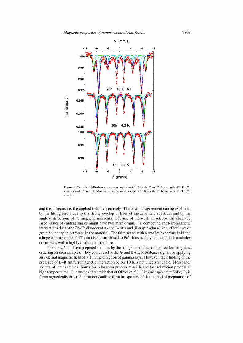
The 4.2 K Mossbauer spectra given in figure 8 for the samples with 11 nm and 22 nm grainsize show that both the powders are magnetically well ordered and the thermal fluctuationsof the magnetization have completely disappeared at this temperature. The shape of theMossbauer spectrum suggests that the Fe sites possess different atomic environments. Thespectra have been well fitted with at least three sextets but fitting models involving moresextets can be also used. The relative line intensities of the sextets were constrained to a 3:2:1ratio as the samples are randomly oriented. Consequently, the refined values of the Mossbauerparameters do not allow at this stage a clear description of the sample. Nevertheless, thevalues of some relevant hyperfine parameters which are listed in table 1 are consistent withthose proposed by Dickof et al on substituted zinc spinel ferrite [19].
The Mossbauer spectrum of the 11 nm grain size powder recorded at 10 K in an externalmagnetic field of 6 T is shown in figure 8. The hyperfine structure reveals a splitting ofoutermost lines and a significant decrease of intermediate line intensities. The first fittingmodel consists in two magnetic components, the inner one with broad lines. The secondmodel which is reported in figure 8 is based on three magnetic components in agreement withthe zero-field model refinement. A discrete distribution of hyperfine fields has been also used,giving rise to similar results. According to the isomer shift values, the effective field and itsorientation respective to the applied field, the two main components are clearly assigned totetrahedral (low isomer shift, high effective field) and octahedral (high isomer shift and loweffective field) iron sites of ZnFe2O4 crystalline grains. The values of hyperfine and effectivefields, quadrupole shift and isomer shift of the various components of the Mossbauer spectraare given in table 2.
The third sextet which arises from Fe3+ ions in the B-sites exhibits broad lines, indicatingdifferent kinds of environment. Our earlier studies on some bulk spinel ferrites also report morethan one sextet from the B-sites due to different local environments of Fe3+ ions [20, 21]. Thenon-vanishing intensity of the second and fifth lines for all the three sextets is clear evidencefor Fe spin canted structures in both A- and B-sites, with respect to the applied field. Theaverage canting angles have been calculated from the ratios of the line intensities of A2,5 toA1,6 and the values are given in table 2.
The following relationship:
〈B〉2 = 〈Beff 〉2 + B2app − 2Bapp〈Beff 〉 cos〈β〉 (1)
allows us to estimate the hyperfine field B where B, Beff , Bapp and β correspond to the meanhyperfine field, the mean effective field (equal to the vectorial sum of the hyperfine and theapplied fields), the external field and the mean angle between the directions of the effective field
Magnetic properties of nanostructured zinc ferrite 7803
-12 -8 -4 0 4 8 12
0,98
0,99
1,00
7h 4.2 K
Tra
nsm
issi
on
V (mm/s)
0,985
0,990
0,995
20h 4.2 K
0,97
0,98
0,99
1,00
-12 -8 -4 0 4 8 12
V (mm/s)
20h 10 K 6T
Figure 8. Zero-field Mossbauer spectra recorded at 4.2 K for the 7 and 20 hours milled ZnFe2O4samples and 6 T in-field Mossbauer spectrum recorded at 10 K for the 20 hours milled ZnFe2O4sample.
and the γ -beam, i.e. the applied field, respectively. The small disagreement can be explainedby the fitting errors due to the strong overlap of lines of the zero-field spectrum and by theangle distributions of Fe magnetic moments. Because of the weak anisotropy, the observedlarge values of canting angles might have two main origins: (i) competing antiferromagneticinteractions due to the Zn–Fe disorder at A- and B-sites and (ii) a spin-glass-like surface layer orgrain boundary anisotropies in the material. The third sextet with a smaller hyperfine field anda large canting angle of 45◦ can also be attributed to Fe3+ ions occupying the grain boundariesor surfaces with a highly disordered structure.
Oliver et al [11] have prepared samples by the sol–gel method and reported ferrimagneticordering for their samples. They could resolve the A- and B-site Mossbauer signals by applyingan external magnetic field of 7 T in the direction of gamma rays. However, their finding of thepresence of B–B antiferromagnetic interaction below 10 K is not understandable. Mossbauerspectra of their samples show slow relaxation process at 4.2 K and fast relaxation process athigh temperatures. Our studies agree with that of Oliver et al [11] in one aspect that ZnFe2O4 isferromagnetically ordered in nanocrystalline form irrespective of the method of preparation of
7804 C N Chinnasamy et al
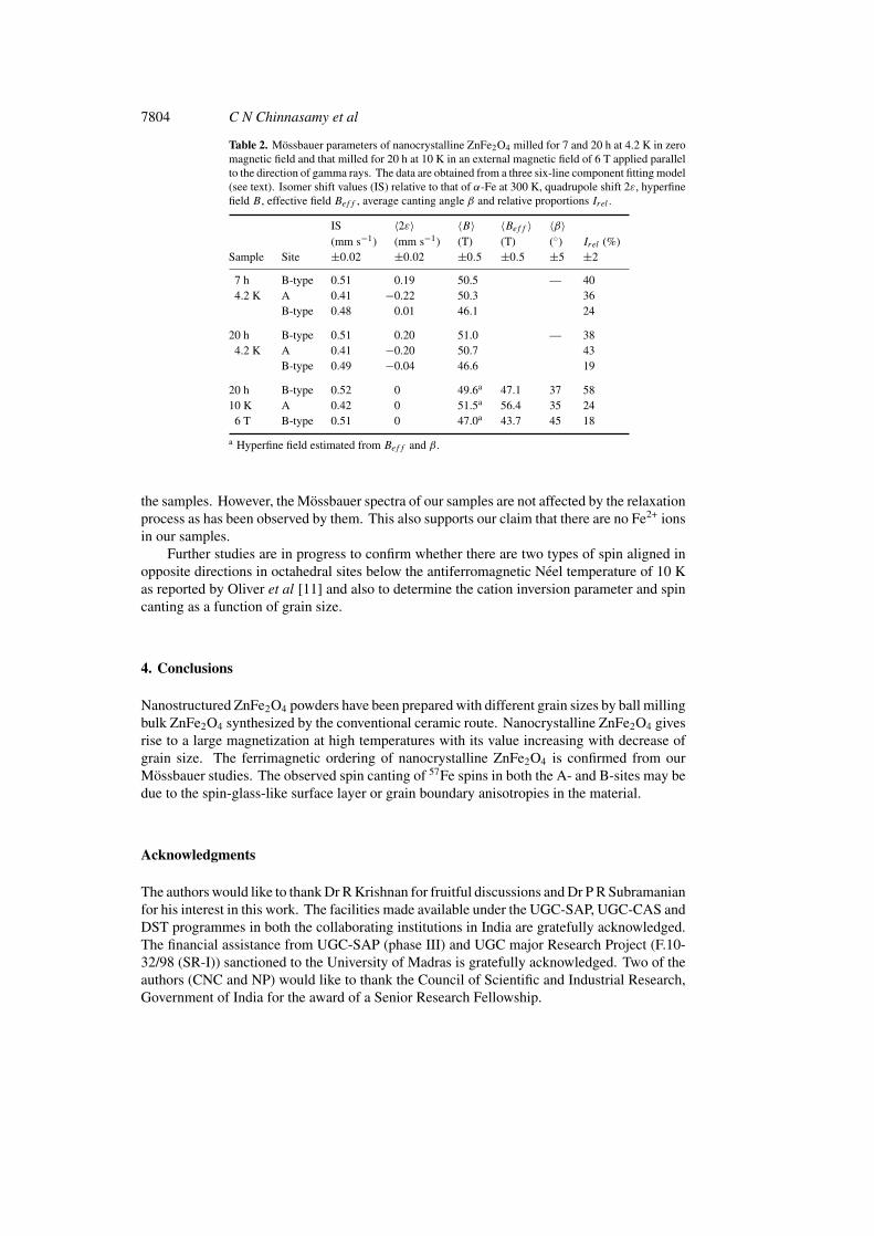
Table 2. Mossbauer parameters of nanocrystalline ZnFe2O4 milled for 7 and 20 h at 4.2 K in zeromagnetic field and that milled for 20 h at 10 K in an external magnetic field of 6 T applied parallelto the direction of gamma rays. The data are obtained from a three six-line component fitting model(see text). Isomer shift values (IS) relative to that of α-Fe at 300 K, quadrupole shift 2ε, hyperfinefield B, effective field Beff , average canting angle β and relative proportions Irel .
IS 〈2ε〉 〈B〉 〈Beff 〉 〈β〉(mm s−1) (mm s−1) (T) (T) (◦) Irel (%)
Sample Site ±0.02 ±0.02 ±0.5 ±0.5 ±5 ±2
7 h B-type 0.51 0.19 50.5 — 404.2 K A 0.41 −0.22 50.3 36
B-type 0.48 0.01 46.1 24
20 h B-type 0.51 0.20 51.0 — 384.2 K A 0.41 −0.20 50.7 43
B-type 0.49 −0.04 46.6 19
20 h B-type 0.52 0 49.6a 47.1 37 5810 K A 0.42 0 51.5a 56.4 35 24
6 T B-type 0.51 0 47.0a 43.7 45 18
a Hyperfine field estimated from Beff and β.
the samples. However, the Mossbauer spectra of our samples are not affected by the relaxationprocess as has been observed by them. This also supports our claim that there are no Fe2+ ionsin our samples.
Further studies are in progress to confirm whether there are two types of spin aligned inopposite directions in octahedral sites below the antiferromagnetic Neel temperature of 10 Kas reported by Oliver et al [11] and also to determine the cation inversion parameter and spincanting as a function of grain size.
4. Conclusions
Nanostructured ZnFe2O4 powders have been prepared with different grain sizes by ball millingbulk ZnFe2O4 synthesized by the conventional ceramic route. Nanocrystalline ZnFe2O4 givesrise to a large magnetization at high temperatures with its value increasing with decrease ofgrain size. The ferrimagnetic ordering of nanocrystalline ZnFe2O4 is confirmed from ourMossbauer studies. The observed spin canting of 57Fe spins in both the A- and B-sites may bedue to the spin-glass-like surface layer or grain boundary anisotropies in the material.
Acknowledgments
The authors would like to thank Dr R Krishnan for fruitful discussions and Dr P R Subramanianfor his interest in this work. The facilities made available under the UGC-SAP, UGC-CAS andDST programmes in both the collaborating institutions in India are gratefully acknowledged.The financial assistance from UGC-SAP (phase III) and UGC major Research Project (F.10-32/98 (SR-I)) sanctioned to the University of Madras is gratefully acknowledged. Two of theauthors (CNC and NP) would like to thank the Council of Scientific and Industrial Research,Government of India for the award of a Senior Research Fellowship.
Magnetic properties of nanostructured zinc ferrite 7805
References
[1] Tkacovba K, Sepelak V, Sterklora N and Noldyrev V V 1996 J. Solid State Chem. 123 100[2] Chinnasamy C N, Narayanasamy A, Ponpandian N and Chattopadhyay K 2000 Mater. Sci. Eng. A at press[3] Schissel W et al 1996 Phys. Rev. B 53 9143[4] Hamdeh H H, Ho J C, Oliver S A, Wiley R J, Oliveri G and Busca G 1997 J. Appl. Phys. 81 1851[5] Clark Ted M and Evans B J 1997 IEEE Trans. Magn. 33 3745[6] Yokayama M, Ohta E, Sato T, Komaba T and Sato T 1997 J. Physique Coll. IV 7 C1 521[7] Battle J, Clark T and Evans B J 1997 J. Physique Coll. IV 7 C1 257[8] Hamdeh H H, Ho J C, Oliver S A, Wiley R J, Kramer J, Chen Y Y, Lin S H, Yao Y D, Datiro M and Busca G
1995 IEEE Trans. Magn. 31 3808[9] Sato T, Haneda K, Seki M and Iijima T 1990 Appl. Phys. A 50 13
[10] Anantharaman M R, Jagatheesan S, Malini K A, Sindhu S, Narayanasamy A, Chinnasamy C N, Jacops J P,Reijne S, Seshan H and Brongersma R H H 1998 J. Magn. Magn. Mater. 189 83
[11] Oliver S A, Hamdeh H H and Ho J C 1999 Phys. Rev. B 60 3400[12] Goya G F and Rechenberg H R 1999 J. Magn. Magn. Mater. 196 191[13] Jiang J Z, Wynn P, Morup S, Okada T and Berry F J 1999 Nanostruct. Mater. 12 737[14] Lutterotti L and Scardi P 1990 J. Appl. Cryst. 23 246[15] Guerault H and Greneche J M 2000 J. Phys.: Condens. Matter 12 4791[16] Jeyadevan B, Tohiji K and Nakatsuka K 1994 J. Appl. Phys. 76 6325[17] Lotzering F K 1996 J. Phys. Chem. Solids 27 139[18] Bean C P and Livingston J D 1959 J. Appl. Phys. 30 120S[19] Dickof P A, Schurer P J and Morrish A H 1980 Phys. Rev. B 22 115[20] Narayanasamy A and Haggstrom L 1983 J. Phys. C: Solid State Phys. 16 591[21] Sundararajan M D, Narayanasamy A, Nagarajan T, Haggstrom L, Swamy C S and Ramanujachary K V 1984
J. Phys. C: Solid State Phys. 17 2953