macromolecules containing metal and metal-like elements || supramolecular structures and functions...
TRANSCRIPT

CHAPTER 1
Supramolecular Structuresand Functions with InorganicBuilding Blocks
Katsuhiko Ariga, Ajayan Vinu, Jonathan P. Hill,
Pavuluri Srinivasu, Somobrata Acharya, and Qingmin Ji
World Premier International (WPI) Research Centerfor Materials Nanoarchitectonics (MANA), NationalInstitute for Materials Science (NIMS), Japan
CONTENTS
I. INTRODUCTION 2
II. HYBRID LIPID THIN FILMS 2
III. LAYER-BY-LAYER ASSEMBLIES 8
IV. STRUCTURE TRANSCRIPTION 13
V. FUNCTIONAL MESOPOROUS HYBRIDS 20
VI. FUTURE PERSPECTIVES 30
VII. ACKNOWLEDGMENTS 30
VIII. REFERENCES 30
Macromolecules Containing Metal and Metal-Like Elements,Volume 9: Supramolecular and Self-Assembled Metal-Containing Materials,
Edited by Alaa S. Abd-El Aziz, Charles E. Carraher Jr., Charles U. Pittman Jr., and Martel Zeldin.Copyright r 2009 John Wiley & Sons, Inc.
1

I. INTRODUCTION
Fabrication of nanometer-scale structures for ultra-small devices has beenpaid a great deal of attention because small machines could provide manysocietal benefits, such as optimization of energy consumption and wastedischarge. Several excellent top-down approaches, including photolithographyand electron-beam lithography, have been so far used to provide fine micro-structures. However, these methods have several drawbacks, including sizelimitation and the paucity of applicable materials. Therefore, an alternate means,known as the bottom-up approach, has been recently intensively investigated.Methods of bottom-up fabrication rely on molecular self-assembly in supramo-lecular processes. Supramolecular chemistry, which was originally a branch offundamental science, has now become an important concept in nanotechnology.
Most of the well-known or well-investigated supramolecular objects areassembled from organic molecules. The class of organic molecules possesses hugestructural diversity and thus can exhibit many interesting chemical, physical, andbiological functions. Probably the main drawback presented by organic species istheir relative physical instability under the harsh conditions currently required forprecise fabrication. This is in contrast to the high mechanical strengths and stabili-ties of most inorganic substances, which make them suitable for material applica-tions. Unfortunately, inorganic materials have not been well investigated in termsof their potential in supramolecular chemistry. A combination of the two worlds oforganic supramolecular chemistry and inorganic materials science should createuseful nano-structured materials concurrently possessing various functionality,fine structural precision, and high mechanical stability. Supramolecular nano-fabrication with inorganic building blocks, including organic–inorganicnano-hybrids and structure-transcribed materials, could be one of the mostpowerful methodologies for satisfying the current demands of nanotechnology.
As has been widely reported, metal-organic complexes, including metal-organic framework materials, are representative examples of supermoleculescontaining inorganic building blocks.1,2 Because this subject has been exten-sively discussed in many forms, we here concentrate on supramolecular objectsconsisting of inorganic building blocks that are based on soft supramolecularassemblies. In this chapter, various recent topics in hybrid lipid thin films,layer-by-layer assemblies, structure transcription, and functional mesoporoushybrids are described (Fig. 1). Of course, not all of the recent work can beintroduced here but the reader should be able to sense the essential recenttrends in the corresponding fields from those examples that have been selected.
II. HYBRID LIPID THIN FILMS
Lipid thin films have been used in many applications, such as permea-tion control, drug delivery, and other medical applications because of their
2 Supramolecular Structures and Functions with Inorganic Building Blocks

biocompatibility and ease of control under physicochemical conditions. How-ever, a limited mechanical strength is sometimes apparent so that the hybrids oflipid thin films and strong inorganic frameworks are anticipated. In this section,several examples of lipid-inorganic hybrid thin films are described.
As stable lipid monolayer structures, self-assembled monolayers (SAMs)have been used widely where covalent bonding between the monolayer andthe substrate afford a satisfactory mechanical strength. Typically, organosi-lane compounds such as n-octadecyltrichlorosilane (OTS) have been used for thepreparation of SAM structures through the formation of covalent Si—O—Silinkages between hydrolyzed silane moieties and solid surfaces, such as glassplates or metal oxide layers. For example, Okahata and co-workers prepared aSAM of dialkylorganosilane on a porous glass plate, and successfully regulated
FIGURE 1. Supramolecular objects consist of inorganic building blocks mainly based
on soft supramolecular assemblies. (a) Hybrid lipid thin films. (b) Layer-by-layer
assemblies. (c) Structure transcription. (d) Functional mesoporous hybrid.
Hybrid Lipid Thin Films 3

material permeation through the glass.3 The SAM structures developed deeplywithin the pores, where permeation of molecules dissolved in water could beregulated by variation of the physical state of the SAM structure.
The same research group developed a more sophisticated technique torealize the first example of permeation control through a single monolayer.4,5
To immobilize a single monolayer on a porous glass surface, the SAM conceptand the Langmuir-Blodgett (LB) technique were combined (Fig. 2). An organo-silane monolayer was first prepared and polymerized on an air–water interfaceand was then transferred for immobilization on a solid substrate. The surfacepressure-molecular area (p-A) isotherm of dialkylalkoxysilane depended signifi-cantly on the pH condition of the water subphase. Condensation at acidic pH 2induced the formation of a well-condensed monolayer with a limiting area of0.54 nm2, which is rather close to the corresponding values from the conven-tional monolayers of dialkyl compounds such as phospholipids. The LB filmsthat were prepared under acidic conditions showed peaks in the infrared spectraat 1026 and 1095 cm�1, which are assignable to Si—O—Si and indicate the pre-sence of an acid catalyst in the subphase, enabling the monolayer to polymerize.
For permeation control experiments, the polymerized monolayer wastransferred onto a porous glass with 5-nm-diameter pores. From the unit areaand degree of polymerization, the area occupied by a single polymer moleculewas estimated to be B150 nm2, which is apparently larger than the pore area ofthe 5-nm glass (20 nm2). The transferred monolayer was covalently immobilizedon the porous glass plate by heat treatment. As illustrated in Figure 3, the glassplate with the immobilized monolayer was attached to the bottom of a poly-ethylene tube and soaked in a 1-cm quartz cell. Permeation of a water-soluble
FIGURE 2. Technique for use of organosilane compounds.
4 Supramolecular Structures and Functions with Inorganic Building Blocks

fluorescent probe was estimated by the increase in fluorescence intensity of thelower solution. It is interesting that permeation through the monolayer exhibiteda discontinuity at around 451C (Fig. 3), which is very close to the phasetransition temperature (crystal state to liquid crystalline state) of the polymerizedmonolayer. This was the first example of permeation control using a 2-nm-thickmonolayer, and so far, it is the thinnest lipid film that operates as a permeationvalve. This demonstration of control of organic thin film permeation becamepossible only through hybridization of the fragile monolayer with the mechani-cally stable inorganic substrate.
Permeation control through monolayer structures has also been demon-strated using electrochemical means (Fig. 4). The same research group similarlyimmobilized monolayers of dialkyl organosilane compounds on SnO2 electro-des by forming covalent linkages; permeability of an electrochemically activeprobe (Fe(CN)6
4�/Fe(CN)63�) through the monolayer was monitored.6 This
monolayer-hybridized electrode suppressed dramatically the redox response ofthe Fe probe and was hardly affected by repeated measurements. In sharpcontrast, the electrode covered noncovalently with monolayers of conventionalfatty acids could not continuously block permeation of the probe molecule.Permeation profiles of Fe(CN)6
4� /Fe(CN)63� probe were evaluated at different
temperatures, indicating that the redox response of the probe increased
FIGURE 3. Permeation of water-soluble fluorescent probe through a glass plate with
the immobilized monolayer.
Hybrid Lipid Thin Films 5

drastically near the phase transition temperature. Maximum permeability to theprobe molecule near the phase transition temperature was probably due to alkylchain disorder during a period of unstable coexistence of crystalline and liquidcrystalline states of the monolayer phase. It is interesting that the addition ofalcohol molecules to the solution blocks the permeation of probe molecules,with a significant dependence on the identity of the added alcohol. It is likelythat the alcohol molecules fill the pinholes formed on the monolayer and shapediscrimination occurs upon alcohol insertion into the monolayer.
The concept of the hybrid electrode was extended to a vitamin-immobilizedsystem by Hisaeda and co-workers (Fig. 5), in which heptapropyl and heptaoctylesters of vitamin B12 derivatives with a Co(II) or Co(III) center were used.7
Investigation of p-A isotherms of these compounds with the alkoxysilanemonolayers suggested that stable accommodation of the vitamin B12 function-ality in the lipid monolayer can be achieved by the introduction of long chains atthe vitamin B12 core. The mixed monolayers of the long chain–substitutedvitamin B12 and organosilane amphiphile were transferred as an x-type LB filmonto a quartz plate and showed reasonable UV absorbance. Monolayerssimilarly immobilized on an indium-tin-oxide (ITO) electrode possessed a
FIGURE 4. Electrochemical investigation of permeation control through monolayer
structure.
6 Supramolecular Structures and Functions with Inorganic Building Blocks

Co(II)/Co(I) redox couple. This indicates that the modified electrode can be usedas a reactive electrode with a vitamin B12 function. To demonstrate thispossibility, the vitamin B12 derivative was hybridized into thicker films, obtainedby the sol-gel process.8 The modified electrode complex exhibited the Co(II)/Co(I) redox couple, and the controlled-potential electrolysis of benzyl bromide inaqueous solution containing 0.1M KCl afforded dehalogenated products,bibenzyl and toluene, with a total turnover number of W1000 for 1 h.
The form of silica-based hybrid lipid assemblies is not limited to planarthin films. Katagiri co-workers have developed the silica-based hybrid vesicle,cerasome (Fig. 6),9,10 which is quite distinct from the above-mentionedapproaches. The unusual term cerasome originates from ‘‘ceramics + soma’’by analogy with liposome (‘‘lipo + soma’’). The cerasome was prepared byvortex mixing of aqueous dispersions of a silane-bearing amphiphile (Si-lipid 1or Si-lipid 2; Fig. 6). Formation of a silica network at the polar head group uponsol-gel reaction together with spontaneous formation of a bilayer structureresulted in cell-like vesicle structures containing a silica network at the outer andinner surfaces. Transmission electron microscopy (TEM) confirmed the forma-tion of multilamellar vesicles (Fig. 7) with a diameter B 200 nm. Aggregated
FIGURE 5. Vitamin B12-immobilized monolayer.
Hybrid Lipid Thin Films 7

vesicle structures were often observed without structural collapse (Fig. 7b),suggesting the superior structural stability of cerasome due to the inorganicframework structure formed at the surface.
III. LAYER-BY-LAYER ASSEMBLIES
To obtain structure-controlled nano-hybrids, preparation of layeredstructures in designed sequences is one of the most practical solutions. Amongseveral methods for the preparation of layered structures, alternate layer-by-layer (LbL) assembly,11–13 which has been rapidly developed, is suitable forinclusion of a variety of organic and inorganic substances. The LbL assemblywas first realized and established by Decher and co-workers14 some time after asuggestive report by Iler.15 An outline of this technique is shown in Figure 8,illustrating that the LbL assembly is conducted mainly through electrostaticinteraction. Excess adsorption of the substances to an oppositely charged
FIGURE 6. Silica-based hybrid vesicle, cerasome.
8 Supramolecular Structures and Functions with Inorganic Building Blocks

surface leads to charge neutralization and resaturation and results in chargereversal. Therefore, continuous assembly between oppositely charged materialscan be conducted continuously with great freedom in the number of layers andlayering sequence. The variability in applicable materials is another of the mostpronounced advantages of this method. Applicable materials are not limited toconventional polyelectrolytes so that not only biochemical materials (including
FIGURE 7. (a) TEM images of cerasome, (b) Its assembly.
FIGURE 8. Outline of LbL assembly.
Layer-by-Layer Assemblies 9

proteins, DNA strands, and virus particles) but also charged inorganicsubstances (such as colloidal nanoparticles, clay, nanosheets, modified zeolitecrystals, two-dimensional perovskite, and polyoxometalates) have beenincluded. Furthermore, charged supramolecular assemblies, such as bolaam-phiphile monolayers, lipid bilayers, LB films, and stacked dye molecules, havealso been used in layer-by-layer approaches. Assemblies with other interactionssuch as hydrogen bonding, metal coordination, and charge transfer interactionshave also been demonstrated.
Typical examples of LbL films containing inorganic building blocks aregiven in Figure 9. The figure shows responses of a quartz crystal microbalance(QCM) on the assembling processes of three sizes of SiO2 particles (25, 45, and78 nm in diameter), where larger and smaller steps correspond to the adsorp-tion of SiO2 particles and poly(diallyldimethylammonium chloride) (PDDA),respectively.16,17 Stepwise regular increases in the film mass were observed forall kinds of SiO2 particles. Figure 10 displays scanning electron microscopy(SEM) images of the SiO2-PDDA LbL film. The SiO2 particles are closelypacked in the layer, but there is no long-range ordering.
This technique can be applied to LbL assembly of cerasomes because, likesilicas, they possess surface charge. Initial attempts entailed LbL assemblybetween cationic polyelectrolyte and anionic cerasomes with monitoring byusing QCM, and where reasonable frequency shifts in the QCM responsewere observed.18 Morphology of the assembled structure was also confirmed
FIGURE 9. QCM responses of the assembling processes of SiO2 particles with
polyelectrolytes. line a, a 25-nm SiO2 particle; line b, a 45-nm SiO2 particle; and line
c, a 78-nm SiO2 particle.
10 Supramolecular Structures and Functions with Inorganic Building Blocks

by atomic force microscopy (AFM). Appropriate design of the silane-bearingamphiphiles, for example Si-lipid 1 and Si-lipid 2, led to the successful prepara-tion of both cationic and anionic cerasomes, which enabled direct assemblyof cerasome particles, thereby avoiding use of interlayer polyelectrolytes(Fig. 11).19 Formation of a smaller size of cationic cerasomes (20–100 nm)
FIGURE 10. A SEM image of the SiO2-PDDA LbL film. (Reprinted with permission
from ref. 17.)
FIGURE 11. LbL assembly between cationic and anionic cerasomes.
Layer-by-Layer Assemblies 11

and a larger size of anionic cerasomes (70–300 nm) was confirmed by TEMobservation. Detailed QCM observation revealed large steps and small steps infrequency change corresponding to adsorption of the anionic and the cationiccerasome, respectively. AFM observations of the surface of the assembledstructures indicated close packing of the cerasome particles like a stonepavement, where the difference in the particle size for each layer clearlyconfirmed successful preparation of layered structures of the cationic andanionic cerasomes. The structures presented can be regarded as novel types ofartificial multicellular systems for use as bioreactors or biosensors. Furtherfunctionalization of the cerasome surface could be used for immobilization ofvarious biomolecules, such as enzymes and antibodies, to create novel bioor-ganic–inorganic nanohybrids.
Appropriate selection of solid supports for the LbL assembly enables usto prepare free-standing films through selective etching of the solid support(sacrificial layer) (Fig. 12). For example, Mamedov and Kotov demonstratedpreparation of free-standing LbL films through assembling films on a celluloseacetate layer that was selectively dissolved later.20 Tsukruk and co-workersused a similar strategy for fabrication of freely suspended, multilayerednanocomposite membranes containing gold nanoparticles.21–24 The films thatwere obtained possessed very stable micromechanical characteristics and asensitivity far surpassing any existing pressure sensors. Preparation of free-standing films of poly(ethylene oxide) (PEO) and poly(acrylic acid) (PAA),which interact with each other through hydrogen bonding, was realized byHammond and co-workers.25 In the films obtained, crystallization of PEO
FIGURE 12. Preparation of free-standing LbL films.
12 Supramolecular Structures and Functions with Inorganic Building Blocks

moieties was suppressed and exhibited properties advantageous for use asultrathin hydrogel membranes and solid-state polymer electrolytes.
The LbL technique is even useful for more advanced microfabrication.An excellent example was report by Lvov and co-workers26 and is summarizedin Figure 13, which illustrates the preparation of an LbL self-assembled ultra-thin micro-cantilever consisting of clay/polymer nanocomposites. The research-ers used sequenced procedures, including patterning, photo-treatment, etching,and LbL assembly. The same research group also proposed a microfluidicapproach for the complex formation of polyelectrolytes and polyelectrolyte/protein microstrips on a glass surface.27 These studies are excellent examples ofresearch into the development of organic–inorganic nanohybrids.
Use of the LbL technique is not restricted to the preparation of planarthin films. One of the most outstanding strategy modifications of the LbLtechnique involves assembly on colloidal particles followed by hollow capsuleformation. For example, Caruso and co-workers reported the formation ofhollow silica vesicles through LbL assembly on colloidal nanoparticle templates(Fig. 14).28 Polyelectrolytes and smaller silica particles were initially formed ona larger colloidal core, which was subsequently selectively destroyed. Calcina-tion of the hybrid vesicles resulted in a hollow vesicle composed of silica.Formation of controlled organic–inorganic layer structures on colloidal parti-cles by LbL assembly also provides media appropriate for investigation offundamental phenomena.
Decher and co-workers exploited the superiority of LbL assembly forstructural design and synthesis to understand energy transfer processes(Fig. 15).29 They prepared LbL films on gold colloids with a diameter of13 nm to fabricate metal-core polymer shell capsules, in which the fluorescentorganic dyes fluorescein isothiocyanate (FITC) and lissamine rhodamine B(LISS) are placed at various distances from the gold core. The gold nanocorequenches the fluorescence of the fluorescein and lissamine dyes situated in theouter polymer layers of the core-shell nanoparticles. Systematic investigationwith control of the distance between dye and metal core provides a directmethod to assess the effect of the metal core on the radiative rates, whichrevealed strongly distance-dependent fluorescence quenching and reduction inthe transition probability for radiative transitions by gold nanoparticles.
IV. STRUCTURE TRANSCRIPTION
Supramolecular structures self-assembled from organic molecules oftenprovide finely tuned structures. Transcription of these structures into mechani-cally stable inorganic substances should be a productive approach for thepreparation of novel functional materials. This concept is sometimes known asstructure transcription. Transcription of fibrous and tubular structures oforganic gels into inorganic substances have has been extensively reported by
Structure Transcription 13

FIGURE 13. Fabrication of LbL self-assembled ultra-thin microcantilever consisting
of clay/polymer nanocomposites.
14 Supramolecular Structures and Functions with Inorganic Building Blocks

groups lead by Shinkai, Shimizu, and Hanabusa.30–32 For example, thecompound shown in Figure 16 forms tubular structures in appropriate media,which can be transcribed as double-walled silica nanotubes through the sol-gelcondensation of the co-reactant tetraethyl orthosilicate, followed by calcinationtreatment.33 Shimizu and co-workers reported metal nanowire formation, bywhich a synthetic glycolipid N-(11-cis-octadecennoyl)-b-D-glucopyranosylaminewas employed for the fabrication of organic tubular structures with a circularhollow cylinder 30–50 nm wide.34 Gold or silver nanoparticles were firstencapsulated in the hollow cylinders of lipid nanotubes using capillary force,followed by removal of the organic components by firing the nanocomposite,which result in gold nanowires with controlled width.
Yamashita demonstrated the use of a biomolecular array for structuretranscription (Fig. 17).35 A Langmuir monolayer of ferritin, which is composedof 24 self-assembled peptide subunits and is capable of including iron oxide,
FIGURE 14. Hollow capsule formation by the LbL method.
Structure Transcription 15

was first transferred onto a hydrophobized silicon substrate to form a two-dimensional hexagonal array of ferritin. Destruction of organic components byUV/ozone treatment and heat treatment under hydrogen flow left iron particleswith diameters o 6 nm in a hexagonal arrangement. Supramolecular arraysprepared from inorganic substances can be used for device preparation.Immobilization of such arrayed structures, for instance as the gate part of atransistor, could lead to the preparation of a floating quantum dot gate transis-tor for multilevel logic gates capable of room temperature operation.
Apart from the previously mentioned approaches, regular inorganicstructures such as porous alumina can be also used as templates forpreparation of fine bioorganic structures. As depicted in Figure 18, LbLassembly of organic components such as polymers, lipids, and proteins withininorganic pores followed by selective removal of the inorganic componentsresults in the formation of self-standing tubular structures. Li et al. appliedthis concept to the fabrication of microtubes from biocomponents such as
FIGURE 15. LbL films on a gold colloid in which the fluorescent organic dyes
fluorescein isothiocyanate (FITC) and lissamine rhodamine B (LISS) are placed at
various distances from the gold core.
16 Supramolecular Structures and Functions with Inorganic Building Blocks

proteins and phospholipids.36 For example, the surface charge of humanserum albumin (HSA) can be modified by variation of pH to be either morepositive or more negative so that pure protein tubes can be prepared by theLbL assembly of HSA under controlled pH conditions. These synthetic HSAmicrotubes have smooth and clean surfaces with a wall thickness of B 30 nm.The length of the tubes is B 60 mm, and they possess a flexibility that istolerant to free bending.
One of the most popular and influential examples of structure transcrip-tion is given by the preparation of mesoporous materials. Mesoporous materialswith regular geometries have recently gained much attention because of theirgreat potential for practical uses such as catalysis, adsorption, separation,sensing, medical usages, environmental applications, and nanotechnology.37,38
Mesoporous silica, which is the most representative mesoporous structure, issynthesized by structure transcription using micelle supramolecular assemblies(Fig. 19a). In 1990, Kuroda and co-workers first reported the preparation ofmesoporous silica of uniform pore size distribution from the layered polysilicatekanemite (FSM-16; folded sheet materials).39,40 A significant breakthrough inmesoporous materials research came when Mobil scientists disclosed the M41Sfamily of materials, which have large uniform pore structures; high specificsurface areas; and specific pore volumes, including hexagonal-MCM-41,41,42
cubic-MCM-48,43 and lamellar-MCM-50.44 These pioneering discoveries were
FIGURE 16. Preparation of a double-walled silica nanotube through the sol-gel
reaction using an organic tube as a template.
Structure Transcription 17

followed by various other kinds of mesoporous materials—for example, hex-agonal mesoporous silica (HMS) prepared using neutral amine as the template45
and MSU-1 (Michigan State University) using polyethylene oxide as a struc-ture directing agent.46 The widely used, highly ordered, large pore mesoporous
FIGURE 18. Fabrication of microtubes from biocomponents using inorganic pore
structures.
FIGURE 17. Preparation of iron particles arrays from a Langmuir monolayer of ferritin.
18 Supramolecular Structures and Functions with Inorganic Building Blocks

silica-15, SBA-15 (Santa Barbara amorphous), with thicker pore walls and two-dimensional hexagonal structure, was prepared using an amphiphilic triblockcopolymer of poly(ethylene oxide) and poly(propylene oxide), P123 (Pluronic)as the structure: directing reagent in a highly acidic media.47,48 Mesocellularform (MCF) materials can be prepared by using triblock copolymer stabilizedoil in water, giving aerogel-like structures, which offer potential as catalystsupports and separation media.49 One recent and unique example introducedchirality as an important morphological aspect of mesoporous materials byusing a chiral surfactant template (Fig. 19b).50A novel synthetic N-acetyl-typealanine-based surfactant was used as the structure-directing reagent, resulting inmesoporous silica materials with regularly twisted rod-like structures ofdiameter 130–180 nm and length 1–6 mm. In the structure obtained, hexagon-ally aligned mesoscopic channels of diameter 2.2 nm were wound together in oneparticular direction.
Several other synthetic strategies have been used for the preparationof mesoporous materials with various components. In particular, production ofcarbon materials containing regular mesopores was begun in the late 1990sthrough a carbonization procedure using mesoporous silica as a regularlystructured template followed by removal of the template silica (Fig. 20).Mesoporous carbon structures were first reported by Ryoo using sucrose as acarbon source and mesoporous silicates, such as MCM-48, SBA-1, and SBA-15,
FIGURE 19. (a) General concept for synthesis of mesoporous silica. (b) Formation of
mesoporous silica materials with a chiral morphological aspect.
Structure Transcription 19

as template. These materials are now known as CMK-x.51,52 Hyeon et al.independently and somewhat later reported a similar approach to well-orderedmesoporous carbon materials with a different designation, SNU-x.53 Morerecently, Vinu and co-workers54 applied this concept to the synthesis ofmesoporous carbon nitride for which combined carbon and nitrogen sourceswere used (Fig. 20). They also pioneered a third method for the synthesis ofmesoporous materials, which they call the ‘‘elemental substitution method.’’55
In this method, component elements are substituted by other elements with theretention of the mesoporous structure. For example, they successfully realizedthe first synthesis of mesoporous boron nitride and mesoporous boron carbonnitride (Fig. 20).56
V. FUNCTIONAL MESOPOROUS HYBRIDS
The pore sizes of mesoporous materials are compatible with those ofcomplex organic molecules and biological materials. Therefore, supramolecularnanofabrication based on mesoporous materials, such as functionalization atthe interiors of mesopores, could be one of the best methods for the creation offunctional materials using media for supramolecular interactions within a well-defined geometry.
To modify the mesoporous materials with organic functional groups,the two major methods of co-condensation and grafting are usually used.57,58
In the co-condensation method, a silica source containing an organic functionalgroup is mixed with a conventional silica source, such as tetraethyl orthosilicate,
FIGURE 20. Syntheses of mesoporous carbon, mesoporous carbon nitride, mesopor-
ous boron nitride, and mesoporous boron carbon nitride.
20 Supramolecular Structures and Functions with Inorganic Building Blocks

during mesoporous silica synthesis (Fig. 21a). This method provides a homo-geneous distribution of organic functional groups within mesopores. Modificationof presynthesized mesoporous silica can be achieved by grafting and is also knownas postsynthesis modification (Fig. 21b). Organic functional groups can beintroduced by direct reaction of organosilanes at the silica surface, and differentfunctionalities can be then attached to the already introduced groups throughcovalent bonding and/or molecular recognition. Similarly, the preintroducedfunctionalities are often converted to other groups by chemical reaction. Suchfunctionalization of mesoporous silica has been used in the development ofvarious functional materials. One example is shown in Figure 22, in which photo-controlled regulation of drug storage and release from mesoporous silica wasachieved in the pioneering work by Fujiwara et al.59 They prepared MCM-41mesoporous silica, which was functionalized with photoactive coumarin that wasgrafted only at the pore outlet. Cholestane (guest drug) was incorporated into themesopores of silica upon exposure to the guest solution. Irradiation by UV light(W 310 nm) caused dimerization of coumarin for trapping of the guest cholestanein the stable form and blocked the leaching of the guest from the mesopores.The dimerized coumarin was cleaved upon irradiation at a different UVwavelength (�250 nm) and resulted in the release of the trapped cholestane.
Ariga and co-workers proposed the synthesis of a mesoporous silicastructure possessing amino acid residues densely packed in its pores using acarefully designed surfactant (Fig. 23).60 During the synthesis of the mesoporousstructure, an amphiphile containing a condensable alkoxysilane group was usedas a template and sol-gel reactions together with tetraethyl orthosilicate resul-ted in mesoporous silica whose channels are filled with the covalently attachedorganic groups (alanine residue) of the template. Cleavage and removal of the
FIGURE 21. Modification of mesoporous silica by (a) co-condensation and (b) grafting.
Functional Mesoporous Hybrids 21

alkyl tail by selective hydrolysis of the ester at the C-terminal leaves open poreswith a surface covalently functionalized by the alanine residue. In this process,the template behaves like a ‘‘lizard’’ because it could be said that it bites thesilica surface with the later cleavage of its tail, which is a defensive mechanismemployed by some species of lizard. Aida and co-workers used the hybridstructure for reactor applications and demonstrated the catalytic capability ofunhydrolyzed materials on the acetalization of ketones, such as cyclohexanone,in ethanol under mild conditions.61 According to their proposed mechanism,hydrophobic and hydrophilic reactants can be incorporated simultaneously intothe amphiphilic core–shell architecture of the immobilized rod-like micellewithin silica channels and then can be activated through hydrogen-bondinginteractions with the peptidic functionalities located at the core–shell interface.The lizard templating method is expected to be used for the fabrication ofmesoporous silica with a variety of biofunctional groups. It could provideartificial active sites for a wide range of biological functions.
Mesoporous pores are interesting media for confinement of functionalorganic molecules under restricted motion and orientation. In particular,confinement of polymeric substances is an attractive research subject because
FIGURE 22. Photo-controlled regulation of drug storage and release from mesoporous
silica.
22 Supramolecular Structures and Functions with Inorganic Building Blocks

the dimensions of many polymers far exceed the dimensions of mesopores,especially the diameter; therefore, their motion would be highly restricted,perhaps resulting in peculiar physicochemical properties. One example of directincorporation of organic polymers into mesoporous silica was reported bySchwartz and co-workers, who incorporated a poly[phenylenevinylene] deriva-tive from solution into a calcined mesoporous silica.62 They demonstrated thatenergy migration along the incorporated polymer is slower than Forster energytransfer between the polymer chains. Because there remains the difficulty ofdiffusion of large polymer molecules into the small mesopores, introductionof monomers and subsequent polymerization of them within the silica nano-channels should be a more reliable method for confinement of the polymers inthe mesopores. Ozin and co-workers reported ring-opening polymerization of[1]silaferrocenophane in MCM-41 to provide poly(ferrocenylsilanes).63 Pyro-lysis of the composite resulted in the formation of iron nanoparticles withsuperparamagnetic properties.
However, postloading methods do not always guarantee confinement ofthe materials. A more efficient method for confining polymeric structures
FIGURE 23. Lizard templating method. (Reprinted with permission from ref. 60.)
Functional Mesoporous Hybrids 23

within mesopores is the introduction of monomer moieties to the templatestructure because template surfactants are definitely located within the meso-pore (Fig. 24). For example, Aida and Tajima used a few kinds of hexadeca-diynyltrimethylammonium bromide as templates to synthesize mesoporoussilica containing poly(diacetylene) as micro-fibers.64 The electronic spectra ofthe silica-polymer composite that were obtained were red shifted possibly dueto confinement of the poly(diacetylene) and an elongated effective conjugationwithin the silica nanochannel. Lu et al. used oligo(ethyleneglycol)-functionalizeddiacetylenic surfactants as structure-directing reagents for mesoporous silica filmsthrough casting, spin-coating, or dip-coating methods.65 Aida and co-workersdeveloped poly(pyrrole)-containing mesoporous silica films.66 Differences wereapparent between the hexagonal and lamellar composites from their spectro-scopic behavior. The polypyrrole chains are highly constrained and insulatedwhen incorporated within hexagonal nanoscopic channels and the possibilityof the polarons recombining into bipolarons is significantly suppressed. Incontrast, the two-dimensional lamellar phase affords spatial freedom for elec-tron recombination. Similarly, Fuhrhop and co-workers reported thiophenepolymerization in mesoporous silica channels.67
Inorganic mesoporous channels also provide a unique environment forsupramolecular assembly. Aida and co-workers reported the first example ofimmobilization of one-dimensional columnar charge-transfer (CT) assembliesin mesoporous silica films through a sol-gel reaction with CT complexesof amphiphilic triphenylene donors and various acceptors (Fig. 25).68
FIGURE 24. Polymerization of template monomers within a mesoporous silica channel.
24 Supramolecular Structures and Functions with Inorganic Building Blocks

The prepared films are highly transparent and their apparent color can be tunedby varying the concentration and/or identity of the intercalating acceptor. It isinteresting that the donor/acceptor molar ratio can be adjusted over a widerange from 1:1 to 9:1, which is advantageous for exploration of their photo-conductive properties. The mesoporous silica wall segregates the individual CTcolumns so that the composite films display neither solvatochromism nor guestexchange activity and exhibit red-shifted absorption bands. The latter phenom-enon may reflect a long-range structural ordering.
Ariga and co-workers developed mesoporous silica with densely packedpeptide segments that are known as proteosilica (Fig. 26).69,70 This nano-composite material provides an asymmetric medium for photochemical reac-tions.71 Photochromic dye molecules such as spiropyran and diarylethene canbe doped into the chiral environments of the proteosilica films, from whichasymmetric photoreaction were demonstrated. Alanylalanine-type surfactantswere used as the host peptides, and the spiropyran guest was co-doped with thesurfactant at various mixing ratios. Isomerization between the spiropyran formand the merocyanine form can be repeated upon alternate irradiation of thefilms with visible light (420 nm) and UV light (280 nm), respectively. Circulardichroism (CD) spectra of the hybrid films containing 5 mol % of spiropyranwere measured. Only negligible CD signals originating from the guest could beobserved for the film containing the merocyanine form, implying that induced
FIGURE 25. Immobilization of one-dimensional columnar charge-transfer assemblies
within mesoporous silica channels.
Functional Mesoporous Hybrids 25

CD of the nonchiral merocyanine was not significant, even in the chiral peptideenvironment. In contrast, the film with the spiropyran form showed clear CDactivity in the region from 250 nm to 400 nm for the L-peptide host. A completemirror image of the CD spectrum was obtained when the D-peptide host wasused as the host surfactant, indicating that the CD signals from the guest weredriven by the chiral environment of the surrounding host. Alternate irradiationwith UV and visible light induced repeated changes in the CD spectra with asmall degradation in the intensity. The reported system could be applied fordevelopment of nondestructive memory devices.
Another unique approach for introduction of organic functionalities intoinorganic mesoporous frameworks can be seen in the synthesis of periodicmesoporous organosilicates (PMOs) (Fig. 27).72–74 This series of materialspossess organic functionalities within a silica framework in their mesoporousstructure and can be synthesized using organic molecules bearing multiplealkoxysilane groups, such as bis(triethoxysilyl)ethene and bis(triethoxysilyl)-benzene. Introduction of various organic components has been reported. One
FIGURE 26. (a) Structure of proteosilica. (b) Photochromic reaction within the
composite.
26 Supramolecular Structures and Functions with Inorganic Building Blocks

of the most impressive findings in PMO technology is formation of a crystallinepore wall.75 Inagaki et al. synthesized PMO materials using benzene-bridgedorganosilane, 1,4-bis(triethoxysilyl)benzene.75 The powder x-ray diffraction(XRD) patterns and a detailed TEM observation confirmed formation of acrystal-like pore-wall structure in which hydrophilic silicate layers and hydro-phobic benzene layers are alternately arrayed.
It is possible to immobilize large biomolecules such as proteins and DNAstrands within mesoporous materials to provide inorganic-bionanohybrids.These materials have been investigated by several research groups. Forexample, Vinu et al. undertook systematic research on protein adsorptiononto mesoporous materials including mesoporous silica and mesoporouscarbon. The results indicated that protein adsorption capability increased aspore volume and/or pore diameter of mesopore materials increased (Fig. 28).76–80
Of various physical parameters, the charge state of the proteins seriouslyaffected adsorption capacities. Proteins lose their net charge at their isoelectricpoint, and at this point most of the proteins exhibit maximum adsorptions.
FIGURE 27. Periodic mesoporous organosilicates with various organic functionalities.
Functional Mesoporous Hybrids 27

Also, hydrophobic mesoporous carbons often display superior adsorptioncapabilities to hydrophilic mesoporous silicates.
Careful selection of the template structure can provide geometricallyengineered carbon materials. Vinu and co-workers recently reported the syn-thesis of the novel nanocarbon, ‘‘carbon nanocages’’81,82 through templatesynthesis using large three-dimensional cage-type face-centered cubic mesopor-ous silica materials (KIT-5) as inorganic templates (Fig. 29). The specificsurface area and specific pore volume of carbon nanocage greatly exceed thosereported for conventional mesoporous carbon materials. Therefore, the capa-city for lysozyme adsorption on the carbon nanocage is much larger than thatobserved with mesoporous carbon CMK-3. The carbon nanocage also exhibitsexcellent capability for separation of small molecules.83 In Figure 30a, thesuperior adsorption capability of the carbon nanocage in the removal of adyestuff (Alizarin Yellow) is demonstrated. With the application of slightpressure, an aqueous solution was passed through a bed of the respective
FIGURE 28. Protein adsorption onto mesoporous carbon.
FIGURE 29. (a) Synthesis of the carbon nanocage. (b) Its TEM image. (Reprinted with
permission from ref. 83.)
28 Supramolecular Structures and Functions with Inorganic Building Blocks
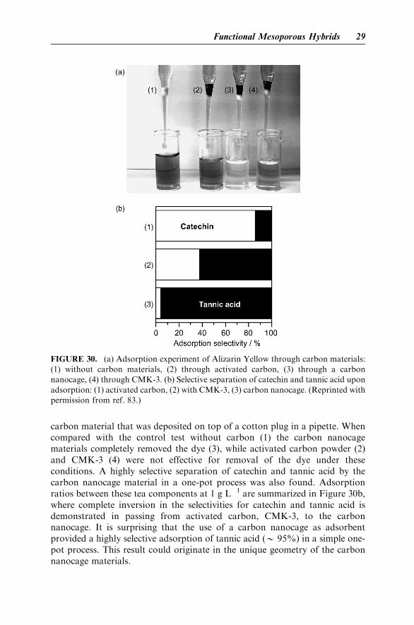
carbon material that was deposited on top of a cotton plug in a pipette. Whencompared with the control test without carbon (1) the carbon nanocagematerials completely removed the dye (3), while activated carbon powder (2)and CMK-3 (4) were not effective for removal of the dye under theseconditions. A highly selective separation of catechin and tannic acid by thecarbon nanocage material in a one-pot process was also found. Adsorptionratios between these tea components at 1 g L�1 are summarized in Figure 30b,where complete inversion in the selectivities for catechin and tannic acid isdemonstrated in passing from activated carbon, CMK-3, to the carbonnanocage. It is surprising that the use of a carbon nanocage as adsorbentprovided a highly selective adsorption of tannic acid (B 95%) in a simple one-pot process. This result could originate in the unique geometry of the carbonnanocage materials.
FIGURE 30. (a) Adsorption experiment of Alizarin Yellow through carbon materials:
(1) without carbon materials, (2) through activated carbon, (3) through a carbon
nanocage, (4) through CMK-3. (b) Selective separation of catechin and tannic acid upon
adsorption: (1) activated carbon, (2) with CMK-3, (3) carbon nanocage. (Reprinted with
permission from ref. 83.)
Functional Mesoporous Hybrids 29

VI. FUTURE PERSPECTIVES
In this chapter, we have described various examples of recent research onsupramolecular structure and function involving inorganic building blocks.Although these examples were limited to the four topics of hybrid lipid thinfilms, layer-by-layer assemblies, structure transcription, and functional meso-porous hybrid, a variety of possibilities in their structural designs and functionsare presented. These supramolecular structures will certainly contribute tofuture nanotechnologies. However, we not only must pursue dreams for the farfuture but also must demonstrate the importance of such supramolecularsystems for uses in the present. For example, the final topic of the final section,tea-component separation by using a nanoengineered carbon nanocage, is agood example for promoting the usefulness of supramolecular structures togeneral audiences. These demonstrations are related to potential real-lifeapplications and are of paramount importance for public understanding andsupport. Sciences and technology in these fields must function practically toensure improvements in our lifestyles and in our society at large.
VII. ACKNOWLEDGMENTS
The research studies described in this chapter were partially supported byGrant-in-Aid for Scientific Research in Priority Area Chemistry of Coordina-tion Space and a Grant-in-Aid for Science Research in Priority Area Super-Hierarchical Structures from the Ministry of Education, Science, Sports, andCulture, Japan, and a Grant-in-Aid for Scientific Research (B) from the JapanSociety for the Promotion of Science.
VIII. REFERENCES
1. O. M. Yaghi, M. O’Keeffe, N. W. Ockwig, H. K. Chae, M. Eddaoudi, and J. Kim,Nature, 2003, 423, 705.
2. S. Kitagawa, R. Kitaura, and S. Noro, Angew. Chem. Int. Ed., 2004, 43, 2334.
3. Y. Okahata, K. Ariga, and O. Shimizu, Langmuir, 1986, 2, 538.
4. Y. Okahata, K. Ariga, H. Nakahara, and K. Fukuda, J. Chem. Soc., Chem. Commun.,1986, 1069.
5. K. Ariga and Y. Okahata, J. Am. Chem. Soc., 1989, 111, 5618.
6. Y. Okahata, M. Yokobori, Y. Ebara, H. Ebato, and K. Ariga, Langmuir, 1990, 6, 1148.
7. K. Ariga, K. Tanaka, K. Katagiri, J. Kikuchi, H. Shimakoshi, E. Oshima, andY. Hisaeda, Phys. Chem. Chem. Phys., 2001, 3, 3442.
8. H. Shimakoshi, A. Nakazato, M. Tokunaga, K. Katagiri, K. Ariga, J. Kikuchi, andY. Hisaeda, Dalton Trans., 2003, 2308.
9. K. Katagiri, K. Ariga, and J. Kikuchi, Chem. Lett., 1999, 661.
30 Supramolecular Structures and Functions with Inorganic Building Blocks

10. K. Katagiri, M. Hashizume, K. Ariga, T. Terashima, and J. Kikuchi, Chem. Eur. J., 2007,13, 5272.
11. G. Decher, Science, 1997, 277, 1232.
12. P. T. Hammond, Adv. Mater., 2004, 16, 1271.
13. K. Ariga, J. P. Hill, and Q. Ji, Phys. Chem. Chem. Phys., 2007, 9, 2319.
14. G. Decher, J. D. Hong, and J. Schmitt, Thin Solid Films, 1992, 210, 831.
15. R. K. Iler, J. Colloid Interface. Sci., 1966, 21, 569.
16. K. Ariga, Y. Lvov, M. Onda, I. Ichinose, and T. Kunitake, Chem. Lett., 1997, 125.
17. Y. Lvov, K. Ariga, M. Onda, I. Ichinose, and T. Kunitake, Langmuir, 1997, 13, 6195.
18. K. Katagiri, R. Hamazaki, K. Ariga, and J. Kikuchi, Langmuir, 2002, 18, 6709.
19. K. Katagiri, R. Hamazaki, K. Ariga, and J. Kikuchi, J. Am. Chem. Soc., 2002, 124, 7892.
20. A. A. Mamedov and N. A. Kotov, Langmuir, 2000, 16, 5530.
21. C. Jiang, S. Markutsya, and V. V. Tsukruk, Adv. Mater., 2004, 16, 157.
22. C. Jiang, S. Markutsya, Y. Pikus, and V. V. Tsukruk, Nat. Mater., 2004, 3, 721.
23. S. Markutsya, C. Jiang, Y. Pikus and V. V. Tsukruk, Adv. Funct. Mater., 2005, 15, 771.
24. C. Jiang and V. V. Tsukruk, Adv. Mater., 2006, 18, 829.
25. J. L. Lutkenhaus, K. D. Hrabak, K. McEnnis, and P. T. Hammond, J. Am. Chem. Soc.,2005, 127, 17228.
26. F. Hua, T. Cui, and Y. M. Lvov, Nano Lett., 2004, 4, 823.
27. D. G. Shchukin, D. S. Kommireddy, Y. Zhao, T. Cui, G. B. Sukhorukov, and Y. M.Lvov, Adv. Mater., 2004, 16, 389.
28. F. Caruso, R. A. Caruso, and H. Mohwald, Science, 1998, 282, 1111.
29. G. Schneider, G. Decher, N. Nerambourg, R. Praho, M. H. V. Werts, and M. Blanchard-Desce, Nano Lett., 2006, 6, 530.
30. Y. Ono, K. Nakashima, M. Sano, Y. Kanekiyo, K. Inoue, J. Hojo, and S. Shinkai, Chem.Commun., 1998, 1477.
31. J. Hwa Jung, Y. Ono, K. Hanabusa, and S. Shinkai, J. Am. Chem. Soc., 2000, 122, 5008.
32. J. H. Jung, H. Kobayashi, M. Masuda, T. Shimizu, and S. Shinkai, J. Am. Chem. Soc.,2001, 123, 8785.
33. J. H. Jung, S.-H. Lee, J. S. Yoo, K. Yoshida, T. Shimizu, and S. Shinkai, Chem. Eur. J.,2003, 9, 5307.
34. B. Yang, S. Kamiya, Y. Shimizu, N. Koshizaki, and T. Shimizu, Chem. Mater., 2004, 16,2826.
35. I. Yamashita, Thin Solid Films, 2001, 393, 12.
36. G. Lu, S. Ai, and J. Li, Langmuir, 2005, 21, 1679.
37. A. Corma, Chem. Rev., 1997, 97, 2373.
38. M. E. Davis, Nature, 2002, 417, 813.
39. T. Yanagisawa, T. Shimizu, K. Kuroda, and C. Kato, Bull. Chem. Soc. Jpn., 1990,63, 988.
40. S. Inagaki, Y. Fukushima, and K. Kuroda, J. Chem. Soc., Chem. Commun., 1993, 680.
41. C. T. Kresge, M. E. Leonowicz, W. J. Roth, J. C. Vartuli, and J. S. Beck, Nature, 1992,359, 710.
42. J. S. Beck, J. C. Vartuli, W. J. Roth, M. E. Leonowicz, C. T. Kresge, K. D. Schmitt, C. T.W. Chu, D. H. Olson, E. W. Sheppard, S. B. McCullen, J. B. Higgins, and J. L.Schlenker, J. Am. Chem. Soc., 1992, 114, 10834.
43. J. C. Vartuli, K. D. Schmitt, C. T. Kresge, W. J. Roth, M. E. Leonowicz, S. B. McCullen,S. D. Hellring, J. S. Beck, J. L. Schlenker, D. H. Olson, and E. W. Sheppard, Chem.Mater., 1994, 6, 2317.
References 31

44. M. Dubois, T. Gulik-Krzywicki, and B. Cabane, Langmuir, 1993, 9, 673.
45. P. T. Tanev and T. J. Pinnavaia, Science, 1995, 267, 865.
46. S. A. Bagshaw, E. Prouset, and T. J. Pinnavaia, Science, 1995, 269, 1242.
47. D. Zhao, J. Feng, Q. Huo, N. Melosh, G. H. Fredickson, B. F. Chmelka, and G. D.Stucky, Science, 1998, 279, 548.
48. D. Zhao, Q. Huo, J. Feng, B. F. Chmelka, and G. D. Stucky, J. Am. Chem. Soc., 1998,120, 6024.
49. P. Schmidt-Winkel, W. W. Lukens, D. Zhao, P. Yang, B. F. Chmelka, and G. D. Stucky,J. Am. Chem. Soc., 1999, 121, 254.
50. S. Che, Z. Liu, T. Ohsuna, K. Sakamoto, O. Terasaki, and T. Tatsumi, Nature, 2004,429, 281.
51. R. Ryoo, S. H. Joo, and S. Jun, J. Phys. Chem. B, 1999, 103, 7743.
52. S. Jun, S. H. Joo, R. Ryoo, M. Kurk, M. Jaroniec, Z. Liu, T. Ohsuna, and O. Terasaki,J. Am. Chem. Soc., 2000, 122, 10712.
53. J. Lee, S. Yoon, T. Hyeon, S. M. Oh, and K. B. Kim, Chem. Commun., 1999, 2177.
54. A. Vinu, K. Ariga, T. Mori, T. Nakanishi, S. Hishita, D. Golberg, and Y. Bando, Adv.Mater., 2005, 17, 1648.
55. A. Vinu, T. Mori, and K. Ariga, Sci. Technol. Adv. Mater., 2006, 7, 753.
56. A. Vinu. M. Terrones, D. Golberg, S. Hishita, K. Ariga, and T. Mori, Chem. Mater.,2005, 17, 5887.
57. A. Vinu, K. Z. Hossain, and K. Ariga, J. Nanosci. Nanotechnol., 2005, 5, 347.
58. K. Ariga, A. Vinu, J. P. Hill, and T. Mori, Coord. Chem. Rev., 2007, 251, 2562.
59. N. K. Mal, M. Fujiwara, and Y. Tanaka, Nature, 2003, 421, 350.
60. Q. Zhang, K. Ariga, A. Okabe, and T. Aida, J. Am. Chem. Soc., 2004, 126, 988.
61. W. Otani, K. Kinbara, Q. Zhang, K. Ariga, and T. Aida, Chem. Eur. J., 2007, 13, 1731.
62. T.-Q. Nguyen, J. Wu, V. Doan, B. J. Schwartz, and S. H. Tolbert, Science, 2000, 288, 652.
63. M. J. MacLachlan, M. Ginzburg, N. Coombs, N. P. Raju, J. E. Greedan, G. A. Ozin, andI. Manners, J. Am. Chem. Soc., 2000, 122, 3878.
64. T. Aida and K. Tajima, Angew. Chem. Int. Ed., 2001, 40, 3803.
65. Y. Lu, Y. Yang, A. Sellinger, M. Lu, J. Huang, H. Fan, R. Haddad, G. Lopez, A. R.Burns, D. Y. Sasaki, J. Shelnutt, and C. J. Brinker, Nature, 2001, 410, 913.
66. M. Ikegame, K. Tajima, and T. Aida, Angew. Chem. Int. Ed., 2003, 42, 2154.
67. G. Li, S. Bhosale, T. Wang, Y. Zhang, H. Zhu, and J.-H. Fuhrhop, Angew. Chem. Int.Ed., 2003, 42, 3818.
68. A. Okabe, T. Fukushima, K. Ariga, and T. Aida, Angew. Chem. Int. Ed., 2002, 41, 3414.
69. K. Ariga, Q. Zhang, M. Niki, A. Okabe, and T. Aida, Stud. Surf. Sci. Catal., 2003,146, 427.
70. K. Ariga, Chem. Rec., 2004, 3, 297.
71. K. Ariga, T. Aimiya, Q. Zhang, A. Okabe, M. Niki, and T. Aida, Int. J. Nanosci., 2002,1, 521.
72. S. Inagaki, S. Guan, Y. Fukushima, T. Ohsuna, and O. Terasaki, J. Am. Chem. Soc.,1999, 121, 9611.
73. T. Asefa, M. J. MacLachlan, N. Coombs, and G. A. Ozin, Nature, 1999, 402, 867.
74. B. J. Melde, B. T. Holland, C. F. Blanford, and A. Stein, Chem. Mater., 1999, 11, 3302.
75. S. Inagaki, S. Guan, T. Ohsuna, and O. Terasaki, Nature, 2002, 416, 304.
76. A. Vinu, C. Streb, V. Murugesan, and M. Hartmann, J. Phys. Chem. B, 2003, 107, 8297.
77. A. Vinu, V. Murugesan, and M. Hartmann, J. Phys. Chem. B, 2004, 108, 7323.
32 Supramolecular Structures and Functions with Inorganic Building Blocks

78. A. Vinu, V. Murugesan, O. Tangermann, and M. Hartmann, Chem. Mater., 2004, 16,3056.
79. A. Vinu, M. Miyahara, and K. Ariga, J. Phys. Chem. B, 2005, 109, 6436.
80. A. Vinu, M. Miyahara, and K. Ariga, J. Nanosci. Nanotechnol., 2006, 6, 1510.
81. A. Vinu, M. Miyahara, V. Sivamurugan, T. Mori, and K. Ariga, J. Nanosci. Nanotech-nol., 2005, 15, 5122.
82. P. Srinivasu, V. V. Balasubramanian, L. Kumaresan, D. P. Sawant, X. Jin, S. Alam, K.Ariga, T. Mori, and A. Vinu, J. Nanosci. Nanotechnol., 2007, 7, 3250.
83. K. Ariga, A. Vinu, M. Miyahara, J. P. Hill, and T. Mori, J. Am. Chem. Soc., 2007, 129,11022.
References 33