m. basler author manuscript nih public access j. j ...faculty.ccbcmd.edu/~gkaiser/bacterial wow...
TRANSCRIPT

Type 6 secretion dynamics within and between bacterial cells
M. Basler1 and J. J. Mekalanos1,*
1Department of Microbiology and Immunobiology, Harvard Medical School, 200 LongwoodAvenue, Boston, MA 02115 U.S.A.
AbstractThe bacterial Type VI Secretion System (T6SS) functions as a virulence factor capable ofattacking both eukaryotic and prokaryotic target cells by a process that involves protein transportthrough a contractile bacteriophage tail-like structure. The T6SS apparatus is composed, in part, ofan exterior sheath wrapped around an interior tube. Here we report that in living cells thecytoplasmic ATPase called ClpV specifically recognizes the contracted T6SS sheath structurecausing its disassembly within seconds. ClpV imaging allowed spatial and temporaldocumentation of cell-cell interactions (termed "T6SS dueling") that likely mark the location ofrepeated T6SS-mediated protein translocation events between bacterial cells.
The bacterial Type 6 Secretion System (T6SS) is a dynamic apparatus that translocatesproteins from predator to prey cells by a mechanism analogous to phage tail contraction (1–3). In Vibrio cholerae, two proteins (VipA and VipB) build a phage tail sheath-like tubularstructure in the cytosol of predator cells that exists in two conformations, extended andcontracted (3). Contraction of the extended VipA/VipB sheath is thought to drive the T6SSspike and inner tube complex out of the effector or 'predator' cell and into an adjacent targetor 'prey' cell (3). Disassembly of the cytoplasmic contracted sheath requires ClpV in vivo(3), a AAA+ ATPase that binds VipA/VipB tubules in vitro and can remodel these structuresin the presence of ATP (4, 5). In Pseudomonas aeruginosa, ClpV1-GFP localizes to discretefoci that depend on T6SS function (6). Although ClpV binds VipA/VipB tubules in vitro (4,5), the ability of this protein to interact with other T6SS components has not beendemonstrated in vivo. Accordingly, we imaged ClpV localization in intact cells to examineits possible association with dynamic T6SS structures in vivo such as extended andcontracted T6SS sheaths and base plates.
We used time-lapse fluorescence microscopy to follow ClpV localization in live V. cholerae2740-80 cells. Functional ClpV-super folder GFP (sfGFP) and mCherry2 fusion proteinsassembled at random times into short structures that disappeared in 10’s of seconds. In theΔVipA background, ClpV was evenly distributed in cytosol suggesting that the short ClpVstructures were dependent on T6SS sheaths (Fig. S1, Video S1). We used functional VipA-sfGFP (3) and ClpV-mCherry2 fusions to image ClpV and T6SS sheaths simultaneously.Extended VipA-sfGFP containing sheaths were not co-localized with ClpV-mCherry2,while contraction of a sheath led to immediate co-localization of ClpV-mCherry2 with thewhole contracted sheath (Fig. 1, Video S2, S3). Half of the ClpV associated with thecontracted sheath between 683 ms and 1273 ms (average 952 ms, standard deviation 164ms, n = 10, Fig. S2, Video S4). The disassembly of the contracted sheath required between22 and 46 seconds (average 32.5 s, standard deviation 6.1 s, n = 40), measured from themoment of contraction to the moment when both ClpV and VipA signal were no longer co-localized to one spot (Video S2 and S3).
*To whom correspondence should be addressed: [email protected].
NIH Public AccessAuthor ManuscriptScience. Author manuscript; available in PMC 2013 February 17.
Published in final edited form as:Science. 2012 August 17; 337(6096): 815. doi:10.1126/science.1222901.
NIH
-PA Author Manuscript
NIH
-PA Author Manuscript
NIH
-PA Author Manuscript
The Y664A mutation in the pore of ClpV blocks VipA/VipB disassembly but still allowsbinding of ClpV to VipB in vitro (4) while the F87R mutation of N-terminal domain ofClpV blocks VipB recognition in vitro (5). We found that in vivo ClpV-Y664A-mCherry2were co-localized with VipA-sfGFP to short non-dynamic structures that were likelycontracted T6SS sheaths (Fig. S3, Video S5). In contrast, ClpV-F87R-mCherry2 wasdistributed uniformly in the cytosol with only VipA-sfGFP localized into contracted non-dynamic sheaths (Fig S3, Video S5). Localization of these ClpV mutants and the change inthe dynamics of VipA-containing structures is consistent with published in vitro biochemicaldata (4, 5) and suggest that in vivo, the N-terminus of VipB is exposed on the surface of thecontracted sheath just prior to its disassembly.
In P. aeruginosa, mutation of the regulatory gene retS allows expression of one of its T6SSloci (6). To assess the dynamics of T6SS in P. aeruginosa we imaged a ClpV1-GFP fusionprotein in a retS mutant (6) by time-lapse fluorescence microscopy. In contrast to V.cholerae, only a subset of P. aeruginosa cells actively formed and disassembled ClpV1-GFPcontaining complexes during the observation period; the formation and dynamics of thesestructures required the VipA homolog PA0083 (Fig. S1, Video S6 and S7). ClpV1-GFPstructures often assembled and disassembled repeatedly in apparently the same subcellularlocation (Fig 1D, Video S7, segments 1–5) indicating that, in contrast to V. cholerae (3),multiple T6SS apparatuses assemble in close proximity, or more likely, T6SS 'base platecomponents' (3) are recycled by P. aeruginosa.
Interestingly, P. aeruginosa cells apparently responded to T6SS activity occurring in aneighboring sister cell with an increase in their own T6SS dynamics (Fig. 1D, Video S6 andS7). Over time the coincidence of T6SS activity between pairs of sister cells (termed "T6SSdueling") became the dominant category of T6SS activity observable in the P. aeruginosapopulation (Table S1, Fig. S4). Spatially concurrent T6SS activity could not be documentedbetween V. cholerae sister cells because this species exhibited much higher levels of T6SSactivity in nearly all cells (Fig. S4). The spatial and temporal coincidence of T6SS activity inadjacent P. aeruginosa cells strongly suggests that a signal was being transferred betweencells precisely at the position of the initial T6SS activity. The P. aeruginosa T6SS is thoughtto transfer peptidoglycan-hydrolyzing T6SS substrates into sister cells that expressimmunity proteins to their action (7). We hypothesize that cellular attack by a T6SSapparatus mediated by translocation of T6SS components (e.g., the spike/inner tubecomplex, or effector proteins) into nearby adjacent sister cells induces local cell envelopealterations (e.g., membrane perturbation, mild peptidoglycan hydrolysis, or proteinphosphorylation (7, 8)) that trigger the formation of a T6SS apparatus in the vicinity of suchalterations (see Fig. S5).
In conclusion, ClpV imaging provides evidence that P. aeruginosa likely recycles T6SSmembrane base plate components and can sense T6SS activity in nearby cells. BecauseT6SS dueling events were spatially and temporally linked, we hypothesize that they likelymark the exact location of T6SS translocation of protein components (e.g., VgrG and/oreffector proteins) between cells. T6SS dueling may reflect social interactions betweenheterologous T6SS+ species that coexist in the same niche.
Supplementary MaterialRefer to Web version on PubMed Central for supplementary material.
Basler and Mekalanos Page 2
Science. Author manuscript; available in PMC 2013 February 17.
NIH
-PA Author Manuscript
NIH
-PA Author Manuscript
NIH
-PA Author Manuscript
AcknowledgmentsWe thank T. G. Bernhardt and N. T. Peters for suggestions on the use of fluorescence microscopy resources and B.Ho and K. Roberts for helpful discussions. This work was supported by NIAID grants AI-018045 and AI-26289 toJ.J.M.
Abbreviations
T6SS Type 6 secretion system
GFP green fluorescent protein
sfGFP super folding green fluorescent protein
ATP Adenosine triphosphate
References1. Pukatzki S, et al. Identification of a conserved bacterial protein secretion system in Vibrio cholerae
using the Dictyostelium host model system. Proc Natl Acad Sci U S A. 2006; 103:1528. [PubMed:16432199]
2. Leiman PG, et al. Type VI secretion apparatus and phage tail-associated protein complexes share acommon evolutionary origin. Proc Natl Acad Sci U S A. 2009; 106:4154. [PubMed: 19251641]
3. Basler M, Pilhofer M, Henderson GP, Jensen GJ, Mekalanos JJ. Type VI secretion requires adynamic contractile phage tail-like structure. Nature. 2012; 483:182. [PubMed: 22367545]
4. Bonemann G, Pietrosiuk A, Diemand A, Zentgraf H, Mogk A. Remodelling of VipA/VipB tubulesby ClpV-mediated threading is crucial for type VI protein secretion. Embo J. 2009; 28:315.[PubMed: 19131969]
5. Pietrosiuk A, et al. Molecular Basis for the Unique Role of the AAA+ Chaperone ClpV in Type VIProtein Secretion. J Biol Chem. 2011; 286:30010. [PubMed: 21733841]
6. Mougous JD, et al. A virulence locus of Pseudomonas aeruginosa encodes a protein secretionapparatus. Science. 2006; 312:1526. [PubMed: 16763151]
7. Russell AB, et al. Type VI secretion delivers bacteriolytic effectors to target cells. Nature. 2011;475:343. [PubMed: 21776080]
8. Silverman JM, et al. Separate inputs modulate phosphorylation-dependent and -independent type VIsecretion activation. Mol Microbiol. 2011; 82:1277. [PubMed: 22017253]
9. Bina JE, Mekalanos JJ. Vibrio cholerae tolC is required for bile resistance and colonization. InfectImmun. 2001; 69:4681. [PubMed: 11402016]
10. Metcalf WW, et al. Conditionally replicative and conjugative plasmids carrying lacZ alpha forcloning, mutagenesis, and allele replacement in bacteria. Plasmid. 1996; 35:1. [PubMed: 8693022]
11. Rietsch A, Vallet-Gely I, Dove SL, Mekalanos JJ. ExsE, a secreted regulator of type III secretiongenes in Pseudomonas aeruginosa. Proc Natl Acad Sci U S A. 2005; 102:8006. [PubMed:15911752]
Basler and Mekalanos Page 3
Science. Author manuscript; available in PMC 2013 February 17.
NIH
-PA Author Manuscript
NIH
-PA Author Manuscript
NIH
-PA Author Manuscript
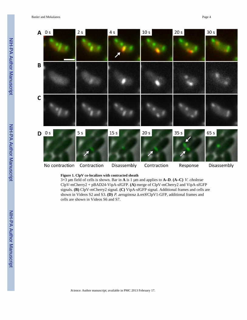
Figure 1. ClpV co-localizes with contracted sheath3×3 µm field of cells is shown. Bar in A is 1 µm and applies to A–D. (A–C) V. choleraeClpV-mCherry2 + pBAD24-VipA-sfGFP. (A) merge of ClpV-mCherry2 and VipA-sfGFPsignals. (B) ClpV-mCherry2 signal. (C) VipA-sfGFP signal. Additional frames and cells areshown in Videos S2 and S3. (D) P. aeruginosa ΔretS/ClpV1-GFP, additional frames andcells are shown in Videos S6 and S7.
Basler and Mekalanos Page 4
Science. Author manuscript; available in PMC 2013 February 17.
NIH
-PA Author Manuscript
NIH
-PA Author Manuscript
NIH
-PA Author Manuscript