lymphomas: hodgkin & non hodgkin by dr. sookun rajeev kumar
TRANSCRIPT

LYMPHOMAS:
Hodgkin & Non Hodgkin
Dr. Sookun Rajeev K
(MD)
Dept of General Medicine
Anna Medical College
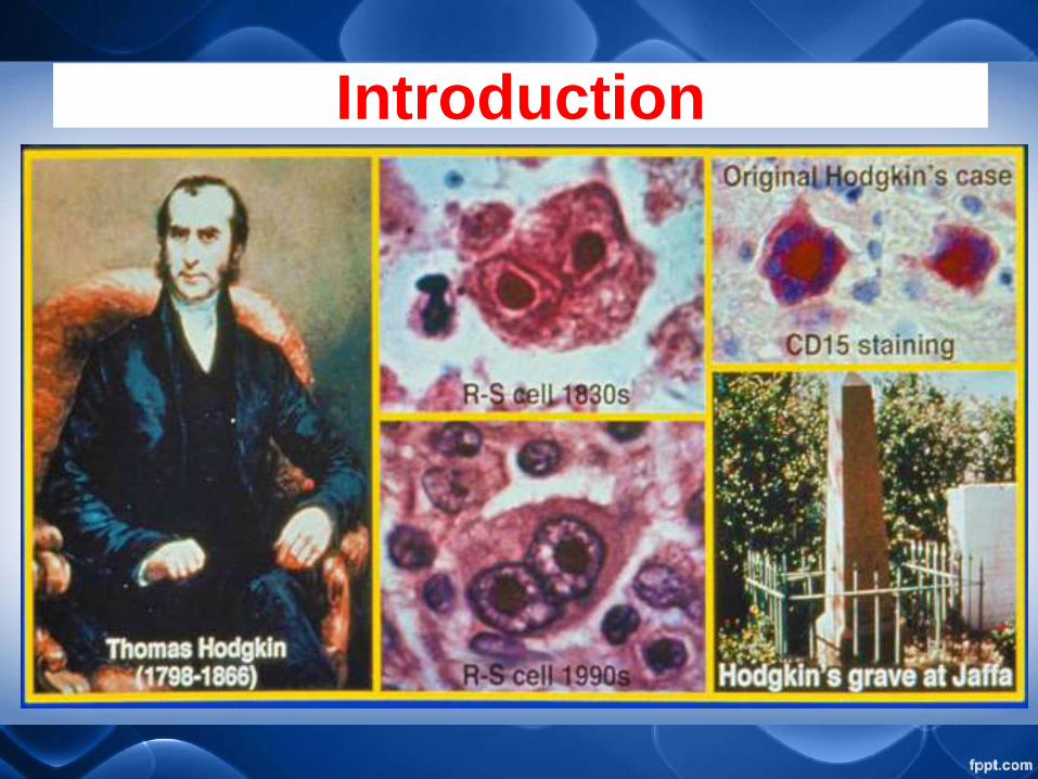
Introduction

Introduction
Lymphoma originates in
Lymphocytes.
Lymphocytes a type of WBC in the
vertebrate immune system.
Lymphomas are part of the broad
group of diseases called
Hematological Neoplasms

DefinitionLYMPHOMA is basically solid tumours
of the lymphoreticular system
Histologically 2 main types:
1. Hodgkin’s disease, characterised by
the presence of multinucleated giant
cells (Reed–Sternberg cells); and
2. Non-Hodgkin’s lymphoma

Pathophysiology Hodgkin’s Lymphoma
The disease is characterised by the
presence of Reed-Sternberg cells.
Spreading in an orderly fashion to
contagious lymph nodes
The cure rate is about 93%, making
it one of the most curable form of
cancer

Red-Sternberg cells
Cells with mirror image nuclei and prominent,
eosinophilic, inclusion-like nuclei.

lacunar cell
(mixed cellularity) (nodular sclerosis) (lymphocyte
predominance)
RS Cell variants
7

EpidemiologyMost frequently (Bimodal)occur in
age group 15 to 35 and >50 yrs
More common in males except
Nodular Sclerosis which is more
common in females
Increase incidence in HIV
infections.

Staging Classification Stage Definition
I Involvement of a single lymph node region or
structure (e.g., spleen)
II Involvement of two or more lymph node regions on
the same side of the diaphragm
III Involvement of lymph node regions or structures on
both sides of the diaphragm
IV Involvement of extranodal site(s) beyond that
designated as ‘E,’ more than one extranodal deposit
at any location, any
involvement of liver or bone marrow

Staging Classification Stage Definition
A No symptoms
B Unexplained weight loss 10% of body weight in last 6 months
Unexplained fever 38°C in the previous month Recurrent
drenching night sweats in the previous month
X Bulky disease: 10 cm maximal diameter of a nodal mass,
mediastinal mass 1/3 chest diameter
E Localized solitary involvement of extralymphatic tissue except
liver and bone marrow: if this is the only site of disease, it is
stage IE
By limited direct extension from a known nodal site Single-
discrete site proximal to a regional involved nodal site (IIE)

Stage I Stage II Stage III Stage IV
Staging of Lymphoma
A: absence of B symptoms
B: fever, night sweats, weight loss

Risk Factors Hodgkin
• Family History
• Environmental – wood workers,
farmers, meat workers
• Diseases- Mononucleosis (EBV
infection), AIDS, Bone marrow
transplant.

Signs & Symptoms Hodgkin1. Painless Lymphadenopathy –
involving superficial lymph nodes
of cervical and supraclavicular
nodes)
• The lymph nodes are swollen and
have a rubbery feeling.

Signs & Symptoms Hodgkin
2. Systemic Symptoms
a) Fevers, Night Sweats and
Weight Loss
b) Pruritus
c) Pel-Ebstein Fever

Signs & Symptoms Hodgkin
3. Pain
a) Alcohol – induced pain
b) Abdominal Pain
c) Bone Pain
d) Neurogenic Pain
e) Back Pain


Non HodgkinThe neoplastic transformation of either
B or T cell lineages of lymphatic cells.
NHL causes the accumulation of
neoplastic cells in both the lymph
nodes as well as more often diffusely
in extralymphatic organs and the
bloodstream.
Absent Reed-Sternberg cells.

Risk Factors Non Hodgkin
i. Diseases- Infections
(HIV,EBV,HepC V,H.Pylori),
RA, SLE, Sjogren’s
syndrome, mixed
Cryoglubulinemia, IBD,
Inherited immune defects
ii. Age > 60
Symptoms Non Hodgkin1. Mass Effect – Lymphadenopathy
(occipital,Posterior auricular,
preauricular, mandibular, submental,
cervical, supra & infraclavicular,
Waldeyer’s ring, Axillary, inguinal,
Popliteal, Hepatosplenimegaly,
mediastinal,Abdominal,Pelvic,testicular,
CNS masses.

Symptoms Non Hodgkin2. Hematologic – Anemia,
Thrombocytopenia,
lymphocytosis
3. Constitutional B-Symptoms-
Fatigue,Anorexia,Pruritus.
4. Paraneoplastic Syndromes

Physical Examination
Evaluate for:
Hepatosplenomegaly
Presence of Effusion
Evidence of Neuropathy
Signs of Obstruction (Extremity
edema, SVC syndrome, Spinal Cord
Comp,Hollow viscera dysfunction)

Physical Examination
Evaluate for:
Lymph Node chains including
Submental, Supraclavicular,
infraclavicular, Infraclavicular,
epitrochlear, iliac, femoral &
popliteal nodes.


Physical Examination
1. Lymph nodes examination for:
i. Size
ii. Multiplicity
iii. Consistency
iv. Tenderness


Physical Examination
2. Tonsils and Oropharynx
Waldeyer’s ring involvement
mandates complete evaluation
of the nasopharynx,
Oropharynx and Hypopharynx
by endoscopy

Investigations
1. Hematologic Tests (Basics)
FBC, Peripheral smear,
Electrolytes, Urea, Creatinine,
AST,ALT, ALP, Bilirubin,
Ca,LDH,ESR, Albumin,Serum
protein electrophoresis, HepC
V,HepB V,HIV

Investigations
1. Hematologic Tests
i. Diagnostically Abnormal
Circulating lymphoid cells or
lymphocytosis
ii. Acute-Phase reactant
iii. Liver function tests

Investigations
iv. Renal Function Tests
v. Serum Uric Acid
vi. Hypercalcemia
vii. Serum Lactate
Dehydrogenase
viii.Serum Immunoglobulins

Investigations
2. Biopsy
i. Peripheral Node Biopsy
ii. Inguinal Lymph Nodes Biopsy
iii. Bone Marrow Biopsy
iv. Endoscopic Gastric Biopsy
v. Trucut Biopsy of Retroperitoneal
& Mesenteric masses
Investigations
3. Mediastinoscopy or Limited
Thoracotomy
4. Laparotomy
5. Laparoscopy
6. Endoscopic Gastroscopy

Investigations
7. Evaluation of the Chest
i. Chest Radiograph
ii. CT Scans
iii. Thoracocentesis and Pleural
Biopsy


Investigations
8. Evaluation of the Abdomen
and Retroperitoneum
i. CT Scans
ii. Bipedal Lymphangiography
iii. Abdominal Ultrasonography

Investigations
9. Evaluation of the
Gastrointestinal Tract
i. Barium Enema
ii. Endoscopic Examination
iii. Biopsy of accessible
abnormalities

Investigations
10. Evaluation of the CNS
i. Spinal Fluid Examination
ii. CT
iii. MRI

Investigations
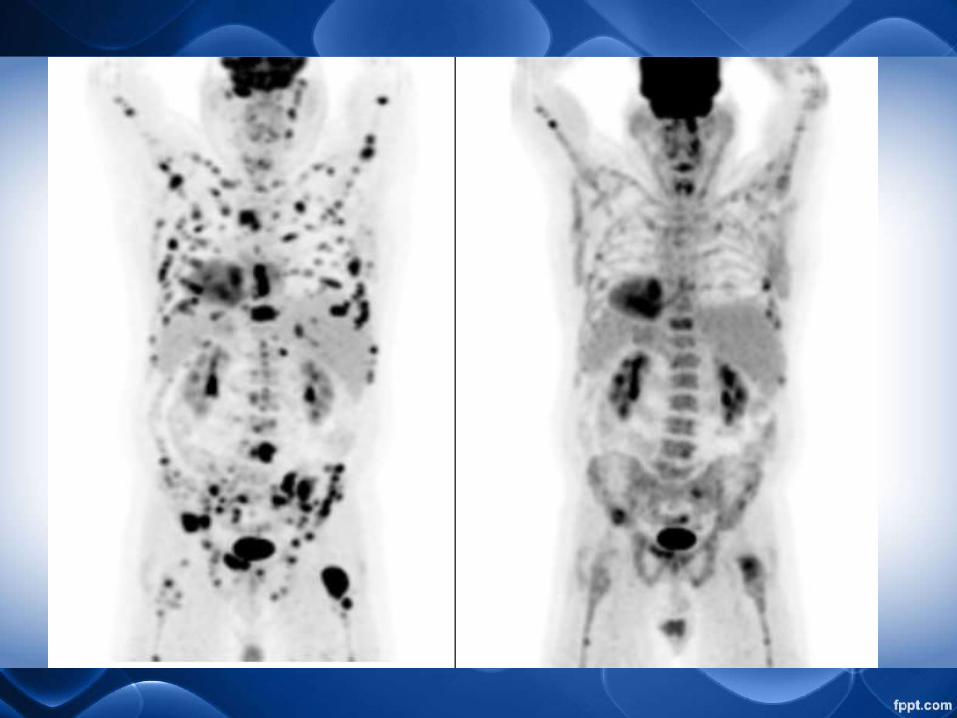
11. Nuclear Scans
i. Positron Emission
Tomography (PET) scan
ii. Gallium Scans

Treatment
i. Surgery
ii. Radiotherapy
iii. Chemotherapy
iv. Combined Chemotherapy.
v. Autologous stem cell
transport
