lymph node dissection for melanoma
TRANSCRIPT


Questions
www.downstatesurgery.org

GOALS OF PRESENTATION • Introduction • Epidemiology and etiology • Risk factors and precursor lesions • Diagnosis and clinical presentation • Staging • Management of local disease • Prognosis • Lymph node biopsy and dissections • Follow-up
www.downstatesurgery.org

INTRODUCTION • Melanoma is a cancer of melanocytes – cells of neural crest origin
www.downstatesurgery.org

EPIDEMIOLOGY • Melanoma accounts for less than 5% of skin cancer cases but
causes the majority of skin cancer deaths • 8790 melanoma-related deaths (2010) • 70,230 new melanoma diagnoses in the US (2010) • Predominantly in Caucasians and least common in African
Americans • Median age is 50 years with greatest incidence in older patients
but is one of the most common cancers in young adults • Incidence rises at a rate of 3% per year
www.downstatesurgery.org

HISTORICAL PERSPECTIVE
• 1787 – Hunter published one of the first accounts of melanoma • 1806 – Laennec discovered metastatic melanoma deposits in the liver
and differentiated them from the more common black tuberculous granulomas and routine carbon deposits
• 1905 – Handley suggested removal of 2 inches of subcutaneous tissue down to the level of the muscle fascia together with radical lymph node removal
www.downstatesurgery.org

HISTORICAL PERSPECTIVE • 1969 – Clark and associates
• Classification based on extent of tumor invasion relative to the anatomic layers of the skin and showed that level of invasion was related to survival
• Not reproducible among pathologists • 1970 – Breslow
• Classification based on tumor thickness in millimeters • Reproducible among pathologists • Excellent correlation with 5-year survival
www.downstatesurgery.org

COMPARISON OF CLARK AND BRESLOW CLASSIFICATIONS
www.downstatesurgery.org

RISK FACTORS • Exact cause of melanoma is still unknown. However…
• Personal history of melanoma or other skin cancers • Family history of melanoma (5-10% of all melanomas) • Fair complexion • Reaction to sun exposure (freckling, sunburns) or intense
intermittent sun exposure • Xeroderma pigmentosum (DNA repair) • UVA and UVB radiation (Tanning beds)
www.downstatesurgery.org

PRECURSOR LESIONS • Dysplastic nevi: 6-15mm flat pigmented lesion with indistinct margins
and variable color
www.downstatesurgery.org

PRECURSOR LESIONS • Giant congenital nevi: > 20 cm in diameter carry 10% lifetime risk of
melanoma
www.downstatesurgery.org

PRECURSOR LESIONS • Spitz nevus (juvenile melanoma): rapidly growing pink/brown benign
lesion
www.downstatesurgery.org
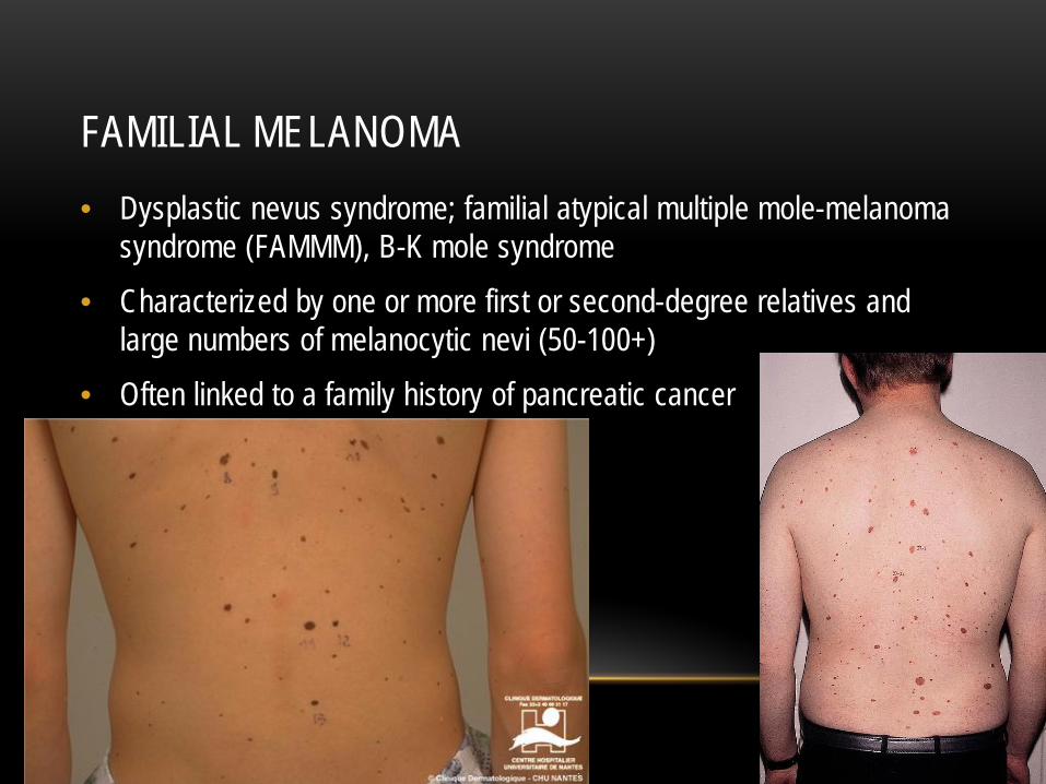
FAMILIAL MELANOMA • Dysplastic nevus syndrome; familial atypical multiple mole-melanoma
syndrome (FAMMM), B-K mole syndrome • Characterized by one or more first or second-degree relatives and
large numbers of melanocytic nevi (50-100+) • Often linked to a family history of pancreatic cancer
www.downstatesurgery.org

DIAGNOSIS
• Asymmetry
• Irregular Border
• Color variations
• Diameter greater than 6mm
• Evolution
www.downstatesurgery.org

DIAGNOSIS • History and physical – Is there a change? • Biopsy – Have a low threshold
• Excisional – most appropriate for pigmented lesions • Incisional – for larger lesions; usually via multiple punch biopsies
including the thickest portion and portion of normal tissue
www.downstatesurgery.org

HISTOLOGY • Four major types based on growth pattern and location
• Superficial spreading melanoma • Lentigo maligna melanoma • Nodular melanoma • Acral lentiginous melanoma
www.downstatesurgery.org

SUPERFICIAL SPREADING MELANOMA
• 70% of melanomas • Arise from preexisting dysplastic nevi • Usually flat and average 2cm in diameter but may become
irregular and elevated in later stages
www.downstatesurgery.org

NODULAR MELANOMA
• 15% of melanomas • Early vertical growth into the dermis with minimal radial
component • Typically blue-black ulcerated and may bleed
www.downstatesurgery.org

LENTIGO MALIGNA MELANOMA
• 10% of melanomas • Lesions appear larger than 3cm and are flat, tan, freckle-like
with marked notching of borders • Occur in sun-exposed areas most commonly in elderly
patients
www.downstatesurgery.org

ACRAL LENTIGINOUS MELANOMA
• 5% of all melanomas • 35-60% in dark-skinned people • Confined to the subungual areas and the glabrous skin of
the palms and soles
www.downstatesurgery.org

PROGNOSIS • Histology itself does not predict survival; however, certain histology
lend toward later diagnosis or early lymphatic spread • Nodular melanomas have an early vertical growth phase • Acral lentiginous melanomas may have a delay in diagnosis • Superficial spreading often develop from precursor lesions
• Number one predictor of survival is lymph node status followed by
tumor depth, ulceration, lymphovascular invasion and mitotic rate
www.downstatesurgery.org

www.downstatesurgery.org

www.downstatesurgery.org

SURVIVAL • Stage IA: The 5-year survival rate is around 97%. The 10-year survival is around 95%. • Stage IB: The 5-year survival rate is around 92%. The 10-year survival is around 86%. • Stage IIA: The 5-year survival rate is around 81%. The 10-year survival is around 67%. • Stage IIB: The 5-year survival rate is around 70%. The 10-year survival is around 57%. • Stage IIC: The 5-year survival rate is around 53%. The 10-year survival is around 40%. • Stage IIIA: The 5-year survival rate is around 78%. The 10-year survival is around 68%. • Stage IIIB: The 5-year survival rate is around 59%. The 10-year survival is around 43%. • Stage IIIC: The 5-year survival rate is around 40%. The 10-year survival is around 24%. • Stage IV: The 5-year survival rate is about 15% to 20%. The 10-year survival is about
10% to 15%. The outlook is better if the spread is only to distant parts of the skin or distant lymph nodes rather than to other organs, and if the blood level of lactate dehydrogenase (LDH) is normal.
www.downstatesurgery.org

MANAGEMENT IN EVOLUTION • 1991 – World Health Organization Melanoma Study
• Wide local excision with 1cm vs. 3cm margin for tumors less than 2mm in thickness • All local recurrence occurred in patients with tumors between 1-2mm in thckness with
a 1cm resection margin • No difference in overall survival
• Intergroup Melanoma Trial • Compared 2cm vs. 4cm margins for tumors 1-4mm thick • Recurrence was the same between groups
• Swedish Melanoma Trial and French Melanoma Trials • Compared 2cm vs. 5cm margins for tumors less than 2mm in thickness • No significant difference in disease-free or overall survival between groups
www.downstatesurgery.org

BIOPSY MARGINS • Lesions with a thickness up to 1mm – 1cm margin • Lesions with thickness 1-2 mm – 1-2 cm margins (slightly higher local
recurrence but no overall survival difference if tissue sparing required) • Lesions with a thickness above 2mm – 2cm margin
• Subungual melanomas are treated with amputation of the distal digit
to provide a 1cm margin; usually involves only the distal phalanx • Large lesions on the face/neck may require tissue-sparing biopsy
www.downstatesurgery.org
LYMPH NODE EVALUATION • 1800s - Lines of Sappey (L2 across the iliac crest to umbillicus) • 1970s – Morton described lymphscintigraphy using technetium 99m-
labeled colloid which was inject intradermally at the primary site • 1980s – Morton injected blue dye intradermally at the primary site
drained to the Sentinel Lymph node • Wounds are also be palpated since nodes obliterated with tumor may
not take up dye or radioisotope
www.downstatesurgery.org

SENTINEL LYMPH NODE BIOPSY • Studies show lower rates of in-basin recurrence after SLNB compared
with TLND; 2-10% vs. 20-50%; suggests that early treatment of regional lymph node metastases promotes regional control
• Preoperative lymphoscintigraphy offers an individualized “road map” of nodal basin drainage
• What to do with a positive SLNB remains controversial
www.downstatesurgery.org

SENTINEL LYMPH NODE BIOPSY • Multicenter Selective Lymphadenectomy Trial-1 (MSLT-1)
• 1269 patients with primary melanoma >1mm • Randomized for either (a) SLNB with completion LN dissection if positive or (b) nodal
observation and therapeutic lymph node dissection for palpable lymphadenopathy • Primary endpoint was melanoma-specific survival • Results:
• No significant melanoma-specific survival at 5 years (87% for both groups) • Small benefit in disease-free survival at 5 years with group A (78% vs 73%)
• MSLT-2: prospective randomized trial comparing SLNB with completion LN
dissection versus observation only; primary endpoint is melanoma-specific survival
www.downstatesurgery.org

SENTINEL LYMPH NODE BIOPSY • Survival benefit to patients with positive sentinel node melanoma after
completion lymph node dissection may be limited to the subgroup with a primary lesion Breslow thickness greater than 1.0 and less than or equal to 4mm (pT2-pT3). • N = 544 • Randomized prospective review • SLNB with completion LN dissection vs. therapeutic LN dissection • Statistically significant OS benefit for the group undergoing SLNB with
CLND at 5 years (57.2% versus 37.9%) in the subgroup of patients with Breslow thickness 1-4mm
Annal Surg Oncol 2008 Aug;15(8) 2223-34
www.downstatesurgery.org

NCCN CLINICAL PRACTICE GUIDELINES FOR MELANOMA AND LYMPH NODES • SLNB should be considered in higher risk stage 1B (thickness
>0.75mm, positive deep margins, lymphovascular invasion • SLNB should be offered in stage II melanoma patients • Consider complete LN dissection in stage III if SLNB is positive or
clinically positive nodes • SLNB should be considered if primary melanomas were
inappropriately diagnosed with shave biopsy and cauterized or underwent cryotherapy
www.downstatesurgery.org

ADJUVANT THERAPY AND TRIALS • Hyperthermic arterial limb perfusion with melphalan
• Alkylating agent used to treat multiple myeloma, ovarian cancer, malignant melanoma
• Considered for patients with multiple in-transit metastases • Interferon alfa-2b
• Prolonged relapse-free survival and overall survival not reproducible
• Vermurafenib, dacarbazine • Radiation therapy has been recommended for head/neck melanomas
and mucosal melanomas in the pelvic region • Intralesional BCG vaccine
www.downstatesurgery.org

POSTOPERATIVE MONITORING • Physical examination is the most important aspect of the return visit
• 3-6 month intervals until 3-5 years from surgery (75% of all recurrences)
• LDH • Complete skin examination • Evaluation for lymphadenopathy • CXR, Head CT and PET-CT
www.downstatesurgery.org

MELANOMA IN PREGNANCY • Reports suggest adverse relationship between pregnancy and
outcome in melanoma. • Recommedation is for early termination and delaying next pregnancy
for 2 years after treatment
www.downstatesurgery.org

SUMMARY • Melanoma is clinically diagnosed with a detailed history and physical
examination • Tissue biopsy will provide histological diagnosis and predictors of survival • Wide local excision is the preferred treatment for all suspicous pigmented
lesions • PET-CT, lymphoscintigraphy and blue dye provide preoperative and
operative localization of sentinel lymph nodes • Lymph node dissection remains controversial given the morbidity of the
procedure. Multiple prospective studies have demonstrated that decreasing microscopic tumor burden there is evidence of preventing further spread in a select group of patients
• SLNB is suggested for stages 1B-IIA/B • Completion LN dissection is suggested for stages III A/B
www.downstatesurgery.org

• Questions?
www.downstatesurgery.org